Embed Size (px)
Citation preview
HAL Id: hal-02403689https://hal.archives-ouvertes.fr/hal-02403689
Submitted on 12 Oct 2021
HAL is a multi-disciplinary open accessarchive for the deposit and dissemination of sci-entific research documents, whether they are pub-lished or not. The documents may come fromteaching and research institutions in France orabroad, or from public or private research centers.
L’archive ouverte pluridisciplinaire HAL, estdestinée au dépôt et à la diffusion de documentsscientifiques de niveau recherche, publiés ou non,émanant des établissements d’enseignement et derecherche français ou étrangers, des laboratoirespublics ou privés.
The hidden side of Parkinson’s disease: Studying pain,anxiety and depression in animal modelsFanny Faivre, Anil Joshi, Erwan Bezard, Michel Barrot
To cite this version:Fanny Faivre, Anil Joshi, Erwan Bezard, Michel Barrot. The hidden side of Parkinson’s disease:Studying pain, anxiety and depression in animal models. Neuroscience and Biobehavioral Reviews,Elsevier, 2019, 96, pp.335-352. �10.1016/j.neubiorev.2018.10.004�. �hal-02403689�
1
The hidden side of Parkinson’s disease:
studying pain, anxiety and depression in animal models
Fanny Faivre a, Anil Joshi a, Erwan Bezard b,c, Michel Barrot a,*
a Centre National de la Recherche Scientifique, Université de Strasbourg, Institut des
Neurosciences Cellulaires et Intégratives, F-67000 Strasbourg, France
b Université de Bordeaux, Institut des Maladies Neurodégénératives, UMR 5293, F-33000
Bordeaux, France;
c Centre National de la Recherche Scientifique, Institut des Maladies Neurodégénératives, UMR
5293, F-33000 Bordeaux, France
* Corresponding author at: Institut des Neurosciences Cellulaires et Intégratives, 5 rue Blaise
Pascal, F-67000 Strasbourg, France
E-mail address: [email protected]
ABSTRACT
Parkinson’s disease is a neurodegenerative disease leading to the loss of midbrain dopamine
neurons. It is well known and characterized by motor symptoms that are secondary to the loss
of dopamine innervation, but it is also accompanied by a range of various non-motor symptoms,
including pain and psychiatric disorders such as anxiety and depression. These non-motor
symptoms usually appear at early stages of the disease, sometimes even before the first motor
symptoms, and have a dramatic impact on the quality of life of the patients. We review here the
present state-of-the-art concerning pain, anxiety and depression-like parameters in animal
models of Parkinson’s disease.
Keywords: Parkinson’s disease; non-motor symptoms; pain; anxiety; depression
© 2018 published by Elsevier. This manuscript is made available under the CC BY NC user licensehttps://creativecommons.org/licenses/by-nc/4.0/
Version of Record: https://www.sciencedirect.com/science/article/pii/S0149763418305050Manuscript_84545c97afe867c04193e99e59f4473c
2
Parkinson’s disease is a neurodegenerative disorder classically known for the loss of dopamine
neurons in the midbrain, and most often accompanied by the presence of cytoplasmic inclusions
of α-synuclein proteins called Lewy bodies. However, although the disease is primarily related
to the loss of the nigrostriatal pathway, several other brain areas are also degenerating (often to
a lesser extent), including the locus coeruleus, the dorsal raphe nucleus, the nucleus basalis of
Meynert, the pedunculopontine nucleus, with a cortical thinning occuring at later
stage(Ehringer and Hornykiewicz, 1960; Halliday et al., 1990; Jellinger, 1991; Zarow et al.,
2003). Synuclein pathology is not restricted either to the nigrostriatal pathway but displays
extensive spread in the nervous system. Indeed, observations of post-mortem human brains
(Braak et al., 2002, 2003, 2004) showed early Lewy bodies inclusions in the IX
(glossopharyngeal) and X (vagal) nerves and in the olfactory bulbs, later spreading beyond
these structures to include the lower raphe nuclei, the magnocellular portion of the reticular
formation and the locus coeruleus, to then affect the substantia nigra pars compacta, and finally
gradually invade the entire neocortex.
Clinically, Parkinson’s disease is classically defined by a triad of motor symptoms:
bradykinesia (i.e. a slowdown to initiate voluntary movements), muscle rigidity and resting
tremors. Beside these classical symptoms, different non-motor symptoms can be present and
sometimes even appear a long time before the first motor symptoms (Bezard and Fernagut,
2014; Blanchet and Brefel-Courbon, 2017; Pont-Sunyer et al., 2015), which may lead to a
misdiagnosis or delayed diagnosis (O’Sullivan et al., 2008) and has a negative impact on the
quality of life of the patients (Nègre-Pagès et al., 2008; Rieu et al., 2016). Three major phases
have thus been proposed to describe the development of Parkinson’s disease (Stern et al., 2012).
The phase 1, the “preclinical Parkinson’s disease”, corresponds to the beginning of the α-
synuclein accumulation in the central and/or the peripheral autonomic nervous system, in the
absence of detectable clinical signs. The second phase, referred to either as “pre-motor” or
3
“prodromal” phase, can exceed ten years before the clinical diagnosis of the disease and is
usually associated to the apparition of non-motor symptoms due in part to extranigral
alterations. It should also be noticed that early subtle motor symptoms can often be present in
this prodromal phase of the disease (Mahlknecht et al., 2015). During this phase, Parkinson’s
disease patient can display higher anxiety as early as 16 years before the diagnosis of the
disease; and depression becomes significantly prevalent among Parkinson’s disease patients in
the last 2-3 years preceding the diagnostic (Darweesh et al., 2017). The phase 3 is the “motor
Parkinson’s disease”, which is the one that is clinically visible and more easily diagnosed.
The non-motor symptoms include notably (but not exclusively) sleep disorders,
gastrointestinal and autonomic symptoms (nausea, constipation), sensory symptoms (olfactory
disturbance), pain and neuropsychiatric symptoms (depression, anxiety) (Barone et al., 2009;
Chaudhuri et al., 2006; O’Sullivan et al., 2008; Park and Stacy, 2009; Pont-Sunyer et al., 2015).
The anxiety, depression and pain symptoms display significantly greater severity than in the
general population (Rana et al., 2016), and an increased frequency of the non-motor symptoms
is associated with Parkinson’s disease duration and severity (Barone et al., 2009).
Understanding the pathophysiological mechanisms underlying non-motor symptoms in
Parkinson’s disease is important, but requires relevant pre-clinical models. Alterations
concerning olfaction, sleep and the gastrointestinal tract are clinically well known (Reichmann,
2017) and addressed in models of the disease (Titova et al., 2017). On the other hand, pain,
anxiety and depression still remain understudied. In this review, we provide an overview of the
present state-of-the-art in the field by rapidly presenting pain and mood non-motor symptoms
in Parkinson’s disease patients, by evoking the different pre-clinical models used in research
and by detailing the literature on measures of nociception, anxiety-like and depression-like
symptoms in rodent and non-human primate models of Parkinson’s disease.
4
1. The non-motor symptoms in Parkinson’s disease patients
This section provides a rapid overview of some clinical data concerning pain, anxiety and
depression in Parkinson’s disease. For more details concerning these clinical aspects, please
refer to the following references (Blanchet and Brefel-Courbon, 2017; Chaudhuri et al., 2006;
Schapira et al., 2017).
1.1.Pain in Parkinson’s disease (Figure 1)
Parkinson’s disease patients exhibit sensory symptoms such as numbness, coldness, burning
or pain (Koller, 1984). Pain is one of the often neglected non-motor symptoms for which there
is no truly effective treatment (Wasner and Deuschl, 2012), even though it was already noted
in the original description of the disorder by James Parkinson. Nevertheless, it is the non-motor
symptoms which is the most frequent in Parkinson’s disease patients (O’Sullivan et al., 2008),
with a prevalence between 30% and 83% of the patients depending on the considered
epidemiological study (Allen et al., 2016; Beiske et al., 2009; Nègre-Pagès et al., 2008; Wasner
and Deuschl, 2012).
Clinical data showed that both pain thresholds and tolerance to pain are significantly lower
in Parkinson’s disease patients compared to the control population (Blanchet and Brefel-
Courbon, 2017; Zambito Marsala et al., 2011), and that these patients can suffer from a variety
of different pain (Beiske et al., 2009; Lee et al., 2006). Because of the variety of pain expressed
by Parkinson’s disease patients, a classification was proposed to identify different types by
using criterions such as “topography, duration, frequency, aggravating factors, temporal and
topographical relationship to Parkinson’s disease, influence of motor complications, influence
of antiparkinson medication, and patient’s opinion about the relationship between pain and
Parkinson’s disease” (Wasner and Deuschl, 2012). Such classification led to separate pain
5
directly/partly related to Parkinson’s disease itself, indirectly related to it (i.e. aggravated by
Parkinson’s disease) and unrelated to the disease itself (Figure 1) (Blanchet and Brefel-
Courbon, 2017; Nègre-Pagès et al., 2008; Wasner and Deuschl, 2012).
Pain in Parkinson’s disease can be a nociceptive pain, defined by the International
Association for the Study of Pain as “pain that arises from actual or threatened damage to non-
neural tissue and is due to the activation of nociceptors” (https://www.iasp-pain.org/). Those
include for example pain that is associated with the motor disturbances and partly due to muscle
rigidity. Indeed, rigidity, akinesia, postural abnormality or dystonia can lead to musculoskeletal
nociceptive pain and back pain (Wasner and Deuschl, 2012). Patients can also display
neuropathic pain, i.e. “pain caused by a lesion or disease of the somatosensory nervous system”,
which may be of peripheral origin, such as radicular pain, or of central one (Blanchet and
Brefel-Courbon, 2017; Wasner and Deuschl, 2012). Miscellaneous pain, which cannot be
classified in the above two categories, can also be observed. Those include for example
akathisia (i.e. restless legs syndrome) and pain associated with depression, which is the one that
appears mostly during the prodromal phase (Schapira et al., 2017; Wasner and Deuschl, 2012)
(Fig. 1). The patients with pain more directly related to Parkinson’s disease itself are often
younger at non-motor and motor disease onsets and show higher parkinsonism severity than
those without pain or with pain unrelated to the disease (Nègre-Pagès et al., 2008; Zambito
Marsala et al., 2011). The gender has also an influence, with women more frequently reporting
pain symptoms (Beiske et al., 2009; Zambito Marsala et al., 2011). The loss of dopamine in the
basal ganglia may partly explain the changes in pain thresholds; however, the absence of a total
recuperation of these symptoms with dopaminergic medication suggests also a role of non-
dopaminergic mechanisms in the appearance or maintenance of pain symptoms (Brefel-
Courbon et al., 2005; Schapira et al., 2017).
6
1.2.Anxiety and depression in Parkinson’s disease
There is a notable comorbidity of mood disorders with Parkinson’s disease (Dissanayaka et
al., 2010). Moreover, the risk to be diagnosed with Parkinson’s disease is significantly increased
in patients that have already been diagnosed with affective disorders (Nilsson et al., 2001),
which reflect the fact that these psychiatric pathologies are part of the early non-motor
symptoms appearing during the prodromal phase of the disease (Jacob et al., 2010). Mood
disorders can indeed appear 5 to 20 years before the motor symptoms (Shiba et al., 2000).
According to the Diagnostic and Statistical Manual of Mental Disorders, 5th edition
(American Psychiatric Association, 2013), “anxiety disorders include disorders that share
features of excessive fear and anxiety and related behavioral disturbances”. 34 to 65% of
Parkinson’s disease patients experience anxiety, including panic disorders, generalized anxiety
disorder or social phobia (Barone et al., 2009; Chaudhuri et al., 2006; Dissanayaka et al., 2010;
Park and Stacy, 2009; Schapira et al., 2017; Szatmari et al., 2017). The risk to develop these
disorders has been observed to be much higher (ninefold) in “younger” patients (<62 years old)
than in older subjects (Dissanayaka et al., 2010). The pathophysiological basis for these
symptoms is however not clearly identified yet. Beside alterations in dopaminergic system,
including a correlation between striatal dopamine transporter availability and anxiety (Erro et
al., 2012; Moriyama et al., 2011; Weintraub et al., 2005), a structural change in amygdala size
has also been proposed to contribute to this symptom (Vriend et al., 2016). Moreover, a loss of
noradrenaline neurons in the locus coeruleus (Bertrand et al., 1997; Delaville et al., 2011;
German et al., 1992; Hughes et al., 1992) and of serotonin cells in the dorsal raphe (Kish, 2003)
has been described in patients, which could also contribute to the presence of anxiety and
depression (Marsh, 2013; Remy et al., 2005; Schapira et al., 2017).
7
Depressive disorders are common psychiatric disorders characterized by the presence of a
sad, empty or irritable mood, accompanied by somatic and cognitive changes that impact
everyday life function. According to the World Health Organization, they affect 300 million
people worldwide (http://www.who.int/). Approximatively 40% of the patients with
Parkinson’s disease display depressive disorders (Burn, 2002; Chaudhuri et al., 2006;
Cummings, 1992; Jacob et al., 2010). Anhedonia and apathy are frequent symptoms that can
occur during the prodromal phase (Ishihara and Brayne, 2006; Pont-Sunyer et al., 2015), which
leads to the fact that, at time of Parkinson’s disease diagnosis, the proportion of patients who
already consulted for depression is more than twice higher than in the control population
(Leentjens et al., 2003). Classical antidepressant drugs as well as deep brain stimulation and L-
DOPA therapy can be useful to improve these symptoms (Czernecki et al., 2002; Schapira et
al., 2017). Even if the detailed mechanism underlying depression is unknown, different
hypotheses have been proposed. Indeed, alterations in the monoaminergic systems (Chaudhuri
et al., 2006; Reichmann, 2017; Schapira et al., 2017), as well as in structural and metabolic
alterations in brain regions known for their implication in affective disorders, such as the
hippocampus and the amygdala (Huang et al., 2013; Surdhar et al., 2012; van Mierlo et al.,
2015), may likely contribute to the depressive symptoms in Parkinson’s disease.
2. Animal models to study Parkinson’s disease
This section mostly focuses on animal models (rodents and monkeys) of Parkinson’s disease
for which data related to pain, anxiety or depression like behaviors are available. For a more
exhaustive view of existing models, readers can consult following reviews (Bastías-Candia et
al., 2018; Bové and Perier, 2012; Creed and Goldberg, 2018; Francardo, 2018; Grandi et al.,
2018; Koprich et al., 2017; Volta and Melrose, 2017).

8
2.1.Toxin-induced Parkinson’s disease models
The more widely used models of Parkinson’s disease are based on 6-hydroxydopamine (6-
OHDA), a drug that selectively acts on catecholamine containing terminals and cells bodies
(Ungerstedt, 1968). Due to its homology with dopamine and noradrenaline, 6-OHDA is caught
by plasma membrane dopamine and noradrenaline transporters to enter into cells. It
accumulates into the cytosol, producing reactive oxygen species that are toxic to the cell and
quinones that inactivate biological macromolecules important to neuronal integrity (Bové and
Perier, 2012; Dauer and Przedborski, 2003; Sachs and Jonsson, 1975). 6-OHDA does not easily
cross the brain-blood barrier, it is thus injected directly into the structure of interest, either in
the area of dopamine cell bodies (substantia nigra pars compacta), in the dopamine passing
fibers (medial forebrain bundle) or at terminal level (striatal complex) (Blum et al., 2001;
Gubellini and Kachidian, 2015; McDowell and Chesselet, 2012). This 6-OHDA administration
induces a degeneration of dopamine neurons, leading to motor impairments that partially mimic
the motor-symptoms of Parkinson’s disease (Bové and Perier, 2012; Ungerstedt, 1968). The
injection can be unilateral, producing a hemiparkinsonism model with asymmetric circling
behaviors that can be used to test treatments for motor symptoms (Bové and Perier, 2012; Dauer
and Przedborski, 2003; Hefti et al., 1980; Ungerstedt and Arbuthnott, 1970). The bilateral
injection in the mesencephalic region of dopamine cell bodies can however induce a notable
mortality of the animals, due to adverse effects such as aphagia, adipsia and seizures (Bezard
and Przedborski, 2011; Bové and Perier, 2012; Ungerstedt, 1971), which limits in part this
direct targeting of cell bodies. As 6-OHDA is toxic for all catecholamine neurons, a
pretreatment with a blocker of noradrenaline re-uptake and/or catabolism (monoamine oxidase
B inhibitor) may be used to protect noradrenaline neurons and have a more specific dopamine
lesion. However, it has also been proposed that administration of 6-OHDA without such
9
neuroprotection may lead to a richer phenotype that better mimics Parkinson’s disease (Bezard
et al., 2013).
The use of 1-methyl-4-phenyl-1,2,3,6-tetrahydropyridine (MPTP) to mimic the disease was
discovered in the 1980’s, after the observation that an accidental production of MPTP during
the manufacture of a synthetic opioid drug could lead to symptoms similar to those observe in
Parkinson’s disease in users of this drug (Blum et al., 2001; Davis et al., 1979; Langston et al.,
1983). In animal research, MPTP is mainly used in non-human primates and in mice, even
though rodents are less sensitive to its toxicity (Schober, 2004), especially rats in which
dopamine neurons are resistant to systemic delivery of MPTP (Betarbet et al., 2002; Bové and
Perier, 2012; Boyce et al., 1984; Chiueh et al., 1984b; Sahgal et al., 1984). However, local
intracerebral delivery of the active metabolite MPP+ can induce dopamine neuron loss in rats
and in guinea-pigs (Chiueh et al., 1984a). The effects of MPTP depend on various parameters,
such as the administration mode, species and age (Blum et al., 2001). Due to its facility to cross
the brain-blood barrier, MPTP is classically administered through systemic injection
(Bankiewicz et al., 2001). After such injection, MPTP enters the brain and is metabolized in 1-
methyl-4-phenyl-2,3-dihydropyridinium ion (MPP+). The MPP+ binding site is located in the
electron leak site, within the complex I of the electron transport chain of the mitochondria
(Betarbet et al., 2002). MPP+ inhibits the complex I of the mitochondrial electron transport
chain, leading to significant ATP depletion, production of reactive oxygen species and cell loss,
particularly in the nigrostriatal pathway which is the brain region the most sensitive to the
compound, but not the only one (Betarbet et al., 2002; Chan et al., 1991; Dauer and Przedborski,
2003; Engeln et al., 2015; Fabre et al., 1999).
The above two toxins are the most used to model Parkinson’s disease, but other drugs can
be found in the literature. Rotenone is a compound used as pesticide which easily crosses the
brain-blood barrier, entering neuron mitochondria through the same mechanism as MPP+ and
10
inhibiting mitochondrial complex I (Dauer and Przedborski, 2003; Heinz et al., 2017; Li et al.,
2003). It produces α-synuclein accumulation and causes a degeneration of the nigrostriatal
dopaminergic pathway following oxidative stress (Betarbet et al., 2000, 2002). In rodents,
rotenone can be administrated by systemic injections or by stereotaxic delivery directly into the
brain. However, its use is strongly limited by the high mortality that follows its administration
at high doses (Antkiewicz-Michaluk et al., 2003). Based on epidemiological toxicology, the
herbicide paraquat (1,1’-dimethyl-4,4’-bypyridinium dichloride), the fungicide maneb (Mn-
ethylene-1,2-bisdithiocarbamate) and the cycad flour (from cycas micronesica) are also used to
model Parkinson’s disease. Paraquat is in fact a structural analog of MPP+, the active metabolite
of MPTP (Betarbet et al., 2002; Bové and Perier, 2012). Its systemic injections induce α-
synuclein accumulation and causes degeneration of dopamine neurons of the substantia nigra
pars compacta (Dauer and Przedborski, 2003; Manning-Bog et al., 2002; McCormack et al.,
2002; McDowell and Chesselet, 2012). Maneb easily crosses the brain-blood barrier neuron
and causes damage in the substantia nigra pars compacta and the striatum, leading to locomotor
and coordination impairments. Combined exposure to maneb potentiates the effect of paraquat
(Bastías-Candia et al., 2015; Thiruchelvam et al., 2003). Finally, mice fed with cycad flour
exhibit α-synuclein accumulation in multiple brain regions and dopamine neuron loss in the
substantia nigra pars compacta, which induces cognitive deficits and Parkinson’s disease-like
symptoms (McDowell and Chesselet, 2012; Wilson et al., 2002).
Moreover, it has been shown that an intranigral, intrastriatal or intra-pallidal injection of
lipopolysaccharides induces an inflammation process associated with the activation of
microglia. This inflammation favors a degeneration of the dopamine neurons of the nigrostriatal
pathway and lead to locomotor impairments similar to the ones seen in other models of
Parkinson’s disease (Castaño et al., 1998; Liu and Bing, 2011; Zhang et al., 2005).
11
Lastly, injections of Lewy body extracts from post mortem Parkinson’s disease patients in
the substantia nigra pars compacta or the striatum in mice or monkey induce a progressive
nigrostriatal degeneration. In mice, this degeneration is accompanied by an astrogliosis in the
substantia nigra and impaired motor coordination and balance in the pole test (Recasens et al.,
2014).
2.2.Genetic models of Parkinson’s disease
While Parkinson’s disease is often sporadic, familial forms have also been described which
are due to autosomal dominant or recessive genetic mutations (Singleton et al., 2013), and
corresponding animal models have been developed.
In 1997, a family form of Parkinson’s disease caused by a mutation of the α-synuclein gene
(SNCA gene) was discovered (Polymeropoulos et al., 1997). This mutation consists of a single
base pair change from a guanine to an adenosine at position 209, leading to an alanine to
threonine substitution in the protein at position 53 (p.A53T) (Polymeropoulos et al., 1997).
Other point mutations of the SNCA gene were also linked to familial forms of Parkinson’s
disease, such as the p.A30P mutation (Krüger et al., 1998) corresponding to an alanine to proline
substitution in the position 30 of the protein and the p.E46K mutation (Zarranz et al., 2004)
corresponding to a glutamic acid to lysine substitution in position 46. All of these mutations
lead to an autosomal dominant form of the disease (Hernandez et al., 2016). Beside these point
mutations, whole locus duplication or triplication of the SNCA gene has also been reported as a
cause of family forms of the disease (Ibáñez et al., 2004; Singleton et al., 2003), the number of
SNCA copies correlating with the early-onset and severity of the disease, suggesting a dose
dependent effect (Ibáñez et al., 2004). The discovery of these different familial forms was the
starting point for the development of transgenic models for the study of Parkinson’s disease.
12
To explore the function of the α-synuclein protein and its implication in the disease, several
lines of transgenic mice expressing either the wild-type or a mutated (p.A53T or p.A30P)
human α-synuclein were produced (for review, see Fernagut and Chesselet, 2004). The first
transgenic mouse overexpressing human α-synuclein used the wild-type form of the protein
under the control of the plateled-derived growth factor β promoter (Fernagut and Chesselet,
2004; Masliah et al., 2000), leading to α-synuclein accumulation in synapses from the
neocortex, the hippocampus, the substantia nigra and the olfactory regions (Masliah et al.,
2000). However, the use of other promoters and/or forms of α-synuclein can lead to different
patterns and levels of α-synuclein expression and of behavioral phenotypes (Fernagut and
Chesselet, 2004). For example, α-synuclein expression driven by the Thy-1 promoter has been
used to produce various transgenic lines that differ in terms of presence (Kahle et al., 2000;
Rockenstein et al., 2002) or absence (Rabl et al., 2017) of α-synuclein accumulation in the
striatum or in the presence (Fleming et al., 2004; Rabl et al., 2017) or absence (Kahle et al.,
2000) of motor impairment. This variability is not only related to the chosen protein form (wild-
type or mutated) but may also be related to the transgene insertion site (Chesselet, 2008).
An expression of the wild-type (Lim et al., 2010; Nuber et al., 2008) or the p.A30P mutated
(Marxreiter et al., 2013) human α-synuclein driven by the calmodulin-dependent protein kinase
IIα promoter induces a degeneration of neurons in the dentate gyrus, the olfactory bulb and in
some midbrain and forebrain regions (Lim et al., 2010; Marxreiter et al., 2013; Nuber et al.,
2008). This α-synuclein accumulation leads to cognitive and progressive motor impairments
(Nuber et al., 2008). Interestingly, if the construct was repressed until weaning, no neuronal
loss was observed in the hippocampal region, indicating that the dentate gyrus neurons are more
vulnerable during development than after maturation (Lim et al., 2010). Transgenic mice
expressing either wild-type or A53T mutated α-synuclein under the mouse prion protein
promoter display age-dependent intracytoplasmic neuronal α-synuclein inclusions (Giasson et
13
al., 2002) and motor impairments that lead to paralysis and death a few days after the first motor
symptoms due to motoneuron loss (Giasson et al., 2002; Giraldo et al., 2018). In this model,
intracerebral injections of preformed α-synuclein fibrils in young asymptomatic mice accelerate
the formation of intracellular inclusions and the development of neurological symptoms (Luk
et al., 2012).
To more specifically target α-synuclein expression to dopamine neurons (which is a useful
model of dopamine cell loss but does not reflect the more widespread expression observed in
both central and peripheral nervous system in patients), lines of transgenic mice with a truncated
wild-type (Tofaris et al., 2006) or mutated (p.A53T) (Wakamatsu et al., 2008) α-synuclein
expression under the control of the tyrosine hydroxylase promoter were developed. These mice
display pathological α-synuclein-positive inclusions in the substantia nigra and in the olfactory
bulb, and a decrease in striatal dopamine levels associated with a progressive reduction of the
spontaneous activity (Tofaris et al., 2006; Wakamatsu et al., 2008).
In addition to the classical transgenic models exposed above, the use of adeno-associated
viral vectors to overexpress α-synuclein has also been developed. This overexpression in the
midbrain induces a nigrostriatal degeneration and the formation of insoluble α-synuclein
aggregates in mice, rats and non-human primates (Bourdenx et al., 2015; Ip et al., 2017; Ulusoy
et al., 2010). This method reproduces many characteristics of the pathology and provides an
interesting model in both rodents and primates (Ulusoy et al., 2010).
Mutations in the PARK8 gene coding for the leucine-rich repeat kinase 2 (LRRK2) protein
have also been described to be associated with familial autosomal-dominant and sporadic forms
of Parkinson’s disease (Funayama et al., 2002; Healy et al., 2008; Paisán-Ruiz et al., 2013).
Mutations of this protein mostly target the GTPase and kinase domains of the protein (Dawson
et al., 2010; Gubellini and Kachidian, 2015) and cause late-onset forms of the disease (Dawson
et al., 2010; Healy et al., 2008). Transgenic mice with bacterial artificial chromosome (BAC)
14
expressing LRRK2 R1441C/G (i.e. mutation in the GTPase domain) have been used to model
Parkinson’s disease, showing age-dependent and progressive motor symptoms but no
nigrostriatal degeneration (Li et al., 2009). The development of LRRK2 knock-in mice also
failed to produce the classical brain alterations of the disease, such as dopamine cell
degeneration or α-synuclein accumulation (Bezard et al., 2013; Dawson et al., 2010; Volta and
Melrose, 2017). More conclusively, a rat LRRK2 model has been developed, using adenoviral-
mediated expression of LRRK2 G2019S mutation (i.e. mutation in the kinase) to induce a
progressive degeneration of nigral dopamine neurons (Dusonchet et al., 2011).
Mutations in the PRKN gene (or PARK2), which encodes for parkin, an E3 ubiquitin ligase,
were identified as a genetic cause of autosomal-recessive and early-onset Parkinson’s disease,
as well as of some forms of sporadic cases (Gubellini and Kachidian, 2015; Dawson et al, 2010).
These parkin mutations lead to a loss-of-function and are responsible for a loss of substantia
nigra pars compacta dopamine neurons without formation of Lewy bodies (Dauer and
Przedborski, 2003). However, mouse models produced by partial or full deletion of the parkin
gene exhibit few or no dopamine loss (Itier et al., 2003; Perez and Palmiter, 2005; Von Coelln
et al., 2004) and only limited behavioral deficits (Perez and Palmiter, 2005). On the other hand,
the overexpression of the mutant human parkin using a BAC transgenic mouse model leads to
a late-onset and progressive degeneration of dopamine neurons, associated with progressive
motor deficits (Dawson et al., 2010; Gubellini and Kachidian, 2015; Lu et al., 2009).
Mutations in the PINK-1 (PTEN-induced putative kinase 1) gene can cause autosomal
recessive forms of the disease (Dawson et al., 2010; Gubellini and Kachidian, 2015; Valente et
al., 2004). PINK-1 interacts with parkin to induce parkin-mediated autophagy of damaged
mitochondria and thus protect cells from mitochondrial dysfunctions (Gubellini and Kachidian,
2015; Lazarou et al., 2013; Narendra et al., 2008). However, similar to parkin null mice, PINK-
1 knock-out mice do not display notable neurodegeneration of substantia nigra dopamine
15
neurons, and show few or no Lewy body-like inclusions (Gispert et al., 2009; Kitada et al.,
2007).
A missense mutation in the DJ-1 gene (PARK7) is responsible for an autosomal recessive
and early-onset form of Parkinson’s disease (Bonifati et al., 2003a, 2003b; Dawson et al., 2010;
Lim and Ng, 2009). DJ-1 knock out mice have been developed, but failed to show nigrostriatal
dopaminergic loss. However, a reduced striatal dopamine release and a decreased locomotor
activity has been observed, making this model interesting as model of early stages of the disease
(Chandran et al., 2008; Chen et al., 2005; Dawson et al., 2010; Goldberg et al., 2005; Lim and
Ng, 2009; Yamaguchi and Shen, 2007).
Beside the above genetic models that are based on human gene mutations associated with
the disease, other transgenic models have also been designed targeting the dopamine system.
One of these models is based on the mutation or the deletion of the homeobox transcription
factor Pitx3 which is known to be selective to dopamine neurons, thus inducing a dopaminergic
loss in the nigrostriatal pathway. The Pitx3-null mice exhibit different markers of the disease,
including a decline in substantia nigra pars compacta cell number, a reduction in striatal
dopamine release, and motor symptoms such as motor coordination impairment, body tremors
and decreased locomotion (Filali and Lalonde, 2016; Le et al., 2015). The Pitx3416insG mutant
mice, relying on a spontaneous mutation (an inserted G in position 416 of the Pitx3 gene in the
chromosome 19), induces microphtalmia or anophtalmia and a loss of dopamine neurons in the
substantia nigra, which induces motor impairment and increased nociceptive response
(Rosemann et al., 2010). Another interesting mouse model, MitoPark, has been obtained by
selectively removing the mitochondrial transcription factor A (Tfam) from dopamine midbrain
neurons. These transgenic mice model an adult-onset of the disease, with a progressive loss of
dopamine cell bodies and terminals, associated with tremors, limb rigidity and a progressive
decline of locomotion and rearing (Ekstrand et al., 2007; Galter et al., 2010).
16
3. Non-motor symptoms in animal models of Parkinson’s disease
3.1.Nociception and pain in rodent Parkinson’s disease models (Figure 2)
According to the definition from the International Association for the Study of Pain, pain is
“an unpleasant sensory and emotional experience associated with actual or potential tissue
damage or described in terms of such damage”. This sensory/emotional duality is a critical
element. It distinguishes pain from nociception, which encompasses “the neural process of
encoding noxious stimuli” and do not necessarily imply the presence of pain. However,
distinguishing pain from nociception remains challenging in animal-based research, and tests
that are referred to as “pain tests” are usually based on reflex responses that may reflect
nociceptive responses (Barrot, 2012). Moreover, because of the implication of reflex motor
responses in most of the tests used to study nociception in rodents and the potential slow-down
of motor reflexes in Parkinson’s disease, it can also be challenging to correctly assess and
interpret some nociceptive responses in rodent models of this disease.
Data reported below show that the nociceptive hypersensitivity observed in patients with
Parkinson’s disease (response to evoked pain) can be reproduced in some of the animal models.
However, the presence of spontaneous pain, which may for example result from rigidity,
akinesia, postural abnormality or dystonia, has not been studied yet in these models. While
challenging, such question could now also be addressed in rodents. Indeed, procedures were
developed in the past decade to test the aversive state induced by ongoing pain in rats and mice
(Barthas et al., 2015; Johansen et al., 2001; King et al., 2009; Qu et al., 2011; Sellmeijer et al.,
2018)
3.1.1. Electrical sensitivity
17
The threshold responses to electrical shocks can be used to assess pain sensitivity. In such
test, electrical stimuli of increasing intensities are delivered through a grid floor and the
threshold intensity eliciting the first response (i.e. flinch, vocalization, jumping or running) is
measured (Barrot, 2012). Rats with a unilateral 6-OHDA lesion of the dopamine neurons show
an hypersensitivity either in the ispsilateral (Chen et al., 2013) of the contralateral body side of
the lesion (Carey, 1986) (Table 1).
3.1.2 Mechanical sensitivity
Although different tests are available to assess the response to mechanical stimuli in rodents
(Barrot, 2012), particularly in rats (von Frey filaments, Randall-Selitto’s paw pressure test…),
most of the data from Parkinson’s disease rodent models found in the literature are based on
the von Frey filaments test. Mechanical nociceptive thresholds are usually lower in animal
models of Parkinson’s disease, regardless of the rodent species (mice or rat) or of the chosen
model (6-OHDA, MPTP, Pitx3) (Cao et al., 2016; Charles et al., 2018; Chudler and Lu, 2008;
Gee et al., 2015; Gómez-Paz et al., 2017; Nascimento et al., 2018; Park et al., 2015; Rosemann
et al., 2010; Saadé et al., 1997; Takeda et al., 2005, 2014; Wang et al., 2017; Zengin-Toktas et
al., 2013). While this mechanical hypersensitivity now appears as well established, there are
some discrepancies among published reports using a unilateral lesion (Charles et al., 2018;
Chudler and Lu, 2008; Gee et al., 2015; Gómez-Paz et al., 2017; Nascimento et al., 2018; Saadé
et al., 1997; Takeda et al., 2005, 2014), with results that may differ regarding the time-course
and/or the laterality of the hypersensivity. Changes in mechanical sensitivity have been
correlated with decreased tyrosine hydroxylase expression in the substantia nigra pars
compacta (Zengin-Toktas et al., 2013) and can be compensated by a neural transplantation of
fetal ventral mesencephalon tissue in lesioned animals (Takeda et al., 2014) (Table 1).
Painful rigidity of the face, chin or jaw and trigeminal neuralgia-like pain can be present in
the prodromal phase of Parkinson’s disease (Waseem and Gwinn-Hardy, 2001). This trigeminal
18
sensitivity can be tested in rodents with different approaches, using procedures assessing static
or dynamic mechanical sensitivity. To test static (pressure) mechanical allodynia of the face,
von Frey filaments can be applied to the vibrissae region. The bilateral lesion of the substantia
nigra pars compacta leads to an increased nociceptive sensitivity of the two side of the face
(Dieb et al., 2016), while a unilateral lesion in the caudate/putamen does not affect this response
(Chudler and Lu, 2008). To test the dynamic mechanical allodynia, gentle air puff is applied on
the animal face. Bilateral lesioned animals display a dynamic mechanical allodynia of the face,
which is inversely correlated with the number of tyrosine hydroxylase positive cells (Dieb et
al., 2014) (Table 1).
3.1.3 Thermal sensitivity
The tail flick test measures the withdrawal latency following tail exposure to a heat stimulus,
by using an infrared heat beam or a warm-controlled water bath (Barrot, 2012; Le Bars et al.,
2001). The response in this test is a nociceptive spinal reflex that is, however, influenced by
descending controls from the brain. Mice with intraperitoneal injections of MPTP have reduced
tail flick latency (Park et al., 2015; Rosland et al., 1992), whereas Pitx3416insG mutant mice
showed no significant difference (Rosemann et al., 2010). On the other hand, the literature
concerning the thermal sensitivity in 6-OHDA models of Parkinson’s disease is controversial,
with opposite results that can be reported: increased latency (Grossmann et al., 1973; Tassorelli
et al., 2007), no difference (Gee et al., 2015; Morgan and Franklin, 1990; Ogata et al., 2015) or
decreased latency (Dolatshahi et al., 2015; Gómez-Paz et al., 2017; Haddadi et al., 2018;
Nascimento et al., 2018; Saadé et al., 1997). A possible explanation for these discrepancies
could be the co-presence of thermal hypersensitivity and of reflex slow-down. Depending on
the location and the extent of the lesion, and on the test procedure and temperature settings, one
or the other aspect may be predominant, which highlights the potential difficulty to interpret
tail-flick results in these models. (Table 1).
19
The hot plate test measures the response to nociceptive heat by placing the animal on a plate
at fixed and controlled temperature, often set in the 52 to 55°C range with a 0.1°C precision.
The latency before the apparition of a withdrawal behavior, such as paw licking, paw
withdrawal or eventually jumping, is measured (Barrot, 2012; Le Bars et al., 2001). Usually,
whatever the model (MPTP or 6-OHDA) and the species (mouse or rat) that is used, the articles
report an increased pain sensitivity (Chen et al., 2013; Dolatshahi et al., 2015; Gee et al., 2015;
Lin et al., 1981; Nascimento et al., 2018; Park et al., 2015; Rosland et al., 1992; Saadé et al.,
1997). In the paw immersion test, consisting in placing the animal’s paw in a water bath at a
fixed and controlled temperature, rats with 6-OHDA lesion in the ventral tegmental area and
the substantia nigra displayed a decreased withdrawal latency, i.e. a hypersensitivity,
concerning the paw contralateral to the lesion (Saadé et al., 1997) (Table 1).
In the radiant heat paw-withdrawal test, often referred to as the Hargreaves’s method
(Hargreaves et al., 1988), a controlled heat beam system is directed toward the plantar surface
of the animal’s hind paws in order to measure the withdrawal latency (Barrot, 2012). In this
test, Pitx3416insG mutant mice (Rosemann et al., 2010), rats at 3 weeks following 6-OHDA lesion
of the caudate/putamen nuclei (Chudler and Lu, 2008) rats with 6-OHDA lesion of the right
medial forebrain bundle (Charles et al., 2018), rats at 4 and 5 weeks following 6-OHDA
bilateral lesion of the striatum (Cao et al., 2016) and rats with bilateral lesion of the substantia
nigra pars compacta at 2 and 4 weeks post-surgery (Wang et al., 2017), displayed hyperalgesia
as illustrated by decreased paw withdrawal latencies (Table 1).
Cold sensitivity is more rarely tested but can be assessed by for example applying a drop of
acetone on the animal’s paw. Rat with a unilateral lesion of the substantia nigra pars compacta,
but not control animals, exhibit nocifensive responses such as paw withdrawal, licking, shaking
or rubbing (Gómez-Paz et al., 2017), showing the presence of a cold allodynia.
3.1.4 Response to nociceptive chemical exposure
20
The intradermal injection of a formalin solution models short-term inflammatory pain. It
results in paw withdrawal, licking, biting or shacking. In rodents, these responses are classically
divided in 2 phases: an initial phase during the first 5 or 10 minutes after the injection, related
to the stimulation of nociceptors; and a second phase, lasting between 20 to 40 min,
corresponding to both inflammatory mechanisms and central sensitization within the dorsal
horn (Barrot, 2012; Le Bars et al., 2001). It has been shown that rats with 6-OHDA lesion,
either in the cell bodies or terminals, displayed enhanced behavioral responses in the formalin
test (Cao et al., 2016; Chudler and Lu, 2008; Gómez-Paz et al., 2017; Ogata et al., 2015;
Tassorelli et al., 2007), thus reflecting hyperalgesia. This hyperalgesia was however detected
in the second phase only (Chudler and Lu, 2008; Ogata et al., 2015) or in both phases (Cao et
al., 2016; Gómez-Paz et al., 2017; Tassorelli et al., 2007), depending on the study. When
formalin was delivered in the face (trigeminal pain) of rats with an unilateral striatal 6-OHDA
lesion, grooming was significantly enhanced during the second (Chudler and Lu, 2008) or both
phases of the test (Maegawa et al., 2015), reflecting an increased inflammatory pain response.
Changes in the formalin test following 6-OHDA lesion could also affect the sensitivity to pain
relief, indeed midbrain 6-OHDA lesions suppressed D-amphetamine and morphine-induced
analgesia in this test (Morgan and Franklin, 1990). On the other hand, in a transgenic LRRK2
model of Parkinson’s disease, mutant mice did not show alteration of formalin test responses
(Bichler et al., 2013), but this model displayed an overall limited range of symptoms. (Table 1)
In the writhing test, the irritant chemical are delivered intraperitoneally, which provokes
abdominal contractions and twisting of dorso-abdominal muscles that can be quantified as an
indicator of peritovisceral nociception (Le Bars et al., 2001). In Pitx3416insG mutant mice,
visceral pain following intraperitoneal acetic acid delivery was significantly enhanced
(Rosemann et al., 2010) (Table 1).
21
3.2.Blink reflex abnormalities in Parkinson’s disease models (Figure 2)
The blink reflex is related to the activity of the orbicularis oculi muscle and may be
considered as a protective nociceptive response. The electrical stimulation of this muscle results
in an electromyographic response composed of 2 different phases, the R1 and R2 components.
The R1 component is the early component, which is only present on the stimulated side. The
R2 component is present in both sides and appears later (Pearce, 2008). In Parkinson’s disease,
patients exhibit a hyperexcitability of the blink reflex, with shorter latency, increased amplitude
and an increased habituation index (Esteban and Giménez-Roldàn, 1975). In 6-OHDA lesioned
rats, as in patients, a hyperexcitability of the blink reflex has also been observed, together with
an impaired blink plasticity and a lower spontaneous blink rate (Basso et al., 1993; Kaminer et
al., 2015) (Table 1).
3.3.Anxiety-like and depression-like behaviors in rodent Parkinson’s disease models (Figure 3)
Animal studies concerning anxiety and depression in Parkinson’s disease mainly focused
on dopaminergic mechanism, which unlikely reflects the complexity underlying the occurrence
of these symptoms in patients. There is still indeed a paucity of studies addressing, beyond
dopamine, the role of the other alterations of the nervous system, in particular for early stages
of the disease.
3.3.1. Anxiety-like behavior in Parkinson’s disease models
Tests for anxiety-like behaviors in rodents, such as the elevated plus maze, the open field
test and the hole-board test, are mostly based on exploratory behaviors in a novel environment.
They more specifically rely on the natural tendency of rodents to explore novel environments
22
and their innate avoidance of open, illuminated and/or elevated environment, and their
behavioral response to anxiolytic drugs leading them to behave against their nature.
In the elevated plus maze, anxiety-like behavior is expressed by an increased time spend in
the closed arms (or decreased time in the open arms) of the test (Pellow et al., 1985). This
classical test has been used in various studies of experimental parkinsonism, but the motor
disturbances present in some models may affect the response to this test, making important to
have a control measure of locomotor activity (such as the number of crossing). Depending on
the model, the species or the lesioned side, different findings were reported. In 6-OHDA
models, most of the literature converge to report an increased anxiety-like profile in lesioned
animals, corresponding to decreased time spent in the open arms of the test (Bonito-Oliva et
al., 2014; Campos et al., 2013; Delaville et al., 2012; Faggiani et al., 2018; Hui et al., 2015;
Jungnickel et al., 2011; Silva et al., 2016; Sun et al., 2015; Tadaiesky et al., 2008); but some
articles reported no difference (Carvalho et al., 2013; Matheus et al., 2016) or even less anxiety-
like behavior (Branchi et al., 2008). In these 6-OHDA models, the present number of studies
does not allow to conclude whether the presence/absence of anxiety could be related to the
lesion side, to its unilateral/bilateral aspect or to the presence or no of a protection of
noradrenergic fibers during the lesion procedure. However, a study showed that a co-lesion of
either noradrenergic or serotonergic systems strongly potentiate anxiety-like behaviors after
dopamine lesion (Delaville et al., 2012). Increased anxiety in the elevated plus maze has also
been reported once in the paraquat (Campos et al., 2013) and lipopolysaccharide (Hritcu and
Gorgan, 2014) models in rats, while the 2 studies in MPTP models reported either a lack (in
mice) (Gorton et al., 2010) or a presence (in rats) (Ho et al., 2011) of increased anxiety. Results
from neurotoxin-based models also mostly reported increased anxiety-like behaviors in other
tests. This is the case for studies assessing social interactions (Branchi et al., 2008; Chiu et al.,
2015; Eskow Jaunarajs et al., 2010; Matheus et al., 2016), for the marble burying test (Gorton
23
et al., 2010), for two third of the literature concerning the open field test (Bonito-Oliva et al.,
2014; Eskow Jaunarajs et al., 2010; Hui et al., 2015; Sun et al., 2015) and for one of the two
publications using the hole-board test (Campos et al., 2013).
Conversely, genetic models mostly showed either a lack of change (Bichler et al., 2013;
Campos et al., 2013; Caudal et al., 2015; Peña-Oliver et al., 2010) or a decrease (George et al.,
2008; Oaks et al., 2013; Rothman et al., 2013; Yamakado et al., 2012) in anxiety-like behavior
in the elevated plus maze test, thus differing from the above mentioned neurotoxin-based
models (Table 2). While changes in behavior were observed in the presence of a predator odor
(TMT) in a genetic model (Marxreiter et al., 2013; Nuber et al., 2008), a lack of increased
anxiety-related responses has also been reported in the light-dark test (Peña-Oliver et al., 2010)
and when looking at changes in body temperature under a stress condition (Caudal et al., 2015;
Paumier et al., 2013). In the open-field test, genetic models were even mostly associated with
an increased time spent in the center area (Oaks et al., 2013; Paumier et al., 2013; Rothman et
al., 2013; Yamakado et al., 2012), which would normally reflect lower anxiety (Belzung, 1999)
(Table 2). However, increased locomotor activity in the open-field, which may interfere with
anxiety-related data in this test, has for example been noted in Thy-1 α-synuclein mice (Lam et
al., 2011). Studies may thus still be needed to understand whether these discrepancies between
neurotoxin-based and genetic models reflect biological differences between models, in
particular in their respective impact on aminergic and limbic systems, or reflect technical
challenges in appropriately testing anxiety-like behaviors.
Lastly, only one article mentioned anxiety-like behaviors in a non-human primate model of
Parkinson’s disease (Niu et al., 2015). Authors analyzed potential ethological signs of anxiety
(walking in circle, sucking on a finger or a toe, self-grasping) in 3 transgenic α-synuclein rhesus
monkeys and observed increased stereotypic behaviors in one of them (Niu et al., 2015) (Table
2). However, such “case report” does not allow concluding between pathological consequence
24
and individual characteristics as explanation. More interestingly, some studies are now
proposing measures of “spontaneous” abnormal or atypical behaviors in order to identify
depressive-like behaviors in non-human primates (Camus et al., 2013a, 2013b, 2014). Using
rhesus monkeys or cynomologus macaques, it proposes to look at inactivity, feeding behaviors,
social behaviors and body postures/orientations in these species. Applying this ethological
approach to Parkinson’s disease models still remains to be done.
3.3.2. Depression-like behavior in Parkinson’s disease models (Figure 4)
The forced swim test consists of placing the animal in a water-filled cylinder with no
possibility to escape. After an initial period of activity, i.e. swimming or attempts at climbing,
the animal will stop to move and only make movements necessary to let its head above water.
This immobility was qualified by Porsolt and colleagues as a characteristic of despair and
resignation, and as a mean to screen antidepressant drugs because they reduce the duration of
immobility in this test (Porsolt et al., 1977). Almost all studies with neurotoxin models of
Parkinson’s disease showed a decreased swimming time and/or increased immobility time
(Berghauzen-Maciejewska et al., 2014; Bonito-Oliva et al., 2014; Branchi et al., 2008; Campos
et al., 2013; Casas et al., 2011; Chiu et al., 2015; Delaville et al., 2012; Hritcu and Gorgan,
2014; Ilkiw et al., 2018; Liu et al., 2015; Matheus et al., 2016; Santiago et al., 2014, 2010,
Tadaiesky et al., 2010, 2008; Tuon et al., 2014). However, this effect is not always seen within
the same time-frame, even in similar models (Berghauzen-Maciejewska et al., 2014; Matheus
et al., 2016). Moreover, due to the potential presence of motor deficits, caution should likely be
present when interpreting forced swim test data in models of Parkinson’s disease. With the use
of transgenic models, data are less consistent, with reports of increased immobility (Caudal et
al., 2015; Taylor et al., 2009), of no difference (Bichler et al., 2013; Campos et al., 2013) or of
decreased immobility (Nuber et al., 2011; Oaks et al., 2013). One hypothesis to explain these
25
discrepancies would be that the deficits observed in genetic models could be progressive and
age-dependent (Taylor et al., 2009) (Table 3).
Similar to the forced swim test, the tail suspension test is also based on an increased
immobility response in a stress situation. In this test, used in mice only, the animal is suspended
by its tail and the immobility time is measured. Acute antidepressant treatment given prior to
the test is able to decrease this immobility (Duman, 2010). If some studies reported longer
immobility time in mouse models of Parkinson’s disease (Antunes et al., 2014; Bonito-Oliva et
al., 2014; Taylor et al., 2009; Vucković et al., 2008), one third of the literature reported no
difference with control animals (Bichler et al., 2013; Gorton et al., 2010). Again, this lack of
phenotype has been proposed to be potentially related to the time-dependent development of
the considered model (Taylor et al., 2009) (Table 3).
The sucrose preference test or sucrose consumption test is classically used as an indicator of
anhedonia (lack of interest in rewarding stimuli), which is one of the symptoms that can be
present in major depressive disorder. In pre-clinical models, the test usually consists in a two-
bottle choice paradigm, with free access to a bottle with sucrose and one of water. A lack of
preference (50% of preference) would be a sign of anhedonia, but the total amount of sucrose
intake can also be considered as a relevant parameter. Some studies reported no difference
between animal models of Parkinson’s disease and their controls (Branchi et al., 2008; Campos
et al., 2013; Gorton et al., 2010; Vucković et al., 2008), but most of published data showed a
significant reduction in either sucrose consumption and/or sucrose preference(Carvalho et al.,
2013; Caudal et al., 2015; Chen et al., 2018; Delaville et al., 2012; Ilkiw et al., 2018; Kamińska
et al., 2017; Krupina et al., 1995; Liu et al., 2015; Matheus et al., 2016; Santiago et al., 2010;
Silva et al., 2016; Tadaiesky et al., 2008; Vecchia et al., 2018). As mentioned above, this
difference can be time-dependent in the considered models (Caudal et al., 2015; Matheus et al.,
2016; Santiago et al., 2010, 2014), and it has been suggested for both the forced swim test and
26
the sucrose preference that adding a co-lesion of noradrenergic and serotonergic systems to the
dopamine lesion favors depression-like behaviors (Delaville et al., 2012).
A recent trend for addressing depression-like phenotypes in rodents is to consider alterations
of naturally occurring behaviors of animals, such as grooming or nesting. The splash test
consists in a pulverization of a sugar solution on the coat of the animal and the measure of the
grooming time. A reduction in this grooming time may relate to apathy in human, which is one
of the symptoms of major depressive disorder. A 6-OHDA lesion of the dorsal striatum
decreases grooming time at one week (but no more at 3 weeks) post-lesion (Matheus et al.,
2016). In A53T mutant mice, a deficit in overall grooming behavior is also present, particularly
at 2 and 6 months of age (Paumier et al., 2013). On the other hand, the nesting test consists in
evaluating the quality of a nest made by the animal, with for example a score ranging from 0 to
4 or 5, 0 corresponding to an absence of nest and the highest score to a fully finished nest. This
test is responsive to antidepressant drugs. A deficit and delay in cotton use for nest building has
been observed in the Thy-1 α-synuclein mice (Fleming et al., 2004), which was proposed to be
related to both deficits in fine motor skills and decreased motivation to build nest. In this test,
both the A53T mutant mice (Paumier et al., 2013) and the MitoPark model (Chen et al., 2018)
also display a significant deficit in nest building (Table 3).
The learned helplessness paradigm consists in the exposure to an inescapable stress (i.e.
footshocks), followed by an active avoidance test. Pre-exposed animals that display a reduced
ability to escape from the shocks (Duman, 2010) are qualified as “helpless”. It is used as a
model of either major depressive disorder or posttraumatic stress disorder, depending on the
considered protocol. Both complete (98%) and partial (75% and 45%) 6-OHDA lesion
increased the latency to escape shock presentation and the proportion of rats meeting “helpless”
criteria (Winter et al., 2007) (Table 3). However, interpretation of those findings is not easy.
Indeed, independently from shock pre-exposure, it has been reported 40 years ago that 6-OHDA
27
injections into dopamine cell bodies or terminals induces a decrease in the number of avoidance
responses in the active avoidance test (Delacour et al., 1977; Lin et al., 1978). While such deficit
can be present even in the absence of significant alteration in locomotor activity (Delacour et
al., 1977), it is unsure whether it is reflecting a deficit in learning the avoidance procedure or it
is reflecting an actual “helpless” state.
4. Conclusion
Beyond dopamine cell loss and motor symptoms, Parkinson’s disease is a complex disease
that leads to a variety of non-motor symptoms, including pain, anxiety and depression in a
notable proportion of patients. For decades, experimental research on Parkinson’s disease has
focused on the motor symptoms as well as on the dopamine system. Indeed, until recently, most
data in animal models were limited to dopaminergic alterations, which could not explain
Parkinson’s disease semiology, particularly in the early stage of the disease. A given symptom,
however, may not necessarily arise from changes in a single given system. It can also be
hypothesized that alterations in different systems (including the dopamine one) may add-on to
a symptom and cumulatively contribute to its severity.
While the search for disease-modifying strategies is intense, the acknowledgment of the
non-motor symptoms’ burden upon Parkinson’s disease patient’s quality of life has only
recently come to experimental researchers’ attention, as witnessed by the relatively sparse and
recent literature (Figure 5). Nevertheless, the present literature suggests that existing models of
Parkinson’s disease allow modeling non-motor symptoms, thus making possible to address
mechanistic aspects and/or take such symptoms into consideration for preclinical testing of new
therapeutic approaches. Preclinical studies of non-motor symptoms will however still require
more systematic characterization to establish their presence/absence and time-line in various
models, in order to provide standardization with robust and reliable outputs for some of them.
28
On the other hand, one might consider that individual variability in the presence/absence of
these symptoms may be naturally present in experimental models as it is in patients, which may
partly contribute to the heterogeneity in reporting these symptoms in experimental studies that
are classically based on small cohorts. Progress would anyway require that these aspects of
Parkinson’s disease are more often tested and studied in rodent and non-human primate models.
5. Acknowledgments
This work was supported by the Centre National de la Recherche Scientifique [contracts
UPR3212 and UMR5293], the University of Strasbourg, the University of Bordeaux, the
Agence Nationale de la Recherche [ANR-15-CE37-0005-02; Euridol ANR-17-EURE-0022],
the Fondation pour la Recherche Médicale [FDT20170437322], the NeuroTime Erasmus
Mondus Joint Doctorate and by a NARSAD distinguish investigator grant from the Brain and
Behavior Research Foundation [24220].
6. Conflict of interest statement
The authors declare no conflict of interest.
7. References
Allen, N.E., Wong, C.M., Canning, C.G., Moloney, N., 2016. The Association Between
Parkinson’s Disease Motor Impairments and Pain. Pain Med. 17, 456–462.
https://doi.org/10.1111/pme.12898
Antkiewicz-Michaluk, L., Karolewicz, B., Romańska, I., Michaluk, J., Bojarski, A.J., Vetulani,
J., 2003. 1-methyl-1,2,3,4-tetrahydroisoquinoline protects against rotenone-induced
mortality and biochemical changes in rat brain. Eur. J. Pharmacol. 466, 263–269.
Antunes, M.S., Goes, A.T.R., Boeira, S.P., Prigol, M., Jesse, C.R., 2014. Protective effect of
29
hesperidin in a model of Parkinson’s disease induced by 6-hydroxydopamine in aged mice.
Nutrition30, 1415–1422. https://doi.org/10.1016/j.nut.2014.03.024
Bankiewicz, K.S., Sanchez-Pernaute, R., Oiwa, Y., Kohutnicka, M., Cummins, A., Eberling,
J., 2001. Preclinical Models of Parkinson’s Disease. Curr. Protoc. Neurosci.
https://doi.org/10.1002/0471142301.ns0904s09
Barone, P., Antonini, A., Colosimo, C., Marconi, R., Morgante, L., Avarello, T.P., Bottacchi,
E., Cannas, A., Ceravolo, G., Ceravolo, R., Cicarelli, G., Gaglio, R.M., Giglia, R.M.,
Iemolo, F., Manfredi, M., Meco, G., Nicoletti, A., Pederzoli, M., Petrone, A., Pisani, A.,
Pontieri, F.E., Quatrale, R., Ramat, S., Scala, R., Volpe, G., Zappulla, S., Bentivoglio, A.R.,
Stocchi, F., Trianni, G., Dotto, P.D., PRIAMO study group, 2009. The PRIAMO study: A
multicenter assessment of nonmotor symptoms and their impact on quality of life in
Parkinson’s disease. Mov. Disord. 24, 1641–1649. https://doi.org/10.1002/mds.22643
Barrot, M., 2012. Tests and models of nociception and pain in rodents. Neuroscience 211, 39–
50. https://doi.org/10.1016/j.neuroscience.2011.12.041
Barthas, F., Sellmeijer, J., Hugel, S., Waltisperger, E., Barrot, M., Yalcin, I., 2015. The anterior
cingulate cortex is a critical hub for pain-induced depression. Biol. Psychiatry 77, 236–245.
https://doi.org/10.1016/j.biopsych.2014.08.004
Basso, M.A., Strecker, R.E., Evinger, C., 1993. Midbrain 6-hydroxydopamine lesions modulate
blink reflex excitability. Exp. Brain Res. 94, 88–96.
Bastías-Candia, S., Di Benedetto, M., D’Addario, C., Candeletti, S., Romualdi, P., 2015.
Combined exposure to agriculture pesticides, paraquat and maneb, induces alterations in
the N/OFQ-NOPr and PDYN/KOPr systems in rats: Relevance to sporadic Parkinson’s
disease. Environ. Toxicol. 30, 656–663. https://doi.org/10.1002/tox.21943
Bastías-Candia, S., Zolezzi, J.M., Inestrosa, N.C., 2018. Revisiting the Paraquat-Induced
Sporadic Parkinson’s Disease-Like Model. Mol. Neurobiol.
30
https://doi.org/10.1007/s12035-018-1148-z
Beiske, A.G., Loge, J.H., Rønningen, A., Svensson, E., 2009. Pain in Parkinson’s disease:
Prevalence and characteristics. Pain 141, 173–177.
https://doi.org/10.1016/j.pain.2008.12.004
Belzung, C., 1999. Chapter 4.11 Measuring rodent exploratory behavior, in: Crusio, W.E.,
Gerlai, R.T. (Eds.), Techniques in the Behavioral and Neural Sciences, Handbook of
Molecular-Genetic Techniques for Brain and Behavior Research. Elsevier, pp. 738–749.
https://doi.org/10.1016/S0921-0709(99)80057-1
Berghauzen-Maciejewska, K., Kuter, K., Kolasiewicz, W., Głowacka, U., Dziubina, A.,
Ossowska, K., Wardas, J., 2014. Pramipexole but not imipramine or fluoxetine reverses the
“depressive-like” behaviour in a rat model of preclinical stages of Parkinson’s disease.
Behav. Brain Res. 271, 343–353. https://doi.org/10.1016/j.bbr.2014.06.029
Bertrand, E., Lechowicz, W., Szpak, G.M., Dymecki, J., 1997. Qualitative and quantitative
analysis of locus coeruleus neurons in Parkinson’s disease. Folia Neuropathol. 35, 80–86.
Betarbet, R., Sherer, T.B., Greenamyre, J.T., 2002. Animal models of Parkinson’s disease.
BioEssays 24, 308–318. https://doi.org/10.1002/bies.10067
Betarbet, R., Sherer, T.B., MacKenzie, G., Garcia-Osuna, M., Panov, A.V., Greenamyre, J.T.,
2000. Chronic systemic pesticide exposure reproduces features of Parkinson’s disease. Nat.
Neurosci. 3, 1301–1306. https://doi.org/10.1038/81834
Bezard, E., Fernagut, P.-O., 2014. Premotor parkinsonism models. Parkinsonism Relat. Disord.
20 Suppl 1, S17-19. https://doi.org/10.1016/S1353-8020(13)70007-5
Bezard, E., Przedborski, S., 2011. A tale on animal models of Parkinson’s disease. Mov. Disord.
26, 993–1002. https://doi.org/10.1002/mds.23696
Bezard, E., Yue, Z., Kirik, D., Spillantini, M.G., 2013. Animal models of Parkinson’s disease:
limits and relevance to neuroprotection studies. Mov. Disord. 28, 61–70.
31
https://doi.org/10.1002/mds.25108
Bichler, Z., Lim, H.C., Zeng, L., Tan, E.K., 2013. Non-motor and motor features in LRRK2
transgenic mice. PloS One 8, e70249. https://doi.org/10.1371/journal.pone.0070249
Blanchet, P.J., Brefel-Courbon, C., 2017. Chronic pain and pain processing in Parkinson’s
disease. Prog. Neuropsychopharmacol. Biol. Psychiatry. In press.
https://doi.org/10.1016/j.pnpbp.2017.10.010
Blum, D., Torch, S., Lambeng, N., Nissou, M., Benabid, A.L., Sadoul, R., Verna, J.M., 2001.
Molecular pathways involved in the neurotoxicity of 6-OHDA, dopamine and MPTP:
contribution to the apoptotic theory in Parkinson’s disease. Prog. Neurobiol. 65, 135–172.
Bonifati, V., Rizzu, P., Baren, M.J. van, Schaap, O., Breedveld, G.J., Krieger, E., Dekker,
M.C.J., Squitieri, F., Ibanez, P., Joosse, M., Dongen, J.W. van, Vanacore, N., Swieten, J.C.
van, Brice, A., Meco, G., Duijn, C.M. van, Oostra, B.A., Heutink, P., 2003a. Mutations in
the DJ-1 Gene Associated with Autosomal Recessive Early-Onset Parkinsonism. Science
299, 256–259. https://doi.org/10.1126/science.1077209
Bonifati, V., Rizzu, P., Squitieri, F., Krieger, E., Vanacore, N., van Swieten, J.C., Brice, A.,
van Duijn, C.M., Oostra, B., Meco, G., Heutink, P., 2003b. DJ-1( PARK7), a novel gene
for autosomal recessive, early onset parkinsonism. Neurol. Sci. 24, 159–160.
https://doi.org/10.1007/s10072-003-0108-0
Bonito-Oliva, A., Masini, D., Fisone, G., 2014. A mouse model of non-motor symptoms in
Parkinson’s disease: focus on pharmacological interventions targeting affective
dysfunctions. Front. Behav. Neurosci. 8, 290. https://doi.org/10.3389/fnbeh.2014.00290
Bourdenx, M., Dovero, S., Engeln, M., Bido, S., Bastide, M.F., Dutheil, N., Vollenweider, I.,
Baud, L., Piron, C., Grouthier, V., Boraud, T., Porras, G., Li, Q., Baekelandt, V., Scheller,
D., Michel, A., Fernagut, P.-O., Georges, F., Courtine, G., Bezard, E., Dehay, B., 2015.
Lack of additive role of ageing in nigrostriatal neurodegeneration triggered by α-synuclein
32
overexpression. Acta Neuropathol. Commun. 3, 46. https://doi.org/10.1186/s40478-015-
0222-2
Bové, J., Perier, C., 2012. Neurotoxin-based models of Parkinson’s disease. Neuroscience 211,
51–76. https://doi.org/10.1016/j.neuroscience.2011.10.057
Boyce, S., Kelly, E., Reavill, C., Jenner, P., Marsden, C.D., 1984. Repeated administration of
N-methyl-4-phenyl 1,2,5,6-tetrahydropyridine to rats is not toxic to striatal dopamine
neurones. Biochem. Pharmacol. 33, 1747–1752.
Braak, H., Del Tredici, K., Bratzke, H., Hamm-Clement, J., Sandmann-Keil, D., Rüb, U., 2002.
Staging of the intracerebral inclusion body pathology associated with idiopathic Parkinson’s
disease (preclinical and clinical stages). J. Neurol. 249 Suppl 3, III/1-5.
Braak, H., Del Tredici, K., Rüb, U., de Vos, R.A.I., Jansen Steur, E.N.H., Braak, E., 2003.
Staging of brain pathology related to sporadic Parkinson’s disease. Neurobiol. Aging 24,
197–211.
Braak, H., Ghebremedhin, E., Rüb, U., Bratzke, H., Del Tredici, K., 2004. Stages in the
development of Parkinson’s disease-related pathology. Cell Tissue Res. 318, 121–134.
https://doi.org/10.1007/s00441-004-0956-9
Branchi, I., D’Andrea, I., Armida, M., Cassano, T., Pèzzola, A., Potenza, R.L., Morgese, M.G.,
Popoli, P., Alleva, E., 2008. Nonmotor symptoms in Parkinson’s disease: investigating
early-phase onset of behavioral dysfunction in the 6-hydroxydopamine-lesioned rat model.
J. Neurosci. Res. 86, 2050–2061. https://doi.org/10.1002/jnr.21642
Brefel-Courbon, C., Payoux, P., Thalamas, C., Ory, F., Quelven, I., Chollet, F., Montastruc,
J.L., Rascol, O., 2005. Effect of levodopa on pain threshold in Parkinson’s disease: a clinical
and positron emission tomography study. Mov. Disord. 20, 1557–1563.
https://doi.org/10.1002/mds.20629
Burn, D.J., 2002. Beyond the iron mask: towards better recognition and treatment of depression
33
associated with Parkinson’s disease. Mov. Disord. 17, 445–454.
https://doi.org/10.1002/mds.10114
Campos, F.L., Carvalho, M.M., Cristovão, A.C., Je, G., Baltazar, G., Salgado, A.J., Kim, Y.-
S., Sousa, N., 2013. Rodent models of Parkinson’s disease: beyond the motor
symptomatology. Front. Behav. Neurosci. 7, 175.
https://doi.org/10.3389/fnbeh.2013.00175
Camus, S.M.J., Blois-Heulin, C., Li, Q., Hausberger, M., Bezard, E., 2013a. Behavioural
profiles in captive-bred cynomolgus macaques: towards monkey models of mental
disorders? PloS One 8, e62141. https://doi.org/10.1371/journal.pone.0062141
Camus, S.M.J., Rochais, C., Blois-Heulin, C., Li, Q., Hausberger, M., Bezard, E., 2014.
Depressive-like behavioral profiles in captive-bred single- and socially-housed rhesus and
cynomolgus macaques: a species comparison. Front. Behav. Neurosci. 8, 47.
https://doi.org/10.3389/fnbeh.2014.00047
Camus, S.M.J., Rochais, C., Blois-Heulin, C., Li, Q., Hausberger, M., Bezard, E., 2013b. Birth
origin differentially affects depressive-like behaviours: are captive-born cynomolgus
monkeys more vulnerable to depression than their wild-born counterparts? PloS One 8,
e67711. https://doi.org/10.1371/journal.pone.0067711
Cao, L.-F., Peng, X.-Y., Huang, Y., Wang, B., Zhou, F.-M., Cheng, R.-X., Chen, L.-H., Luo,
W.-F., Liu, T., 2016. Restoring Spinal Noradrenergic Inhibitory Tone Attenuates Pain
Hypersensitivity in a Rat Model of Parkinson’s Disease. Neural Plast. 2016, 6383240.
https://doi.org/10.1155/2016/6383240
Carey, R.J., 1986. Acute ipsilateral hyperalgesia and chronic contralateral hypoalgesia after
unilateral 6-hydroxydopamine lesions of the substantia nigra. Exp. Neurol. 91, 277–284.
Carvalho, M.M., Campos, F.L., Coimbra, B., Pêgo, J.M., Rodrigues, C., Lima, R., Rodrigues,
A.J., Sousa, N., Salgado, A.J., 2013. Behavioral characterization of the 6-hydroxidopamine
34
model of Parkinson’s disease and pharmacological rescuing of non-motor deficits. Mol.
Neurodegener. 8, 14. https://doi.org/10.1186/1750-1326-8-14
Casas, S., García, S., Cabrera, R., Nanfaro, F., Escudero, C., Yunes, R., 2011. Progesterone
prevents depression-like behavior in a model of Parkinson’s disease induced by 6-
hydroxydopamine in male rats. Pharmacol. Biochem. Behav. 99, 614–618.
https://doi.org/10.1016/j.pbb.2011.06.012
Castaño, A., Herrera, A.J., Cano, J., Machado, A., 1998. Lipopolysaccharide intranigral
injection induces inflammatory reaction and damage in nigrostriatal dopaminergic system.
J. Neurochem. 70, 1584–1592.
Caudal, D., Alvarsson, A., Björklund, A., Svenningsson, P., 2015. Depressive-like phenotype
induced by AAV-mediated overexpression of human α-synuclein in midbrain dopaminergic
neurons. Exp. Neurol. 273, 243–252. https://doi.org/10.1016/j.expneurol.2015.09.002
Chan, P., DeLanney, L.E., Irwin, I., Langston, J.W., Di Monte, D., 1991. Rapid ATP loss
caused by 1-methyl-4-phenyl-1,2,3,6-tetrahydropyridine in mouse brain. J. Neurochem. 57,
348–351.
Chandran, J.S., Lin, X., Zapata, A., Höke, A., Shimoji, M., Moore, S.O., Galloway, M.P., Laird,
F.M., Wong, P.C., Price, D.L., Bailey, K.R., Crawley, J.N., Shippenberg, T., Cai, H., 2008.
Progressive behavioral deficits in DJ-1-deficient mice are associated with normal
nigrostriatal function. Neurobiol. Dis. 29, 505–514.
https://doi.org/10.1016/j.nbd.2007.11.011
Charles, K.-A., Naudet, F., Bouali-Benazzouz, R., Landry, M., De Deurwaerdère, P., Fossat,
P., Benazzouz, A., 2018. Alteration of nociceptive integration in the spinal cord of a rat
model of Parkinson’s disease. Mov. Disord. https://doi.org/10.1002/mds.27377
Chaudhuri, K.R., Healy, D.G., Schapira, A.H.V., National Institute for Clinical Excellence,
2006. Non-motor symptoms of Parkinson’s disease: diagnosis and management. Lancet
35
Neurol. 5, 235–245. https://doi.org/10.1016/S1474-4422(06)70373-8
Chen, C., Li, X., Ge, G., Liu, J., Biju, K.C., Laing, S.D., Qian, Y., Ballard, C., He, Z., Masliah,
E., Clark, R.A., O’Connor, J.C., Li, S., 2018. GDNF-expressing macrophages mitigate loss
of dopamine neurons and improve Parkinsonian symptoms in MitoPark mice. Sci. Rep. 8,
5460. https://doi.org/10.1038/s41598-018-23795-4
Chen, C.-C.V., Shih, Y.-Y.I., Chang, C., 2013. Dopaminergic imaging of nonmotor
manifestations in a rat model of Parkinson’s disease by fMRI. Neurobiol. Dis. 49, 99–106.
https://doi.org/10.1016/j.nbd.2012.07.020
Chen, L., Cagniard, B., Mathews, T., Jones, S., Koh, H.C., Ding, Y., Carvey, P.M., Ling, Z.,
Kang, U.J., Zhuang, X., 2005. Age-dependent motor deficits and dopaminergic dysfunction
in DJ-1 null mice. J. Biol. Chem. 280, 21418–21426.
https://doi.org/10.1074/jbc.M413955200
Chesselet, M.-F., 2008. In vivo alpha-synuclein overexpression in rodents: a useful model of
Parkinson’s disease? Exp. Neurol. 209, 22–27.
https://doi.org/10.1016/j.expneurol.2007.08.006
Chiu, W.-H., Depboylu, C., Hermanns, G., Maurer, L., Windolph, A., Oertel, W.H., Ries, V.,
Höglinger, G.U., 2015. Long-term treatment with L-DOPA or pramipexole affects adult
neurogenesis and corresponding non-motor behavior in a mouse model of Parkinson’s
disease. Neuropharmacology 95, 367–376.
https://doi.org/10.1016/j.neuropharm.2015.03.020
Chiueh, C.C., Markey, S.P., Burns, R.S., Johannessen, J.N., Jacobowitz, D.M., Kopin, I.J.,
1984a. Neurochemical and behavioral effects of 1-methyl-4-phenyl-1,2,3,6-
tetrahydropyridine (MPTP) in rat, guinea pig, and monkey. Psychopharmacol. Bull. 20,
548–553.
Chiueh, C.C., Markey, S.P., Burns, R.S., Johannessen, J.N., Pert, A., Kopin, I.J., 1984b.
36
Neurochemical and behavioral effects of systemic and intranigral administration of N-
methyl-4-phenyl-1,2,3,6-tetrahydropyridine in the rat. Eur. J. Pharmacol. 100, 189–194.
Chudler, E.H., Lu, Y., 2008. Nociceptive behavioral responses to chemical, thermal and
mechanical stimulation after unilateral, intrastriatal administration of 6-hydroxydopamine.
Brain Res. 1213, 41–47. https://doi.org/10.1016/j.brainres.2008.03.053
Creed, R.B., Goldberg, M.S., 2018. New Developments in Genetic rat models of Parkinson’s
Disease. Mov. Disord. 33, 717–729. https://doi.org/10.1002/mds.27296
Cummings, J.L., 1992. Depression and Parkinson’s disease: a review. Am. J. Psychiatry 149,
443–454. https://doi.org/10.1176/ajp.149.4.443
Czernecki, V., Pillon, B., Houeto, J.L., Pochon, J.B., Levy, R., Dubois, B., 2002. Motivation,
reward, and Parkinson’s disease: influence of dopatherapy. Neuropsychologia 40, 2257–
2267. https://doi.org/10.1016/S0028-3932(02)00108-2
Darweesh, S.K.L., Verlinden, V.J.A., Stricker, B.H., Hofman, A., Koudstaal, P.J., Ikram, M.A.,
2017. Trajectories of prediagnostic functioning in Parkinson’s disease. Brain J. Neurol. 140,
429–441. https://doi.org/10.1093/brain/aww291
Dauer, W., Przedborski, S., 2003. Parkinson’s disease: mechanisms and models. Neuron 39,
889–909.
Davis, G.C., Williams, A.C., Markey, S.P., Ebert, M.H., Caine, E.D., Reichert, C.M., Kopin,
I.J., 1979. Chronic Parkinsonism secondary to intravenous injection of meperidine
analogues. Psychiatry Res. 1, 249–254.
Dawson, T.M., Ko, H.S., Dawson, V.L., 2010. Genetic animal models of Parkinson’s disease.
Neuron 66, 646–661. https://doi.org/10.1016/j.neuron.2010.04.034
Delacour, J., Echavarria, M.T., Senault, B., Houcine, O., 1977. Specificity of avoidance deficits
produced by 6-hydroxydopamine lesions of the nigrostriatal system of the rat. J. Comp.
Physiol. Psychol. 91, 875–885.
37
Delaville, C., Chetrit, J., Abdallah, K., Morin, S., Cardoit, L., De Deurwaerdère, P., Benazzouz,
A., 2012. Emerging dysfunctions consequent to combined monoaminergic depletions in
Parkinsonism. Neurobiol. Dis. 45, 763–773. https://doi.org/10.1016/j.nbd.2011.10.023
Delaville, C., Deurwaerdère, P.D., Benazzouz, A., 2011. Noradrenaline and Parkinson’s
disease. Front. Syst. Neurosci. 5, 31. https://doi.org/10.3389/fnsys.2011.00031
Dieb, W., Ouachikh, O., Durif, F., Hafidi, A., 2016. Nigrostriatal dopaminergic depletion
produces orofacial static mechanical allodynia. Eur. J. Pain 20, 196–205.
https://doi.org/10.1002/ejp.707
Dieb, W., Ouachikh, O., Durif, F., Hafidi, A., 2014. Lesion of the dopaminergic nigrostriatal
pathway induces trigeminal dynamic mechanical allodynia. Brain Behav. 4, 368–380.
https://doi.org/10.1002/brb3.214
Dissanayaka, N.N.W., Sellbach, A., Matheson, S., O’Sullivan, J.D., Silburn, P.A., Byrne, G.J.,
Marsh, R., Mellick, G.D., 2010. Anxiety disorders in Parkinson’s disease: prevalence and
risk factors. Mov. Disord. 25, 838–845. https://doi.org/10.1002/mds.22833
Dolatshahi, M., Farbood, Y., Sarkaki, A., Mansouri, S.M.T., Khodadadi, A., 2015. Ellagic acid
improves hyperalgesia and cognitive deficiency in 6-hydroxidopamine induced rat model
of Parkinson’s disease. Iran. J. Basic Med. Sci. 18, 38–46.
Duman, C.H., 2010. Models of depression. Vitam. Horm. 82, 1–21.
https://doi.org/10.1016/S0083-6729(10)82001-1
Dusonchet, J., Kochubey, O., Stafa, K., Young, S.M., Zufferey, R., Moore, D.J., Schneider,
B.L., Aebischer, P., 2011. A rat model of progressive nigral neurodegeneration induced by
the Parkinson’s disease-associated G2019S mutation in LRRK2. J. Neurosci. 31, 907–912.
https://doi.org/10.1523/JNEUROSCI.5092-10.2011
38
Ehringer, H., Hornykiewicz, O., 1960. Distribution of noradrenaline and dopamine (3-
hydroxytyramine) in the human brain and their behavior in diseases of the extrapyramidal
system. Klin. Wochenschr. 38, 1236–1239.
Ekstrand, M.I., Terzioglu, M., Galter, D., Zhu, S., Hofstetter, C., Lindqvist, E., Thams, S.,
Bergstrand, A., Hansson, F.S., Trifunovic, A., Hoffer, B., Cullheim, S., Mohammed, A.H.,
Olson, L., Larsson, N.-G., 2007. Progressive parkinsonism in mice with respiratory-chain-
deficient dopamine neurons. Proc. Natl. Acad. Sci. U. S. A. 104, 1325–1330.
https://doi.org/10.1073/pnas.0605208103
Engeln, M., De Deurwaerdère, P., Li, Q., Bezard, E., Fernagut, P.-O., 2015. Widespread
Monoaminergic Dysregulation of Both Motor and Non-Motor Circuits in Parkinsonism and
Dyskinesia. Cereb. Cortex 1991 25, 2783–2792. https://doi.org/10.1093/cercor/bhu076
Erro, R., Pappatà, S., Amboni, M., Vicidomini, C., Longo, K., Santangelo, G., Picillo, M.,
Vitale, C., Moccia, M., Giordano, F., Brunetti, A., Pellecchia, M.T., Salvatore, M., Barone,
P., 2012. Anxiety is associated with striatal dopamine transporter availability in newly
diagnosed untreated Parkinson’s disease patients. Parkinsonism Relat. Disord. 18, 1034–
1038. https://doi.org/10.1016/j.parkreldis.2012.05.022
Eskow Jaunarajs, K.L., Dupre, K.B., Ostock, C.Y., Button, T., Deak, T., Bishop, C., 2010.
Behavioral and neurochemical effects of chronic L-DOPA treatment on nonmotor sequelae
in the hemiparkinsonian rat. Behav. Pharmacol. 21, 627–637.
https://doi.org/10.1097/FBP.0b013e32833e7e80
Esteban, A., Giménez-Roldàn, S., 1975. Blink reflex in Huntingon’s chorea and Parkinson’s
disease. Acta Neurol. Scand. 52, 145–157.
Fabre, E., Monserrat, J., Herrero, A., Barja, G., Leret, M.L., 1999. Effect of MPTP on brain
mitochondrial H2O2 and ATP production and on dopamine and DOPAC in the striatum. J.
Physiol. Biochem. 55, 325–331.
39
Faggiani, E., Naudet, F., Janssen, M.L.F., Temel, Y., Benazzouz, A., 2018. Serotonergic
neurons mediate the anxiolytic effect of l-DOPA: Neuronal correlates in the amygdala.
Neurobiol. Dis. 110, 20–28. https://doi.org/10.1016/j.nbd.2017.11.001
Fernagut, P.-O., Chesselet, M.-F., 2004. Alpha-synuclein and transgenic mouse models.
Neurobiol. Dis. 17, 123–130. https://doi.org/10.1016/j.nbd.2004.07.001
Filali, M., Lalonde, R., 2016. Neurobehavioral Anomalies in the Pitx3/ak Murine Model of
Parkinson’s Disease and MPTP. Behav. Genet. 46, 228–241.
https://doi.org/10.1007/s10519-015-9753-3
Fleming, S.M., Salcedo, J., Fernagut, P.-O., Rockenstein, E., Masliah, E., Levine, M.S.,
Chesselet, M.-F., 2004. Early and progressive sensorimotor anomalies in mice
overexpressing wild-type human alpha-synuclein. J. Neurosci. 24, 9434–9440.
https://doi.org/10.1523/JNEUROSCI.3080-04.2004
Francardo, V., 2018. Modeling Parkinson’s disease and treatment complications in rodents:
Potentials and pitfalls of the current options. Behav. Brain Res. 352, 142–150.
https://doi.org/10.1016/j.bbr.2017.12.014
Funayama, M., Hasegawa, K., Kowa, H., Saito, M., Tsuji, S., Obata, F., 2002. A new locus for
Parkinson’s disease (PARK8) maps to chromosome 12p11.2-q13.1. Ann. Neurol. 51, 296–
301.
Galter, D., Pernold, K., Yoshitake, T., Lindqvist, E., Hoffer, B., Kehr, J., Larsson, N.-G., Olson,
L., 2010. MitoPark mice mirror the slow progression of key symptoms and L-DOPA
response in Parkinson’s disease. Genes Brain Behav. 9, 173–181.
https://doi.org/10.1111/j.1601-183X.2009.00542.x
Gee, L.E., Chen, N., Ramirez-Zamora, A., Shin, D.S., Pilitsis, J.G., 2015. The effects of
subthalamic deep brain stimulation on mechanical and thermal thresholds in 6OHDA-
lesioned rats. Eur. J. Neurosci. 42, 2061–2069. https://doi.org/10.1111/ejn.12992
40
George, S., van den Buuse, M., San Mok, S., Masters, C.L., Li, Q.-X., Culvenor, J.G., 2008.
Alpha-synuclein transgenic mice exhibit reduced anxiety-like behaviour. Exp. Neurol. 210,
788–792. https://doi.org/10.1016/j.expneurol.2007.12.017
German, D.C., Manaye, K.F., White, C.L., Woodward, D.J., McIntire, D.D., Smith, W.K.,
Kalaria, R.N., Mann, D.M., 1992. Disease-specific patterns of locus coeruleus cell loss.
Ann. Neurol. 32, 667–676. https://doi.org/10.1002/ana.410320510
Giasson, B.I., Duda, J.E., Quinn, S.M., Zhang, B., Trojanowski, J.Q., Lee, V.M.-Y., 2002.
Neuronal alpha-synucleinopathy with severe movement disorder in mice expressing A53T
human alpha-synuclein. Neuron 34, 521–533.
Giraldo, G., Brooks, M., Giasson, B.I., Janus, C., 2018. Locomotor differences in mice
expressing wild-type human α-synuclein. Neurobiol. Aging 65, 140–148.
https://doi.org/10.1016/j.neurobiolaging.2018.01.020
Gispert, S., Ricciardi, F., Kurz, A., Azizov, M., Hoepken, H.-H., Becker, D., Voos, W., Leuner,
K., Müller, W.E., Kudin, A.P., Kunz, W.S., Zimmermann, A., Roeper, J., Wenzel, D.,
Jendrach, M., García-Arencíbia, M., Fernández-Ruiz, J., Huber, L., Rohrer, H., Barrera, M.,
Reichert, A.S., Rüb, U., Chen, A., Nussbaum, R.L., Auburger, G., 2009. Parkinson
phenotype in aged PINK1-deficient mice is accompanied by progressive mitochondrial
dysfunction in absence of neurodegeneration. PloS One 4, e5777.
https://doi.org/10.1371/journal.pone.0005777
Goldberg, M.S., Pisani, A., Haburcak, M., Vortherms, T.A., Kitada, T., Costa, C., Tong, Y.,
Martella, G., Tscherter, A., Martins, A., Bernardi, G., Roth, B.L., Pothos, E.N., Calabresi,
P., Shen, J., 2005. Nigrostriatal dopaminergic deficits and hypokinesia caused by
inactivation of the familial Parkinsonism-linked gene DJ-1. Neuron 45, 489–496.
https://doi.org/10.1016/j.neuron.2005.01.041
Gómez-Paz, A., Drucker-Colín, R., Milán-Aldaco, D., Palomero-Rivero, M., Ambriz-Tututi,
41
M., 2017. Intrastriatal chromospheres’ transplant reduces nociception in hemiparkinsonian
rats. Neuroscience. https://doi.org/10.1016/j.neuroscience.2017.08.052
Gorton, L.M., Vuckovic, M.G., Vertelkina, N., Petzinger, G.M., Jakowec, M.W., Wood, R.I.,
2010. Exercise effects on motor and affective behavior and catecholamine neurochemistry
in the MPTP-lesioned mouse. Behav. Brain Res. 213, 253–262.
https://doi.org/10.1016/j.bbr.2010.05.009
Grandi, L.C., Di Giovanni, G., Galati, S., 2018. Animal models of early-stage Parkinson’s
disease and acute dopamine deficiency to study compensatory neurodegenerative
mechanisms. J. Neurosci. Methods 308, 205–218.
https://doi.org/10.1016/j.jneumeth.2018.08.012
Grossmann, W., Jurna, I., Nell, T., Theres, C., 1973. The dependence of the anti-nociceptive
effect of morphine and other analgesic agents on spinal motor activity after central
monoamine depletion. Eur. J. Pharmacol. 24, 67–77.
Gubellini, P., Kachidian, P., 2015. Animal models of Parkinson’s disease: An updated
overview. Rev. Neurol. (Paris) 171, 750–761. https://doi.org/10.1016/j.neurol.2015.07.011
Haddadi, H., Rajaei, Z., Alaei, H., Shahidani, S., 2018. Chronic treatment with carvacrol
improves passive avoidance memory in a rat model of Parkinson’s disease. Arq.
Neuropsiquiatr. 76, 71–77. https://doi.org/10.1590/0004-282X20170193
Halliday, G.M., Li, Y.W., Blumbergs, P.C., Joh, T.H., Cotton, R.G., Howe, P.R., Blessing,
W.W., Geffen, L.B., 1990. Neuropathology of immunohistochemically identified brainstem
neurons in Parkinson’s disease. Ann. Neurol. 27, 373–385.
https://doi.org/10.1002/ana.410270405
Hargreaves, K., Dubner, R., Brown, F., Flores, C., Joris, J., 1988. A new and sensitive method
for measuring thermal nociception in cutaneous hyperalgesia. Pain 32, 77–88.
Healy, D.G., Falchi, M., O’Sullivan, S.S., Bonifati, V., Durr, A., Bressman, S., Brice, A., Aasly,
42
J., Zabetian, C.P., Goldwurm, S., Ferreira, J.J., Tolosa, E., Kay, D.M., Klein, C., Williams,
D.R., Marras, C., Lang, A.E., Wszolek, Z.K., Berciano, J., Schapira, A.H.V., Lynch, T.,
Bhatia, K.P., Gasser, T., Lees, A.J., Wood, N.W., International LRRK2 Consortium, 2008.
Phenotype, genotype, and worldwide genetic penetrance of LRRK2-associated Parkinson’s
disease: a case-control study. Lancet Neurol. 7, 583–590. https://doi.org/10.1016/S1474-
4422(08)70117-0
Hefti, F., Melamed, E., Sahakian, B.J., Wurtman, R.J., 1980. Circling behavior in rats with
partial, unilateral nigro-striatal lesions: effect of amphetamine, apomorphine, and DOPA.
Pharmacol. Biochem. Behav. 12, 185–188.
Heinz, S., Freyberger, A., Lawrenz, B., Schladt, L., Schmuck, G., Ellinger-Ziegelbauer, H.,
2017. Mechanistic Investigations of the Mitochondrial Complex I Inhibitor Rotenone in the
Context of Pharmacological and Safety Evaluation. Sci. Rep. 7, 45465.
https://doi.org/10.1038/srep45465
Hernandez, D.G., Reed, X., Singleton, A.B., 2016. Genetics in Parkinson disease: Mendelian
versus non-Mendelian inheritance. J. Neurochem. 139 Suppl 1, 59–74.
https://doi.org/10.1111/jnc.13593
Ho, Y.-J., Ho, S.-C., Pawlak, C.R., Yeh, K.-Y., 2011. Effects of D-cycloserine on MPTP-
induced behavioral and neurological changes: potential for treatment of Parkinson’s disease
dementia. Behav. Brain Res. 219, 280–290. https://doi.org/10.1016/j.bbr.2011.01.028
Hritcu, L., Gorgan, L.D., 2014. Intranigral lipopolysaccharide induced anxiety and depression
by altered BDNF mRNA expression in rat hippocampus. Prog. Neuropsychopharmacol.
Biol. Psychiatry 51, 126–132. https://doi.org/10.1016/j.pnpbp.2014.01.016
Huang, C., Ravdin, L.D., Nirenberg, M.J., Piboolnurak, P., Severt, L., Maniscalco, J.S., Solnes,
L., Dorfman, B.J., Henchcliffe, C., 2013. Neuroimaging markers of motor and nonmotor
features of Parkinson’s disease: an 18f fluorodeoxyglucose positron emission computed
43
tomography study. Dement. Geriatr. Cogn. Disord. 35, 183–196.
https://doi.org/10.1159/000345987
Hughes, A.J., Daniel, S.E., Kilford, L., Lees, A.J., 1992. Accuracy of clinical diagnosis of
idiopathic Parkinson’s disease: a clinico-pathological study of 100 cases. J. Neurol.
Neurosurg. Psychiatry 55, 181–184.
Hui, Y.P., Wang, T., Han, L.N., Li, L.B., Sun, Y.N., Liu, J., Qiao, H.F., Zhang, Q.J., 2015.
Anxiolytic effects of prelimbic 5-HT(1A) receptor activation in the hemiparkinsonian rat.
Behav. Brain Res. 277, 211–220. https://doi.org/10.1016/j.bbr.2014.04.053
Ibáñez, P., Bonnet, A.-M., Débarges, B., Lohmann, E., Tison, F., Pollak, P., Agid, Y., Dürr, A.,
Brice, A., 2004. Causal relation between alpha-synuclein gene duplication and familial
Parkinson’s disease. Lancet 364, 1169–1171. https://doi.org/10.1016/S0140-
6736(04)17104-3
Ilkiw, J.L., Kmita, L.C., Targa, A.D.S., Noseda, A.C.D., Rodrigues, L.S., Dorieux, F.W.C.,
Fagotti, J., Dos Santos, P., Lima, M.M.S., 2018. Dopaminergic Lesion in the Olfactory Bulb
Restores Olfaction and Induces Depressive-Like Behaviors in a 6-OHDA Model of
Parkinson’s Disease. Mol. Neurobiol. https://doi.org/10.1007/s12035-018-1134-5
Ip, C.W., Klaus, L.-C., Karikari, A.A., Visanji, N.P., Brotchie, J.M., Lang, A.E., Volkmann, J.,
Koprich, J.B., 2017. AAV1/2-induced overexpression of A53T-α-synuclein in the
substantia nigra results in degeneration of the nigrostriatal system with Lewy-like pathology
and motor impairment: a new mouse model for Parkinson’s disease. Acta Neuropathol.
Commun. 5, 11. https://doi.org/10.1186/s40478-017-0416-x
Ishihara, L., Brayne, C., 2006. A systematic review of depression and mental illness preceding
Parkinson’s disease. Acta Neurol. Scand. 113, 211–220. https://doi.org/10.1111/j.1600-
0404.2006.00579.x
Itier, J.-M., Ibanez, P., Mena, M.A., Abbas, N., Cohen-Salmon, C., Bohme, G.A., Laville, M.,
44
Pratt, J., Corti, O., Pradier, L., Ret, G., Joubert, C., Periquet, M., Araujo, F., Negroni, J.,
Casarejos, M.J., Canals, S., Solano, R., Serrano, A., Gallego, E., Sanchez, M., Denefle, P.,
Benavides, J., Tremp, G., Rooney, T.A., Brice, A., Garcia de Yebenes, J., 2003. Parkin gene
inactivation alters behaviour and dopamine neurotransmission in the mouse. Hum. Mol.
Genet. 12, 2277–2291. https://doi.org/10.1093/hmg/ddg239
Jacob, E.L., Gatto, N.M., Thompson, A., Bordelon, Y., Ritz, B., 2010. Occurrence of depression
and anxiety prior to Parkinson’s disease. Parkinsonism Relat. Disord. 16, 576–581.
https://doi.org/10.1016/j.parkreldis.2010.06.014
Jellinger, K.A., 1991. Pathology of Parkinson’s disease. Changes other than the nigrostriatal
pathway. Mol. Chem. Neuropathol. 14, 153–197.
Johansen, J.P., Fields, H.L., Manning, B.H., 2001. The affective component of pain in rodents:
direct evidence for a contribution of the anterior cingulate cortex. Proc. Natl. Acad. Sci. U.
S. A. 98, 8077–8082. https://doi.org/10.1073/pnas.141218998
Jungnickel, J., Kalve, I., Reimers, L., Nobre, A., Wesemann, M., Ratzka, A., Halfer, N.,
Lindemann, C., Schwabe, K., Töllner, K., Gernert, M., Grothe, C., 2011. Topology of
intrastriatal dopaminergic grafts determines functional and emotional outcome in
neurotoxin-lesioned rats. Behav. Brain Res. 216, 129–135.
https://doi.org/10.1016/j.bbr.2010.07.023
Kahle, P.J., Neumann, M., Ozmen, L., Muller, V., Jacobsen, H., Schindzielorz, A., Okochi, M.,
Leimer, U., van Der Putten, H., Probst, A., Kremmer, E., Kretzschmar, H.A., Haass, C.,
2000. Subcellular localization of wild-type and Parkinson’s disease-associated mutant alpha
-synuclein in human and transgenic mouse brain. J. Neurosci. 20, 6365–6373.
Kaminer, J., Thakur, P., Evinger, C., 2015. Effects of subthalamic deep brain stimulation on
blink abnormalities of 6-OHDA lesioned rats. J. Neurophysiol. 113, 3038–3046.
https://doi.org/10.1152/jn.01072.2014
45
Kamińska, K., Lenda, T., Konieczny, J., Czarnecka, A., Lorenc-Koci, E., 2017. Depressive-like
neurochemical and behavioral markers of Parkinson’s disease after 6-OHDA administered
unilaterally to the rat medial forebrain bundle. Pharmacol. Rep. PR 69, 985–994.
https://doi.org/10.1016/j.pharep.2017.05.016
King, T., Vera-Portocarrero, L., Gutierrez, T., Vanderah, T.W., Dussor, G., Lai, J., Fields, H.L.,
Porreca, F., 2009. Unmasking the tonic-aversive state in neuropathic pain. Nat. Neurosci.
12, 1364–1366. https://doi.org/10.1038/nn.2407
Kish, S.J., 2003. Biochemistry of Parkinson’s disease: is a brain serotonergic deficiency a
characteristic of idiopathic Parkinson’s disease? Adv. Neurol. 91, 39–49.
Kitada, T., Pisani, A., Porter, D.R., Yamaguchi, H., Tscherter, A., Martella, G., Bonsi, P.,
Zhang, C., Pothos, E.N., Shen, J., 2007. Impaired dopamine release and synaptic plasticity
in the striatum of PINK1-deficient mice. Proc. Natl. Acad. Sci. U. S. A. 104, 11441–11446.
https://doi.org/10.1073/pnas.0702717104
Koller, W.C., 1984. Sensory symptoms in Parkinson’s disease. Neurology 34, 957–959.
Koprich, J.B., Kalia, L.V., Brotchie, J.M., 2017. Animal models of α-synucleinopathy for
Parkinson disease drug development. Nat. Rev. Neurosci. 18, 515–529.
https://doi.org/10.1038/nrn.2017.75
Krüger, R., Kuhn, W., Müller, T., Woitalla, D., Graeber, M., Kösel, S., Przuntek, H., Epplen,
J.T., Schöls, L., Riess, O., 1998. Ala30Pro mutation in the gene encoding alpha-synuclein
in Parkinson’s disease. Nat. Genet. 18, 106–108. https://doi.org/10.1038/ng0298-106
Krupina, N.A., Orlova, I.N., Kryzhanovskiĭ, G.N., 1995. [The effect of parlodel on
development of depressive syndrome in rats, caused by administering 1-methyl-4-phenyl-
1,2,3,6-tetrahydropyridine (MPTP)]. Biull. Eksp. Biol. Med. 120, 66–70.
Lam, H.A., Wu, N., Cely, I., Kelly, R.L., Hean, S., Richter, F., Magen, I., Cepeda, C., Ackerson,
L.C., Walwyn, W., Masliah, E., Chesselet, M.-F., Levine, M.S., Maidment, N.T., 2011.
46
Elevated tonic extracellular dopamine concentration and altered dopamine modulation of
synaptic activity precede dopamine loss in the striatum of mice overexpressing human α-
synuclein. J. Neurosci. Res. 89, 1091–1102. https://doi.org/10.1002/jnr.22611
Langston, J.W., Ballard, P., Tetrud, J.W., Irwin, I., 1983. Chronic Parkinsonism in humans due
to a product of meperidine-analog synthesis. Science 219, 979–980.
Lazarou, M., Narendra, D.P., Jin, S.M., Tekle, E., Banerjee, S., Youle, R.J., 2013. PINK1 drives
Parkin self-association and HECT-like E3 activity upstream of mitochondrial binding. J.
Cell Biol. 200, 163–172. https://doi.org/10.1083/jcb.201210111
Le Bars, D., Gozariu, M., Cadden, S.W., 2001. Animal models of nociception. Pharmacol. Rev.
53, 597–652.
Le, W., Zhang, L., Xie, W., Li, S., Dani, J.A., 2015. Pitx3 deficiency produces decreased
dopamine signaling and induces motor deficits in Pitx3(-/-) mice. Neurobiol. Aging 36,
3314–3320. https://doi.org/10.1016/j.neurobiolaging.2015.08.012
Lee, M.A., Walker, R.W., Hildreth, T.J., Prentice, W.M., 2006. A Survey of Pain in Idiopathic
Parkinson’s Disease. J. Pain Symptom Manage. 32, 462–469.
https://doi.org/10.1016/j.jpainsymman.2006.05.020
Leentjens, A.F.G., Van den Akker, M., Metsemakers, J.F.M., Lousberg, R., Verhey, F.R.J.,
2003. Higher incidence of depression preceding the onset of Parkinson’s disease: a register
study. Mov. Disord. 18, 414–418. https://doi.org/10.1002/mds.10387
Li, N., Ragheb, K., Lawler, G., Sturgis, J., Rajwa, B., Melendez, J.A., Robinson, J.P., 2003.
Mitochondrial complex I inhibitor rotenone induces apoptosis through enhancing
mitochondrial reactive oxygen species production. J. Biol. Chem. 278, 8516–8525.
https://doi.org/10.1074/jbc.M210432200
Li, Y., Liu, W., Oo, T.F., Wang, L., Tang, Y., Jackson-Lewis, V., Zhou, C., Geghman, K.,
Bogdanov, M., Przedborski, S., Beal, M.F., Burke, R.E., Li, C., 2009. Mutant
47
LRRK2(R1441G) BAC transgenic mice recapitulate cardinal features of Parkinson’s
disease. Nat. Neurosci. 12, 826–828. https://doi.org/10.1038/nn.2349
Lim, K.-L., Ng, C.-H., 2009. Genetic models of Parkinson disease. Biochim. Biophys. Acta
1792, 604–615. https://doi.org/10.1016/j.bbadis.2008.10.005
Lim, Y., Kehm, V.M., Li, C., Trojanowski, J.Q., Lee, V.M.-Y., 2010. Forebrain overexpression
of alpha-synuclein leads to early postnatal hippocampal neuron loss and synaptic disruption.
Exp. Neurol. 221, 86–97. https://doi.org/10.1016/j.expneurol.2009.10.005
Lin, M.T., Chia, W.Y., Tsai, C.T., Yin, T.H., 1978. Effects of brain monoamine depletion on
thermoregulation, active avoidance, and food and water intake in rats. Experientia 34, 756–
757.
Lin, M.T., Wu, J.J., Chandra, A., Tsay, B.L., 1981. Activation of striatal dopamine receptors
induces pain inhibition in rats. J. Neural Transm. 51, 213–222.
Liu, K.-C., Li, J.-Y., Tan, H.-H., Du, C.-X., Xie, W., Zhang, Y.-M., Ma, W.-L., Zhang, L.,
2015. Serotonin₆ receptors in the dorsal hippocampus regulate depressive-like behaviors in
unilateral 6-hydroxydopamine-lesioned Parkinson’s rats. Neuropharmacology 95, 290–298.
https://doi.org/10.1016/j.neuropharm.2015.03.031
Liu, M., Bing, G., 2011. Lipopolysaccharide Animal Models for Parkinson’s Disease. Park.
Dis. https://doi.org/10.4061/2011/327089
Luk, K.C., Kehm, V.M., Zhang, B., O’Brien, P., Trojanowski, J.Q., Lee, V.M.Y., 2012.
Intracerebral inoculation of pathological α-synuclein initiates a rapidly progressive
neurodegenerative α-synucleinopathy in mice. J. Exp. Med. 209, 975–986.
https://doi.org/10.1084/jem.20112457
Maegawa, H., Morimoto, Y., Kudo, C., Hanamoto, H., Boku, A., Sugimura, M., Kato, T.,
Yoshida, A., Niwa, H., 2015. Neural mechanism underlying hyperalgesic response to
orofacial pain in Parkinson’s disease model rats. Neurosci. Res. 96, 59–68.
48
https://doi.org/10.1016/j.neures.2015.01.006
Mahlknecht, P., Seppi, K., Poewe, W., 2015. The Concept of Prodromal Parkinson’s Disease.
J. Park. Dis. 5, 681–697. https://doi.org/10.3233/JPD-150685
Manning-Bog, A.B., McCormack, A.L., Li, J., Uversky, V.N., Fink, A.L., Di Monte, D.A.,
2002. The herbicide paraquat causes up-regulation and aggregation of alpha-synuclein in
mice: paraquat and alpha-synuclein. J. Biol. Chem. 277, 1641–1644.
https://doi.org/10.1074/jbc.C100560200
Marsh, L., 2013. Depression and Parkinson’s disease: current knowledge. Curr. Neurol.
Neurosci. Rep. 13, 409. https://doi.org/10.1007/s11910-013-0409-5
Marxreiter, F., Ettle, B., May, V.E.L., Esmer, H., Patrick, C., Kragh, C.L., Klucken, J., Winner,
B., Riess, O., Winkler, J., Masliah, E., Nuber, S., 2013. Glial A30P alpha-synuclein
pathology segregates neurogenesis from anxiety-related behavior in conditional transgenic
mice. Neurobiol. Dis. 59, 38–51. https://doi.org/10.1016/j.nbd.2013.07.004
Masliah, E., Rockenstein, E., Veinbergs, I., Mallory, M., Hashimoto, M., Takeda, A., Sagara,
Y., Sisk, A., Mucke, L., 2000. Dopaminergic loss and inclusion body formation in alpha-
synuclein mice: implications for neurodegenerative disorders. Science 287, 1265–1269.
Matheus, F.C., Rial, D., Real, J.I., Lemos, C., Takahashi, R.N., Bertoglio, L.J., Cunha, R.A.,
Prediger, R.D., 2016. Temporal Dissociation of Striatum and Prefrontal Cortex Uncouples
Anhedonia and Defense Behaviors Relevant to Depression in 6-OHDA-Lesioned Rats. Mol.
Neurobiol. 53, 3891–3899. https://doi.org/10.1007/s12035-015-9330-z
McCormack, A.L., Thiruchelvam, M., Manning-Bog, A.B., Thiffault, C., Langston, J.W.,
Cory-Slechta, D.A., Di Monte, D.A., 2002. Environmental risk factors and Parkinson’s
disease: selective degeneration of nigral dopaminergic neurons caused by the herbicide
paraquat. Neurobiol. Dis. 10, 119–127.
McDowell, K., Chesselet, M.-F., 2012. Animal models of the non-motor features of Parkinson’s
49
disease. Neurobiol. Dis. 46, 597–606. https://doi.org/10.1016/j.nbd.2011.12.040
Morgan, M.J., Franklin, K.B., 1990. 6-Hydroxydopamine lesions of the ventral tegmentum
abolish D-amphetamine and morphine analgesia in the formalin test but not in the tail flick
test. Brain Res. 519, 144–149.
Moriyama, T.S., Felicio, A.C., Chagas, M.H.N., Tardelli, V.S., Ferraz, H.B., Tumas, V.,
Amaro-Junior, E., Andrade, L.A.F., Crippa, J.A., Bressan, R.A., 2011. Increased dopamine
transporter density in Parkinson’s disease patients with Social Anxiety Disorder. J. Neurol.
Sci. 310, 53–57. https://doi.org/10.1016/j.jns.2011.06.056
Narendra, D., Tanaka, A., Suen, D.-F., Youle, R.J., 2008. Parkin is recruited selectively to
impaired mitochondria and promotes their autophagy. J. Cell Biol. 183, 795–803.
https://doi.org/10.1083/jcb.200809125
Nascimento, G.C., Bariotto-Dos-Santos, K., Leite-Panissi, C.R.A., Del-Bel, E.A., Bortolanza,
M., 2018. Nociceptive Response to L-DOPA-Induced Dyskinesia in Hemiparkinsonian
Rats. Neurotox. Res. https://doi.org/10.1007/s12640-018-9896-0
Nègre-Pagès, L., Regragui, W., Bouhassira, D., Grandjean, H., Rascol, O., DoPaMiP Study
Group, 2008. Chronic pain in Parkinson’s disease: the cross-sectional French DoPaMiP
survey. Mov. Disord. 23, 1361–1369. https://doi.org/10.1002/mds.22142
Nilsson, F.M., Kessing, L.V., Bolwig, T.G., 2001. Increased risk of developing Parkinson’s
disease for patients with major affective disorder: a register study. Acta Psychiatr. Scand.
104, 380–386.
Niu, Y., Guo, X., Chen, Y., Wang, C.-E., Gao, J., Yang, W., Kang, Y., Si, W., Wang, H., Yang,
S.-H., Li, S., Ji, W., Li, X.-J., 2015. Early Parkinson’s disease symptoms in α-synuclein
transgenic monkeys. Hum. Mol. Genet. 24, 2308–2317.
https://doi.org/10.1093/hmg/ddu748
Nuber, S., Petrasch-Parwez, E., Arias-Carrión, O., Koch, L., Kohl, Z., Schneider, J., Calaminus,
50
C., Dermietzel, R., Samarina, A., Boy, J., Nguyen, H.P., Teismann, P., Velavan, T.P.,
Kahle, P.J., von Hörsten, S., Fendt, M., Krüger, R., Riess, O., 2011. Olfactory neuron-
specific expression of A30P α-synuclein exacerbates dopamine deficiency and
hyperactivity in a novel conditional model of early Parkinson’s disease stages. Neurobiol.
Dis. 44, 192–204. https://doi.org/10.1016/j.nbd.2011.06.017
Nuber, S., Petrasch-Parwez, E., Winner, B., Winkler, J., von Hörsten, S., Schmidt, T., Boy, J.,
Kuhn, M., Nguyen, H.P., Teismann, P., Schulz, J.B., Neumann, M., Pichler, B.J., Reischl,
G., Holzmann, C., Schmitt, I., Bornemann, A., Kuhn, W., Zimmermann, F., Servadio, A.,
Riess, O., 2008. Neurodegeneration and motor dysfunction in a conditional model of
Parkinson’s disease. J. Neurosci. 28, 2471–2484.
https://doi.org/10.1523/JNEUROSCI.3040-07.2008
Oaks, A.W., Frankfurt, M., Finkelstein, D.I., Sidhu, A., 2013. Age-dependent effects of A53T
alpha-synuclein on behavior and dopaminergic function. PloS One 8, e60378.
https://doi.org/10.1371/journal.pone.0060378
Ogata, M., Noda, K., Akita, H., Ishibashi, H., 2015. Characterization of nociceptive response
to chemical, mechanical, and thermal stimuli in adolescent rats with neonatal dopamine
depletion. Neuroscience 289, 43–55. https://doi.org/10.1016/j.neuroscience.2015.01.002
O’Sullivan, S.S., Williams, D.R., Gallagher, D.A., Massey, L.A., Silveira-Moriyama, L., Lees,
A.J., 2008. Nonmotor symptoms as presenting complaints in Parkinson’s disease: a
clinicopathological study. Mov. Disord. 23, 101–106. https://doi.org/10.1002/mds.21813
Paisán-Ruiz, C., Lewis, P.A., Singleton, A.B., 2013. LRRK2: cause, risk, and mechanism. J.
Park. Dis. 3, 85–103. https://doi.org/10.3233/JPD-130192
Park, A., Stacy, M., 2009. Non-motor symptoms in Parkinson’s disease. J. Neurol. 256 Suppl
3, 293–298. https://doi.org/10.1007/s00415-009-5240-1
Park, J., Lim, C.-S., Seo, H., Park, C.-A., Zhuo, M., Kaang, B.-K., Lee, K., 2015. Pain
51
perception in acute model mice of Parkinson’s disease induced by 1-methyl-4-phenyl-
1,2,3,6-tetrahydropyridine (MPTP). Mol. Pain 11, 28. https://doi.org/10.1186/s12990-015-
0026-1
Paumier, K.L., Sukoff Rizzo, S.J., Berger, Z., Chen, Y., Gonzales, C., Kaftan, E., Li, L.,
Lotarski, S., Monaghan, M., Shen, W., Stolyar, P., Vasilyev, D., Zaleska, M., D Hirst, W.,
Dunlop, J., 2013. Behavioral characterization of A53T mice reveals early and late stage
deficits related to Parkinson’s disease. PloS One 8, e70274.
https://doi.org/10.1371/journal.pone.0070274
Pearce, J.M.S., 2008. Observations on the blink reflex. Eur. Neurol. 59, 221–223.
https://doi.org/10.1159/000114053
Pellow, S., Chopin, P., File, S.E., Briley, M., 1985. Validation of open:closed arm entries in an
elevated plus-maze as a measure of anxiety in the rat. J. Neurosci. Methods 14, 149–167.
Peña-Oliver, Y., Buchman, V.L., Stephens, D.N., 2010. Lack of involvement of alpha-
synuclein in unconditioned anxiety in mice. Behav. Brain Res. 209, 234–240.
https://doi.org/10.1016/j.bbr.2010.01.049
Perez, F.A., Palmiter, R.D., 2005. Parkin-deficient mice are not a robust model of parkinsonism.
Proc. Natl. Acad. Sci. U. S. A. 102, 2174–2179. https://doi.org/10.1073/pnas.0409598102
Polymeropoulos, M.H., Lavedan, C., Leroy, E., Ide, S.E., Dehejia, A., Dutra, A., Pike, B., Root,
H., Rubenstein, J., Boyer, R., Stenroos, E.S., Chandrasekharappa, S., Athanassiadou, A.,
Papapetropoulos, T., Johnson, W.G., Lazzarini, A.M., Duvoisin, R.C., Di Iorio, G., Golbe,
L.I., Nussbaum, R.L., 1997. Mutation in the alpha-synuclein gene identified in families with
Parkinson’s disease. Science 276, 2045–2047.
Pont-Sunyer, C., Hotter, A., Gaig, C., Seppi, K., Compta, Y., Katzenschlager, R., Mas, N.,
Hofeneder, D., Brücke, T., Bayés, A., Wenzel, K., Infante, J., Zach, H., Pirker, W., Posada,
I.J., Álvarez, R., Ispierto, L., De Fàbregues, O., Callén, A., Palasí, A., Aguilar, M., Martí,
52
M.J., Valldeoriola, F., Salamero, M., Poewe, W., Tolosa, E., 2015. The onset of nonmotor
symptoms in Parkinson’s disease (the ONSET PD study). Mov. Disord. 30, 229–237.
https://doi.org/10.1002/mds.26077
Porsolt, R.D., Le Pichon, M., Jalfre, M., 1977. Depression: a new animal model sensitive to
antidepressant treatments. Nature 266, 730–732.
Qu, C., King, T., Okun, A., Lai, J., Fields, H.L., Porreca, F., 2011. Lesion of the rostral anterior
cingulate cortex eliminates the aversiveness of spontaneous neuropathic pain following
partial or complete axotomy. Pain 152, 1641–1648.
https://doi.org/10.1016/j.pain.2011.03.002
Rabl, R., Breitschaedel, C., Flunkert, S., Duller, S., Amschl, D., Neddens, J., Niederkofler, V.,
Rockenstein, E., Masliah, E., Roemer, H., Hutter-Paier, B., 2017. Early start of progressive
motor deficits in Line 61 α-synuclein transgenic mice. BMC Neurosci. 18, 22.
https://doi.org/10.1186/s12868-017-0341-8
Rana, A.Q., Qureshi, A.R.M., Rahman, L., Jesudasan, A., Hafez, K.K., Rana, M.A., 2016.
Association of restless legs syndrome, pain, and mood disorders in Parkinson’s disease. Int.
J. Neurosci. 126, 116–120. https://doi.org/10.3109/00207454.2014.994208
Recasens, A., Dehay, B., Bové, J., Carballo-Carbajal, I., Dovero, S., Pérez-Villalba, A.,
Fernagut, P.-O., Blesa, J., Parent, A., Perier, C., Fariñas, I., Obeso, J.A., Bezard, E., Vila,
M., 2014. Lewy body extracts from Parkinson disease brains trigger α-synuclein pathology
and neurodegeneration in mice and monkeys. Ann. Neurol. 75, 351–362.
https://doi.org/10.1002/ana.24066
Reichmann, H., 2017. Premotor Diagnosis of Parkinson’s Disease. Neurosci. Bull. 33, 526–
534. https://doi.org/10.1007/s12264-017-0159-5
Remy, P., Doder, M., Lees, A., Turjanski, N., Brooks, D., 2005. Depression in Parkinson’s
disease: loss of dopamine and noradrenaline innervation in the limbic system. Brain J.
53
Neurol. 128, 1314–1322. https://doi.org/10.1093/brain/awh445
Rieu, I., Houeto, J.L., Pereira, B., De Chazeron, I., Bichon, A., Chéreau, I., Ulla, M., Brefel-
Courbon, C., Ory-Magne, F., Dujardin, K., Tison, F., Krack, P., Durif, F., 2016. Impact of
Mood and Behavioral Disorders on Quality of Life in Parkinson’s disease. J. Park. Dis. 6,
267–277. https://doi.org/10.3233/JPD-150747
Rockenstein, E., Mallory, M., Hashimoto, M., Song, D., Shults, C.W., Lang, I., Masliah, E.,
2002. Differential neuropathological alterations in transgenic mice expressing alpha-
synuclein from the platelet-derived growth factor and Thy-1 promoters. J. Neurosci. Res.
68, 568–578. https://doi.org/10.1002/jnr.10231
Rosemann, M., Ivashkevich, A., Favor, J., Dalke, C., Hölter, S.M., Becker, L., Rácz, I., Bolle,
I., Klempt, M., Rathkolb, B., Kalaydjiev, S., Adler, T., Aguilar, A., Hans, W., Horsch, M.,
Rozman, J., Calzada-Wack, J., Kunder, S., Naton, B., Gailus-Durner, V., Fuchs, H., Schulz,
H., Beckers, J., Busch, D.H., Burbach, J.P.H., Smidt, M.P., Quintanilla-Martinez, L.,
Esposito, I., Klopstock, T., Klingenspor, M., Ollert, M., Wolf, E., Wurst, W., Zimmer, A.,
de Angelis, M.H., Atkinson, M., Heinzmann, U., Graw, J., 2010. Microphthalmia,
parkinsonism, and enhanced nociception in Pitx3 ( 416insG ) mice. Mamm. Genome 21,
13–27. https://doi.org/10.1007/s00335-009-9235-0
Rosland, J.H., Hunskaar, S., Broch, O.J., Hole, K., 1992. Acute and long term effects of 1-
methyl-4-phenyl-1,2,3,6-tetrahydropyridine (MPTP) in tests of nociception in mice.
Pharmacol. Toxicol. 70, 31–37.
Rothman, S.M., Griffioen, K.J., Vranis, N., Ladenheim, B., Cong, W., Cadet, J.-L., Haran, J.,
Martin, B., Mattson, M.P., 2013. Neuronal expression of familial Parkinson’s disease A53T
α-synuclein causes early motor impairment, reduced anxiety and potential sleep
disturbances in mice. J. Park. Dis. 3, 215–229. https://doi.org/10.3233/JPD-120130
Saadé, N.E., Atweh, S.F., Bahuth, N.B., Jabbur, S.J., 1997. Augmentation of nociceptive
54
reflexes and chronic deafferentation pain by chemical lesions of either dopaminergic
terminals or midbrain dopaminergic neurons. Brain Res. 751, 1–12.
Sachs, C., Jonsson, G., 1975. Mechanisms of action of 6-hydroxydopamine. Biochem.
Pharmacol. 24, 1–8.
Sahgal, A., Andrews, J.S., Biggins, J.A., Candy, J.M., Edwardson, J.A., Keith, A.B., Turner,
J.D., Wright, C., 1984. N-methyl-4-phenyl-1,2,3,6-tetrahydropyridine (MPTP) affects
locomotor activity without producing a nigrostriatal lesion in the rat. Neurosci. Lett. 48,
179–184.
Santiago, R.M., Barbieiro, J., Lima, M.M.S., Dombrowski, P.A., Andreatini, R., Vital,
M.A.B.F., 2010. Depressive-like behaviors alterations induced by intranigral MPTP, 6-
OHDA, LPS and rotenone models of Parkinson’s disease are predominantly associated with
serotonin and dopamine. Prog. Neuropsychopharmacol. Biol. Psychiatry 34, 1104–1114.
https://doi.org/10.1016/j.pnpbp.2010.06.004
Santiago, R.M., Barbiero, J., Gradowski, R.W., Bochen, S., Lima, M.M.S., Da Cunha, C.,
Andreatini, R., Vital, M.A.B.F., 2014. Induction of depressive-like behavior by intranigral
6-OHDA is directly correlated with deficits in striatal dopamine and hippocampal serotonin.
Behav. Brain Res. 259, 70–77. https://doi.org/10.1016/j.bbr.2013.10.035
Schapira, A.H.V., Chaudhuri, K.R., Jenner, P., 2017. Non-motor features of Parkinson disease.
Nat. Rev. Neurosci. 18, 509. https://doi.org/10.1038/nrn.2017.91
Schober, A., 2004. Classic toxin-induced animal models of Parkinson’s disease: 6-OHDA and
MPTP. Cell Tissue Res. 318, 215–224. https://doi.org/10.1007/s00441-004-0938-y
Sellmeijer, J., Mathis, V., Hugel, S., Li, X.-H., Song, Q., Chen, Q.-Y., Barthas, F., Lutz, P.-E.,
Karatas, M., Luthi, A., Veinante, P., Aertsen, A., Barrot, M., Zhuo, M., Yalcin, I., 2018.
Hyperactivity of Anterior Cingulate Cortex Areas 24a/24b Drives Chronic Pain-Induced
55
Anxiodepressive-like Consequences. J. Neurosci. 38, 3102–3115.
https://doi.org/10.1523/JNEUROSCI.3195-17.2018
Shiba, M., Bower, J.H., Maraganore, D.M., McDonnell, S.K., Peterson, B.J., Ahlskog, J.E.,
Schaid, D.J., Rocca, W.A., 2000. Anxiety disorders and depressive disorders preceding
Parkinson’s disease: a case-control study. Mov. Disord. 15, 669–677.
Silva, T.P. da, Poli, A., Hara, D.B., Takahashi, R.N., 2016. Time course study of microglial and
behavioral alterations induced by 6-hydroxydopamine in rats. Neurosci. Lett. 622, 83–87.
https://doi.org/10.1016/j.neulet.2016.04.049
Singleton, A.B., Farrer, M., Johnson, J., Singleton, A., Hague, S., Kachergus, J., Hulihan, M.,
Peuralinna, T., Dutra, A., Nussbaum, R., Lincoln, S., Crawley, A., Hanson, M.,
Maraganore, D., Adler, C., Cookson, M.R., Muenter, M., Baptista, M., Miller, D., Blancato,
J., Hardy, J., Gwinn-Hardy, K., 2003. alpha-Synuclein locus triplication causes Parkinson’s
disease. Science 302, 841. https://doi.org/10.1126/science.1090278
Singleton, A.B., Farrer, M.J., Bonifati, V., 2013. The genetics of Parkinson’s disease: progress
and therapeutic implications. Mov. Disord. 28, 14–23. https://doi.org/10.1002/mds.25249
Stern, M.B., Lang, A., Poewe, W., 2012. Toward a redefinition of Parkinson’s disease. Mov.
Disord. 27, 54–60. https://doi.org/10.1002/mds.24051
Sun, Y.-N., Wang, T., Wang, Y., Han, L.-N., Li, L.-B., Zhang, Y.-M., Liu, J., 2015. Activation
of 5-HT₁A receptors in the medial subdivision of the central nucleus of the amygdala
produces anxiolytic effects in a rat model of Parkinson’s disease. Neuropharmacology 95,
181–191. https://doi.org/10.1016/j.neuropharm.2015.03.007
Surdhar, I., Gee, M., Bouchard, T., Coupland, N., Malykhin, N., Camicioli, R., 2012. Intact
limbic-prefrontal connections and reduced amygdala volumes in Parkinson’s disease with
mild depressive symptoms. Parkinsonism Relat. Disord. 18, 809–813.
https://doi.org/10.1016/j.parkreldis.2012.03.008
56
Szatmari, S., Illigens, B.M.-W., Siepmann, T., Pinter, A., Takats, A., Bereczki, D., 2017.
Neuropsychiatric symptoms in untreated Parkinson’s disease. Neuropsychiatr. Dis. Treat.
13, 815–826. https://doi.org/10.2147/NDT.S130997
Tadaiesky, M.T., Dombrowski, P.A., Da Cunha, C., Takahashi, R.N., 2010. Effects of
SR141716A on Cognitive and Depression-Related Behavior in an Animal Model of
Premotor Parkinson’s Disease. Park. Dis. 2010, 238491.
https://doi.org/10.4061/2010/238491
Tadaiesky, M.T., Dombrowski, P.A., Figueiredo, C.P., Cargnin-Ferreira, E., Da Cunha, C.,
Takahashi, R.N., 2008. Emotional, cognitive and neurochemical alterations in a premotor
stage model of Parkinson’s disease. Neuroscience 156, 830–840.
https://doi.org/10.1016/j.neuroscience.2008.08.035
Takeda, R., Ikeda, T., Tsuda, F., Abe, H., Hashiguchi, H., Ishida, Y., Nishimori, T., 2005.
Unilateral lesions of mesostriatal dopaminergic pathway alters the withdrawal response of
the rat hindpaw to mechanical stimulation. Neurosci. Res. 52, 31–36.
https://doi.org/10.1016/j.neures.2005.01.005
Takeda, R., Ishida, Y., Ebihara, K., Abe, H., Matsuo, H., Ikeda, T., Koganemaru, G.,
Kuramashi, A., Funahashi, H., Magata, Y., Kawai, K., Nishimori, T., 2014. Intrastriatal
grafts of fetal ventral mesencephalon improve allodynia-like withdrawal response to
mechanical stimulation in a rat model of Parkinson’s disease. Neurosci. Lett. 573, 19–23.
https://doi.org/10.1016/j.neulet.2014.05.007
Tassorelli, C., Armentero, M.-T., Greco, R., Fancellu, R., Sandrini, G., Nappi, G., Blandini, F.,
2007. Behavioral responses and Fos activation following painful stimuli in a rodent model
of Parkinson’s disease. Brain Res. 1176, 53–61.
https://doi.org/10.1016/j.brainres.2007.08.012
Taylor, T.N., Caudle, W.M., Shepherd, K.R., Noorian, A., Jackson, C.R., Iuvone, P.M.,
57
Weinshenker, D., Greene, J.G., Miller, G.W., 2009. Nonmotor symptoms of Parkinson’s
disease revealed in an animal model with reduced monoamine storage capacity. J. Neurosci.
29, 8103–8113. https://doi.org/10.1523/JNEUROSCI.1495-09.2009
Thiruchelvam, M., McCormack, A., Richfield, E.K., Baggs, R.B., Tank, A.W., Di Monte, D.A.,
Cory-Slechta, D.A., 2003. Age-related irreversible progressive nigrostriatal dopaminergic
neurotoxicity in the paraquat and maneb model of the Parkinson’s disease phenotype. Eur.
J. Neurosci. 18, 589–600.
Titova, N., Schapira, A.H.V., Chaudhuri, K.R., Qamar, M.A., Katunina, E., Jenner, P., 2017.
Nonmotor Symptoms in Experimental Models of Parkinson’s Disease. Int. Rev. Neurobiol.
133, 63–89. https://doi.org/10.1016/bs.irn.2017.05.018
Tofaris, G.K., Garcia Reitböck, P., Humby, T., Lambourne, S.L., O’Connell, M., Ghetti, B.,
Gossage, H., Emson, P.C., Wilkinson, L.S., Goedert, M., Spillantini, M.G., 2006.
Pathological changes in dopaminergic nerve cells of the substantia nigra and olfactory bulb
in mice transgenic for truncated human alpha-synuclein(1-120): implications for Lewy body
disorders. J. Neurosci. 26, 3942–3950. https://doi.org/10.1523/JNEUROSCI.4965-05.2006
Tuon, T., Valvassori, S.S., Dal Pont, G.C., Paganini, C.S., Pozzi, B.G., Luciano, T.F., Souza,
P.S., Quevedo, J., Souza, C.T., Pinho, R.A., 2014. Physical training prevents depressive
symptoms and a decrease in brain-derived neurotrophic factor in Parkinson’s disease. Brain
Res. Bull. 108, 106–112. https://doi.org/10.1016/j.brainresbull.2014.09.006
Ulusoy, A., Decressac, M., Kirik, D., Björklund, A., 2010. Viral vector-mediated
overexpression of α-synuclein as a progressive model of Parkinson’s disease. Prog. Brain
Res. 184, 89–111. https://doi.org/10.1016/S0079-6123(10)84005-1
Ungerstedt, U., 1971. Adipsia and aphagia after 6-hydroxydopamine induced degeneration of
the nigro-striatal dopamine system. Acta Physiol. Scand. Suppl. 367, 95–122.
Ungerstedt, U., 1968. 6-Hydroxy-dopamine induced degeneration of central monoamine
58
neurons. Eur. J. Pharmacol. 5, 107–110.
Ungerstedt, U., Arbuthnott, G.W., 1970. Quantitative recording of rotational behavior in rats
after 6-hydroxy-dopamine lesions of the nigrostriatal dopamine system. Brain Res. 24, 485–
493.
Valente, E.M., Salvi, S., Ialongo, T., Marongiu, R., Elia, A.E., Caputo, V., Romito, L.,
Albanese, A., Dallapiccola, B., Bentivoglio, A.R., 2004. PINK1 mutations are associated
with sporadic early-onset parkinsonism. Ann. Neurol. 56, 336–341.
https://doi.org/10.1002/ana.20256
van Mierlo, T.J., Chung, C., Foncke, E.M., Berendse, H.W., van den Heuvel, O.A., 2015.
Depressive symptoms in Parkinson’s disease are related to decreased hippocampus and
amygdala volume. Mov. Disord. 30, 245–252. https://doi.org/10.1002/mds.26112
Vecchia, D.D., Kanazawa, L.K.S., Wendler, E., de Almeida Soares Hocayen, P., Bruginski, E.,
Campos, F.R., Stern, C.A.J., Vital, M.A.B.F., Miyoshi, E., Wöhr, M., Schwarting, R.K.W.,
Andreatini, R., 2018. Effects of ketamine on vocal impairment, gait changes, and anhedonia
induced by bilateral 6-OHDA infusion into the substantia nigra pars compacta in rats:
Therapeutic implications for Parkinson’s disease. Behav. Brain Res. 342, 1–10.
https://doi.org/10.1016/j.bbr.2017.12.041
Volta, M., Melrose, H., 2017. LRRK2 mouse models: dissecting the behavior, striatal
neurochemistry and neurophysiology of PD pathogenesis. Biochem. Soc. Trans. 45, 113–
122. https://doi.org/10.1042/BST20160238
Von Coelln, R., Thomas, B., Savitt, J.M., Lim, K.L., Sasaki, M., Hess, E.J., Dawson, V.L.,
Dawson, T.M., 2004. Loss of locus coeruleus neurons and reduced startle in parkin null
mice. Proc. Natl. Acad. Sci. U. S. A. 101, 10744–10749.
https://doi.org/10.1073/pnas.0401297101
Vriend, C., Boedhoe, P.S.W., Rutten, S., Berendse, H.W., van der Werf, Y.D., van den Heuvel,
59
O.A., 2016. A smaller amygdala is associated with anxiety in Parkinson’s disease: a
combined FreeSurfer-VBM study. J. Neurol. Neurosurg. Psychiatry 87, 493–500.
https://doi.org/10.1136/jnnp-2015-310383
Vucković, M.G., Wood, R.I., Holschneider, D.P., Abernathy, A., Togasaki, D.M., Smith, A.,
Petzinger, G.M., Jakowec, M.W., 2008. Memory, mood, dopamine, and serotonin in the 1-
methyl-4-phenyl-1,2,3,6-tetrahydropyridine-lesioned mouse model of basal ganglia injury.
Neurobiol. Dis. 32, 319–327. https://doi.org/10.1016/j.nbd.2008.07.015
Wakamatsu, M., Ishii, A., Iwata, S., Sakagami, J., Ukai, Y., Ono, M., Kanbe, D., Muramatsu,
S., Kobayashi, K., Iwatsubo, T., Yoshimoto, M., 2008. Selective loss of nigral dopamine
neurons induced by overexpression of truncated human alpha-synuclein in mice. Neurobiol.
Aging 29, 574–585. https://doi.org/10.1016/j.neurobiolaging.2006.11.017
Wang, C.-T., Mao, C.-J., Zhang, X.-Q., Zhang, C.-Y., Lv, D.-J., Yang, Y.-P., Xia, K.-L., Liu,
J.-Y., Wang, F., Hu, L.-F., Xu, G.-Y., Liu, C.-F., 2017. Attenuation of hyperalgesia
responses via the modulation of 5-hydroxytryptamine signalings in the rostral ventromedial
medulla and spinal cord in a 6-hydroxydopamine-induced rat model of Parkinson’s disease.
Mol. Pain 13, 1744806917691525. https://doi.org/10.1177/1744806917691525
Waseem, S., Gwinn-Hardy, K., 2001. Pain in Parkinson’s disease. Common yet seldom
recognized symptom is treatable. Postgrad. Med. 110, 33–34, 39–40, 46.
Wasner, G., Deuschl, G., 2012. Pains in Parkinson disease--many syndromes under one
umbrella. Nat. Rev. Neurol. 8, 284–294. https://doi.org/10.1038/nrneurol.2012.54
Weintraub, D., Newberg, A.B., Cary, M.S., Siderowf, A.D., Moberg, P.J., Kleiner-Fisman, G.,
Duda, J.E., Stern, M.B., Mozley, D., Katz, I.R., 2005. Striatal dopamine transporter imaging
correlates with anxiety and depression symptoms in Parkinson’s disease. J. Nucl. Med. 46,
227–232.
Wilson, J.M.B., Khabazian, I., Wong, M.C., Seyedalikhani, A., Bains, J.S., Pasqualotto, B.A.,
60
Williams, D.E., Andersen, R.J., Simpson, R.J., Smith, R., Craig, U.-K., Kurland, L.T.,
Shaw, C.A., 2002. Behavioral and neurological correlates of ALS-parkinsonism dementia
complex in adult mice fed washed cycad flour. Neuromolecular Med. 1, 207–221.
https://doi.org/10.1385/NMM:1:3:207
Winter, C., von Rumohr, A., Mundt, A., Petrus, D., Klein, J., Lee, T., Morgenstern, R., Kupsch,
A., Juckel, G., 2007. Lesions of dopaminergic neurons in the substantia nigra pars compacta
and in the ventral tegmental area enhance depressive-like behavior in rats. Behav. Brain
Res. 184, 133–141. https://doi.org/10.1016/j.bbr.2007.07.002
Yamaguchi, H., Shen, J., 2007. Absence of dopaminergic neuronal degeneration and oxidative
damage in aged DJ-1-deficient mice. Mol. Neurodegener. 2, 10.
https://doi.org/10.1186/1750-1326-2-10
Yamakado, H., Moriwaki, Y., Yamasaki, N., Miyakawa, T., Kurisu, J., Uemura, K., Inoue, H.,
Takahashi, M., Takahashi, R., 2012. α-Synuclein BAC transgenic mice as a model for
Parkinson’s disease manifested decreased anxiety-like behavior and hyperlocomotion.
Neurosci. Res. 73, 173–177. https://doi.org/10.1016/j.neures.2012.03.010
Zambito Marsala, S., Tinazzi, M., Vitaliani, R., Recchia, S., Fabris, F., Marchini, C., Fiaschi,
A., Moretto, G., Giometto, B., Macerollo, A., Defazio, G., 2011. Spontaneous pain, pain
threshold, and pain tolerance in Parkinson’s disease. J. Neurol. 258, 627–633.
https://doi.org/10.1007/s00415-010-5812-0
Zarow, C., Lyness, S.A., Mortimer, J.A., Chui, H.C., 2003. Neuronal loss is greater in the locus
coeruleus than nucleus basalis and substantia nigra in Alzheimer and Parkinson diseases.
Arch. Neurol. 60, 337–341.
Zarranz, J.J., Alegre, J., Gómez-Esteban, J.C., Lezcano, E., Ros, R., Ampuero, I., Vidal, L.,
Hoenicka, J., Rodriguez, O., Atarés, B., Llorens, V., Gomez Tortosa, E., del Ser, T., Muñoz,
D.G., de Yebenes, J.G., 2004. The new mutation, E46K, of alpha-synuclein causes
61
Parkinson and Lewy body dementia. Ann. Neurol. 55, 164–173.
https://doi.org/10.1002/ana.10795
Zengin-Toktas, Y., Ferrier, J., Durif, F., Llorca, P.-M., Authier, N., 2013. Bilateral lesions of
the nigrostriatal pathways are associated with chronic mechanical pain hypersensitivity in
rats. Neurosci. Res. 76, 261–264. https://doi.org/10.1016/j.neures.2013.05.003
Zhang, J., Stanton, D.M., Nguyen, X.V., Liu, M., Zhang, Z., Gash, D., Bing, G., 2005.
Intrapallidal lipopolysaccharide injection increases iron and ferritin levels in glia of the rat
substantia nigra and induces locomotor deficits. Neuroscience 135, 829–838.
https://doi.org/10.1016/j.neuroscience.2005.06.049
Figure legends
Fig. 1. Classification of pain in Parkinson’s disease (adapted from Wasner and Deuschl, 2012).
Pain directly or partly related to PD can be either nociceptive pain, neuropathic pain or
miscellaneous pain. Other types of pain include pain indirectly related to PD, pain that can be
aggravated by the disease, and pain unrelated to PD. PD, Parkinson’s disease.
Fig. 2. Nociceptive responses in animal models of Parkinson’s disease. Various nociceptive
modalities have been tested in models of the disease. For a majority of these modalities and
animal models, the existing literature converges to highlight a nociceptive hypersensitivity. The
heat modality, and in particular the tail-flick test, shows the highest heterogeneity in reported
findings, which might be related to potential slow-down of some withdrawal reflexes. “n”
displayed in the figure correspond to the number of publications relative to the test (see Table
1 for more details and references). 6-OHDA, 6-hydroxydopamine; MPTP, 1-methyl-4-phenyl-
1,2,3,6-tetrahydropyridine.
62
Fig. 3. Anxiety-like behaviors in animal models of Parkinson’s disease. While in neurotoxin-
based models of the disease most studies reported increased anxiety-like behaviors, literature is
more controversial concerning genetic-based models of the disease. “n” displayed in the figure
correspond to the number of publications relative to the test (see Table 2 for more details and
references). 6-OHDA, 6-hydroxydopamine; EPM, elevated plus maze; LPS,
lipopolysaccharide; MPTP, 1-methyl-4-phenyl-1,2,3,6-tetrahydropyridine; TMT, 2,3,5-
trimethyl-3-thiazoline (i.e. fox odor).
Fig. 4. Depressive-like behaviors in animal models of Parkinson’s disease. Studies mostly
reported the presence of depressive-like behaviors in models of Parkinson’s disease, even
though FST data are more variable in genetic-based models. “n” displayed in the figure
correspond to the number of publications relative to the test (see Table 3 for more details and
references). 6-OHDA, 6-hydroxydopamine; FST, forced swim test; LPS, lipopolysaccharide;
MPTP, 1-methyl-4-phenyl-1,2,3,6-tetrahydropyridine; TST, tail suspension test.
Fig. 5. Number of publications reporting data on pain, anxiety and depression related
parameters in animal models of Parkinson’s disease.
Tables
Table 1. Nociception in Parkinson’s disease models
Table 2. Anxiety-like behaviors in Parkinson’s disease models
Table 3. Depressive-like behaviors in Parkinson’s disease models
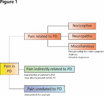

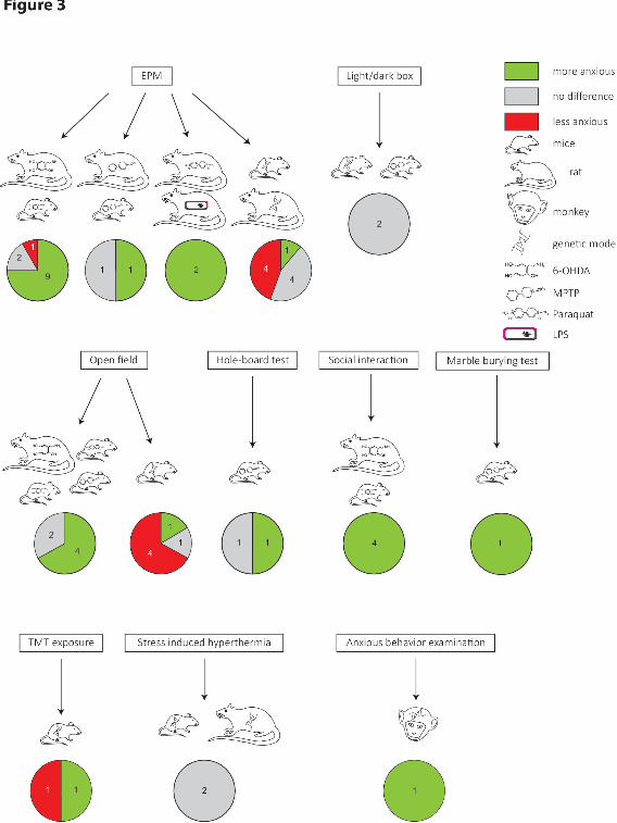
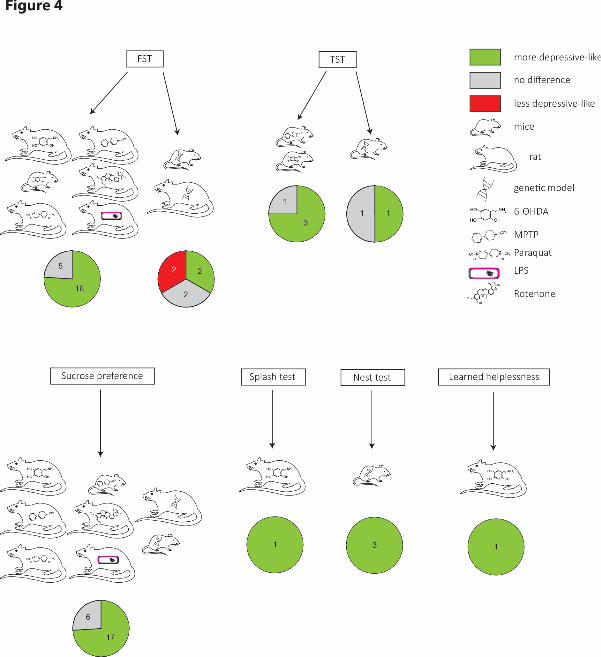
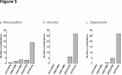
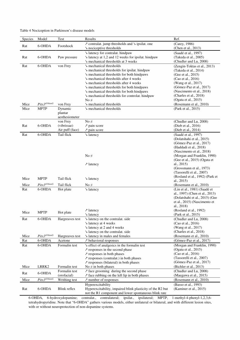
Table 4 Nociception in Parkinson’s disease models
Species Model Test Results Ref.
Rat 6-OHDA Footshock ↗ contralat. jump thresholds and ↘ ipsilat. one ↘ nociceptive thresholds
(Carey, 1986) (Chen et al., 2013)
Rat 6-OHDA Paw pressure ↘ latency for contralat. hindpaw ↘ latency at 1,2 and 12 weeks for ipsilat. hindpaw ↘ mechanical thresholds at 3 weeks
(Saadé et al., 1997) (Takeda et al., 2005) (Chudler and Lu, 2008)
Rat 6-OHDA von Frey ↘ mechanical thresholds ↘ mechanical thresholds for ipsilat. hindpaw ↘ mechanical thresholds for both hindpaws ↘ mechanical thresholds after 4 weeks ↘ mechanical thresholds after 4 weeks ↘ mechanical thresholds for both hindpaws ↘ mechanical thresholds for both hindpaws ↘ mechanical thresholds for controlat. hindpaw No ≠
(Zengin-Toktas et al., 2013) (Takeda et al., 2014) (Gee et al., 2015) (Cao et al., 2016) (Wang et al., 2017) (Gómez-Paz et al., 2017) (Nascimento et al., 2018) (Charles et al., 2018) (Ogata et al., 2015)
Mice Pitx3416insG von Frey ↘ mechanical thresholds (Rosemann et al., 2010) Mice MPTP Dynamic
plantar aesthesiometer
↘ mechanical thresholds (Park et al., 2015)
Rat 6-OHDA von Frey (vibrissae) Air puff (face)
No ≠ ↗ pain score ↗ pain score
(Chudler and Lu, 2008) (Dieb et al., 2016) (Dieb et al., 2014)
Rat 6-OHDA Tail flick ↘ latency No ≠ ↗ latency
(Saadé et al., 1997) (Dolatshahi et al., 2015) (Gómez-Paz et al., 2017) (Haddadi et al., 2018) (Nascimento et al., 2018) (Morgan and Franklin, 1990) (Gee et al., 2015) (Ogata et al., 2015) (Grossmann et al., 1973) (Tassorelli et al., 2007)
Mice MPTP Tail flick ↘ latency (Rosland et al., 1992) (Park et al., 2015)
Mice Pitx3416insG Tail flick No ≠ (Rosemann et al., 2010) Rat 6-OHDA Hot plate ↘ latency (Lin et al., 1981) (Saadé et
al., 1997) (Chen et al., 2013) (Dolatshahi et al., 2015) (Gee et al., 2015) (Nascimento et al., 2018)
Mice MPTP Hot plate ↗ latency ↘ latency
(Rosland et al., 1992) (Park et al., 2015)
Rat 6-OHDA Hargreaves test ↘ latency on the contralat. side ↘ latency at 4 weeks ↘ latency at 2 and 4 weeks ↘ latency on the contralat. side
(Chudler and Lu, 2008) (Cao et al., 2016) (Wang et al., 2017) (Charles et al., 2018)
Mice Pitx3416insG Hargreaves test ↘ latency in males and females (Rosemann et al., 2010) Rat 6-OHDA Acetone ↗ behavioral responses (Gómez-Paz et al., 2017) Rat 6-OHDA Formalin test ↘ effect of analgesics in the formalin test
↗ responses in the second phase ↗ responses in both phases ↗ responses (contralat.) in both phases ↗ responses (bilateral) in both phases
(Morgan and Franklin, 1990) (Ogata et al., 2015) (Cao et al., 2016) (Tassorelli et al., 2007) (Gómez-Paz et al., 2017)
Mice LRRK2 Formalin test No ≠ in both phases (Bichler et al., 2013)
Rat 6-OHDA Formalin test (orofacial)
↗ face grooming during the second phase ↗ face rubbing on the left lip in both phases
(Chudler and Lu, 2008) (Maegawa et al., 2015)
Mice Pitx3416insG Writhing test ↗ number of responses (Rosemann et al., 2010)
Rat 6-OHDA Blink reflex Hyperexcitability Hyperexcitability, impaired blink plasticity of the R2 but not the R1 component and lower spontaneous blink rate
(Basso et al., 1993) (Kaminer et al., 2015)
6-OHDA, 6-hydroxydopamine; contralat., contralateral; ipsilat., ipsilateral; MPTP, 1-methyl-4-phenyl-1,2,3,6-tetrahydropyridine. Note that “6-OHDA” gathers various models, either unilateral or bilateral, and with different lesion sites, with or without neuroprotection of non-dopamine systems.
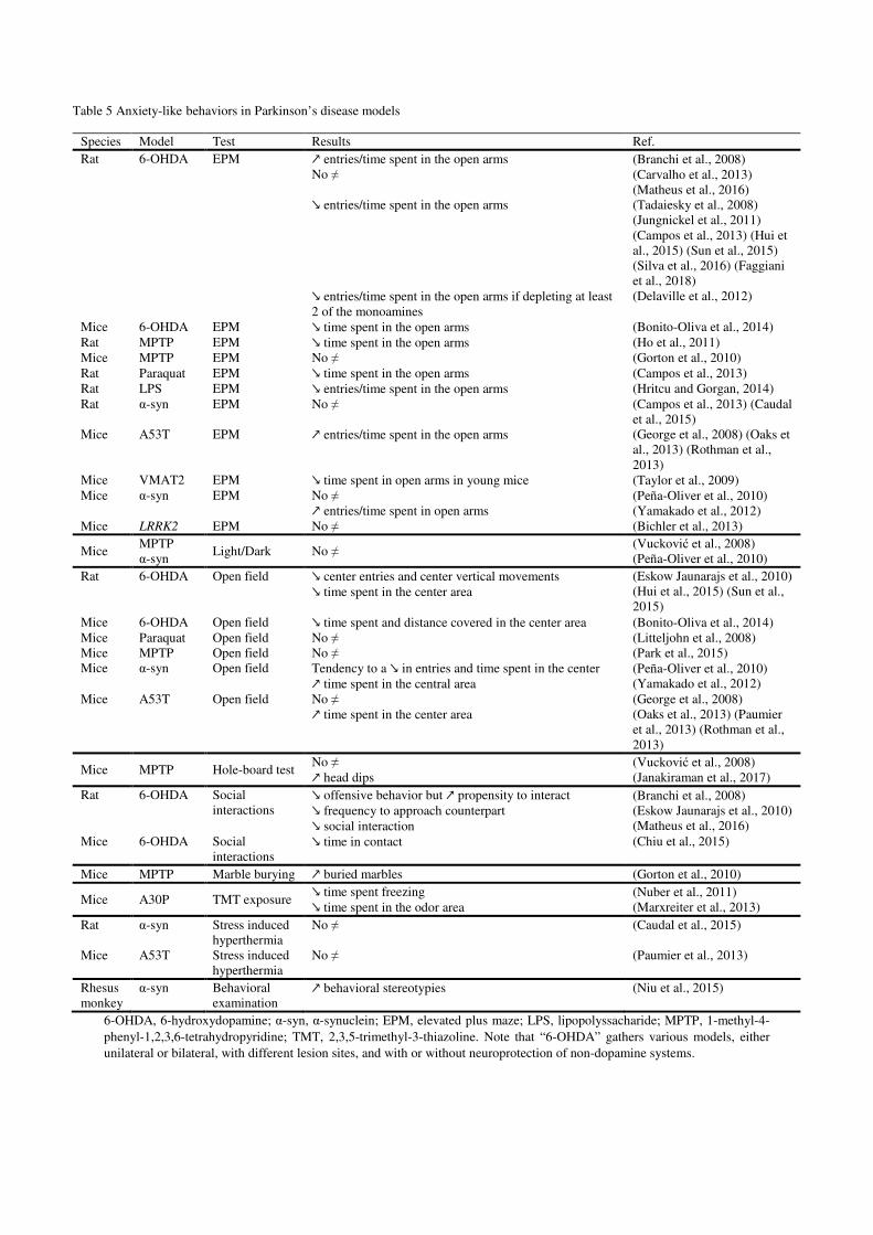
Table 5 Anxiety-like behaviors in Parkinson’s disease models
Species Model Test Results Ref. Rat 6-OHDA EPM ↗ entries/time spent in the open arms
No ≠ ↘ entries/time spent in the open arms ↘ entries/time spent in the open arms if depleting at least 2 of the monoamines
(Branchi et al., 2008) (Carvalho et al., 2013) (Matheus et al., 2016) (Tadaiesky et al., 2008) (Jungnickel et al., 2011) (Campos et al., 2013) (Hui et al., 2015) (Sun et al., 2015) (Silva et al., 2016) (Faggiani et al., 2018) (Delaville et al., 2012)
Mice 6-OHDA EPM ↘ time spent in the open arms (Bonito-Oliva et al., 2014) Rat MPTP EPM ↘ time spent in the open arms (Ho et al., 2011) Mice MPTP EPM No ≠ (Gorton et al., 2010) Rat Paraquat EPM ↘ time spent in the open arms (Campos et al., 2013) Rat LPS EPM ↘ entries/time spent in the open arms (Hritcu and Gorgan, 2014) Rat α-syn EPM No ≠ (Campos et al., 2013) (Caudal
et al., 2015) Mice A53T EPM ↗ entries/time spent in the open arms (George et al., 2008) (Oaks et
al., 2013) (Rothman et al., 2013)
Mice VMAT2 EPM ↘ time spent in open arms in young mice (Taylor et al., 2009) Mice α-syn EPM No ≠
↗ entries/time spent in open arms (Peña-Oliver et al., 2010) (Yamakado et al., 2012)
Mice LRRK2 EPM No ≠ (Bichler et al., 2013)
Mice MPTP α-syn
Light/Dark No ≠ (Vucković et al., 2008) (Peña-Oliver et al., 2010)
Rat 6-OHDA Open field ↘ center entries and center vertical movements ↘ time spent in the center area
(Eskow Jaunarajs et al., 2010) (Hui et al., 2015) (Sun et al., 2015)
Mice 6-OHDA Open field ↘ time spent and distance covered in the center area (Bonito-Oliva et al., 2014) Mice Paraquat Open field No ≠ (Litteljohn et al., 2008) Mice MPTP Open field No ≠ (Park et al., 2015) Mice α-syn Open field Tendency to a ↘ in entries and time spent in the center
↗ time spent in the central area (Peña-Oliver et al., 2010) (Yamakado et al., 2012)
Mice A53T Open field No ≠ ↗ time spent in the center area
(George et al., 2008) (Oaks et al., 2013) (Paumier et al., 2013) (Rothman et al., 2013)
Mice MPTP Hole-board test No ≠ ↗ head dips
(Vucković et al., 2008) (Janakiraman et al., 2017)
Rat 6-OHDA Social interactions
↘ offensive behavior but ↗ propensity to interact ↘ frequency to approach counterpart ↘ social interaction
(Branchi et al., 2008) (Eskow Jaunarajs et al., 2010) (Matheus et al., 2016)
Mice 6-OHDA Social interactions
↘ time in contact (Chiu et al., 2015)
Mice MPTP Marble burying ↗ buried marbles (Gorton et al., 2010)
Mice A30P TMT exposure ↘ time spent freezing ↘ time spent in the odor area
(Nuber et al., 2011) (Marxreiter et al., 2013)
Rat α-syn Stress induced hyperthermia
No ≠ (Caudal et al., 2015)
Mice A53T Stress induced hyperthermia
No ≠ (Paumier et al., 2013)
Rhesus monkey
α-syn Behavioral examination
↗ behavioral stereotypies (Niu et al., 2015)
6-OHDA, 6-hydroxydopamine; α-syn, α-synuclein; EPM, elevated plus maze; LPS, lipopolyssacharide; MPTP, 1-methyl-4-phenyl-1,2,3,6-tetrahydropyridine; TMT, 2,3,5-trimethyl-3-thiazoline. Note that “6-OHDA” gathers various models, either unilateral or bilateral, with different lesion sites, and with or without neuroprotection of non-dopamine systems.
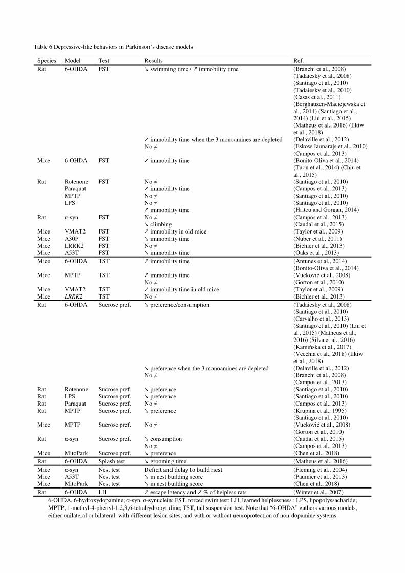
Table 6 Depressive-like behaviors in Parkinson’s disease models
Species Model Test Results Ref.
Rat 6-OHDA FST ↘ swimming time / ↗ immobility time
↗ immobility time when the 3 monoamines are depleted
No ≠
(Branchi et al., 2008)
(Tadaiesky et al., 2008)
(Santiago et al., 2010)
(Tadaiesky et al., 2010)
(Casas et al., 2011)
(Berghauzen-Maciejewska et
al., 2014) (Santiago et al.,
2014) (Liu et al., 2015)
(Matheus et al., 2016) (Ilkiw
et al., 2018)
(Delaville et al., 2012)
(Eskow Jaunarajs et al., 2010)
(Campos et al., 2013)
Mice 6-OHDA FST ↗ immobility time (Bonito-Oliva et al., 2014)
(Tuon et al., 2014) (Chiu et
al., 2015)
Rat Rotenone
Paraquat
MPTP
LPS
FST No ≠
↗ immobility time
No ≠
No ≠
↗ immobility time
(Santiago et al., 2010)
(Campos et al., 2013)
(Santiago et al., 2010)
(Santiago et al., 2010)
(Hritcu and Gorgan, 2014)
Rat α-syn FST No ≠
↘ climbing
(Campos et al., 2013)
(Caudal et al., 2015)
Mice VMAT2 FST ↗ immobility in old mice (Taylor et al., 2009)
Mice A30P FST ↘ immobility time (Nuber et al., 2011)
Mice LRRK2 FST No ≠ (Bichler et al., 2013)
Mice A53T FST ↘ immobility time (Oaks et al., 2013)
Mice 6-OHDA TST ↗ immobility time (Antunes et al., 2014)
(Bonito-Oliva et al., 2014)
Mice MPTP TST ↗ immobility time
No ≠
(Vucković et al., 2008)
(Gorton et al., 2010)
Mice VMAT2 TST ↗ immobility time in old mice (Taylor et al., 2009)
Mice LRRK2 TST No ≠ (Bichler et al., 2013)
Rat 6-OHDA Sucrose pref. ↘ preference/consumption
↘ preference when the 3 monoamines are depleted
No ≠
(Tadaiesky et al., 2008)
(Santiago et al., 2010)
(Carvalho et al., 2013)
(Santiago et al., 2010) (Liu et
al., 2015) (Matheus et al.,
2016) (Silva et al., 2016)
(Kamińska et al., 2017)
(Vecchia et al., 2018) (Ilkiw
et al., 2018)
(Delaville et al., 2012)
(Branchi et al., 2008)
(Campos et al., 2013)
Rat Rotenone Sucrose pref. ↘ preference (Santiago et al., 2010)
Rat LPS Sucrose pref. ↘ preference (Santiago et al., 2010)
Rat Paraquat Sucrose pref. No ≠ (Campos et al., 2013)
Rat MPTP Sucrose pref. ↘ preference (Krupina et al., 1995)
(Santiago et al., 2010)
Mice MPTP Sucrose pref. No ≠ (Vucković et al., 2008)
(Gorton et al., 2010)
Rat α-syn Sucrose pref. ↘ consumption
No ≠
(Caudal et al., 2015)
(Campos et al., 2013)
Mice MitoPark Sucrose pref. ↘ preference (Chen et al., 2018)
Rat 6-OHDA Splash test ↘ grooming time (Matheus et al., 2016)
Mice
Mice
α-syn
A53T
Nest test
Nest test
Deficit and delay to build nest
↘ in nest building score
(Fleming et al., 2004)
(Paumier et al., 2013)
Mice MitoPark Nest test ↘ in nest building score (Chen et al., 2018)
Rat 6-OHDA LH ↗ escape latency and ↗ % of helpless rats (Winter et al., 2007)
6-OHDA, 6-hydroxydopamine; α-syn, α-synuclein; FST, forced swim test; LH, learned helplessness ; LPS, lipopolyssacharide;
MPTP, 1-methyl-4-phenyl-1,2,3,6-tetrahydropyridine; TST, tail suspension test. Note that “6-OHDA” gathers various models,
either unilateral or bilateral, with different lesion sites, and with or without neuroprotection of non-dopamine systems.