Embed Size (px)
Citation preview

www.elsevier.com/locate/phytochem
Phytochemistry 66 (2005) 2800–2814
PHYTOCHEMISTRY
Molecules of Interest
Sugar beet (Beta vulgaris) pectins are covalently cross-linkedthrough diferulic bridges in the cell wall
Marie-Christine Ralet *, Gwenaelle Andre-Leroux 1,Bernard Quemener, Jean-Francois Thibault
Unite Biopolymeres Interactions Assemblages, Institut National de la Recherche Agronomique,
rue de la tsaven Geraudiere B.P. 71627, 44316 Nantes Cedex 03, France
Received 19 July 2005; received in revised form 28 September 2005Available online 17 November 2005
Abstract
Arabinan and galactan side chains of sugar beet pectins are esterified by ferulic acid residues that can undergo in vivo oxidative reac-tions to form dehydrodiferulates. After acid and enzymatic degradation of sugar beet cell walls and fractionation of the solubilized prod-ucts by hydrophobic interaction chromatography, three dehydrodiferulate-rich fractions were isolated. The structural identification ofthe different compounds present in these fractions was performed by electrospray-ion trap-mass spectrometry (before and after 18O label-ing) and high-performance anion-exchange chromatography. Several compounds contained solely Ara (terminal or a-1! 5-linked-dimer) and dehydrodiferulate. The location of the dehydrodiferulate was assigned in some cases to the O-2 and in others to the O-5of non-reducing Ara residues. One compound contained Gal (b-1! 4-linked-dimer), Ara (a-1! 5-linked-dimer) and dehydrodiferulate.The location of the dehydrodiferulate was unambiguously assigned to the O-2 of the non-reducing Ara residue and O-6 of the non-reduc-ing Gal residue. These results provide direct evidence that pectic arabinans and galactans are covalently cross-linked (intra- or inter-molecularly) through dehydrodiferulates in sugar beet cell walls. Molecular modeling was used to compute and structurally characterizethe low energy conformations of the isolated compounds. Interestingly, the conformations of the dehydrodiferulate-bridged arabinanand galactan fragments selected from an energetic criterion, evidenced very nice agreement with the experimental occurrence of the dehy-drodiferulated pectins. The present work combines for the first time intensive mass spectrometry data and molecular modeling to givestructural relevance of a molecular cohesion between rhamnogalacturonan fragments.� 2005 Elsevier Ltd. All rights reserved.
Keywords: Amaranthaceae (Chenopodiaceae); Ferulic acid; Dehydrodimer; Dehydrodiferulate; Pectin; Arabinan; Galactan; Conformational minimum
0031-9422/$ - see front matter � 2005 Elsevier Ltd. All rights reserved.
doi:10.1016/j.phytochem.2005.09.039
Abbreviations: CID, collision-induced dissociation; CW, cell wall;CWM, cell wall material; diAra, a-1! 5-linked dimer; diFA, dehydro-diferulate; diGal, b-1! 4-linked dimer; ESI-IT-MS, electrospray ion-trapmass-spectrometry; EtOH, ethanol; FA, ferulic acid; hpaec, high-performance anion-exchange chromatography; m/z, mass to charge ratio;TFA, trifluoroacetic acid.* Corresponding author. Tel.: +33 240675196; fax: +33 240675084.E-mail address: [email protected] (M.-C. Ralet).
1 Present address: Institut Pasteur, Unite de Biochimie Structurale, 25rue du Dr Roux, 75724 Paris Cedex 15, France.
1. Introduction
Plant cell walls (CW) are complex multifunctional struc-tures constructed principally of polysaccharides. The abun-dance of CW bound ferulic acid (FA) is a distinctivespecific feature of primary CW of species belonging tothe commelinoid group of monocotyledons, the dicotyle-don order Caryophyllales and all gymnosperm families(Carnachan and Harris, 2000). In species of the familyAmaranthaceae (Caryophyllales) such as sugar beet (Betavulgaris), spinach (Spinacia oleracea), glasswort (Salicorniaramosissima) and quinoa (Chenopodium quinoa), it is pecticpolymers that are feruloylated (Fry, 1982; Rombouts and

M.-C. Ralet et al. / Phytochemistry 66 (2005) 2800–2814 2801
Thibault, 1986; Renard et al., 1993a, 1999). Pectin mainstructural features include homogalacturonic and rhamno-galacturonic regions. The latter display some Rha residuessubstituted by Ara- and Gal-containing side chains. FAresidues are mainly ester-linked to O-2 of Ara residues ofthe main core of a-(1! 5)-linked arabinan chains and toO-6 of Gal residues of the main core of b-(1! 4)-linkedtype I galactan chains (Ishii and Tobita, 1993; Colquhounet al., 1994). Recently, minor amounts of FA were assumedto be also ester-linked to O-5 of Ara residues of the maincore of a-(1! 5)-linked arabinan chains, indicating apotential peripheral location of some FA on pectic ‘‘hairy’’regions (Levigne et al., 2004a).
The FA esters are of great interest since they canundergo in vivo oxidative coupling reactions to formdehydrodimers (diFA) (Fry, 1986). Four dimer isomersare present in cell walls (8-O-4 0, 8-5 0, 8-8 0 and 5-5 0)(Fig. 1), their relative proportion varying greatly withrespect to plant origin. In most angiosperm tissues, the 8-O-4 0 and 8-5 0 dimers are the most abundant (Wendeet al., 2000). In vivo diFA formation enables covalentcross-linking of the polysaccharides they esterify. Suchcoupling, that was up to recently thought to take placeexclusively in the CW, was claimed to have a ‘‘tightening’’effect (contribution to wall assembly, promotion of tissuecohesion, restriction of cell expansion, resistance againstfungal penetration etc. . .) (Fry, 1986; Kamisaka et al.,1990; Ralph and Helm, 1993; Brady and Fry, 1997). Itwas, however, lately shown that feruloyl coupling occurrednot only in the CW but also intra-protoplasmically, mostlikely in the Golgi apparatus (Fry et al., 2000; Obel et al.,2003). More balanced hypotheses were then proposed
OH
O
OMe
OH
HO O
OMe
OH
HO O
OMe
OH
OH
O
OMe
O
HO O
OMe
OHHO O
MeO
OH
5-5'8-O-4'
8-5'
OHO
MeO
OH
HO O
OMe
OH
8-8'
Fig. 1. Structure of the main diFA isomers present in plant cell walls.
about the physiological role of such oxidative coupling(Fry et al., 2000). In young maize (Zea mays L.) cultures,much oxidative coupling of feruloyl arabinoxylans wasshown to occur intra-protoplasmically. The majority ofthe arabinoxylan secreted onto the inner surface of theCW was assumed to be present as a pre-cross-linked coag-ulum that could prevent the newly deposited cellulosemicrofibrils to assemble through hydrogen bonds and helpin that way to maintain a high CW extensibility. Oldermaize cultures oxidatively coupled substantially fewer oftheir feruloyl residues intraprotoplasmically. The intenseextraprotoplasmic coupling of feruloyl arabinoxylanswould here tighten the CW (Fry et al., 2000). Additionally,Obel et al. (2003) demonstrated that intracellular dimerformation is confined to specific dimers. 8-5 0 diFA is indeedformed intracellularly while 5-5 0 and 8-O-4 0 diFA are sup-posed to be formed extracellularly.
Fragments composed of a pair of xylan-related oligosac-charides bridged by a 5-5 0 diFA group were isolated frommonocotyledons (Ishii, 1991; Saulnier et al., 1999). Thesefragments are likely to represent inter-polysaccharidecross-links, although the existence of intra-polysaccharideloops cannot be precluded. DiFA have been identifiedand quantified in sugar beet CW, where 15% to over 20%of the quantified alkali-soluble wall-bound phenolics com-prised diFA, indicating a potentially high degree of poly-mer cross-linking (Micard et al., 1997; Waldron et al.,1997). We describe here the isolation of several diFA-bridged oligosaccharides. The structural identification ofthese compounds by high-performance anion-exchangechromatography (hpaec) and electrospray ion-trap mass-spectrometry (ESI-IT-MS) provides direct evidence forcovalent (intra- or inter-molecular) cross-linking of pecticarabinans and galactans through diFA bridges in sugarbeet CW. In an effort to supply high definition of themolecular structures of the identified diFA-bridged arab-inan and galactan fragments, we computed and character-ized their low energy conformations using molecularmodeling techniques. The structures issued from the con-formational study constitute a step forward towards amolecular comprehension of occurence, flexibility andstrength of the diFA bridging effect in the CW.
2. Results
2.1. Isolation and chemical analysis of diFA-rich fractions
A sequential isolation process including acid and enzy-matic hydrolyses and low pressure hydrophobic interactionchromatographic fractionations was followed (Fig. 2).Sugar beet root cell wall material (CWM) (FA 8.2 mg/g;diFA 0.65 mg/g) was hydrolyzed by trifluoroacetic acid(TFA) and the resulting TFA-soluble fraction precipitatedby ethanol (EtOH). It was established in a previous workthat such conditions allowed the recovery of most of theAra residues in the EtOH-soluble fraction (Ralet et al.,

Sugar beet root CWM
TFA
residuesoluble
EtOH-precipitateEtOH-soluble
TFAAmberlite XAD-2
0/100* 15/85* 25/75* 50/50*
Sephadex LH-20
0/100* 15/85* 30/70*
F1F2F3F4F5
F6F7
Ronozym + Driselase
residuesoluble
Amberlite XAD-2
0/100* 15/85* 25/75* 50/50*
Sephadex LH-20
0/100* 15/85* 30/70*
F8F9F10F11
F12
EtOH
Fig. 2. Isolation of diFA-rich fractions (F6, F7 and F12) implementing acid and enzymatic hydrolyses and low pressure hydrophobic interactionchromatographic fractionations (Amberlite XAD-2 and Sephadex LH-20). % EtOH/water (v/v).
2802 M.-C. Ralet et al. / Phytochemistry 66 (2005) 2800–2814
1994). Indeed, �92% of the Ara residues were recovered inthe EtOH-soluble fraction together with �20% of the Galresidues and �55% of the FA and diFA. The repartitionof the different diFA isomers was similar within theEtOH-soluble fraction, EtOH-precipitate and TFA hydro-lysis residue and was representative of the repartitionwithin the initial CWM (5-5 0:8-O-4 0:8-5 0 4:48:48, w/w/w).
The EtOH-soluble fraction was further hydrolyzed withTFA, and loaded onto an Amberlite XAD-2 column elutedsuccessively with water, EtOH:water 15:85 (v/v), EtOH:-water 25:75 (v/v) and EtOH:water 50:50 (v/v). Chromato-graphic yields were >95% for individual sugars, FA anddiFA. The water-eluted fraction contained most of thetotal recovered sugars (97% w/w) and low amounts ofFA and diFA (�1% w/w of the total recovered FA anddiFA). Most of the FA and diFA (67% and 77% w/w ofthe total recovered FA and diFA, respectively) were pres-ent in the EtOH:water 25:75-eluted fraction that was sub-jected to further purification on Sephadex LH-20 elutedsuccessively by water, EtOH:water 15:85 (v/v) and EtOH:-water 30:70 (v/v). Five fractions (F1–F5) were eluted bywater and two fractions were eluted by EtOH:water 15:85(F6, F7). The sugar and phenolic composition of each frac-tion are reported in Table 1. The chromatographic yieldswere close to 100% for individual sugars and FA and�60% for diFA. Most of the diFA was recovered in F2,F6 and F7. F2 contained large amounts of FA (FA/diFAmol/mol of 0.96) and was not further studied. F6 and F7contained Ara as sole sugar and low amounts of FA
(FA/diFA mol/mol of 0.33 and 0 for F6 and F7, respec-tively). The diFA isomers repartition showed the predom-inance of 8-O-4 0 dimer in F6 and the presence of both5-5 0 and 8-O-4 0 dimers in F7. Those two fractions wereselected for further study.
The EtOH-precipitate and TFA hydrolysis residue stillcontained altogether 80% of the Gal, 8% of the Ara and45% of the FA and diFA initially present in the CWM.In order to recover additional diFA-bridged oligosaccha-rides, the EtOH-precipitate and the TFA hydrolysis residuewere hydrolyzed by a mixture of Driselase� and Ron-ozym� two preparations rich in pectolytic activities. Theresulting soluble fraction was loaded onto an AmberliteXAD-2 column as described above. Chromatographicyields were >95% for individual sugars, FA and diFA.The water-eluted fraction contained most of the totalrecovered sugars (95% w/w) and low amounts of FA anddiFA (2.3% and 7.5% w/w of the total recovered FA anddiFA, respectively). Most of the phenolics (71% and 60%w/w of the total recovered FA and diFA, respectively) werepresent in the EtOH:water 25:75-eluted fraction. UnlikeDriselase�, Ronozym� contains some ferulate esteraseactivity and 35% of the FA recovered consisted of freeFA. Interestingly, this ferulate esterase was similarly activeon diFA as 32% of the diFA recovered consisted of freediFA. The EtOH:water 25:75-eluted fraction, that con-tained the major part of diFA, was subjected to furtherpurification on Sephadex LH-20 after removal of free FAand diFA by ether extraction at pH 2. The ether extract

Table 1Composition of the Sephadex LH-20 fractions
Fractions % of total amount elutedfrom LH-20
Sugar repartition within eachfraction (mol%)
diFA repartitionwithin each fraction(mol%)
FA*100/diFA (Ara + Gal)/(FA + diFA)
Sugars FA DiFA GalUA Rha Ara Gal 5-50 8-O-40 8-5 0 (mol%) (mol/mol)
TFA
F1 13 1 2 39 16 11 34 9 52 39 92 12F2 25 13 21 0 1 64 35 8 68 24 96 3F3 20 20 7 0 0 100 0 45 17 38 99 2F4 39 65 9 0 0 100 0 6 41 53 >99 1F5 1 1 6 0 0 96 0 5 32 62 85 2F6 1 <1 34 0 0 100 0 4 78 19 33 2F7 1 0 22 0 0 100 0 41 55 5 0 3
Enzymes
F8 47 8 44 0 13 17 34 11 36 53 85 11F9 9 11 13 0 1 10 74 12 46 43 96 3F10 42 77 24 0 0 3 97 8 32 61 99 2F11 2 4 8 0 0 65 35 9 73 18 94 2F12 <1 0 11 0 0 45 55 79 21 0 20 2
M.-C. Ralet et al. / Phytochemistry 66 (2005) 2800–2814 2803
did not contain any sugar. Four fractions (F8–F11) wereeluted by water and one fraction was eluted by EtOH:water15:85 (F12). The sugar and phenolic composition of eachfraction are reported in Table 1. The chromatographicyields were close to 100% for individual sugars, FA anddiFA. Most of the diFA was recovered in F8, showing thatdiFA were still attached to sugar moieties of high degree ofpolymerization. F9–F11 also contained substantialamounts of diFA but contained also large amounts ofFA (FA/diFA mol/mol of 0.96, 0.99 and 0.94 for F9,F10 and F11, respectively) and were not further studied.F12 contained substantial amounts of diFA, Ara and Galin similar quantities and was lowly contaminated by FA(FA/diFA mol/mol of 0.20). The diFA isomers repartitionshowed the predominance of the 5-5 0 dimer in F12. Thisfraction was selected for further study.
Fig. 3. High performance anion exchange chromatography (pH 13)profiles of the diFA-rich fractions (F6, F7 and F12). a-(1! 5)-linked-arabinan-derived oligosaccharides and b-(1! 4)-linked-galactan-derivedoligosaccharides were used as standards.
2.2. hpaec and MS characterization of diFA-rich fractions
F6, F7 and F12 were analyzed by hpaec at pH 13 (Fig. 3).The ester bond FA residues are released due to the stronglyalkaline conditions used and pure carbohydrates are sepa-rated on the column. By comparison with a-(1! 5)-arabinan- and b-(1! 4)-galactan-derived oligosaccharides,it was shown that F6 and F7 contained solely Ara mono-mers and a-(1! 5)-linked Ara dimers and that F12 con-tained a-(1! 5)-linked Ara dimers and b-(1! 4)-linkedGal dimers. F6, F7 and F12 were further analyzed by nega-tive ESI-IT-MS by direct infusion into the electrospraysource (Fig. 4). Several compounds were detected in F6 withdeprotonated singly charged [M � H]� ions at mass-to-charge ratio (m/z) 517, 649, 781 and 913, that could corre-spond to Ara1-diFA, Ara2-diFA, Ara3-diFA andAra4-diFA, respectively; deprotonated [M � H]� ions atm/z 473, 499, 817 and 1081, that could correspond todecarboxylated Ara1-diFA, dehydrated Ara1-diFA, Ara3-diFA + 2H2O and Ara5-diFA + 2H2O, respectively; depro-
tonated cluster [2M � H]� ions at m/z 1299, that could cor-respond to Ara2-diFA. Major ions at m/z 649 (compound1), 781 (compound 2) and 913 (compound 3) were chosen

200 400 600 800 1000 1200 1400 1600 1800 2000
781
649
517
499 817 913473 1081 1299
0
20
40
60
80
100
Compound 3
Compound 2
Compound 1
F6
200 400 600 800 1000 1200 1400 1600 1800 2000
649
781
517 685
0
20
40
60
80
100
Compound 5
Compound 4F7
200 400 600 800 1000 1200 1400 1600 1800 2000
973
325 841 913563519 11053850
20
40
60
80
100
Compound 6F12
m/z
Relative abundance
Fig. 4. Negative full MS spectra of the diFA-rich fractions (F6, F7 and F12).
2804 M.-C. Ralet et al. / Phytochemistry 66 (2005) 2800–2814
for further structural studies. F7 exhibited two main signalswith deprotonated [M � H]� ions at m/z 649 (compound 4)and 781 (compound 5) that could correspond to Ara2-diFAand Ara3-diFA. Finally, a prominent ion at m/z 973 (com-pound 6) that could correspond to Ara2-Gal2-diFA, wasdetected in F12. After 18O-labeling of the whole fractions,compounds 2–6 gained 4 mass units, in agreement withthe presence of two reducing ends while compound 1 gained2 mass units, in agreement with the presence of one reducingend. Considering the results obtained, the following partialstructure could be assigned to the different compounds:
Compound 1
m/z 649 diFA-Ara-a-(1! 5) Ara Compound 2 m/z 781 Ara-diFA-Ara-a-(1! 5) Ara Compound 3 m/z 913 Ara (5 1)-a-Ara-diFA-Ara-a-(1! 5) Ara
Compound 4
m/z 649 Ara-diFA-Ara Compound 5 m/z 781 Ara-diFA-Ara-a-(1! 5) Ara Compound 6 m/z 973 Gal (4 1)-b-Gal-diFA-Ara-a-(1! 5) Ara
2.3. Full structural assignment by MS fragmentation
techniques
MSn experiments were carried out in order to allow theprecise location of the phenolic moieties onto the sugarrings for the different compounds. MSn experiments wereperformed by negative ESI-IT-MS by direct infusion ofF6, F7 and F12 into the electrospray source. The electro-spray ionization followed by formation of fragment ionsby collision-induced dissociation (CID) allows sensitive

M.-C. Ralet et al. / Phytochemistry 66 (2005) 2800–2814 2805
mapping and sequencing of oligosaccharides. Carbohy-drates undergo two types of fragmentation: those of glyco-sidic cleavages and those of cross-ring fragmentation.According to the cross-ring fragmentation, linkage andbranching patterns can be established (Vakhrushev et al.,2004). For sequencing, glycosidic cleavages along the chainare major tools, especially after 18O labelling of the reduc-ing end. In the present work, 18O labeling was particularlyuseful for establishing whether the losses forming ions con-tained two (**), one (*), or no 18O-labeled reducing end.
2.3.1. Compounds arising from F6
Compounds 1, 2 and 3 exhibited very similar fragmenta-tion patterns and the spectra obtained for compound 3 willbe the ones fully presented here (Fig. 5). Fig. 6 shows thedifferent cleavages observed at each MS step.
After isolation and collision-induced dissociation (CID)of the parent ion at m/z 913 (Ara-Ara-diFA-Ara-Ara) in anMS2 analysis, a series of fragment ions appeared(Fig. 5(a)). The prominent ions produced at m/z 853 and823, corresponding to losses of 60 Da (C2H4O2) and90 Da (C3H6O3), respectively, resulted from cross ringcleavages. A glycosidic fragment ion at m/z 781 (132 Daloss), corresponding to the loss of one Ara unit, was pro-duced. After isolation and CID of the doubly labeled par-ent ion at m/z 917**(*Ara-Ara-diFA-Ara-Ara*), a series ofsingly labeled prominent fragment ions appeared at m/z783*, 825*, and 855*, corresponding to losses of 134 Da(labeled Ara unit), 92 Da (labeled C3H6O3), and 62 Da(labeled C2H4O2), respectively (Fig. 5(a) inset). The frag-mentation pattern observed for labeled and non-labeledcompound 3 is consistent with a linkage through O-5 ofone of the two reducing Ara residues (Harvey, 2000;Quemener and Ralet, 2004; Levigne et al., 2004b) (arbi-trarily quoted Ara1 on Fig. 6), in agreement with hpaecdata. The fragment ion at m/z 781 undergoes further frag-mentations that were assigned to H2O loss (m/z 763) and tocross-ring cleavage fragment ions (m/z 721 and 691, respec-tively). The ion at m/z 649 corresponds to the loss of oneextra Ara residue (132 Da loss). The peak at m/z 631 couldbe assigned to the loss of a water molecule from the ion atm/z 649. The 18O-labeled ion at 783* also undergoes fur-ther fragmentations and one can differentiate between theloss of the second reducing labeled Ara residue (m/z 649,134 Da loss, Ara4 in Fig. 6) and the loss of a non labeledAra residue in ester linkage with the phenolic moiety (m/z 651*, 132 Da loss, Ara2 in Fig. 6). The peak at m/z633* could be assigned to the loss of a non-labeled watermolecule (18 Da loss) from the ion at m/z 651*.
The glycosidic cleavage ion at m/z 781 (Ara-Ara-diFA-Ara ) was selected for further fragmentation in a MS3
CID experiment (m/z 913 > 781 > products) (Figs. 5(b),6(b)). The MS3 trace provided the four fragment ions atm/z 763, 721, 691 and 649 that were already observed inMS2 analysis. The MS3 CID experiment conducted onthe 18O-labeled ion at 783* (*Ara-Ara-diFA-Ara ) (m/z
917** > 783* > products) revealed to be much more infor-mative (Fig. 5(b), inset) with a clear differentiation betweenfragments arising from the second reducing labeled Araresidue (Ara4 in Fig. 6) and the loss of a non labeled Araresidue in ester linkage with the phenolic moiety (Ara2 inFig. 6). Besides glycosidic fragment ions at m/z 649 and651* and consecutive water losses at 631 and 633*, twocross ring cleavage fragment ions at m/z 721 and 691,involving the labeled Ara residue, and one cross ring cleav-age fragment ion at m/z 723*, involving the non labeledAra residue, were observed. The fragmentation patternobserved for the labeled Ara residue (62 and 92 Da losses)is again consistent with a linkage through O-5 of thisreducing Ara residue (Ara4 in Fig. 5), in agreement withhpaec data. The fragmentation observed for the non-labeled Ara residue (Ara2 in Fig. 6) was interpreted bycomparison with CID product-ion patterns obtained forferuloylated arabinose containing mono- and disaccharideswith known linkage configurations (Quemener and Ralet,2004). Indeed, the CID spectrum of an Ara residue substi-tuted on O-2 by a FA residue was shown to be dominatedby a diagnostic cross-ring cleavage ion at (�108 Da) and anintense water loss, while a high relative abundance of thediagnostic cross-ring cleavage ion at (�60 Da) togetherwith moderate water loss were characteristic of linkage ofFA through the O-5 of the Ara unit. The fragmentationobserved for the non-labeled Ara residue in ester linkagewith the phenolic moiety, namely the abundant productionof the ion at m/z 723* (60 Da loss), the absence of 108 Daloss and the absence of water loss, is consistent with thelinkage of the phenolic moiety through the O-5 of theAra unit.
The fragment ions at m/z 649 or m/z 651* were not pro-duced in sufficient quantity (either from MS2 or MS3 anal-yses) to allow further fragmentation. MS3 analysis of theintense ion at m/z 631 (m/z 913 > 631 > products) was ten-tatively performed (Figs. 5(c) and 6(c)). This ion wasshown to arise solely from the loss of two consecutiveAra units (one labeled and one non labeled, quoted Ara1and Ara2 in Fig. 6, respectively) (Ara-Ara-diFA -H2O)(Figs. 5(b) and 6(b)). Beside moderate water loss (m/z613), prominent ions produced at m/z 571 and 541, corre-sponding to losses of 60 and 90 Da, respectively, resultedfrom cross-ring cleavages. A glycosidic fragment ion atm/z 499 (loss 132 Da), corresponding to the loss of oneAra unit (Ara4 in Fig. 6), was produced. This fragmenta-tion pattern confirms the linkage through O-5 of this reduc-ing Ara residue.
MS4 analysis of the glycosidic fragment ion at m/z 499(
�
Ara-diFA -H2O) (m/z 913 > 631 > 499 > products) wasperformed (Figs. 5(d) and 6(d)). The fragmentationobserved for this last Ara residue (Ara3 in Fig. 6), namelythe abundant production of the ion at m/z 439 (60 Da loss),the absence of 108 Da loss and the absence of water loss, isagain consistent with the linkage of the phenolic moietythrough the O-5 of this last Ara unit (Quemener and Ralet,2004).

320 340 360 380 400 420 440 460 480 500
499
367
439
300 350 400 450 500 550 600 650 700 750 800 850 9000
2040
6080
100631 781
649 823 913853691
721 763 895
300 320 340 360 380 400 420 440 460 480 500 520 540 560 580 600 620 640
631
499541
571 613
02040
6080
100
02040
6080
100
02040
6080
100
250 300 350 400 450 500 550 600 650 700 750 800
781
691649
721
763
620 660 700 740 780 820 860 9000
50
100783*
633* 825*917**855*
651*649
620 660 700 740 7800
50
100651*
723*
633*783*691649
631721
m/z
Relative abundance
[M-H]-
[M**-H]-
a
b
c
d
Fig. 5. Negative MSn experiment spectra of compound 3 (Ara-Ara-diFA-Ara-Ara) arising from F6. (a) MS2 experiment (m/z 913 > products), inset: MS2
experiment of 18O-labeled compound (m/z 917** > products); (b) MS3 experiment (m/z 913 > 781 > products), inset: MS3 experiment of 18O-labeledcompound (m/z 917** > 783* > products); (c) MS3 experiment (m/z 913 > 631 > products); (d) MS4 experiment (m/z 913 > 631 > 499 > products).
2806 M.-C. Ralet et al. / Phytochemistry 66 (2005) 2800–2814
Considering the results obtained, the following structurescould be assigned to the main compounds present in F6:
Compound 1
diFA! 5-Ara-a-(1! 5) Ara Compound 2 Ara-(5 diFA! 5)-Ara-a-(1! 5) Ara Compound 3 Ara (5 1)-a-Ara-(5 diFA! 5)-Ara-a-(1! 5) Ara
The 8-O-4 0 diFA isomer was largely predominant in F6.
2.3.2. Compounds arising from F7
After isolation and CID of the doubly labeled parent ionat m/z 785** (*Ara-diFA-Ara-Ara*, compound 5), a seriesof singly labeled prominent fragment ions appeared. Thespectrum was difficult to interpret as both reducing endsgenerated fragment ions. Therefore, some fragment ionswere diagnostic of a linkage through O-5 of one of thetwo reducing Ara residues while others were characteristicof a reducing Ara residue substituted on O-2 by a phenolic
moiety (Quemener and Ralet, 2004). We assumed, consid-ering the fragment ions produced and the hpaec data, thatthe reducing Ara residue in ester linkage with the phenolicmoiety is mainly linked through O-2 although the presenceof minor amounts of O-5 linkage cannot be totally pre-cluded. This was confirmed by the MS4 analysis of the gly-cosidic fragment ion at m/z 517 (
�
diFA-Ara ) (m/z785** > 651* > 517 > products). Indeed, the fragmentationobserved for the Ara residue, namely the abundant produc-tion of the ion at m/z 409 (108 Da loss), the absence of60 Da loss and the abundance of water loss, was consistentwith the linkage of the phenolic moiety through the O-2 ofthis Ara unit (Quemener and Ralet, 2004).
Similarly, compound 4 (Ara-diFA-Ara) exhibited MS2
and MS3 fragmentation patterns that were dominated by108 Da loss fragment ions (base peaks) andwater loss assess-ing the linkage of the phenolic moiety through the O-2 ofAra residues at each side (Quemener and Ralet, 2004).

Fig. 6. Proposed structure of compound 3 (Ara-Ara-diFA-Ara-Ara) arising from F6 and observed cleavages at each MS step. (a) MS2 experiment (m/z913 > products and m/z 917** > products); (b) MS3 experiment (m/z 913 > 781 > products and m/z 917** > 783* > products); (c) MS3 experiment (m/z913 > 631 > products); (d) MS4 experiment (m/z 913 > 631 > 499 > products).
M.-C. Ralet et al. / Phytochemistry 66 (2005) 2800–2814 2807
Considering the results obtained (supplemental dataonline 1 and 2), the following structures could be assignedto the main compounds present in F7:
Compound 4
Ara-(2 diFA! 2)-Ara Compound 5 Ara-(2 diFA! 2)-Ara-a-(1! 5) Ara5-5 0 and 8-O-4 0 diFA isomers were present in roughly sim-ilar amounts in F7.
2.3.3. Compounds arising from F12
MS2 to MS5 CID experiments of the parent ion at m/z973 (Gal-Gal-diFA-Ara-Ara) or of the doubly labeledparent ion at m/z 977** (*Gal-Gal-diFA-Ara- Ara*) pro-vided evidence for a 1! 4 linkage between the two Galresidues and for a 1! 5 linkage between the two Ara res-idues, in agreement with hpaec data. The fragmentationpattern observed for the Gal residue in ester linkage withthe phenolic moiety (prominent 60 Da loss, 90 and120 Da losses) demonstrated that the non-reducing Galunit is linked through O-6 to the phenolic moiety (Queme-ner and Ralet, 2004). The fragmentation pattern observedfor the Ara residue in ester linkage with the phenolic moi-ety, namely the abundant 108 Da loss, the absence of60 Da loss and the abundance of water loss, assessed thelinkage of the phenolic moiety through the O-2 of thisAra unit (Quemener and Ralet, 2004).
Considering the results obtained (supplemental dataonline 3 and 4), the following structure could be assignedto the main compound present in F12:
Compound 6 Gal (4 1)-b-Gal-(6 diFA! 2)-Ara-a-(1! 5) Ara.
The 5-5 0 diFA isomer was largely predominant in F12.
2.4. Molecular modeling
With respect to the type of diFA, the nature of the sugarmoieties and the linkage position of the phenolic moietiesto the sugar rings, four main types of diFA-bridged pecticoligosaccharides were evidenced:
Ara(5 1)-a-Ara-(5 8-O-4 0 ! 5)-Ara-a-(1! 5)Ara.Ara(5 1)-a-Ara-(2 8-O-4 0 ! 2)-Ara-a-(1! 5)Ara.Ara (5 1)-a-Ara-(2 5-5 0 ! 2)-Ara-a-(1! 5) Ara.Gal (4 1)-b-Gal-(6 5-5 0 ! 2)-Ara-a-(1! 5) Ara.
The prerequisite for building and understanding thestructure of these diFA-bridged oligosaccharides was a pre-liminary description of their constitutive disaccharide anddiFA components, namely a-L-Araf-(1! 5)-L-Araf, b-D-Galp-(1! 4)-D-Galp, 8-O-4 0 and 5-5 0. Their main confor-mational features are listed in Table 2.

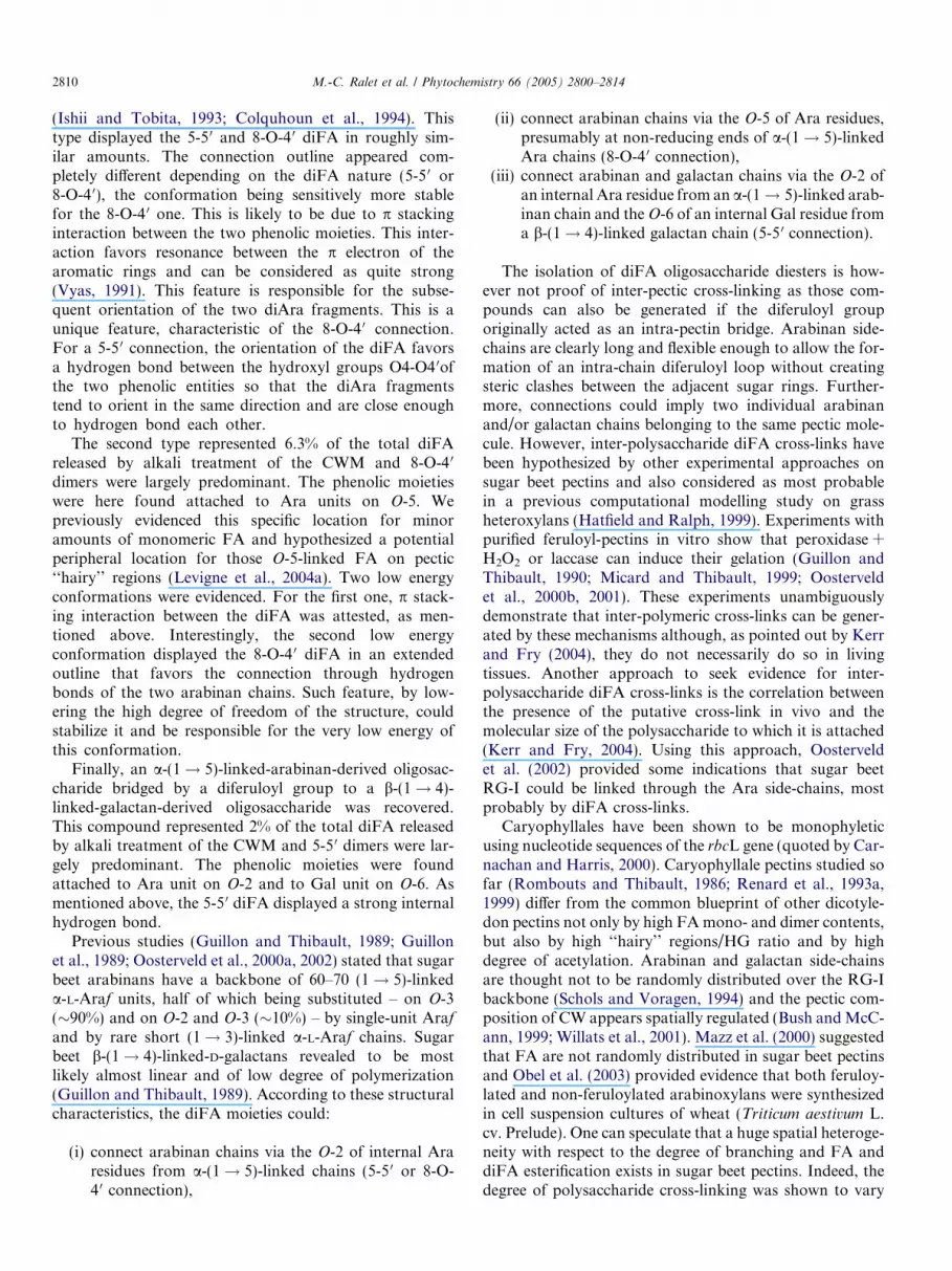
2808 M.-C. Ralet et al. / Phytochemistry 66 (2005) 2800–2814
2.4.1. Conformation of L-Araf-5 1-a-L-Araf-(5 8-O-
4 0 ! 5)-L-Araf-a-(1! 5)-L-Araf
The two conformations that displayed the lowest energywere selected among the numerous probable geometries(Figs. 7A and A 0). The conformation (A) did not showthe diFA in the stacking conformation but on the contraryrather extended. The second minimum (A 0) displayed thestacking interaction. Conformation (A 0) was the one withthe lowest energy compared to other compounds. Thisminimum served as a reference and its energy value wasarbitrarily zeroed to highlight later the energy discrepanciesof its counterparts. The dihedral angles for the first junc-tion were, respectively, C3–C4–C5–O10 at �92�, C4–C5–O10–C9 at �102� and O10–C9–C8–O40 at �176� and the onesfor the second junction were C10–C70–C80–C90 at �83�,C70–C80–C90–O100 at �178� and C80–C90–O100–C
�5 at �175�.
2.4.2. Conformation of L-Araf-(5 1)-a-L-Araf-(2 8-O-
4 0 ! 2)�L-Araf-a-(1! 5)�L-Araf
The two conformations that displayed the lowest energywere selected among the numerous probable geometries. Inthose cases, the diAra and the 8-O-4 0 diFA moietiesadopted their intrinsic low energy conformation. Mark-edly, the diFA moiety displayed a quite compact confor-mation allowed by the p stacking interaction. Thedifference between the two conformations came from thedihedral angles of the junction between the two entities thatshowed two distinct positions after minimization. Theenergy delta was 5.5 kcal/mol. The conformation that ledto the lowest energy is illustrated in Fig. 7B. This was byfar the most compact conformation among the differentcompounds. The dihedrals of the first junction were,respectively, C3–C2–O10–C9 at +38�, C2–O10–C9–C8 at
Table 2Main conformational features of the constitutive disaccharides (diAra and diG
a-L-Araf-(1! 5)-L-ArafTwo minima
Min A �72�; �178�; �158� Extended TG conformMin B �159�; �176�; +180� Very extended TG conResults relevant with Cros et al. (1993, 1994) and Janaswami and ChandrasekSupplementary data online 5A
b-D-Galp-(1! 4)-D-GalpOne large minimum
Min �80�; +128�
Supplementary data online 5B
8-O-40 diFATwo minima
Min with p stacking Compact geometryMin without p stacking Stretched orientationSupplementary data online 6A
5-50 diFAFour minima
Min A � 136� All the conformationsMin B + 46�Min C + 136�Min D � 53�Supplementary data online 6B
�165�, O10–C9–C8–O40 at �26� and C9–C8–O40–C40 at+133� and the ones for the second junction wereC10–C70–C80–C90 at +174�, C70–C80–C90–O100 at +38�,C80–C90–O100–C
�2 at �153� and C90–O100–C
�2–C
�3 at �153�.
2.4.3. Conformation of L-Araf-(5 1)-a-L-Araf-(2 5-5 0 ! 2)�L-Araf-a-(1! 5)�L-Araf
The lowest energy conformation found for that com-pound showed the diFA moiety slightly tilted and thetwo diAra moieties in the minA configuration. The dihe-dral angles for the first junction were, respectively, C3–C2–O10–C9 at �156�, C2–O10–C9–C8 at �165� andO10–C9–C8–C7 at �25� and C9–C8–C7–C1 at +133�. Thedihedral angles for the second junction were C10–C70–C80–C90 at +174�, C70–C80–C90–O100 at +37�, C80–C90–
O100–C�2 at �153� and C90–O100–C
�2–C
�3 at �153� (Fig. 7C).
The energy value was, respectively, 17.4 kcal/mol abovethe reference, where the diFA was 8-O-4 0 linked.
2.4.4. Conformation of D-Galp-(4 1)-b-D-Galp-(6 5-
5 0 ! 2)-L-Araf-a-(1! 5)-L-Araf
Two low energy conformations were found for thatcompound , one of them being illustrated on Fig. 7D.The 5-5 0 diFA was tilted and both the diAra and diGaladopted one more time their lowest energy configuration.The dihedral angles of the first junction were, respectively,C3–C2–O10–C9 at �162�, C2–O10–C9–C8 at �177� and O10–C9–C8–C7 at +4�. The dihedral angles of the second junc-tion were C80–C90–O100–C
�6 at �165�, C90–O100–C
�6–C
�5 at
+165� and O100–C�6–C
�5–C
�4 at +55�.
Computation with the CVFF force field evidenced weakenergy discrepancy between all the conformers of a givencompound. Each oligosaccharide moiety, especially the
al) and diFA components
Hydrogen bondings
ation O2 � � �O30formation O30 � � �O50aran (2005)
O4 � � �O6
O � � �O60
O11 � � �O30None
are energetically comparableO4 � � �O40O4 � � �C50

Fig. 7. Best conformers for each diFA-bridged oligosaccharides. The conformations display compact to extended conformations, combining either strongstacking (B), extensive hydrogen bonding (A 0 and C), both (A) or none (D). Black arrows evidence the junction between the disaccharide and the diFA;dotted lines in green show the hydrogen bond network and the magenta arrow the stacking interaction.
M.-C. Ralet et al. / Phytochemistry 66 (2005) 2800–2814 2809
diAra moiety, contributed for a large part to the high flex-ibility and to the clusterization of isoconformers. For agiven compound, it was thus irrelevant to discriminate aunique geometry from its numerous and equiprobablecounterparts. Since the aim of the study was everythingbut an extensive catalog of all the conformational possibil-ities of the diFA-bridged oligosaccharides, one or two of thelowest energy conformations for each compound were keptfor clear illustration and structural discussion.
3. Discussion
Very few covalent cross-links between CW polymershave been conclusively identified. As mentioned by Mort(2002), cross-links do not need to be very abundant inthe CW as a minimum of two cross-links per polymermolecule is sufficient to form a three-dimensional net-work. Ishii (1991) and Saulnier et al. (1999) succeededin isolating and characterizing compounds containing adiFA bridging two arabinoxylan-derived oligosaccharidesfrom monocotyledon CW, namely bamboo (Phyllostahysedulis) shoots and (Zea mays) maize bran. Saponificationof sugar beet root CW yields not only FA but also severaldiFA (Micard et al., 1997; Waldron et al., 1997; Wende
et al., 1999). In sugar beet root, around 20% of the totalFA is in dimer form, the main dimers being 8-O-4 0 diFAand 8-5 0 diFA (Micard et al., 1997; Waldron et al., 1997).These diFA were claimed to cross-link pectic polysaccha-rides via the Ara-rich and possibly Gal-containing neutralside-chains (Waldron et al., 1997) to which most of themonomeric FA is esterified (Ralet et al., 1994; Colquhounet al., 1994), although no experimental evidence supportedthese locations. In a previous work, we enzymaticallyrecovered and partially characterized a diFA oligoarab-inan from sugar beet root CWM but the exact locationof diFA on arabinan chains could not be elucidated(Levigne et al., 2004b). In the present work, several diFAoligosaccharide diesters were isolated and their structuresdescribed. Besides these original structural assignments,conformational data for diFA moieties and diFA-bridgedoligosaccharides are provided here for the first time. Twotypes of a-(1! 5)-linked-arabinan-derived oligosaccha-rides bridged by a diFA group were recovered.
The first type represented 4% of the total diFA thatwas released by alkali treatment of the CWM. The phe-nolic moieties were found attached to Ara units on O-2,in agreement with the location of monomeric FA resi-dues that are mainly ester-linked to O-2 of Ara residuesof the main core of a-(1! 5)-linked arabinan chains
2810 M.-C. Ralet et al. / Phytochemistry 66 (2005) 2800–2814
(Ishii and Tobita, 1993; Colquhoun et al., 1994). Thistype displayed the 5-5 0 and 8-O-4 0 diFA in roughly sim-ilar amounts. The connection outline appeared com-pletely different depending on the diFA nature (5-5 0 or8-O-4 0), the conformation being sensitively more stablefor the 8-O-4 0 one. This is likely to be due to p stackinginteraction between the two phenolic moieties. This inter-action favors resonance between the p electron of thearomatic rings and can be considered as quite strong(Vyas, 1991). This feature is responsible for the subse-quent orientation of the two diAra fragments. This is aunique feature, characteristic of the 8-O-4 0 connection.For a 5-5 0 connection, the orientation of the diFA favorsa hydrogen bond between the hydroxyl groups O4-O4 0ofthe two phenolic entities so that the diAra fragmentstend to orient in the same direction and are close enoughto hydrogen bond each other.
The second type represented 6.3% of the total diFAreleased by alkali treatment of the CWM and 8-O-4 0
dimers were largely predominant. The phenolic moietieswere here found attached to Ara units on O-5. Wepreviously evidenced this specific location for minoramounts of monomeric FA and hypothesized a potentialperipheral location for those O-5-linked FA on pectic‘‘hairy’’ regions (Levigne et al., 2004a). Two low energyconformations were evidenced. For the first one, p stack-ing interaction between the diFA was attested, as men-tioned above. Interestingly, the second low energyconformation displayed the 8-O-4 0 diFA in an extendedoutline that favors the connection through hydrogenbonds of the two arabinan chains. Such feature, by low-ering the high degree of freedom of the structure, couldstabilize it and be responsible for the very low energy ofthis conformation.
Finally, an a-(1! 5)-linked-arabinan-derived oligosac-charide bridged by a diferuloyl group to a b-(1! 4)-linked-galactan-derived oligosaccharide was recovered.This compound represented 2% of the total diFA releasedby alkali treatment of the CWM and 5-5 0 dimers were lar-gely predominant. The phenolic moieties were foundattached to Ara unit on O-2 and to Gal unit on O-6. Asmentioned above, the 5-5 0 diFA displayed a strong internalhydrogen bond.
Previous studies (Guillon and Thibault, 1989; Guillonet al., 1989; Oosterveld et al., 2000a, 2002) stated that sugarbeet arabinans have a backbone of 60–70 (1! 5)-linkeda-L-Araf units, half of which being substituted – on O-3(�90%) and on O-2 and O-3 (�10%) – by single-unit Arafand by rare short (1! 3)-linked a-L-Araf chains. Sugarbeet b-(1! 4)-linked-D-galactans revealed to be mostlikely almost linear and of low degree of polymerization(Guillon and Thibault, 1989). According to these structuralcharacteristics, the diFA moieties could:
(i) connect arabinan chains via the O-2 of internal Araresidues from a-(1! 5)-linked chains (5-5 0 or 8-O-4 0 connection),
(ii) connect arabinan chains via the O-5 of Ara residues,presumably at non-reducing ends of a-(1! 5)-linkedAra chains (8-O-4 0 connection),
(iii) connect arabinan and galactan chains via the O-2 ofan internal Ara residue from an a-(1! 5)-linked arab-inan chain and theO-6 of an internal Gal residue froma b-(1! 4)-linked galactan chain (5-5 0 connection).
The isolation of diFA oligosaccharide diesters is how-ever not proof of inter-pectic cross-linking as those com-pounds can also be generated if the diferuloyl grouporiginally acted as an intra-pectin bridge. Arabinan side-chains are clearly long and flexible enough to allow the for-mation of an intra-chain diferuloyl loop without creatingsteric clashes between the adjacent sugar rings. Further-more, connections could imply two individual arabinanand/or galactan chains belonging to the same pectic mole-cule. However, inter-polysaccharide diFA cross-links havebeen hypothesized by other experimental approaches onsugar beet pectins and also considered as most probablein a previous computational modelling study on grassheteroxylans (Hatfield and Ralph, 1999). Experiments withpurified feruloyl-pectins in vitro show that peroxidase +H2O2 or laccase can induce their gelation (Guillon andThibault, 1990; Micard and Thibault, 1999; Oosterveldet al., 2000b, 2001). These experiments unambiguouslydemonstrate that inter-polymeric cross-links can be gener-ated by these mechanisms although, as pointed out by Kerrand Fry (2004), they do not necessarily do so in livingtissues. Another approach to seek evidence for inter-polysaccharide diFA cross-links is the correlation betweenthe presence of the putative cross-link in vivo and themolecular size of the polysaccharide to which it is attached(Kerr and Fry, 2004). Using this approach, Oosterveldet al. (2002) provided some indications that sugar beetRG-I could be linked through the Ara side-chains, mostprobably by diFA cross-links.
Caryophyllales have been shown to be monophyleticusing nucleotide sequences of the rbcL gene (quoted by Car-nachan and Harris, 2000). Caryophyllale pectins studied sofar (Rombouts and Thibault, 1986; Renard et al., 1993a,1999) differ from the common blueprint of other dicotyle-don pectins not only by high FAmono- and dimer contents,but also by high ‘‘hairy’’ regions/HG ratio and by highdegree of acetylation. Arabinan and galactan side-chainsare thought not to be randomly distributed over the RG-Ibackbone (Schols and Voragen, 1994) and the pectic com-position of CW appears spatially regulated (Bush andMcC-ann, 1999; Willats et al., 2001). Mazz et al. (2000) suggestedthat FA are not randomly distributed in sugar beet pectinsand Obel et al. (2003) provided evidence that both feruloy-lated and non-feruloylated arabinoxylans were synthesizedin cell suspension cultures of wheat (Triticum aestivum L.cv. Prelude). One can speculate that a huge spatial heteroge-neity with respect to the degree of branching and FA anddiFA esterification exists in sugar beet pectins. Indeed, thedegree of polysaccharide cross-linking was shown to vary

M.-C. Ralet et al. / Phytochemistry 66 (2005) 2800–2814 2811
widely among quinoa and sugar beet tissues with high diFAlevels in roots compared to other tissues (Renard et al., 1999;Wende et al., 2000). Dimers could be present at high concen-trations in epidermal cells and, in that case, might beinvolved in protecting the cells against damage by pathogensand/or soil abrasion (Wende et al., 2000). It was also sug-gested that dimers could be involved in controlling the over-all growth of the plant (Wende et al., 2000; Yang andUchiyama, 2000). Variations in diFA content of sugar beettissues at different stages of development were also investi-gated (Wende et al., 1999, 2000). In the root, tissues laiddown at the end of the growth period investigated (14 weeks)were much poorer in FA and diFA than those depositedearly in root growth. It remains however unclear whetherthe proportion of the different diFA isomers varied duringroot maturation or not. In the present work, we have shownthat in mature sugar beet roots, pectin cross-linking vary notonly with respect to the diFA isomers involved, but also withrespect to the nature of the sugar chains involved and thelinkage position of the phenolic moieties to the sugar rings.This implies, as previously suggested (Wende et al., 2000), aremarkable degree of sophistication in the mechanismswhich control polysaccharide cross-linking through diFAbridges. Furthermore, the dynamic nature of the plantCW includes a changing phenolic content and implies thatthe nature and extent of phenolic cross-linking are underclose metabolic control (Wende et al., 1999). The recent iso-lation of a monoclonal antibody against feruloylated-b-D-(1! 4)-galactan (Clausen et al., 2004) allows to envisionthe production of antibodies against some diFA-bridged oli-gosaccharides – whose structure was assessed in the presentwork – in order to study their distribution in planta.
It is highly unlikely that sugar beet pectins can be exten-sively cross-linked in situ by calcium ions in structuressimilar to egg-boxes (Renard et al., 1993b). Indeed,acetyl-esterification on top of methyl-esterification in theHG domains strongly hinders their associative propertiesthrough calcium ions (Ralet et al., 2003). Cross-linkingthrough diFA bridges could constitute an alternative wayof connecting those peculiar pectic molecules.
4. Experimental
4.1. Materials
The CWM was prepared from fresh mature sugar beetroots as previously described (Levigne et al., 2002). Drise-lase� was obtained from Sigma and Ronozym from Hoff-man-La-Roche (Basel, Switzerland).
4.2. Isolation of diFA-rich fractions
CWM (10 g) was hydrolyzed by 0.05 M TFA (1 l) at100 �C for 90 min. The hydrolysate was filtered on G4 sin-tered glass and the residue rinsed with distilled water.Supernatants were pooled and TFA was removed by three
successive evaporation steps to dryness under vacuum at40 �C. The supernatant was then solubilized in distilledwater, precipitated by 4 volumes of ethanol and left over-night at 4 �C. The EtOH-soluble extract and precipitatewere separated by centrifugation. After removal of EtOHby under vacuum evaporation at 40 �C, EtOH-soluble frac-tion was further hydrolyzed by 0.1 M TFA at 100 �C for3 h. After removal of TFA, the hydrolysate was solubilisedin distilled water and the pH brought to �3 by 0.1 MNaOH. The EtOH-precipitate and the TFA hydrolysis res-idue were suspended in distilled water, brought to pH 5 by0.1 M NaOH and hydrolyzed by a Driselase� (9.5 ml) andRonozym� (0.1 ml) mixture for 24 h at 37 �C. Driselase�
(678 mg) was suspended in distilled water (10 ml) and cen-trifuged. The supernatant solution was used. The hydroly-sate was filtered on G3 sintered glass and the residue rinsedwith distilled water. Supernatants were pooled and concen-trated in vacuum.
Hydrolysates were loaded onto an Amberlite XAD-2column (20 · 1.5 cm) eluted successively by water (3–4column volumes), EtOH:water 15:85 (v/v) (3–4 columnvolumes), EtOH:water 25:75 (v/v) (3–4 column volumes)and EtOH:water 50:50 (v/v) (3–4 column volumes).Aliquots of selected fractions were concentrated to dryness,solubilized in distilled water (10 ml), brought to pH 2 by1 M HCl and extracted twice by ether (25 ml). The aqueousphases were evaporated to dryness, solubilized in water(10 ml), brought to pH 4 by 0.1 M NaOH and loaded ontoa column of Sephadex LH-20 (43 · 1.6 cm) eluted at�35 ml/h successively by water (8 column volumes),EtOH:water 15:85 (4 column volumes) and EtOH:water30:70 (8 column volumes). Fractions (9 ml) were collectedand analyzed for their absorbance at 325 nm and colori-metrically for GalUA (Thibault, 1979) and total neutralsugars (Tollier and Robin, 1979). Selected fractions werepooled, concentrated and kept at �18 �C for furtheranalysis.
4.3. General
The individual sugars were analyzed as their alditol ace-tate derivatives by gas chromatography after hydrolysis by2 M TFA at 121 �C for 2 h (Blakeney et al., 1983). Hpaecwas performed on a Dionex system with pulsed ampero-metric detection. The Carbopac PA1 column was elutedwith 500 mM NaOH, 200 mM sodium acetate and waterat 1 ml/min as follows: initial conditions, 20/30/50;30 min, 20/60/20; 30–34 min, 20/60/20; 35 min, 20/30/50.Oligoarabinans were obtained by mild acid hydrolysis ofsugar beet linearized arabinan (Megazyme) followed bysize-exclusion chromatography (Bio-Gel P-2). Oligogalac-tans were obtained by mild acid hydrolysis of lupin typeI arabinogalactan followed by size-exclusion chromatogra-phy (Bio-Gel P-2). Borwin software (JMBS Developpe-ments, Grenoble, France) was used for data acquisitionand processing. Phenolic compounds were determined byhplc after saponification and ether extraction as previously

2812 M.-C. Ralet et al. / Phytochemistry 66 (2005) 2800–2814
reported (Levigne et al., 2004b). Elution time and responsefactors relative to 3,4,5 trimethoxy-trans-cinnamic acid at320 nm were established using commercial FA and diFAstandards obtained as reported by Saulnier et al. (1999).
4.4. 18O-Labeling
Reducing ends were 18O-labeled by adding �25 ll ofH2
18O to �25 lg of freeze-dried samples. Samples wereincubated with H2
18O for �90 h at ambient temperaturein a dessicator.
4.5. Mass spectrometry
ESI-IT-MS experiments were achieved on an LCQAdvantage ion trap mass spectrometer (ThermoFinnigan,USA) using negative electrospray as the ionization process.Samples (18O-labeled or not) were diluted in order toobtain a final concentration of 20–50 lg/ml in MetOH:water 1:1 (v/v). Infusion was performed at a flow rate of2.5 ll/min. Nitrogen was used as sheath gas. No significantback-exchange of 16O was observed during the analysis ofthe 18O-labeled samples. The MS analyses were carriedout under automatic gain control conditions, using a typi-cal needle voltage of 4 kV and a heated capillary tempera-ture of 200 �C. For MSn experiments, the variousparameters were adjusted for each sample in order to opti-mize signal and get maximal structural information fromthe ion of interest. More than 50 scans were summed forMSn spectra acquisition. The Domon and Costello (1988)nomenclature was used to denote the fragment ions.
4.6. Molecular modeling
Molecular modeling was carried out on SGIs with theAccelrys�package. Molecular displays and energy minimi-zations used Insight II Biopolymer and Discover modules.The conformations were successively minimized using steep-est descent (SD) and conjugate gradient (CG) minimizationalgorithms with the CVFF force field. The potential energycomputed stretching, bending, torsion and electrostatic con-tributions as well as repulsion and dispersion of van derWaals interactions (Yiannikouris et al., 2004). The dielectricconstant e was 1 but all the final compounds were also min-imized with e 80 to compare data with implicite water.
4.6.1. Disaccharide construction
The global shape of a-L-Araf-(1! 5)-L-Araf and b-D-Galp-(1! 4)-D-Galp mainly depends on the rotation ofthe sugar rings around the glycosidic linkage (Perez et al.,2000). For a-L-Araf-(1! 5)-L-Araf, the angles (ua, wa,xa) described the glycosidic linkage with torsions betweenðO–C1–O50–C50Þ; ðC1–O50–C50–C40Þ and ðO50–C50–C40–O0Þ,the prime indicating the reducing end. xa was known toadopt preferentially one of the staggered conformations(i) +60� (GG), (ii) �60� (GT) and (iii) +180� (TG) (Croset al., 1993, 1994). For b-D-Galp-(1! 4)-D-Galp, the (ug,
wg) angles of the glycosidic linkage were torsions betweenðO–C1–O40–C40Þ and ðC1–O40–C40–C50Þ. (ua, wa, GG), (ua,wa, GT), (ua, wa, TG) and (ug, wg) spaces were exploredfrom �180� to +180� with a 10� step. The potential energyvs. torsion bonds was projected on final maps with contourto 10 kcal mol�1 above the minimum and a 2 kcal mol�1
step, then to 50 kcal mol�1 and a 10 kcal mol�1 step. Foreach disaccharide, the low energy conformations were iden-tified and the second hydroxyl groups reconsideredthrough minimization. The final lowest energy conforma-tion was kept further.
4.6.2. DiFA construction
The 8-O-4 0 diFA contained torsion angles around whichthe FA monomers can rotate. The angles c ðC1–C7–C8–O40Þand k ðC7–C8–O40–C40Þ were explored from �180� to +180�with a 10� increment. Once this conformational minimumidentified, the torsion bonds (C2–C1–C7–O8) andðC30–C40–O40–C80Þ were evaluated. That minimum wasfinally minimized (1000 iterations SD plus thousands ofCG until normal completion). The 5-5 0 diFA displayedno torsion bond and was directly minimized with the pro-cedure described above.
4.6.3. Compound construction
To avoid multi dimensional hyperspace impossible tosample, the combination of fragments was stepwise. Anyjunction created between two fragments led to new rota-tional angles, explored from �180� to +180�, with a 10�increment. The lowest energy conformation was selectedbefore the addition and torsion exploration of any ultimatefragment. Eventually, every complete compound was finallyminimized through the procedure (see above). When thethree fragments were bound together within every torsionangle positioned at its best, every complete compound wasfinally minimized through the abovementioned procedure.
Acknowledgments
The authors gratefully acknowledge the skillful technicalassistance of Marie-Jeanne Crepeau. This study has beencarried out with financial support from the Commissionof the European Communities, specific RTD programme‘‘Quality of Life and Management of Living Resources’’,proposal number QLK1-2002-02208 ‘‘Novel cross-linkingenzymes and their consumer acceptance for structure engi-neering of foods’’, acronym CROSSENZ. It does not re-flect its views and in no way anticipates the Commissions�future policy in this area.
Appendix A. Supplementary data
Supplementary data associated with this article can befound, in the online version, at doi:10.1016/j.phytochem.2005.09.039.

M.-C. Ralet et al. / Phytochemistry 66 (2005) 2800–2814 2813
References
Blakeney, A.B., Harris, P.J., Henry, R.J., Stone, B.A., 1983. A simple andrapid preparation of alditol acetates for monosaccharide analysis.Carbohydr. Res. 113, 291–299.
Brady, J.D., Fry, S.C., 1997. Formation of di-isodityrosine and loss ofisodityrosine in the cell walls of tomato cell-suspension cultures treatedwith fungal elicitors or H2O2. Plant Physiol. 115, 87–92.
Bush, M.S., McCann, M.C., 1999. Pectic epitopes are differentiallydistributed in the cell walls of potato (Solanum tuberosum) tubers.Physiol. Plant. 107, 201–213.
Carnachan, S.M., Harris, P.J., 2000. Ferulic acid is bound to the primarycell walls of all gymnosperm families. Biochem. Syst. Ecol. 28, 865–879.
Clausen, M.H., Ralet, M.-C., Willats, W.G.T., McCartney, L., Marcus,S.E., Thibault, J.-F., Knox, J.P., 2004. A monoclonal antibody toferuloylated-(1! 4)-b-D-galactan. Planta 219, 1036–1041.
Colquhoun, I., Ralet, M.-C., Thibault, J.-F., Faulds, C.B., Williamson,G., 1994. Structure identification of feruloylated oligosaccharidesfrom sugar-beet pulp by NMR spectroscopy. Carbohydr. Res. 263,243–256.
Cros, S., Herve du Penhoat, C., Perez, S., Imberty, A., 1993. Modeling ofarabinofuranose and arabinan. Part I: conformational flexibility of thearabinose ring. Carbohydr. Res. 248, 81–93.
Cros, S., Imberty, A., Bouchemal, N., Herve du Penhoat, C., Perez, S.,1994. Modeling of arabinofuranose and arabinan. Part II: nmr andconformational analysis of arabinose and arabinan conformationalflexibility of the arabinose ring. Biopolymers 34, 1433–1447.
Domon, B., Costello, C.E., 1988. A systematic nomenclature for carbo-hydrate fragmentation in FAB-MS/MS spectra conjugates. Glycocon-jugate J. 5, 397–409.
Fry, S.C., 1982. Phenolic components of the primary cell walls: feruloy-lated disaccharides of D-galactose and L-arabinose from spinachpolysaccharide. Biochem. J. 203, 493–504.
Fry, S.C., 1986. Cross linking of matrix polymers in the growing cell wallsof angiosperms. Annu. Rev. Plant Physiol. 37, 165–186.
Fry, S.C., Willis, S.C., Paterson, A.J., 2000. Intraprotoplasmic and wall-localised formation of arabinoxylan-bound diferulates and largerferulate coupling-products in maize cell-suspension cultures. Planta211, 679–692.
Guillon, F., Thibault, J.-F., 1989. Methylation analysis and mild acidhydrolysis of the ‘‘hairy’’ fragments of sugar beet pectins. Carbohydr.Res. 190, 85–96.
Guillon, F., Thibault, J.-F., Rombouts, F.M., Voragen, A.G.J., Pilnik,W., 1989. Enzymic hydrolysis of the ‘‘hairy’’ fragments of sugar-beetpectins. Carbohydr. Res. 190, 97–108.
Guillon, F., Thibault, J.-F., 1990. Oxidative cross-linking of chemicallyand enzymatically modified sugar-beet pectin. Carbohydr. Polym. 12,353–374.
Harvey, D.J., 2000. Collision-induced fragmentation of underivatisedcarbohydrates ionized by electrospray. J. Mass Spectrom. 35, 1178–1190.
Hatfield, R.D., Ralph, J., 1999. Modelling the feasability of intramolec-ular dehydrodiferulate formation in grass walls. J. Sci. Food Agric. 79,425–427.
Ishii, T., 1991. Isolation and characterization of a diferuloyl arabinoxylanhexasaccharide from bamboo shoot cell-walls. Carbohydr. Res. 219,15–22.
Ishii, T., Tobita, T., 1993. Structural characterization of feruloyl oligo-saccharides from spinach-leafs cell walls. Carbohydr. Res. 248, 179–190.
Janaswami, S., Chandrasekaran, R., 2005. Polysaccharide structures frompowder diffraction data: molecular models of arabinan. Carbohydr.Res. 340, 835–839.
Kamisaka, S., Takeda, S., Takahashi, K., Shibata, K., 1990. Diferulic andferulic acid in the cell wall of Avena coleoptiles – their relationships tomechanical properties of the cell wall. Physiol. Plant. 78, 1–7.
Kerr, E.M., Fry, S.C., 2004. Extracellular cross-linking of xylan andxyloglucan in maize cell-suspension cultures: the role of oxidativephenolic coupling. Planta 219, 73–83.
Levigne, S., Ralet, M.-C., Thibault, J.-F., 2002. Characterisation ofpectins extracted from fresh sugar beet under different conditions usingan experimental design. Carbohydr. Polym. 49, 145–153.
Levigne, S.V., Ralet, M.-C.J., Quemener, B.C., Pollet, B.M.-L., Lapierre,C., Thibault, J.-F.J., 2004a. Isolation from sugar beet cell walls ofarabinan oligosaccharides esterified by two ferulic acid monomers.Plant Physiol. 134, 1173–1180.
Levigne, S., Ralet, M.-C., Quemener, B., Thibault, J.-F., 2004b. Isolationof diferulic bridges ester-linked to arabinan in sugar beet cell walls.Carbohydr. Res. 339, 2315–2319.
Mazz, M., McCann, M.C., Kolpak, F., White, A.R., Stacey, N.J.,Roberts, K., 2000. Extraction of pectic polysaccharides from sugar-beet cell walls. J. Sci. Food Agric. 80, 17–28.
Micard, V., Grabber, J.H., Ralph, J., Renard, C.M.G.C., Thibault, J.-F.,1997. Dehydrodiferulic acids from sugar-beet pulp. Phytochemistry 44,1365–1368.
Micard, V., Thibault, J.-F., 1999. Oxidative gelation of sugar-beet pectins:use of laccases and hydration properties of the cross-linked pectins.Carbohydr. Polym. 39, 265–273.
Mort, A.J., 2002. Interactions between pectins and other polymers. In:Seymour, G.B., Knox, J.P. (Eds.), Pectins and their Manipulation.Blackwell Publishing CRC Press, Oxford, pp. 30–51.
Obel, N., Porchia, A.C., Scheller, H.V., 2003. Intracellular feruloylation ofarabinoxylan in wheat: evidence for feruloyl-glucose as precursor.Planta 216, 620–629.
Oosterveld, A., Beldman, G., Schols, H.A., Voragen, A.G.J., 2000a.Characterization of arabinose and ferulic acid rich pectic polysaccha-rides and hemicelluloses from sugar beet pulp. Carbohydr. Res. 328,185–197.
Oosterveld, A., Beldman, G., Voragen, A.G.J., 2000b. Oxidative cross-linking of pectic polysaccharides from sugar beet pulp. Carbohydr.Res. 328, 199–207.
Oosterveld, A., Pol, I.E., Beldman, G., Voragen, A.G.J., 2001. Isolation offeruloylated arabinans and rhamnogalacturonans from sugar beet pulpand their gel forming ability by oxidative cross-linking. Carbohydr.Polym. 44, 9–17.
Oosterveld, A., Beldman, G., Voragen, A.G.J., 2002. Enzymatic modifi-cation of pectic polysaccharides obtained from sugar beet pulp.Carbohydr. Polym. 48, 73–81.
Perez, S., Mazeau, K., Herve du Penhat, C., 2000. The three-dimensionalstructures of the pectic polysaccharides. Plant Physiol. Biochem. 38,37–55.
Quemener, B., Ralet, M.-C., 2004. Evidence for linkage position deter-mination in known feruloylated mono- and disaccharides usingelectrospray ion trap mass spectrometry. J. Mass Spectrom. 39,1153–1160.
Ralet, M.-C., Thibault, J.-F., Faulds, C.B., Williamson, G., 1994.Isolation and purification of feruloylated oligosaccharides from cellwalls of sugar-beet pulp. Carbohydr. Res. 263, 227–241.
Ralet, M.-C., Crepeau, M.-J., Buchholt, H.-C., Thibault, J.-F., 2003.Polyelectrolyte behaviour and calcium binding properties of sugar beetpectins differing in their degrees of methylation and acetylation.Biochem. Eng. J. 16, 191–201.
Ralph, J., Helm, R.F., 1993. Lignin/hydroxycinnamic acid/polysaccharidecomplexes: synthetic models for regio/chemical characterization. In:Jung, H.G., Buxton, D.R., Hatfield, R.D., Ralph, J. (Eds.), ForageCell Wall Structure and Digestibility. ASA-CSSA-SSSA, Madison, pp.201–246.
Renard, C.M.G.C., Champenois, Y., Thibault, J.-F., 1993a. Character-ization of the extractable pectins and hemicelluloses of the cell wall ofglasswort, Salicornia ramosissima. Carbohydr. Polym. 22, 239–245.
Renard, C.M.G.C., Thibault, J.-F., Liners, F., Van Cutsem, P., 1993b.Immunological probing of pectins isolated or in situ. Acta Bot. Neerl.42, 199–204.

2814 M.-C. Ralet et al. / Phytochemistry 66 (2005) 2800–2814
Renard, C.M.G.C., Wende, G., Booth, E.J., 1999. Cell wall phenolics andpolysaccharides in different tissues of quinoa (Chenopodium quinoa
Willd). J. Sci. Food Agric. 79, 2029–2034.Rombouts, F.M., Thibault, J.-F., 1986. Feruloylated pectic substances
from sugar-beet pulp. Carbohydr. Res. 154, 177–187.Saulnier, L., Crepeau, M.-J., Lahaye, M., Thibault, J.-F., Garcia-Conesa,
M.T., Kroon, P.A., Williamson, G., 1999. Isolation and structuraldetermination of two 5-50 diferuloyl oligosaccharides indicate thatmaize heteroxylans are covalently cross-linked by oxidatively coupledferulates. Carbohydr. Res. 320, 82–92.
Schols, H.A., Voragen, A.G.J., 1994. Occurrence of pectic hairy regions invarious plant cell wall materials and their degradability by rhamno-galacturonase. Carbohydr. Res. 256, 83–95.
Thibault, J.-F., 1979. Automated-method for the determination of pecticsubstances. Lebensm.-Wiss. u.-Technol. 12, 247–251.
Tollier, M.-T., Robin, J.-P., 1979. Adaptation of the orcinol-sulfuric acidmethod for the automatic titration of total neutral sugars – conditionsof application to plant extracts. Ann. Technol. Agric. 28, 1–15.
Vakhrushev, S.Y., Zamfir, A., Peter-Katalinic, J., 2004. 0,2An cross-ringcleavage as a general diagnostic tool for glycan assignment inglycoconjugate mixtures. J. Am. Soc. Mass Spectrom. 15, 1863–1868.
Vyas, N.K., 1991. Atomic features of protein–carbohydrate interactions.Curr. Opin. Struct. Biol. 1, 732–740.
Waldron, K.W., Ng, A., Parker, M.L., Parr, A.J., 1997. Ferulic aciddehydrodimers in the cell walls of Beta vulgaris and their possible rolein texture. J. Sci. Food Agric. 74, 221–228.
Wende, G., Waldron, K.W., Smith, A.C., Brett, C.T., 1999. Develop-mental changes in cell-wall ferulate and dehydrodiferulates in sugarbeet. Phytochemistry 52, 819–827.
Wende, G., Waldron, K.W., Smith, A.C., Brett, C.T., 2000. Tissue-specificdevelopmental changes in cell-wall ferulate and dehydrodiferulates insugar beet. Phytochemistry 55, 103–110.
Willats, W.G.T., McCartney, L., Mackie, W., Knox, J.P., 2001. Pectin:cell biology and prospects for functional analysis. Plant Mol. Biol. 47,9–27.
Yang, J.-G., Uchiyama, T., 2000. Hydroxycinnamic acids and their dimersinvolved in the cessation of cell elongation in Mentha suspensionculture. Biosci. Biotech. Bioch. 64, 1572–1579.
Yiannikouris, A., Andre, G., Buleon, A., Jeminet, G., Canet, I., Francois,J.M., Bertin, G., Jouany, J.P., 2004. Comprehensive conformationalstudy of key interactions involved in zearalenone complexation with b-D-glucans. Biomacromolecules 5, 2176–2185.


