Embed Size (px)
Citation preview
ANTIMICROBIAL AGENTS AND CHEMOTHERAPY, Dec. 2010, p. 5201–5208 Vol. 54, No. 120066-4804/10/$12.00 doi:10.1128/AAC.00963-10Copyright © 2010, American Society for Microbiology. All Rights Reserved.
Suitable Disk Antimicrobial Susceptibility Breakpoints DefiningSalmonella enterica Serovar Typhi Isolates with Reduced
Susceptibility to Fluoroquinolones�†�Christopher M. Parry,1,2* Chau Tran Thuy,1 Sabina Dongol,3 Abhilasha Karkey,3 Ha Vinh,1,4
Nguyen Tran Chinh,4 Pham Thanh Duy,1 Tran Vu Thieu Nga,1 James I. Campbell,1,2
Nguyen Van Minh Hoang,1,2 Amit Arjyal,3 Zulfiqar A. Bhutta,5 Sujit K. Bhattacharya,6
Magdarina D. Agtini,7 Baiqing Dong,8 Do Gia Canh,9 Aliya Naheed,10 John Wain,11
Tran Tinh Hien,4 Buddha Basnyat,3 Leon Ochiai,12 John Clemens,12
Jeremy J. Farrar,1,2 Christiane Dolecek,1,2 and Stephen Baker1,2
The Hospital for Tropical Diseases, Wellcome Trust Major Overseas Programme, Oxford University Clinical Research Unit,Ho Chi Minh City, Vietnam1; Centre for Tropical Diseases, University of Oxford, Oxford, United Kingdom2;
Oxford University Clinical Research Unit, Patan Academy of Health Sciences, Kathmandu, Nepal3; TheHospital for Tropical Diseases, Ho Chi Minh City, Vietnam4; Department of Paediatrics, Aga Khan University,Karachi, Pakistan5; National Institute of Cholera and Enteric Diseases, Kolkata, India6; National Institute of
Health Research and Development, Jakarta, Indonesia7; Guangxi Centers for Disease Control andPrevention, Nanning, Guangxi, China8; National Institute of Hygiene and Epidemiology, Hanoi,
Vietnam9; International Centre for Diarrhoeal Disease Research, Dhaka, Bangladesh10;Laboratory for Gastrointestinal Pathogens, HPA Centre for Infections, Colindale,
United Kingdom11; and International Vaccine Institute, Seoul, South Korea12
Received 15 July 2010/Returned for modification 18 August 2010/Accepted 4 September 2010
Infections with Salmonella enterica serovar Typhi isolates that have reduced susceptibility to ofloxacin (MIC >0.25 �g/ml) or ciprofloxacin (MIC > 0.125 �g/ml) have been associated with a delayed response or clinical failurefollowing treatment with these antimicrobials. These isolates are not detected as resistant using current disksusceptibility breakpoints. We examined 816 isolates of S. Typhi from seven Asian countries. Screening for nalidixicacid resistance (MIC > 16 �g/ml) identified isolates with an ofloxacin MIC of >0.25 �g/ml with a sensitivity of97.3% (253/260) and specificity of 99.3% (552/556). For isolates with a ciprofloxacin MIC of >0.125 �g/ml, thesensitivity was 92.9% (248/267) and specificity was 98.4% (540/549). A zone of inhibition of <28 mm around a 5-�gofloxacin disc detected strains with an ofloxacin MIC of >0.25 �g/ml with a sensitivity of 94.6% (246/260) andspecificity of 94.2% (524/556). A zone of inhibition of <30 mm detected isolates with a ciprofloxacin MIC of >0.125�g/ml with a sensitivity of 94.0% (251/267) and specificity of 94.2% (517/549). An ofloxacin MIC of >0.25 �g/ml anda ciprofloxacin MIC of >0.125 �g/ml detected 74.5% (341/460) of isolates with an identified quinolone resistance-inducing mutation and 81.5% (331/406) of the most common mutant (carrying a serine-to-phenylalanine mutationat codon 83 in the gyrA gene). Screening for nalidixic acid resistance or ciprofloxacin and ofloxacin disk inhibitionzone are suitable for detecting S. Typhi isolates with reduced fluoroquinolone susceptibility.
Enteric fever is an infection caused by Salmonella entericaserovars Typhi and Paratyphi A. These human restrictedpathogens are transmitted by the fecal-oral route, and entericfever is common in regions with poor standards of hygiene andsanitation. There are 27 million new enteric fever infectionseach year, of which approximately 200,000 are fatal (16). An-timicrobials are essential for appropriate clinical manage-ment of enteric fever, but antimicrobial resistance in S.Typhi and S. Paratyphi A have become a problem in regions
where they are endemic (6, 8). Multiple-drug-resistant(MDR) S. Typhi and S. Paratyphi A (resistant to chloram-phenicol, trimethoprim-sulfamethoxazole, and ampicillin)are particularly common in some locations in Asia and haveled to large epidemics. An MDR S. Typhi strain was respon-sible for an outbreak in Tajikistan in the late 1990s, causingover 24,000 infections (39).
The occurrence of MDR strains limits the options forantimicrobial therapy of enteric fever. The current WHOguidelines suggest that the fluoroquinolones are the optimalgroup of antimicrobials for the treatment of uncomplicatedtyphoid fever in adults (44). The fluoroquinolones, such asciprofloxacin and ofloxacin, are comparatively inexpensiveand well tolerated and in early randomized clinical trialswere very effective. However, S. Typhi and S. Paratyphi Aisolates with reduced susceptibility to fluoroquinolones havebecome common in Asia and are increasingly common inAfrica (6, 8, 13, 26, 32, 37). Infections with S. Typhi strainswith elevated MICs to ciprofloxacin and ofloxacin have been
* Corresponding author. Mailing address: The Hospital for TropicalDiseases, Wellcome Trust Major Overseas Programme, Oxford Uni-versity Clinical Research Unit, 190 Ben Ham Tu, Quan 5, Ho ChiMinh City, Vietnam. Phone: (84-8) 9 241 761. Fax: (84-8) 9 238 904.E-mail: [email protected].
† Supplemental material for this article may be found at http://aac.asm.org/.
� Published ahead of print on 13 September 2010.� The authors have paid a fee to allow immediate free access to
this article.
5201
associated with the failure of treatment with these antimi-crobials and increased disease severity (15, 30, 33, 36, 43).
Investigations of S. Typhi with reduced susceptibility to fluo-roquinolones has shown the association of elevated MIC withseveral single-base-pair mutations in the DNA gyrase gene,gyrA, and the topoisomerase gene, parC (4, 6, 33, 42). Further-more, extensive genome sequencing and single nucleotidepolymorphism (SNP) investigation of S. Typhi strains havefurther shown the dramatic impact of strains with gyrA muta-tions on the population structure of this monophyletic organ-ism (35). Genotyping studies identified at least 15 independentgyrA mutations that have occurred within a decade and stim-ulated clonal expansion in Asia and Africa (6, 35). These datasuggest that such strains have evolved rapidly and are main-tained by a strong selective pressure.
The laboratory detection and identification of strains with re-duced susceptibility to fluoroquinolones are important for thetreating clinician, but such strains are categorized as susceptibleby the current interpretive guidelines for fluoroquinolone disksusceptibility testing (3, 11, 19). These isolates are invariably re-sistant to nalidixic acid, and susceptibility testing with a nalidixicacid disk has been suggested as a suitable screening method forreduced fluoroquinolone susceptibility (11, 19). The British Soci-ety for Antimicrobial Chemotherapy (BSAC) has recommendedthat for invasive isolates of Salmonella, an MIC for reduced sus-ceptibility to fluoroquinolones should be determined (3).
Here we have examined the relationship between gyrA andparC mutations, nalidixic acid resistance, ofloxacin and cipro-floxacin disk inhibition zone sizes, and MIC for a large numberof S. Typhi clinical isolates from multiple locations in Asia overa 16-year period. We suggest disk susceptibility breakpoints forstrains with reduced susceptibility to ciprofloxacin and ofloxa-cin, which may permit the diagnostic laboratory to detect suchisolates and aid the clinical management of enteric fever.
MATERIALS AND METHODS
S. Typhi strain collection. The S. Typhi strains used in this study were com-prised of isolates collected as part of several independent investigations. Themajority of the strains (516 strains) were collected from randomized controlledtrials conducted between 1992 and 2002 in southern Vietnam. These trials wereconducted using a standard protocol, except for the treatment regimens used,described in detail elsewhere (5, 7, 28, 31, 38, 40, 41). One hundred and four S.Typhi strains were isolated as part of a randomized controlled trial (gatifloxacinversus chloramphenicol [ISRCTN53258327]) at Patan Hospital, Kathmandu,Nepal, for the treatment of uncomplicated enteric fever between 2006 and 2008.The remaining S. Typhi strains (a total of 196) were collected between 2002 and2003 as part of population-based prospective surveillance studies conducted bymultiple teams in Jakarta, Indonesia (n � 27), Dhaka, Bangladesh (n � 40),Hechi City, Guang Xi, China (n � 51), Kolkata, India (n � 25), and Karachi,Pakistan (n � 53) (6).
A subset of the strains described above (n � 100; from Vietnam, Indonesia,China, India, and Pakistan) and a collection of contemporary S. Typhi strains fromVietnam and India (n � 375) were additionally selected for screening for gyrA, gyrB,parC, and parE mutations. These strains are presented in the supplemental material.
Microbiological methods. The isolates were identified by standard biochemicaltests and agglutination with Salmonella-specific antisera (Murex Diagnostics,Dartford, United Kingdom). Antimicrobial susceptibilities were tested at thetime of isolation by the modified Bauer-Kirby disk diffusion method, withzone size interpretation based on CLSI guidelines (9, 11). Antimicrobial diskstested were chloramphenicol (CHL) (30 �g), ampicillin (AMP) (10 �g),trimethoprim-sulfamethoxazole (SXT) (1.25/23.75 �g), ceftriaxone (CRO)(30 �g), ofloxacin (OFX) (5 �g), and nalidixic acid (NAL) (30 �g). Mueller-Hinton agar and antimicrobial discs were purchased from Unipath, Basing-stoke, United Kingdom.
Isolates were stored on Protect beads (Prolabs, Oxford, United Kingdom) at�20°C. The isolates were later subcultured, and the disk antimicrobial susceptibilitytests were repeated on Mueller-Hinton agar by CLSI methods for NAL (30 �g),ciprofloxacin (CIP) (5 �g), and ofloxacin (OFX) (5 �g). The zone of inhibitedgrowth for each antimicrobial was measured by three separate investigators blind tothe result of the measurements of the others. The average zone size recorded by thethree readers was calculated. The MICs for the isolates were determined by thestandard agar plate dilution method according to CLSI guidelines or by Etestaccording to the manufacturer’s recommendations (AB Biodisk, Sweden) (10).
The antimicrobials evaluated were CIP (0.008 �g/ml to 4 �g/ml), OFX (0.008�g/ml to 4 �g/ml), and NAL (0.5 �g/ml to 512 �g/ml). Antimicrobial powders forthe agar plate dilution MICs were purchased from Sigma, United Kingdom. TheMIC end points were read by two independent investigators, each blind to theresult determined by the other. Discrepancies were resolved by discussion. Esch-erichia coli ATCC 25922 and Staphylococcus aureus ATCC 25923 were used ascontrol strains for these assays. The results were interpreted according to currentCLSI guidelines, susceptible being values of �8 �g/ml for nalidixic acid, �2�g/ml for ofloxacin, and �1 �g/ml for ciprofloxacin. An isolate was defined asMDR if it was resistant to chloramphenicol, trimethoprim-sulfamethoxazole, andampicillin by disk susceptibility testing.
PCR amplification and sequencing of gyrA, gyrB, parC, and parE genes in S.Typhi. DNA from the strains that were selected for PCR amplification of thegyrA, gyrB, parC, and parE genes was extracted using the Wizard genomic DNApurification kit (Promega) according to the manufacturer’s recommendations.Briefly, a single colony was inoculated in 1.5 ml of Luria-Bertani broth andincubated overnight at 37°C with shaking at 300 rpm to reach 108 CFU/ml. Oneml of the bacterial culture was transferred to a microcentrifuge tube and cen-trifuged in a microcentrifuge at 13,000 rpm for 2 min. The supernatant wasremoved, and the bacterial pellet was used for DNA extraction. The extractedDNA was stored at �20°C until required.
Oligonucleotide primers for the amplification of the quinolone resistance-determining regions in gyrA, gyrB, parC, and parE genes in S. Typhi were asfollows (6): gyrA, GYRA/P1 (5�-TGTCCGAGATGGCCTGAAGC) andGYRA/P2 (5�-TACCGTCATAAGTTATCCACG) (annealing temperature,55°C); gyrB, StygyrB1 (5�-CAAACTGGCGGACTGTCAGG) and StygyrB2 (5�-TTCCGGCATCTGACGATAGA) (annealing temperature, 62°C); parC,StmparC1 (5�-CTATGCGATGT CAGAGCTGG) and StmparC2 (5� TAACAGCAGCTCGGCGTATT) (annealing temperature; 62°C); and parE,StmparE1 (5�-TCTCTTCCGATGAAGTGCTG) and StmparE2 (5� ATACGGTATAGCGGCGGTAG) (annealing temperature, 62°C).
Predicted PCR amplicon sizes were 347 bp (gyrA), 345 bp (gyrB), 270 bp(parC), and 240 bp (parE). PCRs were performed under the following conditions:30 cycles of 92°C for 45 s, 55°C or 62°C (depending on the primers) for 45 s, andextension at 74°C for 1 min, followed by a final extension step at 74°C for 2 min.
The DNA sequencing reactions were performed using the CEQ DTCS QuickStart kit (Beckman Coulter) and was sequenced using a CEQ 8000 capillarysequencer, and the resulting DNA sequence was analyzed using CEQuenceInvestigator CEQ2000XL (Beckman Coulter). All sequences were verified,aligned, and manipulated using Bioedit software (http://www.mbio.ncsu.edu/BioEdit/bioedit.html). All gyrA, gyrB, parC, and parE sequences were comparedto other gyrA, gyrB, parC, and parE sequences by BLASTn at NCBI. The DNAsequence of the various S. Typhi sequences of gyrA, gyrB, parC, and parE weredownloaded and aligned with the produced sequences.
Data analysis. Zone size interpretive criteria and interpretive discrepancyrates were calculated by the error rate-bounded method of Metzler and DeHaan(27). The MIC breakpoints for reduced susceptibility were �0.25 �g/ml forofloxacin and �0.125 �g/ml for ciprofloxacin. The zone size breakpoints wereadjusted until the number of false-susceptible disk diffusion test results (verymajor discrepancies) and false-resistant disk tests (major discrepancies) wereheld to a minimum. Guidelines for acceptable discrepancy rates were accordingto the CLSI recommendation (12). Normally distributed data were comparedusing the Student t test, nonnormally distributed data using the Mann-WhitneyU test, and proportions by the chi-square test. Statistical analysis was performedusing EpiInfo, version 6 (CDC, Atlanta, GA), and SPSS for Windows version10.1 (SPSS, Inc., Chicago, IL).
RESULTS
Antimicrobial susceptibility testing of S. Typhi isolates. Weinvestigated 816 S. Typhi isolates collected between 1992 and2008 from seven Asian countries: Vietnam, Nepal, Indonesia,India, Bangladesh, Pakistan, and China. Only one isolate (the
5202 PARRY ET AL. ANTIMICROB. AGENTS CHEMOTHER.

strain isolated on admission to the health care facility) fromeach patient was included for microbiological examination andanalysis.
Of the 816 S. Typhi isolates tested, 466 (57.1%) were MDR(resistant to chloramphenicol, ampicillin, and trimethoprim-sulfamethoxazole), while 303/816 (37%) were fully susceptibleto chloramphenicol, ampicillin, and trimethoprim-sulfameth-oxazole. Two hundred fifty-three of the 816 isolates (31%)were resistant to nalidixic acid (MIC, �32 �g/ml), and 4 iso-lates had an MIC of 16 �g/ml (intermediate) to nalidixic acidbut were classified as resistant according to the zone sizes fromdisk susceptibility testing (�13 mm). Of the 466 MDR isolates,145 (31.1%) were additionally resistant to nalidixic acid com-pared to 80/303 (26.4%) isolates that were fully susceptible tochloramphenicol, ampicillin, and trimethoprim-sulfamethox-azole (P � 0.16).
All 816 S. Typhi isolates were classified as susceptible tociprofloxacin according to MIC testing (MIC � 1 �g/ml), yet12 gave a discrepant result with disk testing. These strainsexhibited an inhibition zone size of �20 mm and were, there-fore, classified as intermediate by disk testing. Two of the 816S. Typhi strains were graded with intermediate resistance toofloxacin with an MIC of 4 �g/ml but had inhibition zone sizesof �16 mm and were, therefore, classified as susceptible.
The distribution of the MIC levels to ciprofloxacin andofloxacin for all 816 S. Typhi isolates is presented in Fig. 1. Thehistograms of the levels of MIC to ciprofloxacin and ofloxacinboth demonstrate a bimodal distribution. The two distinctgroups are partially divided by nalidixic acid susceptibility (Fig.1, black shading denotes resistance to nalidixic acid). The 563isolates that were susceptible to nalidixic acid had an MIC90
(range) to ciprofloxacin of 0.03 �g/ml (0.008 to 0.5 �g/ml) andof 0.06 �g/ml (0.016 to 0.5 �g/ml) to ofloxacin. The 253 isolatesthat were resistant to nalidixic acid had an MIC90 (range) tociprofloxacin of 0.5 �g/ml (0.064 to 1 �g/ml) and to ofloxacinof 1.0 �g/ml (0.125 to 4 �g/ml).
Antimicrobial susceptibility test interpretive categories of S.Typhi to ciprofloxacin and ofloxacin. The current CLSI inter-mediate breakpoints are 2 �g/ml and 4 �g/ml, respectively, forciprofloxacin and ofloxacin. Only 2 of the 816 strains tested had
MIC levels greater than or equal to those of the current MICbreakpoints (Fig. 1). The MICs for nalidixic acid were com-pared with those of ofloxacin and ciprofloxacin in scatter plots(Fig. 2). The current interpretive breakpoints are shown in Fig.2 as dark shading in red for ofloxacin and ciprofloxacin and ingray for nalidixic acid. The suggested interpretive breakpointsfor reduced susceptibility are depicted by a broken line with anarrow (Fig. 2). As predicted, there was a linear relationshipbetween the nalidixic acid MIC and the ofloxacin (Fig. 2a) andciprofloxacin MICs (Fig. 2b).
Screening strains using nalidixic acid resistance (MIC � 16�g/ml) for the detection of isolates with an MIC of �0.25�g/ml for ofloxacin had a sensitivity of 97.3% (253/260) and aspecificity of 99.3% (552/556) (Fig. 2a). The number of verymajor discrepancies was 7/260 (2.7%), with none more thantwo dilutions above the breakpoint, and the number of majordiscrepancies was 4/556 (0.7%), with none more than two di-lutions below the breakpoint. Screening for the detection ofisolates with a ciprofloxacin MIC of �0.125 �g/ml, using nali-dixic acid resistance (MIC of �16 �g/ml), was not as reliable asthat for ofloxacin, as it had a sensitivity of 92.9% (248/267) anda specificity of 98.4% (540/549). The number of very majordiscrepancies was 19/267 (7.1%), with 1/267 (0.4%) more thantwo dilutions above the breakpoint, and the number of majordiscrepancies was 9/549 (1.6%), with none more than two di-lutions below the breakpoint.
We explored the relationship between the diameter of thezone of inhibition and the MICs for ciprofloxacin and ofloxa-cin, using 5-�g disks (Fig. 3). A zone of inhibition of �28 mmaround a 5-�g ofloxacin disk correlated with an MIC of �0.25�g/ml, with the least number of discrepancies (Fig. 3a). Thenumber of very major discrepancies was 14/260 (5.4%), withnone more than two dilutions above the breakpoint, and thenumber of major discrepancies was 32/556 (5.7%), with 14/556(2.5%) more than two dilutions below the breakpoint. A zoneof inhibition of �28 mm around a 5-�g ofloxacin disc detectedstrains with an ofloxacin MIC of �0.25 �g/ml, with a sensitivityof 94.6% (246/260) and a specificity of 94.2% (524/556). Azone of inhibition of �30 mm around a 5-�g ciprofloxacin diskcorrelated with an MIC of �0.125 �g/ml, with the least num-
FIG. 1. Fluoroquinolone MIC histograms for 816 S. Typhi isolates from Asia. Histograms showing the distribution of MICs to ofloxacin (a) andciprofloxacin (b) of 816 S. Typhi strains, isolated from patients with enteric fever. Each isolate used for analysis was isolated from an individualenteric fever patient. The MICs are plotted on the x axis, and the numbers of isolates corresponding with particular MICs are plotted on the y axis.The white proportion of the columns indicates the nalidixic acid-susceptible isolates (n � 563). The black proportion of the columns indicates thenalidixic acid-resistant isolates (n � 253). Both histograms show a bimodal distribution, which is partly differentiated by nalidixic acid resistance.
VOL. 54, 2010 S. TYPHI FLUOROQUINOLONE BREAKPOINTS 5203

ber of discrepancies (Fig. 3b). The number of very major dis-crepancies was 16/267 (6.0%), with 4/267 (1.5%) more thantwo dilutions above the breakpoint, and the number of majordiscrepancies was 32/549 (5.8%), with 22/549 (4.0%) morethan two dilutions below the breakpoint. A zone of growthinhibition of �30 mm detected isolates with a ciprofloxacinMIC of �0.125 �g/ml, with a sensitivity of 94.0% (251/267) anda specificity of 94.2% (517/549).
Reduced susceptibility to fluoroquinolones and gyrA, gyrB,parC, and parE mutations. To further define the S. Typhipopulation with reduced susceptibility to fluoroquinolones, weproduced PCR amplicons and then sequenced the quinoloneresistance-determining region in the gyrA, gyrB, parC, and parEgenes from a collection of 475 S. Typhi strains from Vietnam,China, India, Indonesia, and Pakistan. One hundred of thesestrains were described in the previous section, and 375 weremore recent strains from Vietnam and India. The MIC rangeof these strains was 1 to 512 �g/ml to nalidixic acid, 0.008 to 6�g/ml to ciprofloxacin, and 0.03 to 12 �g/ml to ofloxacin. Thesestrains and the corresponding data from these strains are de-scribed in the supplemental material.
Fifteen of the 475 S. Typhi strains examined by PCR andsequencing of gyrA, gyrB, parC, and parE had no mutations inthe quinolone resistance-determining regions of any gene. Nostrains had a mutation in the quinolone resistance-determiningregion of gyrB or parE. Four hundred sixty strains had either asingle mutation or a combination of double or triple mutationsin the gyrA and parC genes. DNA sequencing identified sevendifferent amino acid substitutions: D87A, aspartic acid to as-paragine at codon 87 in the gyrA gene; S83Y, serine to tyrosineat codon 83 in the gyrA gene; S83F, serine to phenylalanine atcodon 83 in the gyrA gene; D87G, aspartic acid to glycine at
codon 87 in the gyrA gene; S83F/D87N, serine to phenylalanineat codon 83 and aspartic acid to asparagine at codon 87 in thegyrA gene; S83F/D87G, serine to phenylalanine at codon 83and aspartic acid to glycine at codon 87 in the gyrA gene; andS83F/D87G/S80I, serine to phenylalanine at codon 83 andaspartic acid to glycine at codon 87 in the gyrA gene and serineto isoleucine at codon 80 in the parC gene. The most com-monly identified amino acid replacement was S83F, constitut-ing (88%) 406/460 strains with a mutation, with S83Y thesecond most common mutant (10%) 46/460.
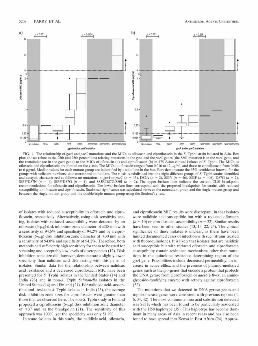
We compared the MICs to ofloxacin and ciprofloxacin of the460 strains with the seven different mutation patterns and the15 strains with no mutation detected (Fig. 4). When groupedinto strains with and without a single mutation in the gyrAgene, the single mutation group had significantly higher MICsto ofloxacin (Fig. 4a) and ciprofloxacin (Fig. 4b) than thosewithout a mutation. The most common amino acid substitu-tion, S83F, had mean MICs of 0.75 �g/ml and 0.33 �g/ml toofloxacin and ciprofloxacin, respectively. Figure 4 also showsthe current CLSI breakpoints and the suggested ofloxacinbreakpoint of 0.25 �g/ml and ciprofloxacin breakpoint of 0.125�g/ml. An MIC of 0.25 �g/ml to ofloxacin and an MIC of 0.125�g/ml to ciprofloxacin detected 74.5% (341/460) of the S.Typhi strains with an identified fluoroquinolone resistance mu-tation and 81.5% (331/406) of the most common S. Typhimutant (S83F) with reduced susceptibility to fluoroquinolones.
DISCUSSION
The increasing recognition that S. Typhi isolates with re-duced susceptibility to ofloxacin and ciprofloxacin may lead totreatment failure has led to calls for a revision of their break-
FIG. 2. Scatter plots relating ofloxacin and ciprofloxacin MICs to nalidixic acid MIC for 816 Asian S. Typhi isolates. Scatter plots comprisedof MIC data from 816 S. Typhi isolates from Nepal (n � 104), India (n � 25), Indonesia (n � 27), Bangladesh (n � 40), Pakistan (n � 53), China(n � 51), and Vietnam (n � 516). Plots show the relationship between the MIC to nalidixic acid (y axis) and the MIC to ofloxacin (a) andciprofloxacin (b) (x axis). The vertical and horizontal shading in each scatter plot indicates the current CLSI recommendations for breakpointsbetween susceptibility (white), intermediate (light gray, nalidixic acid; light red, ofloxacin and ciprofloxacin), and resistance (dark gray, nalidixicacid; dark red, ofloxacin and ciprofloxacin) (nalidixic acid MIC, �8 �g/ml and �32 �g/ml; ofloxacin MIC, �2 �g/ml and �8 �g/ml; andciprofloxacin MIC, �1 �g/ml and �4 �g/ml). The red broken line corresponds to the proposed MIC breakpoint identifying strains with reducedsusceptibility to fluoroquinolones (ofloxacin MIC of �0.25 �g/ml and ciprofloxacin MIC of �0.125 �g/ml).
5204 PARRY ET AL. ANTIMICROB. AGENTS CHEMOTHER.

points. Breakpoints of �0.25 �g/ml for ofloxacin and levofloxa-cin and �0.125 �g/ml for ciprofloxacin and gatifloxacin havebeen suggested (1, 2, 14, 32). Nalidixic acid resistance and disksusceptibility testing have both been proposed as laboratoryscreening methods to detect such isolates. We have explored
the performance of these methods with a large number ofstrains that are representative of S. Typhi isolates circulating incountries in Asia where it is endemic.
Nalidixic acid resistance had a sensitivity of 96.2% and91.8% and a specificity of 99.5% and 98.5% for the detection
FIG. 3. Scatter plots relating ofloxacin and ciprofloxacin MIC to inhibition zone diameter for 816 Asian S. Typhi isolates. Scatter plots for 816S. Typhi isolates comparing the inhibition zone diameters using a 5-�g ciprofloxacin disc (a) and a 5-�g ofloxacin disc (b) (x axis) and thecorresponding MIC of ciprofloxacin (a) and ofloxacin (b) (y axis). The numbers in brackets relate to the 253 nalidixic acid-resistant isolates. Thevertical red shading in each scatter plot is the current CLSI disc zone breakpoint for resistance (ofloxacin inhibition zone diameter, �16 mm;ciprofloxacin inhibition zone diameter, �21 mm). The horizontal red shading distinguishes strains with an MIC of �2 �g/ml for ofloxacin or anMIC of �1 �g/ml for ciprofloxacin. The gray shading is the proposed breakpoint for S. Typhi isolates with reduced susceptibility (ofloxacin MIC,�0.25 �g/ml; ciprofloxacin MIC, �0.125 �g/ml). The red broken line corresponds with the proposed breakpoints for strains with reducedsusceptibility (ofloxacin inhibition zone diameter, �28 mm; ciprofloxacin inhibition zone diameter, �30 mm).
VOL. 54, 2010 S. TYPHI FLUOROQUINOLONE BREAKPOINTS 5205
of isolates with reduced susceptibility to ofloxacin and cipro-floxacin, respectively. Alternatively, using disk sensitivity test-ing, isolates with reduced susceptibility were detected by anofloxacin (5-�g) disk inhibition zone diameter of �28 mm witha sensitivity of 94.6% and specificity of 94.2% and by a cipro-floxacin (5-�g) disk inhibition zone diameter of �30 mm witha sensitivity of 94.0% and specificity of 94.2%. Therefore, bothmethods had sufficiently high sensitivity for them to be used forscreening and acceptably low levels of discrepancies (12). Diskinhibition zone size did, however, demonstrate a slightly lowerspecificity than nalidixic acid disk testing with this panel ofisolates. Similar data for the relationship between nalidixicacid resistance and a decreased ciprofloxacin MIC have beenpresented for S. Typhi isolates in the United States (14) andIndia (23) and in non-S. Typhi Salmonella isolates in theUnited States (14) and Finland (21). For nalidixic acid-suscep-tible and -resistant S. Typhi isolates in India (23), the averagedisk inhibition zone sizes for ciprofloxacin were greater thanthose that we observed here. The non-S. Typhi study in Finlandproposed a ciprofloxacin (5-�g) disk inhibition zone diameterof �37 mm as the breakpoint (21). The sensitivity of thisapproach was 100%, yet the specificity was only 51.9%.
In some isolates in this study, the nalidixic acid, ofloxacin,
and ciprofloxacin MIC results were discrepant, in that isolateswere nalidixic acid susceptible but with a reduced ofloxacin(n � 10) or ciprofloxacin susceptibility (n � 22). Similar resultshave been seen in other studies (13, 15, 22, 26). The clinicalsignificance of these isolates is unclear, as there have beenlimited documented cases of infection with such strains treatedwith fluoroquinolones. It is likely that isolates that are nalidixicacid susceptible but with reduced ofloxacin and ciprofloxacinsusceptibility contain resistance mechanisms other than muta-tions in the quinolone resistance-determining region of thegyrA gene. Possibilities include decreased permeability, an in-crease in active efflux, and the presence of plasmid-mediatedgenes, such as the qnr genes that encode a protein that protectsthe DNA gyrase from ciprofloxacin or aac(6�)-Ib-cr, an amino-glycoside-modifying enzyme with activity against ciprofloxacin(32).
The mutations that we detected in DNA gyrase genes andtopoisomerase genes were consistent with previous reports (4,6, 34, 42). The most common amino acid substitution detectedwas S83F, which has been found to be particularly associatedwith the H58 haplotype (35). This haplotype has become dom-inant in many areas of Asia in recent years and has also beenfound to have spread into Kenya in East Africa (24). Approx-
FIG. 4. The relationship of gyrA and parC mutations and the MICs to ofloxacin and ciprofloxacin in the S. Typhi strain isolated in Asia. Boxplots (boxes relate to the 25th and 75th percentiles) relating mutations in the gyrA and the parC genes (the S80I mutation is in the parC gene, andthe remainder are in the gyrA gene) to the MICs of ofloxacin (a) and ciprofloxacin (b) in 475 Asian clinical isolates of S. Typhi. The MICs toofloxacin and ciprofloxacin are plotted on the y axis. The MICs to ofloxacin ranged from 0.016 to 12 �g/ml, and those to ciprofloxacin from 0.008to 6 �g/ml. Median values for each mutant group are indentified by a solid line in the box. Bars demonstrate the 95% confidence interval for thegroups with sufficient numbers; dots correspond to outliers. The x axis is subdivided into the eight different groups of S. Typhi strains identifiedand assayed, characterized as follows: no mutations in gyrA or parC (n � 15), D87A (n � 2), S83Y (n � 46), S83F (n � 406), D87G (n � 2),S83F/D87N (n � 1), S83F/D87G (n � 1), and S83F/D87G/S80I (n � 2). The upper broken lines indicate the current CLSI breakpointrecommendations for ofloxacin and ciprofloxacin. The lower broken lines correspond with the proposed breakpoints for strains with reducedsusceptibility to ofloxacin and ciprofloxacin. Statistical significance was calculated between the nonmutant group and the single mutant group andbetween the single mutant group and the double/triple mutant group using the Student’s t test.
5206 PARRY ET AL. ANTIMICROB. AGENTS CHEMOTHER.

imately 20 to 25% of the isolates with a gyrA mutation had anMIC below the suggested breakpoints of 0.25 �g/ml for ofloxa-cin and 0.125 �g/ml for ciprofloxacin. The effect on the re-sponse to fluoroquinolone treatment of infection with isolateswith a single gyrA mutation but with an MIC below the sug-gested breakpoints is not known. It is also possible that theisolates with a single gyrA mutation but an MIC above thesuggested breakpoint have additional resistance mechanismspresent (32).
The lack of universally observed guidelines for the detectionof S. Typhi isolates with reduced susceptibility has meant thatsuch isolates are frequently unrecognized by microbiology lab-oratories. Continued use of ciprofloxacin and ofloxacin forthese infections may be driving the emergence of fully fluoro-quinolone-resistant isolates of S. Typhi and S. Paratyphi A (20,25, 34). Gatifloxacin, azithromycin, and ceftriaxone are betteroptions for treating such infections, if the isolates also demon-strate resistance to first-line antimicrobials (7, 17, 18, 29, 31).
The use of nalidixic acid resistance as a surrogate screeningtest is often confusing because it is not used for the treatmentof enteric fever. Furthermore, the emergence of nalidixic acid-susceptible isolates with reduced ofloxacin and ciprofloxacinsusceptibility may mean that some isolates are missed. There-fore, a straightforward solution would be to modify the S.Typhi breakpoints to �30 mm and �28 mm for ciprofloxacinand ofloxacin, respectively. Interpretative breakpoints for thedisk susceptibility tests with the antimicrobials actually used fortreatment will better assist clinicians in the choice of therapyfor enteric fever and will allow the collection of accurate sur-veillance data. Our data suggest disk breakpoints of �30 mmand �28 mm for ciprofloxacin and ofloxacin, respectively.These breakpoints have high specificity and sensitivity, permit-ting the detection of S. Typhi strains that have reduced sus-ceptibility to ciprofloxacin and ofloxacin.
ACKNOWLEDGMENTS
We thank the following for their support of these studies: the direc-tors and the clinical and microbiology staff of the Hospital for TropicalDiseases, Ho Chi Minh City, Vietnam; Vo Anh Ho and colleagues atDong Thap Provincial Hospital, Dong Thap Province, Vietnam; thelate Cao Xuan Thanh Phuong and colleagues at Dong Nai PediatricCentre, Dong Nai Province, Vietnam; Nguyen Van Sach, Tran Thi PhiLa, Nguyen Ngoc Rang, Nguyen Thi Be Bay, and colleagues at the AnGiang Provincial Hospital, Long Xuyen, An Giang Province, Vietnam;and the International Vaccine Institute, Seoul, South Korea.
This work was supported by The Wellcome Trust, Euston Road,London, United Kingdom. S.B. is supported by an OAK FoundationFellowship through Oxford University.
We declare that we have no competing interests.
REFERENCES
1. Aarestrup, F. M., C. Wiuff, K. Mølbak, and E. J. Threlfall. 2003. Is it time tochange fluoroquinolone breakpoints for Salmonella spp.? Antimicrob.Agents Chemother. 47:827–829.
2. Booker, B. M., P. F., Smith, A. Forrest, J. Bullock, P. Kelchlin, S. M.Bhavnani, R. N. Jones, and P. G. Ambrose. 2005. Application of an in vitroinfection model and simulation for re-evaluation of fluoroquinolone break-points for Salmonella enterica serotype Typhi. Antimicrob. Agents Che-mother. 49:1775–1781.
3. British Society for Antimicrobial Chemotherapy. 2010. BSAC methods forantimicrobial susceptibility testing, version 9.1, March 2010. British Societyfor Antimicrobial Chemotherapy, Birmingham, United Kingdom. http://www.bsac.org.uk/_db/_documents/Version_9.1_March_2010_final.pdf.
4. Brown, J. C., P. M. A. Shanahan, M. V. Jesudason, C. J. Thomson, andS. G. B. Aymes. 1996. Mutations responsible for reduced susceptibility to4-quinolones in clinical isolates of multi-resistant Salmonella typhi in India. J.Antimicrob. Chemother. 37:891–900.
5. Cao, X. T. P., R. Kneen, T. A. Nguyen, D. L. Truong, N. J. White, C. M.Parry, and the Dong Nai Paediatric Centre Typhoid Study Group. 1999. Acomparative study of ofloxacin and cefixime for treatment of typhoid fever inchildren. Pediatr. Infect. Dis. J. 18:245–248.
6. Chau, T. T., J. I. Campbell, C. M. Galindo, N. Van Minh Hoang, T. S. Diep,T. T. Nga, N. Van Vinh Chau, P. Q. Tuan, A. L Page, R. L. Ochiai, C.Schultsz, J. Wain, Z. A. Bhutta, C. M. Parry, S. K. Bhattacharya, S. Dutta,M. Agtini, B. Dong, Y. Honghui, D. D. Anh, G. Canh Do, A. Naheed, M. J.Albert, R. Phetsouvanh, P. N. Newton, B. Basnyat, A. Arjyal, T. T. La, N. N.Rang, T. Phuong Le, P. Van Be Bay, L. von Seidlein, G. Dougan, J. D.Clemens, H. Vinh, T. T. Hien, N. T. Chinh, C. J. Acosta, J. Farrar, and C.Dolecek. 2007. Antimicrobial drug resistance of Salmonella serovar Typhi inAsia and molecular mechanism of reduced susceptibility to the fluoroquino-lones. Antimicrob. Agents Chemother. 51:4315–4323.
7. Chinh, N. T., C. M. Parry, N. T. Ly, H. D. Ha, M. X. Thong, T. S. Diep, J.Wain, N. J. White, and J. J. Farrar. 2000. A randomized controlled com-parison of azithromycin and ofloxacin for treatment of multidrug-resistant ornalidixic acid-resistant enteric fever. Antimicrob. Agents Chemother. 44:1855–1859.
8. Chuang, C. H., L. H. Su, J. Perera, C. Carlos, B. H. Tan, G. Kumarasinghe,T. So, P. H. Van, A. Chongthaleong, P. R. Hsueh, J. W. Liu, J. H. Song, andC. H. Chiu. 2009. Surveillance of antimicrobial resistance of Salmonellaserotype Typhi in seven Asian countries. Epidemiol. Infect. 137:266–269.
9. Clinical Laboratory Standards Institute. 2006. Performance standards forantimicrobial disk susceptibility tests, 9th ed. Approved standard. CLSI doc-ument M2-A9. CLSI, Wayne, PA.
10. Clinical Laboratory Standards Institute. 2006. Methods for dilution antimi-crobial susceptibility tests for bacteria that grow aerobically, 7th ed. Ap-proved standard. CLSI document M7-A7. CLSI, Wayne, PA.
11. Clinical Laboratory Standards Institute. 2008. Performance standards forantimicrobial sensitivity testing; disc diffusion. Supplemental tables. M100-S18, vol. 28. CLSI, Wayne, PA.
12. CLSI/NCCLS. 2001. Development of in vitro susceptibility testing criteriaand quality control parameters, 2nd ed. Approved guideline. CLSI/NCCLSdocument M23-A2. CLSI/NCCLS, Wayne, PA.
13. Cooke, F. J., M. Day, J. Wain, L. R. Ward, and E. J. Threlfall. 2007. Casesof typhoid fever imported into England, Scotland and Wales (2000–2003).Trans. R. Soc. Trop. Med. Hyg. 101:398–404.
14. Crump, J. A., T. J. Barrett, J. T. Nelson, and F. J. Angulo. 2003. Re-evaluating fluoroquinolone breakpoints for Salmonella serotype Typhi andfor non-Typhi salmonellae. Clin. Infect. Dis. 37:75–81.
15. Crump, J. A., K. Kretsinger, K. Gay, R. M. Hoekstra, D. J. Vugia, S. Hurd,S. D. Segler, M. Megginson, L. J. Ludeman, B. Shiferaw, S. S. Hanna, K. W.Joyce, E. D. Mintz, Emerging Infections Program FoodNet, and NARMSWorking Groups. 2008. Clinical response and outcome of infection withSalmonella enterica serotype Typhi with decreased susceptibility to fluoro-quinolones: a United States FoodNet multicentre retrospective cohort study.Antimicrob. Agents Chemother. 52:1278–1284.
16. Crump, J. A., S. P. Luby, and E. D. Mintz. 2004. The global burden oftyphoid fever. Bull. World Health Organ. 82:346–353.
17. Dolecek, C., T. T. P. La, N. N. Rang, L. T. Phuong, H. Vinh, P. Q. Tuan, D. C.Du, N. T. B. Bay, D. T. Long, L. B. Ha, N. T. Binh, N. T. A. Hong, P. N. Dung,M. N. Lanh, P. V. B. Bay, V. A. Ho, N. V. M. Hoang, T. T. T. Nga, T. T. Chau,C. Shultsz, S. J. Dunstan, K. Stepniewska, J. I. Campbell., T. S. Diep, B.Basnyat, N. V. V. Chau, N. V. Sach, N. T. Chinh, T. T. Hien, and J. Farrar.2008. A multi-center randomised controlled trial of gatifloxacin versusazithromycin for the treatment of uncomplicated typhoid fever in childrenand adults in Vietnam. PLoS One 3:e2188.
18. Dutta, P., U. Mitra, S. Dutta, A. De, M. K. Chatterjee, and S. K. Bhatta-charya. 2001. Ceftriaxone therapy in ciprofloxacin treatment failure in chil-dren. Indian. J. Med. Res. 113:210–213.
19. European Committee on Antimicrobial Susceptibility Testing. 2010. Breakpointtables for interpretation of MICs and zone diameters. Version 1.1 April2010. http://eucast.www137.server1.mensemedia.net/fileadmin/src/media/PDFs/EUCAST_files/Disk_test_documents/EUCAST_breakpoints_v1.1.pdf.
20. Gaind, R., B. Paglietti, M. Murgia, R. Dawar, S. Uzzau, P. Cappuccinelli, M.Deb, P. Aggarwal, and S. Rubino. 2006. Molecular characterization of cip-rofloxacin-resistant Salmonella serovar Typhi and Paratyphi A causing en-teric fever in India. J. Antimicrob. Chemother. 58:1139–1144.
21. Hakanen, A., P. Kotilainen, J. Jalava, A. Siitonen, and P. Huovinen. 1999.Detection of decreased fluoroquinolone susceptibility in salmonellas andvalidation of nalidixic acid screening test. J. Clin. Microbiol. 37:3572–3577.
22. Hakanen, A., M. Lindgren, P. Huovinen, J. Jalava, A. Siitonen, and P.Kotilainen. 2005. New quinolone resistance phenomena in Salmonella: na-lidixic acid-susceptible isolates with reduced fluoroquinolone susceptibility.J. Clin. Microbiol. 43:5775–5778.
23. Kapil, A., Renuka, and B. Das. 2002. Nalidixic acid susceptibility test toscreen ciprofloxacin resistance in Salmonella typhi. Indian. J. Med. Res.115:49–54.
24. Kariuki, S., G. Revathi, J. Kiiru, D. M. Mengo, J. Mwituria, J. Muyodi, A.Munyalo, Y. Y. Teo, K. Holt, R. A. Kingsley, and G. Dougan. 2010. Typhoidin Kenya is associated with a dominant multi-drug resistant Salmonella
VOL. 54, 2010 S. TYPHI FLUOROQUINOLONE BREAKPOINTS 5207

serovar Typhi haplotype that is also widespread in Southeast Asia. J. Clin.Microbiol. 48:2171–2176.
25. Keddy, K. H., A. M. Smith, A. Sooka, H. Ismail, and S. Oliver. 2010.Fluoroquinolone-resistant typhoid, South Africa. Emerg. Infect. Dis. 16:879–880.
26. Lynch, M. F., E. M. Blanton, S. Bulens, C. Polyak, J. Vojdani, J. Stevenson,F. Medalla, E. Barzilay, K. Joyce, T. Barrett, and E. D. Mintz. 2009. Typhoidfever in the United States, 1999–2006. JAMA 302:859–865.
27. Metzler, D. M., and R. M. DeHaan. 1974. Susceptibility tests of anaerobicbacteria: statistical and clinical considerations. J. Infect. Dis. 130:588–594.
28. Nguyen, T. C., T. Solomon, X. T. Mai, T. L. Nguyen, T. T. Nguyen, J. Wain,S. D. To, M. D. Smith, N. P. Day, T. P. Le, C. Parry, and N. J. White. 1997.Short courses of ofloxacin for the treatment of enteric fever. Trans. R. Soc.Trop. Med. Hyg. 91:347–349.
29. Pandit, A., A. Arjyal, J. N. Day, B. Paudyal, S. Dangol, M. D. Zimmerman,B. Yadav, K. Stepniewska, J. I. Campbell, C. Dolecek, J. J. Farrar, and B.Basnyat. 2007. An open randomised comparison of gatifloxacin versus ce-fixime for the treatment of uncomplicated enteric fever. PLoS One 2:e524.
30. Parry, C. M. 2004. The treatment of multidrug-resistant and nalidixic acid-resistant typhoid fever in Viet Nam. Trans. R. Soc. Trop. Med. Hyg. 98:413–422.
31. Parry, C. M., V. A. Ho, L. T. Phuong, P. V. B. Bay, M. N. Lanh, L. T. Tung,N. T. H. Tham, J. Wain, T. T. Hien, and J. J. Farrar. 2007. Randomizedcontrolled comparison of ofloxacin, azithromycin, and an ofloxacin-azithro-mycin combination for treatment of multidrug-resistant and nalidixic acid-resistant typhoid fever. Antimicrob. Agents Chemother. 51:819–825.
32. Parry, C. M., and E. J. Threlfall. 2008. Antimicrobial resistance in typhoidaland nontyphoidal salmonellae. Curr. Opin. Infect. Dis. 21:531–538.
33. Renuka, K., A. Kapil, S. K. Kabra, N. Wig, B. K. Das, V. V. S. P. Prasad, R.Chaudhry, and P. Seth. 2004. Reduced susceptibility to ciprofloxacin andgyrA gene mutation in North Indian strains of Salmonella enterica serotypeTyphi and serotype Paratyphi A. Microb. Drug Resist. 10:146–153.
34. Renuka, K., S. Sood, B. K. Das, and A. Kapil. 2005. High-level ciprofloxacinresistance in Salmonella enterica serotype Typhi in India. J. Med. Microbiol.54:999–1000.
35. Roumagnac, P., F. X. Weill, C. Dolecek, S. Baker, S. Brisson, N. T. Chinh,
T. A. H. Le, C. J. Acosta, J. Farrar, G. Dougan, and M. Achtman. 2006.Evolutionary history of Salmonella Typhi. Science 314:1301–1304.
36. Rupali, P., O. C. Abraham, M. V. Jesudason, T. J. John, A. Zachariah, S.Sivaram, and D. Mathai. 2004. Treatment failure in typhoid fever withciprofloxacin susceptible Salmonella serotype Typhi. Diagn. Microbiol. In-fect. Dis. 49:1–3.
37. Smith, A. M., N. Govender, and K. H. Keddy. 2010. Quinolone-resistantSalmonella Typhi in South Africa, 2003–2007. Epidemiol. Infect. 138:86–90.
38. Smith, M. D., N. M. Duong, N. T. T. Hoa, J. Wain, H. D. Ha, T. S. Diep,N. P. J. Day, T. T. Hien, and N. J. White. 1994. Comparison of ofloxacin andceftriaxone for short-course treatment of enteric fever. Antimicrob. AgentsChemother. 38:1716–1720.
39. Tarr, P. E., L. Kuppens, T. C. Jones, B. Ivanoff, P. G. Aparin, and D. L.Heymann. 1999. Considerations regarding mass vaccination against typhoidfever as an adjunct to sanitation and public health measures: potential use inan epidemic in Tajikistan. Am. J. Trop. Med. Hyg. 61:163–170.
40. Vinh, H., M. D. Nguyen, T. P. Le, T. T. Nguyen, V. B. B. Phan, J. Wain, T. S.Diep, V. A. Ho, N. J. White, N. P. J. Day, and C. M. Parry. 2005. Comparativetrial of short course ofloxacin for uncomplicated typhoid fever in Vietnamesechildren. Ann. Trop. Paediatr. 25:17–22.
41. Vinh, H., J. Wain, T. N. Vo, N. N. Cao, T. C. Mai, D. Bethell, T. T. Nguyen,S. D. To, M. D. Nguyen, and N. J. White. 1996. Two or three days of ofloxacintreatment for uncomplicated multidrug-resistant typhoid fever in children.Antimicrob. Agents Chemother. 40:958–961.
42. Wain, J., N. T. T. Hoa, N. T. Chinh, H. Vinh, M. J. Everett, T. S. Diep, N. P. J.Day, T. Solomon, N. J. White, L. J. V. Piddock, and C. M. Parry. 1997.Quinolone-resistant Salmonella typhi in Viet Nam: molecular basis of resis-tance and clinical response to treatment. Clin. Infect. Dis. 25:1404–1410.
43. Walia, M., R. Gaind, R. Mehta, P. Paul, P. Aggarwal, and M. Kalaivani.2005. Current perspectives of enteric fever: a hospital-based study fromIndia. Ann. Trop. Paediatr. 25:161–174.
44. World Health Organization. 2003. Background document: the diagnosis,treatment and prevention of typhoid fever. WHO/V&B/03.07. World HealthOrganization, Geneva, Switzerland. http://whqlibdoc.who.int/hq/2003/WHO_V&B_03.07.pdf.
5208 PARRY ET AL. ANTIMICROB. AGENTS CHEMOTHER.


