Embed Size (px)
Citation preview

Toxicology and Applied Pharmacology 247 (2010) 129–137
Contents lists available at ScienceDirect
Toxicology and Applied Pharmacology
j ourna l homepage: www.e lsev ie r.com/ locate /ytaap
Sulforaphane protects Microcystin-LR-induced toxicity through activation of theNrf2-mediated defensive response
Nanqin Gan a, Lixin Mi b, Xiaoyun Sun a, Guofei Dai a, Fung-Lung Chung b, Lirong Song a,⁎a State Key Laboratory of Freshwater Ecology and Biotechnology, Institute of Hydrobiology, CAS, People's Republic of Chinab Department of Oncology, Lombardi Comprehensive Cancer Center, Georgetown University, Washington, DC 20057
Abbreviations: MCs, Microcystins; MC-LR, MicrocystPrimary Liver Cancer; Nrf2, NF-E2-related factor 2; GSHresponse element; ALT, Alanine aminotransferase; ITC, isquinone oxidoreductase 1; HO-1, Heme oxygenase-1; GtBHQ, tert-Butylhydroquinone; siRNA, small interfering RN⁎ Corresponding author. Fax: +86 27 68780871.
E-mail address: [email protected] (L. Song).
0041-008X/$ – see front matter © 2010 Elsevier Inc. Adoi:10.1016/j.taap.2010.06.005
a b s t r a c t
a r t i c l e i n f oArticle history:Received 28 January 2010Revised 7 June 2010Accepted 9 June 2010Available online 21 June 2010
Keywords:Microcystin-LR (MC-LR)Sulforaphane (SFN)NF-E2-related factor 2 (Nrf2)Phase II enzymesGlutathione (GSH)Chemoprevention
Microcystins (MCs), a cyclic heptapeptide hepatotoxins, are mainly produced by the bloom-formingcyanobacerium Microcystis, which has become an environmental hazard worldwide. Long term consumptionof MC-contaminated water may induce liver damage, liver cancer, and even human death. Therefore, inaddition to removal of MCs in drinking water, novel strategies that prevent health damages are urgentlyneeded. Sulforaphane (SFN), a natural-occurring isothiocyanate from cruciferous vegetables, has beenreported to reduce and eliminate toxicities from xenobiotics and carcinogens. The purpose of the presentstudy was to provide mechanistic insights into the SFN-induced antioxidative defense system against MC-LR-induced cytotoxicity. We performed cell viability assays, including MTS assay, colony formation assay andapoptotic cell sorting, to study MC-LR-induced cellular damage and the protective effects by SFN. The resultsshowed that SFN protected MC-LR-induced damages at a nontoxic and physiological relevant dose in HepG2,BRL-3A and NIH 3 T3 cells. The protection was Nrf2-mediated as evident by transactivation of Nrf2 andactivation of its downstream genes, including NQO1 and HO-1, and elevated intracellular GSH level. Resultsof our studies indicate that pretreatment of cells with 10 μM SFN for 12 h significantly protected cells fromMC-LR-induced damage. SFN-induced protective response was mediated through Nrf2 pathway.
in-LR; SFN, Sulforaphane; PLC,, Glutathione; ARE, antioxidantothiocyanate; NQO1, NAD(P)H:STs, glutathione-S-transferases;A; DMSO, dimethyl sulfoxide.
ll rights reserved.
© 2010 Elsevier Inc. All rights reserved.
Introduction
Microcystins (MCs) are a family of cyclic heptapeptide hepato-toxins produced by freshwater species ofmicrocystis. MCs have beenimplicated in the development of liver cancer, necrosis and evendeadly intrahepatic bleeding (Carmichael, 1994). A common featureof these monocyclic peptide toxins is that they share a distinctive20-carbon amino acid, 3-amino-9-methoxy -2,6,8-trimethyl-10-phenyldeca -4,6-dienoic acid (Adda). The Adda component hasbeen known to be a key player for the toxicity of these compounds(Fig. 1A). Also, some intoxication episodes caused by toxiccyanobacterial booms have been reported. Yu et al (Ueno et al.,1996) have found a close correlation between the incidence ofPrimary Liver Cancer (PLC) in Haimen City, Jiangsu Province andMCs in drinking water through a two-year (1993-1994) epidemi-ological survey, and hypothesized that theMCs in the drinking wateris one of the risk factors for the high incidence of PLC in Haimen city.
In 1996, an incident, which caused the death of 50 people, occurredin Caruaru, Brazil due to the use of microcystin contaminatedhemodialysis water (Jochimsen et al., 1998). Mounting evidenceshows that many domestic animals, fish and humans had been takenill or killed by drinking water with cyano-toxin in some cases(Falconer, 2001). Among the Microcystins, Microcystin-LR (MC-LR)is the most toxic MCs variant, and is also the most commonlyencountered in a contaminated aquatic system. Due to its hightoxicity, the World Health Organization (WHO) has established asafe guideline with the recommended upper limit of 1 μg L-1 of MC-LR in the drinking water for human daily consumption (WHO, 1998).
In China, eutrophication in many lakes has become a seriousenvironmental threat. It is estimated that about 85% of lakes areeutrophicated and the rate is still increasing. The dominant species inthese lakes are Microcystins-producing microcystis spp.. Many fresh-water lakes, streams and reservoirs along the Changjiang (Yangtze)River basin are heavily polluted by the increasing economic develop-ment activities. In 2007, a huge blue-green algae bloom in China's third-largest lake (Lake Tai) appeared, which was responsible for thecontaminated drinking water supplies in Wuxi city. The lake pollutionposes a greater nationwide problem; cell density of the cyanobacteriabloom has reached up to more than 2 billion per liter in some of thebiggest lakes in China, such as Lake Dianchi, Lake Chao and Lake Tai.
Clearly, the most effective way to reduce microcystin contamina-tion in drinking water is to inhibit the growth of Microcystis in water

Fig. 1. Chemical structure of microcystins (A) and sulforaphane (B).
130 N. Gan et al. / Toxicology and Applied Pharmacology 247 (2010) 129–137
resources. Sophisticated water treatment to reduce and removemicrocystin is another option. However, these operations are bothtechnically and economically challenging, especially in developingcountries such as China. Therefore, novel strategies to prevent MC-induced health hazards have become important options and areurgently needed. Recently, Vitamin E has been implicated in theprotection against chronic exposure to MC-LR through reducing lipidperoxidation and limiting toxin-induced ALT leakage and GST activityinhibition (Gehringer et al., 2003).
Sulforaphane (Fig. 1B), a natural-occurring isothiocyanate (ITC) fromcruciferous vegetables such as broccoli sprouts, is an effective cancerchemoprotective agent in cell culture and animal studies. Epidemiologicalstudies have also shown that intake of dietary ITC is associated withreduced risk of human cancer in various types, including lung, breast,colon, and prostate (Spitz et al., 2000; Fowke et al., 2003; Lin et al., 1998;Joseph et al., 2004). Several mechanisms, including suppression ofcytochrome P450 enzymes, induction of cell cycle arrest and apoptoticpathways, inhibition of angiogenesis and anti-inflammatory activity, havebeen proposed for chemoprevention activity of SFN. It is believed that amajor mechanism by which SFN protects cells is through NF-E2-relatedfactor 2 (Nrf2)-mediated induction of phase II enzymes that protect cellsfrom oxidative stress and carcinogens (Dinkova-Kostova et al., 2002;Jeong et al., 2006). Nrf2, a transcription factor belonging to the cap'n’collarsubfamily, is pivotal in regulating cellular responses to oxidative andelectrophilic insults. Its target genes are crucial in maintaining cellularredox homeostasis and facilitating xenobiotic metabolism. SFN has beenproposed to release Nrf2 from Keap1-Nrf2 cytoplasmic complex bycovalently binding to some key cysteines in Keap1. Consequently, thereleased Nrf2 translocates into the nucleus, binds to the antioxidantresponse element (ARE), and activates transcription of ARE-dependentphase II enzymes (Dinkova-Kostova et al., 2002). Other studies alsoindicate that SFN stabilizes Nrf2 protein probably through its inhibition ofKeap1-dependent proteasomal degradation (Kobayashi et al., 2004). Ineither case,Nrf2 is essential for SFN-induced antioxidative response.Nrf2-regulated phase II enzymes include NAD(P)H:quinone oxidoreductase 1(NQO1) and Heme oxygenase-1(HO-1). NQO1 catalyzes the reduction ofquinones, protecting cells against redox cycling andoxidative stress (RileyandWorkman, 1992), and reduces the toxicmetabolite of acetaminophenback to the parent compound in vitro (Moffit et al., 2007). HO-1 catalyzesthe breakdown of heme into bilirubin, carbon monoxide, and iron. Thesignificance of Nrf2 as a novel molecular target for cancer chemopreven-
tion has been further confirmed in studies usingNrf2-nullmice. Nrf2-nullmice have reduced basal and induced levels of antioxidative enzymes andaremore susceptible to toxicants and carcinogens (Hayes et al., 2000; Choet al., 2002; Rangasamy et al., 2004; Kensler et al., 2007).
A wide variety of compounds isolated from dietary sources orplants are capable of activating Nrf2 pathway. However, the mostattractive Nrf2 activators are those with less toxic effects. Theseagents can be classified into five categories: 1) phenolic antioxidants(caffeic acid, epigallocatechin-3-gallate, butylated hydroxyanisol); 2)dithiolethiones (oltipraz, 3H-1,2-dithiol-3-thione); 3) triterpenoids(CDDO-Im, Yates et al., 2009; oleanolic acid, Reisman et al., 2009); 4)diterpenoid (Oridonin, Du et al., 2008); and 5) isothiocyanates (SFN).The protective effects of SFN against Arsenic-induced cytotoxitythrough activation of the Nrf2-mediated defensive response havebeen demonstrated in the previous studies (Shinkai et al., 2006;Cornblatt et al., 2007). And SFN is available in people's daily life andless toxic effects with low dose. We hypothesized that SFN, a powerfulindirect antioxidant and strong Nrf2 activator, could prevent MC-LR,an electrophile and reactive oxygen species (ROS) inducer, inducedcell cytotoxicity. In this study, we found that SFN is able to effectivelyprotect MC-LR-induced cell damage in a variety of cell lines at non-toxic and physiological concentrations through the Nrf2-dependentdefensive response, supporting a novel perspective in using SFN as aprotective agent against MC-LR-caused cellular damage.
Materials and methods
Chemicals and reagents. Microcystin-LR was purified from our lab.Sulforaphane (SFN), tert-Butylhydroquinone (tBHQ) and all otherreagents were the highest grade available from Sigma-Aldrich, unlessotherwise noted. MC-LR, SFN, L-Buthionine-sulfoximine (BSO) andtBHQ were dissolved in dimethyl sulfoxide (DMSO) and stored at-20 °C until use.
Cells and culture conditions. HepG2 (Human hepatocellular liver carci-noma cell line), BRL-3A (rat hepatocyte cell line), and NIH 3 T3 (Mouseembryonic fibroblast cell line) cells were obtained from Cell Bank ofChinese Academic of Science and grown in DMEM mediumsupplementedwith 10% (v/v) fetal bovine serum, 100 unit/mL penicillin,and 100 μg/mL streptomycin (Invitrogen) at 37 °C and 5% CO2.

Fig. 2. Cytotoxicity of MC-LRon HepG2 and BRL-3A cells. MC-LR is toxic to HepG2 (A) andBRL-3A (B) cells. Cells seeded in 96-well plates were treated with increasingconcentrations of MC-LR for 24 h before the MTS assay.
131N. Gan et al. / Toxicology and Applied Pharmacology 247 (2010) 129–137
Transient transfection of siRNA. For the small interfering RNA(siRNA) experiments, 20 nM of control siRNA (Qiagen) and Nrf2-specific siRNA (SI03246950, Qiagen) were used. Cells were incubatedwith siRNA or control siRNA in serum-free Opti-MEM withoutantibiotics using Oligofectamine transfection reagent (Invitrogen).After 4 h, Opti-MEM with 2× concentrated serum was added,resulting in a final concentration of 10% FBS. Cells were incubatedfor 48 h before treatment.
Cell lysates preparation and immunoblot analysis. After treatment,cells were lysed in a buffer containing 50 mM Tris-HCl, 10 mM NaCl,5 mM MgCl2, 0.5% NP-40, 1 mM DTT, for 20 min on ice. Cytosolicfractions were obtained as supernatant after centrifugation at 15,000xgfor 10 min at 4 °C. The pellet was resuspended in nuclear extractionbuffer (20 mM Hepes, pH 7.9, 0.5 M NaCl, 1 mM EDTA, 20% glycerol,1 mM DTT) for 20 min on ice, followed by centrifugation at 15,000×gfor 10 min at 4 °C. Proteins (20 μg) were loaded on a SDS-polyacryl-amide gel, separated by electrophoresis, and electroblotted onto PVDFmembranes (Millipore). Immunoblot analysis was performed withspecific antibodies and enhanced chemoluminescence-based detection(Millipore). Antibodies against Nrf2, Keap1, NQO1, and HO-1 werepurchased from Santa Cruz Biotechnology (Santa Cruz, CA, USA). Anti-β-actin was purchased from Sigma (St. Louis, MO).
Measurement of intracellular GSH. The intracellular GSH concentra-tion was measured using Glutathione Assay kit from NanjingJiancheng Bioengineering Institute (Nanjing, China). All the pro-cedures were followed according to the manufacturer's instructions.All the experiments described in this section were performed intriplicates to obtain means and standard deviations.
Cell viability assay (MTS). HepG2 and NIH 3 T3 cells were seeded in96-well plates at a density of 5000 cells/well for 24 h before beingtreated with MC-LR for 24 h in the presence or absence of 10 μM SFNpretreatment for 12 h. The cell viability assay was performed usingthe CellTiter 96 Aqueous non-radioactive cell proliferation assay kit(Promega). The absorbance was read at 490 nm on an ELISA reader(Bio-rad, USA). The data were expressed as percent of cell viabilitycompared with that of the control, which was treated with 0.1 %dimethyl sulfoxide (DMSO) and the percentage of cell survival wasobtained. The values presented are means (n=3)±SD.
Colony Formation Assays. Cells were treated with 10 μM and20 μM MC-LR for 24 h in the presence or absence of 10 μM SFNpretreatment for 12 h, trypsinized, and seeded in plates at 600 cellsfor HepG2 and 500 cells for NIH 3 T3 per plate. Colonies werestained with crystal violet and counted 2–3 weeks after seeding.
Detection of apoptotic cells. Apoptotic cell death was conductedwith an Annexin V-FITC apoptosis detection kit (BD Pharmingen)according to the manufacturer's protocol. A FACScan flow cytometer(Beckman) was used for detection of cell death.
Data analysis. At least three independent experiments wereconducted for all analyses. Data were stored and analyzed on personalcomputers using Excel 2003 (Microsoft) and origin 7.5 (software).Values are expressed asmeans±standard deviation. A student's t-testand the Mann-Whitney test were used to compare averaged valuesand P values ofb0.05 were considered statistically significance.
Results
Cytotoxicity of MC-LR on HepG2 and BRL-3A cells
To study the cytotoxicity of MC-LR to liver cells, we firstcharacterized the concentration dependence of MC-LR-induced cell
death in HepG2 cells, a cell line with a highly responsive Nrf2pathway. As shown in Fig. 2A, treatment with MC-LR for 24 h reducedcell survival in a concentration-dependent manner. MC-LR concen-tration with substantial reduction in cell survival was 10 μM and IC50concentration was at 21.9 μM.We repeated experiments on BRL-3A, arat liver-derived cell line. MC-LR concentration with substantialreduction in cell survival was 5 μM and IC50 concentration was 7.5 μM.It seems that the BRL-3A cell is more sensitive in MC-LR-induced celldeath than HepG2 cells.
SFN pretreatment blocks MC-LR-induced cell death
To demonstrate SFN-induced cytoprotection towards MC-LR-induced cell death, HepG2 cells were treated with 10 μM MC-LR for24 h with and without pretreatment with SFN (1-20 μM) for 12 h.Results from MTS assay (Fig. 3A) show that MC-LR significantlyreduced the cell viability to 64±1.13% (pb0.005). However, pre-treatment with 10 μM SFN for 12 h completely blocked MC-LR causedcell death (100.1±2.04%, pb0.005 . The protection by SFN was alsotime- and dose- dependent (Figs. 3A and B). Substantial protectioncan be achieved by pretreatment with 1 μM SFN for 12 h or with10 μMSFN for 6 h. Similar cytoprotection was also observed in BRL-3Acells (Supplemental Fig. 1). Since BRL-3A is more sensitive to MC-LR-induced cell death, only 5 μM MC-LR was used to induce toxicity.

Fig. 3. SFN protects HepG2 cells from MC-LR-induced cytotoxicity. (A) HepG2 cells were pretreated with the indicated concentrations of SFN for 12 h, and then treated with MC-LRfor 24 h. (B) is the assay results of different pretreatment times. 0 h means cells were treated with SFN and MC-LR at the same time. The cell viability was determined by MTS assay.Each value represents themean±S.D. of three determinations. **, pb0.01. (C) Colony formation assay confirms cytoprotection by SFN. Left, colony formation images in HepG2 clonesafter 2 weeks. Cells were pretreated with or without 10 μM SFN for 12 h and then treated with MC-LR for another 24 h, as indicated in materials and methods. Right, percentage ofviable HepG2 cells after 2 weeks with MC-LR treatment for 24 h in the presence or absence of SFN. Error bars indicate the S.D. (n=3). (D) SFN pretreatment inhibits MC-LR-inducedapoptotic cell death. Cell death in HepG2 cells with orwithout pretreatment with 10 μMSFN for 12 h and then treated withMC-LR for another 24 h. Apoptotic cell death was detectedusing Annexin V-FITC staining and flow cytometry. The bar graph was the proportion of early apoptotic cells plus late apoptotic cells comparing with control; the mean±SD wascalculated from experiments run in triplicate (right).
132 N. Gan et al. / Toxicology and Applied Pharmacology 247 (2010) 129–137
Additionally, SFN (10 μM) and MC-LR (10 μM) were mixed andincubated at 37 °C for 24 h, then analyzed by reverse-phase HPLC. Noadditional peaks were detected and the size of the original peaks werenot changed (data not shown), ruling out possibilities that theinhibition of cell death was due to reaction between SFN and MC-LRand thus lowered cellular uptake of MC-LR.
To study whether SFN induces cytoprotection against MC-LRtoxicity in normal cells, we treated NIH 3 T3 cells, a non-cancerous cellline used to show cell transformation by carcinogens (Sutherlandet al., 1985), with up to 40 μMMC-LR for 24 hwith andwithout 10 μMSFN pretreatment for 12 h. The cell viability assay (Fig. 4A) shows thatSFN was able to protect against all of MC-LR concentrations.

Fig. 4. SFN induces antioxidative protection in NIH 3 T3 cells. (A) SFN pretreatment improves cell viability suppressed by MC-LR. Cell survivals in NIH 3 T3 cells with or withoutpretreatment with 10 μM SFN for 24 h and then treated with MC-LR for another 24 h were measured by the MTS assay. (mean±S.D. of three experiments; *, pb0.05). (B) Colonyformation assay confirms cytoprotection by SFN. Top, colony formation images in NIH 3 T3 clones after 3 weeks. Cells were pretreated with or without 10 μM SFN for 12 h and thentreated with MC-LR for another 24 h, as indicated in materials and methods. Bottom, percentage of viable NIH 3 T3 cells after 3 weeks with MC-LR treatment for 24 h in the presenceor absence of SFN. Error bars indicate the S.D. (n=3). (C) SFN pretreatment inhibits MC-LR-induced apoptotic cell death. Cell death in NIH 3 T3 cells with or without pretreatmentwith 10 μM SFN for 12 h and then treated with MC-LR for another 24 h. Cell death was detected using Annexin V-FITC staining and flow cytometry. The bar graph was the proportionof early apoptotic cells plus late apoptotic cells comparing with control; the mean±SD was calculated from experiments run in triplicate (bottom).
133N. Gan et al. / Toxicology and Applied Pharmacology 247 (2010) 129–137
Interestingly, the SFN protection was more obvious at higher MC-LRconcentrations. SFN pretreatment increased viability of cells treatedwith 40 μM MC-LR from 24% to 53%.
To further confirm the protection, we performed a colony formationassay using HepG2 and NIH 3 T3 cells. Results (Figs. 3C, 4B) show thattreatment with 10 and 20 μMMC-LR dramatically reduced the numberof colonies formed. However, pretreatment of cells with 10 μM SFN for12 h more than doubled the colony numbers at both MC-LRconcentrations.
Additionally, apoptotic cell death by MC-LR with and without SFNpretreatmentwas also quantified using Annexin V-FITC staining assay inHepG2 and NIH 3 T3 cells. Fig. 3D shows that treatment in HepG2 cellswith 5 μM MC-LR for 24 h increased the percentage of apoptotic cells,whereas pretreatment followedby cotreatmentwith10 μMSFNreducedapoptotic cell death (5.74 in control, 11.5 in MC-LR treated, 8.25 incotreated). Fig. 4C also shows that the proportion of apoptotic cells wasdecreased by cotreatment with 10 μM SFN and 5 μMMC-LR in NIH 3 T3cells (5.74 in control, 9.51 in MC-LR treated, 7.50 in cotreated). It shouldbe noted that SFN alone at 10 μM only induced apoptosis slightly.
SFN activates Nrf2-mediated phase II enzymes and GSH
SFN is known to protect cells from oxidative stress or electrophilicattack by activating Nrf2-mediated signaling pathway (Prestera andTalalay, 1995; Dinkova-Kostova et al., 2002). To understand whetherSFN-induced cytoprotection against MC-LR is also done through thispathway, we treated HepG2 cells with SFN and examined the Nrf2levels in both cytoplasmic and nuclear fractions. Results (Fig. 5A)show that Nrf2 was rapidly enriched in the nuclear fraction after 1 htreatment with 10 μM SFN and the enrichment lasted for at least 8 h.Separate experiments indicate that substantial enrichment of Nrf2 innucleus was observed in cells treated with as low as 5 μM SFN for 2 h(Fig. 5B). The Nrf2 enrichment was dose-dependent up to 20 μM. Incontrast, the band intensity of Nrf2 in the cytoplasmic fraction of celllysate was clearly reduced in agreement with the translocationfollowed by accumulation of Nrf2 in nucleus. The level of Keap1 inboth fractions were not significantly changed, consistent with aprevious study showing that SFN was unable to induce a switch ofubiquitination/degradation from Nrf2 to Keap1 (Zhang et al., 2005).
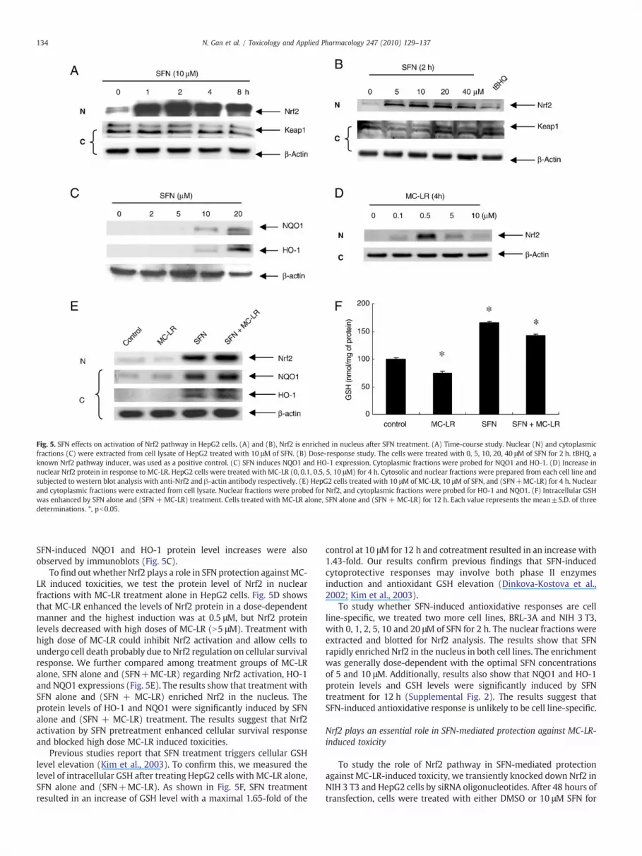
Fig. 5. SFN effects on activation of Nrf2 pathway in HepG2 cells. (A) and (B), Nrf2 is enriched in nucleus after SFN treatment. (A) Time-course study. Nuclear (N) and cytoplasmicfractions (C) were extracted from cell lysate of HepG2 treated with 10 μM of SFN. (B) Dose-response study. The cells were treated with 0, 5, 10, 20, 40 μM of SFN for 2 h. tBHQ, aknown Nrf2 pathway inducer, was used as a positive control. (C) SFN induces NQO1 and HO-1 expression. Cytoplasmic fractions were probed for NQO1 and HO-1. (D) Increase innuclear Nrf2 protein in response to MC-LR. HepG2 cells were treated with MC-LR (0, 0.1, 0.5, 5, 10 μM) for 4 h. Cytosolic and nuclear fractions were prepared from each cell line andsubjected to western blot analysis with anti-Nrf2 and β-actin antibody respectively. (E) HepG2 cells treated with 10 μM of MC-LR, 10 μM of SFN, and (SFN+MC-LR) for 4 h. Nuclearand cytoplasmic fractions were extracted from cell lysate. Nuclear fractions were probed for Nrf2, and cytoplasmic fractions were probed for HO-1 and NQO1. (F) Intracellular GSHwas enhanced by SFN alone and (SFN + MC-LR) treatment. Cells treated with MC-LR alone, SFN alone and (SFN + MC-LR) for 12 h. Each value represents the mean±S.D. of threedeterminations. *, pb0.05.
134 N. Gan et al. / Toxicology and Applied Pharmacology 247 (2010) 129–137
SFN-induced NQO1 and HO-1 protein level increases were alsoobserved by immunoblots (Fig. 5C).
To find out whether Nrf2 plays a role in SFN protection againstMC-LR induced toxicities, we test the protein level of Nrf2 in nuclearfractions with MC-LR treatment alone in HepG2 cells. Fig. 5D showsthat MC-LR enhanced the levels of Nrf2 protein in a dose-dependentmanner and the highest induction was at 0.5 μM, but Nrf2 proteinlevels decreased with high doses of MC-LR (N5 μM). Treatment withhigh dose of MC-LR could inhibit Nrf2 activation and allow cells toundergo cell death probably due to Nrf2 regulation on cellular survivalresponse. We further compared among treatment groups of MC-LRalone, SFN alone and (SFN+MC-LR) regarding Nrf2 activation, HO-1and NQO1 expressions (Fig. 5E). The results show that treatment withSFN alone and (SFN + MC-LR) enriched Nrf2 in the nucleus. Theprotein levels of HO-1 and NQO1 were significantly induced by SFNalone and (SFN + MC-LR) treatment. The results suggest that Nrf2activation by SFN pretreatment enhanced cellular survival responseand blocked high dose MC-LR induced toxicities.
Previous studies report that SFN treatment triggers cellular GSHlevel elevation (Kim et al., 2003). To confirm this, we measured thelevel of intracellular GSH after treating HepG2 cells with MC-LR alone,SFN alone and (SFN+MC-LR). As shown in Fig. 5F, SFN treatmentresulted in an increase of GSH level with a maximal 1.65-fold of the
control at 10 μM for 12 h and cotreatment resulted in an increase with1.43-fold. Our results confirm previous findings that SFN-inducedcytoprotective responses may involve both phase II enzymesinduction and antioxidant GSH elevation (Dinkova-Kostova et al.,2002; Kim et al., 2003).
To study whether SFN-induced antioxidative responses are cellline-specific, we treated two more cell lines, BRL-3A and NIH 3 T3,with 0, 1, 2, 5, 10 and 20 μM of SFN for 2 h. The nuclear fractions wereextracted and blotted for Nrf2 analysis. The results show that SFNrapidly enriched Nrf2 in the nucleus in both cell lines. The enrichmentwas generally dose-dependent with the optimal SFN concentrationsof 5 and 10 μM. Additionally, results also show that NQO1 and HO-1protein levels and GSH levels were significantly induced by SFNtreatment for 12 h (Supplemental Fig. 2). The results suggest thatSFN-induced antioxidative response is unlikely to be cell line-specific.
Nrf2 plays an essential role in SFN-mediated protection against MC-LR-induced toxicity
To study the role of Nrf2 pathway in SFN-mediated protectionagainst MC-LR-induced toxicity, we transiently knocked down Nrf2 inNIH 3 T3 and HepG2 cells by siRNA oligonucleotides. After 48 hours oftransfection, cells were treated with either DMSO or 10 μM SFN for

Fig. 6.Nrf2 pathway is specifically responsible for SFN-induced cytoprotection. (A) Nrf2 siRNA is effective in silencing Nrf2 translocation and activation of target genes. NIH 3 T3 cellswere transfectedwith either control oligos or Nrf2 siRNA. After transfection, cells were exposed to SFN for 4 h. Nuclear extracts were prepared to analyze the distribution of Nrf2, andcytoplasmic extracts were prepared to analyze the NQO1, HO-1 and β-actin expression. (B) Nrf2 silencing voids SFN-induced cytoprotection against MC-LR toxicity. NIH 3 T3 cellswere transfected with Nrf2-siRNA for 48 h before treated with either MC-LR alone or a combination of MC-LR and SFN for 24 h. The cell survival rate was measured by the MTS assay.Error bars indicate the S.D. (n=3). *pb0.05. (C) The specificity of Nrf2 pathway in SFN-induced cytoprotection is confirmed by Annexin V-FITC assay. More apoptotic cells wereobserved in NIH 3 T3 cells transfected with Nrf2 siRNA and treated with SFN and MC-LR than those with only MC-LR.
135N. Gan et al. / Toxicology and Applied Pharmacology 247 (2010) 129–137
4 h. Gene silencing against Nrf2 suppressed SFN-induced Nrf2translocation and expression of NQO1 and HO-1 in NIH 3 T3(Fig. 6A) and HepG2 (Supplemental Fig. 3A). The cell viability assayindicates that the selective silencing of Nrf2 abolished the cytoprotec-tion by SFN againstMC-LR-induced cell death in NIH 3 T3 (Fig. 6B) andHepG2 (Supplemental Fig. 3B). These results indicate that Nrf2pathway plays a role in the SFN-induced cytoprotection. This notionwas also supported by the assay assessing the proportion of apoptoticcells by MC-LR in NIH 3 T3 cells (Fig. 6C). Cells transfected with Nrf2-siRNA had more apoptotic cells when treated with a combination ofSFN and MC-LR than MC-LR alone, contrary to the results indicated inFig. 4C. Nrf2 is a key factor mediated the protective antioxidantresponse. Knockdown of Nrf2 mRNA inhibits the protection onoxidative stress, which was proved by MC-LR treated alone on Nrf2cells and Nrf2-deficient cells (9.51 on NIH 3 T3 cells, 13.54 on NIH
3 T3-Nrf2 deficient cells). And more apoptosis was obtained withcotreatment of SFN and MC-LR in Nrf2-deficient cells (13.54 in MC-LRtreated, 14.3 in cotreated). Therefore, SFN pretreatment aggravatedMC-LR-induced cell death in Nrf2-deficient cells probably due to theloss of Nrf2-dependent defensive response.
Discussion
MC-LR, a main poison in the cyanobacerium Microcystis-contam-inated lakes and rivers, poses an imminent threat to public healtharound the world. Chronically drinking water with trace amounts ofMC-LR may cause severe organ damage to both animals and humans.In addition to sophisticated and costly water treatment to removeeither Microcystis or its metabolic product-MCs, strategies tominimize its poisonous effects on animal and human health by

136 N. Gan et al. / Toxicology and Applied Pharmacology 247 (2010) 129–137
enhancing detoxifying systems are very important, but rarelyinvestigated. In this study, we demonstrated that pretreatment of avariety of cultured cells with SFN effectively induced antioxidativeprotection against cytotoxicity caused by MC-LR, providing apromising prospect that SFN may be used as a protective agentagainst MC-LR-contaminated drinking water.
Previous studies indicated that MC-LR, an electrophile, rapidlyconjugates with GSH. In fact, the detoxification of MC-LR in the liver isknown to occur via conjugation to glutathione with and withoutglutathione-S-transferases (GSTs) (Pflugmacher et al., 1998; Take-naka, 2001; Gehringer et al., 2004). Consequently, cellular GSH level issignificantly reduced with elevated oxidative stress (Zegura et al.,2006). Therefore, the best approach to protect cells from MC-LR is toenhance antioxidative activities. It has been reported that SFN caneffectively reduce cytotoxicity caused by a variety of compounds, likemenadione,tert-butyl hydroperoxide, 4-hydroxynonenal, and arsenictrioxide (Gao et al., 2001; Shinkai et al., 2006; Yoon et al., 2008).
This study shows that SFN protects cells from MC-LR-inducedtoxicity through activation of the Nrf2 pathway. The Nrf2 signalingpathway has been reported to confer protection against chemicalcarcinogens-induced cytotoxicity (Shinkai et al., 2006; William andThomas, 2008; Du et al., 2008). Here, we confirmed in several cell linesthat SFN induced Nrf2 translocation to the nucleus and activated itsdownstream target genes encoding proteins, such as phase II enzymesNQO1 and HO-1. Induction of NQO1 and HO-1 has been established asa strong cellular defense against oxidative stress (Zhang, 2006). Also,we observed an intracellular GSH level increase after SFN treatment.Since γ-glutamylcysteine synthetase, a phase II enzyme regulated byNrf2 pathway, is the rate-limiting enzyme in GSH biosynthesis, theGSH level increase may reflect the induction of γ-glutamylcysteinesynthetase by SFN (Kong et al., 2001; Brook et al., 2001). Interestingly,SFN-induced antioxidative activities were abolished when Nrf2 wasknocked down, indicating a specific role of Nrf2 pathway in SFN-induced cytoprotection against MC-LR. The discovery of the targetpathway is significant because it provides a signal that leads toidentify compounds with better efficacy. In this study, we found thatthe cytoprotection induced by SFN is evident at low concentrationranges of below 10 μM, which is physiologically attainable. LimitedNrf2 accumulation and cytoprotection were observed at highconcentrations of SFN (40 μM for HepG2 cells, 20 μM for NIH 3 T3cells), consistent with previous findings that SFN can inducesubstantial apoptosis in cells at high concentrations (Kong et al.,2001; Kim et al., 2003). Therefore, it is conceivable that lower SFNconcentration may achieve optimal Nrf2-mediated antioxidativeprotection. Our data also show that substantial protection by SFNagainst MC-LR is time-dependent, and thus it is promising that longterm protection against trace amounts of MC-LR in drinking water isachievable by daily consumption of SFN-rich vegetables. However,further preclinical and clinical studies on the protective activity of SFNon MC-LR toxicity are needed.
Acknowledgments
This work was supported by grants from “973” program(2008CB418000), Natural Science Foundation of China-Yunnan Project(U0833604), Chinese Academy of Sciences (KZCX1-YW-14-1) andNational water pollution control and management technology majorprojects (2009ZX07527-005).
Appendix A. Supplementary data
Supplementary data associated with this article can be found, inthe online version, at doi:10.1016/j.taap.2010.06.005.
References
Brook, J.D., Paton, V.G., Vidanes, G., 2001. Potent induction of phase 2 enzymes inhuman prostate cells by Sulforaphane. Cancer Epidemiol. Biomark. Prev. 10,949–954.
Carmichael, W.W., 1994. The toxins of cyanobacteria. Sci. Am. 270, 78–86.Cho, H.Y., Jedlicka, A.E., Reddy, S.P., Kensler, T.W., Yamamoto, M., Zhang, L.Y.,
Kleeberger, S.R., 2002. Role of NRF2 in protection against hyperoxic lung injuryin mice. Am. J. Respir. Cell Mol. Biol. 26, 175–182.
Cornblatt, B.S., Ye, L.X., Dinkova-Kostova, A.T., Erb, M., 2007. Preclinical and clinicalevaluation of sulforaphane for chemoprevention in the breast. Carcinogenesis 28,1485–1490.
Dinkova-Kostova, A.T., Holtzclaw,W.D., Cole, R.N., Itoh, K., Wakabayashi, N., Katoh, Y., etal., 2002. Direct evidence that sulfhydryl groups of Keap1 are the sensors regulatinginduction of phase 2 enzymes that protect against carcinogens and oxidants. Proc.Natl. Acad. Sci. U. S. A. 99, 11908–11913.
Du, Y., Villeneuve, N.F., Wang, X.J., Sun, Z.H., Chen, W.M., Li, J.X., et al., 2008. OridoninConfers Protection against Arsenic-Induced Toxicity through Activation of the Nrf2-Mediated Defensive Response. Environ. Health Perspect. 116, 1154–1161.
Falconer, I.R., 2001. Toxic cyanobacterial bloom problems in Australian waters: risksand impacts on human health. Phycologia 40, 228–233.
Fowke, J.H., Chung, F.L., Jin, F., Qi, D., Cai, Q., et al., 2003. Urinary isothiocyanate levels,brassica, and human breast cancer. Cancer Res. 63, 3980–3986.
Gao, X., Dinkova-Kostova, A.T., Talalay, P., 2001. Powerful and prolonged protection ofhuman retinal pigment epithelial cells, keratinocytes, and mouse leukemia cellsagainst oxidative damage: the indirect antioxidant effects of sulforaphane. Proc.Natl. Acad. Sci. U. S. A. 98, 15221–15226.
Gehringer, M.M., Govender, S., Shah, M., 2003. An investigation of the role of vitamin E inthe protection of mice against microcystin toxicity. Environ. Toxicol. 18, 142–148.
Gehringer, M.M., Shephard, E.G., Downing, T.G., Wiegand, C., Neilan, B.A., 2004. Aninvestigation into the detoxification of microcystin-LR by the glutathione pathwayin Balb/c mice. Int. J. Biochem. Cell Biol. 36, 931–941.
Hayes, J.D., Chanas, S.A., Henderson, C.J., McMahon, M., Sun, C., Moffat, G.J., et al., 2000.The Nrf2 transcription factor contributes both to the basal expression ofglutathione S-transferases in mouse liver and to their induction by thechemopreventive synthetic antioxidants, butylated hydroxyanisole and ethoxy-quin. Biochem. Soc. Trans. 28, 33–41.
Jeong, W.S., Jun, M., Kong, A.N., 2006. Nrf2: a potential molecular target for cancerchemoprevention by natural compounds. Antioxid. Redox Signal. 8, 99–106.
Jochimsen, E.M., Carmichael, W.W., Jisi, A., 1998. Liver failure and death after exposureto microcystins at a hemodialysis center in Brazil. New Engl. J Med. 338, 873–878.
Joseph, M.A., Moysich, K.B., Freudenheim, J.L., Shields, P.G., Bowman, E.D., et al., 2004.Cruciferous vegetables, genetic polymorphisms in glutathione S-transferases M1and T1, and prostate cancer risk. Nutr. Cancer 50, 206–213.
Kensler, T.W., Wakabayashi, N., Biswal, S., 2007. Cell Survival Responses toEnvironmental Stresses Via the Keap1-Nrf2-ARE Pathway. Annu. Rev. Pharmacol.Toxicol. 47, 89–116.
Kim, B.R., Hu, R., Keum, Y.S., Hebbar, V., Shen, G., Nair, S.S., Kong, A.N., 2003. Effects ofglutathione on antioxidant response element-mediated gene expression andapoptosis elicited by sulforaphane. Cancer Res. 63, 7520–7525.
Kobayashi, A., Kang, M.I., Okawa, H., Ohtsuji, M., Zenke, Y., Chiba, T., et al., 2004.Oxidative stress sensor Keap1 functions as an adaptor for Cul3-based E3 ligase toregulate proteasomal degradation of Nrf2. Mol. Cell. Biol. 24, 7130–7139.
Kong, A.N., Yu, R., Hebbar, V., Chen, C., Owuor, E., Hu, R., et al., 2001. Signal transductionevents elicited by cancer prevention compounds. Mutat. Res. 480–481, 231–241.
Lin, H.J., Probst-Hensch, N.M., Louie, A.D., Kau, I.H., Witte, J.S., et al., 1998. Glutathionetransferase null genotype, broccoli, and lower prevalence of colorectal adenomas.Cancer Epidemiol. Biomark. Prev. 7, 647–652.
Moffit, J.S., Aleksunes, L.M., Kardas, M.J., Slitt, A.L., Klaassen, C.D., Manautou, J.E., 2007.Role of NAD(P)H:quinone oxidoreductase 1 in clofibrate-mediated hepatoprotec-tion from acetaminophen. Toxicology 230, 197–206.
Pflugmacher, S., Wiegand, C., Oberemm, A., Beattie, K.A., Krause, E., Codd, G.A., et al.,1998. Identification of an enzymatically formed glutathione conjugate of thecyanobacterial hepatotoxin microcystin LR: the first step in detoxification. Biochim.Biophys. Acta 1425, 527–533.
Prestera, T., Talalay, P., 1995. Electrophile and antioxidant regulation of enzymes thatdetoxify carcinogens. Proc. Natl. Acad. Sci. U. S. A. 92, 8965–8969.
Rangasamy, T., Cho, C.Y., Thimmulappa, R.K., Zhen, L., Srisuma, S.S., Kensler, T.W., et al.,2004. Genetic ablation of Nrf2 enhances susceptibility to cigarette smoke-inducedemphysema in mice. J. Clin. Invest. 114, 1248–1259.
Reisman, S.A., Aleksunes, L.M., Klaassen, C.D., 2009. Oleanolic acid activates Nrf2 andprotects from acetaminophen hepatotoxicity via Nrf2-dependent and Nrf2-independent processes. Biochem. Pharmacol. 77, 1273–1282.
Riley, R.J., Workman, P., 1992. DT-diaphorase and cancer chemotherapy. Biochem.Pharmacol. 43, 1657–1669.
Shinkai, Y., Sumi, D., Fukami, I., Ishii, T., Kumagai, Y., 2006. Sulforaphane, an activator ofNrf2, suppresses cellular accumulation of arsenic and its cytotoxicity in primarymouse hepatocytes. FEBS Lett. 580, 1771–1774.
Spitz, M.R., Duphorne, C.M., Detry, M.A., Pillow, P.C., Amos, C.I., et al., 2000. Dietaryintake of isothiocyanates: evidence of a joint effect with glutathione S-transferase polymorphisms in lung cancer risk. Cancer Epidemiol. Biomark.Prev. 9, 1017–1020.
Sutherland, B.M., Bennett, P.V., Freeman, A.G., Moore, S.P., Strickland, P.T., 1985.Transformation of human cells by DNAs ineffective in transformation of NIH 3 T3cells. Proc. Natl. Acad. Sci. U. S. A. 82, 2399–2403.

137N. Gan et al. / Toxicology and Applied Pharmacology 247 (2010) 129–137
Takenaka, S., 2001. Covalent glutathione conjugation to cyanobacterial hepatotoxinmicrocystin LR by F344 rat cytosolic and microsomal glutathione S-transferases.Environ. Toxicol. Pharmacol. 9, 135–139.
Ueno, Y., Nagata, S., Tsutsumi, T., Yu, S.Z., 1996. Detection of microcystins, a blue-greenalgal hepatotoxin, in drinking water sample in Haimen and Fusui, endemic areas ofPrimary Liver Cancer in China, by highly sensitive immunoassay. Carcinogenesis 17,1317–1321.
WHO, 1998.Guidelines fordrinking-waterquality.AddendumtoVolume2.HealthCriteriaand Other Supporting Information.Word Health Organization, Genveva, Switzerland.
William, O.O., Thomas, W.K., 2008. Nrf2 signaling: An adaptive response pathway forprotection against environmental toxic insults. Mutat. Res./Rev. Mutat. Res. 659,31–39.
Yates, M.S., Tran, Q.T., Dolan, P.M., Osburn, W.O., Shin, S., et al., 2009. Genetic versuschemoprotective activation of Nrf2 signaling: overlapping yet distinct gene
expression profiles between Keap1 knockout and triterpenoid-treated mice.Carcinogenesis 30, 1024–1031.
Yoon, H.Y., Kang, N.I., Lee, H.K., Jang, K.Y., Park, J.W., Park, B.H., 2008. Sulforaphaneprotects kidneys against ischemia-reperfusion injury through induction of theNrf2-dependent phase 2 enzyme. Biochem. Pharmacol. 75, 2214–2223.
Zegura, B., Lah, T.T., Filipic, M., 2006. Alteration of intracellular GSH levels and its role inmicrocystin- LR-induced DNA damage in human hepatoma HepG2 cells. Mutat.Res. 611, 25–33.
Zhang, D.D., Lo, S.C., Sun, Z.H., Habib, G.M., Lieberman, M.W., Hannink, M., 2005.Ubiquitination of Keap1, a BTB-Kelch Substrate Adaptor Protein for Cul3, TargetsKeap1 for Degradation by a Proteasome-independent Pathway. J. Biol. Chem. 280,30091–30099.
Zhang, D.D., 2006. Mechanistic studies of the Nrf2-Keap1 signaling pathway. DrugMetab. Rev. 38, 769–789.


