Embed Size (px)
Citation preview

RESEARCH ARTICLE Open Access
Superoxide dismutase is upregulated inStaphylococcus aureus following protoporphyrin-mediated photodynamic inactivation and doesnot directly influence the response tophotodynamic treatmentJoanna Nakonieczna*, Ewelina Michta, Magda Rybicka, Mariusz Grinholc, Anna Gwizdek-Wiśniewska,Krzysztof P Bielawski
Abstract
Background: Staphylococcus aureus, a major human pathogen causes a wide range of disease syndromes. Themost dangerous are methicillin-resistant S. aureus (MRSA) strains, resistant not only to all b-lactam antibiotics butalso to other antimicrobials. An alarming increase in antibiotic resistance spreading among pathogenic bacteriainclines to search for alternative therapeutic options, for which resistance can not be developed easily. Amongothers, photodynamic inactivation (PDI) of S. aureus is a promising option. Photodynamic inactivation is based on aconcept that a non toxic chemical, called a photosensitizer upon excitation with light of an appropriatewavelength is activated. As a consequence singlet oxygen and other reactive oxygen species (e.g. superoxideanion) are produced, which are responsible for the cytotoxic effect towards bacterial cells. As strain-dependence inphotodynamic inactivation of S. aureus was observed, determination of the molecular marker(s) underlying themechanism of the bacterial response to PDI treatment would be of great clinical importance. We examined therole of superoxide dismutases (Sod) in photodynamic inactivation of S. aureus as enzymes responsible for oxidativestress resistance.
Results: The effectiveness of photodynamic inactivation towards S. aureus and its Sod isogenic mutants deprivedof either of the two superoxide dismutase activities, namely SodA or SodM or both of them showed similar results,regardless of the Sod status in TSB medium. On the contrary, in the CL medium (without Mn++ ions) the doubleSodAM mutant was highly susceptible to photodynamic inactivation. Among 8 clinical isolates of S. aureusanalyzed (4 MRSA and 4 MSSA), strains highly resistant and strains highly vulnerable to photodynamic inactivationwere noticed. We observed that Sod activity as well as sodA and sodM transcript level increases afterprotoporphyrin IX-based photodynamic treatment but only in PDI-sensitive strains.
Conclusions: We confirmed that porphyrin-based photokilling efficacy is a strain-dependent phenomenon. Weshowed that oxidative stress sensitivity caused by the lack of both Sod enzymes can be relieved in the presence ofMn ions and partially in the presence of Fe ions. The fact that Sod activity increase is observed only in PDI-susceptible cells emphasizes that this is probably not a direct factor affecting S. aureus vulnerability to porphyrin-based PDI.
* Correspondence: [email protected] Faculty of Biotechnology University of Gdansk and MedicalUniversity of Gdansk, Kladki 24, 80-822 Gdansk, Poland
Nakonieczna et al. BMC Microbiology 2010, 10:323http://www.biomedcentral.com/1471-2180/10/323
© 2010 Nakonieczna et al; licensee BioMed Central Ltd. This is an Open Access article distributed under the terms of the CreativeCommons Attribution License (http://creativecommons.org/licenses/by/2.0), which permits unrestricted use, distribution, andreproduction in any medium, provided the original work is properly cited.

BackgroundStaphylococcus aureus, a major human pathogen causesa wide range of disease syndromes, including life-threa-tening endocarditis, meningitidis and pneumonia.According to the Centers for Disease Control and Pre-vention this bacterium has been reported to be the mostsignificant cause of serious infections in the UnitedStates [1]. S. aureus is able to cause and develop aninfection very efficiently due to its ability to produce afew dozen of virulence factors, on one hand, and anease of antibiotic resistance development, on the other.The most dangerous are methicillin-resistant S. aureus(MRSA) strains, constituting 50% of hospital-aquiredisolates as well as emerging vancomycin-resistant var-iants, isolated from some hospital settings [2].Among several virulence factors, S. aureus produces
enzymes responsible for resistance against oxidativestress, like catalase and superoxide dismutase (Sod). Sodconverts superoxide anion (O2·
-) into hydrogen peroxide(H2O2), a less potent biological oxidant, which is furtherdecomposed by catalase to water and ground state oxy-gen. Sod enzyme is produced in response to the pre-sence of reactive oxygen species (ROS) generatedendogenously as an effect of oxygen metabolism or, exo-genously produced by neutrophils and macrophages.Superoxide anion, which is the product of oxygenreduction, reacts with hydrogen peroxide within thebacterial cell and produces free hydroxyl radical (.OH),the most dangerous oxygen species able to interact withvirtually any organic substance in the cell. Superoxideanion can reduce hypochlorus acid (HOCl) arose as aresult of H2O2 interaction with phagocyte-derived per-oxidases, and further form .OH [3].The classification of Sod enzymes is based on the type
of transition metal present in their active center, includ-ing manganese (Mn), iron (Fe), copper (Cu) and a fewyears ago a nickel (Ni)-containing Sod was described,originally isolated from the cytoplasm of Streptomycesseoulensis [4,5]. In the Escherichia coli bacterium model,the presence of three Sods were described: Fe- andMn- Sods localized in the cytoplasm, whereas in theperiplasm copper-zinc (Cu-Zn) SOD was detected [6].S. aureus produces three Sod enzymes, encoded by twogenes, sodA and sodM [7,8]. The particular subunitsform two kinds of Sod homodimers, i.e. SodA-SodA andSodM-SodM as well as SodA-SodM heterodimers, easilydistinguishable on native gels stained for Sod activity[8]. Both, SodA and SodM subunits are believed to pos-sess Mn ions as a cofactor in the active site. Manganeseis now believed to play a crucial role in a variety of cel-lular processes including stress responses [9]. In arange of bacterial pathogens, Mn is recognized as havinga major effect on virulence [10,11]. Apart from
participating in several enzyme functions, Mn complexeswith phosphate and lactate were demonstrated to sca-venge ROS [12].The role of Sod in the pathogenesis of many bacteria
was proved. In S. aureus however, the results are notunambiguous. The very first analyses of antioxidantenzymes and staphylococcal virulence showed no corre-lation [13]. Similarly, in a mouse abscess model resultingfrom S. aureus infection, inactivation of sodA gene,recognized as the main Sod activity in S. aureus, had noimpact on staphylococcal virulence [7]. Moreover,mouse kidney infection was not attenuated after sodMgene inactivation [14]. On the other hand, examinationof a range of virulent versus non-virulent S. aureus clini-cal isolates, showed statistically significant higher Sodactivity in the first group studied [15]. Karavolos et al.tested the role of Sod in a mouse subcutaneous modelof infection and claimed that mutants deprived of eitherSodA, SodM or both activities had significantly reducedvirulence compared to S. aureus wild-type SH1000strain [16].As bacteria replicate very quickly, the possibility of
mutant selection which effectively deals with antibiotictreatment rises. An alarming increase in antibiotic resis-tance spreading among pathogenic bacteria inclines tosearch for alternative therapeutic options, for whichresistance cannot be developed easily. One such optionis photodynamic inactivation of bacteria (PDI). Thismethod involves the use of non toxic dyes, so calledphotosensitizers (PS), which become excited upon visi-ble light of an appropriate wavelength and eventually anumber of ROS are formed [17]. As a consequence ofROS action, which are known to cause severe damageto DNA, RNA, proteins, and lipids, bacterial cells die.Two oxidative mechanisms can occur after light activa-tion of a photosensitizer. When the photosensitizerinteracts with a biomolecule, free radicals (type Imechanism), and/or singlet molecular oxygen (1O2)(type II mechanism) are produced, which are responsiblefor cell inactivation [18]. In the case of porphyrin-basedphotosensitizers, 1O2 seems to be the main ROS gener-ated upon photoexcitation, although O2
.-, .OH are alsoimplicated [19]. In a very elegant study by Hoebeke etal., the photochemical action of bacteriochlorin a, astructural analog of protoporphyrin IX, was also demon-strated to be based on both, type I and type II mechan-ism of action in a 1:1 proportion [20]. Several lines ofevidence indicate the effectiveness of PDI in vitroagainst both Gram-positive and -negative species[21,22]. It was also demonstrated that photodynamicinactivation may be applied to inactivate bacterial viru-lence factors, which represents an advantage over topicalantibiotic treatments [23].
Nakonieczna et al. BMC Microbiology 2010, 10:323http://www.biomedcentral.com/1471-2180/10/323
Page 2 of 14

In our previous reports we observed that the S. aureusresponse to PDI is strain-dependent. Among clinical iso-lates some were killed in 99,999%, whereas others inonly about 20% in protoporphyrin-based PDI [24]. Tounderstand if the antioxidant enzyme status may beinvolved in the S. aureus response to PDI, we checkedthe survival rate of the isogenic sod mutants of S. aureusand compared the activities of Sods in response to PDIon the protein as well as gene expression level.
ResultsPDI effectiveness towards wild type Staphylococcus aureusand its sod isogenic mutantsWith the use of type I or type II oxidative stressquenching agents, we checked that PpIX-mediated PDIis involved in the type I mechanism of oxidative stressinduction (production of free radicals) (data not shown).This gave us a rationale to study the influence of Sodon the PDI outcome. In order to check superoxide dis-mutases’ role in photodynamic inactivation we first ofall checked whether S. aureus RN6390 strain deprivedof either SodA, SodM or both of the activities differen-tially responded to photodynamic inactivation. In ourstudy we used protoporphyrin IX (PpIX) as a photosen-sitizer. Treatment of S. aureus RN6390 and its isogenicsod mutants with 0-50 μM PpIX and an irradiation doseof 12 J/cm2 resulted in a weak response to PDI in TSBmedium. Wild-type RN6390 showed 1.85 log10 unitssurvival reduction in comparison to non PDI-treatedcells. In the case of the single SodA and SodM mutantsthe survival rate accounted for 2.0 log10 units reductionand 1.55 log10 units reduction, respectively (Figure 1).The double SodAM mutant reduced its survival rate byonly 1.3 log10 units. Statistical analysis performed on sixindependent sets of measurements revealed no correla-tion between the Sod status and PDI response, at leastin TSB medium. The observed phototoxic effect was ineach case PpIX-concentration dependent in a range of0-50 μM. We chose one light dose of 12 J/cm2 in allexperiments concerning killing data based on our pre-viously published results [24,25].
Effect of divalent ions on PDI effectiveness towards wildtype RN6390 and its sod isogenic mutantsAs S. aureus Sod enzymes are recognized as Mn-con-taining proteins, we further checked the influence ofMn ion depletion on PDI effectiveness. After cells werecultured in a chemically defined CL medium with andwithout 20 μM MnSO4, PDI procedure was performedaccording to the Methods section, similarly as with TSBmedium. When the CL medium was supplemented withMn ions the survival rate of S. aureus RN6390 andsodA, sodM, sodAM mutants was in general higher incomparison to non Mn-supplemented medium. The
values ranged between 0.5 log10 units reduction forwild-type RN6390, through 0.6 and 0.9 log10 units forthe two single sodM and sodA mutants, respectively, to1.3 log10 units reduction observed in the case of thedouble sodAM mutant (Figure 2A). When the PDI stu-dies were performed in the absence of Mn ions, the sur-vival rate of the three analyzed mutants, but not thewild-type RN6390, decreased. In the case of the sodMmutant we observed a 0.9 log10 unit reduction in survi-val rate and 1.3 log10 unit reduction when the sodAS. aureus was analyzed. For those differences, however,no statistical relevance was proved. Significant differencewas observed for the double mutant, whose survival ratedropped by 4.1 log10 units (Figure 2B). This result wasstatistically confirmed. The obtained results suggest thata single Sod activity is sufficient to combat oxidativestress conditions resulting from PDI, whereas S. aureuscells without any Sod activity can be rescued by the pre-sence of Mn++ ions. Based on the presented results itcan be assumed that oxidative stress sensitivity causedby the lack of both Sod enzymes can be overcame inthe presence of Mn ions.In order to check whether other divalent ions are able
to cause such an effect we performed analogous experi-ments with 20 μM FeSO4. Supplementation of CL med-ium with iron ions resulted in partial restoration ofoxidative stress resistance but only in sodAM mutant,where the drop in survival rate increased from 4.1 log10units to 2.4 log10 units, respectively in CL mediumwithout and supplemented with divalent metal ions(Additional file 1).
PDI effectiveness towards clinical Staphylococcus aureusisolatesIn order to check PpIX-based PDI effectiveness towardsS. aureus strains isolated from patients, we chose 4strains characterized as methicillin resistant (MRSA)and 4 methicillin susceptible strains (MSSA). Examina-tion of the survival rate of the chosen strains resulted inan observation that the response to PDI treatment isstrain-dependent. In both groups, MRSA and MSSA, weobserved strains which were killed very inefficiently(Figure 3), eg. strains 1397 and 2002 reduced their sur-vival rate only by 0.2 log10 units. On the contrary, inde-pendent of the methicillin-resistance status we observedstrains highly susceptible to PpIX-based photokilling, eg.strains 472, 80/0 and 2288, which reduced their survivalrate by 3.4 log10 units, 2.4 log10 units and 2.5 log10units, respectively. One-way analysis of variance test ofthe survival of the studied clinical isolates (at 50 μMPpIX concentration) showed statistically significant dif-ferences (F = 88,3 p < 0.05). Based on the Tukey post-hoc test, a decrease in the survival of the 4246 straindid not differ from the strains 7259, 2002 and 1397, and
Nakonieczna et al. BMC Microbiology 2010, 10:323http://www.biomedcentral.com/1471-2180/10/323
Page 3 of 14

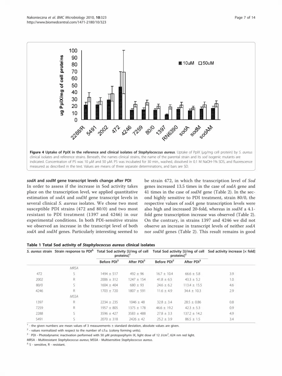
further those strains were classified as one group. Thisgroup was considered by us as PDI-resistant with thesurvival decrease not exceeding 1.5 log10 units. The nextfour bacterial isolates (5491, 2288, 80/0, 472) wererecognized as PDI-sensitive with the survival decrease ofmore than 1.5 log10 units. It is believed that the effec-tiveness of PDI depends on the ability of cells to uptakethe photosensitizer. We checked whether there are anydifferences among S. aureus strains in PpIX uptake intothe cell. Protoporphirin IX uptake in the tested strainsdid not show much differentiation. It is worth mention-ing, however, that in the case of the most PDI-vulner-able 472 strain, PpIX uptake value was 47.4 μg/mg andon the contrary, only 7.3 μg/mg in the case of the mostresistant 1397 strain. We observed no apparent correla-tion between PS uptake and PDI effectiveness. In thecase of RN6390 and its isogenic sod mutants the uptakewas very balanced and ranged between 13.1 and16.2 μg/mg for the wild type and the mutants (Figure 4).
Sod activity increases after PDIIn order to assess the amount of Sod activity in strain-dependent response to PpIX-based photodynamictreatment, we measured total Sod activity in S. aureus
isolates before and after PDI treatment. The activitywas measured with the NBT reduction method andwas expressed as Sod units per mg of total cell proteincontent (Methods section). Firstly, the basic level ofSod activity was estimated. Among 8 clinical isolatestested Sod activity was similar and ranged between1495 U/mg and 2234 U/mg, with the exception of2288 strain, where the observed activity was the high-est and amounted to 3597 U/mg. However, whenmean activity values were normalized with respect tothe number of c.f.u. (colony forming units), theyslightly differed for PDI-susceptible and PDI-resistantstrains (23.6 ± 4 U/mg and 33.2 ± 15 U/mg, respec-tively) (Table 1). These differences appeared muchgreater when bacterial cells were exposed to PDI.After photosensitization with 50 μM PpIX and illumi-nation with 12 J/cm2 red light, the total Sod activityraised to the mean value of 100.9 ± 30 U/mg in thecase of PDI-susceptible strains, whereas only a minorincrease in the Sod activity level was observed in PDI-resistant strains (37.1 ± 7 U/mg). This indicates thatoxidative stress generated in our experimental condi-tions greatly induced Sod activity in PDI-susceptiblestrains (Table 1).
Figure 1 Protoporphyrin IX-mediated PDI against reference strains in TSB medium. The bacterial suspensions were illuminated after darkincubation for 30 min. at 37°C with different concentrations of PpIX (up to 50 μM). PDI was tested against reference strains of S. aureus: RN6390,RN6390sodA, RN6390sodM, RN6390sodAM. Bacteria were illuminated with 12 J/cm2 624 ± 18 nm light, and survival fractions were determined asdescribed in Methods. Values are means of at least three separate experiments.
Nakonieczna et al. BMC Microbiology 2010, 10:323http://www.biomedcentral.com/1471-2180/10/323
Page 4 of 14

Figure 2 Mn ions influence on protoporphyrin IX-mediated PDI against reference strains. The bacterial suspensions were illuminated afterdark incubation for 30 min. at 37°C with different concentrations of PpIX (up to 50 μM). PDI was tested against reference strains of S. aureus:RN6390, RN6390sodA, RN6390sodM, RN6390sodAM in Mn-supplemented medium (A) and Mn-depleted medium (B). Bacteria were illuminatedwith 12 J/cm2 624 ± 18 nm light, and survival fractions were determined as described in Methods. Values are means of three separateexperiments, and bars are SD. * indicates statistically significant difference in survival drop between RN6390sodAM and each of the followingstrains RN6390, RN6390sodA, RN6390sodM at each tested concentration (p < 0.05).
Nakonieczna et al. BMC Microbiology 2010, 10:323http://www.biomedcentral.com/1471-2180/10/323
Page 5 of 14

Figure 3 Protoporphyrin IX-mediated PDI against clinical strains. The bacterial suspensions were illuminated after dark incubation for30 min. at 37°C with different concentrations of PpIX (up to 50 μM). PDI was tested against clinical S. aureus strains: MRSA, MSSA. Bacteria wereilluminated with 12 J/cm2 624 ± 18 nm light, and survival fractions were determined as described in Methods. Values are means of threeseparate experiments, and bars are SD.
Nakonieczna et al. BMC Microbiology 2010, 10:323http://www.biomedcentral.com/1471-2180/10/323
Page 6 of 14
sodA and sodM gene transcript levels change after PDIIn order to assess if the increase in Sod activity takesplace on the transcription level, we applied quantitativeestimation of sodA and sodM gene transcript levels inseveral clinical S. aureus isolates. We chose two mostsusceptible PDI strains (472 and 80/0) and two mostresistant to PDI treatment (1397 and 4246) in ourexperimental conditions. In both PDI-sensitive strainswe observed an increase in the transcript level of bothsodA and sodM genes. Particularly interesting seemed to
be strain 472, in which the transcription level of Sodgenes increased 13.5 times in the case of sodA gene and41 times in the case of sodM gene (Table 2). In the sec-ond highly sensitive to PDI treatment, strain 80/0, therespective values of sodA gene transcription levels werealso high and increased 20-fold, whereas in sodM a 4.1-fold gene transcription increase was observed (Table 2).On the contrary, in strains 1397 and 4246 we did notobserve an increase in transcript levels of neither sodAnor sodM genes (Table 2). This result remains in good
Figure 4 Uptake of PpIX in the reference and clinical isolates of Staphylococcus aureus. Uptake of PpIX (μg/mg cell protein) by S. aureusclinical isolates and reference strains. Beneath, the names clinical strains, the name of the parental strain and its sod isogenic mutants areindicated. Concentration of PS was 10 μM and 50 μM. PS was incubated for 30 min., washed, dissolved in 0.1 M NaOH-1% SDS, and fluorescencemeasured as described in the text. Values are means of three separate determinations, and bars are SD.
Table 1 Total Sod activity of Staphylococcus aureus clinical isolates
S. aureus strain Strain response to PDIΔ Total Sod activity [U/mg of cellproteins]1
Total Sod activity [U/mg of cellproteins]2
Sod activity increase [× fold]
Before PDI3 After PDI3 Before PDI3 After PDI3
MRSA
472 S 1494 ± 517 492 ± 96 16.7 ± 10.4 66.6 ± 5.8 3.9
2002 R 2006 ± 312 1247 ± 154 41.8 ± 6.5 43.3 ± 5.2 1.0
80/0 S 1604 ± 404 680 ± 93 24.6 ± 6.2 113.4 ± 15.5 4.6
4246 R 1703 ± 720 1807 ± 591 11.6 ± 4.9 34.4 ± 10.3 2.9
MSSA
1397 R 2234 ± 235 1046 ± 48 32.8 ± 3.4 28.5 ± 0.86 0.8
7259 R 1957 ± 805 1375 ± 178 46.6 ± 19.2 42.3 ± 5.3 0.9
2288 S 3596 ± 427 3583 ± 488 27.8 ± 3.3 137.2 ± 14.2 4.9
5491 S 2070 ± 318 2426 ± 42 25.2 ± 3.9 86.5 ± 1.5 3.41 - the given numbers are mean values of 3 measurements ± standard deviation, absolute values are given.2 - values normalized with respect to the number of c.f.u. (colony forming units).3 - PDI - Photodynamic inactivation performed with 50 μM protoporphyrin IX, light dose of 12 J/cm2, 624 nm red light.
MRSA - Multiresistant Staphylococcus aureus; MSSA - Multisensitive Staphylococcus aureus.Δ S - sensitive, R - resistant.
Nakonieczna et al. BMC Microbiology 2010, 10:323http://www.biomedcentral.com/1471-2180/10/323
Page 7 of 14

agreement with the total Sod activity rise after PDItreatment (Table 1).
DiscussionStaphylococcus aureus is one of the most commonhuman pathogens. It infects tissues locally, howeverthrough the action of a range of pyrogenic toxins andsuperantigens, bacteria can spread easily making theinfection generalized [26]. The most dangerous thera-peutically problematic are methicillin-resistant Staphy-lococcus aureus (MRSA), which are resistant not onlyto methicillin itself but also to all b-lactams as well asother groups of antimicrobial chemotherapeutics, likemacrolides, lincosamides, aminoglycosides [27]. The lat-est epidemiological data indicates that the prevalence ofMRSA in Europe seems to be low but is increasing,moreover European strains are very heterogeneous asopposed to USA-derived MRSA [28]. As the multiresis-tance spread is apparent among S. aureus strains inhospital settings, and becoming more evident in thecommunity (so called community-aquired MRSA), sev-eral attempts are taken to develop strategies againstthese bacteria. The most popular and main-streamareas of research are new antimicrobial therapeutics[29]. However, alternative therapeutic options are alsounder investigation, to name: antimicrobial naturalcompounds [30,31], cationic antimicrobial peptides[32], the use of protection strategy to biofilm formation[33,34] or bacteriophage-based approaches [35,36].Among the listed, photodynamic inactivation (PDI) ofS. aureus is also a promising option. Photodynamicinactivation is based on a concept that a non toxic che-mical, named a photosensitizer upon excitation withlight of an appropriate wavelength is activated. As aconsequence singlet oxygen and other reactive oxygenspecies are produced, which are responsible for the
cytotoxic effect towards bacterial cells [37,38]. It is ofgreat clinical importance and an advantage of PDI thatS. aureus isolates, both MRSA and MSSA, can beeffectively killed [39]. Previous reports of our groupemphasized that S. aureus response to PDI is a strain-dependant phenomenon, which from the clinical pointof view warrants attention [24]. Among 80 MRSA andMSSA strains some were ultra-sensitive to protopor-phyrin IX diarginate-based PDI, whereas others exertedcomplete resistance to such treatment. The same ten-dency was observed in the presented results with theuse of protoporphyrin IX as a photosensitizer (Figure 3).In our attempts to determine the molecular marker ofstrain-dependent response to PDI, we found that bio-film producing strains were killed less efficiently incomparison to non biofilm-producing strains [24],whereas efflux pumps, eg. NorA had no influence onthe efficacy of photokilling [25].
Sod status and PDI responseIn the presented work we focused on the role of super-oxide dismutases in the response of S. aureus to PDI.Superoxide dismutase constitutes the first line of bacter-ial defense against oxidative stress, therefore it wasexpected that the correlation may exist between the Sodstatus in the cell and response to PDI. Statistical analysisrevealed no substantial difference in the survival rateamong the four reference strains in TSB medium. In thestudy by Valderas and Hart, the same strains, deprivedof either of the two Sods or both of them, were analyzedin conditions of methyl viologen (MV)-generated oxida-tive stress. They noticed that the highest drop in viabi-lity was observed in the case of SodAM double mutantsgrown in TSB medium [8]. On the contrary, the groupof Foster, found that similar strains (i.e. analogues Sodmutants but with different genetic background) due to
Table 2 Transcript level of the sodA, sodM genes in Staphylococcus aureus clinical isolates
S. aureus strain Strain response to PDI Sod genes transcript level[copies/μl]1
Sod genes transcript level[copies/μl]2
Transcript level increase [× fold]
Before PDI3 After PDI3 Before PDI3 After PDI3
SodA
472 sensitive 372150 396674 418.1 5666.7 13.5
80/0 sensitive 1671 3136 2.5 52.2 20
1397 resistant 450267 24647 662.1 68.4 0.1
4246 resistant 4978943 1482683 3387.0 2745.7 0.8
SodM
472 sensitive 59205 194245 66.5 2774.9 41
80/0 sensitive 56789 21804 87.3 363.4 4.1
1397 resistant 123025 45475 279.6 119.6 0.4
4246 resistant 286623 198523 267.8 208.9 0.81 - absolute number of amplified mRNA fragment.2 - values normalized with respect to the number of c.f.u. (colony forming units).3 - PDI - Photodynamic inactivation performed with 50 μM protoporphyrin IX, light dose of 12 J/cm2, 624 nm red light.
Nakonieczna et al. BMC Microbiology 2010, 10:323http://www.biomedcentral.com/1471-2180/10/323
Page 8 of 14

the action of internally-generated superoxide anion, via-bility drops in the case of both, SodA and SodAM dou-ble mutants in the Chelex treated BHI medium withoutMn++ ions. They also observed that upon supplementa-tion of the medium with Mn++ the viability of the men-tioned mutants increased. When the same strains werechallenged with externally generated superoxide anionin the stationary phase of growth, only the double Sodmutant was more susceptible to such treatment in com-parison to the wild type SH1000 strain, moreover suchan effect was not dependent on Mn++ presence [16]. Weperformed statistical analysis of the data presented inFigure 1 and found no statistically relevant differenceexisting among the four strains analyzed (i.e. wild typeRN6390, RN6390sodA::tet, RN6390sodM::erm,RN6390sodM::erm sodA::tet), what is seen in Figure 1.Our results differ from the one presented by Hart [8],which may be attributed to the differences in types ofoxidative stress generated as a result of photodynamicaction versus methyl viologen-induced oxidative stressused by Hart group. Methyl viologen is believed toinduce internal oxidative stress. Our previous resultsshowed that PDI-induced oxidative stress is mainlyexternal [25]. In our previous work, when PpIX waswashed away from the cell suspension before illumina-tion, the photodynamic effect was abolished. Thus wecan speculate that oxidative stress associated toxicity isa result of cell wall and bacterial membrane damage,which eventually leads to loss of cell viability. We canhypothesize that in our experimental conditions we useda more complex oxidative stress generating system thanthat used by Hart or Foster group. It is known that dur-ing photodynamic inactivation a number of reactive oxy-gen species are generated. This phenomenon isdependent on the type of photosensitizer used as well asmedium conditions. For example, it was shown for full-erol c60, a recently studied photosensitizer, that depend-ing on the medium used, either singlet oxygen alone orsinglet oxygen together with superoxide anion were pro-duced in a phototoxic process [40]. Different species ofROS produced in various media may affect the photo-toxic effect on the same strain. We can speculate thatapart from singlet oxygen and superoxide anion, otherROS can be generated in PpIX-mediated photodynamicprocess, which can affect either SodA or SodM regula-tory pathways. The regulation of Sod activity in bacterialcells is very complex and yet not fully understood. Diva-lent metal ions, eg. Mn, Fe play a crucial role in theseprocesses as enzyme or transcription factor regulatorcofactors [16,41,42]. It is known that homeostasis of Mnand Fe are intertwined and most likely the manipulationof one of them greatly alters the uptake, storage andregulation of the other. It was shown that direct elemen-tal superoxide scavenging by Mn occurs in S. aureus
[12]. This effect was also clearly visible in our experi-mental data, where the survival rate of the double S.aureus sodAM mutant increased from 4.1 log10 unitsreduction in the Mn-depleted medium to 1.3 log10 unitsin the Mn-supplemented one (Figure 2) as a response tooxidative stress generating PDI. The comparison of thesurvival fraction of wild type RN6390 and sod mutantsamong each other as well as between conditions of Mnpresence and absence in the medium explicitly indicatesthat Mn++ ions influence the efficacy of bacteria killingbut based on our results this seems to be regardless ofthe Sod activity.
Clinical isolates of Staphyloccocus aureus diverselyrespond to PDIEight S. aureus strains isolated from hospitalizedpatients (4 MRSA and 4 MSSA) examined with respectof their ability to survive after PDI treatment, showeddifferent pattern of response. Based on statistical analy-sis we divided those strains into two groups: sensitiveand resistant to PDI. In the group of resistant strains(2002, 4246, 1397, 7259) the drop in the survival ratedid not exceed 1.5 log10 units. In the second group ofstrains, called sensitive, (472, 80/0, 2288, 5491) the dropin survival rate was at least 1.5 log10 units reduction inviable counts. In our previous reports we alreadyshowed a strain-dependent response to PDI targeted S.aureus cells, where the observed efficacy of photokillingreached even 5 log10 units reduction. The differencesbetween our previous studies and the one presentedhere might have probably resulted from a differentphotosensitizer used - PpIX vs. protoporphyrin IX dia-rginate (PpIXArg2) [24,25]. Other groups also observedthe phenomenon of PDI-strain dependence, however,the mechanism underlying the diverse response to PDIwas not explored [43,44]. Our data shows that at lowerconcentration of a photosensitizer (10 μM) a substantialdrop in bacterial survival occurred, whereas at higherconcentrations (25-50 μM), no further decrease in survi-val was noticed. We associate this phenomenon withpoor solubility of PpIX in water solutions but the solu-bility itself does not justify the observed variability inkilling curves. Similar results were obtained by thegroup of Wilson (2008). In the study they used anotheranionic photosensitizer, indocyanine green (ICG) againstS. aureus and observed that the concentration of 25 μg/ml resulted in 6 log10 units reduction in viable counts,but higher ICG concentrations (50 and 100 μg/ml),resulted in lesser, about 4 and 5 log10 units reduction insurvival counts, respectively [45]. Possible explanation ofthis phenomenon may be the self shielding effect of thenon-bound PS in solution at higher concentrations.Effective photodynamic therapy is a result of a combina-tion of several factors. Beside the biophysical properties
Nakonieczna et al. BMC Microbiology 2010, 10:323http://www.biomedcentral.com/1471-2180/10/323
Page 9 of 14

of a sensitizer itself, also total light delivered, time ofincubation with a photosensitizer, presence of additionalproteins are crucial. In our work we did not focused onexamining the dependence of killing rate vs. light dose.We performed all photodynamic inactivation studies onone light dose (12 J/cm2) chosen as optimal based onour previously published data concerning S. aureusphotoinactivation as well as phototoxicity assays per-formed on dermal human fibroblasts [46,47].In our previous attempts to explore the differences of
porphyrin-based photokilling towards S. aureus cells, wefound biofilm production ability to correlate with higherresistance to PDI treatment. However, it was also notedthat among S. aureus isolates with elevated resistance toPDI, biofilm non-producing strains were also observed.This points out that biofilm production is not the onlyfactor responsible for the observed phenotype [24].Results published by Gad et al. indicated that extracellu-lar slime significantly influences PS uptake by S. aureuscells, however an unambiguous conclusion was not pos-sible due to the significant differences in both theuptake and PDI efficacy of the three PS tested, namelychlorinee6, poly-L-lysine-chlorinee6 and methylene blue[48]. S. aureus strains tested in our experimental condi-tions expressed no statistical correlation between PSuptake and PDI effectiveness, nevertheless the highestaccumulation of PS was observed for the most efficientlykilled strain 472 (3.4 log10 reduction in viable countunits), as well as the lowest PS accumulation wasobserved in the case of the most resistant to PDI -strain 1397 (0.2 log10 reduction in viable count units)(Figure 3). The mean uptake level was 47.4 μg/mg oftotal protein content and 7.3 μg/mg of total proteincontent, for strains 472 and 1397, respectively. Theresults concerning uptake level in strains 472 and 1397remain in a good agreement with our previous reports,where the same set of clinical isolates was analyzed butwith the use of a different PS, namely PpIXArg2 [25].Based on our previous and present results we concludethat the PS uptake process is not the main determinantof PDI effectiveness, at least for the porphyrin-basedphotokilling. We and other authors propose subsequentfactors which may contribute and explain the differencesin PDI efficacy of bacteria [25,49], eg. cellular repair sys-tems or level of antioxidant enzymes.
Sod activity and transcript level increase after PDI in PDI-susceptible strainsThe participation of superoxide dismutase in oxidativestress resistance, and also in photodynamically generatedreactive oxygen species is obvious. However, the role ofSod activity in PDI of bacteria has not been studied sofar. There is few literature data on the association ofSod activity and photodynamic inactivation studies, and
to the best of our knowledge they all concern eukaryoticcells. It was proposed for example that inhibition ofMn-Sod activity potentiates the antitumor effectivenessof photodynamic therapy in several cell lines and also ina mouse model of tumorigenesis [50]. Our attempt wasto assess Sod activity in clinical isolates of S. aureus andto compare its basic level between PDI-resistant andPDI-susceptible bacteria. Basic Sod activity levels dif-fered only slightly between PDI-resistant and PDI-sus-ceptible strains (33.2 ± 15 U/mg and 23.6 ± 4 U/mg,respectively), which can be expected as S. aureus is notconstantly exposed to elevated levels of oxidative stressAfter PDI treatment we observed about a 4-fold increaseof Sod activity but only in strains susceptible to PDI.Sod expression is probably induced by a particular sig-nal. The result published by the Foster group showedthat when examining lacZ fusions with SodA genes,exposition of the cells to methyl viologen (internal oxi-dative stress generating agent) the level of SodAincreased. The increase of SodM level was also observed,but only when cells were exposed to externally gener-ated oxidative stress (xanthine/xanthine oxidase) [16].Summarizing, although we did observe some differencesof the basic Sod activity levels in PDI-susceptible vs.PDI-resistant strains, their statistical relevance is notobvious and does not explain the huge differences inPDI-based bactericidal efficacy (Table 2).The reports previously published by our group showed
that the bactericidal effect of PpIXArg2-based photokill-ing was almost completely abolished, when PS waswashed away after incubation (before light exposure)[25]. This indicated that externally generated ROS areresponsible for bacterial cell destruction. In regard toour currently presented results we also noticed thatsome amount of PS enters the cell and influences thetranscription of certain genes, eg. sodA and sodM. Weobserved an increase in sodA and sodM transcript levelsbut only in 472 and 80/0, PDI-susceptible strains (Table2). The strains recognized as PDI-resistant, namely 1397and 2002, did not demonstrate higher sodA nor sodMtranscript levels. These results correlate very well withSod activity measurements observed in these strains.However, Sod activity increase in only susceptible cells
proves that this is probably not the only factor affectingS. aureus vulnerability to porphyrin-based PDI.
ConclusionsWe confirmed in the presented study that the protopor-phyrin-based photokilling efficacy is a strain-dependentphenomenon. We showed that oxidative stress sensitiv-ity caused by the lack of both Sod enzymes can berelieved in the presence of Mn ions and partially in thepresence of Fe ions. The fact that Sod activity increaseis observed only in PDI-susceptible cells emphasizes
Nakonieczna et al. BMC Microbiology 2010, 10:323http://www.biomedcentral.com/1471-2180/10/323
Page 10 of 14

that this is probably not the only factor affecting S. aur-eus vulnerability to porphyrin-based PDI.
MethodsLight sourceBioStimul Lamp which emits polarized (96% level ofpolarization) monochromatic light (624 nm ± 18 nm)(BIOTHERAPY, Czech Republic) was used for all irra-diation experiments. The power of the lamp was mea-sured using a light power meter (model LM1, CARLZEISS, Jena, Germany). The delivered light energy wasapprox. 0.2 J/cm2 per minute.
PhotosensitiserProtoporphyrin IX (MP Biomedicals) stock solution wasprepared in 100% dimethyl sulfoxide (DMSO) (Sigma-Aldrich) to the final concentration of 10 mM and keptin the dark at room temperature.
Bacterial strainsIn this investigation we used the reference S. aureusstrains: RN6390, RN6390 sodA:: tet (lack of SodA activ-ity), RN6390 sodM::erm (lack of SodM activity), RN6390sodM::erm sodA:: tet (lack of SodA and SodM activities).These strains were obtained from the collection of Dr.Mark Hart from University of Arkansas, USA [8]. Wealso investigated eight S. aureus clinical strains isolatedfrom patients from the Provincial Hospital in Gdansk,Poland. Among the clinical strains were four methicil-lin-sensitive Staphylococcus aureus (MSSA): 7259, 5491,2288, 1397 and four methicillin-resistant Staphylococcusaureus (MRSA): 80/0, 2002, 4246, 472. The isolateswere characterized by Gram-staining and their ability toproduce coagulase and clumping factor using SlidexStaph Plus (BioMerieux). Additionally, the species wereidentified using the biochemical identification system ID32 Staph (BioMerieux).
Growth conditionsStrains were stored at 4°C on TSA plates (TSB contain-ing 1.5% agar). For experimental purposes, a few colo-nies were inoculated into 5 ml of trypcase soy broth(TSB, BioMerieux) or Chelex-treated chemically definedmetal limitation medium (CL) containing 400 μMMgSO4 and 1% glucose. Such broth cultures weregrown overnight (18-24 h) at 37°C with rotation(250 rpm). After overnight growth, the optical densitywas adjusted to 0.055-0.06 at 600 nm, corresponding toapproximately 1 × 10⁷ colony forming units (c.f.u.) perml. CL medium was prepared by adding 20 g Chelex-100 1-1 and stirring at room temperature for 6 h priorthe removal by filtration [41]. When needed 20 μMMnSO4, or FeSO4 was added to CL medium. Antibiotic-resistant S. aureus strains were maintained in the
presence of either erythromycin or tetracycline (FlukaBioChemika) at the final antibiotic concentration of 5μg/ml.
Photodynamic inactivation studiesA photosensitizer solution, was added to 0.8 ml of thebacterial culture (OD600 = 0.055-0.06) to achieve thedesired final concentration, from 10 to 50 μM. The cul-ture was incubated at 37°C for 30 min. in the darknessand then loaded into a 96-well plate and irradiated. Thetotal volume of the culture in each well was 0.1 ml. Anidentical microplate was incubated in the darkness inthe same conditions and served as a control. After theillumination, aliquots (10 μl) were taken from each wellto determine the number of colony-forming units (c.f.u.). The aliquots were serially diluted 10-fold in sterilephosphate buffered saline (PBS) to give dilutions from10-1 to 10-4. Aliquots (10 μl) of each of the dilutionswere streaked horizontally on trypticase soy agar (TSA)(BioMerieux). After 18-24 h of incubation at 37°C in thedarkness the formed colonies were counted and theresults were analyzed statistically. There were threetypes of controls: bacteria untreated with photosensitizer(PS) and light, bacteria incubated with PS but kept inthe darkness for the duration of the illumination, andbacteria exposed to light in the absence of PS. Eachexperiment was repeated three times. Decimal logarithmof c.f.u./ml was counted and normalized with respect toc.f.u./ml of control cells (untreated with PpIX). Theresults were shown as fractions of 1 in log10 scale.
Preparation of cell lysatesCell lysates were prepared from broth cultures of S. aur-eus. Cells were harvested by centrifugation (10,000 × gfor 10 min at 4°C), washed with 1 ml of sterile PBS sup-plemented with 2 mM EDTA (ethylenediaminetetraace-tic acid) and 1 mM PMSF (phenylmethylsulfonylfluoride). Approximately 25 mg of glass beads (Sigma-Aldrich) were added to the cell suspension. The tubeswere placed into a FastPrep (Bio 101) homogenizer andagitated at 6 m/s for 40 s. The lysates were cleared bycentrifugation (12,000 × g, for 20 min at 4°C). Thesupernatant was recovered as 180 μl portions and storedat -20°C. Protein concentration was determined usingthe Bradford assay [51]. The experiment was repeatedthree times.
SOD activity assayThe S. aureus clinical strains, during various phases ofgrowth, were tested for SOD activity. Overnight (18-24h) cultures were used to inoculate 5 ml of fresh TSB in1:25 ratio. Cultures were incubated at 37°C with rotation(250 rpm). In order to assess Sod activity in cellextracts, samples were taken directly after PDI
Nakonieczna et al. BMC Microbiology 2010, 10:323http://www.biomedcentral.com/1471-2180/10/323
Page 11 of 14

treatment. The proteins were extracted from lysate andthe concentration was determined using Bradford assay[51]. The total SOD activity was determined by the inhi-bition of nitro blue tetrazolium (NBT) reduction [52],using 10 μl of protein sample per assay. The experimentwas repeated three times.
PpIX uptake studiesOvernight (18-24 h) cultures of S. aureus strains wereinoculated to fresh TSB medium (OD600 = 0.3). Oneand a half ml of fresh bacteria suspensions were incu-bated in the dark at 37°C, 1 h with the final PpIX con-centration of 10 μM or 50 μM. After incubation, thecell suspensions were centrifuged (1 min, 9000 rpm)and cells were washed twice with 1.5 ml of sterile PBSand centrifuged (1 min, 9000 rpm). Finally, the bacteriawere lysed by digestion in 1 ml of 0.1 M NaOH-1% SDS(sodium dodecyl sulfate) for 24 h at room temperatureto obtain a homogenous solution of the cell extracts.The fluorescence of the cell extracts was measured witha microplate reader (Victor, EG&G Wallac) in theamount of 0.1 ml per well. Separate fluorescence cali-bration curves were prepared with known amounts ofPS dissolved in 0.1 M NaOH-1% SDS. The protein con-tent of the entire cell extract was then determined by amodified Lowry method [51], using serum albumin dis-solved in 0.1 M NaOH-1% SDS to construct calibrationcurve. Results were expressed as μg of PS per mg of cellprotein [48].
RNA extractionTotal RNA from PDI-treated cells was isolated directlyafter 60 min of illumination. Total RNA was isolatedwith the RNeasy Mini kit (QIAgen, Hamburg, Ger-many). S. aureus isolates were grown in 5 ml of trypticsoy broth (TSB) after 18 h of incubation with agitationat 37°C, (optical density OD600 = 2.0). Colony-formingunits (c.f.u.) were measured by inoculating serial dilu-tions from the bacterial suspensions onto tryptic soyagar plates (TSA). A volume of 0.5 ml of the bacterialsuspension was incubated with 1 ml of RNA Later™(Ambion, Inc.) for 5 min. at room temperature. Cellswere then centrifuged at 5000 rpm, 10 min. and thepellet was suspended in the commercial RTL buffer (QIA-gen, Hamburg, Germany). About 25 mg of acid-washed
glass beads were added to the solution. The tubes wereplaced into a FastPrep (Bio 101) homogenizer and agitatedtwice at 6 m/s for 40 s. with 1 min-interval on ice. Thenext steps were performed according to manufacturer’sinstructions. Finally, RNA samples were dissolved in 30 μlof RNase-free water. RNA integrity was tested with elec-trophoresis on 1% agarose gel. RNA quantification wasperformed measuring the absorbance at 260 nm. Nucleicacid purity was assessed measuring A260/A280 ratio (accep-table ratio was between 1.8 and 2.0).
cDNA synthesisReverse transcription was performed with the use ofcommercially available QuantiTect Reverse Transcrip-tion kit (QIAgen, Hamburg, Germany). Firstly, 100 ngof total RNA was incubated with 2 μl of Wipeout buffer(QIAgen, Hamburg, Germany), containing RNase-freeDNase, for 5 min. at 42°C. cDNA synthesis reaction wasperformed in a final volume of 20 μl, containing 100 ngof total RNA, 50 ng of random hexamer primers andthe QuantiTect Reverse Transcriptase in RT buffer(QIAgen, Hamburg, Germany) according to the manu-facturer’s instructions for the first-strand cDNAsynthesis.
Quantitative real-time PCR conditionsThe expression level of sodA and sodM genes werequantified using real-time RT-PCR (LightCycler® Fas-tStart DNA Master SYBR Green I; Roche Diagnostics).Two μl of cDNA were subjected to amplification in a20-μl volume containing 5 μM concentration of eachprimer (Table 3), 3 mM of MgCl2 and 2 μl of ready-to-use Light Cycler® DNA Master SYBR Green I(Roche Diagnostics). Pre-incubation step (95°C for 10min.) was initially performed to activate FastStart DNApolymerase and to denature the template DNA. Thefollowing cycling conditions were used in the reaction:amplification and quantification program repeated50 times (95°C for 5 s, 66°C for 15 s and 10 s exten-sion at 72°C with a single fluorescence measurement),melting curve program (65-95°C with a heating rate of0.2°C per second and a continuous fluorescence mea-surement) and finally a cooling step to 40°C. Specificityof the PCR products was confirmed by analysis of thedissociation curves.
Table 3 Primer sequence used for real-time PCR
Gene name* Primer sequence (5’-3’) Amplification product size Identification number of the gene
sodA for TGC ACG CTT TGG TTC AGG TTG GG 177 b.p. NCTC 8325 ID 3920105
sodA rev GCG CCA ATG TAG TCA GGG CGT TTG
sodM for CCG GAA GCG ATG AGG ATG TCA GTC 132 b.p. NCTC 8325 ID 3919804
sodM rev TGC CCC ACT GCG CTT TGA TGT C
* - for: forward primer, rev: reverse primer.
Nakonieczna et al. BMC Microbiology 2010, 10:323http://www.biomedcentral.com/1471-2180/10/323
Page 12 of 14

Expression levels of sodA and sodM genes were mea-sured using an absolute quantification method thatallows to determine the exact copy concentration of tar-get gene by relating the Ct value to a standard curve. Ctvalue is defined as the point at which the fluorescencerises appreciably above the background fluorescence.Standard curve was constructed by amplifying knownamounts of target DNA. Standard curves for sodA andsodM genes were generated using serial dilutions of astandard sample (calibrator): 1×, 0.5×, 0.2×, 0.1×. As acalibrator, genomic DNA extracted from RN6390 strain(12.34 ng/μl) was used. In the case of sodA transcriptquantification, amplification of sodA gene fragment wasused, and similarly, to quantify sodM transcript level,sodM gene fragment from genomic DNA was used ascalibrator. The quantitative data was generated based ondifferent PCR kinetics of samples with different levels oftarget gene expression. The expression levels of sodA andsodM genes were compared to the data from a standardcurve. The standard sample was included in every PCRrun to control intra-assay variability.
Statistical analysisEach experiment was performed at least in triplicate. Allprimary data are presented as means with standarddeviations of the mean. Statistical analysis was per-formed with one-way analysis of variance (ANOVA)with Tukey post-hoc test. Hypothesis were tested at sig-nificant level of 0.05. All analysis were performed usingthe STATISTICA version 8.0 software (StatSoft Inc.2008, data analysis software system, Tulsa, USA).
Additional material
Additional file 1: Fe ions influence on protoporphyrin IX-mediatedPDI against reference strains. The bacterial suspensions wereilluminated after dark incubation for 30 min. at 37°C with differentconcentrations of PpIX (up to 50 μM). PDI was tested against referencestrains of S. aureus: RN6390, RN6390sodA, RN6390sodM, RN6390sodAM inFe-supplemented CL medium. Bacteria were illuminated with 12 J/cm2
624 ± 18 nm light, and survival fractions were determined as describedin Methods. Values are means of three separate experiments, and barsare SD.
List of abbreviationsSod: (superoxide dismutase); PpIX: (protoporphyrin IX); MRSA: (multi-resistantStaphylococcus aureus); MSSA: (multi-sensitive Staphylococcus aureus); ROS:(reactive oxygen species); PDI: (photodynamic inactivation); PS:(photosensitizer).
AcknowledgementsThe authors wish to thank Dr. Mark Hart from the University of Arkansas forkindly providing the reference S. aureus strains. This work was supported bythe University of Gdansk grant no. M030-5-0584-0 (J.N.) and the Ministry ofScience and Higher Education grant no. NN 405164039 (J.N.). Criticalcomments on the manuscript by Dr. Joanna Zawacka-Pankau isacknowledged.
Authors’ contributionsJN: conceived the study, carried out the experimental work, analyzed theresults and drafted the manuscript. EM: carried out experiments. MR:performed real-time PCR experiments. MG: provided technical support andhelped to draft the manuscript. AGW: performed statistical analysis. KPB:helped to draft the manuscript. All authors read and approved the finalmanuscript.
Received: 12 July 2010 Accepted: 17 December 2010Published: 17 December 2010
References1. Klevens RM, Morrison MA, Nadle J, Petit S, Gershman K, Ray S, et al:
Invasive methicillin-resistant Staphylococcus aureus infections in theUnited States. JAMA 2007, 298:1763-1771.
2. Chang S, Sievert DM, Hageman JC, Boulton ML, Tenover FC, Downes FP,et al: Infection with vancomycin-resistant Staphylococcus aureuscontaining the vanA resistance gene. N Engl J Med 2003, 348:1342-1347.
3. Candeias LP, Patel KB, Stratford MR, Wardman P: Free hydroxyl radicals areformed on reaction between the neutrophil-derived species superoxideanion and hypochlorous acid. FEBS Lett 1993, 333:151-153.
4. Youn HD, Kim EJ, Roe JH, Hah YC, Kang SO: A novel nickel-containingsuperoxide dismutase from Streptomyces spp. Biochem J 1996, 318(Pt3):889-896.
5. Dupont CL, Neupane K, Shearer J, Palenik B: Diversity, function andevolution of genes coding for putative Ni-containing superoxidedismutases. Environ Microbiol 2008, 10:1831-1843.
6. Benov LT, Fridovich I: Escherichia coli expresses a copper- and zinc-containing superoxide dismutase. J Biol Chem 1994, 269:25310-25314.
7. Clements MO, Watson SP, Foster SJ: Characterization of the majorsuperoxide dismutase of Staphylococcus aureus and its role instarvation survival, stress resistance, and pathogenicity. J Bacteriol 1999,181:3898-3903.
8. Valderas MW, Hart ME: Identification and characterization of a secondsuperoxide dismutase gene (sodM) from Staphylococcus aureus. JBacteriol 2001, 183:3399-3407.
9. Papp-Wallace KM, Maguire ME: Manganese transport and the role ofmanganese in virulence. Annu Rev Microbiol 2006, 60:187-209.
10. Kehres DG, Maguire ME: Emerging themes in manganese transport,biochemistry and pathogenesis in bacteria. FEMS Microbiol Rev 2003,27:263-290.
11. Jakubovics NS, Jenkinson HF: Out of the iron age: new insights into thecritical role of manganese homeostasis in bacteria. Microbiology 2001,147:1709-1718.
12. Horsburgh MJ, Wharton SJ, Karavolos M, Foster SJ: Manganese: elementaldefence for a life with oxygen. Trends Microbiol 2002, 10:496-501.
13. Mandell GL: Catalase, superoxide dismutase, and virulence ofStaphylococcus aureus. In vitro and in vivo studies with emphasis onstaphylococcal–leukocyte interaction. J Clin Invest 1975, 55:561-566.
14. Schneider WP, Ho SK, Christine J, Yao M, Marra A, Hromockyj AE: Virulencegene identification by differential fluorescence induction analysis ofStaphylococcus aureus gene expression during infection-simulatingculture. Infect Immun 2002, 70:1326-1333.
15. Kanafani H, Martin SE: Catalase and superoxide dismutase activities invirulent and nonvirulent Staphylococcus aureus isolates. J Clin Microbiol1985, 21:607-610.
16. Karavolos MH, Horsburgh MJ, Ingham E, Foster SJ: Role and regulation ofthe superoxide dismutases of Staphylococcus aureus. Microbiology 2003,149:2749-2758.
17. Dai T, Huang YY, Hamblin MR: Photodynamic therapy for localizedinfections–state of the art. Photodiagnosis Photodyn Ther 2009, 6:170-188.
18. Wainwright M: Photodynamic antimicrobial chemotherapy (PACT).J Antimicrob Chemother 1998, 42:13-28.
19. Chekulayeva LV, Shevchuk IN, Chekulayev VA, Ilmarinen K: Hydrogenperoxide, superoxide, and hydroxyl radicals are involved in thephototoxic action of hematoporphyrin derivative against tumor cells.J Environ Pathol Toxicol Oncol 2006, 25:51-77.
20. Hoebeke M, Schuitmaker HJ, Jannink LE, Dubbelman TM, Jakobs A, Van d V:Electron spin resonance evidence of the generation of superoxideanion, hydroxyl radical and singlet oxygen during the photohemolysis
Nakonieczna et al. BMC Microbiology 2010, 10:323http://www.biomedcentral.com/1471-2180/10/323
Page 13 of 14

of human erythrocytes with bacteriochlorin a. Photochem Photobiol 1997,66:502-508.
21. Maisch T, Bosl C, Szeimies RM, Love B, Abels C: Determination of theantibacterial efficacy of a new porphyrin-based photosensitizer againstMRSA ex vivo. Photochem Photobiol Sci 2007, 6:545-551.
22. Tseng SP, Teng LJ, Chen CT, Lo TH, Hung WC, Chen HJ, et al: Toluidineblue O photodynamic inactivation on multidrug-resistant Pseudomonasaeruginosa. Lasers Surg Med 2009, 41:391-397.
23. Tubby S, Wilson M, Nair SP: Inactivation of staphylococcal virulencefactors using a light-activated antimicrobial agent. BMC Microbiol 2009,9:211.
24. Grinholc M, Szramka B, Kurlenda J, Graczyk A, Bielawski KP: Bactericidaleffect of photodynamic inactivation against methicillin-resistant andmethicillin-susceptible Staphylococcus aureus is strain-dependent.J Photochem Photobiol B 2008, 90:57-63.
25. Grinholc M, Zawacka-Pankau J, Gwizdek-Wisniewska A, Bielawski KP:Evaluation of the role of the pharmacological inhibition of S. aureusmultidrug resistance pumps and the variable levels of the uptake of thesensitizer in the strain-dependent response of S. aureus to PPArg2-basedphotodynamic inactivation. Photochem Photobiol 2010, 5:1118-1126.
26. Appelbaum PC: MRSA–the tip of the iceberg. Clin Microbiol Infect 2006,12(Suppl 2):3-10.
27. Kurlenda J, Grinholc M: MRSA: The Virulence, Epidemiology andPerspective Diagnostics and Therapy. In Methycillin-ResistantStaphylococcus Aureus (MRSA): Etiology, At-Risk Populations And Treatment.Edited by: Kolendi CL. New York: Nova Sciences Publishers, Inc;2010:211-256.
28. Otter JA, French GL: Molecular epidemiology of community-associatedmeticillin-resistant Staphylococcus aureus in Europe. Lancet Infect Dis2010, 10:227-239.
29. Manfredi R, Sabbatani S: Novel pharmaceutical molecules againstemerging resistant gram-positive cocci. Braz J Infect Dis 2010, 14:96-108.
30. Kokai-Kun JF, Walsh SM, Chanturiya T, Mond JJ: Lysostaphin creameradicates Staphylococcus aureus nasal colonization in a cotton ratmodel. Antimicrob Agents Chemother 2003, 47:1589-1597.
31. Oh S, Kim SH, Ko Y, Sim JH, Kim KS, Lee SH, et al: Effect of bacteriocinproduced by Lactococcus sp. HY 449 on skin-inflammatory bacteria.Food Chem Toxicol 2006, 44:1184-1190.
32. Stryjewski ME, Hall RP, Chu VH, Kanafani ZA, O’Riordan WD, Weinstock MS,et al: Expression of antimicrobial peptides in the normal and involvedskin of patients with infective cellulitis. J Infect Dis 2007, 196:1425-1430.
33. Cirioni O, Giacometti A, Ghiselli R, Dell’Acqua G, Orlando F, Mocchegiani F,et al: RNAIII-inhibiting peptide significantly reduces bacterial load andenhances the effect of antibiotics in the treatment of central venouscatheter-associated Staphylococcus aureus infections. J Infect Dis 2006,193:180-186.
34. Balaban N, Cirioni O, Giacometti A, Ghiselli R, Braunstein JB, Silvestri C, et al:Treatment of Staphylococcus aureus biofilm infection by the quorum-sensing inhibitor RIP. Antimicrob Agents Chemother 2007, 51:2226-2229.
35. Sulakvelidze A, Alavidze Z, Morris JG Jr: Bacteriophage therapy. AntimicrobAgents Chemother 2001, 45:649-659.
36. Capparelli R, Parlato M, Borriello G, Salvatore P, Iannelli D: Experimentalphage therapy against Staphylococcus aureus in mice. Antimicrob AgentsChemother 2007, 51:2765-2773.
37. Romanova NA, Brovko LY, Moore L, Pometun E, Savitsky AP, Ugarova NN,et al: Assessment of photodynamic destruction of Escherichia coli O157:H7 and Listeria monocytogenes by using ATP bioluminescence. ApplEnviron Microbiol 2003, 69:6393-6398.
38. Sharma M, Visai L, Bragheri F, Cristiani I, Gupta PK, Speziale P: Toluidineblue-mediated photodynamic effects on staphylococcal biofilms.Antimicrob Agents Chemother 2008, 52:299-305.
39. Grinholc M, Szramka B, Olender K, Graczyk A: Bactericidal effect ofphotodynamic therapy against methicillin-resistant Staphylococcusaureus strain with the use of various porphyrin photosensitizers. ActaBiochim Pol 2007, 54:665-670.
40. Brunet L, Lyon DY, Hotze EM, Alvarez PJ, Wiesner MR: Comparativephotoactivity and antibacterial properties of C60 fullerenes and titaniumdioxide nanoparticles. Environ Sci Technol 2009, 43:4355-4360.
41. Horsburgh MJ, Ingham E, Foster SJ: In Staphylococcus aureus, fur is aninteractive regulator with PerR, contributes to virulence, and Is
necessary for oxidative stress resistance through positive regulation ofcatalase and iron homeostasis. J Bacteriol 2001, 183:468-475.
42. Ballal A, Manna AC: Regulation of superoxide dismutase (sod) genes bySarA in Staphylococcus aureus. J Bacteriol 2009, 191:3301-3310.
43. Embleton ML, Nair SP, Heywood W, Menon DC, Cookson BD, Wilson M:Development of a novel targeting system for lethal photosensitizationof antibiotic-resistant strains of Staphylococcus aureus. Antimicrob AgentsChemother 2005, 49:3690-3696.
44. Lambrechts SA, Demidova TN, Aalders MC, Hasan T, Hamblin MR:Photodynamic therapy for Staphylococcus aureus infected burn woundsin mice. Photochem Photobiol Sci 2005, 4:503-509.
45. Omar GS, Wilson M, Nair SP: Lethal photosensitization of wound-associated microbes using indocyanine green and near-infrared light.BMC Microbiol 2008, 8:111.
46. Jurczak A, Szramka B, Grinholc M, Legendziewicz J, Bielawski KP:Photodynamic effect of lanthanide derivatives of meso-tetra(N-methyl-4-pyridyl)porphine against Staphylococcus aureus. Acta Biochim Pol 2008,55:581-585.
47. Grinholc M, Kawiak A, Kurlenda J, Graczyk A, Bielawski KP: Photodynamiceffect of protoporphyrin diarginate (PPArg2) on methicillin-resistantStaphylococcus aureus and human dermal fibroblasts. Acta Biochim Pol2008, 55:85-90.
48. Gad F, Zahra T, Hasan T, Hamblin MR: Effects of growth phase andextracellular slime on photodynamic inactivation of gram-positivepathogenic bacteria. Antimicrob Agents Chemother 2004, 48:2173-2178.
49. Tegos GP, Masago K, Aziz F, Higginbotham A, Stermitz FR, Hamblin MR:Inhibitors of bacterial multidrug efflux pumps potentiate antimicrobialphotoinactivation. Antimicrob Agents Chemother 2008, 52:3202-3209.
50. Golab J, Nowis D, Skrzycki M, Czeczot H, Baranczyk-Kuzma A, Wilczynski GM,et al: Antitumor effects of photodynamic therapy are potentiated by 2-methoxyestradiol. A superoxide dismutase inhibitor. J Biol Chem 2003,278:407-414.
51. Olson BJ, Markwell J: Assays for determination of protein concentration.Current Protocols in Protein Science New York: John Wiley; 2007.
52. Beauchamp C, Fridovich I: Superoxide dismutase: improved assays and anassay aplicable to acrylamide gels. Anal Biochem 1971, 276-287.
doi:10.1186/1471-2180-10-323Cite this article as: Nakonieczna et al.: Superoxide dismutase isupregulated in Staphylococcus aureus following protoporphyrin-mediated photodynamic inactivation and does not directly influencethe response to photodynamic treatment. BMC Microbiology 2010 10:323.
Submit your next manuscript to BioMed Centraland take full advantage of:
• Convenient online submission
• Thorough peer review
• No space constraints or color figure charges
• Immediate publication on acceptance
• Inclusion in PubMed, CAS, Scopus and Google Scholar
• Research which is freely available for redistribution
Submit your manuscript at www.biomedcentral.com/submit
Nakonieczna et al. BMC Microbiology 2010, 10:323http://www.biomedcentral.com/1471-2180/10/323
Page 14 of 14


