Embed Size (px)
Citation preview

Western UniversityScholarship@Western
University of Western Ontario - Electronic Thesis and Dissertation Repository
January 2015
Surfactant and Matrix Metalloproteinase 3 in thePathogenesis of Acute Lung InjuryValeria PuntorieriThe University of Western Ontario
SupervisorDr. Jim Lewis and Dr. Ruud VeldhuizenThe University of Western Ontario
Follow this and additional works at: http://ir.lib.uwo.ca/etd
Part of the Circulatory and Respiratory Physiology Commons
This Dissertation/Thesis is brought to you for free and open access by Scholarship@Western. It has been accepted for inclusion in University ofWestern Ontario - Electronic Thesis and Dissertation Repository by an authorized administrator of Scholarship@Western. For more information,please contact [email protected].
Recommended CitationPuntorieri, Valeria, "Surfactant and Matrix Metalloproteinase 3 in the Pathogenesis of Acute Lung Injury" (2015). University of WesternOntario - Electronic Thesis and Dissertation Repository. Paper 2661.

SURFACTANT AND MATRIX METALLOPROTEINASE 3
IN THE PATHOGENESIS OF ACUTE LUNG INJURY
(Thesis format: Integrated-Article)
by
Valeria Puntorieri
Graduate Program in Physiology
A thesis submitted in partial fulfillment
of the requirements for the degree of
Doctor of Philosophy
The School of Graduate and Postdoctoral Studies
The University of Western Ontario
London, Ontario, Canada
© Valeria Puntorieri 2015

ii
ABSTRACT:
Acute Lung Injury (ALI) is a pulmonary inflammatory disorder resulting in respiratory
failure that is initiated by a number of different insults to the lung. Despite very high
mortality, there are still no effective pharmacological therapies for this disease, and the
main supportive treatment, mechanical ventilation (MV), can further lung injury and
inflammation, contributing to ALI progression.
The overall objective of this work was, therefore, to broaden the knowledge of ALI
pathophysiology in an attempt to improve outcomes for this disorder. To this end, the roles
of two key players in the disease process were evaluated, namely: i) lung surfactant, a
material essential for minimizing the work of breathing and for pulmonary immuno-
modulation, and ii) matrix metalloproteinase 3 (MMP-3), protease involved in the
inflammatory response associated with ALI. The experimental approach consisted of
exposing mice to different models of ALI, in order to investigate: i) the effects of
exogenous surfactant administration on lung inflammation and injury progression
associated with MV, ii) the role of MMP-3 in the pulmonary inflammatory response
associated with ALI, and iii) the potential interactions between MMP-3-related
inflammatory changes, surfactant function, and pulmonary mechanics in ALI.
The results demonstrated that exogenous surfactant treatment did not impact inflammatory
outcomes of ALI that are associated, clinically, with mortality. Further research is therefore
required to improve such potential therapy. The data also illustrated the contribution of
MMP-3 to the pulmonary inflammation associated with ALI, specifically in female mice.
Furthermore, the complexity of the interactions between lung inflammation, surfactant
function, and mechanics of the lung was demonstrated.
Overall, this evidence underscored the challenges faced in the treatment of ALI;
nonetheless, a broader knowledge of ALI complex pathophysiology will be beneficial to
the design of new therapies and the improvement of ALI outcomes.

iii
KEYWORDS:
Acute Lung Injury, lung inflammation, cytokines, chemokines, mechanical ventilation,
lung surfactant, exogenous surfactant, matrix metalloproteinase-3, lipopolysaccharide,
acid-induced lung injury, respiratory mechanics.

iv
CO-AUTHORSHIP STATEMENT:
Chapters 2, 3, and 4 describe experimental studies performed by Valeria Puntorieri under
the supervision of Dr. Jim Lewis and Dr. Ruud Veldhuizen.
Both Dr. Ruud Veldhuizen and Dr. Jim Lewis provided intellectual contribution to all of
the studies described in this thesis, participating in experimental design, data analysis and
interpretation, and manuscript review.
Lynda McCaig provided general assistance with animal work in all of the studies.
Li-Juan Yao helped with the Milliplex assays and cell counting in Chapters 2 and 3.
In Chapter 2, Josh Qua Hiansen provided assistance with the collection of lavage and
perfusate samples and with data analysis. He also performed the analysis of surfactant
function on the captive bubble surfactometer.
In Chapter 3, the contribution of Dr. Cory Yamashita was intellectual in nature, and
consisted of the participation in experimental design, data interpretation, and manuscript
review.
In Chapter 4, Scott Milos analyzed the surfactant samples on the constrained sessile drop
surfactometer. Stephanie Aigbe performed measurements of surfactant pool sizes in
lavage, while Dr. Chris Howlett prepared and analyzed the histological sections.

v
ACKNOWLEDGMENTS:
I can hardly express how much gratitude I have towards the Lung Lab for welcoming me
as a lab member years ago, and for what has truly been a life-changing experience. The
Lung Lab has been my family away from home, and the most scientifically stimulating
environment I had the pleasure to join. I would also like to thank the Department of
Physiology and Pharmacology, as well as the members of my advisory committee, Dr.
Gedas Cepinskas, Dr. Sean Gill, and Dr. Donglin Bai, for the support and insight they
provided throughout the years. My supervisors, Dr. Ruud Veldhuizen and Dr. Jim Lewis,
are two outstanding scientists who have shown, time and again, to have the best interest of
their students and collaborators at heart. My thanks for the all the support I received as part
of the Lung Lab begin with them.
Rudy, your mentorship, help, and friendship have been indispensable throughout these
years. I admire your scientific rigor, dedication, and your humble attitude, all of which are,
for me, an example to follow. Thank you for the enjoyable chats about science, and for
your words of encouragement during my moments of anxiety and stress. You have taught
me more than good science and research by sharing some ‘life insights’ over a beer at the
Grad Club, and by encouraging me to push past my boundaries. Thank you for always
being there, even through difficult times, during all these years.
Jim, over the years I have had the pleasure to know what a great scientist and wonderful
person you are. I truly admire your commitment to science and medicine, and I can only
wish to become half as knowledgeable as you are. Thank you for giving me this unique
opportunity, and for taking the time to provide me with feedback and guidance on my work
throughout the years.
I would also like to thank Dr. Cory Yamashita: you are such a perfect match for the lab,
and I am glad I had the chance to work with you. Thank you, Cory, for the scientific
discussions, the ideas, and the feedback you have shared with me. Even though you were
not my official supervisor, you have made yourself available on multiple occasions, and
that has really meant a lot to me.

vi
My experience in the lab would not have been the same without the indispensable technical
and moral support of two fantastic women: Lynda McCaig and Li-Juan Yao. What would
I have done without you? You both have been excellent teachers, and I only have deep
admiration for all your competence and knowledge. I hope you realize how much respect
and gratitude I have for you both. Lynda, your whole family has adopted me from the very
beginning, making my transition into this new environment so far away from home, so
much easier. You have never left me alone, and I truly appreciate all of your kindness and
friendship. Thank you for playing such an essential role throughout my experience, on so
many fronts of my life. Also, life in the lab would have not been as smooth and as fun
without Yao Meister around. Thank you, Yao, for keeping your promise not to retire before
I had finished my experiments. I am grateful for all your precious help. I will remember
the joy you shared with me over the picture of your grandkids, and our great swimming
nights at the pool.
Additionally, my thanks go out to all of the past and present members of the Lung Lab.
Josh Qua Hiansen, Brandon Banaschewski, Scott Milos, Stephanie Aigbe, and many more
of you have made this experience easier not only through your help around the lab, but also
through sharing lots of good times and good memories that I will always keep with me.
I am also glad I had the opportunity to meet and spend time with such amazing people as
Rosige Zeiten, Shannon Seney, Christine Beamish, among many others: thank you for the
chats and the house parties; in one way or another, you all helped to bring balance into my
life. I would especially like to thank Lenny (a.k.a. Leonardo) Guizzetti – you have been
and still are such a great friend! Thank you for listening to me when I needed to vent my
frustrations, and for the great evenings spent with you and Heather. A special thanks also
goes to Erica Martin-Conte, for being such an amazing friend throughout the years despite
the geographical distance separating us, and for all the support (moral and material!) that
you have given me. It meant a lot.
A lot of gratitude goes to Chris Langlois. You are an incredible man. You have been an
invaluable support, and I do not think I could have made it through these last few months
without your encouragement, your understanding, and your attention. Thank you for your
patience. I hope you know how much you (and the cats) mean to me.

vii
Lastly, a few words for my family. I do not know where to begin to thank you all. You
have always accepted my choices, even when they seemed a bit radical and crazy, and have
always unconditionally supported me. Despite the distance, you have made sure to be close
to me with your thoughts and words when I needed them the most, and encouraged me to
persevere during the toughest times. I hope I will always be worthy of all your love.
Mamma, Papa’, Patrizia, Donatella, Salvatore, Sandro, Giulia, Claudio, Luca, Maria Sofia:
GRAZIE.
Chapter 2: The authors would like to thank bLES Biochemical Inc. London, Ontario for
generously providing bLES, and Shannon Seney for her help with the Milliplex cytokine/
chemokine analysis.
Chapter 3: The authors would like to thank Arian Gholami for technical assistance with
cell counting, and Shannon Seney for her help with the Milliplex cytokine/ chemokine
analysis.
Chapter 4: The authors would like to thank Wenjia Zhou and Manon Need for assistance
with the surfactant analysis.

viii
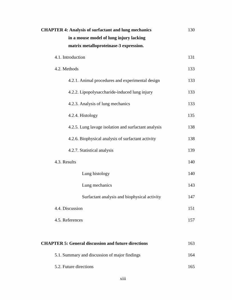
TABLE OF CONTENTS:
ABSTRACT ii
KEYWORDS iii
CO-AUTHORSHIP STATEMENT iv
ACKNOWLEDGMENTS v
TABLE OF CONTENTS viii
LIST OF TABLES xv
LIST OF FIGURES xvii
LIST OF APPENDICES xix
LIST OF ABBREVIATIONS xx
CHAPTER 1: General introduction and literature review 1
1.1. General overview 2
1.2. Lung function and structure 4
1.3. Lung mechanics 5
1.4. Lung insults: the Acute Lung Injury / 6
Acute Respiratory Distress Syndrome (ALI/ARDS) paradigm.
1.4.1. ALI/ARDS definition 6
1.4.2. ALI overview 7
1.4.3. Treatment of ALI 9
1.4.4. Development of ALI: the ALI paradigm 10

ix
1.4.4.1. Primary insults and ALI pathogenesis 11
1.4.4.2. Secondary insult, systemic inflammation 12
and multi-organ failure
1.5. Inflammation and inflammatory mediators in acute lung injury 13
1.5.1. Cellular components in the inflammatory process 14
1.5.2. Overview of soluble inflammatory mediators 15
1.5.2.1. Cytokines and chemokines 17
1.5.2.2. Lipid mediators in ALI 18
1.5.3. Targeting inflammation as a therapy for ALI 19
1.6. The pulmonary surfactant system 20
1.6.1. Surfactant composition 20
1.6.2. Surfactant metabolism 21
1.6.3. Surfactant function 22
1.6.4. Surfactant alterations in ALI 24
1.6.5. Exogenous surfactant treatment in ALI 26
1.7. Overview on matrix metalloproteinases (MMPs) 28
1.8. Matrix metalloproteinase-3 (MMP-3)
1.8.1. Characteristics of MMP-3 29
1.8.2. MMP-3 in the inflammatory process 30
1.8.3. MMP-3 in ALI 30
1.8.4. Study tool: Mmp3 knock-out mouse 31

x
1.9. Animal models of ALI 32
1.9.1. Hallmarks of ALI in animal models 32
1.9.2. Hydrochloric acid-induced lung injury 34
1.9.3. Lipopolysaccharide-induced lung injury 35
1.9.4. Ventilation associated lung injury and ex vivo ventilation 35
1.9.4.1. Measurements of lung mechanics in small rodents 37
1.10. Summary and overall objective 37
1.11. References 39
CHAPTER 2: The effects of exogenous surfactant administration 63
on ventilation-induced inflammation in mouse
models of lung injury
2.1. Introduction 64
2.2. Materials and methods 66
2.2.1. Experimental design and ethics statement 66
2.2.2. Intra-tracheal hydrochloric acid instillation 67
2.2.3. Intra-tracheal surfactant instillation 67
2.2.4. Isolated and Perfused Mouse Lung setup 68
2.2.5. Surfactant and total lung lavage protein measurements 68
2.2.6. Biophysical functional analysis of surfactant 69
2.2.7. Measurement of inflammatory mediators 69
2.2.8. Statistical analysis 70

xi
2.3. Results 71
2.3.1. Experiment 1. 71
Lavage Analysis 72
Perfusate Analysis 73
2.3.2. Experiment 2. 75
Lavage Analysis 77
Perfusate Analysis 79
2.4. Discussion 83
2.5. References 87
CHAPTER 3: Lack of matrix metalloproteinase-3 in mouse models 92
of lung injury ameliorates the pulmonary inflammatory
response in female but not in male mice
3.1. Introduction 93
3.2. Methods 95
3.2.1. Animal procedures and experimental design 95
3.2.2. Experiment 1: Lipopolysaccharide induced lung injury 95
3.2.3. Experiment 2: Acid-induced lung injury 96
3.2.4. Lung lavage isolation and total protein analysis 97
3.2.5. Lavage cell analysis 97
3.2.6. Measurements of MMP-3 and 98
inflammatory mediators in lung lavage

xii
3.2.7. Experiment 3: Isolation of Mmp3+/+ and Mmp3-/- 98
bone marrow-derived macrophages
3.2.8. In vitro stimulation of Mmp3+/+ and Mmp3-/- 99
bone marrow-derived macrophages
3.2.9. Statistical analysis 100
3.3. Results 101
3.3.1. Experiment 1: Lipopolysaccharide-induced lung injury 101
Lavage MMP-3 101
Lavage protein 103
Lavage inflammatory cells 103
Lavage inflammatory mediators 106
3.3.2. Experiment 2: Acid-induced lung injury 109
Lavage MMP-3 109
Lavage protein 109
Lavage inflammatory cells 110
Lavage inflammatory mediators 112
3.3.3. Experiment 3: In vitro stimulation of Mmp3+/+ 115
and Mmp3-/- bone marrow-derived macrophages
3.4. Discussion 117
3.5. References 122
xiii
CHAPTER 4: Analysis of surfactant and lung mechanics 130
in a mouse model of lung injury lacking
matrix metalloproteinase-3 expression.
4.1. Introduction 131
4.2. Methods 133
4.2.1. Animal procedures and experimental design 133
4.2.2. Lipopolysaccharide-induced lung injury 133
4.2.3. Analysis of lung mechanics 133
4.2.4. Histology 135
4.2.5. Lung lavage isolation and surfactant analysis 138
4.2.6. Biophysical analysis of surfactant activity 138
4.2.7. Statistical analysis 139
4.3. Results 140
Lung histology 140
Lung mechanics 143
Surfactant analysis and biophysical activity 147
4.4. Discussion 151
4.5. References 157
CHAPTER 5: General discussion and future directions 163
5.1. Summary and discussion of major findings 164
5.2. Future directions 165

xiv
5.2.1. Exogenous surfactant as a vehicle 165
for anti-inflammatory molecules
5.2.2. Sex differences and the inflammatory response 167
5.2.3. MMP-3 and Surfactant 169
5.3. Concluding remarks 170
5.4. References 171
APPENDIX 1: UWO animal use sub-committee protocol approval 175
Protocol 2010-272 176
Protocol 2006-124-12 177
APPENDIX 2: Information about copyright release for publication 178
BMC Pulmonary Medicine 179
CURRICULUM VITAE 180

xv
LIST OF TABLES:
Table 1.1: Summary of inflammatory mediators involved in ALI 16
and most relevant in this thesis
Table 1.2: Examples of inflammatory phenotypes described in 32
Mmp3-/- mice
Table 2.1: Experiment 1. Total protein levels and IL-6 concentrations 72
in lung lavage at the end of MV
Table 2.2: Experiment 1. Cytokine and chemokine analysis 75
in lung perfusate at the end of MV
Table 2.3: Experiment 2. Total protein levels and IL-6 concentrations 78
measured in lung lavage at the end of MV
Table 2.4: Experiment 2. Cytokine and chemokine measured 81
in lung perfusate at the end of MV
Table 2.5: Experiment 2. Concentrations of prostaglandin E2, 82
leukotriene B4, thromboxane B2 and 8-isoprostane
in lung perfusate
Table 3.1: Experiment 1. Total protein content in lavage 103
from Mmp3+/+ and Mmp3-/- mice of either sex
Table 3.2: Experiment 2. Total protein content in lavage 110
from female and male mice of both genotypes
Table 4.1: Summary of the maneuvers and outcomes 135
of respiratory mechanics relevant in this thesis
Table 4.2: Scoring system utilized for the assessment 137
of lung injury on H&E stained lung sections

xvi
Table 4.3: Surface activity of LA samples at cycle #1, #5, and #10 150
of dynamic compression-expansion cycles
Table 4.4: Percent area compression at cycle #10 of dynamic 150
compression-expansion

xvii
LIST OF FIGURES:
Figure 1.1: Multiple hit paradigm of ALI development 11
Figure 1.2: Schematic of surfactant metabolism 21
Figure 1.3: Schematic of surfactant alterations in ALI 25
Figure 2.1: Experiment 1. Perfusion pressure measured throughout MV 71
Figure 2.2: Experiment 1. Surfactant recovery in lung lavage 73
and surface activity of LA
Figure 2.3: Experiment 1. IL-6 levels measured in lung perfusate 74
at 60, 90 and 120 min.
Figure 2.4: Experiment 2. Peak Inspiratory Pressure measured 76
over the course of MV
Figure 2.5: Experiment 2. Perfusion pressure measured throughout MV 77
Figure 2.6: Experiment 2. Surfactant recovery in lung lavage 79
and surface activity of crude LA
Figure 2.7: Experiment 2. IL-6 levels measured in lung perfusate 80
at 0, 30, 60, 90, 120 min
Figure 3.1: Experiment 1. MMP-3 levels in lung lavage samples 102
from Mmp3+/+ mice
Figure 3.2: Experiment 1. Total and differential cell counts 105
in lavage samples from Mmp3+/+ and Mmp3-/-
female and male mice
Figure 3.3: Experiment 1. Cytokine and chemokine levels 107
in lung lavage from Mmp3+/+ and Mmp3-/- female mice

xviii
Figure 3.4: Experiment 1. Cytokine and chemokine levels 108
in lung lavage from Mmp3+/+ and Mmp3-/- male mice
Figure 3.5: Experiment 2. MMP-3 levels measured in lavage samples 109
from female and male Mmp3+/+ mice
Figure 3.6: Experiment 2. Total number of PMN neutrophils 111
in lavage samples from female and male mice
of both genotypes
Figure 3.7: Experiment 2. Cytokine and chemokine levels 113
in lung lavage from Mmp3+/+ and Mmp3-/- female mice
Figure 3.8: Experiment 2. Cytokine and chemokine levels 114
in lung lavage from Mmp3+/+ and Mmp3-/- male mice
Figure 3.9: Experiment 3. Stimulation of BMDMs isolated 116
from Mmp3+/+ and Mmp3-/- mice
Figure 4.1: Representative images of the histology score utilized 137
for the analyses of lung sections
Figure 4.2: Hematoxylin-eosin stained pictures representative of 141
lung sections from female Mmp3+/+ and Mmp3-/- mice
Figure 4.3: Hematoxylin-eosin stained pictures representative of 142
lung sections from male Mmp3+/+ and Mmp3-/- mice
Figure 4.4: Analysis of respiratory mechanics in female 144
Mmp3+/+ and Mmp3-/- mice
Figure 4.5: Analysis of respiratory mechanics in male 146
Mmp3+/+ and Mmp3-/- mice
Figure 4.6: Surfactant pool sizes and percent large aggregates 148
in female and male Mmp3+/+ and Mmp3-/- mice

xix
LIST OF APPENDICES:
Appendix 1: UWO animal use sub-committee protocol approval 175
Appendix 2: Information about copyright release for publication 178

xx
LIST OF ABBREVIATIONS:
ALI acute lung injury
AM alveolar macrophages
ANOVA analysis of variance
ARDS acute respiratory distress syndrome
bLES bovine lipid extract surfactant
BMDM bone marrow derived macrophage
CBS captive bubble surfactometer
Crs compliance of whole respiratory system
Cst quasi-static compliance
DPPC dipalmitoylphosphatidylcholine
E2 17β-estradiol
ECM extracellular matrix
ELISA enzyme-linked immune-sorbent assay
Ers elastance of whole respiratory system
Est quasi-static elastance
FIO2 fraction of inspired oxygen
FOT forced oscillation technique
G tissue damping
G-CSF granulocyte colony-stimulating factor
GM-CSF granulocyte macrophage colony-stimulating factor
H tissue elastance
H&E hematoxylin & eosin
HCl hydrochloric acid
IC inspiratory capacity
IL-10 interleukin-10
IL-1β interleukin-1β
IL-6 interleukin-6
IL-8 interleukin-8

xxi
IP-10 interferon-γ-induced protein-10
IPML isolated perfused mouse lung
KC keratinocyte chemoattractant
LA large aggregate
LIX lipopolysaccharide-induced CXC chemokine
LPS lipopolysaccharide
LTB4 leukotriene B4
MCP-1 monocyte chemotactic protein-1
MIP-2 macrophage inflammatory protein-2
MMP matrix metalloproteinase
MMP-3 matrix metalloproteinase-3
MOF multiple organ failure
MV mechanical ventilation
NRDS neonatal respiratory distress syndrome
PaO2 partial pressure of oxygen
PC phosphatidylcholine
PEEP positive end expiratory pressure
PGE2 prostaglandin E2
PIP peak inspiratory pressure
PL phospholipid
PMN polymorphonuclear
PV pressure-volume
RN airway resistance (Newtonian resistance)
RR respiratory rate
Rrs resistance of whole respiratory system
SA small aggregate
SEM standard error of the mean
SP-A surfactant protein A
SP-B surfactant protein B
SP-C surfactant protein C
SP-D surfactant protein D

xxii
TIMPs tissue inhibitor of metalloproteinases
TLC total lung capacity
TLR-4 toll-like receptor 4
TNF-α tumor necrosis factor-α
TS total surfactant
TXA2 thromboxane A2
TXB2 thromboxane B2
VALI ventilation-associated lung injury
VFD ventilator-free days
VILI ventilation-induced lung injury
Vt tidal volume

1
CHAPTER 1:
General introduction and literature review

2
1.1. General overview
Most of the metabolic processes taking place in the human body require oxygen and
generate carbon dioxide as a waste product that needs to be eliminated. The lung is the
organ responsible for exchange of gases, providing oxygen to the blood for delivery to all
systems in the body, and allowing for carbon dioxide removal to the external environment
[1]. This process, essential for life, can become impaired as a result of a variety of diseases
affecting the lung, such as asthma, chronic bronchitis, fibrosis and, central to this thesis,
acute lung injury.
Acute Lung Injury/Acute Respiratory Distress Syndrome (ALI/ARDS) is a pulmonary
disorder with a complex pathophysiology and no proven therapeutic option available for
its treatment [2]. Throughout the years, understanding of this disorder has been hampered
by the multiplicity of lung insults potentially causing ALI/ARDS, the wide range of patient
population, which includes both pediatric and adult subjects, and a complex disease
progression [3, 4]. Two aspects of its pathophysiology and treatment support, however, are
common to all patients with ALI/ARDS: i) the use of mechanical ventilation, necessary to
support impaired gas exchange, and ii) the development of an overwhelming pulmonary
inflammatory response.
As explained more extensively throughout this doctoral thesis, persisting and
overwhelming lung inflammation negatively affects lung function, and has been shown to
correlate with poor prognosis and outcome in patients with ALI/ARDS [5, 6]. This scenario
is further aggravated by the effects of mechanical ventilation on the injured lung.
Mechanical ventilation has been shown to alter lung surfactant [7], a substance lining the
inner pulmonary surface, with biophysical and immune-modulatory properties essential for
lung function [8, 9]. Ventilation-induced impairment of lung surfactant leads to impaired
lung function [10], which can often be rescued by administration of exogenous surfactant
[11]. The effects of mechanical ventilation, however, are not limited to surfactant, as
ventilation can further pulmonary inflammation, and participate in the progression toward
the development of an inflammatory response in the systemic circulation [12–14]. These
events can, ultimately, affect distal organ function causing multi-organ failure, the major
cause of death in ALI/ARDS patients [15, 16].

3
In light of these issues, the first scientific problem addressed in chapter two is related to
the assessment of a lung-targeted strategy aimed at mitigating the effects of
ventilation on the inflamed lung. The strategy of interest is exogenous surfactant
administration. The objective of chapter two is to determine whether exogenous surfactant
administration can mitigate ventilation-induced pulmonary and systemic inflammation, in
different mouse models of ALI/ARDS.
The second aspect highlighted by the lack of suitable treatment is the need for a better
understanding of ALI/ARDS pathophysiology, specifically focused on identifying key
mediators in the pulmonary inflammatory response that may serve as future potential
therapeutic targets. Among the multiple mediators involved in ALI/ARDS, chapter 3
focuses on the protease matrix metalloproteinase-3 for its role in inflammation and
inflammatory diseases [17]. The objective of chapter three is to assess the role of matrix
metalloproteinase-3 in the development of the pulmonary inflammation associated with
ALI/ARDS. The results of matrix metalloproteinase-3 contribution to inflammation in two
different mouse models of lung injury prompted the investigation of further aspects of
disease development.
Chapter four focuses on the examination of the potential role of matrix
metalloproteinase-3 in the interplay between pulmonary inflammation and lung
function. The objective of chapter four is in fact to investigate whether such protease,
mediator of lung inflammation, can affect the surfactant system and overall lung function.
The remaining of this first chapter provides general information on lung structure and
function, and illustrates in greater detail the pathophysiology of ALI/ARDS, touching on
the inflammatory response, lung surfactant, and matrix metalloproteinase-3, before closing
with a brief description of the animal models available for the study of this disorder.

4
1.2. Lung function and structure
The primary function of the lung is gas exchange. During breathing, oxygen entering the
lungs diffuses into the blood, while carbon dioxide diffuses from the blood into the lung to
be exhaled in the external environment. Lung function is facilitated by a number of
anatomical features, namely the presence of an extremely large pulmonary surface area
available for diffusion, close proximity of the inhaled air to blood vessels at the alveolar
level, and the very low thickness of the alveolo-capillary barrier through which oxygen and
carbon dioxide diffuse [1].
Such anatomical features are the final result of a tree-like structure that starts at the nose
and mouth, and proceeds within the thoracic cavity via a semi-flexible tube, the trachea,
which divides into left and right main primary bronchi. Each bronchus branches multiple
times into progressively narrower and shorter bronchi/bronchioles down to the terminal
bronchioles, thereby generating a very large number of conducting airways. The process
of subsequent divisions continues further into the distal regions of the lung, until millions
of individual lung units, known as alveoli, generate a very large surface area suitable for
gas exchange. Diffusion of oxygen and carbon dioxide is also favored by the vast network
of capillaries wrapped around the alveoli, with the capillary endothelial cells laying in very
close proximity to the epithelial cells of the alveoli [1].
As mentioned, epithelial cells form the alveolar wall; precisely, alveoli are mainly made of
flat, squamous type I alveolar epithelial cells and some cuboidal type II epithelial cells.
The type II cells produce and secrete pulmonary surfactant at the air-liquid interface [18].
Surfactant, a protein-lipid mixture, is very important for reducing the surface tension in the
alveoli, thereby optimizing lung compliance and facilitating the work of breathing [8]. In
addition to type I and type II epithelial cells, some resident alveolar macrophages (AM) are
found within the alveolar space, where they act as a first line defense against pathogens
and participate in surfactant metabolism [19, 20].

5
1.3. Lung mechanics
In addition to the structure of the lung, the process of ventilation is important for gas
exchange. Ventilation refers to the process by which a volume of air, known as tidal
volume, enters and leaves the lung with each breathing cycle. The mechanics of this
process are closely linked to the properties of the lungs and those of the thoracic cavity in
which the lungs are located. Within this enclosed space, the base of the lungs comes in
contact with the diaphragm, a dome-shaped muscle that separates the lungs from the
abdominal contents. Pleural membranes surround the outer surface of each lung and line
the inside of the chest wall, forming a thin intra-pleural space that is normally filled with a
small volume of fluid. The intra-pleural space provides connection between the lung and
the chest wall, and given the natural tendency of the chest wall to expand and the lung to
collapse, this space has a slightly sub-atmospheric pressure or, in other words, a negative
pressure [1].
During inspiration, contraction of the inspiratory muscles (diaphragm and external
intercostals) causes an increase in the volume of the thoracic cavity. With the increase in
volume, the negative intra-pleural pressure becomes more negative leading to the
expansion of the lung and a fall in alveolar pressure to a slightly sub-atmospheric value.
This pressure gradient promotes flow of air from the atmosphere to the alveoli. In tidal
breathing (non-exertional), expiration, unlike inspiration, is a passive process resulting
from the relaxation of the inspiratory muscles, with subsequent decrease in the thoracic
and lung volumes. These changes affect the alveolar pressure, now slightly greater than
atmospheric pressure, providing the driving force necessary for air to flow from the alveoli
back to the atmosphere [1].
The work of breathing can be affected by two main factors: the resistance to airflow within
the conducting airways and the distensibility of the lung tissue. In healthy subjects, the
medium to larger airways offer a negligible degree of resistance to the airflow. Narrowing
of these airways, however, as a result of bronchial constriction or obstruction such as in
asthma, will increase the resistance and impair airflow [21].

6
The distensibility of the lung tissue, known as compliance, refers to the ability of the lung
to inflate and stretch during inspiration. Lung compliance is an indicator of the stiffness of
the lungs, and it is defined as volume change per unit pressure change [1]. Two principal
factors can influence compliance: the elastic properties of the lung and the surface tension
of the alveolar lining fluid. The elastic properties can be described essentially as the
tendency of the lung to recoil to the resting volume after distention. Lung elasticity arises
from the elastin and collagen fibers in the pulmonary tissue, and any alteration of these
fibers can cause changes in lung compliance [1]. As mentioned, compliance is also
influenced by the surface tension arising from attractive forces between water molecules
at the air-liquid interface within the alveoli. In healthy lungs, surface tension has very low
values attributable to the presence of a material lining the alveoli called pulmonary
surfactant. Secreted by alveolar type II cells at the air-liquid interface, lung surfactant is a
protein-lipid mixture that lowers surface tension, thereby stabilizing the alveoli and
reducing the work of breathing [8, 22]. Whole lung compliance is therefore strongly
affected by the presence of a functioning surfactant system. The relationship between an
impaired surfactant system and consequently poor lung compliance is most evident in a
disease called the neonatal respiratory distress syndrome, where preterm infants are born
with insufficient amounts of surfactant and struggle to breath [23]. Alterations of the
surfactant system in a mature lung can occur in the acute respiratory distress syndrome,
which is the focus of this thesis.
A more detailed overview of surfactant composition, function and alterations during
disease will be provided in the following sections of this thesis.
1.4. Lung insults: the Acute Lung Injury / Acute Respiratory Distress Syndrome
(ALI/ARDS) paradigm.
1.4.1. ALI/ARDS definition
This thesis focuses on the clinical problem of acute lung injury (ALI) and on ALI’s more
severe form, the acute respiratory distress syndrome (ARDS). An adult respiratory-distress

7
syndrome was first described in 1967 by Ashbaugh in twelve adults presenting with acute
onset of rapid breathing, hypoxia, and poor lung distensibility [24]. The extensive research
that followed improved the knowledge of this disease’s pathophysiology, leading to a
renaming of the disease to acute respiratory distress syndrome and a clear clinical definition
in 1994 by the American-European Consensus Conference. ARDS and ALI were defined
as acute in onset and characterized by bilateral radiologic infiltrates with no evidence of
heart failure, and hypoxemia, as determined by the ratio of arterial partial pressure of
oxygen (PaO2) to fraction of inspired oxygen (FIO2). Specifically, ALI was defined by a
PaO2/ FIO2 ≤ 300mmHg, while the cut off for the more severe ARDS was a PaO2/ FIO2 ≤
200mmHg [25]. In more recent years, some of the limitations of this definition have been
addressed with the updated “Berlin definition”, which has removed the term ALI and
instead distinguishes between three mutually exclusive ARDS subgroups (mild, moderate,
or severe ARDS) based on the severity of the hypoxemia [26]. While this new definition
constitutes an improvement to the clinical practice allowing for better stratification of
patients in clinical trials, it poses some challenges in its application to animal models of
lung injury since there is no reference to underlying pathophysiology. The terminology
acute lung injury, instead, traditionally includes a broader spectrum of the disease,
encompassing both patients and experimental models. For this reason, the term ALI will
be used to refer to the acute lung injury/acute respiratory distress syndrome throughout the
remainder of this thesis.
A broadly accepted definition of this disease has allowed for the collection of useful
epidemiological information. Recent estimates suggest an ALI incidence of approximately
60-80 new cases per 100,000 person-years in the United States [4, 27]. Importantly,
mortality from ALI is still very high at 40%, with distal organ failure, rather than
respiratory failure, being the main cause of death for these patients [15, 16, 28].
1.4.2. ALI overview
Despite a relatively simple clinical definition based on physiological parameters, ALI is a
complex pulmonary disorder characterized by decreased compliance, persistent and
elevated lung inflammation, a high mortality, and no available therapeutic options [29–31].

8
After many years of clinical and basic research, many aspects of ALI pathophysiology are
still elusive and challenge the development of effective pharmacological therapies. To add
to the complexity, disease progression is associated with the development of systemic
inflammation and multi-organ failure which is, as previously mentioned, the most common
cause of death in ALI [15, 32].
Affecting patients of all ages, ALI is initiated by a variety of lung insults of different origin
[2]. For example, potential threats and pathogens can come from the external environment,
to which the lung is continuously exposed [33, 34]. On the other hand, the lung receives
the entire cardiac output through its vasculature and can therefore be affected indirectly by
damage-associated molecules or invading organisms found in the pulmonary circulation
[33, 34]. In most cases, the lung can manage and effectively clear such threats. On occasion,
however, the host response to a lung insult can become maladaptive. As a result, patients
with ALI present with lung edema, altered pulmonary surfactant, decreased compliance
with an associated hypoxemia, and a sustained pulmonary inflammatory response [2, 35,
36].
The progressive hypoxemia affecting these patients ultimately requires the use of
mechanical ventilation (MV), the main supportive treatment for this disorder. Even though
essential, MV can contribute to lung injury and inflammation, thereby promoting ALI
progression [12]. The role of MV in ALI became strongly evident in a large, multi-center
randomized clinical trial conducted in 2000 [37]. In this study, the ARDS Network assessed
the effect of ventilation using different tidal volumes in patients with ALI. Patients received
MV with either a ‘conventional’ tidal volume (Vt=12mL/kg predicted body weight) or a
low tidal volume (Vt=6mL/kg predicted body weight) strategy. The trial was stopped early
due to the significantly lower mortality in the low Vt group (31.0%) compared to the group
receiving conventional Vt ventilation (39.8%) [37]. The underlying pathophysiological
mechanism responsible for such outcome was suggested to stem from the effects of MV
on the inflammatory response in ALI. An earlier clinical study by Ranieri et al. had in fact
shown that, at 36 hours post randomization, more injurious ventilation strategies caused
greater increases in both pulmonary and systemic inflammation in ALI patients, compared
to concentrations at study entry and in patients ventilated with lung-protective MV [12].

9
Overall, these clinical studies have highlighted that MV is a potential contributor to disease
progression, by enhancing lung inflammation and leading to a systemic inflammatory
response and peripheral organ failure. As such, MV could represent an ultimately effective
target in ALI treatment.
Additionally, this evidence shifted the focus from oxygenation, the traditional “clinical
outcome” of patients with ALI/ARDS, to inflammation, as it became clear that a persistent,
excessive lung inflammatory response is the culprit for disease progression [5, 6]. In this
respect, Meduri et al. demonstrated that at the onset of ARDS, non-survivors had
significantly higher pulmonary inflammatory mediators (i.e. IL6, IL8, TNF-α) levels than
survivors, stressing the association between lung inflammation and disease outcome [6].
Lastly, the evidence of a central role of inflammation in ALI would suggest that a lung-
targeted treatment, aimed at reducing pulmonary and systemic inflammation, could be
extremely beneficial and more effective than strategies merely aimed at improving
oxygenation. In this sense, exogenous surfactant administration is a lung-targeted
treatment whose role in affecting the inflammatory response associated with ALI has been
insufficiently characterized. Moreover, the study of key mediators of pulmonary
inflammation (such as, for example, matrix metalloproteinase-3) could help identify
new potential therapeutic targets for this disorder.
1.4.3. Treatment of ALI
Treatment of ALI is based on supporting gas exchange through MV, careful monitoring
and stabilization of these critically ill patients, and management of the initiating insult
when possible [38]. Unfortunately, no pharmacological treatment is yet available for this
disorder, and even though many therapies have been promising experimentally, they have
failed to improve outcomes clinically [39]. Among these, exogenous surfactant
administration led to exciting improvements in oxygenation and compliance for ALI
patients in Phase 2 trials and smaller Phase 3 trials [40, 41]. This treatment, however, did
not appear to improve mortality in more recent, larger Phase 3 trials, possibly due to
surfactant administration occurring too late in the paradigm of ALI development [40]. A

10
more in depth description of surfactant in ALI pathogenesis and treatment will be described
in section 1.6.
To date, the only approach shown to reduce mortality in ALI is the use of low tidal volume/
protective MV, likely due to lower pulmonary and systemic inflammation elicited by this
strategy compared to higher Vt ventilation [37]. Since ALI is characterized by a persistent,
excessive pulmonary inflammatory response [42, 43], anti-inflammatory treatments such
as corticosteroids, statins and activated protein C could theoretically decrease mortality.
The results from clinical trials however have been disappointing, showing no clear benefits
of such treatments [44, 45]. It is therefore imperative to expand our knowledge of ALI
pathophysiology and the associated inflammatory response, as well as to re-examine some
of its treatments, in order to effectively interfere with disease progression and improve
mortality.
In summary, examination of the clinical studies performed to date suggest that ALI,
although defined by physiological criteria, is a complex inflammatory disease in which its
essential therapeutic intervention (MV) can actually contribute to disease progression.
Despite the complex pathophysiology of ALI, recent animal and clinical studies have
started to provide insight into the development of this disease. The current state of
knowledge, and areas requiring further research, are described below with a specific focus
on inflammation.
1.4.4. Development of ALI: the ALI paradigm
The current model for ALI development and progression, based on clinical, in vivo, and in
vitro studies, is shown in figure 1.1. Briefly, the model illustrates how multiple insults (or
hits) to a normal lung can lead to the development of ALI, and exemplifies the disease
progression from lung injury to multi-organ failure, which is the main cause of death for
ALI patients. More detailed information on the different steps in the multiple hit paradigm
of ALI is provided in the upcoming sections.

11
Figure 1.1: Multiple hit paradigm of ALI development.
1.4.4.1. Primary insults and ALI pathogenesis
In the multiple hit paradigm of ALI, a normal lung is first exposed to an initiating or
primary insult, which can be classified as either direct or indirect (Fig. 1.1) [2]. Indirect
insults such as sepsis or trauma primarily affect the pulmonary vasculature, given the
presence in the circulation of pathogens and inflammatory molecules from various
potential sources. On the other hand, insults such as pneumonia or gastric acid aspiration
represent direct injuries to the lung, and are the main focus of this thesis [2]. Numerous
animal studies have shown that such direct injuries cause greater damage to the alveolar
epithelium, and a more robust inflammatory response within the alveolar space compared
to indirect insults [33, 46]. Overall, the response of the lung to any insult is a rather

12
complicated process involving a multitude of soluble mediators, multiple cells types, and
a complex integration of intracellular pathways. For this reason, a simplification of the
major pathophysiological steps involved in this disease is necessary to better understand
ALI.
Following the initial lung insult, the disruption of the alveolo-capillary barrier allows the
abnormal leakage of a protein-rich edema into the alveolar space [29, 47]. This, in turn,
inhibits surfactant function [48] and leads to a profound decrease in lung compliance [49].
Importantly, alveolar macrophages and, to some extent, epithelial cells respond to the
injurious event by mounting an inflammatory response within the alveolar space [50]. Up-
regulation of pulmonary cytokines and chemokines results in the recruitment of
polymorphonuclear neutrophils (PMNs), cells of the innate immune system that are first
responders in tissue injury and infection [51]. While this initial inflammatory response is a
homeostatic process important for injury resolution, the severity and persistence of
inflammation in ALI can cause tissue injury, impair effective resolution, and correlate with
poor outcomes. Occurrence of the latter processes may be promoted if the lung is exposed
to a secondary insult.
1.4.4.2. Secondary insult, systemic inflammation and
multi-organ failure
The elevated pulmonary inflammation, alveolar flooding and surfactant alterations that
follow a primary insult make the lung susceptible (predisposed lung) to the effects of other
secondary insults, such as sepsis, trauma or, most commonly, mechanical ventilation (Fig.
1.1). As mentioned earlier, decreased compliance and hypoxemia are hallmarks of ALI
[25], and MV is necessary to support gas exchange. The contribution of MV as a secondary
insult has been extensively highlighted in both clinical studies and animal models of lung
injury [12, 14, 52, 53], many of which have also shed light on the three different ways MV
participates in ALI progression.
First, MV contributes to lung surfactant inactivation, with consequent alveolar collapse and
worsening lung compliance [7, 54]. Second, due to the overstretching of the more aerated
and compliant alveolar units, MV causes increased release of inflammatory mediators,

13
thereby exacerbating lung injury [55–57]. Third, ventilation itself is an important
contributor in ALI progression toward systemic inflammation (Fig. 1.1) [13, 58].
A vast body of experimental evidence has in fact shown that MV promotes the de-
compartmentalization of pulmonary inflammatory mediators into the systemic circulation,
and that exacerbation of pulmonary inflammation by MV enhances the development of
systemic inflammation [58–60]. Importantly, the severity of the systemic inflammatory
response correlates with mortality in ALI [37]. Inflammatory molecules in the circulation
are, in fact, biologically active and exert a pathogenic role on extra-pulmonary organs (i.e.,
liver and kidneys) ultimately leading to multi-organ failure [14, 52, 61–63]. An interesting
study from our lab has in fact demonstrated that circulating inflammatory mediators
released from injured lungs can activate distal cell populations, namely mouse liver
endothelial cells and leukocytes, leading to a pro-inflammatory and pro-adhesive
phenotype in these cells [64].
Since inflammation is a crucial component of ALI pathophysiology, an overview of the
inflammatory response associated with ALI is necessary, before addressing in greater
details the specific variables manipulated for the study of lung injury in this thesis.
1.5. Inflammation and inflammatory mediators in acute lung injury
As mentioned above, inflammation plays a central role in ALI. Inflammation is a complex,
highly regulated adaptive response to tissue injury or infection, mounted to re-establish
homeostatic conditions. The inflammatory response involves a variety of cellular and
soluble mediators cooperatively working to eliminate the detrimental stimuli, thereby
progressing toward phases of resolution and tissue repair [65]. Sometimes, however, for
reasons that are not yet clear, the inflammatory process persists, becomes maladaptive, and
may lead to organ injury and dysfunction as in the case of ALI.

14
1.5.1. Cellular components in the inflammatory process
Among the different cell types participating in the inflammatory process, neutrophils are
particularly relevant in the settings of ALI. Neutrophils are recruited to the injured lung
and migrate into the air space shortly after a primary insult [51]. These cells are essential
players in the innate immune response to injury and/or infection, and within the alveolar
environment these cells can contribute to the development of ALI through the release of
pro-inflammatory cytokines and production of reactive oxygen species [51, 66]. Moreover,
activated neutrophils can secrete potent proteolytic enzymes, such as elastase, collagenase
(i.e., matrix metalloproteinase-8) and gelatinases (i.e., matrix metalloproteinase-9),
potentially responsible for alterations of the lung extracellular matrix [66]. The pathogenic
role of neutrophils in ALI has been shown in animal models of lung injury [67, 68];
moreover, pulmonary accumulation and persistence of neutrophils appear to correlate with
disease severity in patients with ALI [69]. Nonetheless, the evidence that neutropenic
patients can develop lung injury as well [70], suggests that other cell populations may be
involved in ALI. In fact, important contributors to this elaborate inflammatory response
are also the parenchymal cells, namely endothelial cells, alveolar epithelial cells,
fibroblasts, and the alveolar macrophages [50].
As a first line of defense, the alveolar macrophages phagocytose pathogens and dead cells,
can secrete anti-microbial peptides, and release proteases such as matrix metalloproteinases
(including matrix metalloproteinase-3), thereby orchestrating the inflammatory and
immune response, and contributing to the later reparative phase [71]. Additionally, alveolar
macrophages can also release a variety of soluble mediators of inflammation,
proteinaceous and/or lipidic in nature, responsible for many of the pathophysiological
events occurring in ALI, including neutrophils recruitment [71].
Migration and influx of leukocytes to the injured lung is also facilitated by the activated
endothelium, which expresses surface adhesion molecules necessary for cell to cell
interaction [72]. Epithelial cells participate to the immune response in ALI through the
secretion of collectins (SP-A, SP-D) associated with surfactant and, additionally, via the
release of inflammatory mediators (i.e. IL-1β, IL-8, TNF-α) in response to multiple stimuli,
including stretch associated with MV [73–76].

15
1.5.2. Overview of soluble inflammatory mediators
In addition to the cellular component, soluble mediators are a second key pathological
feature in the development and progression of ALI. Following insults to the lung (Fig. 1.1),
a broad variety of pro- and anti-inflammatory molecules are released from the
aforementioned cellular sources within the lung and in the bloodstream. For the sake of
brevity, only inflammatory signals relevant to this thesis will be reviewed in this section.
Mediators of interest include: i) cytokines, ii) chemokines, and iii) lipid mediators. A list
of such mediators with an indication of their main respective biological functions is given
in Table 1.1.

16
Inflammatory Mediators Biological Activity
Cytokines
G-CSF Granulocyte survival/growth
GM-CSF
Host defense;
granulocyte/monocyte/AM survival &
growth
IL-1β Pro-inflammatory; fever; neutrophil
migration
IL-6
Pro-inflammatory; acute-phase
response; leukocytes
growth/differentiation
IL-10 Dual role; pro- and anti- inflammatory
IL-13 Anti-inflammatory; asthma and allergic
disease
TNF-α Pro-inflammatory; hypotension/shock;
cell cytotoxicity; fever
Chemokines
(alternative name)
Eotaxin (CCL11)
Pro-inflammatory; chemoattractant for
eosinophils & basophils; allergic
airways inflammation
IP-10 (CXCL10) Pro-inflammatory; chemoattractant for
activated T cells
KC (CXCL1) Pro-inflammatory; chemoattractant for
neutrophils
LIX (CXCL5) Pro-inflammatory; chemoattractant &
activator of neutrophils
MCP-1 (CCL2) Pro-inflammatory; chemoattractant for
monocytes
MIP-2 (CXCL2) Pro-inflammatory; neutrophils
chemoattractant/activator
Lipid Mediators
8-Isoprostane Marker of oxidative stress
Leukotriene B4 Pro-inflammatory; neutrophils
chemoattractant/activator
Prostaglandin E2 Inflammation; vascular tone &
permeability
Thromboxane A2 Pro-inflammatory; neutrophils
chemoattractant/activator

17
Table 1.1: Summary of inflammatory mediators involved in ALI and most relevant in this
thesis. KC, MIP-2 are considered murine equivalent of human IL-8. For further
information, see Bathia M. et al. [43], Puneet P. et al. [77], and “Principles of internal
medicine”, Harrison, 15th edition [78]. AM, alveolar macrophages.
G-CSF = granulocyte colony-stimulating factor, GM-CSF = granulocyte-macrophage
CSF, IL-6 = interleukin-6, IP-10 = interferon-γ-induced protein 10, KC = keratinocyte
chemoattractant, LIX = lipopolysaccharide-induced CXC chemokine, MCP-1 = monocyte
chemotactic protein-1, MIP-2 = macrophage inflammatory protein 2 and TNF-α = tumor
necrosis factor-alpha.
1.5.2.1. Cytokines and chemokines
Cytokines and chemokines are small proteins secreted by immune and non-immune cells.
Once released in the extracellular environment, cytokines will affect the activity and
function of other, target cells [65]. Chemokines work as chemoattractant and activators of
leukocytes, and are generally classified based on the position of the first two cysteine
residues at the N-terminal: CC chemokines for adjacent residues, CXC if an amino acid
separates them [77]. Together, cytokines and chemokines coordinate the inflammatory
response through cell activation, changes in gene expression, and recruitment of
inflammatory cells to the site of injury. These events are extremely significant in the
pathogenesis of ALI, where increases of chemokine levels in alveolar fluid lead to massive
recruitment and infiltration of neutrophils, potentially contributing to lung dysfunction [42,
66]. In the multiple hit paradigm of ALI development, changes in pulmonary cytokine and
chemokine levels are induced at first by a primary insult of variable nature [2]; invariably,
however, MV is applied to the predisposed lung contributing to overwhelming cytokine
release. Numerous experimental studies confirm the exacerbation of lung inflammation by
MV and point out the ventilation-induced de-compartmentalization of mediators such as
TNF-α, IL-8, MCP-1 in the systemic circulation, with consequent development of distal
organ failure [13, 58, 60, 61, 79]. It appears, therefore, that alterations of the inflammatory
milieu resulting from MV can worsen ALI outcomes. The pathogenic role of cytokines and
chemokines in ALI is in fact substantiated by clinical evidence that elevated lavage and

18
plasma levels of IL-6, TNF-α, IL-1β, and IL-8 correlate with disease gravity and poor
outcomes in these patients [6, 12, 80–82]. Importantly, the concentration of these pro-
inflammatory cytokines was found persistently elevated in the lung of non-survivors, while
ALI survivors observed lower IL-6, TNF-α, IL-1β, and IL-8 levels at the onset of ALI, and
over the course of the disease [6]. Overall, this evidence indicates the necessity to modulate
pulmonary and systemic cytokine/ chemokine levels in order to improve ALI outcomes.
1.5.2.2. Lipid mediators in ALI
The complex inflammatory scenario associated with ALI is not limited to the above-
mentioned small, protein mediators, but also includes lipid mediators of inflammation
(Table 1.1). Lipid mediators, or eicosanoids, are derived from the metabolism of
arachidonic acid by different enzymes: the cyclooxygenases pathway leads to the
production of mediators such as isoprostanes, prostaglandin E2 (PGE2) and thromboxane
A2 (TXA2), while the activity of lipoxygenases generates, among others, leukotriene B4
(LTB4) [83]. The contribution of these mediators to ALI pathogenesis can be inferred by
both clinical and experimental data.
Elevated concentrations of leukotrienes have been detected in lavage samples from
ALI/ARDS patients [84]; moreover, LTB4 levels have been shown to correlate with the
occurrence of lung injury in trauma patients [85]. Eicosanoids play a role in vascular tone,
activation and permeability, and serve as potent chemoattractants and activators for
neutrophils. In this regard, Zarbock et al. have demonstrated that TXA2 is responsible for
the recruitment and accumulation of neutrophils to the injured lung in a mouse model of
acid-induced ALI [67]. Moreover, experimental work performed by Jaecklin et al. has
shown the pathogenic role of circulating lipid mediators in a model of ventilation-induced
lung injury. The group observed that, when perfused in the pulmonary circulation of
recipient mice, lung-derived mediators isolated from mice with ventilation-induced lung
injury could worsen permeability and compliance of recipient mice ventilated with non-
injurious modes of MV [86]. Subsequent analysis of these soluble mediators revealed their
protein and lipid nature, thereby strengthening the role of cytokines, chemokines and
eicosanoids in ALI pathogenesis.

19
1.5.3. Targeting inflammation as a therapy for ALI
Despite the convincing evidence that overwhelming inflammation is an important
pathophysiological feature contributing to the progression of ALI, interfering with this
process has not been successful to date. For example, strategies involving the depletion of
inflammatory cell types (i.e. alveolar macrophages or neutrophils) not only have shown
conflicting experimental results [87–90], but would translate poorly into clinical practice
due to issues of safety and feasibility, given the undeniable importance of innate immunity
in host defense and tissue repair.
The rather intuitive approach of targeting individual cytokines or chemokines to down-
modulate the inflammatory response has also proven ineffective in both experimental and
clinical studies. The work of Nakamura et al. showed that mice lacking the expression of
the cytokine IL-6 had similar physiological impairments than wild type mice, following
exposure to three different models of ALI [91]. A more recent study by Markovic et al.
demonstrated that, while solutions containing lung-derived inflammatory mediators caused
liver endothelial cell dysfunction, the neutralization of IL-6 or TNF-α found in such
solutions was ineffective in rescuing the alterations in endothelial cells [64]. Clinically, an
antibody against TNF-α or administration of IL-1β Receptor Antagonist have been tested
for the treatment of severe sepsis, a known cause of ALI (Fig. 1.1), but failed at reducing
mortality in this patient population [92, 93].
Attempts at modulating the inflammatory response associated with ALI have also been
made with the clinical evaluation of corticosteroids. As emerges from a recent meta-
analysis of several clinical studies, corticosteroid therapy in ALI showed no effect on long
term mortality, and even appeared to significantly harm patients with influenza-related
lung injury [94].
Taken together, this evidence underscores the necessity for further understanding of the
inflammatory response and the need for new therapeutic strategies. In this regard, we
believe that lung surfactant and the protease matrix metalloproteinase-3 might be such
potential strategies. An overview dedicated to their role in inflammation and ALI is given
in the following sections of this introductory chapter.

20
1.6. The pulmonary surfactant system
Being an important contributor in the pathogenesis of ALI and a marker of disease
progression, the pulmonary surfactant system has been the focus of intense in vitro, in vivo,
and clinical research over the last five decades.
As mentioned earlier, the biophysical role of pulmonary surfactant is to reduce the surface
tension at the air-liquid interface, thereby ensuring optimal lung compliance [1]. The
decrease in pulmonary compliance typical of ALI generally results from the impairment of
surfactant activity [95].
In addition to its surface tension reducing properties, lung surfactant has also an important
role in immune modulation and host defense within the alveolar environment [9].
Interestingly, less is known about this function in the context of ALI.
1.6.1. Surfactant composition
The composition of surfactant is conserved across mammalian species and consists of
approximately 90% lipids, primarily phospholipids, and 10% surfactant associated proteins
[8]. The most abundant phospholipid component is phosphatidylcholine (PC), half of
which is dipalmitoylphosphatidycholine (DPPC), a disaturated species important for
achieving low surface tension values at the end of expiration [8].
The protein components include four surfactant associated proteins: surfactant protein-A
(SP-A), surfactant protein-B (SP-B), surfactant protein-C (SP-C) and surfactant protein-D
(SP-D) [96, 97]. The large, hydrophilic SP-A and SP-D proteins are members of the
collectin family and participate in the innate immune response [98]. SP-B and SP-C are
small, highly hydrophobic proteins very intimately associated with the lipids. SP-B and
SP-C proteins are important for promoting the formation of the surface film and supporting
its biophysical function [99].

21
1.6.2. Surfactant metabolism
All of the surfactant components are synthesized and secreted by the alveolar type II cells
(Fig. 1.2) [100]. Surfactant is exocytosed into the alveolar space from storage organelles,
the lamellar bodies, found within type II cells [101]. SP-B and SP-C are assembled and
secreted together with the lipids, while synthesis and release of SP-A and SP-D is mainly
independent from lipid metabolism [102, 103]. There is some evidence, however, that SP-
A may be secreted in association with the lamellar bodies as well (Fig. 1.2) [104, 105].
Figure 1.2: Surfactant metabolism. Surfactant is a protein-lipid mixture synthesized by
type II alveolar cells, stored in lamellar bodies and secreted into the liquid hypophase.
Tubular myelin is then generated and adsorption of phospholipids to the interface creates
a surface film enriched in DPPC. Breathing motion causes formation of small vesicles,
which are up-taken by type II cells or cleared by alveolar macrophages (AM). LA, large
aggregates; SA, small aggregates.

22
Following secretion, the surfactant from the lamellar bodies undergoes reorganization into
tubular myelin, a lattice-like structure that is then adsorbed rapidly to the air-liquid
interface where it forms a monolayer film [102]. Upon ventilation, the changes in the
alveolar surface area favor the conversion of surfactant into small vesicles with poor
biophysical activity. Pulmonary surfactant can be collected through lung lavage, and the
vesicular forms can be subsequently isolated via differential centrifugation of lung lavage
samples. The process leads to the separation of a larger, heavier sub-fraction named large
aggregates (LA) and small, lighter vesicles called small aggregates (SA) [106, 107]. The
LA component consists of structures from the lamellar bodies, tubular myelin, surfactant
proteins SP-A, SP-B, and SP-C and has excellent surface tension reducing properties [108,
109]. As mentioned, changes in the pulmonary surface area determine the conversion of
large aggregates into SA, the latter being the biophysically inactive sub-fraction with lower
content of surfactant-associated proteins [108]. Lastly, clearance of inactive surfactant
occurs via reuptake and recycling from alveolar type II cells, or through phagocytosis and
degradation within alveolar macrophages (Fig. 1.2) [102, 103].
1.6.3. Surfactant function
As previously mentioned, the biophysical function of surfactant consists in lowering the
surface tension at the air-liquid interface [8]. Without surfactant, the water molecules at the
very surface of the liquid hypophase would experience a net inward force, attracting them
to the bulk of the liquid. In this situation, as the surface area decreases (ie. during
exhalation), the high surface tension would resist lung expansion and a relatively high
pressure would be necessary to re-open the lung. This would decrease alveolar stability
and promote alveolar collapse. The presence of the surfactant film at the air-liquid interface
assures a reduction of the surface tension to values near zero mN/m, with DPPC being the
primary component contributing to alveoli stabilization at low lung volume [110, 111].
Importantly, the hydrophobic proteins SP-B and SP-C also participate in this process by
promoting lipid adsorption at the interface and facilitating the re-spreading of surfactant
during inspiration (corresponding to an expansion in surface area) [22, 99, 112]. The roles
of SP-B and SP-C in surfactant function become particularly evident when analyzing the
phenotype of mice in which the expression of either protein had been knocked out. Mice

23
lacking SP-B are not viable and die shortly after birth due to respiratory distress. Lack of
SP-B impairs the generation of lamellar bodies and tubular myelin, highlighting the
essential role of this surfactant protein in lipid organization [99, 113]. Conversely, the
phenotype of mice with SP-C deficiency is not as dramatically altered, with pulmonary
surfactant from these mice exhibiting minor biophysical changes at low lung volume,
thereby supporting the importance of SP-C in film stabilization at the end of expiration
[114].
The collectin SP-A contributes as well to the biophysical function of surfactant. SP-A aids
in lipid adsorption and in the structural organization of the surfactant film undergoing
cycles of compression and expansion [115, 116]. This surfactant-associated protein is also
important for limiting the impairment in biophysical function consequent to intra-
pulmonary leakage of serum albumin [117].
SP-A, however, together with SP-D, is primarily involved in the immuno-modulatory
functions of surfactant [9]. SP-A and SP-D can opsonise viruses and bacteria and promote
their clearance via phagocytosis by inflammatory cells within the lung; in line with this
evidence, mice genetically modified to lack SP-A or SP-D expression succumb more easily
to bacterial infection [118–120]. In addition to such activities, SP-A can influence the
secretion of inflammatory mediators by peripheral immune cells and alveolar
macrophages, the production of reactive oxygen species, and can inhibit lymphocyte
proliferation [76, 121].
Furthermore, SP-B, SP-C, and some of the surfactant phospholipids have been shown to
contribute to the immuno-modulatory properties of surfactant, as they can regulate the
inflammatory response elicited by a variety of stimuli both in vitro and in vivo [122–125].
Of note, the aforementioned SP-C deficient mice appear to mount a more robust
inflammatory response, and to be more severely impacted by bacterial or viral infections
than wild type mice, thereby unraveling the anti-microbial and anti-inflammatory
properties of SP-C [126, 127].

24
Overall, both the immuno-modulatory and biophysical properties of surfactant are essential
for lung homeostasis, and the importance of a functional surfactant system is particularly
relevant in the pathogenesis of ALI.
1.6.4. Surfactant alterations in ALI
Surfactant impairment can be considered one of the hallmarks of ALI. Analyses of lung
lavage samples from numerous animal models and patients with ALI have shown changes
in phospholipid composition with decreased DPPC, decreased levels of SP-A, SP-B, SP-
C, and higher conversion of large aggregates into functionally inactive SA (Fig 1.3) [95,
128–131]. These alterations are likely dependent on a number of factors.
Firstly, injury to the alveolar type II cells during ALI can hinder any of the steps in
surfactant metabolism, affecting phospholipid species, surfactant protein levels, and
availability of functional large aggregates [128, 132].
Secondly, mechanical ventilation and the broad milieu of inflammatory mediators within
the alveolar space contribute to the aforementioned alterations. Specifically, it has been
observed that the changes in alveolar surface area associated with MV increase the
conversion of LA into SA pools, and this is particularly relevant when ventilation with high
tidal volumes is utilized [133–135]. The inflammatory mediators in the injured lung can
degrade the different surfactant components via phospholipases and proteases released by
immune cells or invading pathogens. For example, enzymes secreted by Pseudomonas
Aeruginosa have been shown to degrade surfactant lipids as well as SP-A, SP-B and SP-
D, and increase LA to SA conversion in vitro [136–138].
Lastly, leakage of serum proteins into the lung due to the more permeable alveolo-capillary
barrier considerably contributes to surfactant alterations in ALI. Albumin, hemoglobin, and
the accumulation of fibrin rich material favored by reduced fibrinolytic activity largely
contribute to surface film impairment (Fig. 1.3) [48, 139].

25
Figure 1.3: Surfactant alterations in ALI. The figure represents the increased conversion
of LA into SA, dysfunction of type 2 cells, and inhibition of surfactant due to leakage of
serum proteins.
The functional implications of surfactant alterations in ALI result in higher surface tension,
lower lung compliance, hypoxemia, and possible loss of surfactant immuno-modulatory
and host defense properties. It is important to note that the alterations of surfactant are not
just the consequence of the disease process, but they also directly contribute to injury
progression. Surfactant changes occur, in fact, relatively early in ALI pathophysiology, as
demonstrated by the work of Maruscak and colleagues [10]. In this study, surfactant
dysfunction was assessed in rats exposed to one hour or two hours of high Vt MV. Changes
such as increase in SA content, decrease in percent LA, and impaired ability of isolated

26
surfactant to reduce surface tension occurred within the first hour of injurious MV, in the
early stages of injury and before any physiological dysfunction (such as hypoxemia or low
compliance) was detectable [10]. This experimental evidence is also supported by clinical
observations, in which lung lavage collected from ALI patients within 24 hours from
intubation showed already significantly lower PC and DPPC levels, lower SP-A/SP-B/SP-
C, and higher minimum surface tension compared to healthy, spontaneously breathing
controls [131].
Overall, this evidence suggests that alterations and impairment of the endogenous
surfactant system contributes to lung dysfunction in ALI resulting in decreased compliance
and hypoxemia. These findings constitute the main rationale for exogenous surfactant
administration as a potential treatment of this disorder.
1.6.5. Exogenous surfactant treatment in ALI
The concept of exogenous surfactant treatment in ALI stems from the necessity to
overcome endogenous surfactant alterations impairing the function of the lung. Throughout
the years, different surfactant preparations have been investigated in both experimental and
clinical settings. Such preparations differ in terms of source of surfactant (natural versus
synthetic, or different animal origin) and content of surfactant associated proteins;
however, none of them contains SP-A or SP-D, for reasons related to the purification
process of natural surfactants or commercial considerations. Natural surfactant
preparations are derived from either porcine (Curosurf, HL-10) or bovine (bLES,
Alveofact, Infasurf, Survanta) sources, and contain natural lipids as well as SP-B and SP-
C, while synthetic surfactants are protein-free (Exosurf and ALEC) or contain recombinant
SP-C protein or a SP-B like peptide (Venticute and KL4, respectively) [40].
Extensive animal studies have been performed to evaluate the efficacy of exogenous
surfactant administration in models of ALI, leading to exciting results with improvements
in oxygenation and compliance in the treated animals [11, 140–142]. In line with the
experimental findings, initial case reports and small clinical trials evidenced a beneficial
effect of exogenous surfactant on oxygenation in patients with ALI [40, 143]. The
subsequent controlled, multi-center, prospective, randomized trials focused on clinical

27
outcomes such as ventilator-free days (VFD) and mortality. Even though surfactant
treatment proved to be safe and led to acute improvements in oxygenation, these trials
showed no change in VFD or mortality in the surfactant treated patients compared to
patients exposed to standard care [40, 144–146].
The factors that may have affected the outcomes of exogenous surfactant treatment are
multiple, and include: the dose and method of surfactant delivery, the type of surfactant
preparation used, and the severity of the underlying injury.
An additional factor that may have influenced the efficacy of surfactant treatment is the
timing of administration [40]. It is in fact possible that exogenous surfactant was
administered too late into ALI progression, when systemic inflammation and distal organ
failure may have already developed (Fig. 1.1); therefore, administration at earlier time
points may prove more beneficial.
Lastly, it is important to note that improvements in oxygenation following surfactant
treatment did not correlate with a mortality benefit. Remarkably, the only approach (low
Vt MV) achieving a decrease in mortality was associated with lower systemic
inflammation [12, 37]. It would be, therefore, of interest to investigate the relationship
between exogenous surfactant treatment and inflammation during ALI. In this respect,
recent data from our lab suggests that elevated endogenous surfactant pool sizes can
mitigate the inflammation associated with ALI, in mice exposed to injurious MV only or
to a combination of lipopolysaccharide instillation and MV [147, 148]. The intriguing
question of whether exogenous surfactant treatment can mirror those findings and have an
impact on the inflammatory response during ALI will be addressed in chapter 2 of this
thesis.

28
1.7. Overview on matrix metalloproteinases (MMPs)
As previously discussed, mitigation of the inflammation in ALI is central to hindering
disease progression. As mediators of inflammation and potential therapeutic targets, matrix
metalloproteinases are of particular interest, with one of these proteases, namely MMP-3,
being the experimental variable under investigation in this thesis (chapters 3 and 4).
The matrix metalloproteinase family consists of 25 zinc-dependent endopeptidases
produced by immune cells, epithelial cells, and fibroblasts, and characterized by similar
structural features [17, 149–151]. The initial view on the function of MMPs was that of
proteases exclusively dedicated to matrix remodeling. A growing body of research,
however, has now shown that a large part of the MMP substrates are non-matrix molecules,
and that MMPs play important roles in many different physiologic and pathologic
processes. The functions of MMPs span, in fact, from organogenesis to wound repair,
fibrosis, and, above all, inflammation [152–154]. Importantly, and contrary to the original
view, advancements in the biology of MMPs suggest distinct and non-overlapping
functions for these proteases in vivo. The original misconception mainly originated from
in vitro observations, where different MMPs were shown to cleave the same substrates.
The case against such redundant/overlapping roles for MMPs is supported by differences
in MMPs cell expression and their diverse pericellular localization, which affects substrate
availability in vivo. Additionally, it should be noted that there are emerging non-enzymatic
activities for several MMPs that further undermine the concept of functional overlap for
these enzymes [155–157].
Due to the potential effects of MMPs on multiple molecules and biological events, their
activity undergoes different levels of regulation. For instance, MMPs are released as latent
pro-enzymes and, once activated, their activity is limited by alpha 2 macroglobulin or
specific endogenous inhibitors such as the tissue inhibitors of metalloproteinases (TIMPs).
MMPs are also regulated at the transcriptional level, with their expression being typically
low in healthy tissue, and up-regulated in pathological conditions such as infection, injury
and, most importantly, inflammation [17, 158].

29
Elevated levels and activity of various MMPs have been observed in inflammatory
conditions such as atherosclerosis, asthma, sepsis, and acute lung injury [159–162]. Far
from being simple by-standers, these enzymes actively participate in the inflammatory
process, through regulation of the activation and availability of several cytokines and
chemokines [152].
Several clinical studies have in fact observed elevated levels of some MMPs, including
MMP-1, -2, -3, -8, and -9, in lavage fluids of patients with ALI [163–165]. Importantly,
one of this studies showed that MMP-3 levels correlated with disease severity, incidence
of multi-organ failure, and mortality [163]. In light of this evidence, it is of interest to
specifically investigate MMP-3 contribution to the pathophysiology of ALI.
1.8. Matrix metalloproteinase-3 (MMP-3)
1.8.1. Characteristics of MMP-3
Matrix metalloproteinase-3, also called stromelysin-1, is an extracellular, secreted MMP
whose levels are low in healthy tissue, but increase following injury or inflammation.
Within the lung, MMP-3 is expressed by epithelial cells, alveolar macrophages, and
fibroblasts [151, 166].
MMP-3 structure consists of a pro-domain, a catalytic domain, a hinge region and a
hemopexin domain. The pro-domain contains a conserved cysteine residue, whose thiol
group interacts with a zinc (Zn2+) ion coordinated by the catalytic domain [167, 168]. The
interaction between the thiol group of the conserved cysteine and the Zn2+ maintains MMP-
3 in a zymogen state, and activation occurs through the proteolytic removal of the pro-
domain or the disruption of the Zn2+-cysteine interaction via a chaotropic agent [169]. As
mentioned, MMP-3 also has a flexible hinge region, and a C-terminal hemopexin-like
domain that is important in substrate recognition [168].
Since its first description in the ‘70s and isolation in the mid ‘80s, the knowledge about
MMP-3 in physiology and disease has substantially increased, and new emerging roles for
this protease are still being discovered. Like other members of this family, MMP-3 has

30
multiple and diverse substrates. It can degrade components of the extracellular matrix
(ECM), such as cross-linked fibrin, fibrinogen, fibronectin, elastin, laminin, collagens and
proteoglycans [162, 170–172]. Consistent with a role in ECM remodeling, MMP-3
participates in wound healing where it regulates wound contraction and speed of wound
closure [173, 174]. Beside the ECM, MMP-3 has also been shown to cleave non-matrix
substrates. For example, it degrades protease inhibitors, and takes part in the proteolytic
activation of other MMPs such as MMP-1, -8 and MMP-9 [162, 175–178].
Cell-to-cell contacts are also affected by MMP-3 through cleavage of E-cadherin, protein
situated at the junctions between epithelial cells and important for maintenance of the
integrity of the alveolo-capillary barrier [151, 170]. While shedding of E-cadherin could
contribute to the development of lung injury, MMP-3 role in the regulation of inflammation
is of greater importance in the pathogenesis of ALI.
1.8.2. MMP-3 in the inflammatory process
MMP-3 has a widespread effect on the inflammatory signaling. MMP-3 can proteolytically
activate the latent forms of TGF-β and TNF-α, and is responsible for both activation and
subsequent degradation of IL-1β [17, 179, 180]. Moreover, MMP-3 cleaves several CC
and CXC chemokines generating receptor antagonists (RANTES, MCP-1, MCP-2, MCP-
3, and MCP-4), leading to inactivation (SDF-1), or generating chemotactic gradients [152].
It has in fact been shown that MMP-3 is essential to the generation of an unknown
macrophage chemo-attractant in a model of herniated disc resorption and may participate,
among other MMPs, to the activation of NAP-2, a potent neutrophils chemo-attractant
[181, 182]. This considerable involvement in the inflammatory response is possibly the
reason for a role of MMP-3 in various inflammatory diseases, such as atherosclerosis,
endometriosis, sepsis and, importantly, ALI.
1.8.3. MMP-3 in ALI
The involvement of MMP-3 in ALI is suggested not only by the aforementioned clinical
evidence, but also by some experimental data. These studies have utilized Mmp3 wild type
(Mmp3+/+) and knock out mice (Mmp3-/-) to specifically assess the role of such protease in
injury development. In the study by Warner et al, Mmp3+/+ and Mmp3-/- mice were exposed

31
to a model of acute alveolitis; subsequently, histological analysis and measurements of
protein content and cellularity in lavage were performed [183]. The authors observed
reduced protein levels and neutrophils number in the lavage of injured Mmp3-/- mice
compared to Mmp3+/+ controls, thereby suggesting a role for MMP-3 in the development
of injury [183]. These findings are supported by a later study in which lung injury was
induced through the intra-tracheal instillation of the chemokine CXCL2/MIP-2 [184]. In
this model as well, Mmp3-/- mice appeared to develop milder injury, indicating a role for
MMP-3 in lung permeability and neutrophil recruitment following an inflammatory injury
[184].
Even though these experimental findings are of great interest, they are characterized by
some important limitations. First, these models of lung injury lack clinical relevance, since
the primary insults utilized to initiate ALI do not closely resemble common ALI etiologies
[2]. Second, both models are essentially based on neutrophilic infiltration, while models
reflecting more of the hallmarks of ALI (see section 1.9) would be desirable. Lastly, in-
depth analyses of pulmonary inflammation and ALI outcomes such as surfactant alterations
and changes in lung mechanics have not yet been performed and are necessary to the
understanding of MMP-3 role in such complex disease.
1.8.4. Study tool: Mmp3 knock-out mouse
As pointed out previously, the use of genetically modified mice lacking the expression of
Mmp3 is a powerful tool to investigate, specifically, MMP-3 contribution to injury and
inflammation.
The first MMP-knock out to be developed, Mmp3-/- mice are viable, fertile, and have
normal lung structure and function under unchallenged conditions (chapter 4 of this thesis
and [151]). Following injury, however, Mmp3-/- mice present different phenotype from
wild type mice, and examples of the inflammatory phenotype of Mmp3-/- mice in several
injury models are listed in Table 1.2.
Overall, the Mmp3-/- mouse appears to be an adequate and reliable tool to expand the
understanding of MMP-3 role in the pathogenesis of ALI and, for such reasons, has been
the model of choice in our studies (chapters 3 and 4).
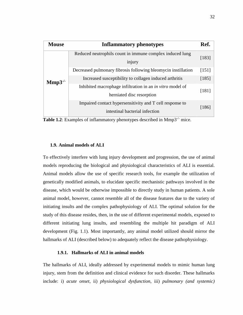
32
Mouse Inflammatory phenotypes Ref.
Mmp3-/-
Reduced neutrophils count in immune complex induced lung
injury [183]
Decreased pulmonary fibrosis following bleomycin instillation [151]
Increased susceptibility to collagen induced arthritis [185]
Inhibited macrophage infiltration in an in vitro model of
herniated disc resorption [181]
Impaired contact hypersensitivity and T cell response to
intestinal bacterial infection [186]
Table 1.2: Examples of inflammatory phenotypes described in Mmp3-/- mice.
1.9. Animal models of ALI
To effectively interfere with lung injury development and progression, the use of animal
models reproducing the biological and physiological characteristics of ALI is essential.
Animal models allow the use of specific research tools, for example the utilization of
genetically modified animals, to elucidate specific mechanistic pathways involved in the
disease, which would be otherwise impossible to directly study in human patients. A sole
animal model, however, cannot resemble all of the disease features due to the variety of
initiating insults and the complex pathophysiology of ALI. The optimal solution for the
study of this disease resides, then, in the use of different experimental models, exposed to
different initiating lung insults, and resembling the multiple hit paradigm of ALI
development (Fig. 1.1). Most importantly, any animal model utilized should mirror the
hallmarks of ALI (described below) to adequately reflect the disease pathophysiology.
1.9.1. Hallmarks of ALI in animal models
The hallmarks of ALI, ideally addressed by experimental models to mimic human lung
injury, stem from the definition and clinical evidence for such disorder. These hallmarks
include: i) acute onset, ii) physiological dysfunction, iii) pulmonary (and systemic)

33
inflammation, iv) increased pulmonary permeability, and v) histological evidence of lung
injury [187, 188].
Specifically, in valid models of ALI lung injury should develop shortly after the exposure
to the insults (acute onset), and lead to the manifestation of physiological dysfunction. The
latter implies the presence of pulmonary surfactant alterations, impaired gas exchange, and
changes in lung mechanics [2]. Analyses of pulmonary surfactant can be readily performed
on isolated samples of lung lavage undergoing differential centrifugation. Following
isolation of total surfactant, large, and small aggregates, the abundance of each sub-fraction
can be determined through well-established biochemical assays [189, 190]. Moreover,
surfactant biophysical properties can be evaluated in vitro, by monitoring the minimum
surface tension achieved by LA samples exposed to repeated cycles of compression and
expansion via captive bubble surfactometer [191] (chapter 2) or constrained sessile drop
surfactometer [192] (chapter 4). With the exception of surfactant analyses, assessment of
physiological dysfunction can pose a technical challenge, especially in mice, and may not
be always feasible. Measurements of oxygenation often require the use of invasive arterial
catheters, and are limited by a small total blood volume. Physiological parameters are also
hard to measure in spontaneously breathing animals, but can be assessed in mechanically
ventilated mice. MV allows in vivo and ex vivo monitoring of changes to peak pressure
and/or lung compliance [193]; furthermore, as previously mentioned, MV augments the
pulmonary inflammation associated with ALI, and contributes to the systemic
inflammatory response [13, 58].
Pulmonary inflammation, a definite hallmark of ALI, comprises increased lavage
inflammatory mediators and neutrophil numbers, and can be effectively measured in lung
lavage samples through commercially available assays and analysis of total and differential
cell counts. Lung lavage samples can also be utilized to evaluate increases in total protein
content, thereby illustrating, with some limitations, increases in pulmonary permeability,
another characterizing aspect of lung injury [188].
Lastly, a good animal model of ALI should provide histological evidence of lung injury,
since histological analyses can give both qualitative and quantitative information on the

34
extent and uniformity of the injury, cellularity, sites of edema accumulation, and gross
structural abnormalities [188].
In conclusion, it should be noted that most animal models of ALI present at least a few of
the hallmarks described above. While a description of all the models available for the study
of this disease goes beyond the scope of this chapter, the main characteristics of the lung
injury models relevant to this thesis are described in the following paragraphs.
1.9.2. Hydrochloric acid-induced lung injury
Gastric acid aspiration refers to the unintentional inhalation of the gastric content, and is
one of the direct pulmonary insults predisposing the lung to ALI (Fig. 1.1). It accounts for
about 30% of all the deaths due to anesthesia, and can also occur in subjects experiencing
drug overdose or seizures [194]. Even though gastric aspiration exposes the lung to both
particulate matter and acidic juices from the stomach, this injury has been widely modelled
through the intra-tracheal instillation of hydrochloric acid (HCl) solutions with low pH [58,
195, 196].
Animal models of acid-induced ALI are characterized by acute pulmonary changes,
detectable as early as four hours post HCl instillation (chapters 2 and 3) [58]. Lung injury
results from the chemical burn of the pulmonary parenchyma, subsequently leading to
elevated lavage concentrations of inflammatory cytokines and chemokines involved in
neutrophils recruitment. Acid-induced ALI, in fact, appears to be neutrophil dependent, as
demonstrated by marked pulmonary infiltrates and, conversely, by the development of
milder injury when neutrophils accumulation in the lung is inhibited or decreased [67, 197,
198]. While it is not entirely clear how acid injury activates the immune system, some
recent study suggests that the Toll-like receptor 4 (TLR4) pathway may be involved [199].
Being a direct lung insult, the alveolar epithelium is especially affected, with loss of
epithelial cells, alterations of the alveolo-capillary barrier and consequent surfactant
impairment [187]. Additional signs of physiological dysfunction, such as increased peak
pressure and decreased compliance, have also been observed in this model of ALI [58,
196].
35
1.9.3. Lipopolysaccharide-induced lung injury
Bacterial pneumonia is one of the most common causes of ALI [2]. This injury has been
extensively reproduced in experimental models through the intra-nasal or intra-tracheal
administration of bacteria or lipopolysaccharide (LPS), an immunogenic molecule found
in the outer membrane of Gram negative bacteria. While administration of live bacteria
constitutes a valid alternative in modeling pneumonia, LPS has the advantage of being easy
to prepare and administer, and ensures a good degree of reproducibility [187].
LPS instilled intra-tracheally directly affects both the alveolar macrophages and epithelium
by binding to its receptor, TLR4, on these cells and activating intracellular pathways
leading to synthesis and release of multiple cytokines and chemokines [200]. LPS-induced
lung injury then develops presenting most of the hallmarks of ALI described above, as
highlighted by the analyses of pulmonary permeability and inflammation, physiological
dysfunction and histological damage described throughout chapters 3 and 4 of this thesis.
For such reasons, LPS-induced ALI is a very useful and informative model in the study of
this disorder.
1.9.4. Ventilation associated lung injury and ex vivo ventilation
As previously mentioned, mechanical ventilation is the main supportive treatment in ALI
necessary to improve oxygenation, and a crucial component of ALI pathophysiology as a
potential secondary insult. Experimentally, the role of MV in ALI has been investigated in
isolation, overstretching the lung by applying high tidal volume MV to healthy lungs. This
injury model is referred to as ventilator-induced lung injury (VILI) and allows the study of
specific alterations in pulmonary function, inflammation and cell signaling resulting from
excessive mechanical stretch and strain implemented on otherwise normal lungs [201,
202].
While VILI is an experimental tool of great analytical value, in a clinical setting ventilation
is utilized to support lung function, which is already impaired due to a pre-existing injury.
The study of ventilation in clinically relevant, in vivo or ex vivo models is therefore
performed by implementing MV on lungs that have been pre-exposed to a clinically
significant insult, such as aspiration or pneumonia. Such models of ventilation-associated

36
lung injury (VALI) directly examine the role of MV as a secondary insult in propagating
lung injury and in contributing to the development of systemic inflammation and associated
distal organ failure [14, 52, 58]. Overall, models of VILI and VALI have been essential in
demonstrating that MV can lead to damage via over-distention and repeated
opening/closing of alveolar units, and have convincingly pointed out that the extent of the
injury is proportional to the degree of mechanical stress applied to the lung.
The mechanisms through which ventilation can cause and exacerbate injury are barotrauma
(gross lung injury due to excessive airway pressure), volutrauma (excessive volume
applied to the lungs) and, particularly relevant for this thesis, biotrauma [55, 203]. The
concept of biotrauma refers to the release of biologically active mediators from lungs
exposed to injurious modalities of MV, mediators that can potentially translocate into the
circulation and damage distal organs [56]. Evidence for the biotrauma hypothesis stems
from a vast body of in vitro, in vivo and ex vivo research. Numerous studies have in fact
shown release of inflammatory molecules and changes in gene expression in alveolar
epithelial cells and macrophages exposed to cycles of mechanical stretch [56, 204, 205].
These in vitro findings have been mirrored by in vivo experimental observations, in which
high pressure or high volume ventilation caused elevated pulmonary and systemic levels
of cytokines and chemokines, often in combination with changes in peripheral organs [14,
58, 79]. Notably, Imai and colleagues have demonstrated that, in a model of acid-induced
ALI followed by injurious MV, lung derived mediators in the systemic circulation led to
increased apoptosis in the kidneys and small intestine of the experimental animals, thereby
strongly supporting the biotrauma concept [61].
A particularly useful tool to study biotrauma and the effects of MV on the lung in isolation
is a system for ex vivo ventilation, namely the isolated and perfused mouse lung (IPML)
set up [59]. The IPML set up allows for ex vivo MV of mouse lungs and simultaneous re-
circulation of a cell free solution through the pulmonary blood vessels. One of the main
advantages of this system resides in the possibility to collect and analyze inflammatory
mediators specifically released from the lung into the circulation throughout MV [59, 86].
Importantly, the IPML system allows the monitoring of potential changes in the levels of

37
these circulating, lung-derived mediators resulting from lung specific treatments, as
illustrated in chapter 2 of this thesis.
1.9.4.1. Measurements of lung mechanics in small rodents
An undoubtable advantage associated with the use of MV in small rodents is the possibility
to evaluate lung mechanics. Measurement of physiological parameters in spontaneously
breathing animals is, in fact, quite difficult and usually not very precise [206]. For example,
whole-body plethysmography can be used in unrestrained, conscious animals; this
technique, however, provides only general information about the pattern of ventilation, and
it can generate errors and artifacts. On the other hand, MV is a more invasive procedure,
requiring restrain, surgery and anesthesia management for in vivo models, but providing
the valuable advantage of reliable, reproducible, and accurate measurements [207]. While
in vivo MV was not utilized in this thesis work, models of ex vivo ventilation and ex vivo
measurements of lung function were applied to terminally sacrificed mice [59, 208, 209].
These strategies allowed the monitoring of peak inspiratory pressure changes throughout
MV (chapter 2) and the specific assessment of several parameters, such as: compliance,
resistance, and elastance for the whole respiratory system; quasi-static compliance and
elastance; central airway resistance (RN); tissue resistance or tissue damping (G); and tissue
elastance (H) (chapter 4); thereby providing complete and novel information on
physiological dysfunction in our models of ALI.
1.10. Summary and overall objective
The lung is the organ responsible for gas exchange, a vital function that is severely impaired
during acute lung injury. This life threatening condition can be initiated by multiple and
diverse insults to the lung, resulting in profound alterations of the alveolar
microenvironment. Among such alterations, the impairment of the surfactant system and
the development of an extensive pulmonary inflammatory response are key events in the
progression of the disease. Despite extensive research, the highly complex
pathophysiology of ALI has hindered the development of an effective therapeutic treatment
capable of reducing the high mortality associated with this disorder. Further investigation

38
is, therefore, necessary to improve applicable interventions and identify key mediators of
lung injury that could serve as potential therapeutic targets.
The overarching hypothesis of this thesis work is that interventions targeting the
inflammatory response associated with ALI can improve outcomes. The overall objective
is then to expand our understanding of the ALI pathophysiology, in order to better interfere
with disease progression and provide potential future alternatives in the treatment of ALI.

39
1.11. References
1. West JB: Respiratory Physiology: The Essentials. 5th edition. Lippincott Williams &
Wilkins; .
2. Ware LB, Matthay MA: The Acute Respiratory Distress Syndrome. N Engl J Med
2000, 342:1334–1349.
3. Wheeler AP, Bernard GR: Acute lung injury and the acute respiratory distress
syndrome: a clinical review. Lancet 2007, 369:1553–1564.
4. Walkey AJ, Summer R, Ho V, Alkana P: Acute respiratory distress syndrome:
epidemiology and management approaches. Clin Epidemiol 2012, 4:159–169.
5. Parsons PE, Eisner MD, Thompson BT, Matthay MA, Ancukiewicz M, Bernard GR,
Wheeler AP, and the NHLBI Acute Respiratory Distress Syndrome Clinical Trials
Network: Lower tidal volume ventilation and plasma cytokine markers of
inflammation in patients with acute lung injury. Crit Care Med 2005, 33:1–6.
6. Meduri GU, Kohler G, Headley S, Tolley E, Stentz F, Postlethwaite A: Inflammatory
Cytokines in the BAL of Patients with ARDS. Persistent Elevation Over Time
Predicts Poor Outcome. Chest 1995, 108:1303–1314.
7. Veldhuizen RAW, Welk B, Harbottle R, Hearn S, Nag K, Petersen N, Possmayer F:
Mechanical ventilation of isolated rat lungs changes the structure and biophysical
properties of surfactant. J Appl Physiol 2002, 92:1169–1175.
8. Goerke J: Pulmonary surfactant: functions and molecular composition. Biochim
Biophys Acta 1998, 1408:79–89.
9. Wright JR: Immunomodulatory functions of surfactant. Physiol Rev 1997, 77:931–
962.

40
10. Maruscak AA, Vockeroth DW, Girardi B, Sheikh T, Possmayer F, Lewis JF,
Veldhuizen RAW: Alterations to surfactant precede physiological deterioration
during high tidal volume ventilation. Am J Physiol Lung Cell Mol Physiol 2008,
294:L974–L983.
11. Rasaiah VP, Malloy JL, Lewis JF, Veldhuizen RAW: Early surfactant
administration protects against lung dysfunction in a mouse model of ARDS. Am J
Physiol Cell Mol Physiol 2003, 284:L783–L790.
12. Ranieri VM, Suter PM, Tortorella C, De Tullio R, Dayer JM, Brienza A, Bruno F,
Slutsky AS: Effect of mechanical ventilation on inflammatory mediators in patients
with acute respiratory distress syndrome: a randomized controlled trial. JAMA 1999,
282:54–61.
13. Chiumello D, Pristine G, Slutsky AS: Mechanical ventilation affects local and
systemic cytokines in an animal model of acute respiratory distress syndrome. Am J
Respir Crit Care Med 1999, 160:109–116.
14. Dhanireddy S, Altemeier WA, Matute-Bello G, O’Mahony DS, Glenny RW, Martin
TR, Liles WC: Mechanical ventilation induces inflammation, lung injury, and extra-
pulmonary organ dysfunction in experimental pneumonia. Lab Invest 2006, 86:790–
799.
15. Montgomery AB, Stager MA, Carrico CJ, Hudson LD: Causes of mortality in
patients with the adult respiratory distress syndrome. Am Rev Respir Dis 1985,
132:485–489.
16. Stapleton RD, Wang BM, Hudson LD, Rubenfeld GD, Caldwell ES, Steinberg KP:
Causes and timing of death in patients with ARDS. Chest 2005, 128:525–532.
17. Parks WC, Wilson CL, López-Boado YS: Matrix metalloproteinases as modulators
of inflammation and innate immunity. Nat Rev Immunol 2004, 4:617–629.
18. Mason RJ: Biology of alveolar type II cells. Respirology 2006, 11 Suppl:S12–S15.

41
19. Du Bois RM: The alveolar macrophage. Thorax 1985, 40:321–7.
20. Wright JR: Clearance and recycling of pulmonary surfactant. Am J Physiol 1990,
259:L1–L12.
21. West JB: Pulmonary Pathophysiology: The Essentials. 5th edition. Lippincott Williams
& Wilkins.
22. Veldhuizen EJ, Haagsman HP: Role of pulmonary surfactant components in surface
film formation and dynamics. Biochim Biophys Acta 2000, 1467:255–270.
23. Avery ME, Mead J: Surface properties in relation to atelectasis and hyaline
membrane disease. AMA J Dis Child 1959, 97:517–523.
24. Ashbaugh DG, Bigelow DB, Petty TL, Levine BE: Acute respiratory distress in
adults. Lancet 1967, 2:319–323.
25. Bernard GR, Artigas A, Brigham KL, Carlet J, Falke K, Hudson L, Lamy M, Legall
JR, Morris A, Spragg R: The American-European Consensus Conference on ARDS.
Definitions, mechanisms, relevant outcomes, and clinical trial coordination. Am J
Respir Crit Care Med 1994, 149(3 Pt 1):818–824.
26. Ranieri VM, Rubenfeld GD, Thompson BT, Ferguson ND, Caldwell E, Fan E,
Camporota L, Slutsky AS: Acute respiratory distress syndrome: the Berlin Definition.
JAMA 2012, 307:2526–2533.
27. Rubenfeld GD, Caldwell E, Peabody E, Weaver J, Martin DP, Neff M, Stern EJ,
Hudson LD: Incidence and outcomes of acute lung injury. N Engl J Med 2005,
353:1685–1693.
28. Phua J, Badia JR, Adhikari NKJ, Friedrich JO, Fowler R a, Singh JM, Scales DC,
Stather DR, Li A, Jones A, Gattas DJ, Hallett D, Tomlinson G, Stewart TE, Ferguson ND:
Has mortality from acute respiratory distress syndrome decreased over time?: A
systematic review. Am J Respir Crit Care Med 2009, 179:220–227.

42
29. Matthay MA, Ware LB, Zimmerman GA: The acute respiratory distress syndrome.
J Clin Invest 2012, 122:2731–2740.
30. Bellingan G.J.: The pulmonary physician in critical care * 6 : The pathogenesis of
ALI/ARDS. Thorax 2002, 57:540–546.
31. Johnson ER, Matthay MA: Acute lung injury: Epidemiology, Pathogenesis, and
Treatment. J Aerosol Med Pulm Drug Deliv 2010, 23:243–252.
32. Del Sorbo L, Slutsky AS: Acute respiratory distress syndrome and multiple organ
failure. Curr Opin Crit Care 2011, 17:1–6.
33. Pelosi P, D’Onofrio D, Chiumello D, Paolo S, Chiara G, Capelozzi V, Barbas C,
Chiaranda M, Gattinoni L: Pulmonary and extrapulmonary acute respiratory distress
syndrome are different. Eur Respir J 2003, 22:48s–56s.
34. Rocco PRM, Pelosi P: Pulmonary and extrapulmonary acute respiratory distress
syndrome: myth or reality?. Curr Opin Crit Care 2008, 14:50–55.
35. Castro CY: ARDS and diffuse alveolar damage: a pathologist’s perspective. Semin
Thorac Cardiovasc Surg 2006, 18:13–19.
36. Petty TL, Silvers GW, Paul GW, Stanford RE: Abnormalities in lung elastic
properties and surfactant function in adult respiratory distress syndrome. Chest 1979,
75:571–574.
37. The Acute Respiratory Distress Syndrome Network: Ventilation with lower tidal
volumes as compared with traditional tidal volumes for acute lung injury and the
acute respiratory distress syndrome. The Acute Respiratory Distress Syndrome
Network. N Engl J Med 2000, 342:1301–1308.
38. Brower RG: Treatment of ARDS. Chest 2001, 120:1347–1367.

43
39. Levitt JE, Matthay MA: Treatment of acute lung injury: historical perspective and
potential future therapies. Semin Respir Crit Care Med 2006, 27:426–437.
40. Lewis JF, Veldhuizen R: The role of exogenous surfactant in the treatment of acute
lung injury. Annu Rev Physiol 2003, 65:613–642.
41. Davidson WJ, Dorscheid D, Spragg R, Schulzer M, Mak E, Ayas NT: Exogenous
pulmonary surfactant for the treatment of adult patients with acute respiratory
distress syndrome: results of a meta-analysis. Crit Care 2006, 10:R41.
42. Goodman RB, Pugin J, Lee JS, Matthay MA: Cytokine-mediated inflammation in
acute lung injury. Cytokine Growth Factor Rev 2003, 14:523–535.
43. Bhatia M, Moochhala S: Role of inflammatory mediators in the pathophysiology of
acute respiratory distress syndrome. J Pathol 2004, 202:145–56.
44. Bosma KJ, Taneja R, Lewis JF: Pharmacotherapy for prevention and treatment of
acute respiratory distress syndrome: current and experimental approaches. Drugs
2010, 70:1255–1282.
45. Yamashita CM, Lewis JF: Emerging therapies for treatment of acute lung injury
and acute respiratory distress syndrome. Expert Opin Emerg Drugs 2012, 17:1–4.
46. Pelosi P, Caironi P, Gattinoni L: Pulmonary and extrapulmonary forms of acute
respiratory distress syndrome. Semin Respir Crit Care Med 2001, 22:259–268.
47. Bhattacharya J, Matthay MA: Regulation and repair of the alveolar-capillary
barrier in acute lung injury. Annu Rev Physiol 2013, 75:593–615.
48. Seeger W, Grube C, Gunther A, Schmidt R: Surfactant inhibition by plasma
proteins: differential sensitivity of various surfactant preparations. Eur Respir J 1993,
6:971–977.

44
49. Da Silva K, McCaig L, Veldhuizen RAW, Possmayer F: Protein inhibition of
surfactant during mechanical ventilation of isolated rat lungs. Exp Lung Res 2005,
31:745–758.
50. Pittet JF, Mackersie RC, Martin TR, Matthay MA: Biological markers of acute lung
injury: prognostic and pathogenetic significance. Am J Respir Crit Care Med 1997,
155:1187–1205.
51. Witko-Sarsat V, Rieu P, Descamps-Latscha B, Lesavre P, Halbwachs-Mecarelli L:
Neutrophils: molecules, functions and pathophysiological aspects. Lab Invest 2000,
80:617–653.
52. O’Mahony DS, Liles WC, Altemeier WA, Dhanireddy S, Frevert CW, Liggitt D,
Martin TR, Matute-Bello G: Mechanical ventilation interacts with endotoxemia to
induce extrapulmonary organ dysfunction. Crit Care 2006, 10:R136.
53. Altemeier WA, Matute-Bello G, Gharib SA, Glenny RW, Martin TR, Liles WC:
Modulation of lipopolysaccharide-induced gene transcription and promotion of lung
injury by mechanical ventilation. J Immunol 2005, 175:3369–3376.
54. Nakamura T, Malloy J, McCaig L, Yao L, Joseph M, Lewis J, Veldhuizen R:
Mechanical ventilation of isolated septic rat lungs : effects on surfactant and
inflammatory cytokines. J Appl Physiol 2001, 91:811–820.
55. Tremblay LN, Slutsky AS: Ventilator-induced injury: from barotrauma to
biotrauma. Proc Assoc Am Physicians 1998, 110:482–488.
56. Dos Santos CC, Zhang H, Liu M, Slutsky AS: Bench-to-bedside review: Biotrauma
and modulation of the innate immune response. Crit Care 2005, 9:280–286.
57. Wilson MR, Choudhury S, Goddard ME, O’Dea KP, Nicholson AG, Takata M: High
tidal volume upregulates intrapulmonary cytokines in an in vivo mouse model of
ventilator-induced lung injury. J Appl Physiol 2003, 95:1385–1393.

45
58. Walker MG, Yao LJ, Patterson EK, Joseph MG, Cepinskas G, Veldhuizen RA, Lewis
JF, Yamashita CM: The effect of tidal volume on systemic inflammation in Acid-
induced lung injury. Respiration 2011, 81:333–342.
59. Von Bethmann AN, Brasch F, Nusing R, Vogt K, Volk HD, Muller KM, Wendel A,
Uhlig S: Hyperventilation induces release of cytokines from perfused mouse lung. Am
J Respir Crit Care Med 1998, 157:263–272.
60. Haitsma JJ, Uhlig S, Goggel R, Verbrugge SJ, Lachmann U, Lachmann B: Ventilator-
induced lung injury leads to loss of alveolar and systemic compartmentalization of
tumor necrosis factor-alpha. Intensive Care Med 2000, 26:1515–1522.
61. Imai Y, Parodo J, Kajikawa O, De Perrot M, Fischer S, Edwards V, Cutz E, Liu M,
Keshavjee S, Martin TR, Marshall JC, Ranieri VM, Slutsky AS: Injurious mechanical
ventilation and end-organ epithelial cell apoptosis and organ dysfunction in an
experimental model of acute respiratory distress syndrome. JAMA 2003, 289:2104–
2112.
62. Plötz FB, Slutsky AS, van Vught AJ, Heijnen CJ: Ventilator-induced lung injury and
multiple system organ failure: a critical review of facts and hypotheses. Intensive Care
Med 2004, 30:1865–1872.
63. Slutsky AS, Tremblay LN: Multiple system organ failure. Is mechanical ventilation
a contributing factor?. Am J Respir Crit Care Med 1998, 157(6 Pt 1):1721–1725.
64. Markovic N, McCaig LA, Stephen J, Mizuguchi S, Veldhuizen RA, Lewis JF,
Cepinskas G: Mediators released from LPS-challenged lungs induce inflammatory
responses in liver vascular endothelial cells and neutrophilic leukocytes. Am J Physiol
liver Physiol 2009, 297:G1066–1076.
65. Medzhitov R: Origin and physiological roles of inflammation. Nature 2008,
454:428–435.

46
66. Grommes J, Soehnlein O: Contribution of neutrophils to acute lung injury. Mol
Med 2011, 17:293–307.
67. Zarbock A, Singbartl K, Ley K: Complete reversal of acid-induced acute lung injury
by blocking of platelet-neutrophil aggregation. J Clin Invest 2006, 116:3211–3219.
68. Riva CM, Morganroth ML, Ljungman AG, Schoeneich SO, Marks RM, Todd RF 3rd,
Ward PA, Boxer LA: Iloprost inhibits neutrophil-induced lung injury and neutrophil
adherence to endothelial monolayers. Am J Respir Cell Mol Biol 1990, 3:301–309.
69. Lee CT, Fein AM, Lippmann M, Holtzman H, Kimbel P, Weinbaum G: Elastolytic
activity in pulmonary lavage fluid from patients with adult respiratory-distress
syndrome. N Engl J Med 1981, 304:192–196.
70. Braude S, Apperley J, Krausz T, Goldman JM, Royston D: Adult respiratory distress
syndrome after allogeneic bone-marrow transplantation: evidence for a neutrophil-
independent mechanism. Lancet 1985, 1:1239–1242.
71. Aggarwal NR, King LS, D’Alessio FR: Diverse macrophage populations mediate
acute lung inflammation and resolution. Am J Physiol Lung Cell Mol Physiol 2014,
306:L709–L725.
72. Kuebler WM: Inflammatory pathways and microvascular responses in the lung.
Pharmacol reports 2005, 57 Suppl:196–205.
73. Dos Santos CC, Han B, Andrade CF, Bai X, Uhlig S, Hubmayr R, Tsang M, Lodyga
M, Keshavjee S, Slutsky AS, Liu M: DNA microarray analysis of gene expression in
alveolar epithelial cells in response to TNFalpha, LPS, and cyclic stretch. Physiol
Genomics 2004, 19:331–342.
74. Vlahakis NE, Schroeder MA, Limper AH, Hubmayr RD: Stretch induces cytokine
release by alveolar epithelial cells in vitro. Am J Physiol 1999, 277(1 Pt 1):L167–L173.

47
75. Manicone AM: Role of the pulmonary epithelium and inflammatory signals in
acute lung injury. Expert Rev Clin Immunol 2009, 5:63–75.
76. Wright JR: Immunoregulatory functions of surfactant proteins. Nat Rev 2005,
5:58–68.
77. Puneet P, Moochhala S, Bhatia M: Chemokines in acute respiratory distress
syndrome. Am J Physiol Lung Cell Mol Physiol 2005, 288:L3–L15.
78. Braunwald E, Fauci AS, Kasper DL, Hauser SL, Longo DL, Jameson JL: Harrison’s
Principles of Internal Medicine. 15th edition. McGraw Hill.
79. Gurkan OU, O’Donnell C, Brower R, Ruckdeschel E, Becker PM: Differential effects
of mechanical ventilatory strategy on lung injury and systemic organ inflammation
in mice. Am J Physiol Cell Mol Physiol 2003, 285:L710–L718.
80. Meduri G, Headley S, Kohler G, Stentz F, Tolley E, Umberger R, Leeper K: Persistent
elevation of inflammatory cytokines predicts a poor outcome in ARDS. Plasma IL-1
beta and IL-6 levels are consistent and efficient predictors of outcome over time.
CHEST J 1995, 107:1062–1073.
81. Goodman RB, Strieter RM, Martin DP, Steinberg KP, Milberg JA, Maunder RJ,
Kunkel SL, Walz A, Hudson LD, Martin TR: Inflammatory cytokines in patients with
persistence of the acute respiratory distress syndrome. Am J Respir Crit Care Med
1996, 154(3 Pt 1):602–611.
82. Donnelly SC, Strieter RM, Kunkel SL, Walz A, Robertson CR, Carter DC, Grant IS,
Pollok AJ, Haslett C: Interleukin-8 and development of adult respiratory distress
syndrome in at-risk patient groups. Lancet 1993, 341:643–647.
83. McCarthy MK, Weinberg JB: Eicosanoids and respiratory viral infection:
coordinators of inflammation and potential therapeutic targets. Mediators Inflamm
2012, 2012:1–13.

48
84. Stephenson AH, Lonigro AJ, Hyers TM, Webster RO, Fowler AA: Increased
concentrations of leukotrienes in bronchoalveolar lavage fluid of patients with ARDS
or at risk for ARDS. Am Rev Respir Dis 1988, 138:714–719.
85. Auner B, Geiger E V, Henrich D, Lehnert M, Marzi I, Relja B: Circulating leukotriene
B4 identifies respiratory complications after trauma. Mediators Inflamm 2012, 2012:1–
8.
86. Jaecklin T, Engelberts D, Otulakowski G, O’Brodovich H, Post M, Kavanagh BP:
Lung-derived soluble mediators are pathogenic in ventilator-induced lung injury. Am
J Physiol Cell Mol Physiol 2011, 300:L648–L658.
87. Traeger T, Kessler W, Hilpert A, Mikulcak M, Entleutner M, Koerner P, Westerholt
A, Cziupka K, van Rooijen N, Heidecke C-D, Maier S: Selective depletion of alveolar
macrophages in polymicrobial sepsis increases lung injury, bacterial load and
mortality but does not affect cytokine release. Respiration 2009, 77:203–213.
88. Eyal FG, Hamm CR, Parker JC: Reduction in alveolar macrophages attenuates
acute ventilator induced lung injury in rats. Intensive Care Med 2007, 33:1212–1218.
89. Kotani M, Kotani T, Ishizaka A, Fujishima S, Koh H, Tasaka S, Sawafuji M, Ikeda E,
Moriyama K, Kotake Y, Morisaki H, Aikawa N, Ohashi A, Matsushima K, Huang Y-CT,
Takeda J: Neutrophil depletion attenuates interleukin-8 production in mild-
overstretch ventilated normal rabbit lung. Crit Care Med 2004, 32:514–519.
90. Gavett SH, Carakostas MC, Belcher LA, Warheit DB: Effect of circulating neutrophil
depletion on lung injury induced by inhaled silica particles. J Leukoc Biol 1992,
51:455–461.
91. Nakamura T, Moyer BZ, Veldhuizen RA, Lewis JF: Interleukin-6 has no effect on
surfactant or lung function in different lung insults. Exp Lung Res 2006, 32:27–42.
49
92. Abraham E, Anzueto A, Gutierrez G, Tessler S, San Pedro G, Wunderink R, Dal
Nogare A, Nasraway S, Berman S, Cooney R, Levy H, Baughman R, Rumbak M, Light
RB, Poole L, Allred R, Constant J, Pennington J, Porter S: Double-blind randomised
controlled trial of monoclonal antibody to human tumour necrosis factor in treatment
of septic shock. NORASEPT II Study Group. Lancet 1998, 351:929–933.
93. Opal SM, Fisher CJ, Dhainaut JF, Vincent JL, Brase R, Lowry SF, Sadoff JC, Slotman
GJ, Levy H, Balk RA, Shelly MP, Pribble JP, LaBrecque JF, Lookabaugh J, Donovan H,
Dubin H, Baughman R, Norman J, DeMaria E, Matzel K, Abraham E, Seneff M:
Confirmatory interleukin-1 receptor antagonist trial in severe sepsis: a phase III,
randomized, double-blind, placebo-controlled, multicenter trial. The Interleukin-1
Receptor Antagonist Sepsis Investigator Group. Crit Care Med 1997, 25:1115–1124.
94. Ruan S-Y, Lin H-H, Huang C-T, Kuo P-H, Wu H-D, Yu C-J: Exploring the
heterogeneity of effects of corticosteroids on acute respiratory distress syndrome: a
systematic review and meta-analysis. Crit Care 2014, 18:R63.
95. Veldhuizen RA, McCaig LA, Akino T, Lewis JF: Pulmonary surfactant subfractions
in patients with the acute respiratory distress syndrome. Am J Respir Crit Care Med
1995, 152(6 Pt 1):1867–1871.
96. Possmayer F: A proposed nomenclature for pulmonary surfactant-associated
proteins. Am Rev Respir Dis 1988, 138:990–998.
97. Kuroki Y, Voelker DR: Pulmonary surfactant proteins. J Biol Chem 1994,
269:25943–25946.
98. Sano H, Kuroki Y: The lung collectins, SP-A and SP-D, modulate pulmonary innate
immunity. Mol Immunol 2005, 42:279–287.
99. Weaver TE, Conkright JJ: Function of surfactant proteins B and C. Annu Rev Physiol
2001, 63:555–578.

50
100. Wright JR, Hawgood S: Pulmonary surfactant metabolism. Clin Chest Med 1989,
10:83–93.
101. Wright JR, Dobbs LG: Regulation of pulmonary surfactant secretion and
clearance. Annu Rev Physiol 1991, 53:395–414.
102. Perez-Gil J, Weaver TE: Pulmonary surfactant pathophysiology: current models
and open questions. Physiology (Bethesda) 2010, 25:132–141.
103. Hawgood S, Poulain FR: The pulmonary collectins and surfactant metabolism.
Annu Rev Physiol 2001, 63:495–519.
104. Froh D, Gonzales LW, Ballard PL: Secretion of surfactant protein A and
phosphatidylcholine from type II cells of human fetal lung. Am J Respir Cell Mol Biol
1993, 8:556–561.
105. Fisher AB, Dodia C, Ruckert P, Tao JQ, Bates SR: Pathway to lamellar bodies for
surfactant protein A. Am J Physiol Cell Mol Physiol 2010, 299:L51–L58.
106. Gross NJ, Narine KR: Surfactant subtypes in mice: characterization and
quantitation. J Appl Physiol 1989, 66:342–349.
107. Gross NJ, Narine KR: Surfactant subtypes of mice: metabolic relationships and
conversion in vitro. J Appl Physiol 1989, 67:414–421.
108. Brackenbury AM, Malloy JL, McCaig LA, Yao LJ, Veldhuizen RA, Lewis JF:
Evaluation of alveolar surfactant aggregates in vitro and in vivo. Eur Respir J 2002,
19:41–46.
109. Putz G, Goerke J, Clements JA: Surface activity of rabbit pulmonary surfactant
subfractions at different concentrations in a captive bubble. J Appl Physiol 1994,
77:597–605.

51
110. Possmayer F, Yu SH, Weber JM, Harding PG: Pulmonary surfactant. Can J
Biochem Cell Biol 1984, 62:1121–1133.
111. Veldhuizen R, Nag K, Orgeig S, Possmayer F: The role of lipids in pulmonary
surfactant. Biochim Biophys Acta 1998, 1408:90–108.
112. Veldhuizen EJ, Batenburg JJ, van Golde LM, Haagsman HP: The role of surfactant
proteins in DPPC enrichment of surface films. Biophys J 2000, 79:3164–3171.
113. Clark JC, Wert SE, Bachurski CJ, Stahlmant MT, Stripp BR, Weaver TE, Whitsett
JA: Targeted disruption of the surfactant protein B gene disrupts surfactant
homeostasis , causing respiratory failure in newborn mice. Proc Natl Acad Sci U S A
1995, 92(August):7794–7798.
114. Glasser SW, Burhans MS, Korfhagen TR, Na CL, Sly PD, Ross GF, Ikegami M,
Whitsett JA: Altered stability of pulmonary surfactant in SP-C-deficient mice. Proc
Natl Acad Sci U S A 2001, 98:6366–6371.
115. McCormack FX: Structure, processing and properties of surfactant protein A.
Biochim Biophys Acta 1998, 1408:109–131.
116. Veldhuizen RAW, Yao L, Hearn SA, Possmayer F, Lewis JF: Surfactant-associated
Protein A is important for maintaining surfactant large-aggregate forms during
surface-area cycling. Biochem J 1996, 313:835–840.
117. Cockshutt AM, Weitz J, Possmayer F: Pulmonary surfactant-associated protein A
enhances the surface activity of lipid extract surfactant and reverses inhibition by
blood proteins in vitro. Biochemistry 1990, 29:8424–8429.
118. LeVine AM, Kurak KE, Wright JR, Watford WT, Bruno MD, Ross GF, Whitsett JA,
Korfhagen TR: Surfactant protein-A binds group B streptococcus enhancing
phagocytosis and clearance from lungs of surfactant protein-A-deficient mice. Am J
Respir Cell Mol Biol 1999, 20:279–286.

52
119. Crouch E, Wright JR: Surfactant proteins A and D and pulmonary host defense.
Annu Rev Physiol 2001, 63:521–554.
120. McCormack FX, Whitsett JA: The pulmonary collectins, SP-A and SP-D,
orchestrate innate immunity in the lung. J Clin Invest 2002, 109:707–712.
121. Borron PJ, Mostaghel EA, Doyle C, Walsh ES, McHeyzer-Williams MG, Wright JR:
Pulmonary surfactant proteins A and D directly suppress CD3+/CD4+ cell function:
evidence for two shared mechanisms. J Immunol 2002, 169:5844–5850.
122. Wolfson MR, Wu J, Hubert TL, Gregory TJ, Mazela J, Shaffer TH: Lucinactant
attenuates pulmonary inflammatory response, preserves lung structure, and
improves physiologic outcomes in a preterm lamb model of RDS. Pediatr Res 2012,
72:375–383.
123. Van Iwaarden JF, Claassen E, Jeurissen SH, Haagsman HP, Kraal G: Alveolar
macrophages, surfactant lipids, and surfactant protein B regulate the induction of
immune responses via the airways. Am J Respir Cell Mol Biol 2001, 24:452–458.
124. Numata M, Chu HW, Dakhama A, Voelker DR: Pulmonary surfactant
phosphatidylglycerol inhibits respiratory syncytial virus-induced inflammation and
infection. Proc Natl Acad Sci U S A 2010, 107:320–325.
125. Abate W, Alghaithy AA, Parton J, Jones KP, Jackson SK: Surfactant lipids regulate
LPS-induced interleukin-8 production in A549 lung epithelial cells by inhibiting
translocation of TLR4 into lipid raft domains. J Lipid Res 2010, 51:334–344.
126. Glasser SW, Senft AP, Whitsett JA, Maxfield MD, Ross GF, Richardson TR, Prows
DR, Xu Y, Korfhagen TR: Macrophage dysfunction and susceptibility to pulmonary
Pseudomonas aeruginosa infection in surfactant protein C-deficient mice. J Immunol
2008, 181:621–628.

53
127. Glasser SW, Witt TL, Senft AP, Baatz JE, Folger D, Maxfield MD, Akinbi HT,
Newton DA, Prows DR, Korfhagen TR: Surfactant protein C-deficient mice are
susceptible to respiratory syncytial virus infection. Am J Physiol Cell Mol Physiol 2009,
297:L64–L72.
128. Lewis JF, Ikegami M, Jobe AH: Altered surfactant function and metabolism in
rabbits with acute lung injury. J Appl Physiol 1990, 69:2303–2310.
129. Lewis JF, Veldhuizen R, Possmayer F, Sibbald W, Whitsett J, Qanbar R, McCaig L:
Altered alveolar surfactant is an early marker of acute lung injury in septic adult
sheep. Am J Respir Crit Care Med 1994, 150:123–130.
130. Gregory TJ, Longmore WJ, Moxley MA, Whitsett JA, Reed CR, Fowler a a, Hudson
LD, Maunder RJ, Crim C, Hyers TM: Surfactant chemical composition and biophysical
activity in acute respiratory distress syndrome. J Clin Invest 1991, 88:1976–1981.
131. Schmidt R, Markart P, Ruppert C, Wygrecka M, Kuchenbuch T, Walmrath D, Seeger
W, Guenther A: Time-dependent changes in pulmonary surfactant function and
composition in acute respiratory distress syndrome due to pneumonia or aspiration.
Respir Res 2007, 8:55.
132. Holm BA, Matalon S, Finkelstein JN, Notter RH: Type II pneumocyte changes
during hyperoxic lung injury and recovery. J Appl Physiol 1988, 65:2672–2678.
133. Ito Y, Veldhuizen RA, Yao LJ, McCaig LA, Bartlett AJ, Lewis JF: Ventilation
strategies affect surfactant aggregate conversion in acute lung injury. Am J Respir Crit
Care Med 1997, 155:493–499.
134. Veldhuizen RA, Yao LJ, Lewis JF: An examination of the different variables
affecting surfactant aggregate conversion in vitro. Exp Lung Res 1999, 25:127–141.
135. Veldhuizen RA, Ito Y, Marcou J, Yao LJ, McCaig L, Lewis JF: Effects of lung injury
on pulmonary surfactant aggregate conversion in vivo and in vitro. Am J Physiol 1997,
272(5 Pt 1):L872–L878.

54
136. Malloy JL, Veldhuizen RAW, Thibodeaux BA, Callaghan RJO, Wright JR:
Pseudomonas aeruginosa protease IV degrades surfactant proteins and inhibits
surfactant host defense and biophysical functions. Am J Physiol Lung Cell Mol Physiol
2005, 288:L409–L418.
137. Beatty AL, Malloy JL, Wright JR: Pseudomonas aeruginosa Degrades Pulmonary
Surfactant and Increases Conversion In Vitro. Am J Respir Cell Mol Biol 2005, 32:128–
134.
138. Mariencheck WI, Alcorn JF, Palmer SM, Wright JR: Pseudomonas aeruginosa
Elastase Degrades Surfactant Proteins A and D. Am J Respir Cell Mol Biol 2003,
28:528–537.
139. Seeger W, Elssner A, Günther A, Krämer HJ, Kalinowski HO: Lung surfactant
phospholipids associate with polymerizing fibrin: loss of surface activity. Am J Respir
Cell Mol Biol 1993, 9:213–220.
140. Welk B, Malloy JL, Joseph M, Yao LJ, Veldhuizen A: Surfactant treatment for
ventilation-induced lung injury in rats: effects on lung compliance and cytokines. Exp
Lung Res 2001, 27:505–520.
141. Häfner D, Germann PG, Hauschke D, Kilian U: Effects of early treatment with rSP-
C surfactant on oxygenation and histology in rats with acute lung injury. Pulm
Pharmacol Ther 1999, 12:193–201.
142. Vreugdenhil HA, Lachmann B, Haitsma JJ, Zijlstra J, Heijnen CJ, Jansen NJ, van
Vught AJ: Exogenous surfactant restores lung function but not peripheral
immunosuppression in ventilated surfactant-deficient rats. Exp Lung Res 2006, 32:1–
14.
143. Lewis JF, Brackenbury A: Role of exogenous surfactant in acute lung injury. Crit
Care Med 2003, 31(4 Suppl):S324–S328.
55
144. Anzueto A, Baughman RP, Guntupalli KK, Weg JG, Wiedemann HP, Raventos AA,
Lemaire F, Long W, Zaccardelli DS, Pattishall EN: Aerosolized surfactant in adults with
sepsis-induced acute respiratory distress syndrome. Exosurf Acute Respiratory
Distress Syndrome Sepsis Study Group. N Engl J Med 1996, 334:1417–1421.
145. Gregory TJ, Steinberg KP, Spragg R, Gadek JE, Hyers TM, Longmore WJ, Moxley
MA, Cai GZ, Hite RD, Smith RM, Hudson LD, Crim C, Newton P, Mitchell BR, Gold AJ:
Bovine surfactant therapy for patients with acute respiratory distress syndrome. Am
J Respir Crit Care Med 1997, 155:1309–1315.
146. Spragg RG, Lewis JF, Walmrath HD, Johannigman J, Bellingan G, Laterre PF, Witte
MC, Richards GA, Rippin G, Rathgeb F, Hafner D, Taut FJ, Seeger W: Effect of
recombinant surfactant protein C-based surfactant on the acute respiratory distress
syndrome. N Engl J Med 2004, 351:884–892.
147. Walker MG, Tessolini JM, McCaig L, Yao LJ, Lewis JF, Veldhuizen RA: Elevated
endogenous surfactant reduces inflammation in an acute lung injury model. Exp Lung
Res 2009, 35:591–604.
148. Yamashita C, Forbes A, Tessolini JM, Yao LJ, Lewis JF, Veldhuizen RA: Protective
effects of elevated endogenous surfactant pools to injurious mechanical ventilation.
Am J Physiol Cell Mol Physiol 2008, 294:L724–L732.
149. Brinckerhoff CE, Matrisian LM: Matrix metalloproteinases: a tail of a frog that
became a prince. Nat Rev Mol Cell Biol 2002, 3:207–214.
150. Webster NL, Crowe SM: Matrix metalloproteinases, their production by
monocytes and macrophages and their potential role in HIV-related diseases. J Leukoc
Biol 2006, 80:1052–1066.

56
151. Yamashita CM, Dolgonos L, Zemans RL, Young SK, Robertson J, Briones N, Suzuki
T, Campbell MN, Gauldie J, Radisky DC, Riches DW, Yu G, Kaminski N, McCulloch
CA, Downey GP: Matrix metalloproteinase 3 is a mediator of pulmonary fibrosis. Am
J Pathol 2011, 179:1733–1745.
152. Gill SE, Parks WC: Metalloproteinases and their inhibitors: regulators of wound
healing. Int J Biochem Cell Biol 2008, 40:1334–1347.
153. Manicone AM, McGuire JK: Matrix metalloproteinases as modulators of
inflammation. Semin Cell Dev Biol 2008, 19:34–41.
154. Page-McCaw A, Ewald AJ, Werb Z: Matrix metalloproteinases and the regulation
of tissue remodelling. Nat Rev cell Biol 2007, 8:221–233.
155. Iyer RP, Patterson NL, Fields GB, Lindsey ML: The history of matrix
metalloproteinases: milestones, myths, and misperceptions. Am J Physiol Heart Circ
Physiol 2012, 303:H919–H930.
156. Kessenbrock K, Dijkgraaf GJP, Lawson DA, Littlepage LE, Shahi P, Pieper U, Werb
Z: A role for matrix metalloproteinases in regulating mammary stem cell function via
the Wnt signaling pathway. Cell Stem Cell 2013, 13:300–313.
157. Correia AL, Mori H, Chen EI, Schmitt FC, Bissell MJ: The hemopexin domain of
MMP3 is responsible for mammary epithelial invasion and morphogenesis through
extracellular interaction with HSP90β. Genes Dev 2013, 27:805–817.
158. Khokha R, Murthy A, Weiss A: Metalloproteinases and their natural inhibitors in
inflammation and immunity. Nat Rev Immunol 2013, 13:649–665.
159. Silence J, Lupu F, Collen D, Lijnen HR: Persistence of atherosclerotic plaque but
reduced aneurysm formation in mice with stromelysin-1 (MMP-3) gene inactivation.
Arterioscler Thromb Vasc Biol 2001, 21:1440–1445.

57
160. Gueders MM, Foidart J-M, Noel A, Cataldo DD: Matrix metalloproteinases
(MMPs) and tissue inhibitors of MMPs in the respiratory tract: potential implications
in asthma and other lung diseases. Eur J Pharmacol 2006, 533:133–144.
161. Yazdan-Ashoori P, Liaw P, Toltl L, Webb B, Kilmer G, Carter DE, Fraser DD:
Elevated plasma matrix metalloproteinases and their tissue inhibitors in patients with
severe sepsis. J Crit Care 2011, 26:556–565.
162. Davey A, McAuley DF, O’Kane CM: Matrix metalloproteinases in acute lung
injury: mediators of injury and drivers of repair. Eur Respir J 2011, 38:959–970.
163. Fligiel SE, Standiford T, Fligiel HM, Tashkin D, Strieter RM, Warner RL, Johnson
KJ, Varani J: Matrix metalloproteinases and matrix metalloproteinase inhibitors in
acute lung injury. Hum Pathol 2006, 37:422–430.
164. Kong MYF, Gaggar A, Li Y, Winkler M, Blalock JE, Clancy JP: Matrix
metalloproteinase activity in pediatric acute lung injury. Int J Med Sci 2009, 6:9–17.
165. O’Kane CM, McKeown SW, Perkins GD, Bassford CR, Gao F, Thickett DR,
McAuley DF: Salbutamol up-regulates matrix metalloproteinase-9 in the alveolar
space in the acute respiratory distress syndrome. Crit Care Med 2009, 37:2242–2249.
166. Warner RL, Bhagavathula N, Nerusu KC, Lateef H, Younkin E, Johnson KJ, Varani
J: Matrix metalloproteinases in acute inflammation: induction of MMP-3 and MMP-
9 in fibroblasts and epithelial cells following exposure to pro-inflammatory mediators
in vitro. Exp Mol Pathol 2004, 76:189–195.
167. Nagase H, Woessner JF: Matrix metalloproteinases. J Biol Chem 1999, 274:21491–
21494.
168. Massova I, Kotra LP, Fridman R, Mobashery S: Matrix metalloproteinases:
structures, evolution, and diversification. FASEB J 1998, 12:1075–1095.
58
169. Van Wart HE, Birkedal-Hansen H: The cysteine switch: a principle of regulation
of metalloproteinase activity with potential applicability to the entire matrix
metalloproteinase gene family. Proc Natl Acad Sci U S A 1990, 87:5578–5582.
170. Lochter A, Galosy S, Muschler J, Freedman N, Werb Z, Bissell MJ: Matrix
metalloproteinase stromelysin-1 triggers a cascade of molecular alterations that leads
to stable epithelial-to-mesenchymal conversion and a premalignant phenotype in
mammary epithelial cells. J Cell Biol 1997, 139:1861–1872.
171. Sympson CJ, Talhouk RS, Alexander CM, Chin JR, Clift SM, Bissell MJ, Werb Z:
Targeted expression of stromelysin-1 in mammary gland provides evidence for a role
of proteinases in branching morphogenesis and the requirement for an intact
basement membrane for tissue-specific gene expression. J Cell Biol 1994, 125:681–693.
172. Bini A, Itoh Y, Kudryk BJ, Nagase H: Degradation of cross-linked fibrin by matrix
metalloproteinase 3 (stromelysin 1): hydrolysis of the gamma Gly 404-Ala 405 peptide
bond. Biochemistry 1996, 35:13056–13063.
173. Bullard KM, Lund L, Mudgett JS, Mellin TN, Hunt TK, Murphy B, Ronan J, Werb
Z, Banda MJ: Impaired wound contraction in stromelysin-1-deficient mice. Ann Surg
1999, 230:260–265.
174. Bullard KM, Mudgett J, Scheuenstuhl H, Hunt TK, Banda MJ: Stromelysin-1-
Deficient Fibroblasts Display Impaired Contraction in Vitro. J Surg Res 1999, 84:31–
34.
175. Lijnen HR, Arza B, Van Hoef B, Collen D, Declerck PJ: Inactivation of plasminogen
activator inhibitor-1 by specific proteolysis with stromelysin-1 (MMP-3). J Biol Chem
2000, 275:37645–50.
176. Knäuper V, Wilhelm SM, Seperack PK, DeClerck YA, Langley KE, Osthues A,
Tschesche H: Direct activation of human neutrophil procollagenase by recombinant
stromelysin. Biochem J 1993, 295:581–586.

59
177. Suzuki K, Enghild JJ, Morodomi T, Salvesen G, Nagase H: Mechanisms of
activation of tissue procollagenase by matrix metalloproteinase 3 (stromelysin).
Biochemistry 1990, 29:10261–10270.
178. Ogata Y, Enghild JJ, Nagase H: Matrix metalloproteinase 3 (stromelysin) activates
the precursor for the human matrix metalloproteinase 9. J Biol Chem 1992, 267:3581–
3584.
179. Maeda S, Dean DD, Gomez R, Schwartz Z, Boyan BD: The first stage of
transforming growth factor beta1 activation is release of the large latent complex
from the extracellular matrix of growth plate chondrocytes by matrix vesicle
stromelysin-1 (MMP-3). Calcif Tissue Int 2002, 70:54–65.
180. Schönbeck U, Mach F, Libby P: Generation of biologically active IL-1 beta by
matrix metalloproteinases: a novel caspase-1-independent pathway of IL-1 beta
processing. J Immunol 1998, 161:3340–3346.
181. Haro H, Crawford HC, Fingleton B, MacDougall JR, Shinomiya K, Spengler DM,
Matrisian LM: Matrix metalloproteinase-3-dependent generation of a macrophage
chemoattractant in a model of herniated disc resorption. J Clin Invest 2000, 105:133–
141.
182. Kruidenier L, MacDonald TT, Collins JE, Pender SLF, Sanderson IR: Myofibroblast
matrix metalloproteinases activate the neutrophil chemoattractant CXCL7 from
intestinal epithelial cells. Gastroenterology 2006, 130:127–136.
183. Warner RL, Beltran L, Younkin EM, Lewis CS, Weiss SJ, Varani J, Johnson KJ: Role
of stromelysin 1 and gelatinase B in experimental acute lung injury. Am J Respir Cell
Mol Biol 2001, 24:537–544.
184. Nerusu KC, Warner RL, Bhagavathula N, McClintock SD, Johnson KJ, Varani J:
Matrix metalloproteinase-3 (stromelysin-1) in acute inflammatory tissue injury. Exp
Mol Pathol 2007, 83:169–176.

60
185. Mudgett JS, Hutchinson NI, Chartrain NA, Forsyth AJ, Mcdonnell J, Singer II, Bayne
EK, Flanagan J, Kawka D, Shen CF, Stevens K, Chen H, Trumbauer M, Visco DM:
Susceptibility of stromelysin 1-deficient mice to collagen-induced arthritis and
cartilage destruction. Arthritis Rheum 1998, 41:110–121.
186. Li CKF, Pender SLF, Pickard KM, Chance V, Holloway J a, Huett A, Goncalves NS,
Mudgett JS, Dougan G, Frankel G, MacDonald TT, Gonçalves NS: Impaired immunity
to intestinal bacterial infection in stromelysin-1 (matrix metalloproteinase-3)-
deficient mice. J Immunol 2004, 173:5171–5179.
187. Matute-Bello G, Frevert CW, Martin TR: Animal models of acute lung injury. Am
J Physiol Cell Mol Physiol 2008, 295:L379–L399.
188. Matute-Bello G, Downey G, Moore BB, Groshong SD, Matthay MA, Slutsky AS,
Kuebler WM, Group ALI in AS: An official American Thoracic Society workshop
report: features and measurements of experimental acute lung injury in animals. Am
J Respir Cell Mol Biol 2011, 44:725–738.
189. Duck-Chong CG: A rapid sensitive method for determining phospholipid
phosphorous involving digestion with magnesium nitrate. Lipids 1979, 14:492–497.
190. Bligh EG, Dyer WJ: A rapid method of total lipid extraction and purification. Can
J Biochem Physiol 1959, 37:911–917.
191. Schurch S, Bachofen H, Possmayer F: Surface activity in situ, in vivo, and in the
captive bubble surfactometer. Comp Biochem Physiol A, Mol Integr Physiol 2001,
129:195–207.
192. Yu LMY, Lu JJ, Chan YW, Ng A, Zhang L, Hoorfar M, Policova Z, Grundke K,
Neumann AW: Constrained sessile drop as a new configuration to measure low surface
tension in lung surfactant systems. J Appl Physiol 2004, 97:704–715.
193. Schwarte LA, Zuurbier CJ, Ince C: Mechanical ventilation of mice. Basic Res
Cardiol 2000, 95:510–520.

61
194. Marik PE: Aspiration Pneumonitis and Aspiration Pneumonia. N Engl J Med
2001, 344:665.
195. Brackenbury AM, Puligandla PS, McCaig LA, Nikore V, Yao LJ, Veldhuizen RA,
Lewis JF: Evaluation of exogenous surfactant in HCL-induced lung injury. Am J
Respir Crit Care Med 2001, 163:1135–1142.
196. Aspros AJ, Coto CG, Lewis JF, Veldhuizen RA: High-frequency oscillation and
surfactant treatment in an acid aspiration model. Can J Physiol Pharmacol 2010,
88:14–20.
197. Modelska K, Pittet JF, Folkesson HG, Courtney Broaddus V, Matthay MA: Acid-
induced lung injury. Protective effect of anti-interleukin-8 pretreatment on alveolar
epithelial barrier function in rabbits. Am J Respir Crit Care Med 1999, 160(5 Pt
1):1450–1456.
198. St John RC, Mizer LA, Kindt GC, Weisbrode SE, Moore SA, Dorinsky PM: Acid
aspiration-induced acute lung injury causes leukocyte-dependent systemic organ
injury. J Appl Physiol 1993, 74:1994–2003.
199. Imai Y, Kuba K, Neely GG, Yaghubian-Malhami R, Perkmann T, van Loo G,
Ermolaeva M, Veldhuizen R, Leung YHC, Wang H, Liu H, Sun Y, Pasparakis M, Kopf
M, Mech C, Bavari S, Peiris JSM, Slutsky AS, Akira S, Hultqvist M, Holmdahl R, Nicholls
J, Jiang C, Binder CJ, Penninger JM: Identification of oxidative stress and Toll-like
receptor 4 signaling as a key pathway of acute lung injury. Cell 2008, 133:235–249.
200. Sender V, Stamme C: Lung cell-specific modulation of LPS-induced TLR4
receptor and adaptor localization. Commun Integr Biol 2014, 7:1–9.
201. Vadász I, Brochard L: Update in acute lung injury and mechanical ventilation
2011. Am J Respir Crit Care Med 2012, 186:17–23.

62
202. Herold S, Gabrielli NM, Vadász I: Novel concepts of acute lung injury and
alveolar-capillary barrier dysfunction. Am J Physiol Lung Cell Mol Physiol 2013,
305:L665–L681.
203. Slutsky AS: Lung injury caused by mechanical ventilation. Chest 1999, 116(1
Suppl):9S–15S.
204. Vlahakis NE, Schroeder MA, Limper AH, Hubmayr RD: Stretch induces cytokine
release by alveolar epithelial cells in vitro. Am J Physiol Lung Cell Mol Physiol 2011,
227:L167–L173.
205. Frank JA, Wray CM, McAuley DF, Schwendener R, Matthay MA: Alveolar
macrophages contribute to alveolar barrier dysfunction in ventilator-induced lung
injury. Am J Physiol Lung Cell Mol Physiol 2006, 291:L1191–L1198.
206. Irvin CG, Bates JHT: Measuring the lung function in the mouse: the challenge of
size. Respir Res 2003, 4:4.
207. Glaab T, Taube C, Braun A, Mitzner W: Invasive and noninvasive methods for
studying pulmonary function in mice. Respir Res 2007, 8:63.
208. Manali ED, Moschos C, Triantafillidou C, Kotanidou A, Psallidas I, Karabela SP,
Roussos C, Papiris S, Armaganidis A, Stathopoulos GT, Maniatis NA: Static and dynamic
mechanics of the murine lung after intratracheal bleomycin. BMC Pulm Med 2011,
11:33.
209. Martin EL, Truscott EA, Bailey TC, Leco KJ, McCaig LA, Lewis JF, Veldhuizen RA:
Lung mechanics in the TIMP3 null mouse and its response to mechanical ventilation.
Exp Lung Res 2007, 33:99–113.

63
CHAPTER 2:
The effects of exogenous surfactant administration on ventilation-
induced inflammation in mouse models of lung injury
A version of this chapter has been published:
Puntorieri V, Hiansen JQ, McCaig LA, Yao LJ, Veldhuizen RAW, Lewis JF. The
effects of exogenous surfactant administration on ventilation-induced inflammation
in mouse models of lung injury. BMC Pulm Med. 2013 Nov 20; 13:67.

64
2.1. Introduction
Pulmonary surfactant is a mixture of phospholipids, surfactant-associated proteins and
neutral lipids which has an important role in the lung in both host defence mechanisms
such as modulating pulmonary inflammation and in stabilizing the alveoli by reducing
surface tension [1, 2]. Both biophysical and immuno-modulatory properties of
endogenous surfactant are essential for normal lung function. Importantly, both properties
are severely impaired during the course of acute lung injury (ALI) [3, 4].
ALI is a life threatening condition characterized by bilateral pulmonary infiltrates on
chest radiograph, alveolar edema and hypoxemia [5]. Mortality is approximately 30-40%,
with the main cause of death resulting from multiple organ failure (MOF) rather than
respiratory failure. The former is thought to develop in large part due to the release of
inflammatory mediators from the lung into the circulation thereby contributing to
excessive systemic inflammation. This, in turn, causes MOF and death [6-8].
The main supportive therapy required to maintain adequate oxygenation for patients with
ALI is mechanical ventilation (MV). Unfortunately, this intervention is also an important
component of the complex pathophysiology of ALI, since it can increase pulmonary
inflammation and contribute to the development of the associated systemic inflammation
leading to MOF [9-13]. A pharmacological therapy capable of mitigating the specific
inflammatory effects of MV thereby reducing the contribution of the lung to the systemic
inflammation is needed. Based on the known properties of surfactant within the lung, the
current study investigated on such potential therapy namely exogenous surfactant
administration.
Exogenous surfactant has been investigated as a possible therapy for ALI in many
experimental and clinical studies [14-17]. Traditionally surfactant treatment has been
administered to improve the biophysical function of this material within the lung.
Although extensive research has shown improvements in physiological and biophysical
outcomes following surfactant treatment, there was no effect on mortality [18].
Contrasting this extensively investigated approach, only a limited number of studies have
evaluated surfactant with the aim to down-regulate the systemic inflammation associated

65
with ALI and MV. Previous studies in our laboratory demonstrated that elevated
endogenous surfactant pool sizes prior to MV attenuated the development of pulmonary
and systemic inflammation in animal models where injurious MV was applied to normal
lungs [19] or conventional ventilation was applied to lungs with a pre-existing injury
(lipopolysaccharide-induced ALI) [20]. Whether exogenous surfactant can mirror these
observations obtained with elevated endogenous surfactant is not known. It was therefore
hypothesized that administration of exogenous surfactant prior to MV would reduce the
systemic inflammation associated with lung injury.
To test this hypothesis, two separate mouse models were utilized: i) a model of
mechanical ventilation in animals with otherwise normal lungs and ii) a model of acid-
induced lung injury followed by MV. For both experiments, exogenous surfactant was
administered prior to MV, and the ventilation was performed ex vivo using an isolated
and perfused mouse lung (IPML) setup. The inflammatory mediators released by the
lungs into the circulation were collected (via left ventricle) in perfusate and re-circulated
(via pulmonary artery) throughout MV. This ex vivo circulatory system in the IPML setup
allowed us to isolate the contribution of mechanically ventilated lungs to the systemic
system, with perfusate representing a surrogate of systemic inflammation.

66
2.2. Materials and methods
2.2.1. Experimental design and ethics statement
A total of 36 male 129X1/SVJ mice (Jackson Laboratories, Bar Harbor, Me., USA) were
utilized for two separate animal experiments. All procedures were approved by the
Animal Use Subcommittee at the University of Western Ontario (Permit Number: 2010-
272) and, whenever necessary, adequate anesthetic regimen was used to minimize
suffering. For both experiments, mice were allowed to acclimatize for a minimum period
of 72 hours in an animal facility, during which time they were allowed free access to
water and standard chow.
In order to test our hypothesis of an anti-inflammatory role of surfactant toward the
effects of MV, administration of exogenous surfactant was performed in two separate
models of lung injury: experiment 1 involved the use of MV only and experiment 2
involved the use of intra-tracheal (i.t.) instillation of hydrochloric acid (HCl) followed by
conventional MV.
In experiment 1, mice were anaesthetized and subsequently randomized to either
exogenous surfactant administration or no treatment. After the completion of the i.t.
surfactant instillation, mice were connected to the IPML setup and exposed immediately
following re-perfusion to MV with a tidal volume (Vt) of 20ml/kg, a positive end
expiratory pressure (PEEP) of 3 cmH2O, and a respiratory rate (RR) of 30 breaths/min.
This resulted in the randomization of a total of 12 mice to one of the two experimental
conditions: i) No Treatment group or ii) bLES group.
In experiment 2, a total of 24 male 129X1/SVJ mice were anaesthetized and then
randomized to receive an intra-tracheal instillation of HCl or air. Four hours after the
development of acid-induced lung injury, mice were randomized to receive an intra-
tracheal exogenous surfactant administration (or no treatment) before ex vivo, in situ MV.
The IPML setup was used to ventilate these animals with the following ventilation
parameters: Vt=5ml/kg, PEEP=3 cmH2O, RR=60 breaths/min. This resulted in the
following experimental conditions: i) air + no treatment; ii) air + bLES; iii) acid + no
treatment; iv) acid + bLES.

67
2.2.2. Intra-tracheal hydrochloric acid instillation
Mice were randomized to receive either an intra-tracheal (i.t.) administration of HCl or
air as a control, as previously described [9]. Briefly, mice were anesthetised with an intra-
peritoneal injection of ketamine (130 mg/kg; Sandoz, Quebec, Que., Canada) and
xylazine (6 mg/kg; Bayer, Toronto, Ont., Canada). Once the proper depth of anesthesia
was reached, mice were positioned dorsally on a vertical stand and their trachea was
intubated with a 20-gauge catheter coupled with a fiber-optic stylet (BioLite intubation
system for small rodents, BioTex, Inc., Houston, Tex., USA). Animals randomized to the
acid instillation group were given 50 µl of 0.05 Ν HCl in a drop-wise fashion through the
endotracheal tube. Animals randomized to the control group were intubated as described
and allowed to breathe spontaneously through the tube. The total procedure took
approximately 5 minutes. Mice were then extubated, positioned on a horizontal inclined
stand and administered sub-cutaneous injections of buprenorphine (0.05-0.1 mg/kg) and
1ml of sterile normal saline. Subsequently, mice were returned to the cage and allowed to
recover for 4 hours with free access to water and food. Mice were carefully monitored
during the 4 hours recovery period.
2.2.3. Intra-tracheal surfactant instillation
Mice were anesthetised with an intra-peritoneal (i.p.) injection of ketamine (130 mg/kg)
and xylazine (6 mg/kg). Animals were then positioned dorsally on a vertical rodent stand
and the trachea was intubated trans-orally with a 20-gauge catheter coupled with a fiber-
optic stylet (BioLite intubation system for small rodents, BioTex, Inc., Houston, Tex.,
USA). Mice randomized to the surfactant administration group were given 50 mg/kg
bLES (BLES Biochemicals, London, Ont., Canada) in a drop wise fashion through the
endotracheal tube. This natural, bovine lipid extracted surfactant is composed of
approximately 97% phospholipids, 3% neutral lipids, and about 1% by weight proteins
[21]. After the surfactant was spontaneously inhaled by the animals, mice were
extubated, positioned on a horizontal inclined stand. To allow for peripheral surfactant
distribution, based on preliminary experiments, mice were allowed to spontaneously
breathe for 12-15 minutes before MV. Animals randomized to the no treatment group
were intubated as described and allowed to breathe spontaneously.

68
2.2.4. Isolated and Perfused Mouse Lung setup
Mice were ventilated for a total of 2 hours using the IPML setup. Following exogenous
surfactant administration (or no treatment), the anesthetised mice were sacrificed with an
additional i.p. injection of ketamine (200 mg/kg) and xylazine (10 mg/kg). A
tracheostomy tube was then inserted and secured in the trachea, and the animals were
subsequently connected to the IPML apparatus as described by Von Bethmann et al. [22].
Briefly, the heart and lungs were surgically exposed and the lungs were ventilated with a
volume cycled, positive pressure ventilator (Flexivent, Scireq, Montreal, Que., Canada)
with different ventilation strategies as described in detail under the experimental design
section. Perfusate (RPMI lacking phenol red + 2% w/v low endotoxin grade Bovine
Serum Albumin; Sigma, St. Louis, Mo., USA) was circulated into the pulmonary
vasculature through a catheter inserted in the pulmonary artery and collected by a second
catheter in the left ventricle. Once the lungs were cleared of all the blood, perfusate was
delivered in a re-circulating fashion (rate 1ml/min) during the 2 hours of MV. One
milliliter of perfusate was collected at baseline (time 0, immediately after vascular
clearing and before perfusate re-circulation) and every 30 minutes of MV thereafter.
Samples were frozen and stored at -80ºC for subsequent measurement of inflammatory
mediators. Physiological parameters such as peak inspiratory pressure (PIP) and
perfusion pressure were monitored throughout ventilation utilizing Chart v.4.12 software
(AD Instruments, Castle Hill, Australia).
2.2.5. Surfactant and total lung lavage protein measurements
Immediately after MV using the IPML setup, lungs were lavaged with 3 x 1ml aliquots of
0.9% NaCl solution with each aliquot instilled and withdrawn 3 times. The total lavage
volume was recorded and average recoveries of lavage fluid were 2.7 mL and 2.8 mL for
experiment 1 and experiment 2, respectively. Total lavage was then immediately
centrifuged at 380 g for 10 min. at 4ºC to remove the cellular component, and the
collected supernatant was termed total surfactant (TS). A 1 ml aliquot of TS was stored at
-80ºC for cytokine and protein analysis. In order to separate the small aggregate sub-
fraction (SA) from the large aggregate (LA) sub-fraction, 1 ml of TS was centrifuged at
40,000 g for 15 min at 4ºC. The LA pellet was then re-suspended in 0.3 ml of 0.9% NaCl,

69
while the supernatant represented the SA fraction. The leftover volume of TS was used
for analysis of total surfactant pool size. TS, LA and SA were frozen and stored at -80ºC.
Measurement of the phospholipid content in TS, LA and SA was performed by
phosphorous assay on chloroform-methanol extracted samples, as previously described
[23, 24]. Total protein content in lavage was assessed using a Micro BCA protein assay
kit (Pierce, Rockford, Ill., USA) according to manufacturer’s instructions.
2.2.6. Biophysical functional analysis of surfactant
LA sub-fractions from animals within each experimental group were pooled together for
functional analysis. An aliquot from each pooled sample was utilized to measure the total
phospholipid content by phosphorous assay, while the remaining pooled LA was
centrifuged at 40,000g for 15 min at 4ºC. The supernatant was then discarded and the
purified LA pellet re-suspended in a buffer solution (1.5 mM CaCl2, 5 mM TRIS) to a
final phospholipid concentration of 5 mg/ml. The surface activity of the LA samples was
assessed using a computer-controlled captive bubble surfactometer (CBS, 3 runs for each
pooled sample) as previously described [25, 26].
2.2.7. Measurement of inflammatory mediators
Interleukin-6 (IL-6) levels were measured in aliquots of lung lavage and in perfusate
aliquots obtained at different time points using an enzyme-linked immunosorbent assay
(ELISA) kit following manufacturer’s instructions (BD Biosciences, San Diego, CA.,
USA). A broader array of inflammatory mediators was measured in perfusate samples
collected at the end of MV using a Milliplex Map mouse cytokine/chemokine panel
(MPXMCYTO-70K-12; Millipore Corporation, Billerica, MA, USA) for the following
12 analytes: eotaxin, granulocyte colony-stimulating factor (G-CSF), granulocyte-
macrophage colony-stimulating factor (GM-CSF), IL-1β, IL-6, IL-13, interferon-γ-
induced protein 10 (IP-10), keratinocyte chemoattractant (KC), lipopolysaccharide-
induced CXC chemochine (LIX), monocyte chemotactic protein-1 (MCP-1), macrophage
inflammatory protein 2 (MIP-2) and tumor necrosis factor-alpha (TNF-α). Samples were
analyzed utilizing the Luminex® xMAP® detection system on the Luminex100 (Linco
Research, St. Charles, Mo., USA) according to the manufacturer’s instructions. Perfusate

70
samples collected at the end of MV in experiment 2 were further analyzed for eicosanoids
levels (8-isoprostane, prostaglandin E2, leukotriene B4, thromboxane B2) using
colorimetric competitive enzyme immunoassay (EIA) kits (Cayman Chemical Company,
Ann Arbor, MI, USA) according to manufacturer’s instructions.
2.2.8. Statistical analysis
All data are expressed as mean ± standard error of the mean (SEM). Statistical analyses
were performed using the GraphPad Prism statistical software (GraphPad Software, Inc.,
La Jolla, CA., USA). Data were analysed with a t-test or one way ANOVA with a
Tukey’s post hoc test when appropriate (experiment 1). For experiment 2, a two-way
ANOVA (variables: presence of primary insult and treatment effects) followed by a one-
way ANOVA with a Tukey’s post hoc test was used to analyse the data. A repeated
measures two-way ANOVA was performed when appropriate with a Bonferroni post hoc
test. P < 0.05 was considered statistically significant.

71
2.3. Results
2.3.1. Experiment 1.
In experiment 1 the effects of exogenous surfactant administration on lung and systemic
inflammation during MV of otherwise normal lungs were determined. Peak inspiratory
pressure (PIP) was recorded throughout MV. PIP ranged between 20.62±1.6 cmH2O and
22.6±2.7 cmH2O for the No Treatment group (time 0 and time 120min, respectively) and
varied between 22.6±2.7 cmH2O and 26.3±2.7 cmH2O for the bLES group (time 0 and
time 120min, respectively). Exogenous surfactant administration did not reduce PIP
values in the surfactant treated group compared to No Treatment. Perfusion pressure was
also monitored throughout MV and maintained between 4 and 6 mmHg for both groups
(Fig. 2.1).
Figure 2.1 Experiment 1. Perfusion pressure measured throughout MV. Values are
expressed as mean ± SEM; n=6 per group.

72
Lavage Analysis
Results reflecting local inflammation, as assessed by pulmonary permeability changes
and inflammatory markers are shown in Table 2.1. The total protein content and IL-6
levels in lung lavage collected at the end of MV were not affected by surfactant
treatment, with no statistically significant differences noted in these values between bLES
treated and non-treated groups. Recoveries of lung lavage fluid were not statistically
significant between groups (data not shown).
Mechanical Ventilation
No treatment bLES
Total lavage protein
(mg/kg body weight) 13.4 ± 1.2 34.6 ± 18.9
Lavage IL-6 (pg/mL) 136.8 ± 31.4 474.1 ± 233.8
Table 2.1 Experiment 1. Total protein levels and IL-6 concentrations in lung lavage at
the end of MV. Values are expressed as mean ± SEM; n=6 per group.
Surfactant pool sizes of TS, LA and SA sub-fractions isolated from lung lavage for the
two groups are shown in Figure 2.2A. As expected, TS pools were significantly higher in
the bLES treated group compared to No Treatment mice. Similarly to TS values, LA and
SA pools were significantly higher in the bLES group compared to the No Treatment
group (Fig. 2.2A). The functional activity of the LA samples measured during four
different dynamic compression-expansion cycles is shown in Figure 2.2B for each
experimental group. No significant differences in surface tension were found between
bLES treated and No Treatment mice for any of the cycles. Within each group, the
minimum achievable surface tension was significantly higher during cycle 10 compared
with cycles 1 and 2 (Fig. 2.2B).

73
Figure 2.2 Experiment 1. Surfactant recovery in lung lavage and surface activity of LA.
A: surfactant pool size of TS, LA and SA sub-fractions measured by phosphorous assay.
Data are expressed as amount of phospholipids/kg body weight. Within each sub-
fraction, *p<0.05 vs the No Treatment condition. B: minimum surface tension of pooled
LA samples during different dynamic compression-expansion cycles. #p<0.05 versus
cycle 1 and 2 within each experimental conditions. Values are expressed as mean ±
SEM.; n=6 per group.
Perfusate Analysis
The concentration of IL-6 was measured in perfusate samples in order to assess the
effects of exogenous surfactant on the development of systemic inflammation (Figure
2.3). IL-6 levels were not detectable within the first 30 minutes of MV (time 0 and 30
min; data not shown). A gradual increase in perfusate IL-6 was measured at 60 and 90
minutes in both groups; however, there was no statistically significant difference in this
cytokine level between bLES treated and No Treatment mice at any time point
throughout MV.

74
Figure 2.3 Experiment 1. IL-6 levels measured in lung perfusate at 60, 90 and 120 min.
Values are expressed as mean ± SEM.; n=6 per group.
Perfusate concentrations of 11 cytokines/chemokines measured at the end of MV by
multiplex assay are shown in Table 2.2. Perfusate IL-13 levels were not detectable (data
not shown). There was no statistically significant effect of exogenous surfactant
administration on cytokines/chemokines concentrations in perfusate, with no differences
between No treatment and bLES groups.

75
Mechanical Ventilation
Mediator (pg/ml) No treatment bLES
Eotaxin 42.8±3.6 42.7±6.5
G-CSF 9.5±2.0 11.7±2.8
GM-CSF 1.6±1.6 5.1±2.4
IL-6 520.6±117.2 463.0±75.2
IL-1 β 0.8±0.4 0.6±0.3
KC 868.6±254.3 853.8±222.6
LIX 71.3±14.8 66.1±11.4
MCP-1 12.2±2.7 7.5±1.8
MIP-2 753.9±193.8 658.5±167.8
TNF-α 23.4±7.8 17.4±7.0
IP-10 39.9±5.4 37.8±6.2
Table 2.2 Experiment 1. Cytokine and chemokine analysis in lung perfusate at the end of
MV. Data are expressed as mean ± SEM; n=6 per group.
G-CSF=granulocyte colony-stimulating factor, GM-CSF= granulocyte-macrophage CSF,
IL-6=interleukin-6, IP-10=interferon-γ-induced protein 10, KC=keratinocyte
chemoattractant, LIX=lipopolysaccharide-induced CXC chemokine, MCP-1=monocyte
chemotactic protein-1, MIP-2=macrophage inflammatory protein 2 and TNF-α=tumor
necrosis factor-alpha.
2.3.2. Experiment 2.
In experiment 2, the effect of exogenous surfactant on systemic inflammation during MV
was assessed in the presence of a pre-existing acid-induced lung injury/inflammation.
Physiological parameters such as peak inspiratory pressure and perfusion pressure were
monitored throughout ventilation as in experiment 1, and PIP values are shown in Figure
2.4. Although all experimental groups were exposed to the same ventilation strategy, the
peak inspiratory pressure was significantly higher in Acid injured mice compared to the

76
respective Air groups (Acid No Treatment vs Air No Treatment; Acid bLES vs Air
bLES). Exogenous surfactant administration led to a significant increase in PIP values
during the first hour of MV (10 to 75 min) in the Air bLES group compared to Air No
Treatment group and, importantly, did not reduce PIP values in the Acid bLES group
compared to Acid No Treatment group at any time point. Perfusion pressure was
monitored during MV and maintained between 5 and 7 mmHg for all groups (Fig. 2.5).
Figure 2.4 Experiment 2. Peak Inspiratory Pressure (PIP) was measured over the course
of MV. Values are expressed as mean ± SEM. +p<0.05 versus Air No Treatment at the
specific time point indicated, *p<0.05 versus the respective Air control at each time
point; n=6 per group.

77
Figure 2.5 Experiment 2. Perfusion pressure measured throughout MV. Values are
expressed as mean ± SEM; n=6 per group.
Lavage Analysis
Lung permeability, as reflected by total protein content in lung lavage (Table 2.3), was
significantly higher in the acid injured animals versus the air control groups, whether they
were given surfactant or not (Acid No Treatment vs Air No Treatment; Acid bLES vs Air
bLES). No significant difference was noted between Air bLES versus Air No Treatment
and Acid bLES versus Acid No Treatment. Similar results were observed for IL-6
concentration in lung lavage (Table 2.3). Acid–instilled animals showed greater IL-6
levels in lavage compared to the respective air-instilled controls. Exogenous surfactant
did not affect lavage IL-6 levels in both air groups (Air bLES vs Air No Treatment);
however, there was a significantly higher cytokine concentration in the lavage of Acid
bLES mice compared to the Acid No Treatment group. Recoveries of lung lavage fluid
were not statistically significant between groups (data not shown).

78
Air Acid
No
treatment bLES
No
treatment bLES
Total lavage protein
(mg/kg body weight) 46.3 ± 8.1 32.8 ± 5.6 215.4 ± 21.1*
194.3 ±
26.9*
Lavage IL-6
(pg/mL) 237.8 ± 72.9
635.1 ±
120.2
5034.9 ±
653.4*
6775.5 ±
1476.1*,#
Table 2.3 Experiment 2. Total protein levels and IL-6 concentrations were measured in
lung lavage at the end of MV. Data are expressed as mean ± SEM; n=6 per group.
*p<0.05 versus the respective Air control, #p<0.05 versus Acid no Treatment.
Surfactant sub-fractions and the surface activity of isolated LA are shown in Figures 2.6A
and B respectively. Acid instillation did not change TS, LA and SA pool sizes compared
to their respective Air control groups (Fig. 2.6A). This was similar for both not treated
and surfactant treated groups. As expected and observed in experiment 1, total surfactant
and LA values were significantly higher in surfactant treated groups than non-surfactant
treated controls (Air bLES vs Air No Treatment; Acid bLES vs Acid No Treatment).
There was no difference in SA values among the various experimental groups.
There were no statistically significant differences noted between any of the experimental
groups in the biophysical activity of the LA samples (Fig. 2.6B). Within some of the
groups, however, significant differences in surface tension were measured between the
different dynamic cycles. In particular, surface tension was significantly higher during
compression-expansion of cycles 5 and 10 when compared to cycle 1 within the acid
instilled groups (in both Acid No Treatment and Acid bLES). LA from the Air No
Treatment and Air bLES groups maintained low surface tension values throughout the 10
dynamic compression-expansion cycles.
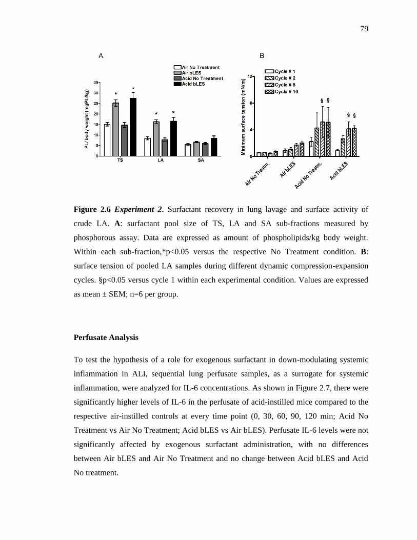
79
Figure 2.6 Experiment 2. Surfactant recovery in lung lavage and surface activity of
crude LA. A: surfactant pool size of TS, LA and SA sub-fractions measured by
phosphorous assay. Data are expressed as amount of phospholipids/kg body weight.
Within each sub-fraction,*p<0.05 versus the respective No Treatment condition. B:
surface tension of pooled LA samples during different dynamic compression-expansion
cycles. §p<0.05 versus cycle 1 within each experimental condition. Values are expressed
as mean ± SEM; n=6 per group.
Perfusate Analysis
To test the hypothesis of a role for exogenous surfactant in down-modulating systemic
inflammation in ALI, sequential lung perfusate samples, as a surrogate for systemic
inflammation, were analyzed for IL-6 concentrations. As shown in Figure 2.7, there were
significantly higher levels of IL-6 in the perfusate of acid-instilled mice compared to the
respective air-instilled controls at every time point (0, 30, 60, 90, 120 min; Acid No
Treatment vs Air No Treatment; Acid bLES vs Air bLES). Perfusate IL-6 levels were not
significantly affected by exogenous surfactant administration, with no differences
between Air bLES and Air No Treatment and no change between Acid bLES and Acid
No treatment.

80
Figure 2.7 Experiment 2. IL-6 levels measured in lung perfusate at 0, 30, 60, 90, 120
min. Data are expressed as mean ± SEM. *p<0.05 versus respective Air control at each
time point; n=6 per group.
Lung perfusate samples collected at 120 min were further analyzed for a wider array of
cytokines/chemokines. Among the 12 mediators measured (Table 2.4), IL-13 levels were
not detectable (data not shown), while there were significantly greater levels of eotaxin,
IL-6, KC, MIP-2 in acid-instilled animals compared to the respective air instilled control.
Overall, exogenous surfactant administration did not affect eotaxin, GM-CSF, IL-6, IL-
1β, KC, TNF-α and IP-10 levels, with no statistical difference between the bLES and No
Treatment group in both Air and Acid instilled mice.
A statistically significant increase of MIP-2 levels in the perfusate of Acid bLES mice
was determined compared to Acid No Treatment, as well as significantly higher perfusate
levels of G-CSF, LIX and MCP-1 in acid injured mice treated with surfactant compared
to the Air bLES.

81
Air Acid
Mediator
(pg/ml)
No
treatment bLES No treatment bLES
Eotaxin 17.4±1.3 25.7±0.9 135.2±13.6* 142.8±15.9*
G-CSF 45.9±10.5 59.4±3.4 890.8±75.8 1270.3±187.8*
GM-CSF 0 0 13.4±2.8 16.3±2.2
IL-6 567.7±62.7 1119.0±99.1 8303.2±323.6* 10720.1±764.2*
IL-1 β 1.0±0.40 0.2±0.1 0.3±0.1 3.2±0.6
KC 579.2±65.8 907.0±75.4 3617.2±174.3* 6212.7±504.8*
LIX 113.7±11.1 98.3±13.0 284.6±17.9 447.4±31.9*
MCP-1 12.9±1.8 30.9±4.9 337.4±26.2 558.0±71.5*
MIP-2 584.4±68.2 725.7±41.9 1840.2±76.7* 3113.4±204.8*,#
TNF-α 63.4±11.6 72.2±7.0 119.7±3.1 143.3±8.1
IP-10 32.9±3.6 40.8±1.7 391.3±80.8 283.3±27.7
Table 2.4 Experiment 2. Cytokine and chemokine measured in lung perfusate at the end
of MV. Data are expressed as mean ± SEM; n=6 per group. *p<0.05 versus respective
Air control, #p<0.05 versus Acid No Treatment.
G-CSF=granulocyte colony-stimulating factor, GM-CSF= granulocyte-macrophage CSF,
IL-6=interleukin-6, IP-10=interferon-γ-induced protein 10, KC=keratinocyte
chemoattractant, LIX=lipopolysaccharide-induced CXC chemokine, MCP-1=monocyte
chemotactic protein-1, MIP-2=macrophage inflammatory protein 2 and TNF-α=tumor
necrosis factor-alpha.

82
Finally, in order to further characterize the effect of exogenous surfactant administration
on lung-derived mediators in perfusate, eicosanoids levels were also measured at the 120
min. time point (Table 2.5). Although increased levels of thromboxane B2 and
prostaglandin E2 were recorded in the perfusate of acid-instilled animals compared to
their respective Air controls, these changes did not reach statistical significance.
Perfusate concentrations of 8-isoprostane were significantly higher in the acid injured
groups compared to air controls. Surfactant treatment did not affect thromboxane B2 and
8-isoprostane concentrations. Prostaglandin E2 levels were significantly elevated only in
the perfusate of Acid bLES mice compared to Air bLES controls. Leukotriene B4 levels
were increased in the perfusate of Acid bLES mice but this difference failed to be
statistically significant.
Air Acid
Mediator (pg/ml) No
treatment bLES
No
treatment bLES
Prostaglandin E2 14.3±2.0 26.8±3.9 147.4±40.3 221.7±89.2*
Leukotriene B4 14.4±5.2 10.6±5.3 13.1±7.3 41.9±12.3
Thromboxane B2 52.7±10.1 67.8±12.9 109.9±24.8 134.9±43.6
8-Isoprostane 11.3±1.6 19.7±3.0 47.2±8.1* 70.4±12.5*
Table 2.5 Experiment 2. Concentrations of prostaglandin E2, leukotriene B4,
thromboxane B2 and 8-isoprostane measured in lung perfusate samples collected at the
end of MV. Data are expressed as mean ± SEM; n=6 per group. *p<0.05 versus the
respective Air control.

83
2.4. Discussion
The overall objective of this study was to evaluate the anti-inflammatory effects of
exogenous surfactant when administered prior to mechanical ventilation, either in the
absence (experiment 1) or in the presence (experiment 2) of an initiating pulmonary
insult. For both lung injury models, the IPML setup was utilized to specifically evaluate
the contribution of ventilation to the development of systemic inflammation. MV of
normal lungs resulted in the release of IL-6 (locally) into the airspace and several
mediators (systemically) in the perfusate. Surfactant administration, however, was not
effective in reducing the systemic inflammation associated with MV. Conventional
ventilation of HCl instilled mice led to higher levels of both IL-6 and total protein in
lavage, and significantly higher levels of pro-inflammatory mediators in perfusate
without any beneficial effect of bLES instillation. Notably, significantly higher lavage
IL-6 and perfusate MIP-2 concentrations were observed in acid-injured mice receiving
bLES, compared to Acid-No Treatment controls. Based on these results, it was concluded
that administration of exogenous surfactant prior to MV does not reduce the systemic
inflammation associated with lung injury in these models.
An important feature of the current study was to examine the effects of surfactant therapy
in two different models. Analysis of the data showed important differences between the
models, such as the degree of lung edema. Mechanical stretch of uninjured lungs did not
affect lung permeability, whereas acid injured mice had increased total lavage proteins
after two hours of MV. Another aspect that distinguishes the two models is represented
by different levels of pulmonary and perfusate inflammation, which becomes particularly
evident when comparing cytokine levels measured in the perfusate of the MV only, No
Treatment group to the cytokine levels of the Acid No Treatment group. For example,
MV of normal lungs caused a moderate increase in circulating Eotaxin, IL-6, KC and
MIP-2, while acid instilled animals subjected to conventional MV had perfusate
concentrations of these mediators that were at least two times greater. Given the greater
inflammation characterizing the acid-injury model and the important role of lipid
mediators in the development and progression of lung injury [27-32], eicosanoids levels
were analyzed only on samples from experiment 2. Unambiguous conclusions about the

84
effects of exogenous surfactant on systemic inflammation were therefore inferred from
two experimental models with very different characteristics. This allowed us to rule out
possible causes for the lack of efficacy of our treatment (such as presence/lack of pre-
existing injury, specific effects of ventilation), and strengthened the understanding of the
biological response.
Exogenous surfactant administration has been extensively investigated as a potential
adjunctive therapy in acute lung injury [33-36]. The traditional approach with surfactant
treatment has been to evaluate its efficacy in terms of physiological and biophysical
improvements. Many experimental studies have in fact demonstrated that exogenous
surfactant instilled after the onset of ventilation improved oxygenation, lung volume and
compliance; moreover, it improved the surface tension reducing properties of the
surfactant recovered from lung lavage subsequent to administration [15, 37, 38].
Nevertheless, despite this encouraging experimental evidence, clinical trials showed no
improvement in mortality in surfactant treated patients even in the presence of an initial
improvement in oxygenation [16, 18]. It is possible that surfactant treatment in the
previous studies was administered too late into ALI development; therefore earlier
surfactant administration prior to or at the onset of MV could be more effective at
mitigating disease progression. Since mortality can be improved by ameliorating
ventilation – induced systemic inflammation [39], it was our interest to investigate
whether exogenous surfactant could mitigate the effects of MV thereby down-modulating
inflammation.
To our knowledge, the effect of surfactant on ventilation-induced release of inflammatory
mediators in perfusate of an IPML model has been specifically addressed in two previous
studies. Stamme and colleagues [40] showed elevated TNFα and IL-6 concentrations in
the perfusate of surfactant treated animals compared to controls, in their mouse model of
high pressure ventilation. In contrast, our group has shown a reduced level of
inflammatory cytokines in perfusate due to elevated endogenous surfactant in an LPS
model of injury [20]. Together with the current study in which surfactant did not impact
inflammation in two models of injury, these data illustrate the complexity of surfactant
treatment in which specific details of the experimental model may impact outcome.

85
Furthermore, such details are obviously important to understand in the context of a
potential clinical utilization of surfactant treatment to down-regulate systemic
inflammation as well as to understand the mechanisms by which surfactant may affect
inflammation.
Despite the lack of effect of surfactant treatment in our study, we speculate that
mitigation of MV induced inflammation is still the best approach for an early
intervention. Our data support earlier studies which showed that cytokines can be
detected in perfusate rapidly after the onset of ventilation [22, 41]. This loss of alveolar
and systemic cytokine compartmentalization can lead to peripheral organ dysfunction, a
condition of difficult clinical management. Therefore, targeting the lung with anti-
inflammatory agents prior to MV may be a successful treatment option leading to
improved outcomes. In this respect, surfactant could be utilized as a carrier for delivering
lung specific anti-inflammatory agents prior to MV in future studies.
Along with the strengths of the present study, some limitations need to be addressed. Due
to the lack of blood perfusion in the IPML setup, the lungs were not exposed during ex
vivo MV to circulating soluble factors and immune cells which could have affected the
progression of the injury. Moreover, ex vivo ventilation of perfused lungs did not favor
the use of severe lung injury models, due to potential technical failure of the preparation.
Consequently, the injury from ventilation was mild to moderate, thereby explaining the
lack of change in surface tension or surfactant pool sizes. It is believed, however, that
these limitations of the IPML setup were counter balanced by the advantage of
specifically isolating lung-derived mediators released into the circulation, without the
confounding contribution of systemic factors to the development of inflammation. Intra-
tracheal instillation was also used for administering surfactant, ensuring the presence of
large amounts of active material in the airspace before ventilation, as shown by higher
levels of TS and LA in the lung lavage of treated animals. It should be acknowledged,
however, that some inadequate distribution of surfactant might have occurred following
instillation. Obstruction of smaller airways, with consequent heterogeneous lung inflation
and regional over-distension might have been responsible for the increase in PIP
(experiment 2, Air treated groups), and for the non-significant trend towards higher
86
lavage IL-6 levels in the surfactant treated groups. Nevertheless, the instilled surfactant
retained excellent biophysical properties as shown by the very low minimum surface
tension achieved during dynamic compression-expansion of the crude LA. Overall, we
believe that instillation did not account for the lack of efficacy of our treatment.
In conclusion, this study expands the knowledge about exogenous surfactant treatment. It
specifically focuses on the anti-inflammatory effects of a lung targeted therapy
administered prior to MV on the development of systemic inflammation using two
different mouse models. Although our data suggest a lack of efficacy for exogenous
surfactant in down-modulating inflammation, future studies might focus on surfactant as
a carrier for anti-inflammatory drugs or antibiotics in order to better interfere with ALI
progression.

87
2.5. References
1. Goerke J: Pulmonary surfactant: functions and molecular composition. Biochim
Biophys Acta 1998, 1408:79-89.
2. Wright JR: Immunomodulatory functions of surfactant. Physiol Rev 1997,
77(4):931-962.
3. Lewis JF, Jobe AH: Surfactant and the adult respiratory distress syndrome. Am
Rev Respir Dis 1993, 147(1):218-233.
4. Veldhuizen RA, McCaig LA, Akino T, Lewis JF: Pulmonary surfactant
subfractions in patients with the acute respiratory distress syndrome. Am J Respir
Crit Care Med 1995, 152(6 Pt 1):1867-1871.
5. Ware LB, Matthay MA: The acute respiratory distress syndrome. N Engl J Med
2000, 342(18):1334-1349.
6. Rubenfeld GD, Caldwell E, Peabody E, Weaver J, Martin DP, Neff M, Stern EJ,
Hudson LD: Incidence and outcomes of acute lung injury. N Engl J Med 2005,
353(16):1685-1693.
7. Stapleton RD, Wang BM, Hudson LD, Rubenfeld GD, Caldwell ES, Steinberg KP:
Causes and timing of death in patients with ARDS. Chest 2005, 128(2):525-532.
8. Montgomery AB, Stager MA, Carrico CJ, Hudson LD: Causes of mortality in
patients with the adult respiratory distress syndrome. Am Rev Respir Dis 1985,
132(3):485-489.
9. Walker MG, Yao LJ, Patterson EK, Joseph MG, Cepinskas G, Veldhuizen RA, Lewis
JF, Yamashita CM: The effect of tidal volume on systemic inflammation in Acid-
induced lung injury. Respiration 2011, 81(4):333-342.

88
10. Gurkan OU, O'Donnell C, Brower R, Ruckdeschel E, Becker PM: Differential
effects of mechanical ventilatory strategy on lung injury and systemic organ
inflammation in mice. Am J Physiol Lung Cell Mol Physiol 2003, 285(3):L710-8.
11. Dhanireddy S, Altemeier WA, Matute-Bello G, O'Mahony DS, Glenny RW, Martin
TR, Liles WC: Mechanical ventilation induces inflammation, lung injury, and extra-
pulmonary organ dysfunction in experimental pneumonia. Lab Invest 2006,
86(8):790-799.
12. O'Mahony DS, Liles WC, Altemeier WA, Dhanireddy S, Frevert CW, Liggitt D,
Martin TR, Matute-Bello G: Mechanical ventilation interacts with endotoxemia to
induce extrapulmonary organ dysfunction. Crit Care 2006, 10(5):R136.
13. Ranieri VM, Suter PM, Tortorella C, De Tullio R, Dayer JM, Brienza A, Bruno F,
Slutsky AS: Effect of mechanical ventilation on inflammatory mediators in patients
with acute respiratory distress syndrome: a randomized controlled trial. JAMA
1999, 282(1):54-61.
14. Aspros AJ, Coto CG, Lewis JF, Veldhuizen RA: High-frequency oscillation and
surfactant treatment in an acid aspiration model. Can J Physiol Pharmacol 2010,
88(1):14-20.
15. Vazquez de Anda GF, Lachmann RA, Gommers D, Verbrugge SJ, Haitsma J,
Lachmann B: Treatment of ventilation-induced lung injury with exogenous
surfactant. Intensive Care Med 2001, 27(3):559-565.
16. Spragg RG, Lewis JF, Walmrath HD, Johannigman J, Bellingan G, Laterre PF, Witte
MC, Richards GA, Rippin G, Rathgeb F, Hafner D, Taut FJ, Seeger W: Effect of
recombinant surfactant protein C-based surfactant on the acute respiratory distress
syndrome. N Engl J Med 2004, 351(9):884-892.

89
17. Spragg RG, Taut FJ, Lewis JF, Schenk P, Ruppert C, Dean N, Krell K, Karabinis A,
Gunther A: Recombinant surfactant protein C-based surfactant for patients with
severe direct lung injury. Am J Respir Crit Care Med 2011, 183(8):1055-1061.
18. Davidson WJ, Dorscheid D, Spragg R, Schulzer M, Mak E, Ayas NT: Exogenous
pulmonary surfactant for the treatment of adult patients with acute respiratory
distress syndrome: results of a meta-analysis. Crit Care 2006, 10(2):R41.
19. Yamashita C, Forbes A, Tessolini JM, Yao LJ, Lewis JF, Veldhuizen RA: Protective
effects of elevated endogenous surfactant pools to injurious mechanical ventilation.
Am J Physiol Lung Cell Mol Physiol 2008, 294(4):L724-L732.
20. Walker MG, Tessolini JM, McCaig L, Yao LJ, Lewis JF, Veldhuizen RA: Elevated
endogenous surfactant reduces inflammation in an acute lung injury model. Exp
Lung Res 2009, 35(7):591-604.
21. Yu S, Harding PG, Smith N, Possmayer F: Bovine pulmonary surfactant: chemical
composition and physical properties. Lipids 1983, 18(8):522-529.
22. von Bethmann AN, Brasch F, Nusing R, Vogt K, Volk HD, Muller KM, Wendel A,
Uhlig S: Hyperventilation induces release of cytokines from perfused mouse lung.
Am J Respir Crit Care Med 1998, 157(1):263-272.
23. Bligh EG, Dyer WJ: A rapid method of total lipid extraction and purification.
Can J Biochem Physiol 1959, 37(8):911-917.
24. Duck-Chong CG: A rapid sensitive method for determining phospholipid
phosphorous involving digestion with magnesium nitrate. Lipids 1979, 14(5):492-497.
25. Vockeroth D, Gunasekara L, Amrein M, Possmayer F, Lewis JF, Veldhuizen RA:
Role of cholesterol in the biophysical dysfunction of surfactant in ventilator-induced
lung injury. Am J Physiol Lung Cell Mol Physiol 2010, 298(1):L117-L125.

90
26. Gunasekara L, Schoel WM, Schurch S, Amrein MW: A comparative study of
mechanisms of surfactant inhibition. Biochim Biophys Acta 2008, 1778(2):433-444.
27. Ngiam N, Peltekova V, Engelberts D, Otulakowski G, Post M, Kavanagh BP: Early
growth response-1 worsens ventilator-induced lung injury by up-regulating
prostanoid synthesis. Am J Respir Crit Care Med 2010, 181(9):947-956.
28. Jaecklin T, Engelberts D, Otulakowski G, O'Brodovich H, Post M, Kavanagh BP:
Lung-derived soluble mediators are pathogenic in ventilator-induced lung injury.
Am J Physiol Lung Cell Mol Physiol 2011, 300(4):L648-L658.
29. Zarbock A, Singbartl K, Ley K: Complete reversal of acid-induced acute lung
injury by blocking of platelet-neutrophil aggregation. J Clin Invest 2006,
116(12):3211-3219.
30. Stephenson AH, Lonigro AJ, Hyers TM, Webster RO, Fowler AA: Increased
concentrations of leukotrienes in bronchoalveolar lavage fluid of patients with
ARDS or at risk for ARDS. Am Rev Respir Dis 1988, 138(3):714-719.
31. Auner B, Geiger EV, Henrich D, Lehnert M, Marzi I, Relja B: Circulating
leukotriene B4 identifies respiratory complications after trauma. Mediators Inflamm
2012, 2012:536156.
32. Eun JC, Moore EE, Banerjee A, Kelher MR, Khan SY, Elzi DJ, McLaughlin NJ,
Silliman CC: Leukotriene b4 and its metabolites prime the neutrophil oxidase and
induce proinflammatory activation of human pulmonary microvascular endothelial
cells. Shock 2011, 35(3):240-244.
33. Lewis JF, Veldhuizen R: The role of exogenous surfactant in the treatment of
acute lung injury. Annu Rev Physiol 2003, 65:613-642.

91
34. Ito Y, Manwell SE, Kerr CL, Veldhuizen RA, Yao LJ, Bjarneson D, McCaig LA,
Bartlett AJ, Lewis JF: Effects of ventilation strategies on the efficacy of exogenous
surfactant therapy in a rabbit model of acute lung injury. Am J Respir Crit Care Med
1998, 157(1):149-155.
35. Brackenbury AM, Puligandla PS, McCaig LA, Nikore V, Yao LJ, Veldhuizen RA,
Lewis JF: Evaluation of exogenous surfactant in HCL-induced lung injury. Am J
Respir Crit Care Med 2001, 163(5):1135-1142.
36. Lewis JF, Goffin J, Yue P, McCaig LA, Bjarneson D, Veldhuizen RA: Evaluation of
exogenous surfactant treatment strategies in an adult model of acute lung injury. J
Appl Physiol 1996, 80(4):1156-1164.
37. Welk B, Malloy JL, Joseph M, Yao LJ, Veldhuizen AW: Surfactant treatment for
ventilation-induced lung injury in rats: effects on lung compliance and cytokines.
Exp Lung Res 2001, 27(6):505-520.
38. Rasaiah VP, Malloy JL, Lewis JF, Veldhuizen RA: Early surfactant administration
protects against lung dysfunction in a mouse model of ARDS. Am J Physiol Lung Cell
Mol Physiol 2003, 284(5):L783-L790.
39. The Acute Respiratory Distress Syndrome Network: Ventilation with lower tidal
volumes as compared with traditional tidal volumes for acute lung injury and the
acute respiratory distress syndrome. N Engl J Med 2000, 342(18):1301-1308.
40. Stamme C, Brasch F, von Bethmann A, Uhlig S: Effect of surfactant on ventilation-
induced mediator release in isolated perfused mouse lungs. Pulm Pharmacol Ther
2002, 15(5):455-461.
41. Truscott EA, McCaig LA, Yao LJ, Veldhuizen RA, Lewis JF: Surfactant protein-A
reduces translocation of mediators from the lung into the circulation. Exp Lung Res
2010, 36(7):431-439.

92
CHAPTER 3:
Lack of matrix metalloproteinase-3 in mouse models of lung injury
ameliorates the pulmonary inflammatory response in female but not in
male mice.
Valeria Puntorieri, Lynda A. McCaig, Li-Juan Yao, James F. Lewis, Cory M. Yamashita,
and Ruud A.W. Veldhuizen.

93
3.1. Introduction
Acute lung injury (ALI) is a pulmonary inflammatory disorder with a mortality of
approximately 40% [1, 2]. Clinically, ALI is defined by the presence of bilateral infiltrates
on chest radiograph, alveolar edema in the absence of cardiac failure, and hypoxemia [3,
4]. Despite a relatively simplistic clinical definition, ALI pathophysiology is complex, with
a variable disease progression, and many different insults, such as bacterial pneumonia or
gastric acid aspiration, capable of initiating lung injury [5, 6]. Within this complex disease
process, pulmonary inflammation represents a key aspect of ALI pathophysiology that is
common to all patients regardless of the initiating insult [7, 8], and which is strongly related
to outcomes of ALI [9]. Numerous experimental studies have shown that strategies capable
of modulating the lung inflammatory response greatly affect disease progression to
systemic inflammation and consequent multi-organ failure, the main cause of death in ALI
[10–15].
Clinical evidence for the importance of lung inflammation in ALI stems from clinical trials
over the past decade, which consistently demonstrated that lower indices of lung
inflammatory mediators were associated with diminished systemic inflammation, and
subsequent lower mortality [16, 17]. In these trials, the decrease in pulmonary
inflammation resulted from the use of non-injurious strategies of mechanical ventilation,
the main supportive therapy in ALI [16, 17]. Since then, however, no other lung-targeted
pharmacological or anti-inflammatory treatment has been identified that is capable of
reducing mortality [18, 19], thereby stressing the importance of further research into key
modulators of pulmonary inflammation in ALI.
One potential key mediator of pulmonary inflammation is matrix metalloproteinase-3
(MMP-3), a protease expressed in the lung by alveolar macrophages (AM), alveolar
epithelial cells, and fibroblasts [20–22]. MMP-3 is a member of the matrix
metalloproteinases (MMPs) family, which consists of over 20 extracellular, zinc-
dependent proteolytic enzymes involved in several physiological processes such as matrix
turnover, tissue repair, and inflammation [23–25]. Interestingly, metalloproteinase activity
and expression levels are often elevated in injured tissues and in inflammatory conditions
such as asthma, sepsis, and ALI [26–30]. Specifically, recent clinical studies have

94
confirmed elevated MMPs levels in lavage samples from patients with ALI; importantly,
detectable lavage levels of a specific member of this family, namely MMP-3, were
associated with severity of ALI, distal organ failure, and mortality [27, 31]. In addition to
this clinical evidence, two experimental studies suggest a potential role for MMP-3 in ALI
[32, 33]. These studies showed that, compared to wild type animals, mice lacking Mmp3
expression had lower neutrophils and total protein accumulation within the lung following
intra-tracheal instillation of the chemokine MIP-2 and in a model of immunoglobulin G
immune-complex-induced lung injury [32, 33]. Although supportive of a role for MMP-3
in ALI, these studies have utilized neutrophil-dependent models of ALI that lack clinical
relevance, since they do not resemble any of the most common initiating insults in the
pathogenesis of this disorder. It is, therefore, necessary to investigate the role of MMP-3
in clinically relevant models of ALI, with a specific focus on pulmonary inflammation, as
it appears to be a key driving factor in disease progression.
It was hypothesized that MMP-3 is a key mediator in the pathogenesis of ALI by affecting
pulmonary inflammation. To test this hypothesis, male and female wild type mice
(Mmp3+/+) and mice lacking Mmp3 expression (Mmp3-/-) were exposed to two clinically
relevant models of lung injury: i) a model of lipopolysaccharide-induced lung injury,
resembling bacterial pneumonia, and ii) a model of acid-induced ALI, which mirrors lung
injury due to gastric acid aspiration. Each model reproduces major pathophysiological
events in ALI, such as the intra-pulmonary accumulation of proteinaceous edema fluid, the
increased pulmonary release of cytokines and chemokines by resident alveolar
macrophages and lung epithelial cells, and the recruitment of inflammatory neutrophils to
the injured lung [5].
Interestingly, initial analyses showed a more prominent role of MMP-3 in female mice;
therefore, data from female mice and from male mice have been analyzed separately, as
distinct data sets. A sex-dependent role of MMP-3 in lung inflammation was observed in
both ALI models, which led to initial in vitro investigations into mechanisms and
inflammatory cell population (AM) potentially contributing to these findings. Experiments
were performed using bone marrow-derived macrophages (BMDMs) as a surrogate for
AM. Questions on possible inherent differences in the inflammatory response of

95
male/female Mmp3+/+ and Mmp3-/- BMDMs, and on the potential effect of 17β-estradiol
(E2), were preliminarily addressed in this work.
3.2. Methods
3.2.1. Animal procedures and experimental design
Mice lacking the expression of Mmp3 (Mmp3-/-) were originally generated as described by
Mudgett JS et al. [34].; breeding pairs of Mmp3-/- mice were kindly provided by Dr. G.P.
Downey (National Jewish Hospital, Denver, Colorado) to initiate and re-derive a pathogen
free colony of Mmp3+/+ and Mmp3-/- mice. All the animals were group housed in a research
animal facility, exposed daily to a 12 hours light/dark cycle and allowed unrestricted access
to standard chow food and water.
A total of 109 female and male Mmp3+/+ and Mmp3-/- mice were utilized for three separate
experiments – a model of lipopolysaccharide-induced lung injury (experiment 1), a model
of acid-induced lung injury (experiment 2), and BMDMs isolation (experiment 3) – and all
animal procedures were approved by the Animal Use Subcommittee at Western University
(protocol number: 2010-272).
3.2.2. Experiment 1: Lipopolysaccharide induced lung injury
A total of 47 Mmp3+/+ and Mmp3-/-, female and male mice (13.9 ± 0.4 weeks old; average
± sem) were randomized to receive an intra-tracheal (i.t.) instillation of lipopolysaccharide
(LPS from E.Coli, 0111:B4, 20 µg/mouse; Sigma, St. Louis, Mo., USA) or saline (0.15M
NaCl solution) as a control for this model. Considering that the data were analyzed by sex,
for both female and male mice the following 4 experimental groups were obtained: i)
Mmp3+/+ Saline, ii) Mmp3+/+ LPS, iii) Mmp3-/- Saline, iv) Mmp3-/- LPS.
Mice were anesthetized with an intraperitoneal injection of ketamine (100 mg/kg; Sandoz,
Quebec, Que., Canada) and medetomidine (1 mg/kg; Orion Corporation, Espoo, Finland).
Once adequate sedation was achieved, mice were placed dorsally on a vertical, slightly
96
inclined stand, and the oral cavity was opened to visualize the vocal cords. Subsequently,
a 20-gauge catheter combined with a fiber-optic stylet (BioLite intubation system for small
rodents, BioTex, Inc., Houston, Tex., USA) was inserted through the vocal cords into the
trachea. Mice randomized to the LPS group received 40 µL of a 0.5 mg/mL LPS solution
in sterile saline (0.15M NaCl), instilled drop-wise through the endotracheal tube. Mice
randomized to the Saline group were intubated and received 40 µL of a sterile 0.15M NaCl
solution. The whole procedure required approximately 15 minutes. Mice were then
extubated, injected subcutaneously with 0.5 mL sterile saline and immediately returned to
their cage. Preliminary studies performed during development of the model determined
that mice did not require any analgesia following instillation. Within 30 minutes from the
injection of the anesthetic mix, mice received an intra-peritoneal injection of atipamezole
(1 mg/kg; Zoetis, Florham Park, NJ, USA), the reversal agent for medetomidine. Mice were
left to recover for a total of 18 hours with free access to water and food. At the end of the
18 hours, mice were sacrificed with an intraperitoneal injection of sodium pentobarbital
(110 mg/kg; Lundbeck, Valby, Denmark) followed by exsanguination, and processed for
various analyses as described in sections 3.2.4 to 3.2.7.
3.2.3. Experiment 2: Acid-induced lung injury
A total of 44 Mmp3+/+ and Mmp3-/-, female and male mice (12.7 ± 0.5 weeks old) were
randomized to receive an intra-tracheal instillation of hydrochloric acid (HCl) or air as a
control. Similar to Experiment 1, the data from each sex were analyzed as different data
sets. For both female and male mice, the following 4 experimental groups were analyzed:
i) Mmp3+/+ Air, ii) Mmp3+/+ Acid, iii) Mmp3-/- Air, iv) Mmp3-/- Acid.
An intraperitoneal injection of ketamine (100 mg/kg; Sandoz, Quebec, Que., Canada) and
xylazine (5 mg/kg; Bayer, Toronto, Ont., Canada) was given to anesthetize mice and
subsequently proceed with the endotracheal intubation as described above (under
‘lipopolysaccharide-induced lung injury’). Mice randomized to acid instillation received
50 µL of 0.05N HCl, instilled intra-tracheally in a drop-wise fashion. Mice randomized to
the air group were intubated as described and allowed to spontaneously breathe through
the tube. Overall, the procedure lasted about 15 minutes. Mice were then extubated, put on
an inclined stand and injected subcutaneously with buprenorphine (0.05-0.1 mg/kg

97
Animalgesic Labs Inc., Millersville, MD, USA) and 0.3 mL sterile saline. Mice were then
returned to their cage and 30 minutes after the injection of anesthetic, they were injected
intra-peritoneally with atipamezole (1 mg/kg), reversal agent for xylazine.
Mice were allowed to recover for 4 hours, with free access to food and water, and sacrificed
at the end of the recovery period with an intraperitoneal injection of a ketamine (200
mg/kg)-xylazine (10 mg/kg) solution followed by exsanguination.
3.2.4. Lung lavage isolation and total protein analysis
At the end of their respective recovery periods (18 hours for LPS-induced lung injury, 4
hours for acid-induced lung injury), mice were given a euthanizing dose of anesthetic as
previously described, and placed in a supine position. When deep anesthesia was attained,
the abdominal content and thorax were exposed through an abdominal midline incision,
and exsanguination was performed through excision of the inferior vena cava. A
tracheostomy tube was subsequently inserted and secured in the trachea, the diaphragm cut
and the chest wall opened via a midline incision to expose the lungs. Lungs were then
lavaged with 3 × 1 mL aliquots of saline, and each aliquot was instilled and withdrawn 3
times. The total volume of lavage fluid collected from each mouse was recorded. Lung
lavage was immediately centrifuged at 380 g for 10 minutes at 4˚C to isolate the cell pellet;
the supernatant was collected and 4 × 0.25 mL aliquots were frozen at -80˚C for analyses
of inflammatory mediators.
Measurements of total protein content in lavage were also performed on this supernatant
using a Micro BCA protein assay kit (Pierce, Rockford, Ill., USA), as per manufacturer’s
instructions.
3.2.5. Lavage cell analysis
Following centrifugation of lavage samples, the cellular component was isolated and
resuspended in Plasmalyte (300 to 500 µL depending on cell density). An aliquot of cell
suspension was mixed with an equal volume of trypan blue to assess viability through
trypan blue exclusion, and subsequently utilized to determine total cell counts with a
hemocytometer and light microscopy. Aliquots of the resuspended cell pellet were also

98
spun down on cytospin slides at 1000 rpm for 6 minutes at room temperature, stained with
Hemacolor® stain (Harleco, EMD Chemicals Inc., Gibbstown, NJ, USA) and utilized to
perform differential cell counts under light microscopy. Specifically, 5 fields were counted
and averaged for each slide, and the relative percentage of each inflammatory cell type was
calculated. The absolute number of neutrophils in lavage was then obtained by multiplying
percentages by the total cell number previously determined.
3.2.6. Measurements of MMP-3 and inflammatory mediators in lung lavage
Total MMP-3 levels were measured in lavage aliquots from experiment 1 and 2, using a
commercially available enzyme-linked immune-sorbent assay (ELISA) according to
manufacturer’s instructions (R&D Systems, Minneapolis, MN, USA).
Lavage interleukin-6 (IL-6) levels were also assessed through the use of a mouse IL-6
ELISA (BD Biosciences, San Diego, Ca., USA) according to manufacturer’s instructions.
An array of inflammatory cytokines and chemokines was measured in lung lavage samples
from both injury models. For both experiment 1 and 2, mouse Milliplex multi-analyte
panels (Map) (MCYTOMAG-70K, Millipore Corporation, Billerica, MA, USA) were
utilized to measure lavage levels of the following cytokine/chemokine: granulocyte colony
stimulating factor (G-CSF), interleukin 10 (IL-10), keratinocyte chemoattractant (KC),
monocyte chemotactic protein 1 (MCP-1), macrophage inflammatory protein 2 (MIP-2),
and tumor necrosis factor alpha (TNF-α). Samples analysis was performed on the
Luminex® xMAP® detection system on the Luminex100 (Linco Research, St. Charles, Mo.,
USA), as per manufacturer’s protocol.
3.2.7. Experiment 3: Isolation of Mmp3+/+ and Mmp3-/- bone marrow-derived
macrophages
Bone marrow-derived macrophages (BMDMs) were isolated from the femurs and tibias of
mice as previously described [35]. Briefly, a total of 18 Mmp3+/+ and Mmp3-/- mice (19.9
± 1.7 weeks old) were sacrificed with an intraperitoneal injection of sodium pentobarbital
(110 mg/kg) and exsanguinated. The femur and tibia of both hind legs were isolated, the
soft tissue was removed, and the ends of each bone were cut off before placing each pair

99
of long bones in a 0.65 mL Eppendorf tube with a punctured bottom. Each one of these
tubes was then placed into a 1.5 mL Eppendorf, thereby allowing for the collection of the
bone marrow cell pellet upon centrifugation of the bones at 2000 g for 1 minute at 4˚C.
After lysis of red blood cells and subsequent wash with cold, sterile 1x PBS, bone marrow
samples from each mouse were pulled together and centrifuged at 400 g, for 10 minutes,
4˚C, and the resulting cell pellet was then resuspended in macrophage differentiation media
(Mac media: RPMI 1640, 10% fetal bovine serum, 2 mM glutamine, 50 units/mL
penicillin, 50 µg/mL streptomycin, and 30% L-cell supernatant as a source of monocyte-
colony stimulating factor). Bone marrow-derived cells were seeded and cultured in a 100
mm Petri dish at 37˚C with 5% CO2 for 24 hours; at the end of this time, the non-adherent
and loosely adherent cells were transferred to a 150 mm tissue culture dish with fresh Mac
media, and cultured as previously described. Following a 7 days culture period, cells were
fully differentiated into BMDMs and ready for experimental use.
All reagents used for the culture of bone marrow derived macrophages were provided by
Gibco® (Life Technologies, Grand Island, NY, USA), unless otherwise specified. L-cell
supernatant was a generous gift of Dr. Gill (Western University, London ON, Canada).
3.2.8. In vitro stimulation of Mmp3+/+ and Mmp3-/- bone marrow-derived
macrophages
The effects of LPS or a combination of LPS and estrogen were assessed in vitro on BMDMs
isolated as described above. Fully differentiated BMDMs from male and female Mmp3+/+
and Mmp3-/- mice were harvested and 2×10⁵ cells per well were seeded in a 24 well plate.
The following day, macrophage differentiation media was substituted with stimulation
media (RPMI 1640, 10% fetal bovine serum, 2 mM glutamine, 50 units/mL penicillin, 50
µg/mL streptomycin) and BMDMs were exposed to one of the following conditions: i)
control – media only, ii) 0.1µg/mL LPS (E.Coli, 0111:B4, Sigma, St. Louis, Mo., USA),
iii) 1µg/mL LPS, iv) 10µg/mL LPS. BMDMs were stimulated at 37˚C, 5% CO2 for 18
hours. At the end of the stimulation period, individual cell supernatants were collected,
spun at 380 g for 10 minutes at 4˚C and frozen at -80 ˚C for subsequent analysis of IL-6
levels. BMDMs from the stimulated 24 well plate were lysed on ice in lysis buffer (0.5%
Sodium dodecyl sulfate, 50 mM Tris pH 7.5, 1 mM EDTA) with protease inhibitor

100
(cOmplete, Mini, EDTA-free, Roche Diagnostic GmbH, Mannheim, Germany) for
measurements of total protein content (Micro BCA protein assay kit, Pierce, Rockford, Ill.,
USA). IL-6 levels of each individual cell supernatant were then normalized by the
corresponding total protein content, to account for differences in total cell numbers
between wells. A total of three independent experiments were performed.
In a second set of experiments, differentiated BMDMs from female Mmp3+/+ and Mmp3-/-
mice were harvested and 2×10⁵ cells per well were seeded in a 24 well plate to be
stimulated in the presence of 17-β estradiol (E2, Sigma, St. Louis, Mo., USA). On the next
day, stimulation media was utilized to expose BMDMs to one of the following conditions:
i) control media (with vehicle), ii) 0.1nM E2, iii) 1nM E2, iv) 10nM E2, v) 1µg/mL LPS,
vi) 0.1nM E2 + 1µg/mL LPS, vii) 1nM E2 + 1µg/mL LPS, and viii) 10nM E2 + 1µg/mL
LPS. Following 18 hours, cell supernatants were collected for IL-6 analysis and total
protein content was assessed on cell lysates, as previously described. A total of three
independent experiments were performed.
3.2.9. Statistical analysis
All data are expressed as mean ± standard error of the mean (SEM). GraphPad Prism
statistical software (La Jolla, CA, USA) was utilized to perform statistical analyses. Data
were analyzed using a two-way ANOVA (variables: genotype and treatment) followed by
a one-way ANOVA with a Tukey’s post-hoc test. When appropriate, an unpaired two tailed
Student’s t-test was utilized for analysis. P<0.05 was considered statistically significant.

101
3.3. Results
3.3.1. Experiment 1: Lipopolysaccharide-induced lung injury
Lavage MMP-3
The role of MMP-3 in ALI was first assessed utilizing an 18-hour model of LPS-induced
lung injury, in which Mmp3+/+ and Mmp3-/- mice of both sexes received an intra-tracheal
administration of LPS or saline as a control.
Total MMP-3 was measured in lavage samples from Mmp3+/+ mice. The results in figure
3.1A show significantly higher lavage MMP-3 concentrations in LPS-injured mice
compared to Saline controls. Given the use of both female and male mice for this study,
the data was subsequently analyzed by sex. Notably, female Mmp3+/+ mice showed
significantly elevated total MMP-3 following LPS injury compared to Saline (Fig. 3.1B);
however MMP-3 concentrations in male, LPS-instilled Mmp3+/+ mice were not
significantly higher than the respective Saline control (Fig. 3.1C). In light of this difference
in results from female and male mice, the remaining of the experimental data was analyzed
separately by sex.

102
Figure 3.1 Experiment 1: MMP-3 levels measured in lung lavage samples from Mmp3+/+
mice, 18 hours after intra-tracheal saline or LPS instillation. A: Lavage MMP-3 levels
assessed in all Mmp3+/+ mice, n=11-12 per group; B: in female Mmp3+/+ mice, n=5-6 per
group; C: in male Mmp3+/+ mice, n=6 per group. Data are expressed as mean ± SEM;
*p<0.05 vs Saline control.
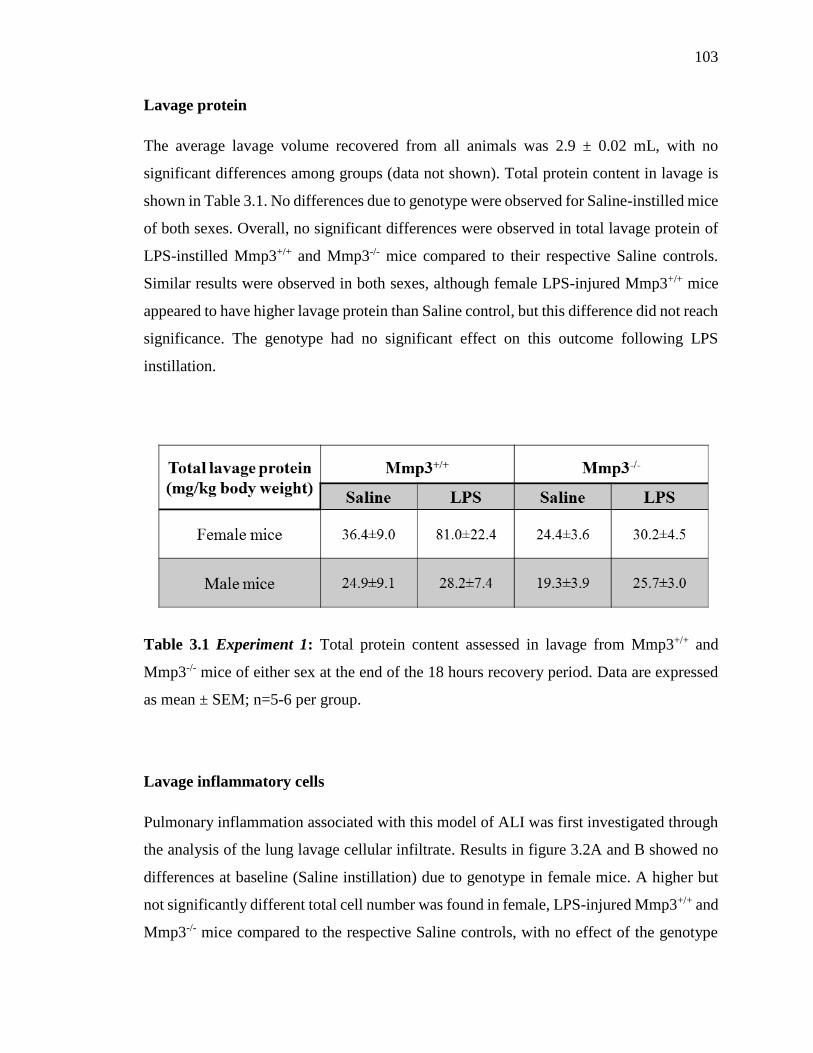
103
Lavage protein
The average lavage volume recovered from all animals was 2.9 ± 0.02 mL, with no
significant differences among groups (data not shown). Total protein content in lavage is
shown in Table 3.1. No differences due to genotype were observed for Saline-instilled mice
of both sexes. Overall, no significant differences were observed in total lavage protein of
LPS-instilled Mmp3+/+ and Mmp3-/- mice compared to their respective Saline controls.
Similar results were observed in both sexes, although female LPS-injured Mmp3+/+ mice
appeared to have higher lavage protein than Saline control, but this difference did not reach
significance. The genotype had no significant effect on this outcome following LPS
instillation.
Table 3.1 Experiment 1: Total protein content assessed in lavage from Mmp3+/+ and
Mmp3-/- mice of either sex at the end of the 18 hours recovery period. Data are expressed
as mean ± SEM; n=5-6 per group.
Lavage inflammatory cells
Pulmonary inflammation associated with this model of ALI was first investigated through
the analysis of the lung lavage cellular infiltrate. Results in figure 3.2A and B showed no
differences at baseline (Saline instillation) due to genotype in female mice. A higher but
not significantly different total cell number was found in female, LPS-injured Mmp3+/+ and
Mmp3-/- mice compared to the respective Saline controls, with no effect of the genotype

104
(Fig. 3.2A). Similarly, determination of total PMN cells in lavage showed a non-significant
trend toward higher neutrophils in female Mmp3+/+ and Mmp3-/- mice exposed to LPS,
with no differences due to genotype (Fig. 3.2B).
In male mice, no differences due to genotype were found in the total number of cells (Fig.
3.2C) and of PMN (Fig. 3.2D) in the lavage of Saline-instilled controls. The increase in
lavage cell number was statistically significant in male Mmp3+/+ and Mmp3-/- mice exposed
to LPS-induced lung injury compared to controls, with no effect of the genotype (Fig.
3.2C). Male mice of both genotypes had also significantly higher lavage numbers of
neutrophils following LPS-induced lung injury compared to Saline controls, with no effect
stemming from lack of Mmp3 expression (Fig. 3.2D).

105
Figure 3.2 Experiment 1: Total (A, C) and differential (B, D) cell counts performed in
lavage samples 18 hours post Saline or LPS intra-tracheal instillation. Total lavage cell
numbers were determined in Mmp3+/+ and Mmp3-/- female (A) and male (C) mice.
Following differential cell counts performed on cytospin slides, the total number of PMN
neutrophils in lavage was obtained for female (B) and male (D) mice of both genotypes.
Data are expressed as mean ± SEM; n=5-6 per group. *p<0.05 vs Saline control within the
genotype.

106
Lavage inflammatory mediators
To further characterize the role of MMP-3 in the pulmonary inflammatory response
associated with ALI, the concentration of several cytokines and chemokines was measured
in lung lavage. Data from Mmp3+/+ and Mmp3-/- female mice are shown in figure 3.3A to
F, while results for male mice are shown in figure 3.4A to F. Overall, lavage concentrations
of cytokines and chemokines did not differ between genotypes of Saline-instilled mice
from both sexes (Figures 3.3 & 3.4).
In female mice, LPS instillation led to significantly higher lavage concentrations of MIP-
2 in Mmp3+/+ mice compared to Saline controls. Significantly elevated lavage
concentrations of IL-6, G-CSF, KC, MCP-1, and TNF-α were found in female, LPS-
instilled Mmp3+/+ and Mmp3-/- mice compared to the respective Saline controls (Fig. 3.3A-
F). Significantly lower lavage concentrations of IL-6, G-CSF, MIP-2, TNF-α, were
observed in female, LPS-injured Mmp3-/- mice compared to the LPS-instilled Mmp3+/+
group, while differences in these experimental groups did not reach significance for the
KC concentration (Fig. 3.3A-F). No differences were found for IL-10 (data not shown).
The same array of inflammatory mediators was measured in the lavage of male mice and
showed that LPS-induced injury caused significant increases in IL-6, G-CSF, KC, MIP-2,
and TNF-α in Mmp3+/+ and Mmp3-/- mice compared to Saline mice, with no effect of the
genotype (Fig. 3.4A-F) . No differences were detected in the lavage concentrations of
MCP-1 (Fig. 3.4D) and IL-10 (data not shown).

107
Figure 3.3 Experiment 1: Cytokine and chemokine levels measured in lung lavage from
Mmp3+/+ and Mmp3-/- female mice (A-F), 18 hours after LPS or saline instillation. Data
are expressed as mean ± SEM; n=5-6 per group. *p<0.05 vs Saline control within the
genotype; #p<0.05 vs Mmp3+/+ LPS group.
IL-6= interleukin-6, G-CSF= granulocyte colony stimulating factor, KC= keratinocyte
chemoattractant, MCP-1= monocyte chemotactic protein-1, MIP-2= macrophage
inflammatory protein 2, and TNF-α= tumor necrosis factor alpha.

108
Figure 3.4 Experiment 1: Cytokine and chemokine levels measured in lung lavage from
Mmp3+/+ and Mmp3-/- male mice (A-F), 18 hours after LPS or saline instillation. Data are
expressed as mean ± SEM; n=5-6 per group. *p<0.05 vs Saline control within the genotype;
#p<0.05 vs Mmp3+/+ LPS group.
IL-6= interleukin-6, G-CSF= granulocyte colony stimulating factor, KC= keratinocyte
chemoattractant, MCP-1= monocyte chemotactic protein-1, MIP-2= macrophage
inflammatory protein 2, and TNF-α= tumor necrosis factor alpha.

109
3.3.2. Experiment 2: Acid-induced lung injury
Lavage MMP-3
The role of MMP-3 in ALI was also investigated in a model of acid-induced lung injury,
in which female and male Mmp3+/+ and Mmp3-/- mice were exposed to an intra-tracheal
instillation of HCl or Air, and sacrificed after a period of 4 hours. Similar to Experiment 1,
the data have been separated by sex.
Analysis of total lavage MMP-3 levels (Fig. 3.5) showed that acid injury led to significantly
higher MMP-3 levels in the lavage of female and male Mmp3+/+ mice (Fig. 3.5A, B),
compared to the respective Air controls.
Figure 3.5 Experiment 2: MMP-3 levels measured in lavage samples from female (A) and
male (B) Mmp3+/+ mice, 4 hours after intra-tracheal instillation of HCl or Air as a control.
Data are expressed as mean ± SEM; n=5-6 per group. *p<0.05 vs Air control.
Lavage protein
The average lavage recovery volume from all animals was 2.9 ± 0.01 mL, with a significant
difference only observed in male acid-instilled Mmp3-/- mice compared to Mmp3-/- Air
control (2.99 ± 0.55 mL versus 2.81 ± 0.03 mL, respectively). Total protein content was
measured in the lavage of female and male, Mmp3+/+ and Mmp3-/- mice (Table 3.2). When
comparing Air-instilled mice within each sex, no differences in total protein content were

110
observed due to genotype. Overall, total protein content was significantly higher in mice
exposed to acid-induced ALI compared to Air controls. These results were observed in
both female and male mice, with no effect of the Mmp3 genotype (Table 3.2).
Table 3.2 Experiment 2: Total protein content measured in lavage samples from female
and male mice of both genotypes. Data are expressed as mean ± SEM; n=5-6 per group.
*p<0.05 vs Air control within the genotype.
Lavage inflammatory cells
The total number of neutrophils in the lavage of Air-instilled mice did not differ between
genotypes in both female (Fig. 3.6A) and male mice (Fig. 3.6B). The results in figure 3.6A
showed a significantly higher number of neutrophils in female, acid-injured Mmp3+/+ mice
compared to Air control. Nonetheless, the increase in lavage neutrophil numbers in female,
acid-injured Mmp3-/- mice was not significant compared to the respective Air mice. Lower
neutrophil abundance was observed in the lavage of female, acid-injured Mmp3-/- mice
compared to Mmp3+/+ counterpart (Fig. 3.6A); however this difference did not reach
significance.
In male mice the increases in lavage neutrophils following acid injury were not statistically
significant, and no effect stemmed from the difference in genotype (Fig. 3.6B).

111
Figure 3.6 Experiment 2: Total number of PMN neutrophils in lavage samples from
female (A) and male (B) mice of both genotypes, 4 hours post air or acid instillation. Data
are expressed as mean ± SEM; n=5-6 per group. *p<0.05 vs Air control.

112
Lavage inflammatory mediators
Lavage concentrations of inflammatory mediators from Air-instilled mice of either sex did
not show any difference due to genotype (Figures 3.7 & 3.8).
In female mice (Fig. 3.7A-F), acid instillation led to significant increases in lavage IL-6,
G-CSF, KC, MCP-1, MIP-2, and TNF-α in Mmp3+/+ mice compared to Mmp3+/+ Air mice;
however in acid-injured Mmp3-/- mice such increases only reached significance for MIP-2
and TNF-α (Fig 3.7A-F) compared to Air controls. Although non-significant, female acid-
injured Mmp3-/- mice appeared to have lower lavage concentrations of IL-6, G-CSF, KC,
MCP-1, and had significantly lower concentrations of MIP-2 and TNF-α compred to acid-
instilled Mmp3+/+ mice (Fig 3.7A-F). IL-10 levels were increased following acid injury,
with no effect of the genotype (data not shown).
Analysis of the same inflammatory mediators in the lavage of male mice showed
significantly higher concentrations of IL-6, MCP-1, and TNF-α in acid-instilled Mmp3+/+
mice compared to Air controls (Fig 3.8A-F). Significant increases in IL-6, MCP-1, G-CSF,
KC, and MIP-2 were also observed in acid-injured Mmp3-/- mice compared to Air-instilled
controls (Fig 3.8A-F). No differences were observed due to the Mmp3 genotype in male
mice (Fig 3.8A-F) following acid injury; moreover, no effect of the genotype was noticed
in IL-10 lavage levels (data not shown).

113
Figure 3.7 Experiment 2: Cytokine and chemokine levels in lavage samples from Mmp3+/+
and Mmp3-/- female mice (A-F), 4 hours following acid injury or air instillation. Data are
expressed as mean ± SEM; n=5-6 per group. *p<0.05 vs Air control within the genotype;
#p<0.05 vs Mmp3+/+ Acid group.
IL-6= interleukin-6, G-CSF= granulocyte colony stimulating factor, KC= keratinocyte
chemoattractant, MCP-1= monocyte chemotactic protein-1, MIP-2= macrophage
inflammatory protein 2, and TNF-α= tumor necrosis factor alpha.

114
Figure 3.8 Experiment 2: Cytokine and chemokine levels in lavage samples from Mmp3+/+
and Mmp3-/- male mice (A-F), 4 hours following acid injury or air instillation. Data are
expressed as mean ± SEM; n=5-6 per group. *p<0.05 vs Air control within the genotype;
#p<0.05 vs Mmp3+/+ Acid group.
IL-6= interleukin-6, G-CSF= granulocyte colony stimulating factor, KC= keratinocyte
chemoattractant, MCP-1= monocyte chemotactic protein-1, MIP-2= macrophage
inflammatory protein 2, and TNF-α= tumor necrosis factor alpha.

115
3.3.3. Experiment 3: In vitro stimulation of Mmp3+/+ and Mmp3-/- bone
marrow-derived macrophages
In order to investigate the observed MMP-3-related sex differences in pulmonary
inflammation, inflammatory cells such as bone marrow-derived macrophages were isolated
from female and male Mmp3+/+ and Mmp3-/- mice. Stimulation of these BMDMs with
media only as a control or increasing doses of LPS (0.1µg/mL, 1µg/mL, 10µg/mL) led to
step-wise increases in IL-6 in cell culture supernatant, with no significant differences
related to the sex of the mice from which the cells were derived, or Mmp3 genotype (Fig.
3.9A).
A potential role of 17β-estradiol (E2) in the MMP-3 related differences in inflammatory
response was addressed in BMDMs from female Mmp3+/+ and Mmp3-/- mice. The results
in figure 3.8B showed that stimulation of BMDMs with 1µg/mL LPS caused increased
release of IL-6 in cell culture supernatant, with no additional effect stemming from E2 co-
stimulation, and no differences associated with lack of Mmp3 expression (Fig. 3.9B).

116
Figure 3.9 Experiment 3: Stimulation of BMDMs isolated from Mmp3+/+ and Mmp3-/-
mice. (A) BMDMs from female and male mice of both genotypes were stimulated with
different doses of LPS (E. Coli 0111:B4) for 18 hours. IL-6 measurements in cell culture
supernatant were normalized by values for female Mmp3+/+ BMDMs at 0.1µg/mL LPS.
(B) BMDMs from female Mmp3+/+ and Mmp3-/- mice were stimulated for 18 hours with
1µg/mL LPS alone, or LPS and three different doses of E2. IL-6 values in cell culture
supernatant were normalized by values from female Mmp3+/+ BMDMs at 1µg/mL LPS. In
both experiments, release of IL-6 in the control conditions was minimal and therefore not
visible on the graphs. Data are expressed as mean ± SEM and representative of three
experiments performed on three independent cell isolations.

117
3.4. Discussion
The overall objective of this study was to investigate the role of MMP-3 in the
inflammatory response associated with ALI. To this end, models of LPS- (experiment 1)
and acid-induced lung injury (experiment 2) were utilized in mice lacking the expression
of Mmp3, and in wild type controls. Consistent with previous experimental and clinical
observations of increased MMPs expression during injury and inflammation [23, 24, 36],
total MMP-3 levels were found elevated in the lavage of Mmp3+/+ mice following LPS or
acid injury. In our models, lack of Mmp3 expression did not alter baseline responses in any
of the measured outcomes. Following injury, lack of Mmp3 expression did not affect total
protein in lavage and did not significantly alter the recruitment of neutrophils to the injured
lung. Importantly, however, the release of cytokines and chemokines in response to the
different injuries appeared to be consistently mitigated by the genetic ablation of Mmp3 in
female mice, while male LPS- and acid-injured Mmp3-/- mice showed concentrations of
pulmonary inflammatory mediators comparable to male injured Mmp3+/+ mice. Based on
these data, it is concluded that MMP-3 plays a role in the pathogenesis of ALI by affecting
pulmonary inflammation in a sex-dependent fashion.
The role of MMP-3 in lung injury has been previously investigated in neutrophils-
dependent models of ALI, in which mice lacking Mmp3 expression demonstrated lower
neutrophils infiltration and lower intra-pulmonary accumulation of proteinaceous edema
in response to injury [32, 33]. Our data did not fully replicate those findings, due in part to
differences in the nature of the injuries and their development, as well as potential technical
limitations related to the retrieval of activated inflammatory cells and the sensitivity of the
cell counting technique. In contrast to those previous studies, our experiments utilized
mouse models that resembled two of the most common causes of lung injury in the patient
population [5, 37]. Furthermore, in light of the association between lung cytokines/
chemokines and mortality in ALI [9, 38], the present research extended the previous
observations on inflammation by analyzing the concentrations of several pulmonary
inflammatory mediators. Overall, this study has investigated outcomes that are relevant in
the pathogenesis of ALI, such as the development of pulmonary inflammation, and has
explored sex differences in the response to lung insults. The current work, therefore,

118
provides an overall stronger clinical correlate about the role of MMP-3 in ALI than
previously assessed.
An unanticipated but interesting outcome of the current study were the MMP-3 related sex
differences in the inflammatory response. Of interest, the sex of the mice previously
utilized to investigate MMP-3 in ALI was not clearly reported in those studies [32, 33]. In
a general sense, therefore, our findings underscore the upcoming policy changes announced
by the National Institue of Health, which recommend the inclusion of both sexes in
experimental studies [39]. Such changes have been prompted by the observation that the
almost exclusive use of male mice in research may have led to an under-estimation of sex-
driven differences in pathophysiology, and may have negatively impacted the translation
of experimental findings into clinical practice [39]. Recent studies however started to
demonstrate marked sex differences in the inflammatory response, with females mounting
a better and more pronounced immune response to viral and bacterial infections, and being
substantially more inclined to develop autoimmune disorders than males [40, 41]. In terms
of pulmonary pathophysiology, sex differences have been observed in patients affected by
cystic fibrosis (CF) or chronic obstructive pulmonary disease (COPD). Specifically,
prepubertal female patients with CF have a higher mortality rate than peers of the opposite
sex; females also exhibit higher prevalence and risk of hospitalization due to COPD than
males [42, 43]. In patients with ALI, sex differences in mortality have not been observed
[2]; however, a clinical study by Heffernan et al. indicated that, following traumatic injury,
female patients are more likely to develop ALI [44].
The underlying pathology leading to sex differences in the inflammatry responses in
general may encompass gonadal hormones, sex chromosomes, and anatomical differences
[45]. In the context of the current study, our in vivo models of ALI demonstrated that a lack
of Mmp3 dampened the concentrations of lavage inflammatory mediators only in female
mice, with no effect in males. These differences could be related to possible inflammatory
dissimilarities inherent to the response of male/female Mmp3+/+ and Mmp3-/- mice,
however there is also evidence of a potential relationship between sex hormones,
specifically 17β-estradiol, and MMP-3. Different studies have in fact shown that exposure
to E2 leads to up-regulation of Mmp3 expression by primary human osteoblasts, rat uterine

119
stromal cells, and human synovial fibroblasts [46–48]. These observations prompted our
preliminary in vitro investigation into the sex-differences in the MMP-3-related
inflammatory response, as shown in experiment 3. Based on the evidence of Mmp3
expression in AM [20], and the role of AM in the progression and resolution of lung injury
[49, 50], two experiments were performed on isolated BMDM to evaluate: i) inherent
differences in the macrophage-driven inflammatory response of male/female Mmp3+/+ and
Mmp3-/- mice; and ii) the possibility of an hormonal influence on the MMP-3 related sex
differences. The data suggested that, with respect to IL-6 secretion, there were no inherent
sex-related differences between female and male derived BMDM in response to LPS and/
or 17β-estradiol.
Although those preliminary results suggest that BMDMs do not demonstrate sex
differences, these experiments have some limitations and further research is required to
elucidate the specific mechanisms by which the MMP-3-related sex-differences occur. For
example, even though BMDMs provide a convenient, readily available and widely
accepted in vitro model for alveolar macrophages [51, 52], some evidence suggests that
AM may have properties and patterns of gene expression divergent from the ones
characterizing BMDMs [53]. There is, of course, also the possibility that such MMP-3
related sex differences do not involve the AM, but rather other cell populations within the
lung (i.e. alveolar type II cells, fibroblasts) or more complex, systemic interactions. Future
studies may address this aspect through broader in vitro analyses. Additionally,
ovariectomized Mmp3+/+ and Mmp3-/- mice would be a useful model to determine whether
estrogen- and, more generally speaking, sex hormones- have a role in MMP-3 related
differences in the inflammatory response.
As mentioned, the use of two different models of ALI is a strength of the study and an
important aspect in the analysis of MMP-3 role. The rationale for such experimental
approach resides, firstly, in the complex pathophysiology of ALI, which is associated with
multiple and different initiating insults [5, 54]. Secondly, various models of ALI could help
understand whether the role of MMP-3 differs depending on the lung insult. There is indeed
some evidence that other MMPs, such as MMP-8 and -9, may have roles in ALI that vary
depending on the nature of the initiating insult. For example, studies on Mmp9 knock-out

120
mice have shown that MMP-9 plays a pathogenic role during immunoglobulin G immune-
complex-induced lung injury [32], but appears to be protective from the development of
ventilation-induced lung injury (VILI). Mice lacking Mmp9 expression were in fact more
susceptible to the effects of injurious mechanical ventilation than wild type controls,
presenting significant impairment in lung function and greater tissue damage [55].
Similarly, studies on MMP-8 have demonstrated that this protease has a protective role in
models of LPS- and hyperoxia-induced lung injury while, on the other hand, it appears to
favor the development of VILI [56, 57]. Overall, what emerges is the possibility that the
role of matrix metalloproteinases in the pathogenesis of ALI may be dependent on the
aetiology of lung injury. This evidence, combined with the multiplicity of causes leading
to lung injury, undercores the necessity to perform ALI research through different injury
models.
In this regard, our ALI models are characterized at different time points (18 hours in
experiment 1, 4 hours in experiment 2), and by different initiating insults with dissimilar
features. Intra-tracheal instillation of LPS (experiment 1) has been described to cause the
activation of specific intra-cellular inflammatory pathways, with patchy areas of
neutrophils infiltration and limited epithelial damage [58], as demonstrated by the mild
changes in lung permeability in LPS-injured mice observed in experiment 1 (table 3.1).
Conversely, intra-tracheal instillation of HCl (experiment 2) resembles more of a chemical
burn, and the subsequent lung injury was associated with damage of the alveolar
epithelium, neutrophil infiltration, and extensive pulmonary edema as reflected by the
significant changes in total protein content shown in table 3.2. In light of such differences,
it is noteworthy that the involvement of MMP-3 in pulmonary inflammation in female mice
was observed consistently in both models, thereby strengthening our conclusions.
This important observation of reduced lavage cytokine and chemokine concentrations
following lung injury in female Mmp3-/- mice has potential clinical relevance. The
possibility of targeting MMP-3 to lower pulmonary levels of inflammation could improve
outcomes of ALI. Within the pathophysiology of this disorder, the development of an
overwhelming inflammatory response is indeed predictive of poor outcome [38, 59].
Experimental and clinical studies have shown that lung-derived inflammatory mediators

121
de-compartmentalized into the systemic circulation can affect the function of peripheral
organs, with progression to multi organ failure and, eventually, death [10, 13, 60]. In this
sense, a therapeutic approach capable of modulating the inflammation associated with lung
injury would be an ideal treatment, and our study is a first step to assess whether MMP-3
could be such key target.
Aside from the aforementioned strengths, our study also has some weaknesses. For
example, there may be intrinsic limitations related to the use of a mouse model that lacks
Mmp3 expression as a result of gene disruption. Nonetheless, Mmp3-/- mice are fertile and
viable, show no phenotypical alteration and appear to have normal lung function when
unchallenged (chapter 4 and [20]), thereby representing a useful and valid tool for the study
of MMP-3 in ALI.
Additionally, the present study did not explore the potential mechanisms responsible for
the decreased inflammation in female Mmp3-/- mice with ALI. It has been described that
MMP-3 plays a role in inflammation through direct cleavage of specific inflammatory
mediators, resulting in activation, potentiation of their activity, or inactivation of such
molecules, and also through the generation of chemotactic gradients for neutrophils and
macrophages [23]. Our data leave open the possibility that the effect of MMP-3 on ALI-
associated inflammation may result from direct or indirect proteolytic activity on a variety
of inflammatory substrates. Importantly, however, new and non-enzymatic roles are
emerging for MMP-3 in several disease processes [61, 62], thereby outlining a possibly
more complex scenario than traditionally anticipated. More mechanistic insight about the
role of MMP-3 in ALI could be gained in the future through the development of a
catalytically inactive Mmp3 knock-in mouse or, when available, the administration of a
highly selective MMP-3 inhibitor in models of lung injury.
In conclusion, this study expanded the knowledge of ALI pathophysiology, addressing the
role of MMP-3 in the context of two, clinically relevant models of lung injury. Our findings
demonstrated a sex-dependent role for MMP-3 in the overwhelming lung inflammatory
response associated with ALI, and highlighted the need for a better understanding of the
disease processes in the search for future therapeutic approaches.

122
3.5. References
1. Rubenfeld GD, Caldwell E, Peabody E, Weaver J, Martin DP, Neff M, Stern EJ, Hudson
LD: Incidence and outcomes of acute lung injury. N Engl J Med 2005, 353:1685–1693.
2. Phua J, Badia JR, Adhikari NKJ, Friedrich JO, Fowler RA, Singh JM, Scales DC, Stather
DR, Li A, Jones A, Gattas DJ, Hallett D, Tomlinson G, Stewart TE, Ferguson ND: Has
mortality from acute respiratory distress syndrome decreased over time?: A
systematic review. Am J Respir Crit Care Med 2009, 179:220–227.
3. Bernard GR, Artigas A, Brigham KL, Carlet J, Falke K, Hudson L, Lamy M, Legall JR,
Morris A, Spragg R: The American-European Consensus Conference on ARDS.
Definitions, mechanisms, relevant outcomes, and clinical trial coordination. Am J
Respir Crit Care Med 1994, 149(3 Pt 1):818–824.
4. Ranieri VM, Rubenfeld GD, Thompson BT, Ferguson ND, Caldwell E, Fan E,
Camporota L, Slutsky AS: Acute respiratory distress syndrome: the Berlin Definition.
JAMA 2012, 307:2526–2533.
5. Ware LB, Matthay MA: The Acute Respiratory Distress Syndrome. N Engl J Med
2000, 342:1334–1349.
6. Matthay MA, Ware LB, Zimmerman GA: The acute respiratory distress syndrome. J
Clin Invest 2012, 122:2731–2740.
7. Bhatia M, Moochhala S: Role of inflammatory mediators in the pathophysiology of
acute respiratory distress syndrome. J Pathol 2004, 202:145–56.
8. Goodman RB, Pugin J, Lee JS, Matthay MA: Cytokine-mediated inflammation in
acute lung injury. Cytokine Growth Factor Rev 2003, 14:523–535.
9. Meduri GU, Kohler G, Headley S, Tolley E, Stentz F, Postlethwaite A: Inflammatory
Cytokines in the BAL of Patients with ARDS. Persistent Elevation Over Time
Predicts Poor Outcome. Chest 1995, 108:1303–1314.

123
10. Imai Y, Parodo J, Kajikawa O, de Perrot M, Fischer S, Edwards V, Cutz E, Liu M,
Keshavjee S, Martin TR, Marshall JC, Ranieri VM, Slutsky AS: Injurious mechanical
ventilation and end-organ epithelial cell apoptosis and organ dysfunction in an
experimental model of acute respiratory distress syndrome. JAMA 2003, 289:2104–
2112.
11. O’Mahony DS, Liles WC, Altemeier WA, Dhanireddy S, Frevert CW, Liggitt D,
Martin TR, Matute-Bello G: Mechanical ventilation interacts with endotoxemia to
induce extrapulmonary organ dysfunction. Crit Care 2006, 10:R136.
12. Dhanireddy S, Altemeier WA, Matute-Bello G, O’Mahony DS, Glenny RW, Martin
TR, Liles WC: Mechanical ventilation induces inflammation, lung injury, and extra-
pulmonary organ dysfunction in experimental pneumonia. Lab Invest 2006, 86:790–
799.
13. Walker MG, Yao LJ, Patterson EK, Joseph MG, Cepinskas G, Veldhuizen RA, Lewis
JF, Yamashita CM: The effect of tidal volume on systemic inflammation in Acid-
induced lung injury. Respiration 2011, 81:333–342.
14. Chiumello D, Pristine G, Slutsky AS: Mechanical ventilation affects local and
systemic cytokines in an animal model of acute respiratory distress syndrome. Am J
Respir Crit Care Med 1999, 160:109–116.
15. Stapleton RD, Wang BM, Hudson LD, Rubenfeld GD, Caldwell ES, Steinberg KP:
Causes and timing of death in patients with ARDS. Chest 2005, 128:525–532.
16. Ranieri VM, Suter PM, Tortorella C, De Tullio R, Dayer JM, Brienza A, Bruno F,
Slutsky AS: Effect of mechanical ventilation on inflammatory mediators in patients
with acute respiratory distress syndrome: a randomized controlled trial. JAMA 1999,
282:54–61.
124
17. The Acute Respiratory Distress Syndrome Network: Ventilation with lower tidal
volumes as compared with traditional tidal volumes for acute lung injury and the
acute respiratory distress syndrome. The Acute Respiratory Distress Syndrome
Network. N Engl J Med 2000, 342:1301–1308.
18. Cepkova M, Matthay MA: Pharmacotherapy of acute lung injury and the acute
respiratory distress syndrome. J Intensive Care Med 2006, 21:119–143.
19. Bosma KJ, Taneja R, Lewis JF: Pharmacotherapy for prevention and treatment of
acute respiratory distress syndrome: current and experimental approaches. Drugs
2010, 70:1255–1282.
20. Yamashita CM, Dolgonos L, Zemans RL, Young SK, Robertson J, Briones N, Suzuki
T, Campbell MN, Gauldie J, Radisky DC, Riches DW, Yu G, Kaminski N, McCulloch
CA, Downey GP: Matrix metalloproteinase 3 is a mediator of pulmonary fibrosis. Am
J Pathol 2011, 179:1733–1745.
21. Davey A, McAuley DF, O’Kane CM: Matrix metalloproteinases in acute lung
injury: mediators of injury and drivers of repair. Eur Respir J 2011, 38:959–970.
22. Warner RL, Bhagavathula N, Nerusu KC, Lateef H, Younkin E, Johnson KJ, Varani J:
Matrix metalloproteinases in acute inflammation: induction of MMP-3 and MMP-9
in fibroblasts and epithelial cells following exposure to pro-inflammatory mediators
in vitro. Exp Mol Pathol 2004, 76:189–195.
23. Parks WC, Wilson CL, López-Boado YS: Matrix metalloproteinases as modulators
of inflammation and innate immunity. Nat Rev Immunol 2004, 4:617–629.
24. Manicone AM, McGuire JK: Matrix metalloproteinases as modulators of
inflammation. Semin Cell Dev Biol 2008, 19:34–41.
25. Gill SE, Parks WC: Metalloproteinases and their inhibitors: regulators of wound
healing. Int J Biochem Cell Biol 2008, 40:1334–1347.

125
26. Yazdan-Ashoori P, Liaw P, Toltl L, Webb B, Kilmer G, Carter DE, Fraser DD:
Elevated plasma matrix metalloproteinases and their tissue inhibitors in patients with
severe sepsis. J Crit Care 2011, 26:556–565.
27. O’Kane CM, McKeown SW, Perkins GD, Bassford CR, Gao F, Thickett DR, McAuley
DF: Salbutamol up-regulates matrix metalloproteinase-9 in the alveolar space in the
acute respiratory distress syndrome. Crit Care Med 2009, 37:2242–2249.
28. Greenlee KJ, Werb Z, Kheradmand F: Matrix metalloproteinases in lung: multiple,
multifarious, and multifaceted. Physiol Rev 2007, 87:69–98.
29. Parks WC, Shapiro SD: Matrix metalloproteinases in lung biology. Respir Res 2001,
2:10–19.
30. Kong MYF, Gaggar A, Li Y, Winkler M, Blalock JE, Clancy JP: Matrix
metalloproteinase activity in pediatric acute lung injury. Int J Med Sci 2009, 6:9–17.
31. Fligiel SE, Standiford T, Fligiel HM, Tashkin D, Strieter RM, Warner RL, Johnson KJ,
Varani J: Matrix metalloproteinases and matrix metalloproteinase inhibitors in acute
lung injury. Hum Pathol 2006, 37:422–430.
32. Warner RL, Beltran L, Younkin EM, Lewis CS, Weiss SJ, Varani J, Johnson KJ: Role
of stromelysin 1 and gelatinase B in experimental acute lung injury. Am J Respir Cell
Mol Biol 2001, 24:537–544.
33. Nerusu KC, Warner RL, Bhagavathula N, McClintock SD, Johnson KJ, Varani J:
Matrix metalloproteinase-3 (stromelysin-1) in acute inflammatory tissue injury. Exp
Mol Pathol 2007, 83:169–176.
34. Mudgett JS, Hutchinson NI, Chartrain NA, Forsyth AJ, Mcdonnell J, Singer II, Bayne
EK, Flanagan J, Kawka D, Shen CF, Stevens K, Chen H, Trumbauer M, Visco DM:
Susceptibility of stromelysin 1-deficient mice to collagen-induced arthritis and
cartilage destruction. Arthritis Rheum 1998, 41:110–121.

126
35. Manicone AM, Birkland TP, Lin M, Betsuyaku T, van Rooijen N, Lohi J, Keski-Oja J,
Wang Y, Skerrett SJ, Parks WC: Epilysin (MMP-28) restrains early macrophage
recruitment in Pseudomonas aeruginosa pneumonia. J Immunol 2009, 182:3866–3876.
36. Khokha R, Murthy A, Weiss A: Metalloproteinases and their natural inhibitors in
inflammation and immunity. Nat Rev Immunol 2013, 13:649–665.
37. Erickson SE, Martin GS, Davis JL, Matthay MA, Eisner MD: Recent trends in acute
lung injury mortality: 1996-2005. Crit Care Med 2009, 37:1574–1579.
38. Parsons PE, Eisner MD, Thompson BT, Matthay MA, Ancukiewicz M, Bernard GR,
Wheeler AP, and the NHLBI Acute Respiratory Distress Syndrome Clinical Trials
Network: Lower tidal volume ventilation and plasma cytokine markers of
inflammation in patients with acute lung injury. Crit Care Med 2005, 33:1–6.
39. Clayton JA, Collins FS: Policy: NIH to balance sex in cell and animal studies. Nature
2014, 509:282–283.
40. Klein SL: Immune cells have sex and so should journal articles. Endocrinology
2012, 153:2544–2550.
41. Furman D, Hejblum BP, Simon N, Jojic V, Dekker CL, Thiébaut R, Tibshirani RJ,
Davis MM: Systems analysis of sex differences reveals an immunosuppressive role for
testosterone in the response to influenza vaccination. Proc Natl Acad Sci U S A 2014,
111:869–874.
42. Rosenfeld M, Davis R, FitzSimmons S, Pepe M, Ramsey B: Gender gap in cystic
fibrosis mortality. Am J Epidemiol 1997, 145:794–803.
43. Prescott E, Bjerg AM, Andersen PK, Lange P, Vestbo J: Gender difference in
smoking effects on lung function and risk of hospitalization for COPD : results from
a Danish longitudinal population study. Eur Respir J 1997, 10:822–827.

127
44. Heffernan DS, Dossett LA, Lightfoot MA, Fremont RD, Ware LB, Sawyer RG, May
AK: Gender and acute respiratory distress syndrome in critically injured adults: a
prospective study. J Trauma 2011, 71:878–883.
45. Fish EN: The X-files in immunity: sex-based differences predispose immune
responses. Nat Rev Immunol 2008, 8(September):737–744.
46. Garcia AJ, Tom C, Guemes M, Polanco G, Mayorga ME, Wend K, Miranda-Carboni
GA, Krum SA: ERα signaling regulates MMP3 expression to induce FasL cleavage
and osteoclast apoptosis. J bone Miner Res 2013, 28:283–290.
47. Russo LA, Peano BJ, Trivedi SP, Cavalcanto TD, Olenchock BA, Caruso JA, Smolock
AR, Vishnevsky O, Gardner RM: Regulated expression of matrix metalloproteinases,
inflammatory mediators, and endometrial matrix remodeling by 17beta-estradiol in
the immature rat uterus. Reprod Biol Endocrinol 2009, 7:124.
48. Yamaguchi A, Nozawa K, Fujishiro M, Kawasaki M, Takamori K, Ogawa H, Sekigawa
I, Takasaki Y: Estrogen inhibits apoptosis and promotes CC motif chemokine ligand
13 expression on synovial fibroblasts in rheumatoid arthritis. Immunopharmacol
Immunotoxicol 2012, 34:852–857.
49. Aggarwal NR, King LS, D’Alessio FR: Diverse macrophage populations mediate
acute lung inflammation and resolution. Am J Physiol Lung Cell Mol Physiol 2014,
306:L709–L725.
50. Frank JA, Wray CM, McAuley DF, Schwendener R, Matthay MA: Alveolar
macrophages contribute to alveolar barrier dysfunction in ventilator-induced lung
injury. Am J Physiol Lung Cell Mol Physiol 2006, 291:L1191–L1198.
51. Tanino Y, Chang MY, Wang X, Gill SE, Skerrett S, McGuire JK, Sato S, Nikaido T,
Kojima T, Munakata M, Mongovin S, Parks WC, Martin TR, Wight TN, Frevert CW:
Syndecan-4 regulates early neutrophil migration and pulmonary inflammation in
response to lipopolysaccharide. Am J Respir Cell Mol Biol 2012, 47:196–202.

128
52. Gill SE, Gharib SA, Bench EM, Sussman SW, Wang RT, Rims C, Birkland TP, Wang
Y, Manicone AM, McGuire JK, Parks WC: Tissue inhibitor of metalloproteinases-3
moderates the proinflammatory status of macrophages. Am J Respir Cell Mol Biol
2013, 49:768–777.
53. Kapetanovic R, Fairbairn L, Downing A, Beraldi D, Sester DP, Freeman TC, Tuggle
CK, Archibald AL, Hume DA: The impact of breed and tissue compartment on the
response of pig macrophages to lipopolysaccharide. BMC Genomics 2013, 14:581.
54. Wheeler AP, Bernard GR: Acute lung injury and the acute respiratory distress
syndrome: a clinical review. Lancet 2007, 369:1553–1564.
55. Albaiceta GM, Gutiérrez-Fernández A, Parra D, Astudillo A, García-Prieto E, Taboada
F, Fueyo A: Lack of matrix metalloproteinase-9 worsens ventilator-induced lung
injury. Am J Physiol Lung Cell Mol Physiol 2008, 294:L535–L543.
56. Quintero PA, Knolle MD, Cala LF, Zhuang Y, Owen CA: Matrix metalloproteinase-
8 inactivates macrophage inflammatory protein-1 alpha to reduce acute lung
inflammation and injury in mice. J Immunol 2010, 184:1575–1588.
57. Albaiceta GM, Gutierrez-Fernández A, García-Prieto E, Puente XS, Parra D, Astudillo
A, Campestre C, Cabrera S, Gonzalez-Lopez A, Fueyo A, Taboada F, López-Otin C:
Absence or inhibition of matrix metalloproteinase-8 decreases ventilator-induced
lung injury. Am J Respir Cell Mol Biol 2010, 43:555–563.
58. Matute-Bello G, Frevert CW, Martin TR: Animal models of acute lung injury. Am J
Physiol Cell Mol Physiol 2008, 295:L379–L399.
59. Parsons PE, Matthay MA, Ware LB, Eisner MD: Elevated plasma levels of soluble
TNF receptors are associated with morbidity and mortality in patients with acute
lung injury. Am J Physiol Lung Cell Mol Physiol 2005, 288:L426–L431.

129
60. Markovic N, McCaig LA, Stephen J, Mizuguchi S, Veldhuizen RA, Lewis JF,
Cepinskas G: Mediators released from LPS-challenged lungs induce inflammatory
responses in liver vascular endothelial cells and neutrophilic leukocytes. Am J Physiol
liver Physiol 2009, 297:G1066–G1076.
61. Kessenbrock K, Dijkgraaf GJP, Lawson DA, Littlepage LE, Shahi P, Pieper U, Werb
Z: A role for matrix metalloproteinases in regulating mammary stem cell function via
the Wnt signaling pathway. Cell Stem Cell 2013, 13:300–313.
62. Correia AL, Mori H, Chen EI, Schmitt FC, Bissell MJ: The hemopexin domain of
MMP3 is responsible for mammary epithelial invasion and morphogenesis through
extracellular interaction with HSP90β. Genes Dev 2013, 27:805–817.

130
CHAPTER 4:
Analysis of surfactant and lung mechanics in a mouse model of lung
injury lacking matrix metalloproteinase-3 expression.
Valeria Puntorieri, Lynda A. McCaig, Scott Milos, Stephanie Aigbe, Chris Howlett,
James F. Lewis, and Ruud A.W. Veldhuizen.

131
4.1. Introduction
Acute lung injury (ALI) is a life threatening disorder initiated by a variety of insults to the
lung, among which bacterial pneumonia is one of the most common [1]. No effective
pharmacological therapy is currently available for the treatment of patients with ALI, and
mortality is still extremely high at approximately 40% [2, 3]. The clinical criteria of ALI
include an acute onset (within days) of respiratory failure due to pulmonary edema with no
evidence of left ventricular failure, and hypoxemia, the latter of which is defined by the
ratio of partial pressure of arterial oxygen over the fraction of inspired oxygen (PaO2 / FIO2)
being lower than 300 mmHg [4].
In order to develop novel therapies for ALI, better insight into the various complex
pathophysiological alterations is required. One of the biggest challenges in this regard is
the poor understanding of the relationship between the primary physiological alterations
related to lung mechanics and oxygenation, as determined clinically as part of the definition
of ALI, and the underlying inflammatory processes that appear to influence the mortality
associated with the disease [1, 5–7]. The objective of this study was to utilize an
experimental model, namely the Mmp3 deficient mouse, in which altered inflammatory
processes in response to lung injury have been demonstrated [8, 9], and study the potential
changes in lung mechanics and its associated changes to pulmonary surfactant in this
setting.
MMP-3 belongs to a family of zinc-dependent proteases that have important roles in innate
immunity and inflammation [10, 11]. In the previous chapter, the role of this protease in
lung injury was investigated in mice lacking Mmp3 expression (Mmp3-/-) and in wild type
controls (Mmp3+/+) exposed to two different models of ALI (chapter 3, page 92). The
results from both injury models highlighted a role for MMP-3 in the pathogenesis of this
disorder. Specifically, the data showed a sex-dependent effect of MMP-3 on the pulmonary
inflammatory response, with female Mmp3-/- mice having lower lavage concentrations of
cytokines and chemokines following injury than female Mmp3+/+ controls in both models
of ALI.

132
Although these data constitute an important step towards a better understanding of the
inflammatory response occurring in ALI, the role of MMP-3 in other aspects of the
complex pathophysiology of ALI, such as lung mechanics, still needs to be investigated. It
is clear that the hypoxemia defining ALI stems from many physiological and biochemical
processes occurring within the lung, including decreased lung distensibility and increased
work of breathing, which together with pulmonary edema, result in poor oxygenation [12].
The underlying mechanisms responsible for the alterations in the mechanical properties of
the lung reside, to some extent, in the dysfunction of lung surfactant, an important
substance that prevents alveolar collapse at the end of expiration [1, 13].
Lung surfactant is essential to facilitate the work of breathing [13]. Surfactant is a lipid-
protein mixture secreted into the alveolar space, where it exists in two distinct structural
forms: the functionally active large aggregates, and the inactive small aggregates, the latter
existing as vesicular structures within the airspace [14]. Adsorbing at the air-liquid
interface, the surfactant film reduces surface tension, thereby ensuring stability of the
alveoli at low lung volumes and maintaining lung compliance [13]. The relationship
between inactivation of lung surfactant and physiological alterations of lung function in
ALI has been demonstrated in many different animal models of lung injury [12, 15–17]. In
conjunction with in vitro analyses of surfactant samples isolated from injured lungs, these
studies also have shown that the impairment in the surface tension reducing properties of
the large aggregates, and the increased proportion of small aggregates relative to LA as
injury progresses are associated with lower compliance and oxygenation [12, 15–17]. The
mechanisms responsible for these surfactant alterations are several, and include functional
inactivation by extravasated plasma proteins such as albumin and fibrinogen, and
degradation and inactivation of surfactant components by proteases secreted by bacteria or
inflammatory cells [18–20].
Based on this information, it was hypothesized that MMP-3 contributes to alterations to
lung mechanics and surfactant function during ALI. This hypothesis was tested by
exposing Mmp3+/+ and Mmp3-/- mice of both sexes to an 18-hour model of
lipopolysaccharide (LPS)-induced lung injury, followed by histological assessment of lung
injury, measurement of pulmonary mechanics, and in vitro analyses of lung surfactant.

133
4.2. Methods
4.2.1. Animal procedures and experimental design
Mice lacking the expression of Mmp3 (Mmp3-/-) were originally generated as previously
described by Mudgett JS et al [21]. Two breeding pairs were kindly donated to our
laboratory by Dr. G.P. Downey (National Jewish Hospital, Denver, Colorado), and utilized
to obtain a pathogen-free colony via back-crossing with Mmp3 wild type mice (Mmp3+/+)
on a C57BL/6 background. All mice were housed in an animal research facility, with ad
libitum access to food and water and daily exposed to a 12 hours light/dark cycle. All
procedures were reviewed and approved by the Animal Use Subcommittee at Western
University (protocol #: 2010-272).
A total of 39 Mmp3+/+ and Mmp3-/- mice (17.9 ± 0.4 weeks old) of both sexes were exposed
to an 18-hour model of LPS-induced lung injury previously described (chapter 3) and
utilized for the analyses of lung mechanics and histology. Surfactant analyses were
performed on samples isolated from a cohort of mice (14.5 ± 0.4 weeks old) utilized in
chapter 3 (details in section 3.2) and exposed to the same injury model.
4.2.2. Lipopolysaccharide-induced lung injury
Female and male Mmp3+/+ and Mmp3-/- mice were randomized to receive an intratracheal
(i.t.) instillation of LPS (from E.Coli, 0111:B4, 20 µg/mouse; Sigma, St. Louis, Mo., USA),
or saline (0.15M NaCl solution) as a control. Based on the evidence from our previous
study (chapter 3), data from female and male mice were analyzed separately leading, for
each sex, to the following experimental groups: i) Mmp3+/+ Saline, ii) Mmp3+/+ LPS, iii)
Mmp3-/- Saline, iv) Mmp3-/- LPS.
For a more detailed description of the instillation and other experimental procedures related
to this model, please refer to chapter 3, section 3.2, page 95.
4.2.3. Analysis of lung mechanics
Eighteen hours after LPS instillation, mice received an intraperitoneal injection of sodium
pentobarbital (110 mg/kg; Lundbeck, Valby, Denmark) with a euthanasing dose. When the

134
appropriate depth of anesthesia was reached, mice were placed in a supine position, and
the lower abdomen was exposed through a small midline incision. The inferior vena cava
was identified, and 50 µL of heparin (Sandoz, Boucherville, QC, Canada) were injected
intravenously over a 1 minute-period, followed by mice exsanguination. A tracheostomy
was subsequently performed, and an endotracheal tube was secured in the trachea. Mice
were then returned to a prone position, and connected to a FlexiVent rodent ventilator
(Scireq, Montreal, Quebec, Canada) for ex vivo measurements of lung function, via
Flexiware software controlled maneuvers. Immediately after connecting the endotracheal
tube to the FlexiVent ventilator, mice were exposed to 2 minutes of ex vivo mechanical
ventilation (Vt = 10 mL/kg, RR =150 breaths/min, PEEP = 0 cmH2O), before being
exposed to 4 different maneuvers. These software-controlled procedures were performed
with 10 seconds intervals of ex vivo ventilation. In order to standardize volume history, the
first maneuver performed was a deep inflation of the lungs from PEEP value to a maximal
pressure of 30 cmH2O. This maneuver, representing the total lung capacity (TLC), also
allowed for the determination of the inspiratory capacity (IC). A single frequency (150 bpm
or 2.5Hz) forced oscillation technique (FOT) was subsequently performed, in which a
sinusoidal waveform was over imposed on the mouse lungs. The signal output of this
analysis can be implemented onto a single compartment model using linear regression,
leading to determination of compliance (Crs), elastance (Ers), and resistance (Rrs) of the
whole respiratory system. The third maneuver consisted of the application of a broad-band
FOT (over a range of mutually prime frequencies); the resulting input impedance of the
respiratory system was fit onto the constant phase model to determine values of airway
resistance (RN), tissue damping (G), and tissue elastance (H). Lastly, software-controlled
pressure-volume (PV) curves were performed via step-wise increases in pressure, in order
to calculate quasi-static lung compliance (Cst) and elastance (Est). An overview of
maneuvers and outcomes of interest in this study is provided in Table 4.1. Once all
measurements had been performed, ex vivo ventilation was delivered for an additional 10
seconds, before disconnecting the endotracheal tube from the ventilator and proceeding
with lung isolation for histological analysis.

135
Table 4.1: Summary of the maneuvers and outcomes of respiratory mechanics relevant in
this thesis work. TLC, total lung capacity; FOT, forced oscillation technique; PV curve,
pressure-volume curve.
4.2.4. Histology
Following measurements of lung mechanics, the lungs and heart were isolated en block.
Briefly, the diaphragm was cut and a midline incision was performed to open the rib cage
and expose the lungs. Lungs, heart, and trachea with the previously placed endotracheal
tube intact were carefully removed, connected to a manometer and air inflated to a pressure
of 15 cmH2O. A tight knot was then performed to occlude the trachea, the endotracheal
tube removed, and the isolated heart-lungs block immersed in a fixative solution of 4%
paraformaldehyde for at least 24 hours. Fixed lungs were rinsed twice, for 20 minutes and
under gentle rocking, with phosphate buffered saline, before being placed in increasingly
higher concentrations of ethanol (v/v %; 30% then 50% ethanol for 20 minutes each, at
room temperature with gentle rocking), and kept in 70% ethanol at 4˚C until further
processing, kindly performed by Dr. Chris Howlett (Dept. of Pathology and Laboratory
Medicine, Western University, Canada). Once wax embedded, lungs were sectioned into
slices and stained with hematoxylin and eosin (H&E) stain. The development of lung injury

136
was subsequently assessed through analysis and scoring of the lung tissue slides by a
pathologist blinded to the experimental conditions.
The scoring system applied to the histological sections for the semi-quantitative assessment
of lung injury is shown in Table 4.2, while figure 4.1 (A-D) provides representative
pictures, from lung sections used in this experiment, exemplifying the degree of lung injury
for each point of the scoring system. The lowest score assigned was 0, representing a
normal histological section with no significant inflammatory or structural alterations (Fig.
4.1A), proceeding up to 3, which was assigned to lung sections with substantial neutrophil
infiltrate and structural damage (Fig. 4.1D).

137
Table 4.2: Scoring system utilized for the assessment of lung injury on H&E stained lung
sections.
Figure 4.1: Representative images of the histology score utilized for the analyses of lung
sections isolated from female and male Mmp3+/+ and Mmp3-/- mice, 18 hours post intra-
tracheal instillation of LPS or saline. (A) Score 0, (B) score 1, (C) score 2, (D) score 3.
Slides stained with hematoxylin and eosin; magnification, 40x. Scale bar = 100µm shown
in the bottom right corner of panel D.

138
4.2.5. Lung lavage isolation and surfactant analysis
Lung lavage samples were isolated from the cohort of mice described in chapter 3.
Experimental details about lung lavage isolation are provided in chapter 3, section 3.2.4,
page 97. Following centrifugation of whole lung lavage at 380 g at 4˚C for 10 min., the
supernatant was collected and termed total surfactant (TS). Part of TS was aliquoted and
utilized for the measurements of inflammatory mediators and protein content described in
chapter 3; 1 mL of TS was centrifuged at 40,000 g at 4˚C for 15 min. to separate the heavier
large aggregate (LA) sub-fraction (pellet) from the small aggregate (SA) sub-fraction
(supernatant). The LA pellet was resuspended in 0.3 mL of 0.9% NaCl. The remaining
volume of TS was utilized for analysis of total surfactant pool sizes. TS, LA, and SA were
stored frozen at -80˚C until further analysis. The phospholipid content of TS, LA, and SA
was determined via phosphorous assay on the lipid component extracted by the Bligh and
Dyer method, as previously described [22, 23], and corrected by body weight. The percent
LA indicates the percent proportion of LA over the sum of LA and SA.
4.2.6. Biophysical analysis of surfactant activity
Individual crude LA samples were centrifuged at 21,000 g at 4˚C for 15 minutes, and the
resulting pellets were resuspended in a buffer solution (2.5 mM HEPES, 1.5 mM CaCl2,
pH 7.2) to a final phospholipid concentration of 1 mg/mL. For each of these LA samples,
9 to 10 µL were utilized for the analysis of their surface tension reducing properties through
a computer controlled constrained sessile drop surfactometer, as previously described [24].
This system is composed of a light source (Advanced Illuminator CS410, Rochester, VT,
USA) and a microscope connected to a camera (1.3 Megapixel CMOS monochrome
camera), all directed at a pedestal on which LA samples are deposited, thereby forming a
drop. The pedestal, which has the top and lateral sides arranged in an approximately 60˚
angle, has a sharp edge that prevents the drop from spilling over. It is also provided with a
central hole, which is connected to a motor-driven syringe controlled by a computer
software and utilized for the cyclic compression and expansion of the surfactant drop.
Twenty-five to thirty cycles were performed per minute, at 37˚C, for each LA sample. All
processes of image recording and image analysis were automated. Images were recorded
at 10 frames per second throughout the duration of the thirty cycles. All images were
139
sequentially analyzed via the axisymmetric drop shape analysis, providing values of
surface tension and surface area for each picture during the complete dynamic cycles.
Values of minimum surface tension at cycles 1-10, 15, 20, and 25 were subsequently
analyzed. The percent surface area compression necessary to reach minimum surface
tension at cycle 10 was also measured for each drop.
4.2.7. Statistical analysis
All data are expressed as mean ± standard error of the mean (SEM). The GraphPad
statistical software (La Jolla, CA, USA) was used to perform statistical analyses. A two-
way ANOVA (variables: genotype and treatment) was utilized to analyze the data,
followed by a one-way ANOVA with a Tukey’s post-hoc test. P<0.05 was considered
statistically significant.

140
4.3. Results
Lung histology
Representative pictures of lung histology with a summary of the histological score are
shown in Figure 4.2 for female mice, and Figure 4.3 for male mice. Overall, no differences
were found in the lung histology of Saline-instilled female (Fig. 4.2A, C, E) or male mice
(Fig. 4.3A, C, E), indicating that the genotype itself had no effect on the histological
indications of lung injury.
Based on the scoring system outlined in table 4.2, the analysis of lung histological sections
from female Mmp3+/+ and Mmp3-/- mice showed a significantly higher histology score in
LPS-instilled mice compared to their respective Saline controls (Fig. 4.2E), with increased
cellularity and parenchymal damage following LPS injury (Fig. 4.2B, D) than after saline
instillation (Fig. 4.2A, C). No effect of the genotype was observed (Fig. 4.2 A-E).
In male mice, the increases in the histology score of LPS-injured mice reached significance
only in the Mmp3-/- group compared to Saline controls (Fig. 4.3E). The extent of lung
injury in LPS-instilled male Mmp3+/+ and Mmp3-/- mice was mild to moderate, with
peribronchial infiltrates, thickening of the septa and/or patchy areas of more pronounced
inflammation following injury (Fig. 4.3B, D) compared to saline instillation (Fig. 4.3 A,
C). No effect of the genotype was observed (Fig. 4.3E).

141
Figure 4.2: Hematoxylin-eosin stained pictures representative of lung sections from
female Mmp3+/+ (A, B) and Mmp3-/- (C, D) mice, 18 hours after saline (A, C) or LPS (B,
D) instillation. Magnification, 100x. (E) Quantitative histological assessment of lung
injury. Analyses were performed on lung sections from female Mmp3+/+ and Mmp3-/- mice,
according to the scoring system described above. Data are expressed as mean ± SEM; n=4-
6 per group; *p<0.05 vs Saline control. Scale bar = 100µm shown in the bottom right corner
of panel D.

142
Figure 4.3: Representative hematoxylin-eosin stained pictures of lung sections from male
Mmp3+/+ (A, B) and Mmp3-/- (C, D) mice, 18 hours after saline (A, C) or LPS (B, D)
instillation. Magnification, 100x. (E) Quantitative histological assessment of lung injury.
Analyses were performed on lung sections from male Mmp3+/+ and Mmp3-/- mice,
according to the scoring system described above. Data are expressed as mean ± SEM; n=4-
5 per group; *p<0.05 vs Saline control. Scale bar = 100µm shown in the bottom right corner
of panel D.

143
Lung mechanics
Results from the assessment of respiratory mechanics in female Mmp3+/+ and Mmp3-/- mice
are shown in figure 4.4A-I. No differences due to genotype were observed in Saline
controls, for any of the outcomes. LPS-instilled mice showed a reduction in inspiratory
capacity (IC; Fig. 4.4G) that reached statistical significance only in the LPS-injured
Mmp3-/- group, compared to the Saline controls. Analysis of dynamic compliance (Crs),
dynamic elastance (Ers), and dynamic resistance (Rrs) for the whole respiratory system
demonstrated alterations of these parameters only in Mmp3-/- mice following LPS injury.
Specifically, Crs was significantly reduced, while Ers and Rrs were significantly increased
in female LPS-injured Mmp3-/- mice compared to Saline (Fig. 4.4A, B, C respectively).
LPS-induced lung injury also led to a significant reduction in quasi-static compliance (Cst;
Fig. 4.4D) and a significant increase in quasi-static elastance (Est; Fig. 4.4E) in female
Mmp3+/+ and Mmp3-/- mice, compared to Saline-instilled controls. Lastly, no changes were
observed overall for measurements of central airway resistance (RN; Fig. 4.4F), while LPS
instillation led to significantly higher tissue resistance (tissue damping, G; Fig 4.4H) and
elastance (H; Fig. 4.4I) only in female Mmp3-/- mice compared to Saline controls. No
statistical differences due to genotype were observed in LPS-instilled female mice, for any
of the measurements performed (Fig. 4.4A-I).

144
Figure 4.4: Analysis of respiratory mechanics in female Mmp3+/+ and Mmp3-/- mice, at 18
hours post LPS or saline instillation. (A) Lung compliance (Crs), (B) elastance (Ers), and
(C) resistance (Rrs) of the whole respiratory system were obtained through a single
frequency forced oscillation technique (FOT). (D) Quasi-static compliance (Cst) and (E)
elastance (Est) resulted from P-V curves performed via step-wise increases in pressure.
Broadband FOT was utilized to obtain values of (F) central airway resistance (RN), (H)
tissue damping (G), and (I) tissue elastance (H). (G) IC, inspiratory capacity. Data are
expressed as mean ± SEM; n=4-6 per group; *p<0.05 vs Saline control.

145
Respiratory mechanics were also analyzed in male Mmp3+/+ and Mmp3-/- mice (Fig. 4.5A-
I). No differences were present due to genotype between Saline controls. Following LPS
instillation, no significant changes were observed in inspiratory capacity (Fig. 4.5G),
dynamic compliance, elastance, and resistance of the respiratory system (Fig. 4.5A, B, and
C respectively) compared to Saline-instilled mice in both genotypes. A decrease in quasi-
static compliance and an increase in quasi-static elastance was observed in male, LPS-
instilled Mmp3+/+ and Mmp3-/- mice compared to Saline controls (Fig. 4.5D, E); however,
these differences did not reach statistical significance. Additionally, measurements of
airway resistance (Fig. 4.5F), tissue damping indicative of tissue resistance (Fig. 4.5H),
and tissue elastance (Fig. 4.5I) did not show any significant change due to LPS injury.
Lastly, no effect of the genotype was observed for any of the performed measurements in
male, LPS-injured mice (Fig. 4.5A-I).

146
Figure 4.5: Analysis of respiratory mechanics in male Mmp3+/+ and Mmp3-/- mice, at 18
hours post LPS or saline instillation. (A) Lung compliance (Crs), (B) elastance (Ers), and
(C) resistance (Rrs) of the whole respiratory system were obtained through a single
frequency forced oscillation technique (FOT). (D) Quasi-static compliance (Cst) and (E)
elastance (Est) resulted from P-V curves performed via step-wise increases in pressure.
Broadband FOT was utilized to obtain values of (F) central airway resistance (RN), (H)
tissue damping (G), and (I) tissue elastance (H). (G) IC, inspiratory capacity. Data are
expressed as mean ± SEM; n=4-6 per group; *p<0.05 vs Saline control.

147
Surfactant analysis and biophysical activity
The analysis of surfactant sub-fractions is shown in figure 4.6A-D for both sexes. Overall,
there were no differences in surfactant pool sizes between Saline instilled mice as a result
of the different genotype. This was observed in both female (Fig. 4.6A, B) and male mice
(Fig. 4.6C, D).
In female mice, no differences were found in TS and SA pool sizes following LPS injury,
or as a result of lacking Mmp3 expression (Fig. 4.6A). The amount of LA (Fig. 4.6A) and
the percent LA (Fig. 4.6B) were significantly higher in female, LPS-injured Mmp3+/+ mice
compared to Saline control, while no increases in the LA sub-fraction (Fig. 4.6A) and
percent LA (Fig. 4.6B) were found in female, Mmp3-/- mice exposed to LPS injury.
Furthermore, female LPS-instilled Mmp3-/- mice had significantly lower LA amounts than
LPS-injured Mmp3+/+ mice (Fig. 4.6A).
In male mice, no significant differences were observed in the amounts of TS, LA, or SA
between LPS-injured and Saline-instilled mice; moreover, there was no effect of the
genotype on male surfactant pool sizes (Fig. 4.6C). In line with these observations, there
were no significant changes in the percent LA retrieved following LPS injury or in mice
lacking Mmp3 (Fig. 4.6D).

148
Figure 4.6: Surfactant pool sizes (A, C) and percent large aggregates (B, D) measured in
female (A, B) and male (C, D) Mmp3+/+ and Mmp3-/- mice, 18 hours after LPS or saline
instillation. (A, C) Total surfactant (TS), large aggregate (LA), and small aggregate (SA)
sub-fractions were measured in isolated lung lavage samples. The data represent the
amount of phospholipids normalized by body weight. Within each sub-fraction, *p<0.05
vs Saline control of the same genotype; #p<0.05 vs Mmp3+/+ LPS. (B, D) The data
represent the proportion of LA over the sum of LA and SA. *p<0.05 vs Saline control of
the same genotype. Data are expressed as mean ± SEM; n=5-6 in each group.

149
The biophysical activity of LA samples was evaluated using a constrained sessile drop
surfactometer. The minimum surface tension achieved by LA samples during repeated
cycles of compression-expansion is shown in Table 4.3. Minimum surface tension of LA
samples from Saline instilled mice did not differ between genotypes in both female and
male mice.
No differences were found in the surface activity of LA samples from female, LPS-injured
mice compared to Saline controls at cycles 1, 5, and 10 of dynamic compression-expansion
(Table 4.3). Furthermore, no effect of the genotype on LA biophysical activity was
observed for any of the cycles (Table 4.3).
LA samples from male, LPS-injured mice also reached values of minimum surface tension
that were not significantly different from Saline-instilled controls (Table 4.3); moreover,
lack of Mmp3 expression did not affect the surface activity of LA in male mice at cycles
1, 5, and 10 (Table 4.3).
The surface tension reducing properties of LA samples were further assessed through
measurements of the percent area compression necessary to achieve minimum surface
tension at a given cycle of compression-expansion (cycle 10). The data in Table 4.4 show
no significant differences in percent area compression at baseline between Saline-instilled
mice, and in LPS-instilled mice of either sex compared to the respective Saline-instilled
controls. Additionally, no effect of the genotype was observed in LPS-instilled female or
male mice (Table 4.4).

150
Table 4.3: Surface activity of LA samples at cycle #1, #5, and #10 of dynamic
compression-expansion cycles, performed via a constrained sessile drop surfactometer.
Data are expressed as mean ± SEM; n=5-6 in each group.
Table 4.4: Percent area compression at cycle #10 of dynamic compression-expansion,
needed to achieve minimum surface tension. Measurements performed via a constrained
sessile drop surfactometer. Data are expressed as mean ± SEM; n=5-6 in each group.

151
4.4. Discussion
The objective of the present study was to investigate whether MMP-3, previously shown
to modulate the pulmonary inflammatory response in models of lung injury (Chapter 3),
could affect the changes in histology, respiratory mechanics, and surfactant function
associated with ALI. Analysis of lung histological sections from female and male Mmp3+/+
and Mmp3-/- mice demonstrated alterations in the alveolar architecture and increased
neutrophil infiltration in response to LPS instillation, which was consistent with previous
observations of this injury model. Notably, lungs from female mice exposed to LPS were
less compliant and had greater elastic recoil compared to Saline controls, while such
changes in respiratory mechanics were less prominent and not statistically significant in
male mice given LPS versus control. Lastly, isolated surfactant samples retained good
biophysical properties across all experimental groups. No effect of Mmp3 ablation was
observed for the majority of the measured outcomes following LPS instillation, with the
exception of LA pool size in female, LPS-injured mice. Furthermore, no differences due
to genotype were observed at baseline between Saline-instilled mice in both sexes. Based
on these data, and on the observations from our previous study (chapter 3), it was concluded
that although MMP-3 impacts inflammation in LPS-induced injury in female mice, this
protease does not impact pulmonary mechanics and surfactant function associated with
lung injury.
The contribution of MMP-3 to lung injury has been previously demonstrated in models of
ALI initiated by intra-tracheal instillation of immunoglobulin G, or instillation of the
chemokine MIP-2 [8, 9]. In those models, lack of Mmp3 mitigated the development of
injury mainly through reduced recruitment of inflammatory neutrophils to the lung [8, 9].
In our previous study (chapter 3), lack of Mmp3, while not affecting lavage neutrophil
numbers, led to decreased inflammatory mediators in the lavage of female mice exposed
to LPS-induced ALI. This same injury model has been utilized in the current work to
investigate the interplay between pulmonary inflammation, surfactant, and lung mechanics
in order to gain a better understanding of ALI pathophysiology. In ALI, this relationship
appears to be very complex with various experimental evidence supporting an important
role for surfactant in modulating pulmonary inflammation, but with other evidence

152
supporting a marked effect of inflammation on surfactant composition and function and,
consequently, lung mechanics. Furthermore, in some other studies, inflammation and
surfactant alterations appear to occur independently.
For example, modalities of mechanical ventilation (MV), the main supportive therapy in
ALI, that are considered to be injurious for the lung can lead to surfactant impairment and
decreased compliance, and have been associated with increased pulmonary inflammation
[12, 25, 26]. Interestingly, maintaining an active surfactant system in this scenario has been
shown to reduce pulmonary inflammation [27]. Lung surfactant, comprised of
phospholipids and surfactant associated proteins (surfactant proteins A-D), also has
important immuno-modulatory properties within the alveolar space [13, 28]. Specifically,
surfactant protein C (SP-C) has been recently shown to have an anti-inflammatory role in
a model of chronic LPS exposure, as mice lacking SP-C expression had more intense lung
inflammation compared to wild type [29]. Additionally, numerous evidence support the
role of SP-A and SP-D in host defense and inflammation. SP-A has been shown to
modulate levels of inflammatory mediators such as TNF-α and, interestingly, mice lacking
the expression of SP-A have higher lavage concentrations of IL-6 and TNF-α following
lung injury [28, 30]. Thus, there is strong evidence supporting the concept that pulmonary
surfactant can impact inflammatory responses.
Conversely, and more relevant to the current study, inflammatory mediators released in the
alveolar space during ALI can affect lung surfactant metabolism, and inhibit the expression
of surfactant proteins such as SP-A and SP-B with potential consequences on surfactant
biophysical function [31–33]. Moreover, proteases from inflammatory cells or bacteria
present within the alveolar space have been shown to degrade surfactant associated
proteins, thereby altering aggregate conversion and increasing surfactant minimum surface
tension values [19, 20, 34]. Whether any of the four surfactant associated proteins could be
a potential substrate for MMP-3 is currently not known. In our experiment, the lower
concentration of inflammatory mediators released in the alveolar space of female Mmp3-/-
mice after LPS injury (Chapter 3) did not appear to have a major impact on surfactant
function and lung mechanics when compared to Mmp3+/+ mice. These data, in part,
reproduced the findings by Nakamura et al. which demonstrated that mice lacking the

153
expression of Il6, a major cytokine and marker of ALI, had changes in surfactant and lung
function following lung injury that were not different from the ones affecting injured, wild
type animals [35]. Yamashita et al. also showed that, in mice with aberrant inflammatory
processes due to absence of Apolipoprotein E, the elevated lavage concentrations of IL-6,
KC, MCP-1, and TNF-α after lung injury did not correlate with physiological outcomes
measured in the study [36]. Indeed, surfactant pool sizes, percent LA, oxygenation, and
lung distensibility were not different from the ones measured in control injured mice, which
experienced only minor inflammatory changes [36]. Together, these data underscore the
complex nature of the relationship between inflammation and surfactant. It is likely that
the specific cause of lung inflammation, the different cell types involved, as well as the
nature and concentrations of the inflammatory mediators have all an impact on these
processes; future studies are required to clarify these aspects.
Although no major effect of genotype was observed in the present study, the biochemical
and functional changes to lung surfactant in this model are of general interest to our
understanding of ALI. In our analysis of surfactant pool sizes, a significant increase in the
active LA sub-fraction and percent LA was observed in female, LPS-instilled Mmp3+/+
compared to Mmp3+/+ Saline control, while female LPS-instilled Mmp3-/- mice maintained
LA pool sizes comparable to Mmp3-/- Saline control. Similar observations were reported
by Nakamura et al. and Malloy et al. in spontaneously breathing animals exposed to a cecal
ligation and perforation model of sepsis-induced ALI [16, 26, 37, 38]. In these studies, no
differences were found in LA pool sizes between control and septic animals; moreover, as
reported by Malloy and colleagues, the percent LA relative to TS was significantly
increased in spontaneously breathing septic mice compared to uninjured controls [16].
Interestingly, the application of mechanical ventilation to septic lungs led to alterations in
surfactant aggregates more reflective of the surfactant alterations observed in ALI patients
all of whom are, in fact, mechanically ventilated [16, 39]. Such changes in aggregates
consist in a relative decrease in LA and increase in SA. This has been shown to result from
the increased conversion of LA into SA during lung injury, process that is influenced by
changes in tidal volumes and consequent changes in the alveolar surface area [40]. In other
words, the larger tidal volumes delivered with mechanical ventilation, compared to
spontaneous breathing, cause larger surface area changes and greater LA to SA conversion,

154
leading to decreased percent LA. Even though tidal volumes were not measured in the
current work, it could be speculated that the tidal volume in female mice was reduced
following LPS injury, thereby preserving LA pool sizes.
Importantly, preservation of the LA pool sizes may have partially overcome inhibition of
surfactant by leaked serum proteins, or released proteases. In this sense, our biophysical
surfactant analyses demonstrated that LA samples from mice of both sexes and genotypes
retained good surface tension reducing properties, even following LPS injury. These
findings seemingly conflict with the notion that decreased compliance in ALI results from
impaired surfactant function; however, regional variability in LPS-induced lung injury
should also be taken into consideration when interpreting these data. As surfactant is
routinely isolated through lavage of the whole lung, impaired material originating from the
more injured areas may ultimately combine with surfactant from functional, non-injured
alveoli. It becomes more difficult, therefore, to detect surfactant impairments, when
present, under all the aforementioned circumstances.
Despite such limitations, the biophysical analysis of surfactant constitutes an innovative
aspect of this study. This is indeed the first time that the surface activity of surfactant
samples from Mmp3-/- mice has been analyzed, both at baseline (Saline group) and post-
lung injury (LPS group). Moreover, accurate and sensitive determination of surface tension
was obtained through the use of a constrained sessile drop surfactometer, a new technology
that also provides rigorous control of experimental settings such as temperature and
humidity of the chamber where the sessile drop is contained [24, 41]. Most importantly,
the constrained sessile drop surfactometer requires the use of small sample volumes,
thereby overcoming the necessity of sample-pooling that is typical of traditional systems
of surfactant analysis (see chapter 2, captive bubble surfactometer) and allowing
measurements of surface tension in individual mouse LA samples.
In addition to analyzing surfactant, one of the goals of this study was to expand the
knowledge of the respiratory mechanics in Mmp3-/- mice, both at baseline and following
lung injury. Previous measurements of lung compliance were performed in Mmp3-/- mice
following bleomycin-induced fibrosis [42]. That study showed greater compliance in
bleomycin-instilled Mmp3-/- mice compared to Mmp3+/+ controls, and highlighted a role

155
for MMP-3 in the development of lung fibrosis following acute inflammation [42]. Aside
from this particular study, the information related to the changes in respiratory mechanics
associated with lung injury in Mmp3-/- mice is very limited. Our investigation addressed
this knowledge gap through the use of the FlexiVent mechanical ventilator, a system that
provides reliable and reproducible measurements of lung mechanics, task that is otherwise
difficult to accurately perform in spontaneously breathing mice [43, 44]. In general, it was
observed that lack of Mmp3 did not affect respiratory mechanics at baseline or following
injury in both sexes. Measurements of quasi-static compliance and quasi-static elastance
in our study illustrated changes in female, LPS instilled mice that were consistent with
mechanical alterations typical of ALI. Additionally, a significant decrease in dynamic
compliance (Crs), and significant increases in elastance and resistance of the whole
respiratory system (Ers, Rrs) were only observed in female, LPS injured Mmp3-/- mice
compared to Saline Mmp3-/- controls, but not within the Mmp3+/+ groups. These
mechanical changes were possibly related to modifications in the lung parenchyma, as
suggested by the increases in tissue resistance (G) and tissue elastance (H), which are
representative of the dissipative and elastic properties of the lung tissue, but no significant
changes in the resistance of the central airways (RN). It could be speculated that such
changes observed in female, LPS injured Mmp3-/- mice compared to Saline Mmp3-/-
controls stemmed from a slower repair process in the Mmp3-/- mice. It has been previously
shown that early contraction of dermal wounds occurred more slowly in vivo in Mmp3-/-
mice compared to Mmp3+/+ mice, and that Mmp3-/- lung fibroblasts demonstrated impaired
contraction in vitro [45, 46]. Nevertheless, Mmp3-/- mice exposed to bleomycin injury were
protected from developing pulmonary fibrosis, suggesting that the overall repair process is
ultimately functional in these mice [42].
Lastly, while LPS-induced lung injury appeared to elicit significant responses in female
mice for most of the measured outcomes, instillation of LPS in male mice did not always
yield significant changes compared to their respective Saline controls, as particularly
evident in the analyses of respiratory mechanics and surfactant pool sizes. These findings
are consistent with observations noted in chapter 3, in which the inflammatory response of
male mice exposed to the same 18 hour model of LPS-induced lung injury appeared to be
of lower magnitude than the one elicited in Mmp3+/+ female mice. A formal statistical

156
comparison between male and female mice, however, has not been performed in either
studies, and further investigations are required to appropriately address the issue of sex
differences in the response to injury.
In conclusion, this study investigated the interplay between pulmonary inflammation, lung
mechanics and surfactant function in a mouse model of ALI, specifically expanding our
knowledge of the role of MMP-3 in the modulation of physiological outcomes of ALI. The
results from the current study suggest that MMP-3 did not appear to participate in such
modulation, and that the decrease in lavage inflammatory mediators (i.e. IL-6, G-CSF,
MIP-2, and TNF-α) previously observed in female Mmp3-/- mice with lung injury (Chapter
3) did not correlate with superior lung mechanics and surfactant function following LPS-
induced ALI. The specific interplay between alveolar cytokines and chemokines, lung
surfactant and pulmonary function requires further investigation. Given the correlation
between soluble inflammatory mediators and outcomes of ALI, a better understanding of
these relationships could ultimately result in more effective treatments and an improved
mortality.

157
4.5. References
1. Ware LB, Matthay MA: The Acute Respiratory Distress Syndrome. N Engl J Med
2000, 342:1334–1349.
2. Rubenfeld GD, Caldwell E, Peabody E, Weaver J, Martin DP, Neff M, Stern EJ, Hudson
LD: Incidence and outcomes of acute lung injury. N Engl J Med 2005, 353:1685–1693.
3. Rubenfeld GD, Herridge MS: Epidemiology and outcomes of acute lung injury. Chest
2007, 131:554–562.
4. Bernard GR, Artigas A, Brigham KL, Carlet J, Falke K, Hudson L, Lamy M, Legall JR,
Morris A, Spragg R: The American-European Consensus Conference on ARDS.
Definitions, mechanisms, relevant outcomes, and clinical trial coordination. Am J
Respir Crit Care Med 1994, 149(3 Pt 1):818–824.
5. Ranieri VM, Suter PM, Tortorella C, De Tullio R, Dayer JM, Brienza A, Bruno F,
Slutsky AS: Effect of mechanical ventilation on inflammatory mediators in patients
with acute respiratory distress syndrome: a randomized controlled trial. JAMA 1999,
282:54–61.
6. The Acute Respiratory Distress Syndrome Network: Ventilation with lower tidal
volumes as compared with traditional tidal volumes for acute lung injury and the
acute respiratory distress syndrome. The Acute Respiratory Distress Syndrome
Network. N Engl J Med 2000, 342:1301–1308.
7. Stapleton RD, Wang BM, Hudson LD, Rubenfeld GD, Caldwell ES, Steinberg KP:
Causes and timing of death in patients with ARDS. Chest 2005, 128:525–532.
8. Nerusu KC, Warner RL, Bhagavathula N, McClintock SD, Johnson KJ, Varani J:
Matrix metalloproteinase-3 (stromelysin-1) in acute inflammatory tissue injury. Exp
Mol Pathol 2007, 83:169–176.
9. Warner RL, Beltran L, Younkin EM, Lewis CS, Weiss SJ, Varani J, Johnson KJ: Role
of stromelysin 1 and gelatinase B in experimental acute lung injury. Am J Respir Cell
Mol Biol 2001, 24:537–544.

158
10. Parks WC, Wilson CL, López-Boado YS: Matrix metalloproteinases as modulators
of inflammation and innate immunity. Nat Rev Immunol 2004, 4:617–629.
11. Manicone AM, McGuire JK: Matrix metalloproteinases as modulators of
inflammation. Semin Cell Dev Biol 2008, 19:34–41.
12. Maruscak AA, Vockeroth DW, Girardi B, Sheikh T, Possmayer F, Lewis JF,
Veldhuizen RAW: Alterations to surfactant precede physiological deterioration
during high tidal volume ventilation. Am J Physiol Lung Cell Mol Physiol 2008,
294:L974–L983.
13. Goerke J: Pulmonary surfactant: functions and molecular composition. Biochim
Biophys Acta 1998, 1408:79–89.
14. Brackenbury AM, Malloy JL, McCaig LA, Yao LJ, Veldhuizen RA, Lewis JF:
Evaluation of alveolar surfactant aggregates in vitro and in vivo. Eur Respir J 2002,
19:41–46.
15. Bailey TC, Maruscak AA, Petersen A, White S, Lewis JF, Veldhuizen RA:
Physiological effects of oxidized exogenous surfactant in vivo: effects of high tidal
volume and surfactant protein A. Am J Physiol Cell Mol Physiol 2006, 291:L703–L709.
16. Malloy JL, Veldhuizen RAW, Lewis JF: Effects of ventilation on the surfactant
system in sepsis-induced lung injury. J Appl Physiol 2000, 88:401–408.
17. Welk B, Malloy JL, Joseph M, Yao LJ, Veldhuizen A: Surfactant treatment for
ventilation-induced lung injury in rats: effects on lung compliance and cytokines. Exp
Lung Res 2001, 27:505–520.
18. Seeger W, Grube C, Gunther A, Schmidt R: Surfactant inhibition by plasma
proteins: differential sensitivity of various surfactant preparations. Eur Respir J 1993,
6:971–977.

159
19. Malloy JL, Veldhuizen RAW, Thibodeaux BA, Callaghan RJO, Wright JR:
Pseudomonas aeruginosa protease IV degrades surfactant proteins and inhibits
surfactant host defense and biophysical functions. Am J Physiol Lung Cell Mol Physiol
2005, 288:L409–L418.
20. Beatty AL, Malloy JL, Wright JR: Pseudomonas aeruginosa Degrades Pulmonary
Surfactant and Increases Conversion In Vitro. Am J Respir Cell Mol Biol 2005, 32:128–
134.
21. Mudgett JS, Hutchinson NI, Chartrain NA, Forsyth AJ, Mcdonnell J, Singer II, Bayne
EK, Flanagan J, Kawka D, Shen CF, Stevens K, Chen H, Trumbauer M, Visco DM:
Susceptibility of stromelysin 1-deficient mice to collagen-induced arthritis and
cartilage destruction. Arthritis Rheum 1998, 41:110–121.
22. Bligh EG, Dyer WJ: A rapid method of total lipid extraction and purification. Can
J Biochem Physiol 1959, 37:911–917.
23. Duck-Chong CG: A rapid sensitive method for determining phospholipid
phosphorous involving digestion with magnesium nitrate. Lipids 1979, 14:492–497.
24. Yu LMY, Lu JJ, Chan YW, Ng A, Zhang L, Hoorfar M, Policova Z, Grundke K,
Neumann AW: Constrained sessile drop as a new configuration to measure low surface
tension in lung surfactant systems. J Appl Physiol 2004, 97:704–15.
25. Chiumello D, Pristine G, Slutsky AS: Mechanical ventilation affects local and
systemic cytokines in an animal model of acute respiratory distress syndrome. Am J
Respir Crit Care Med 1999, 160:109–116.
26. Nakamura T, Malloy J, Mccaig L, Yao L, Joseph M, Lewis J, Veldhuizen R:
Mechanical ventilation of isolated septic rat lungs : effects on surfactant and
inflammatory cytokines. J Appl Physiol 2001, 91:811–820.
27. Yamashita C, Forbes A, Tessolini JM, Yao LJ, Lewis JF, Veldhuizen RA: Protective
effects of elevated endogenous surfactant pools to injurious mechanical ventilation.
Am J Physiol Cell Mol Physiol 2008, 294:L724–L732.

160
28. Wright JR: Immunoregulatory functions of surfactant proteins. Nat Rev 2005,
5:58–68.
29. Glasser SW, Maxfield MD, Ruetschilling TL, Akinbi HT, Baatz JE, Kitzmiller JA,
Page K, Xu Y, Bao EL, Korfhagen TR: Persistence of LPS-induced lung inflammation
in surfactant protein-C-deficient mice. Am J Respir Cell Mol Biol 2013, 49:845–854.
30. LeVine AM, Whitsett JA, Gwozdz JA, Richardson TR, Fisher JH, Burhans MS,
Korfhagen TR: Distinct effects of surfactant protein A or D deficiency during bacterial
infection on the lung. J Immunol 2000, 165:3934–3940.
31. Ikegami M, Whitsett JA, Chroneos ZC, Ross GF, Reed JA, Bachurski CJ, Jobe AH:
IL-4 increases surfactant and regulates metabolism in vivo. Am J Physiol Lung Cell
Mol Physiol 2000, 278:L75–L80.
32. Mallampalli RK, Ryan AJ, Salome RG, Jackowski S: Tumor necrosis factor-alpha
inhibits expression of CTP:phosphocholine cytidylyltransferase. J Biol Chem 2000,
275:9699–9708.
33. Pryhuber GS, Khalak R, Zhao Q: Regulation of surfactant proteins A and B by TNF-
alpha and phorbol ester independent of NF-kappa B. Am J Physiol 1998, 274(2 Pt
1):L289–L295.
34. Mariencheck WI, Alcorn JF, Palmer SM, Wright JR: Pseudomonas aeruginosa
Elastase Degrades Surfactant Proteins A and D. Am J Respir Cell Mol Biol 2003,
28:528–537.
35. Nakamura T, Moyer BZ, Veldhuizen RA, Lewis JF: Interleukin-6 has no effect on
surfactant or lung function in different lung insults. Exp Lung Res 2006, 32:27–42.
36. Yamashita CM, Fessler MB, Vasanthamohan L, Lac J, Madenspacher J, McCaig L,
Yao L, Wang L, Puntorieri V, Mehta S, Lewis JF, Veldhuizen RA: Apolipoprotein E-
deficient mice are susceptible to the development of acute lung injury. Respiration
2014, 87:416–427.

161
37. Malloy J, McCaig L, Veldhuizen R, Yao LJ, Joseph M, Whitsett J, Lewis J: Alterations
of the endogenous surfactant system in septic adult rats. Am J Respir Crit Care Med
1997, 156(2 Pt 1):617–623.
38. Malloy JL, Veldhuizen RAW, McCormack FX, Korfhagen TR, Whitsett J a, Lewis JF:
Pulmonary surfactant and inflammation in septic adult mice: role of surfactant
protein A. J Appl Physiol 2002, 92:809–816.
39. Veldhuizen RA, McCaig LA, Akino T, Lewis JF: Pulmonary surfactant subfractions
in patients with the acute respiratory distress syndrome. Am J Respir Crit Care Med
1995, 152(6 Pt 1):1867–1871.
40. Ito Y, Veldhuizen RA, Yao LJ, McCaig LA, Bartlett AJ, Lewis JF: Ventilation
strategies affect surfactant aggregate conversion in acute lung injury. Am J Respir Crit
Care Med 1997, 155:493–499.
41. Zuo YY, Veldhuizen RAW, Neumann AW, Petersen NO, Possmayer F: Current
perspectives in pulmonary surfactant--inhibition, enhancement and evaluation.
Biochim Biophys Acta 2008, 1778:1947–1977.
42. Yamashita CM, Dolgonos L, Zemans RL, Young SK, Robertson J, Briones N, Suzuki
T, Campbell MN, Gauldie J, Radisky DC, Riches DW, Yu G, Kaminski N, McCulloch
CA, Downey GP: Matrix metalloproteinase 3 is a mediator of pulmonary fibrosis. Am
J Pathol 2011, 179:1733–1745.
43. Irvin CG, Bates JHT: Measuring the lung function in the mouse: the challenge of
size. Respir Res 2003, 4:4.
44. Glaab T, Taube C, Braun A, Mitzner W: Invasive and noninvasive methods for
studying pulmonary function in mice. Respir Res 2007, 8:63.
45. Bullard KM, Lund L, Mudgett JS, Mellin TN, Hunt TK, Murphy B, Ronan J, Werb Z,
Banda MJ: Impaired wound contraction in stromelysin-1-deficient mice. Ann Surg
1999, 230:260–265.

162
46. Bullard KM, Mudgett J, Scheuenstuhl H, Hunt TK, Banda MJ: Stromelysin-1-
Deficient Fibroblasts Display Impaired Contraction in Vitro. J Surg Res 1999, 84:31–
34.
163
CHAPTER 5:
General discussion and future directions

164
5.1. Summary and discussion of major findings
The overall objective of this work was to advance the knowledge of ALI, a pulmonary
inflammatory disorder associated with a complex pathophysiology and lacking any
effective pharmacological therapy [1, 2]. One of the major challenges facing the
development of treatments for this disease is to effectively compromise between supporting
the impaired lung function through mechanical ventilation (MV) on one hand, and limiting
the propagation of lung inflammation, which is necessary to improve outcomes of ALI, on
the other hand [3–5].
In an attempt to pursue our objective and alter ALI progression, attention was placed on
two key elements of ALI pathophysiology that are common to all ALI patients: the effects
of MV on the injured lung, and the development of a pulmonary inflammatory response
following a lung insult. In order to perform our investigations, we utilized clinically
relevant models of ALI (chapters 2, 3, and 4) resembling gastric acid aspiration and
bacterial infection, two of the most common causes of direct lung injury [6]. Ex vivo MV
was also utilized for part of our research, as ventilation can worsen lung injury and
contribute to the severity of pulmonary and systemic inflammation which, in turn, is highly
correlated with mortality in ALI [4, 7].
In chapter 2, exogenous surfactant administration was investigated as a lung-targeted
therapy potentially mitigating the pro-inflammatory effects of MV. The results from this
chapter convincingly demonstrated that, even though exogenous surfactant was safe and
overall well-tolerated, it was not effective in down-modulating the inflammatory response
associated with ventilation in our models of ALI.
These observations have led us to some important considerations. Firstly, the inability of
exogenous surfactant to reduce lung and systemic inflammation in ALI may offer insight
into the results of clinical trials in which surfactant treatment failed to improve mortality
[8]. In light of the association between inflammation and mortality in ALI, however, it
appeared necessary to broaden our research interest to identify other molecules, such as
matrix metalloproteinase-3 (MMP-3), involved in inflammatory processes that could be
targeted to limit the overwhelming inflammation in ALI. Secondly, while the results from

165
chapter 2 were consistent with previous experimental studies on exogenous surfactant [9,
10], they conflicted with evidence suggesting that elevated endogenous surfactant has the
potential to dampen the inflammation associated with different models of lung injury, while
also preserving compliance and oxygenation [11, 12]. Therefore, there appeared to be
complex inter-connections between pulmonary inflammation, lung surfactant, and
respiratory mechanics, which we believed required further elucidation.
As mentioned, the role MMP-3 was investigated in the context of LPS-induced and acid-
induced ALI (chapter 3) on the basis of a well described role for this protease in
inflammation and repair [13]. Our findings confirmed a role for MMP-3 in the pulmonary
inflammatory response associated with ALI, but also pointed out unanticipated sex
differences in such response. Specifically, lack of Mmp3 expression reduced the
concentrations of alveolar cytokines and chemokines following lung injury only in female
mice, but not in males. Importantly, results from chapter 4 suggested that the mitigation
of the inflammatory process observed in female, Mmp3-/- mice did not seem to correlate
with improvements in physiological parameters related to respiratory mechanics and lung
surfactant. The alteration observed in surfactant and lung mechanics were consistent with
models of LPS-induced ALI, with no differences between genotypes.
Based on the knowledge acquired from these series of studies, the purpose of this final
chapter is to discuss observations not addressed in the previous chapters, and to briefly
provide an overview of potential future directions that could complement and expand the
findings of this thesis work.
5.2. Future directions
5.2.1. Exogenous surfactant as a vehicle for anti-inflammatory molecules
The lack of efficacy of exogenous surfactant administration in our models of lung injury,
and indeed in clinical trials of patients with ALI, has been disappointing. Nevertheless,
these findings do not preclude the potential usefulness of this therapy in the treatment of
lung injury. Exogenous surfactant can improve compliance and gas exchange, and these
features are quite important within the pathogenesis of a disorder associated with

166
endogenous surfactant impairment such as ALI. As noted above, however, down-
regulation of the overwhelming inflammatory response is also required for optimal therapy.
In this regard, combination therapy of surfactant with anti-inflammatory agents may be a
novel therapeutic approach for ALI. In this approach, the ability of surfactant preparations
to reach the more distal regions of the lung and spread across the alveolar surface will
enhance the delivery of anti-inflammatory molecules to the distal areas of the injured lung
[14].
In the context of ALI, an anti-inflammatory molecule that could be utilized to improve the
immuno-modulatory properties of exogenous surfactant is SP-A. This collectin has
important functions in innate immunity and inflammation, and mice lacking SP-A
expression experience higher lung concentrations of inflammatory mediators in models of
lung injury [15, 16]. Importantly, SP-A is not present in exogenous surfactant preparations
due to the purification process. The lack of SP-A in the exogenous surfactant utilized in
our experiments may account, at least in part, for the absence of beneficial effects from
surfactant treatment on inflammation observed in chapter 2. Future studies could therefore
evaluate the efficacy of an exogenous surfactant preparation supplemented with SP-A in
clinically relevant models of LPS-induced ALI or acid injury. Interestingly, both the
oxidative stress, generated during acid injury, and LPS can activate TLR-4, a cell surface
receptor expressed by alveolar macrophages and epithelial cells and involved in the
production of pro-inflammatory cytokines [17, 18]. This is important, since SP-A has been
shown to interact with TLR-4 preventing binding of smooth LPS, and it can also modulate
TLR-4 expression and cellular localization [17, 19]. Supplementing exogenous surfactant
with SP-A, therefore, could provide superior immuno-modulation associated with lower
pulmonary and systemic inflammation, and consequently improved outcomes of ALI. It
should be noted, however, that obtaining sufficient amounts of SP-A for clinical usage may
be problematic as animal sources may lead to immunological issues, and synthesis of
human SP-A, which is a complex glycosylated octadecamer, is currently not feasible.
Despite the above limitations, it would be of interest to complement our findings from the
study in chapter 2 by testing the effects of an exogenous surfactant-SP-A preparation on
the inflammatory response subsequent to acid injury and MV. Considering that a recent

167
study suggested a role for SP-A in limiting the translocation of inflammatory mediators
from the lung to the circulation [20], the isolated and perfused mouse lung set up would be
ideal to deliver ex vivo MV. This system, in fact, allows for the collection and measurement
of the lung-derived mediators released in the circulation throughout MV, alongside the
monitoring of respiratory mechanics and lung inflammation.
Whereas SP-A is an endogenous anti-inflammatory component of surfactant, it is also
possible to supplement exogenous surfactant with other drugs to allow for optimal delivery.
An important aspect of these approaches is to establish that the drug does not interfere with
the function of surfactant and, vice versa, that the surfactant does not alter the properties
of the anti-inflammatory drug. An example of this approach is currently being investigated
in our laboratory as a potential therapy for the treatment of Pseudomonas Aeruginosa lung
infection. In these studies, an exogenous surfactant is fortified with a small anti-microbial,
anti-inflammatory peptide, capable of directly killing bacteria without causing antibiotic
resistance [21]. Preliminary experiments appear promising, showing maintenance of
surfactant and antimicrobial properties, safety and tolerability of this “fortified” surfactant,
and excellent efficacy in the clearance of bacterial infection (B. Banaschewski, personal
communication).
Although a specific MMP-3 inhibitor has not yet been reported, a similar approach could
be used if such compound became available. In this scenario exogenous surfactant could
be used to deliver an MMP-3 inhibitor specifically to the injured lung, assuming that the
surfactant-inhibitor preparation maintains both surfactant properties and pharmacological
inhibition. Such an approach would overcome inherent limitations stemming from the use
of a knock out mouse model. Importantly, this would allow us to study the effect of MMP-
3 inhibition on lung inflammation after the development of lung injury, thereby resembling
a more clinically relevant scenario.
5.2.2. Sex differences and the inflammatory response
One of the most interesting observations stemming from this thesis work was related to the
sex-differences observed in our models of ALI (chapters 3 & 4). Aside from the MMP-3
related differences already discussed in chapter 3, a general examination of our data led to

168
the observation of different inflammatory responses to lung injury in wild type female
compared to wild type male mice. As the a priori experimental questions in those studies
were related to MMP-3, the female-male differences were not analyzed statistically;
nonetheless, our results would suggested the presence of sexual dimorphism in the response
to lung injury, which could be interesting to investigate in future studies.
Male-female differences in immunity and inflammation have been described in several
clinical and experimental studies. These investigations have demonstrated that, following
infections, female mice have higher lung concentrations of inflammatory mediators, and
mount a more robust innate and humoral responses than males [22–24]. This greater
responsiveness in female subjects may favor a faster and more effective clearance of the
invading pathogens than occurs in males, providing lower frequency of disease. With
respect to pulmonary involvement, some studies have in fact shown greater susceptibility
and incidence of respiratory infections [25], and greater risk of post trauma pneumonia in
male patients compared to female patients [26, 27]. Interestingly, the risk of death among
patients with post-injury pneumonia was greater in women than men [27]. Similar findings
have also been described by Sakr and colleagues, who reported that female gender was an
independent risk factor associated with death among patients with severe sepsis [28]. This
clinical evidence suggests that the heightened immune response in females can be a
‘double-edge sword’, as it can potentially exacerbate injury and lead to poorer outcomes.
As pneumonia and sepsis are two of the most common causes of ALI, there is a good
rationale for more experimental investigations aimed at exploring sex differences in ALI.
Some indications in this regard can be found in our studies, in which a seemingly more
robust pulmonary release of cytokines and chemokines such as IL-6, G-CSF, and TNF-α
was observed in wild type females compared to males after LPS-induced lung injury, and
was even more pronounced in mice exposed to acid instillation. To draw definitive
conclusions and complement the observations of this thesis, the rigorous exploration of
male-female differences in inflammation during ALI should be the objective of future
studies. The first step would consist in exposing female and male mice to either LPS- or
acid-induced ALI, and then analyzing outcomes of lung injury similar to the ones described
throughout this thesis. Multiple cytokines and chemokines would be assayed in lavage

169
samples to evaluate sex differences in inflammation; moreover, time-course analyses could
shed light onto possible variability in lung injury development. If the presence of sexual
dimorphism in the inflammatory response to lung injury is confirmed, it would then be
important to understand the underlying biological and physiological mechanisms, which
could be related to the activities of steroid hormones [22]. To this end, outcomes of lung
injury and inflammation would be measured in both sham and gonadectomized mice
exposed to LPS and acid instillation. Subsequently, differences in the inflammatory
response could be investigated following hormonal replacement. Altogether, these
approaches would be helpful for unravelling the potential interactions between gonadal
hormones and inflammatory processes in the pathogenesis of ALI. If such interactions truly
occur, it will then be necessary to consider sex and gender as important variables within
the treatment of this disease.
5.2.3. MMP-3 and Surfactant
While the above proposed studies would primarily focus on the effects of surfactant therapy
or sex differences on the inflammatory response, the possibility that mediators of
inflammation involved in ALI may affect surfactant (and therefore, lung function) should
also be considered. This issue of the complex and elusive relationship between the different
components – inflammation, surfactant, and lung mechanics – participating in the
pathogenesis of ALI has been addressed in the context of the altered inflammatory response
to injury in Mmp3-/- mice (chapter 4). One aspect, however, that has not been investigate
yet is the specific interaction of the protease MMP-3 with surfactant proteins, such as
SP-A. As mentioned, this collectin has an important role in innate host defense; however
SP-A also contributes to the biophysical properties of surfactant, by reducing surfactant
inhibition stemming from plasma protein or oxidative alterations [29, 30]. In this sense,
alterations and/ or degradation of SP-A may not only affect the host defense properties, but
can also lead to poorer biophysical function of surfactant, as shown by Malloy et al. in
relation to the effects of P. Aeruginosa protease IV on SP-A [31].
These interesting observations prompted preliminary in vitro experiments in our lab,
performed through incubation of human SP-A with recombinant human MMP-3. Our
preliminary results suggest that this protease can degrade free SP-A, but cannot cleave

170
SP-A when bound to surfactant lipids (data not shown). These initial findings are
intriguing, and it would be interesting to validate them further through a series of in vitro
experiments assessing, for example, the time course and dose response of SP-A
degradation by MMP-3. Moreover, to answer the question of in vivo relevance of such
potential interaction, lavage samples from Mmp3+/+ and Mmp3-/- mice exposed to lung
injury could be analyzed to assess the state of SP-A degradation after injury-driven
increases in lavage MMP-3, and the effect on overall surfactant function. Given the
important roles of MMP-3 and SP-A in the modulation of the inflammatory response in
ALI, and the biophysical alterations stemming from SP-A degradation, a more in-depth
knowledge of such interplay could be beneficial to a better treatment of this disorder.
5.3. Concluding remarks
The Acute Lung Injury/ Acute Respiratory Distress Syndrome is a life- threatening
condition with a mortality of 40%, high morbidity, and an incidence around 60-80 new
cases for 100,000 persons every year. Despite many decades of intensive research, the main
treatment in ALI is only supportive and involved in the disease pathogenesis.
The findings presented in this thesis have contributed to the knowledge of ALI
pathophysiology by: i) evaluating potential treatments in relation to outcomes of ALI
clinically related to mortality; ii) identifying a potential target (MMP-3) in the regulation
of the lung inflammatory response; iii) outlining possible sex-differences in the response
to lung injury. Overall, these findings underscored the challenges faced in the identification
of suitable targets and therapies for the treatment of this disorder. Such challenges result in
part from the very complex and mutual influences between inflammation, lung surfactant,
and overall lung function within the pathophysiology of ALI. Further experimental and
clinical efforts, therefore, need to be channeled into ALI/ ARDS research to provide more
effective treatment of this disorder and decrease mortality.

171
5.4. References
1. Ware LB, Matthay MA: The Acute Respiratory Distress Syndrome. N Engl J Med
2000, 342:1334–1349.
2. Yamashita CM, Lewis JF: Emerging therapies for treatment of acute lung injury
and acute respiratory distress syndrome. Expert Opin Emerg Drugs 2012, 17:1–4.
3. Goligher E, Ferguson ND: Mechanical ventilation: epidemiological insights into
current practices. Curr Opin Crit Care 2009, 15:44–51.
4. Ranieri VM, Suter PM, Tortorella C, De Tullio R, Dayer JM, Brienza A, Bruno F,
Slutsky AS: Effect of mechanical ventilation on inflammatory mediators in patients
with acute respiratory distress syndrome: a randomized controlled trial. JAMA 1999,
282:54–61.
5. Parsons PE, Matthay MA, Ware LB, Eisner MD: Elevated plasma levels of soluble
TNF receptors are associated with morbidity and mortality in patients with acute
lung injury. Am J Physiol Lung Cell Mol Physiol 2005, 288:L426–L431.
6. Erickson SE, Martin GS, Davis JL, Matthay MA, Eisner MD: Recent trends in acute
lung injury mortality: 1996-2005. Crit Care Med 2009, 37:1574–1579.
7. The Acute Respiratory Distress Syndrome Network: Ventilation with lower tidal
volumes as compared with traditional tidal volumes for acute lung injury and the
acute respiratory distress syndrome. The Acute Respiratory Distress Syndrome
Network. N Engl J Med 2000, 342:1301–1308.
8. Kesecioglu J, Haitsma JJ: Surfactant therapy in adults with acute lung injury/acute
respiratory distress syndrome. Curr Opin Crit Care 2006, 12:55–60.
9. Wu H, Kobayashi T, Wan Q, Shi W, Qian H, Cui X, Li W: Effects of surfactant
replacement on alveolar overdistension and plasma cytokines in ventilator-induced
lung injury. Acta Anaesthesiol Scand 2010, 54:354–361.

172
10. Welk B, Malloy JL, Joseph M, Yao LJ, Veldhuizen A: Surfactant treatment for
ventilation-induced lung injury in rats: effects on lung compliance and cytokines. Exp
Lung Res 2001, 27:505–520.
11. Yamashita C, Forbes A, Tessolini JM, Yao LJ, Lewis JF, Veldhuizen RA: Protective
effects of elevated endogenous surfactant pools to injurious mechanical ventilation.
Am J Physiol Cell Mol Physiol 2008, 294:L724–L732.
12. Walker MG, Tessolini JM, McCaig L, Yao LJ, Lewis JF, Veldhuizen RA: Elevated
endogenous surfactant reduces inflammation in an acute lung injury model. Exp Lung
Res 2009, 35:591–604.
13. Parks WC, Wilson CL, López-Boado YS: Matrix metalloproteinases as modulators
of inflammation and innate immunity. Nat Rev Immunol 2004, 4:617–629.
14. Haitsma JJ, Lachmann U, Lachmann B: Exogenous surfactant as a drug delivery
agent. Adv Drug Deliv Rev 2001, 47:197–207.
15. Borron P, McIntosh JC, Korfhagen TR, Whitsett JA, Taylor J, Wright JR: Surfactant-
associated protein A inhibits LPS-induced cytokine and nitric oxide production in
vivo. Am J Physiol Lung Cell Mol Physiol 2000, 278:L840–L847.
16. LeVine AM, Whitsett JA, Gwozdz JA, Richardson TR, Fisher JH, Burhans MS,
Korfhagen TR: Distinct effects of surfactant protein A or D deficiency during bacterial
infection on the lung. J Immunol 2000, 165:3934–3940.
17. Sender V, Stamme C: Lung cell-specific modulation of LPS-induced TLR4 receptor
and adaptor localization. Commun Integr Biol 2014, 7:1–9.
18. Imai Y, Kuba K, Neely GG, Yaghubian-Malhami R, Perkmann T, van Loo G,
Ermolaeva M, Veldhuizen R, Leung YHC, Wang H, Liu H, Sun Y, Pasparakis M, Kopf
M, Mech C, Bavari S, Peiris JSM, Slutsky AS, Akira S, Hultqvist M, Holmdahl R, Nicholls
J, Jiang C, Binder CJ, Penninger JM: Identification of oxidative stress and Toll-like
receptor 4 signaling as a key pathway of acute lung injury. Cell 2008, 133:235–249.
173
19. Yamada C, Sano H, Shimizu T, Mitsuzawa H, Nishitani C, Himi T, Kuroki Y:
Surfactant protein A directly interacts with TLR4 and MD-2 and regulates
inflammatory cellular response. Importance of supratrimeric oligomerization. J Biol
Chem 2006, 281:21771–21780.
20. Truscott EA, McCaig LA, Yao L-JJ, Veldhuizen RAW, Lewis JF: Surfactant protein-
A reduces translocation of mediators from the lung into the circulation. Exp Lung Res
2010, 36:431–439.
21. Veldhuizen EJA, Brouwer EC, Schneider VAF, Fluit AC: Chicken cathelicidins
display antimicrobial activity against multiresistant bacteria without inducing strong
resistance. PLoS One 2013, 8:e61964.
22. Klein SL: Immune cells have sex and so should journal articles. Endocrinology
2012, 153:2544–2550.
23. Fish EN: The X-files in immunity: sex-based differences predispose immune
responses. Nat Rev Immunol 2008, 8(September):737–744.
24. Robinson DP, Lorenzo ME, Jian W, Klein SL: Elevated 17β-estradiol protects
females from influenza A virus pathogenesis by suppressing inflammatory responses.
PLoS Pathog 2011, 7:e1002149.
25. Falagas ME, Mourtzoukou EG, Vardakas KZ: Sex differences in the incidence and
severity of respiratory tract infections. Respir Med 2007, 101:1845–1863.
26. Gannon CJ, Pasquale M, Tracy JK, McCarter RJ, Napolitano LM: Male gender is
associated with increased risk for postinjury pneumonia. Shock 2004, 21:410–414.
27. Napolitano LM, Greco ME, Rodriguez A, Kufera JA, West RS, Scalea TM: Gender
differences in adverse outcomes after blunt trauma. J Trauma 2001, 50:274–280.

174
28. Sakr Y, Elia C, Mascia L, Barberis B, Cardellino S, Livigni S, Fiore G, Filippini C,
Ranieri VM: The influence of gender on the epidemiology of and outcome from severe
sepsis. Crit Care 2013, 17:R50.
29. Cockshutt AM, Weitz J, Possmayer F: Pulmonary surfactant-associated protein A
enhances the surface activity of lipid extract surfactant and reverses inhibition by
blood proteins in vitro. Biochemistry 1990, 29:8424–8429.
30. Bailey TC, Maruscak AA, Petersen A, White S, Lewis JF, Veldhuizen RA:
Physiological effects of oxidized exogenous surfactant in vivo: effects of high tidal
volume and surfactant protein A. Am J Physiol Cell Mol Physiol 2006, 291:L703–L709.
31. Malloy JL, Veldhuizen RAW, Thibodeaux BA, Callaghan RJO, Wright JR:
Pseudomonas aeruginosa protease IV degrades surfactant proteins and inhibits
surfactant host defense and biophysical functions. Am J Physiol Lung Cell Mol Physiol
2005, 288:L409–L418.
175
APPENDIX 1:
UWO animal use sub-committee protocol approval

176

177

178
APPENDIX 2:
Information about copyright release for publication
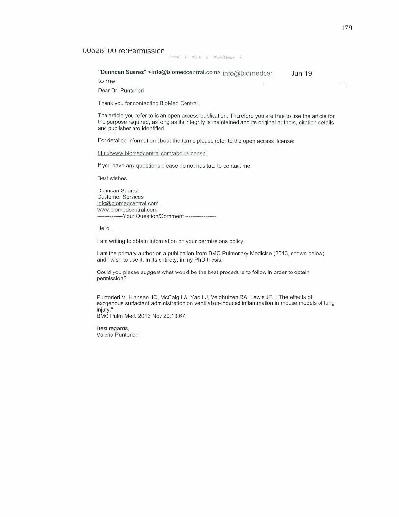
179

180
CURRICULUM VITAE
Name: Valeria Puntorieri
Post-secondary University of Turin
Education and Turin, Italy
Degrees: 2000-2003 B.Sc. Biotechnology
University of Turin
Turin, Italy
2003-2005 M.Sc. Medical Biotechnology
The University of Western Ontario
London, Ontario, Canada
2009- Ph.D. Candidate
Honours and Lawson Internal Research Fund award -
Awards: Lawson Health Research Institute, 2009
First place Poster Presentation Award– Research Day,
Department of Medicine, 2010
Teaching Assistant Award Nominee – Western University
2010/2011/2013
Leadership Award Nominee – Lawson Health Research Institute
2011
Related Work Research Assistant – University of Turin Experience Department of Anesthesiology and Intensive Care
2005-2008
Teaching Assistant –Physiology 3130y Department of Physiology and Pharmacology
Western University
2009-2011
Teaching Assistant –Human Physiology 2130 Department of Physiology and Pharmacology
Western University
2011-2014

181
Publications:
Manuscripts
Valeria Puntorieri, Lynda A. McCaig, Li-Juan Yao, James F. Lewis, Cory M.
Yamashita, and Ruud A.W. Veldhuizen. Lack of matrix metalloproteinase-3 in mouse
models of lung injury ameliorates the pulmonary inflammatory response in females
but not in male mice. In preparation
Cory M Yamashita, Michael B Fessler, Lakshman Vasanthamohan, Joanne Lac,
Jennifer Madenspacher, Lynda McCaig, Lijuan Yao, Lefeng Wang, Valeria Puntorieri,
Sanjay Mehta, Jim F Lewis, Ruud A W Veldhuizen. Apolipoprotein E-deficient mice are
susceptible to the development of acute lung injury. Respiration. 2014 Mar 22.
Valeria Puntorieri, Josh Qua Hiansen, Lynda A McCaig, Li-Juan Yao, Ruud AW
Veldhuizen and James F Lewis. The effects of exogenous surfactant administration on
ventilation-induced inflammation in mouse models of lung injury. BMC Pulmonary
Medicine, 2013 Nov 20;13(1):67.
Fanelli V, Puntorieri V, Assenzio B, Martin EL, Elia V, Bosco M, Delsedime L,
Del Sorbo L, Ferrari A, Italiano S, Ghigo A, Slutsky AS, Hirsch E, Ranieri VM.
Pulmonary-derived phosphoinositide 3-kinase gamma (PI3Kγ) contributes to
ventilator-induced lung injury and edema. Intensive Care Med. 2010 Nov; 36(11):1935-
45. Epub 2010 Aug 19.
Martin EL, Souza DG, Fagundes CT, Amaral FA, Assenzio B, Puntorieri V, Del
Sorbo L, Fanelli V, Bosco M, Delsedime L, Pinho JF, Lemos VS, Souto FO, Alves-Filho
JC, Cunha FQ, Slutsky AS, Ruckle T, Hirsch E, Teixeira MM, Ranieri VM.
PI3K{gamma} kinase activity contributes to sepsis and organ damage by altering
neutrophil recruitment. Am J Respir Crit Care Med., 2010 Sep 15;182(6):762-73.
Fanelli V., Mascia L., Puntorieri V., Assenzio B., Elia V., Fornaio G., Martin E.L.,
Grasso S., Bosco M., Delsedime L., Ranieri V.M. Pulmonary atelectasis during low
stretch ventilation: "open lung versus "lung rest" strategy. Crit Care Med., 2009 Mar;
37(3):1046-53.
Cantaluppi V., Assenzio B., Pasero D., Mauriello-Romanazzi G., Beltramo S.,
Figliolini F., Pacitti A., Lanfranco G., Puntorieri V., Martin E.L., Mascia L., Biancone L.,
Segoloni G.P., Camusi G., Ranieri V.M. Polymyxin-B hemoperfusion inactivates
circulating proapoptotic factors. Intensive Care Medicine, 2008 Sep; 34(9):1638-45.
Fonsato V., Buttiglieri S., Deregibus MC., Puntorieri V., Bussolati B., Camussi
G. (2006). Expression of Pax2 in human renal tumor-derived endothelial cells sustains
apoptosis resistance and angiogenesis. American Journal of Pathology, 2006 Feb;
168(2):706-13.

182
Abstracts
Valeria Puntorieri, Lynda A. McCaig, Li-Juan Yao, Stephanie Aigbe, Jim Lewis,
Cory M. Yamashita, Ruud A.W. Veldhuizen. The role of matrix metalloproteinase 3 in
the Acute Respiratory Distress Syndrome. Matrix Metalloproteinases Gordon Research
Conference, Barga (Italy), May 2013.
Valeria Puntorieri, LeFeng Wang, Li-Juan Yao, Sanjay Mehta, Rudolf A.W.
Veldhuizen, James F. Lewis, Cory Yamashita. The effect of oxidized low-density
lipoprotein on pulmonary endothelial function in vitro. Am. J. Respir. Crit. Care Med.,
May 2012; 185: A2108.
Valeria Puntorieri, Lynda A. McCaig, LiJuan Yao, Ruud A. Veldhuizen, and
James F. Lewis. Systemic inflammation in acute lung injury using an isolated and
perfused mouse lung model. Am. J. Respir. Crit. Care Med., May 2011; 183: A5236.
Puntorieri V., Martin E. L., Pasero D., Del Sorbo L., Ranieri V. M. PMX
treatment of septic patients improved viability of pulmonary epithelial cells. Intensive
Care Medicine 2008
Puntorieri V., Martin E. L., Del Sorbo L., Ranieri V. M.. Lipopolysaccharide
induces alterations in lung epithelial cells indirectly through macrophages. Intensive
Care Medicine 2008
Puntorieri V, Martin EL, Assenzio B, Del Sorbo L, Ranieri VM. Indirect effects
of LPS on pulmonary A549 epithelial cells via THP-1 differentiated macrophages.
Am. J. Crit. Care Med., 2007, 177: A623.


