Embed Size (px)
Citation preview
SARA
TORN
BERG SU
RGICA
LLY TREATED REN
AL CELL CA
RCINO
MA
: PROG
NO
STIC FACTO
RS AN
D O
UTCO
MES O
F TREATMEN
T

Faculty of Medicine University of Helsinki
SURGICALLY TREATED RENAL CELL CARCINOMA: PROGNOSTIC
FACTORS AND OUTCOMES OF TREATMENT
Sara Tornberg
ACADEMIC DISSERTATION
To be presented with the permission of the Faculty of Medicine of the University of Helsinki, from Doctoral Programme in Clinical
Research, for public examination in Athena 107 auditorium,
on the 4th of September 2020 at 12 o’clock
Helsinki 2020
Supervised by: Harry Nisén, MD, Adjunct Professor
Department of Urology
University of Helsinki and
Helsinki University Hospital, Finland
Kimmo Taari, MD, Professor
Department of Urology
University of Helsinki and
Helsinki University Hospital, Finland
Revised by: Markku Vaarala, MD, Adjunct Professor
Department of Urology
University of Oulu and
Oulu University Hospital, Finland
Jukka Häkkinen, MD, Adjunct Professor
Department of Urology
University of Tampere
Opponent: Martti Nurmi, MD, Adjunct Professor
Department of Urology
University of Turku
The Faculty of Medicine uses the Urkund system (plagiarism recognition) to
examine all doctoral dissertations.
ISBN 978-951-51-6381-3 (paperback)
ISBN 978-951-51-6382-0 (PDF)
Painosalama Oy
Turku 2020

Dedicated to patients suffering from renal cell carcinoma.
4
ABSTRACT
Background
Kidney cancer is the 12th most common malignancy worldwide, accounting for
over 400,000 new cases in 2018 (1). As renal cell carcinoma (RCC) incidence
and mortality, as well as treatment patterns, vary widely in Europe, to plan
strategies for the future, we need to comprehend the current situation in
Finland.
Accurate prognostic tools are essential for detecting cancers amongst the
tumours noted in imaging studies and choosing optimal treatment for cancer
patients. The Tumor, Node, Metastasis (TNM) staging system and
International Society of Urologic Pathology (ISUP)/Fuhrman grading system
are the most commonly used prognostic parameters for RCC. Currently, risk
stratification relies on prognostic nomograms or risk stratification tools
combining clinical, anatomical and histopathological data. However, these
models have well-known limitations.
Treatment for RCC is changing. Over the last decades, more incidental
RCCs were found, and more minor lesions were operated on using less invasive
techniques. At the opposite end of the disease spectrum, selected metastatic
RCC patients receive a combined treatment consisting of nephrectomy,
metastasectomy and oncologic therapies. Surgery for locally advanced and
metastasised tumours must be justified by the prospect of an improved
outcome or quality of life. Decisions to operate on metastatic RCCs are
currently based on expert opinions and nomograms designed for targeted
therapy survival estimations only. Thus, better prognostic markers and
diagnostic tools are needed.
Aims
The aims of this PhD study were to evaluate the current changes in the clinical
picture, treatment and outcomes of RCC in Helsinki University Hospital
district. Further analysis was done to determine the clinical outcomes of
surgically treated RCC with tumour thrombus and metastasised RCC (mRCC).
The authors aimed to externally validate the performance of the Leuven-Udine
(LU) prognostic group model for mRCC and to evaluate the prognostic value
5
of serum concentration of tumour-associated trypsin inhibitor (TATI). The
performance of renal tumour diameter and parenchymal invasion depth was
compared with more complex classifications to assess their accuracy in
predicting the nephrectomy performed.
Patients and methods
All patients studied were either suspected to have RCC or had RCC, and the
majority of patients underwent nephrectomy at the Helsinki University
Hospital (HUH). There were 1,719 patients with tumours suspected of RCC
evaluated in four periods from 2006 to 2016 for clinical characteristics and
treatments offered. From 2006–2014, 142 RCC patients with tumour
thrombus (TT) were operated on at HUH. In total, using computed
tomography (CT) or magnetic resonance imaging (MRI) images of 915
patients, tumour maximum diameter, depth of invasion, Preoperative Aspects
and Dimensions Used for an Anatomical (PADUA) score and Renal Tumour
Invasion Index (RTII) were estimated. There were 97 patients with metastatic
RCC undergoing surgery for metastases. Preoperative and postoperative serum
levels of tumour associated trypsin inhibitor (S-TATI) of 132 RCC patients
were determined by time-resolved immunofluorescence assay in 2006-2010.
Main results and conclusions
During the study period, the proportions of frail and co-morbid patients
increased significantly as did the percentage of small (diameter ≤4 cm) and
asymptomatic tumours. The use of surveillance as treatment increased
significantly while the use of cytoreductive nephrectomies (CNs) decreased to
54%. However, CN combined with tyrosine kinase inhibitors remained the
primary option in patients with metastatic RCC. However, the changing
landscape of RCCs has already affected and will increasingly affect the
treatments given. For RCC patients with TT, no statistically significant difference in survival
was found amongst the different levels of the venous extension. The prognosis
for operated RCC patients with TT was good in the absence of papillary
histology of primary tumour, lymphoid or distant metastases. Surgery remains
a feasible option for selected patients in the era of modern oncologic therapy.
6
In predicting the type of nephrectomy, partial or radical, the simple
measurements of tumour diameter and parenchymal invasion, were superior
to the more complex classification. Hence, all of them were significant
predictors for nephrectomy type. Our results recommend that potential
anatomical classifications should be tested against these user-friendly
measurements, diameter and parenchymal invasion.
Overall survival (OS) was more favourable for patients undergoing
complete metastasectomy than patients with non-complete metastasectomy
and time to systemic therapy was longer. Patients with skeletal metastases had
shorter survival than patients with other metastatic sites whereas patients with
lung metastases had the most favourable prognosis. In this study population,
the performance of the LU prognostic group model could not be validated.
Despite the abundant amount of inauspicious prognostic factors in our patient
cohort, survival rates were reasonable.
Significant associations with preoperative S-TATI and Chronic Kidney
Disease Stage (CKD grade), tumour stage, lymph-node involvement,
metastatic status and preoperative C-reactive protein (CRP) level were noted.
S-TATI, as a continuous variable, however, significantly predicted OS and
cancer-specific survival (CSS). Prognostic significance of S-TATI should be
further studied in larger patient cohorts and prospective settings.
7
TABLE OF CONTENTS
ABSTRACT ............................................................................................................4
TABLE OF CONTENTS ........................................................................................ 7
LIST OF ORIGINAL PUBLICATIONS .............................................................. 10
ABBREVIATIONS .............................................................................................. 11
1 INTRODUCTION ..................................................................................... 13
2 REVIEW OF LITERATURE .................................................................... 15
2.1 Epidemiology of renal cell carcinoma .......................................... 15
2.2 Etiology and risk factors of Renal cell carcinoma ....................... 18
2.3 Diagnosing renal cell carcinoma .................................................. 18
2.3.1 Clinical presentation ........................................................ 18
2.3.2 Radiological examinations .............................................. 19
2.3.3 Histolopathological classifications ................................ 20
2.4 Survival and prognostic factors ................................................... 22
2.4.1 Anatomical prognostic factors ....................................... 22
2.4.2 Histological prognostic factors .......................................25
2.4.3 Clinical prognostic factors ............................................... 27
2.4.4 Prognostic biomarkers .................................................... 27
2.4.5 Prognostic systems and nomograms .............................. 31
2.4.5.1 Prognostic systems and nomograms in
localised disease ................................................ 32
2.4.5.2 Prognostic systems and nomograms for
metastasised disease ......................................... 34
2.4.6 Complete resectability and surgical results .................... 35
2.5 Treatment options for local and locally advanced renal cell
carcinoma ..................................................................................... 36
2.5.1 Active Surveillance.......................................................... 36
2.5.2 Operative treatment of RCC ............................................ 37
2.5.3 Renal carcinoma with tumour thrombus ...................... 39
2.5.4 Ablative therapies ........................................................... 40
2.5.5 Adjuvant therapy ............................................................ 40
8
2.6 Treatment of metastasised renal cell carcinoma .........................41
2.6.1 Cytoreductive nephrectomy.............................................41
2.6.2 Metastasectomy and metastases-directed therapies..... 43
2.6.3 Systemic therapy ............................................................. 46
2.6.4 Precision medicine – the way of the future ................... 47
3 AIMS OF THE STUDY ............................................................................ 49
4 PATIENTS AND METHODS .................................................................. 50
4.1 Study cohorts and timelines ........................................................ 50
4.1.1 Study I .............................................................................. 50
4.1.2 Study II ............................................................................ 52
4.1.3 Study III ........................................................................... 53
4.1.4 Study IV ........................................................................... 53
4.1.5 Study V ............................................................................. 54
4.2 Statistical analysis ........................................................................ 54
4.3 Ethics ............................................................................................. 55
5 RESULTS ................................................................................................. 56
5.1 Study I ........................................................................................... 56
5.2 Study II .......................................................................................... 57
5.3 Study III ........................................................................................ 59
5.4 Study IV ......................................................................................... 63
5.5 Study V .......................................................................................... 65
6 DISCUSSION ........................................................................................... 68
6.1 Suspicious mass in kidney: any changes in treatment
patterns? ....................................................................................... 68
6.2 Tumour size, does it matter? ....................................................... 69
6.3 S-TATI, should we measure it? .................................................... 70
6.4 Locally advanced, who should be operated? ............................... 72
6.5 Metastasised RCC and modern systemic therapy, any point of
operating? ..................................................................................... 74
6.6 Big operations, big centres and big risks? ................................... 77
6.7 Limitations .................................................................................... 78
6.8 RCC, what’s ahead? ...................................................................... 79
6.9 Future scientific considerations .................................................. 81

9
7 CONCLUSIONS ...................................................................................... 83
8 ACKNOWLEDGEMENTS ...................................................................... 85
9 REFERENCES ......................................................................................... 88
10 ORIGINAL PUBLICATIONS ................................................................. 101
10
LIST OF ORIGINAL PUBLICATIONS
This thesis is based on the following publications:
I Erkkilä K, Tornberg SV, Järvinen P, Järvinen R, Kilpeläinen TP, Visapää
H, Hervonen P, Taari K, Nisen H. Evolving clinical picture of renal cell
carcinoma: A population-based study from Helsinki. Urol Int. 102(4):
390–398, 2019.
II Tornberg SV, Nisen H, Visapää H, Kilpeläinen TP, Järvinen R, Mirtti T,
Kantonen I, Simpanen J, Bono P, Taari K, Järvinen P. Outcome of
surgery for patients with renal cell carcinoma and tumour thrombus in
the era of modern targeted therapy. Scand J Urol. Oct; 50(5): 380–386,
2016.
III Tornberg SV, Kilpeläinen TP, Järvinen P, Visapää H, Järvinen R, Taari
K, Nisén H. Renal tumor invasion depth and diameter are the two most
accurate anatomical features regarding the choice of radical versus
partial Nephrectomy. Scand J Surg. Mar; 107(1): 54–61, 2018.
IV Tornberg SV, Visapää H, Kilpeläinen TP, Taari K, Järvinen R, Erkkilä K,
Nisen H, Järvinen P. Surgery for metastases of renal cell carcinoma:
Outcome of treatments and preliminary assessment of Leuven-Udine
prognostic groups in the targeted therapy era. Scand J Urol. Oct-Dec;
52(5-6): 419–426, 2018.
V Tornberg SV, Nisen H, Järvinen P, Järvinen R, Kilpeläinen TP, Taari K,
Stenman U-H, Visapää H. Serum tumour associated trypsin as a
biomarker for survival in renal cell carcinoma. Accepted for publication
in Scand J Urol 15.7.2020.
The publications are referred to in the text by their roman numerals.
The original publications have been reproduced with the permission of the
copyright holders. Study I is included here with permission of the first author.
11
ABBREVIATIONS
ANCR Association of the Nordic Cancer Registries ASSURE Adjuvant Sorafenib or Sunitinib for Unfavourable Renal
Carcinoma AUC Area under the curve BHD Birt-Hogg-Dubé syndrome CAIX Carbonic anhydrase IX ccRCC Clear cell renal cell carcinoma CEUS Contrast-enhanced ultrasound chroRCC Chromophobe renal cell carcinoma CI Confidence interval CKD Chronic kidney disease CN Cytoreductive nephrectomy CRP C-reactive protein CSA Contact surface area CSS Cancer-specific survival CT Computed tomography CTLA-4 Cytotoxic T-lymphocyte associated antigen 4 DAP Diameter-axial-polar DISSRM Delayed Intervention and Surveillance for Small Renal
Masses EAU European Association of Urology ECOG Eastern Cooperative Oncology Group EORTC European Organisation for Research and Treatment of Cancer GFR Glomerular filtration rate HIF1 α Hypoxia-induced 1 alpha HLRCC Hereditary leiomyomatosis and renal cell cancer HPRC Hereditary papillary renal cancer HU Hounsfield units HUH Helsinki University Hospital IARC International Agency for Research on Cancer IMDC International Metastatic Renal Cancer Database Consortium IQR Interquartile range ISUP International Society of Urological Pathology LDH Lactate dehydrogenase LU Leuven-Udine MDT Metastasis-directed therapy miRNA MicroRNA mRCC Metastatic renal cell carcinoma
12
MSKCC Memorial Sloan Kettering Cancer Center mTOR Mammalian target of rapamycin NePhRO Nearness, Physical location, Radius and Organization of
tumour NLR Neutrophil-lymphocyte ratio NSAID Nonsteroidal anti-inflammatory drug OR Odds ratio OS Overall survival PADUA Preoperative Aspects and Dimensions Used for an
Anatomical Classification PD-1 Programmed death-1 receptor PDGF Platelet-derived growth factor PD-L1 Programmed death-ligand 1 PFS Progression-free survival PN Partial nephrectomy pRCC Papillary renal cell carcinoma PSM Positive surgical margin PTEN Phosphatase and tensin homolog QOL Quality of life RAIV Resected and ischemic volume RCC Renal cell carcinoma RFS Recurrence-free survival RN Radical nephrectomy ROC Receiver operating characteristic RTB Renal tumour biopsy RTII Renal tumour invasion index SARR Surgical approach renal ranking SRM Small renal mass SSIGN Stage, size, grade and necrosis S-TATI Serum tumour-associated trypsin inhibitor S-TRAC Sunitinib as Adjuvant Treatment for Patients at High Risk of
Recurrence of Renal Cell Carcinoma Following Nephrectomy TATI Tumour-associated trypsin inhibitor TKI Tyrosine kinase inhibitor TNM Tumour, node and metastasis TSC Tuberous sclerosis complex TT Tumour thrombus UISS University of California Integrated Staging System VEGF Vascular endothelial growth factor VHL Von Hippel-Lindau WHO World Health Organization
13
1 INTRODUCTION
Renal cell carcinoma (RCC) is the most lethal urological malignancy; however,
reliable prognostic tools to recognise the ‘killer tumours’ from indolent ones
are lacking. Being a highly heterogenic disease, the clinical course of RCC is
strikingly unpredictable.
As up to half of newly diagnosed RCCs are incidental findings (2, 3), we are
often faced with the clinical challenge of an unclassified tumour susceptible to
malignancy which is found in an imaging study. Peak incidence of RCC is from
60 to 70 years of age, meaning that RCC patients are often frail, have co-
morbidities and a limited life expectancy. Therefore, operations for indolent or
slowly progressing tumours may easily turn into overtreatment. Renal tumour
biopsies (RTBs) are used to distinguish malignant tumours from benign ones.
The sensitivity of an RTB for detecting renal malignancy is excellent in
experienced centres (93-99.5%) (4, 5), but the accuracy of defining the grade
is poor (67%) (4). Although severe complications associated with RTBs are rare
(4), there is a definite need for less invasive and more precise tools to predict
the course of yet unclassified renal tumours.
The five year overall survival (OS) for all RCCs was reported to be as low as
40% by a Swedish population-based study and merely 13% for metastatic RCC
(6). Of local tumours operated on with curative intention, 20-40% commonly
recur (7). When considering who benefits from nephrectomy or
metastasectomy, estimating survival is important. The tumour, node and
metastasis (TNM) classification is one of the most common and robust
predictors of oncological outcomes (8). TNM classification and other
prognostic factors for RCC, such as Fuhrman grade, tumour necrosis,
histologic subtype and performance status of the patient, are not sufficiently
accurate prognosticators when used alone (9). Hence, these prognostic factors
are combined to numerous prognostic models or nomograms both for localised
and metastatic RCCs. The follow-up recommendations and treatment
decisions are based on these nomograms and models estimating recurrence
and survival. Still, as clinicians and researchers, we are in great need of more
accurate prognostic information to facilitate patient counselling, plan
individual surveillance schemes and select optimal treatments for each patient
(9).
14
To improve the prognostic accuracy, the further research and development
of prognostic biomarkers has become a priority. The increased understanding
of gene technology, proteomics, molecular biology and immunology of cancer
has raised great hope for finding a ‘true prognostic biomarker’. Despite the
exhaustive efforts made in marker research, no molecular biomarker has been
able to significantly improve the prognostic accuracy of existing models (10).
Surgery is always prone to complications, and complication rates vary
amongst centres and procedures. Knowing the results of one’s own centre is a
benefit when planning more complicated surgery. After recent improvements
in targeted therapy and immuno-oncology, doubts have been raised about the
role and sequence of surgery in metastasised cases (11, 12). As metastasectomy
or cytoreductive nephrectomy may also be used for palliation, the risk of
complications, the recovery time from surgery and delay in commencing
systematic treatments must be tolerable. For local tumours, mini-invasive
surgery is the treatment of choice in all T1 tumours (13) as the oncologic safety
of partial nephrectomy (PN) seems to be similar when compared with radical
nephrectomy (RN) (13). Still, 22% of pT1a tumours are removed by RN in
Northern European countries (14), inferring that more than just the diameter
affects our decisions.
15
2 REVIEW OF LITERATURE
2.1 EPIDEMIOLOGY OF RENAL CELL CARCINOMA
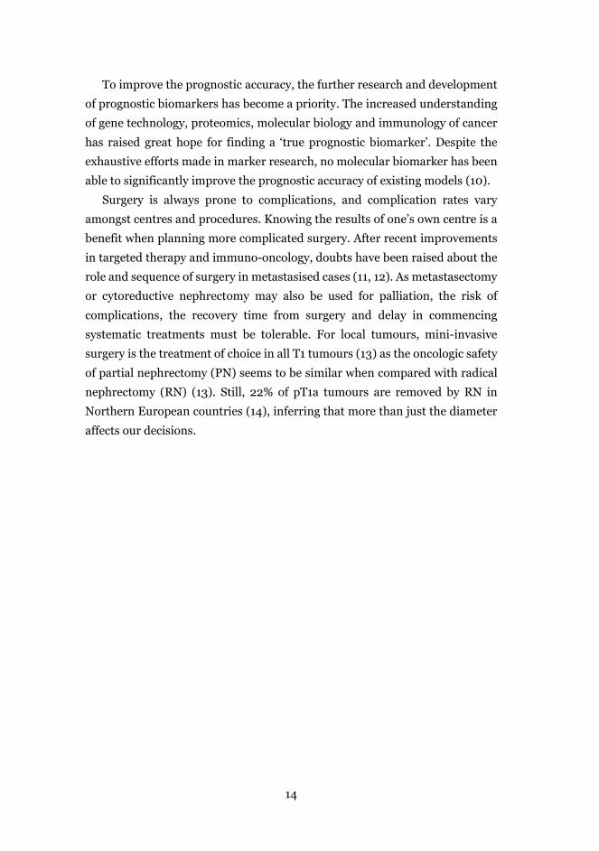
RCC accounts for 2-3% of all cancers (15, 16), representing the ninth most
common cancer in men and the sixteenth in women (17). For men, the lifetime
risk for developing kidney cancer is about 1 in 48. In Finland, the reported
number of new cases of RCC in 2018 was 1,010, ranking 10th in all newly
diagnosed cancers (18).
Figure 1 Estimated age-standardized rates of incidence for both sexes (per 100,000
persons) in 2018. In developed countries, the incidence is generally higher than in developing countries. Reprinted with the permission from the World Health Organization.
The number of incidental RCCs, usually smaller and of lower stage, have
increased due to the increased use of imaging techniques. The proportion of
small renal masses (SRMs) is currently up to 40% of overall incidence (19).
Over the last decade, the RCC incidence has been rising worldwide, although
this is less pronounced in women (18). However, a great disparity exists
concerning RCC incidence globally (Figure 1). The incidence of RCC is highest
in Western countries (16, 18). Differences are also profound in Europe: male
incidence in Sweden and Malta is as low as 7.1/100,000 but is 22/100,000 in
16
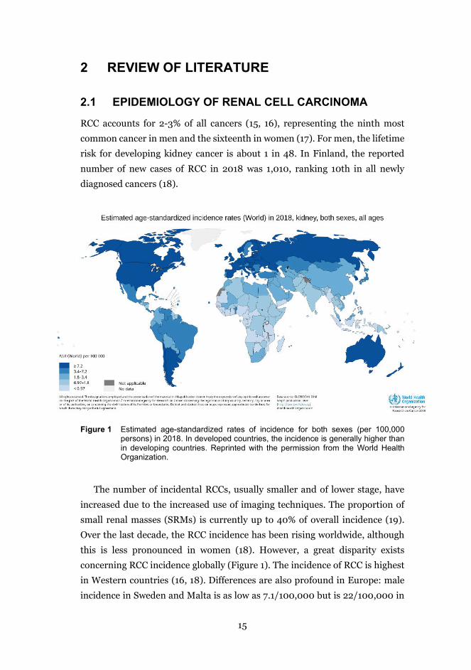
the Czech Republic (16). Amongst the countries of Northern Europe, only in
Finland and Estonia is male RCC incidence stabilised (18) (Figure 2).
Figure 2 RCC incidence by age group in Finland from 1953 to 2018. Data from Finnish
Cancer Registry. https://tilastot.syoparekisteri.fi/syovat, data from 2020-04-02, version 2020-04-17-004. Reprinted with the permission of Finnish Cancer Registry.
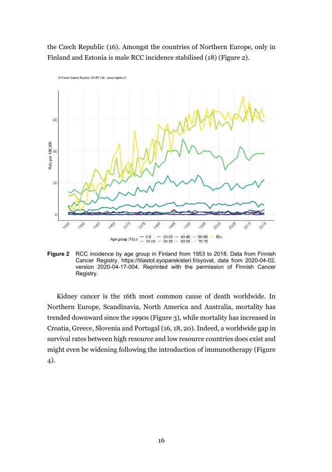
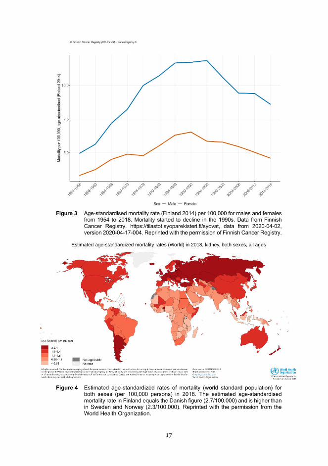
Kidney cancer is the 16th most common cause of death worldwide. In
Northern Europe, Scandinavia, North America and Australia, mortality has
trended downward since the 1990s (Figure 3), while mortality has increased in
Croatia, Greece, Slovenia and Portugal (16, 18, 20). Indeed, a worldwide gap in
survival rates between high resource and low resource countries does exist and
might even be widening following the introduction of immunotherapy (Figure
4).
17
Figure 3 Age-standardised mortality rate (Finland 2014) per 100,000 for males and females
from 1954 to 2018. Mortality started to decline in the 1990s. Data from Finnish Cancer Registry. https://tilastot.syoparekisteri.fi/syovat, data from 2020-04-02, version 2020-04-17-004. Reprinted with the permission of Finnish Cancer Registry.
Figure 4 Estimated age-standardized rates of mortality (world standard population) for
both sexes (per 100,000 persons) in 2018. The estimated age-standardised mortality rate in Finland equals the Danish figure (2.7/100,000) and is higher than in Sweden and Norway (2.3/100,000). Reprinted with the permission from the World Health Organization.
18
2.2 ETIOLOGY AND RISK FACTORS OF RENAL CELL CARCINOMA
Known risk factors for RCC are age, with peaks at 60-70 years, and gender,
with a 1.5:1 male predominance. Common risk factors found in epidemiological
studies include cigarette smoking (21), obesity (22), hypertension (21) and
greater adult attained height (1). However, the importance of these risk factors
may be biased by incidental cancer detection in imaging done due to illnesses
associated with these risk factors (23). The presence of kidney disease, viral
hepatitis, urinary stone in male patients and continuous use of paracetamol or
non-aspirin non-steroidal anti-inflammatory drugs (NSAIDs) seem to increase
the risk of RCC (21, 24). Having a first-degree relative with RCC has been
associated in meta-analysis with a 2.2-fold and, in case-control analysis, a 4.3-
fold significantly increased risk for RCC, 95% CI [1.6-2.9] and [1.6-11.9],
respectively (25).
Kidney transplantation, end-stage renal disease and dialysis predispose
patients to RCC (26-28). Several hereditary syndromes elevate the risk of RCC,
accounting for 4% of all RCCs (25). Von Hippel-Lindau (VHL) disease, Birt-
Hogg-Dubé syndrome (BHD), hereditary leiomyomatosis and renal cell cancer
(HLRCC), hereditary papillary renal cancer (HPRC) and tuberous sclerosis
complex (TSC) are the five most common autosomal dominantly inherited
syndromes with distinct clinical manifestations and genetic alterations (29).
However, most cases of TSC occur as sporadic cases, due to de novo mutation.
Physical activity and consumption of cruciferous vegetables associate with
a lower risk of RCC (30, 31) as well as moderate alcohol consumption relative
to abstinence (1, 32).
2.3 DIAGNOSING RENAL CELL CARCINOMA
2.3.1 CLINICAL PRESENTATION
Being located retroperitoneally and surrounded by fat, tumours of the kidney may
enlarge significantly without presenting any symptoms. Due to the lack of early
warning signs, as many as 25-30% of RCCs have already metastasised by the time
of diagnosis (33). However, the recent population-based data from Sweden show
the percentage of synchronous metastatic RCC to be as low as 19% (2).
19
The ‘classic triad’ of RCC, which includes abdominal pain, haematuria and
palpable mass in the flank or abdomen, is especially rare today. Earlier, 6-10%
of patients presented this triad (34, 35). Currently, up to 50% of RCC is
detected incidentally (2, 3). However, the disease is sometimes accompanied
by paraneoplastic symptoms such as fever, malaise, erythrocytosis and
hypercalcemia.
No laboratory examinations, serum or urine are helpful in diagnosing
whether a renal cell carcinoma exists. However, laboratory parameters, i.e.
haemoglobin, neutrophils, thrombocytes, lactate dehydrogenase and calcium,
are used to estimate prognosis of metastasised RCC.
2.3.2 RADIOLOGICAL EXAMINATIONS
Most renal masses are primarily diagnosed by imaging. Ultrasound imaging
often raises suspicion about RCC, which is then followed by further imaging
with computed tomography (CT) or magnetic resonance imaging (MRI). The
paramount criterion for malignancy is the enhancement of contrast material
within the tumour (36). An increase of 15 or more Hounsfield units (HUs)
indicates solid tissue, which is most often malignant (37). However, fat-free
angiomyolipomas and oncocytomas cannot be reliably differentiated from
RCC. Recently, contrast-enhanced ultrasound (CEUS) has been used to
differentiate cystic lesions from solid ones and has high sensitivity and
specificity in characterising renal masses (38). For the purpose of staging RCC,
thorax CT added to renal imaging by CT or MRI is recommended (13). CT of
head and bone scans are performed only in the presence of symptoms or
particular signs (13, 39). Position-emission tomography has a low sensitivity
and specificity for detecting RCC and is not recommended by European
Association of Urology (EAU) Guidelines (13) .
RTB is often considered non-necessary if the patient will undergo an
operation based on radiology (13). Renal biopsies are strongly recommended
prior to ablative treatment and for matching patients with the best medical
therapy scheme, and they can be used to guide patient selection to active
surveillance (13). Use of ultrasound or CT guidance provides similar results
(40, 41). Even in experienced centres, up to 22% of biopsies are non-diagnostic,
however (4, 42). Researchers found that the diagnostic rate was lowest when
there was a long distance (>10 cm) from skin to tumour (73.1%), tumour
20
diameter was smaller than 4 cm (86.2%), enhancement on contrasted CT was
less than 20 HU (57.9%) or the tumour was cystic (60.2%) (43). Also, biopsies
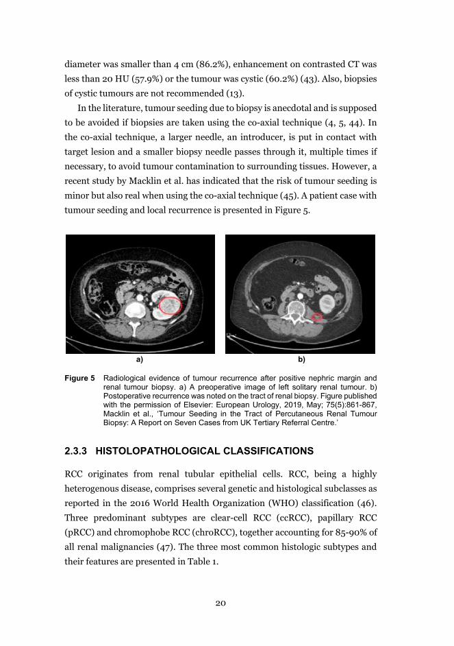
of cystic tumours are not recommended (13). In the literature, tumour seeding due to biopsy is anecdotal and is supposed
to be avoided if biopsies are taken using the co-axial technique (4, 5, 44). In
the co-axial technique, a larger needle, an introducer, is put in contact with
target lesion and a smaller biopsy needle passes through it, multiple times if
necessary, to avoid tumour contamination to surrounding tissues. However, a
recent study by Macklin et al. has indicated that the risk of tumour seeding is
minor but also real when using the co-axial technique (45). A patient case with
tumour seeding and local recurrence is presented in Figure 5.
a) b)
Figure 5 Radiological evidence of tumour recurrence after positive nephric margin and renal tumour biopsy. a) A preoperative image of left solitary renal tumour. b) Postoperative recurrence was noted on the tract of renal biopsy. Figure published with the permission of Elsevier: European Urology, 2019, May; 75(5):861-867, Macklin et al., ‘Tumour Seeding in the Tract of Percutaneous Renal Tumour Biopsy: A Report on Seven Cases from UK Tertiary Referral Centre.’
2.3.3 HISTOLOPATHOLOGICAL CLASSIFICATIONS
RCC originates from renal tubular epithelial cells. RCC, being a highly
heterogenous disease, comprises several genetic and histological subclasses as
reported in the 2016 World Health Organization (WHO) classification (46).
Three predominant subtypes are clear-cell RCC (ccRCC), papillary RCC
(pRCC) and chromophobe RCC (chroRCC), together accounting for 85-90% of
all renal malignancies (47). The three most common histologic subtypes and
their features are presented in Table 1.
21
Oncocytomas comprise 7% of all renal tumours (48). As there are cases
where oncocytoma has been noted to invade vascular structures and
perinephric fat, although without altering the benign prognosis of oncocytoma,
it could be considered as a tumour of a very low potential of malignancy instead
of a strictly benign tumour (49) (50). Indeed, the EAU recommends that active
surveillance should be offered as a treatment alternative for biopsy proven
oncocytomas, as only 64.6% of those remained histologically oncocytomas
after surgery, while 31% were classified as cancers (51).
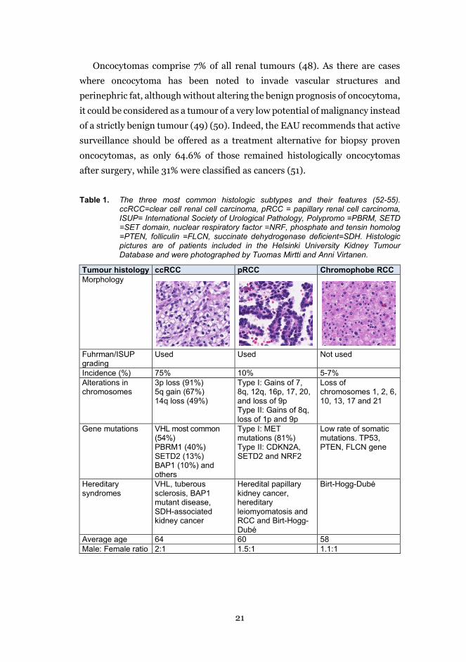
Table 1. The three most common histologic subtypes and their features (52-55). ccRCC=clear cell renal cell carcinoma, pRCC = papillary renal cell carcinoma, ISUP= International Society of Urological Pathology, Polypromo =PBRM, SETD =SET domain, nuclear respiratory factor =NRF, phosphate and tensin homolog =PTEN, folliculin =FLCN, succinate dehydrogenase deficient=SDH. Histologic pictures are of patients included in the Helsinki University Kidney Tumour Database and were photographed by Tuomas Mirtti and Anni Virtanen.
Tumour histology ccRCC pRCC Chromophobe RCC Morphology
Fuhrman/ISUP grading
Used Used Not used
Incidence (%) 75% 10% 5-7% Alterations in chromosomes
3p loss (91%) 5q gain (67%) 14q loss (49%)
Type I: Gains of 7, 8q, 12q, 16p, 17, 20, and loss of 9p Type II: Gains of 8q, loss of 1p and 9p
Loss of chromosomes 1, 2, 6, 10, 13, 17 and 21
Gene mutations VHL most common (54%) PBRM1 (40%) SETD2 (13%) BAP1 (10%) and others
Type I: MET mutations (81%) Type II: CDKN2A, SETD2 and NRF2
Low rate of somatic mutations. TP53, PTEN, FLCN gene
Hereditary syndromes
VHL, tuberous sclerosis, BAP1 mutant disease, SDH-associated kidney cancer
Heredital papillary kidney cancer, hereditary leiomyomatosis and RCC and Birt-Hogg-Dubé
Birt-Hogg-Dubé
Average age 64 60 58 Male: Female ratio 2:1 1.5:1 1.1:1
22
The remaining histologic subtypes are rare, each accounting for
approximately 1% of total incidence (46). These minor subtypes include
collecting duct RCCs, medullary RCC, clear-cell papillary RCC,
microphthalmia-associated transcription factor (MiT) family translocation
RCCs, hereditary leiomyomatosis and RCC, acquired cystic disease-associated
RCC, tubulocystic RCC, succinate dehydrogenase-deficient RCC and mucinous
tubular and spindle cell RCC. Up to 4% of RCCs fail to fit into any of these
categories and are labelled as unclassified RCCs (52).
Pathological diagnosis does not only determine the subtypes of RCC, but
also interprets the nuclear grade, tumour necrosis, lymphovascular invasion,
sarcomatoid features and invasion to perirenal fat or venous system stage (53).
2.4 SURVIVAL AND PROGNOSTIC FACTORS
The clinical course of RCC is variable. After complete, curative-intended,
surgical resection of local RCC, up to 30% recurred, according to a five-year
follow-up, after being considered disease-free (56). The most fundamental
prognosticator is whether the RCC is localised or advanced, at the time of
diagnosis, as the hazard ratio of cancer-specific survival (CSS) in metastatic
(M1) disease is 33.23 (95% CI [28.18-39.18]) compared with T1N0M0 disease
(57). Prognostic factors can be labelled as clinical, anatomical,
histopathological and molecular. Prognostic models which combine individual
prognostic factors are needed to predict individual likelihood of recurrence and
death when counselling patients and selecting patients for adjuvant therapy
trials. Accurate and easy-to-use prognostic markers and models are needed, as
detecting cancer as early as possible, finding the ones that will recur and
deciding the right individual treatment for cancer patients are some of the
great challenges of medicine today.
2.4.1 ANATOMICAL PROGNOSTIC FACTORS
2.4.1.1 TNM classification
The classic anatomic prognostic system is TNM classification (58) (Table 2). It
was composed for scientific and clinical use (59). The current TNM
classification was constructed in 1997 with global consensus (60) and is
continuously updated by the Union for International Cancer Control. The
23
latest update was done in 2017. Tumour size, invasion of the renal capsule or
venous system, adrenal involvement, lymph node status and existence of
distant metastasis, all important prognostic factors, are included in TNM
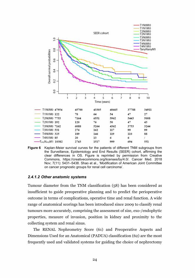
staging. The TNM classification is one of the most solid and reliable predictors
of oncologic outcomes, as OS (Figure 6) and recurrence (8). The TNM
classification from 2010 divided T2 into T2a and T2b and changed the pT3a
and pT3b classifications. However, these changes have led to only modest
improvements in predictive accuracy (8).
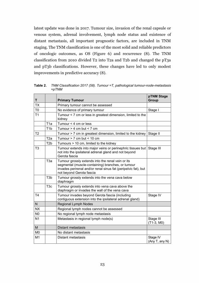
Table 2. TNM Classification 2017 (58). Tumour =T, pathological tumour-node-metastasis =pTNM
T Primary Tumour pTNM Stage Group
TX Primary tumour cannot be assessed T0 No evidence of primary tumour Stage I T1 Tumour < 7 cm or less in greatest dimension, limited to the
kidney
T1a Tumour < 4 cm or less T1b Tumour > 4 cm but < 7 cm T2 Tumour > 7 cm in greatest dimension, limited to the kidney Stage II T2a Tumour > 7 cm but < 10 cm T2b Tumours > 10 cm, limited to the kidney T3 Tumour extends into major veins or perinephric tissues but
not into the ipsilateral adrenal gland and not beyond Gerota fascia
Stage III
T3a Tumour grossly extends into the renal vein or its segmental (muscle-containing) branches, or tumour invades perirenal and/or renal sinus fat (peripelvic fat), but not beyond Gerota fascia
T3b Tumour grossly extends into the vena cava below diaphragm
T3c Tumour grossly extends into vena cava above the diaphragm or invades the wall of the vena cava
T4 Tumour invades beyond Gerota fascia (including contiguous extension into the ipsilateral adrenal gland)
Stage IV
N Regional Lymph Nodes NX Regional lymph nodes cannot be assessed N0 No regional lymph node metastasis N1 Metastasis in regional lymph node(s) Stage III
(T1-3, M0) M Distant metastasis M0 No distant metastasis M1 Distant metastasis Stage IV
(Any T, any N)
24
Figure 6 Kaplan‐Meier survival curves for the patients of different TNM subgroups from
the Surveillance, Epidemiology and End Results (SEER) cohort, affirming the clear differences in OS. Figure is reprinted by permission from Creative Commons, https://creativecommons.org/licenses/by/4.0/. Cancer Med. 2018 Nov; 7(11): 5431–5438. Shao et al., ‘Modification of American Joint Committee on cancer prognostic groups for renal cell carcinoma’.
2.4.1.2 Other anatomic systems
Tumour diameter from the TNM classification (58) has been considered as
insufficient to guide preoperative planning and to predict the perioperative
outcome in terms of complications, operative time and renal function. A wide
range of anatomical scorings has been introduced since 2009 to classify renal
tumours more accurately, comprising the assessment of size, exo-/endophytic
properties, measure of invasion, position in kidney and proximity to the
collecting system and renal sinus.
The RENAL Nephrometry Score (61) and Preoperative Aspects and
Dimensions Used for an Anatomical (PADUA) classification (62) are the most
frequently used and validated systems for guiding the choice of nephrectomy
25
and to predict complications, length of operation and hospital stay (63, 64).
Other scoring systems are the zonal NePhRO scoring system (65), the surgical
approach renal ranking (SARR) (66), centrality index (67), the Renal Tumor
Invasion Index (RTII) (68), diameter-axial-polar (DAP) nephrometry (69), the
renal tumour contact surface area (CSA) (70) and resected and ischemic
volume (RAIV) (71), to name a few.
2.4.2 HISTOLOGICAL PROGNOSTIC FACTORS
The survival rates for ccRCC are the lowest, followed by pRCC and chroRCC
(72, 73), but when stratifying histology to tumour grade or tumour stage (T-
stage), the survival rate differences disappear in multivariable analysis,
indicating that stage and grade determine prognosis more than histological
subtype (74). Also, pRCCs consist of type 1 and type 2, with the first group
being of lower grade and of favourable outcome, while latter group’s tumours
are of higher grade with an increased metastatic potential (75, 76). The
prognostic difference between this subtyping is however debatable (75).
Median survival of a rare subtype, carcinoma of the collecting duct, is 13
months, remarkably lower than for ccRCC, and the majority of tumours (70%)
have metastasised by the time of diagnosis (77).
The presence of necrosis, sarcomatoid or rhabdoid features, microscopic
venous or lymphaneous invasion and invasion in the collecting system are all
associated with worse outcomes (78). Having a median survival of 4 to 13
months after diagnosis, sarcomatous carcinoma has a discouraging prognosis
(79, 80). Of the relevant histological features, sarcomatoid differentiation also
predicts a worse prognosis in metastatic renal cell carcinoma (mRCC) (81) as
well as does non-ccRCC histology (82). For patients with rhabdoid features
containing carcinoma, median survival is slightly better (8 to 45 months) than
for sarcomatotic features. Rhabdoid features, as independent prognostic
factors apart from Fuhrman grade, were not found to be associated with higher
mortality (80, 83). Necrosis, however, is a predictor of CSS (84), recurrence
(85) and progression to metastasis (86)
2.4.2.1 Fuhrman/ISUP grading system
The Fuhrman grading system has been one of most generally agreed upon
independent prognostic markers for RCC (9, 87). Even though it is the most
26
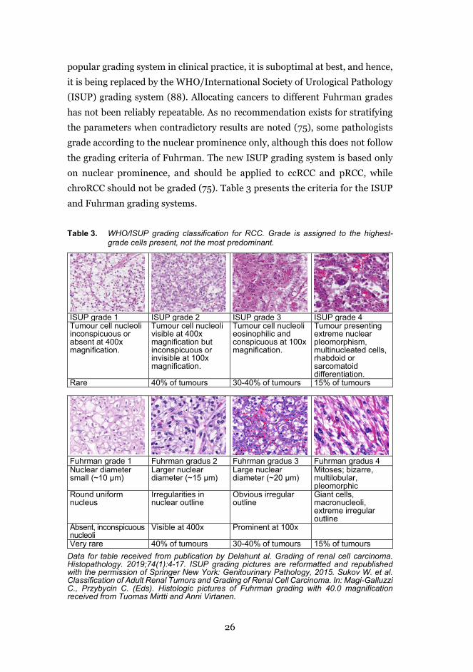
popular grading system in clinical practice, it is suboptimal at best, and hence,
it is being replaced by the WHO/International Society of Urological Pathology
(ISUP) grading system (88). Allocating cancers to different Fuhrman grades
has not been reliably repeatable. As no recommendation exists for stratifying
the parameters when contradictory results are noted (75), some pathologists
grade according to the nuclear prominence only, although this does not follow
the grading criteria of Fuhrman. The new ISUP grading system is based only
on nuclear prominence, and should be applied to ccRCC and pRCC, while
chroRCC should not be graded (75). Table 3 presents the criteria for the ISUP
and Fuhrman grading systems.
Table 3. WHO/ISUP grading classification for RCC. Grade is assigned to the highest-grade cells present, not the most predominant.
ISUP grade 1 ISUP grade 2 ISUP grade 3 ISUP grade 4 Tumour cell nucleoli inconspicuous or absent at 400x magnification.
Tumour cell nucleoli visible at 400x magnification but inconspicuous or invisible at 100x magnification.
Tumour cell nucleoli eosinophilic and conspicuous at 100x magnification.
Tumour presenting extreme nuclear pleomorphism, multinucleated cells, rhabdoid or sarcomatoid differentiation.
Rare 40% of tumours 30-40% of tumours 15% of tumours
Fuhrman grade 1 Fuhrman gradus 2 Fuhrman gradus 3 Fuhrman gradus 4 Nuclear diameter small (~10 µm)
Larger nuclear diameter (~15 µm)
Large nuclear diameter (~20 µm)
Mitoses; bizarre, multilobular, pleomorphic
Round uniform nucleus
Irregularities in nuclear outline
Obvious irregular outline
Giant cells, macronucleoli, extreme irregular outline
Absent, inconspicuous nucleoli
Visible at 400x Prominent at 100x
Very rare 40% of tumours 30-40% of tumours 15% of tumours Data for table received from publication by Delahunt al. Grading of renal cell carcinoma. Histopathology. 2019;74(1):4-17. ISUP grading pictures are reformatted and republished with the permission of Springer New York: Genitourinary Pathology, 2015. Sukov W. et al. Classification of Adult Renal Tumors and Grading of Renal Cell Carcinoma. In: Magi-Galluzzi C., Przybycin C. (Eds). Histologic pictures of Fuhrman grading with 40.0 magnification received from Tuomas Mirtti and Anni Virtanen.
27
2.4.3 CLINICAL PROGNOSTIC FACTORS
With RCC, being symptomatic at the time of diagnosis is associated with worse
survival (89). Moreover, cachexia, anaemia and low physical performance are
independent markers for poor prognosis (90). The Eastern Cooperative
Oncology Group (ECOG) or the Karnofsky scale are used to assess the
performance status, and strongly correlate with prognosis (73).
Age is not an independent prognostic factor of RCC for surgically treated,
localised RCC (91), and it is excluded from the commonly used prognostic
models such as the Stage, Size, Grade and Necrosis (SSIGN) score and
Memorial Sloan Kettering Cancer Center (MSKCC) nomogram. Age is strongly
associated with other-cause mortality, meaning that septuagenarians, being at
high risk for small renal tumour/RCC diagnosis, are also at high risk of other-
cause mortality.
As age is only a number, the co-morbidity and disability of patients
potentially play a more significant role when determining the outcomes of
cancer treatments. In particular, the condition of frailty, meaning when an
elderly person is a state of major vulnerability due to adverse health status
changes because of a diminished physiological reserve capacity, is a major
contributor to health outcomes. Studies of other cancers (92, 93) have pointed
out that frailty is a predictor of complications after elective surgery, intolerance
to chemotherapy, progression of disease and worse survival. To the best of my
knowledge, no studies on the relationship between frailty and RCC exist at this
point.
For mRCC patients, there is some evidence that socioeconomic status, older
age and marital status (widowed, divorced or separated) are associated with
higher cancer-specific mortality but not with overall mortality (94). However,
these results may be biased by other factors, e.g. those who are married and
have better socioeconomic status are more likely to receive cytoreductive
nephrectomy (CN) (95, 96).
2.4.4 PROGNOSTIC BIOMARKERS
Cancer biomarkers are often defined as molecules that raise the suspicion of
cancer or predict the future prognosis of cancer. But, a biomarker can be any
medical sign that can be objectively measured and reproduced. Serum
28
prostate-specific antigen (S-PSA) is one of most widely used serum biomarkers
for cancer, just as body temperature is a common biomarker for fever. Of
commonly used laboratory markers, low haemoglobin level and high corrected
serum calcium predict poor survival in patients with advanced RCC (97), and
they are currently integrated into standard of care survival calculators. In
addition, for patients receiving targeted therapy, hypertension and
neutropenia are associated with favourable outcomes (98) and hyponatremia
with poor outcomes (99).
Despite the increased interest in molecular biomarkers and the noted
promising associations with outcomes, no biomarker has yet been externally
validated or found to clearly improve the accuracy beyond the commonly used
prognostic factors. Thus, they have not been accepted for use in routine clinical
practice but are used in experimental settings only.
2.4.4.1 Markers of hypoxia-induced pathway
Proneness to inherited RCC arises from genes that participate in regulating
cellular metabolism. In sporadic ccRCC, some molecular alterations are very
common: both the inactivation of the von Hippel-Lindau tumour suppressor
(VHL) gene and the loss of chromosome 3p (site of the VHL gene) are found in
the majority of cases (100, 101). The VHL gene, which controls oxygen sensing,
is encoded to protein (pVHL), which targets hypoxia-induced factor 1 alpha
(HIF-1α). Accumulation of the hypoxia-inducible factors (HIF) leads to
overexpression of angiogenic factors, platelet-derived growth factor (PDGF)
and vascular endothelial growth factor (VEGF), which promote
neoangiogenesis (102). Commonly, HIF is inactive in an oxygenated
environment but in cancer cells, with VHL mutation, HIF stays active although
no hypoxemia exists.
The VHL alterations and HIF-1α have been associated with both better
(103) and worse survival (104). High carbonic anhydrase IX (CAIX), a HIF-1 α
regulated protein, has been demonstrated to predict better prognosis (105).
VEGFs, working as regulators of angiogenesis, have been associated with worse
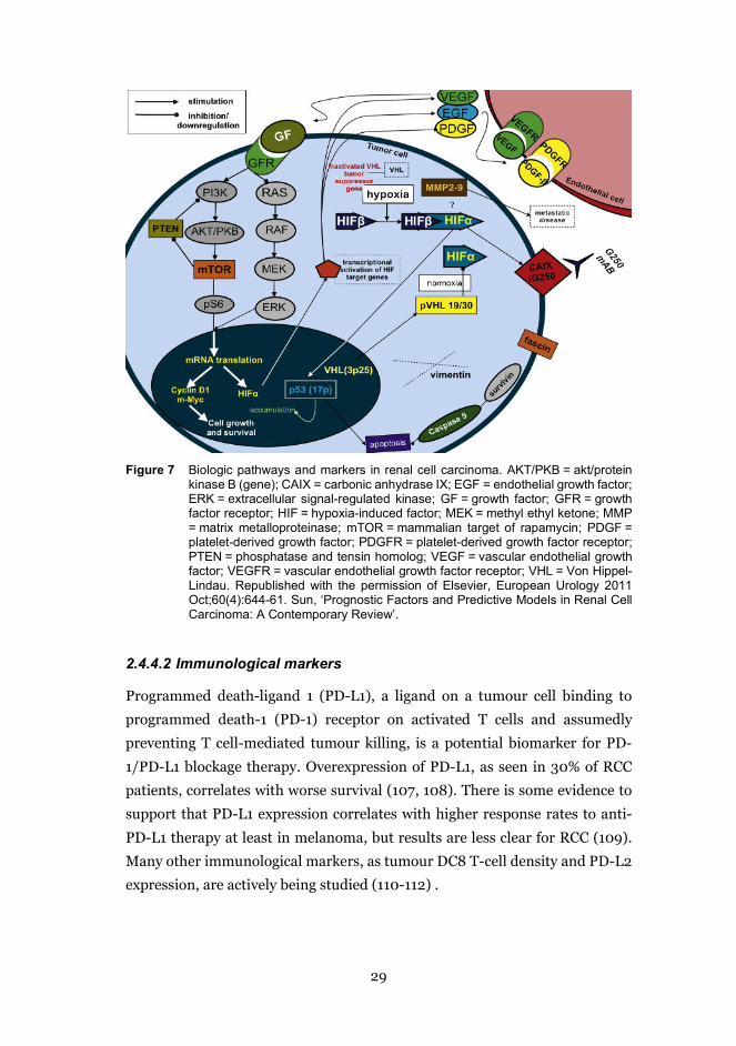
survival (106). The best known stimulative and inhibitive pathways for RCC
are presented in Figure 7.
29
Figure 7 Biologic pathways and markers in renal cell carcinoma. AKT/PKB = akt/protein
kinase B (gene); CAIX = carbonic anhydrase IX; EGF = endothelial growth factor; ERK = extracellular signal-regulated kinase; GF = growth factor; GFR = growth factor receptor; HIF = hypoxia-induced factor; MEK = methyl ethyl ketone; MMP = matrix metalloproteinase; mTOR = mammalian target of rapamycin; PDGF = platelet-derived growth factor; PDGFR = platelet-derived growth factor receptor; PTEN = phosphatase and tensin homolog; VEGF = vascular endothelial growth factor; VEGFR = vascular endothelial growth factor receptor; VHL = Von Hippel-Lindau. Republished with the permission of Elsevier, European Urology 2011 Oct;60(4):644-61. Sun, ‘Prognostic Factors and Predictive Models in Renal Cell Carcinoma: A Contemporary Review’.
2.4.4.2 Immunological markers
Programmed death-ligand 1 (PD-L1), a ligand on a tumour cell binding to
programmed death-1 (PD-1) receptor on activated T cells and assumedly
preventing T cell-mediated tumour killing, is a potential biomarker for PD-
1/PD-L1 blockage therapy. Overexpression of PD-L1, as seen in 30% of RCC
patients, correlates with worse survival (107, 108). There is some evidence to
support that PD-L1 expression correlates with higher response rates to anti-
PD-L1 therapy at least in melanoma, but results are less clear for RCC (109).
Many other immunological markers, as tumour DC8 T-cell density and PD-L2
expression, are actively being studied (110-112) .

30
2.4.4.3 Inflammatory markers of RCC
Some tumours are heavily infiltrated by immune cells of the host, and this
process seems to mimic inflammatory responses of normal tissue (113).
Previously, this immune reaction was seen as an attempt to erode tumours.
However, currently, the inflammation process is considered to have the
paradoxical effect of facilitating the tumour growth and progression (114).
Inflammation is sometimes present in the earliest stages of progression from
neoplasia to cancer (115, 116), suggesting that inflammation laboratory exams
are possible prognostic markers.
A meta-analysis by Hu et al. noted that elevated C-reactive protein (CRP)
correlated with poorer CSS and OS (117). The new and promising inflammation
marker, neutrophil-lymphocyte ratio (NLR), was found to be associated with
poorer prognosis in a recent meta-analysis (118). Elevated platelet and
neutrophil counts are also suggested to be independent predictors for poor
prognosis (119). In addition, the C-reactive protein to albumin ratio has been
associated with worse OS and disease-free survival (DFS) in a recent meta-
analysis (120).
Consequently, there is still interest in searching for other acute phase
reactants that have a prognostic impact for RCC, such as lactate dehydrogenase
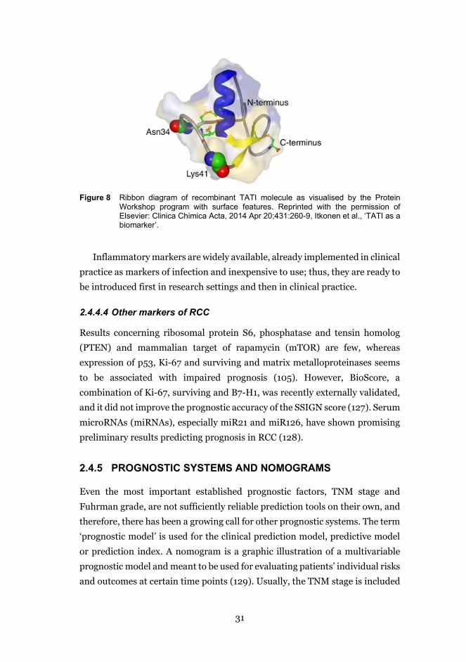
(LD) (121) and tumour-associated trypsin inhibitor (TATI) (122, 123). TATI, a
6-kDa peptide, occurs in high concentrations in the pancreas and pancreatic
fluid and in several tumours. As pancreatectomised patients have normal TATI
concentrations (124), the liver is considered as a main source of TATI (122).
Increased serum levels are noted in renal, ovarian, colorectal, bladder and
pancreatic cancer and are associated with adverse outcomes (122, 123, 125).
Renal excretion removes TATI from circulation. When renal dysfunction
exists, serum TATI (s-TATI) becomes markedly elevated (126). The TATI
molecule is shown in Figure 8.
31
Figure 8 Ribbon diagram of recombinant TATI molecule as visualised by the Protein
Workshop program with surface features. Reprinted with the permission of Elsevier: Clinica Chimica Acta, 2014 Apr 20;431:260-9, Itkonen et al., ‘TATI as a biomarker’.
Inflammatory markers are widely available, already implemented in clinical
practice as markers of infection and inexpensive to use; thus, they are ready to
be introduced first in research settings and then in clinical practice.
2.4.4.4 Other markers of RCC
Results concerning ribosomal protein S6, phosphatase and tensin homolog
(PTEN) and mammalian target of rapamycin (mTOR) are few, whereas
expression of p53, Ki-67 and surviving and matrix metalloproteinases seems
to be associated with impaired prognosis (105). However, BioScore, a
combination of Ki-67, surviving and B7-H1, was recently externally validated,
and it did not improve the prognostic accuracy of the SSIGN score (127). Serum
microRNAs (miRNAs), especially miR21 and miR126, have shown promising
preliminary results predicting prognosis in RCC (128).
2.4.5 PROGNOSTIC SYSTEMS AND NOMOGRAMS
Even the most important established prognostic factors, TNM stage and
Fuhrman grade, are not sufficiently reliable prediction tools on their own, and
therefore, there has been a growing call for other prognostic systems. The term
‘prognostic model’ is used for the clinical prediction model, predictive model
or prediction index. A nomogram is a graphic illustration of a multivariable
prognostic model and meant to be used for evaluating patients’ individual risks
and outcomes at certain time points (129). Usually, the TNM stage is included
32
in all models. The competence of a model is estimated by measuring
discrimination using the concordance index (C-index) or area under the curve
(AUC). Mathematic values of discrimination vary from pure hazard (0.5) to
perfect prediction (1.0). However, no recommendation of an acceptable level
of accuracy for allowing models to be introduced to clinical practice exists.
2.4.5.1 Prognostic systems and nomograms in localised disease
The first models to predict recurrence, the Kattan nomogram (130) and
Cindolo model (89), included clinical presentation (asymptomatic vs
symptomatic) at the time of diagnosis. More current models have excluded
symptoms as the presentation of RCC has changed completely. Currently, up
to 60% patients are asymptomatic (131), while two decades ago, when Cindolo
model was introduced, up to 62% of patients were symptomatic at the time of
diagnosis. (89)
Prognostic models that predict CSS, such as the SSIGN score (84) and the
Karakiewicz nomogram (132), including all TNM stages, usually report better
discrimination than models excluding the metastatic patients, since the
metastatic stage is the most powerful prognosticator of CSS. The SSIGN score
and Karakiewicz nomogram have a report accuracy of over 82-88% (133, 134)
and 87-89% (132), respectively, thus being superior to the TNM stage alone
(77%) (135). Despite the better (135, 136) discrimination, the models including
all stages are mainly used for research purposes only because the endpoint
(CSS) for such a variety of diseases should not guide clinical decision-making.
The Leibovich prognostic score (86) and University of California Integrated
Staging System (UISS) (136) are the most generally used postoperative
prognostic systems. The original purpose of the UISS score was to assess
survival (136), but after few years, the UISS score was adopted to estimate
recurrence-free survival (56), while the Leibovich score was used to estimate
the metastasis-free survival. The Leibovich includes N+ patients and only
ccRCC, but the UISS considers N+ patients as metastatic and accepts all RCC
patients. The most common prognostic models both for local and metastasised
RCC are shown in Table 4.
33
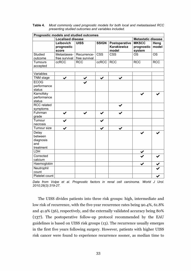
Table 4. Most commonly used prognostic models for both local and metastasised RCC presenting studied outcomes and variables included.
Prognostic models and studied outcomes Localised disease Metastatic disease Leibovich
prognostic score
UISS SSIGN Postoperative Karakiewicz model
MKSCC prognostic system
Heng model
Studied outcome
Metastases-free survival
Recurrence-free survival
CSS CSS OS OS
Tumours accepted
ccRCC RCC ccRCC RCC RCC RCC
Variables TNM stage ✔ ✔ ✔ ✔ ECOG performance status
✔
Karnofsky performance status
✔ ✔
RCC related symptoms
✔
Fuhrman grade
✔ ✔ ✔ ✔
Tumour necrosis
✔ ✔
Tumour size ✔ ✔ ✔ Delay between diagnosis and treatment
✔ ✔
LDH ✔ Corrected calcium
✔ ✔
Haemoglobin ✔ ✔ Neutrophil count
✔
Platelet count ✔
Data from Volpe et al. Prognostic factors in renal cell carcinoma. World J Urol. 2010;28(3):319-27.
The UISS divides patients into three risk groups: high, intermediate and
low risk of recurrence, with the five-year recurrence rates being 90.4%, 61.8%
and 41.9% (56), respectively, and the externally validated accuracy being 80% (137). The postoperative follow-up protocol recommended by the EAU
guidelines is based on UISS risk groups (13). The recurrence usually emerges
in the first five years following surgery. However, patients with higher UISS
risk cancer were found to experience recurrence sooner, as median time to
34
recurrence in high, intermediate and low risk groups were 9.5 months (mean
21.9 ± SD: 26.2), 17.8 months (mean 25.5 ± SD: 23.9) and 28.9 months (mean
26.5 ± SD: 17.1), respectively (56). In addition, in a RECUR database analysis,
52.8% (n=28) of Leibovich low-risk patients recurring in a five-year follow-up
time, 37.1% (n=39) of intermediate risk and 30.5% (n=39) of high-risk patients
were considered potentially curable at the time of recurrence (138).
2.4.5.2 Prognostic systems and nomograms for metastasised disease
The earliest version of MSKCC prognostic model dates back to 1999 (139), and
was updated in 2002 and 2004 (97, 140). In the era of interferon-α, high serum
corrected calcium, high serum lactate dehydrogenase (LDH), low
haemoglobin, low Karnofsky performance status and absence of prior
nephrectomy were all recognised to be independent markers for poor
prognosis (139). They all, except the absence of prior nephrectomy, combined
with delay between diagnosis and treatment, have also continued to be
markers of worse OS after the introduction of tyrosine kinase inhibitor (TKI)
therapy. The importance of performance status assessed by the ECOG scale or
Karnofsky index must be highlighted, as it is the most important patient-
derived prognostic factor in mRCC. Criteria for grading the ECOG scale and
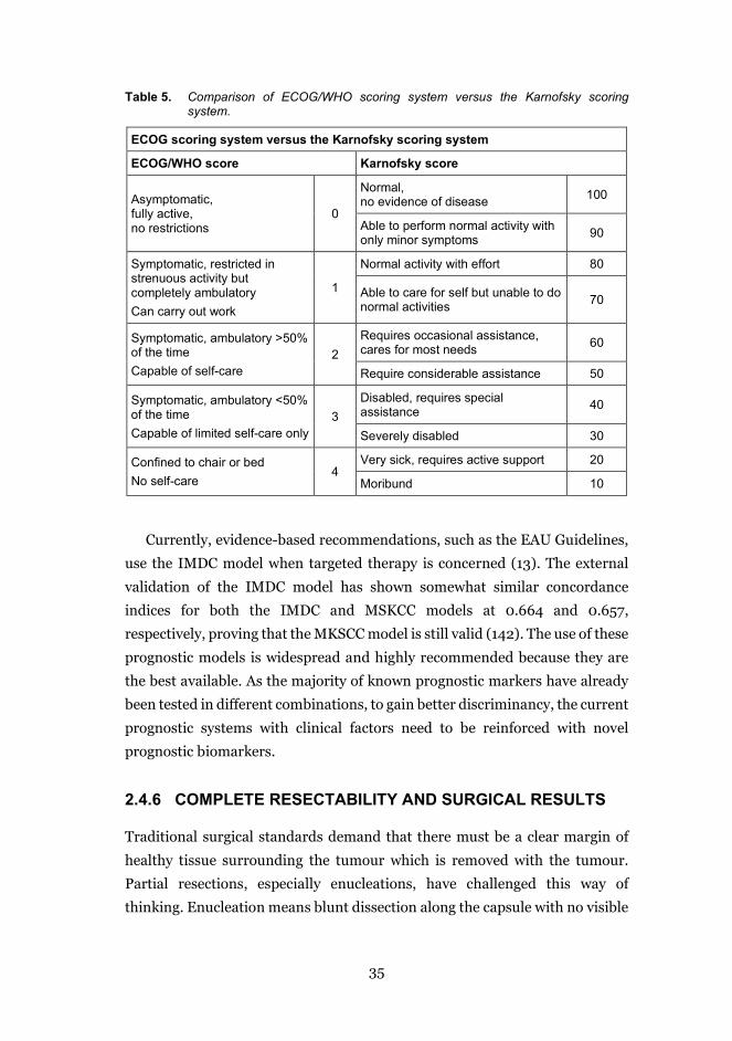
Karnofsky index are presented in Table 5.
The MSKCC model is still used in recommendations of the EAU for CN, for
example; however, as it was developed before the targeted therapy era, it is
considered outdated by some (10). Choueiri et al. combined prognostic factors
to predict progression-free survival after targeted therapy (141).
Heng et al. used the International Metastatic Renal Cancer Database
Consortium (IMDC) data and developed Choueiri’s model, proposing six
prognostic factors for OS and dividing patients into three prognostic groups
(poor, intermediate and good prognosis). The median OS for poor-risk,
intermediate-risk and low-risk groups were determined to be 7.8, 22.5 and
43.3 months, respectively (142).
35
Table 5. Comparison of ECOG/WHO scoring system versus the Karnofsky scoring system.
ECOG scoring system versus the Karnofsky scoring system
ECOG/WHO score Karnofsky score
Asymptomatic, fully active, no restrictions
0
Normal, no evidence of disease 100
Able to perform normal activity with only minor symptoms 90
Symptomatic, restricted in strenuous activity but completely ambulatory Can carry out work
1
Normal activity with effort 80
Able to care for self but unable to do normal activities 70
Symptomatic, ambulatory >50% of the time Capable of self-care
2 Requires occasional assistance, cares for most needs 60
Require considerable assistance 50
Symptomatic, ambulatory <50% of the time Capable of limited self-care only
3 Disabled, requires special assistance 40
Severely disabled 30
Confined to chair or bed No self-care
4 Very sick, requires active support 20
Moribund 10
Currently, evidence-based recommendations, such as the EAU Guidelines,
use the IMDC model when targeted therapy is concerned (13). The external
validation of the IMDC model has shown somewhat similar concordance
indices for both the IMDC and MSKCC models at 0.664 and 0.657,
respectively, proving that the MKSCC model is still valid (142). The use of these
prognostic models is widespread and highly recommended because they are
the best available. As the majority of known prognostic markers have already
been tested in different combinations, to gain better discriminancy, the current
prognostic systems with clinical factors need to be reinforced with novel
prognostic biomarkers.
2.4.6 COMPLETE RESECTABILITY AND SURGICAL RESULTS
Traditional surgical standards demand that there must be a clear margin of
healthy tissue surrounding the tumour which is removed with the tumour.
Partial resections, especially enucleations, have challenged this way of
thinking. Enucleation means blunt dissection along the capsule with no visible
36
layer of normal tissue covering the tumour. Also, in partial resections, the
healthy tissue margin has been getting smaller. Recently, a meta-analysis
confirmed that enucleation is as safe as PN concerning progression-free
survival (PFS) and CSS in a relatively short follow-up time (7.2-54.4 months in
studies of meta-analysis) and in T1 tumours (143, 144).
Positive surgical margins (PSMs), usually meaning that cancer cells are
present in an inked surface, is seen as a risk of oncologic failure, recurrence or
metastasis. Avoiding PSM is of paramount importance in all cancer surgery.
However, results are confusing. Some studies have argued that PSM might be
prognostically significant only with aggressive cancers, if at all (145). Yet, some
recent studies have been able to demonstrate PSM to be a significant factor
predicting recurrence (146). The role of PSMs remains debatable, but until
more conclusive evidence exists, they should be vigorously avoided.
2.5 TREATMENT OPTIONS FOR LOCAL AND LOCALLY ADVANCED RENAL CELL CARCINOMA
2.5.1 ACTIVE SURVEILLANCE
More widespread use of imaging has led to an increasing number of
incidentally diagnosed SRMs. Non-symptomatic tumours, less than 4 cm in
size, are considered as SRMs. Treating all these SRMs aggressively would lead
to overtreatment as approximately 20-30% of them are of benign origin (147).
Most SRMs are diagnosed in the elderly and co-morbid population, making
postponing the treatment a possible option. Active surveillance is a treatment
plan for monitoring patients’ condition but giving treatment only if the
surveilled condition deteriorates. To be included in active surveillance,
patients have to be sufficiently fit to endure the operative or other active
treatment.
The success story of active surveillance of prostatic grade group 1 prostate
cancer has prompted the interest in surveilling low-stage RCC patients. Both
cancers are local, slowly growing tumours which are more prevalent in the
elderly. But distinct features do differ. In active surveillance of SRMs, patients
are monitored by serial abdominal imaging. Information on histology, grade
and pathologic stage might be missing as renal biopsy is not mandatory before
designating patients with SRMs for active surveillance. No serum test, such as
37
S-PSA for prostate cancer, is available for follow-up. Added to this,
intervention for prostate cancer causes more functional consequences while
RN or PN is considered having only a minimal effect on quality of life (QOL)
(148). However, it has been affirmed that preserving nephrons by PN leads to
better QOL (149) (150). This might suggest that, by choosing not to operate,
i.e. ‘choosing the ultimate nephron sparing treatment’, might have a beneficial
influence on certain aspects of QOL. Additionally, no adverse changes in
mental health were noted when comparing active surveillance to operative
surgery, according to one multicentre study (151).
Despite fervent discussions, no agreed-on instructions as to when to move
from active surveillance to active treatment exist. Arbitrary values of SRM
diameter growing larger than 4 cm and growing faster than 0.5 cm/year have
been used (152, 153). Recent results of the multi-institutional Delayed
Intervention and Surveillance for Small Renal Masses (DISSRM) registry
affirmed that active surveillance, when tumour diameter is less than 4 cm, was
not predictive for cancer-specific or OS during a 5-year follow-up time (152).
However, a meta-analysis by Smaldone et al. determined that almost half
of the actively surveilled patients (45.4%) ended up having delayed
intervention at a mean of 30.5 months (range 6-143 months, SD 21.8 months),
mainly due to patients’ wishes (57.2%), but also as a result of tumour growth
(35.7%) (154). Of the SRMs actively surveilled, 2% metastasised at a mean of
40.2 months (154).
Currently, according to EAU guidelines, following young and healthy
patients with active surveillance protocol is against recommendations.
2.5.2 OPERATIVE TREATMENT OF RCC
Over a century, operative removal of local and locally advanced renal tumour
has been the treatment of choice. In 1876, Carl Johan Langenbuch performed
the first successful nephrectomy on a human patient because of a malignant
tumour. It was not long after this that Spencer Wells in 1884 and Vincenz
Cerny in 1887 published the first PN results. Although active surveillance has
been introduced and focal therapies exist, surgery is still the first-line
treatment for local tumours.
RN entails the removal of the entire kidney and surrounding perinephric
fat. PN is recommended for T1 tumours and is an option for larger tumours if
38
technically feasible (155, 156). The indication and possibly gained benefit from
lymphadenectomy is still controversial. In a recent systematic review by Bindi,
lymphadenactomy was found not to increase survival either in M0 or M1 RCC
(157), but contributing studies were sparse and contained risk of bias as only
one randomised controlled trial was investigated (158). However, when
adverse clinical features exist, extended lymphadenectomy should be
considered according to EAU guidelines (13) as a knowledge gap of possible
survival benefit for subgroups of high-risk M0 RCC still exists.
The actual procedures (RN or PN) can be performed as conventional open
procedures or mini-invasive ones using laparoscopic technology with or
without the 3D technique, robotic assistance or the more rarely used hand
assistance. Significant debate and research have been ongoing about the best
practice, previously about the RN vs PN and more recently about the open or
mini-invasive approaches.
As was made evident in a randomised, multicentre study by van Poppel et
al., both PN and RN provide excellent and equal results in terms of oncological
safety (156). PN, compared with RN, is superior when it comes to renal
function and QOL after the operation, but similar to CSS and recurrence-free
survival (RFS), although the OS question remains unanswered (13). Since
oncologic results are pointed out to be similar, and RN deteriorates kidney
function more, PN is the first-line treatment for all T1 tumours if no
contraindications exist (13). In 2015, in all Nordic countries, 55% of all kidney
tumour treatments were RNs and only 37% PNs (14).
In comparing mini-invasive with open RN, researchers found no differences
in CSS, OS or RFS (159, 160). The length of hospital stay, blood loss during
operation and requirements for anaesthetics were lower for laparoscopic RN
patients (160). In experienced centres, conventional laparoscopic and robot-
assisted PN yielded similar results to open PN when looking at OS, RFS and
severe complications (159, 161). Blood loss was lower in mini-invasive PN (161)
(159). Operation time and warm ischaemia time were longer in laparoscopic PN
but not in robot-assisted PN compared with open PN (159, 161, 162). Results
concerning glomerular filtration rate (GFR) decline were somewhat
contradictory: at least no long-term difference in GFR decline was reported (163,
164). However, the conventional laparoscopic PN is a challenging procedure and
comes with a long surgical learning curve. Robot-assisted PN, with articulated
39
wrist instrument motion, has lessened these technical challenges, reduced the
learning curve and reduced the use on laparoscopic PN (165, 166). Robot-
assisted PN must still be regarded as a demanding procedure. As shown by
Larcher et al., the learning curve for robot-assisted PN, with respect to
complications, appears to be endless, without reaching a plateau even after 300
cases (167). Also, amongst hospital regions, there is assumedly a marked
variance in the use of mini-invasive techniques, as the choice is dependent of
financial resources, patient volume and experience of surgeons.
2.5.3 RENAL CARCINOMA WITH TUMOUR THROMBUS
Tumour thrombus (TT) reaching the vena cava inferior is a considerable
adverse prognostic factor for RCC. At the time of diagnosis, up to 10% of RCC
patients have venous extension added to the primary tumour (168).
For treating RCC with TT, nephrectomy, combined with thrombectomy, is
considered the only curative option and sizable surgical resections are accepted
for curative intention. Evidence to support this is based only on case series with
limited sample sizes, often with a single-centre design and inhomogeneous
population (169-171). Results concerning the prognostic significance of the TT
level have been somewhat controversial as some studies have found the level
of TT as an independent prognostic factor (172, 173), while some have not (174,
175). In the presence of non-metastatic disease, surgical removal of the tumour
and TT are strongly recommended (13), while systemic treatment has been
reserved for metastatic disease only.
In a recent retrospective series by Field et al., patients with locally-
advanced or metastatic RCC with TT had improved CSS and reduction in
tumour and thrombus size after receiving neoadjuvant sunitinib (176). Older
studies, with smaller patient populations and with a selection of targeted
therapy offered, have had contrasting results (177, 178), however. More studies
are needed to confirm if patients who are not able to undergo up-front surgical
treatment could benefit from neoadjuvant sunitinib prior to surgery.
Compared with that of localised disease, long-term survival of TT patients
remains poor (169-171). Survival has been outstandingly poor for patients with
lymph node metastases. Isolated lymph node disease seemed to predict shorter
survival (CSS) than single distant metastasis in a large multicentre database
study by Tilki (170), with the 5-year CSS estimates for lymph node positive
40
disease and single distant metastases being 17.3 (95% CI [9.3–27.4]) and
36.8% (95% CI [27.0–46.5]), respectively. Patients with TT and tumours with
papillary histology seemed to be associated with significantly worse outcomes
as 5-year estimates for pRCC and ccRCC were 36.8% (95% CI [27.0–46.5]) and
54.8% (95% CI [51.8–57.8]), respectively (172).
Being major surgery, the surgery of RCC with TT is prone to complications.
According to a large study by Tilki et al., the overall 30-day postoperative
complication rate was 34%, and the major complication rate (Clavien 3-5) was
13% (169).
2.5.4 ABLATIVE THERAPIES
Ablation techniques have not gained wide popularity in Finland nor in Nordic
Countries as initial treatment for kidney tumours (5% and 8%, respectively)
(14). Ablation therapy options are cryoablation and radiofrequency ablation,
done percutaneously or laparoscopically. High-quality data to prove oncologic
outcome or morbidity of ablative therapies is lacking (13). Population-based
study results about oncologic safety are mixed, but no study has proven
ablative therapies to be superior to PN (179, 180). A recent meta-analysis found
that lower morbidity rates and lower GFR reduction favour ablative therapies,
while CSS and OS do favour PN, and local recurrences and appearances of
metastasis do not differ between the two treatments (181). Ablative therapies
are recommended for old and fragile patients as an alternative to PN or active
surveillance (13).
2.5.5 ADJUVANT THERAPY
Surgery is the therapy of choice in non-metastasised RCC. Survival after
surgery, in locally advanced disease, remains modest, however, since 5-year
disease-free survival (DFS) for UISS intermediate and high-risk patients has
been determined to be 64% and 37%, respectively, after nephrectomy (182).
Also, a number of targeted therapies have been studied for reducing recurrence
of cancer.
The findings of the newly published, important trials for Adjuvant
Sorafenib or Sunitinib for Unfavourable Renal Carcinoma (ASSURE) (183),
Sunitinib as Adjuvant Treatment for Patients at High Risk of Recurrence of
41
Renal Cell Cancer (S-TRAC) (184), PROTECT (185) and ATLAS (186) are
mixed. The largest trials to date, ASSURE and PROTECT defined no
differences between the placebo and treatment arm in DFS or OS, but S-TRAC
could prove a DFS difference. ASSURE also enrolled patients with T1b and pT2
disease, whereas S-TRAC only accepted patients with pT3-4 disease. The EAU
guidelines recommend not to offer adjuvant therapy with sorafenib, pazopanib
or axitinib (strength of rating: strong) and recommend against sunitinib
adjuvant therapy in surgically resected high-grade ccRCC (strength of rating:
weak). Recent meta-analysis did prove a DFS benefit, but without a significant
improvement in OS, for patients treated with adjuvant TKIs (187). This meta-
analysis comprised all these four studies, also the ATLAS study which was
missing from the most recent version of guidelines. Patients with greater
tumour size, T3-T4 tumours and/or nodal metastases did benefit more (187).
The biological rationale behind the effect of TKIs in an adjuvant setting is
unknown as is whether or not adjuvant TKI just delays metastases or can if it
actually prevent recurrence and metastasis. Immune check-point inhibitors
have been proven to have promising efficacy in metastatic settings, and several
adjuvant studies on immune check-point inhibitors (PROSPER, IMMotion,
KEYNOTE and CheckMate) are still recruiting or ongoing, and their results
scheduled for 2022 to 2024 are eagerly awaited.
2.6 TREATMENT OF METASTASISED RENAL CELL CARCINOMA
2.6.1 CYTOREDUCTIVE NEPHRECTOMY
Prior to immunotherapy, nephrectomy for metastatic RCC patients was used
for the palliation of symptoms (e.g. unendurable pain, bleeding, uncontrolled
hypertension or hypercalcemia). As well as the option of palliative
nephrectomy, mRCC patients can undergo cytoreductive nephrectomy, meant
for reducing of tumour burden, or nephrectomy combined with
metastasectomy for oligo-metastatic disease aiming to reach a state where
there is no evidence of disease status.
During the last decade, the efficacy of CN was proven in two trials by the
former Southwest Oncology Group (SWOG) (188) and the European
Organisation for Research and Treatment of Cancer (EORTC), respectively
42
(189). CN followed by interferon α-2b improved survival compared with
interferon treatment alone (OS 11.1 vs 8.1 months and 17 vs 7 months,
respectively). Since then, CN has been a routine procedure for patients with a
large primary tumour, restricted amount of metastases and good performance
status. However, the use of CN has declined over time. According to recent
Swedish Cancer Registry, 55% of mRCC patients underwent nephrectomy (2).
However, contemporary demographic data from the US has indicated that
distinctly fewer patients, only 30% of those receiving targeted therapy,
underwent CN (96).
In the era of targeted therapy, the role and sequence of CN remains an
unanswered question. Two large prospective and randomised trials,
CARMENA (11) and EORTC SURTIME (43), tried to answer this question.
CARMENA compared the efficiency of sunitinib alone with immediate CN
followed by sunitinib in mRCC patients. Although the trial failed to reach its
accrual goals, it still had enough power in the intention-to-treat population to
prove the non-inferiority of sunitinib alone vs CN followed by sunitinib.
Although OS for sunitinib alone patients was longer (18.4 vs 13.9 months), the
results of CARMENA cannot prove superiority of sunitinib alone.
Nevertheless, the CARMENA trial lacks real-world applicability, as operated
patients were mainly MSKCC intermediate and high-risk patients. The
proportion of high-risk patients was as high as 43%, although, according to
earlier evidence, CN for poor-risk patients is discouraged (190). Also, the
groups did differ as to the proportion of locally advanced tumours (T3 and T4),
51% and 70.1% for sunitinib alone vs CN-sunitinib group, respectively.
Bex A et al. studied immediate CN followed by sunitinib versus sunitinib
followed by deferred CN in a SURTIME trial (43). Results of SURTIME lack
statistical power to draw any conclusions because of early termination due to
slow accrual. The main finding of SURTIME was that patients undergoing
deferred compared with immediate nephrectomy had longer OS, suggesting
that selecting patients for nephrectomy with a trial phase sunitinib treatment
might be useful. Because of slow recruiting, the time has almost passed for
these results from CARMENA and SURTIME to be of any relevance as a newer
and potentially more efficient option, immunotherapy, has been invented and
trials with immunotherapy are now ongoing.
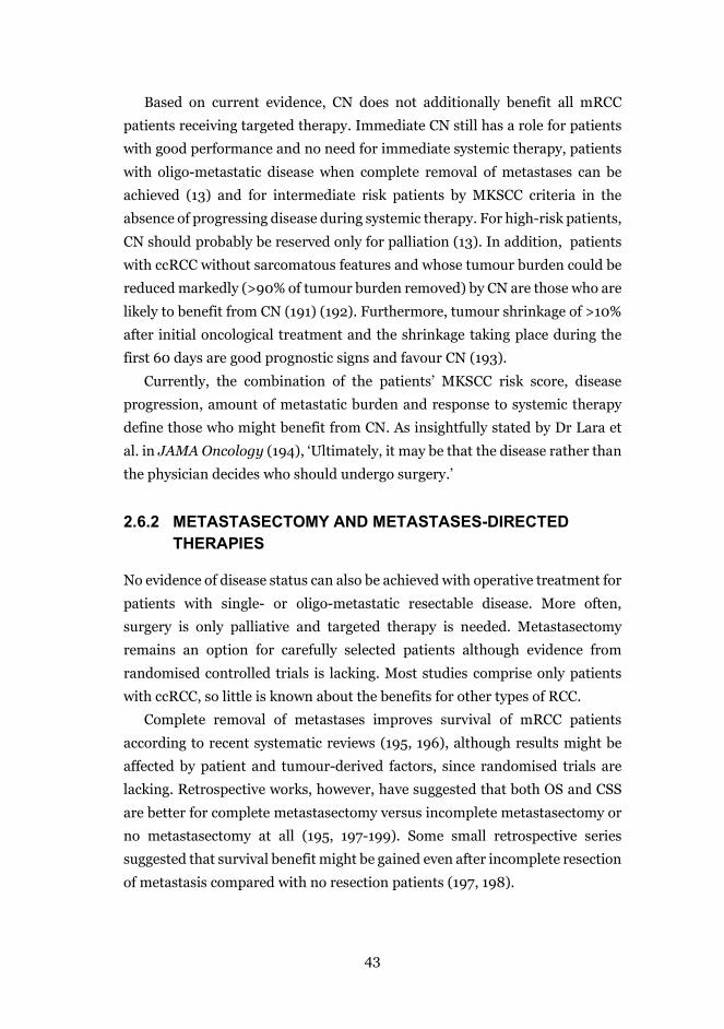
43
Based on current evidence, CN does not additionally benefit all mRCC
patients receiving targeted therapy. Immediate CN still has a role for patients
with good performance and no need for immediate systemic therapy, patients
with oligo-metastatic disease when complete removal of metastases can be
achieved (13) and for intermediate risk patients by MKSCC criteria in the
absence of progressing disease during systemic therapy. For high-risk patients,
CN should probably be reserved only for palliation (13). In addition, patients
with ccRCC without sarcomatous features and whose tumour burden could be
reduced markedly (>90% of tumour burden removed) by CN are those who are
likely to benefit from CN (191) (192). Furthermore, tumour shrinkage of >10%
after initial oncological treatment and the shrinkage taking place during the
first 60 days are good prognostic signs and favour CN (193).
Currently, the combination of the patients’ MKSCC risk score, disease
progression, amount of metastatic burden and response to systemic therapy
define those who might benefit from CN. As insightfully stated by Dr Lara et
al. in JAMA Oncology (194), ‘Ultimately, it may be that the disease rather than
the physician decides who should undergo surgery.’
2.6.2 METASTASECTOMY AND METASTASES-DIRECTED THERAPIES
No evidence of disease status can also be achieved with operative treatment for
patients with single- or oligo-metastatic resectable disease. More often,
surgery is only palliative and targeted therapy is needed. Metastasectomy
remains an option for carefully selected patients although evidence from
randomised controlled trials is lacking. Most studies comprise only patients
with ccRCC, so little is known about the benefits for other types of RCC.
Complete removal of metastases improves survival of mRCC patients
according to recent systematic reviews (195, 196), although results might be
affected by patient and tumour-derived factors, since randomised trials are
lacking. Retrospective works, however, have suggested that both OS and CSS
are better for complete metastasectomy versus incomplete metastasectomy or
no metastasectomy at all (195, 197-199). Some small retrospective series
suggested that survival benefit might be gained even after incomplete resection
of metastasis compared with no resection patients (197, 198).
44
Metastasectomy does also have a role in palliation and postponing the
initiation of targeted treatment to avoid associated toxicity (195, 200). To avoid
the complications of surgery, non-surgical metastases directed-therapies, i.e.
stereotactic radiotherapy and thermal ablation, have been used to treat
metastases and symptoms of metastases.
The most common sites of metastasis in RCC are the lungs (45%), bones
(30%), lymph nodes (22%), liver (20%), brain (8%) and adrenal glands (8%),
but any site is practically possible (201). The primary recurrence sites are the
lungs (54-64%), lymph nodes (22%) and bones (15-20%) (138, 202). Less
common sites include distant lymph nodes, adrenal glands, local recurrences,
pancreas, liver and brain. Metastasectomy is the most relevant local treatment
to all metastatic sites, except brain and possibly bones. Many prognostic
factors for metastasectomy are presented in Figure 9, including patient-
derived factors, features of cancer and completeness of surgery (195) (203).
Factors associated with favourable outcomes after metastasectomy include
good performance status and cancer risk status of good or intermediate risk
(by MSKCC or Heng risk score), metastasis being metachronous with primary
tumour, recurrence-free interval being over two years, metastasis being
solitary or oligo-metastatic, metastasis emerging in a single organ, absence of
nodal metastasis, clear-cell histology, absence of a sarcomatoid component
and low-to-moderate Fuhrman grade (199).
EAU guidelines recommend metastasectomy if complete resection is
achievable and beneficial prognostic factors exist or for mere palliation (13).
Other local therapies can be used for palliative purposes; for example,
stereotactic radiotherapy comes with low toxicity and can significantly
improve local symptoms caused by brain and bone metastasis (204).
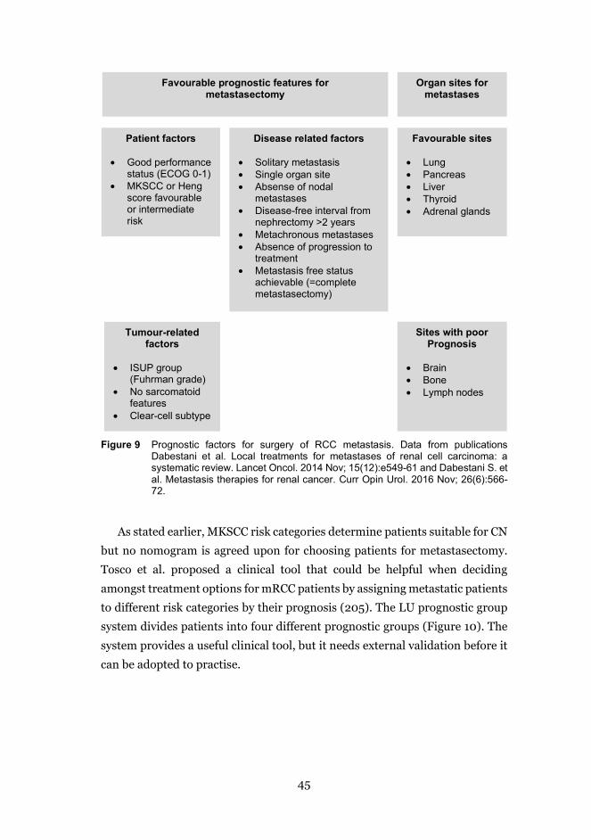
45
Figure 9 Prognostic factors for surgery of RCC metastasis. Data from publications
Dabestani et al. Local treatments for metastases of renal cell carcinoma: a systematic review. Lancet Oncol. 2014 Nov; 15(12):e549-61 and Dabestani S. et al. Metastasis therapies for renal cancer. Curr Opin Urol. 2016 Nov; 26(6):566-72.
As stated earlier, MKSCC risk categories determine patients suitable for CN
but no nomogram is agreed upon for choosing patients for metastasectomy.
Tosco et al. proposed a clinical tool that could be helpful when deciding
amongst treatment options for mRCC patients by assigning metastatic patients
to different risk categories by their prognosis (205). The LU prognostic group
system divides patients into four different prognostic groups (Figure 10). The
system provides a useful clinical tool, but it needs external validation before it
can be adopted to practise.
Favourable prognostic features for metastasectomy
Patient factors
• Good performance status (ECOG 0-1)
• MKSCC or Heng score favourable or intermediate risk
Disease related factors
• Solitary metastasis • Single organ site • Absense of nodal
metastases • Disease-free interval from
nephrectomy >2 years • Metachronous metastases • Absence of progression to
treatment • Metastasis free status
achievable (=complete metastasectomy)
Tumour-related factors
• ISUP group
(Fuhrman grade) • No sarcomatoid
features • Clear-cell subtype
Favourable sites
• Lung • Pancreas • Liver • Thyroid • Adrenal glands
Organ sites for metastases
Sites with poor Prognosis
• Brain • Bone • Lymph nodes
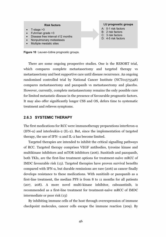
46
Figure 10 Leuven-Udine prognostic groups.
There are some ongoing prospective studies. One is the RESORT trial,
which compares complete metastasectomy and targeted therapy vs
metastasectomy and best supportive care until disease recurrence. An ongoing
randomised controlled trial by National Cancer Institute (NCT01575548)
compares metastasectomy and pazopanib vs metastasectomy and placebo.
However, currently, complete metastasectomy remains the only possible cure
for limited metastatic disease in the presence of favourable prognostic factors.
It may also offer significantly longer CSS and OS, defers time to systematic
treatment and relieves symptoms.
2.6.3 SYSTEMIC THERAPY
The first medications for RCC were immunotherapy preparations interferon-α
(IFN-α) and interleukin-2 (IL-2). But, since the implementation of targeted
therapy, the use of IFN- α and IL-2 has become limited.
Targeted therapies are intended to inhibit the critical signalling pathways
of RCC. Targeted therapy comprises VEGF antibodies, tyrosine kinase and
multikinase inhibitors and mTOR inhibitors (206). Sunitinib and pazopanib,
both TKIs, are the first-line treatment options for treatment-naïve mRCC of
IMDC favourable risk (13). Targeted therapies have proven survival benefits
compared with IFN-α, but durable remissions are rare (206) as cancer finally
develops resistance to these medications. With sunitinib or pazopanib as a
first-line treatment, the median PFS is from 8 to 11 months for all patients
(207, 208). A more novel multi-kinase inhibitor, cabozantinib, is
recommended as a first-line treatment for treatment-naïve mRCC of IMDC
intermediate or poor risk (13)
By inhibiting immune cells of the host through overexpression of immune
checkpoint molecules, cancer cells escape the immune reaction (209). By
LU prognostic groups A: 0-1 risk factors B: 2 risk factors C: 3 risk factors D: 4-5 risk factors
Risk factors • T-stage >3 • Fuhrman grade >3 • Disease free interval ≤12 months • Nonpulmonary metastases • Multiple mestatic sites
47
weakening the negative regulation of immune defence, the immune reactions
of the host are boosted to fight intruding cells (210). Cytotoxic T-lymphocyte
associated antigen 4 (CTLA-4) is a receptor, which turns off the activation of T
cells to prevent excessive reaction. A more physiological and less expensive way
is to unwind the downregulation of readily activated T cells by selectively
blocking the interaction between programmed death 1 (PD1) receptor and
programmed death-ligand 1 (PD-L1) (210).
The phase III trial CheckMate 214 (NCT 02231749) proved that a
combination of ipilimumab (CTLA-4 inhibitor) and nivolumab (a PD-1
immune checkpoint inhibitor) in treatment-naïve advanced or mRCC patients
to be superior to sunitinib in terms of response rate and OS but also in health-
related QOL assessment (211). Therefore, the most recent recommendation by
EAU is to use ipilimumab plus nivolumab as a treatment for IMDC
intermediate and poor-risk previously untreated patients (13). However,
widely removing the downregulation of the immune system is not innocuous:
even life-threatening autoimmune reactions may happen, and more selective
immunological medication is highly anticipated.
2.6.4 PRECISION MEDICINE – THE WAY OF THE FUTURE
Over its history, cancer has been perceived as a disease of an organ, and
oncologic treatment has been chosen for the organ involved, e.g. TKIs for RCC.
In addition, treatments have been traditionally designed for the ‘average
patient’. Consequently, treatments have been effective for some but not for all,
and the effect of the treatment has been tested with a trial period for each
cancer patient. By contrast, precision medicine, e.g. personalised medicine, is
treatment taking into consideration genetics as well as molecular and cellular
features of the tumour (212)
RCC is, however, not a single entity, but includes various subtypes of
tumours. Also, intra- and inter-tumour heterogeneity is huge, being a major
obstacle to selecting individual, and effective, treatment for mRCC. As
demonstrated by Saaed et al., even cells form different tumour regions and
respond differently to cancer medication (213). In clinical research, the bulk
tumour has already been handled with surgery or one drug, and tumour
subclones simultaneously with some other drug (214). Some promising tissue-
based predictive biomarker studies for mRCC exist, but to validate the results,

48
larger studies are needed. Due to the heterogeneity of the index tumour and
metastases, precision medicine will be essentially dependent on tissue samples
for now until more accessible biomarkers emerge.
49
3 AIMS OF THE STUDY
Currently, the prognosis of an individual patient with a renal tumour still
cannot be accurately predicted due to the heterogenous behaviour of tumours.
The ongoing question concerning which patients should be designated for an
operation is more relevant than ever when tumours are ranging from tiny,
indolent tumours to life-threatening metastasised disease. Additionally,
patients are getting older and more vulnerable. Thus, the low number of
complications, good surgical quality and reasonably accurate estimate of life
expectancy are of utmost importance when deciding on the treatment.
The aims of this study were to evaluate the contemporary landscape and
quality of surgical treatment in the Helsinki metropolitan area and identify
prognostic factors of RCC.
The specific aims were as follows:
1) To evaluate changes in the clinical picture and treatment of RCC (I)
2) To evaluate the efficacy of surgery in RCC patients with TT (II)
3) To ensure that anatomical factors and models predict the type of
nephrectomy, and that they have impact on recurrence (III)
4) To evaluate the outcome of metastasectomy of RCC patients and assess
the performance of the LU prognostic model (IV)
5) To appraise the value of s-TATI as a prognostic marker (IV).
50
4 PATIENTS AND METHODS
4.1 STUDY COHORTS AND TIMELINES
The patient cohorts are described in detail in the original publications (I-V).
All patients studied had the clinical condition of RCC (II-V) or were suspected
to have RCC (I). All patients in studies II-V underwent nephrectomy at the
Helsinki University Hospital (HUH).
We searched the consecutive patients for each study from either the
Helsinki University Kidney Tumour Database or Helsinki University Hospital
urological and oncological outpatient registries. Statistics Finland provided the
death certificates, but all other information was gathered from patient
registries.
Tumours were histologically classified according to either WHO 2004
classification (II and III) (46) or WHO/ISUP 2013 classification (studies I, IV
and V) (88) and staging of tumours according to TNM 2009 classification (58).
For studies II and IV, we recorded postoperative complications occurring in
the first 30 days after operation and graded them according to the Clavien-
Dindo classification of surgical complications published by Dindo et al. (215).
Patient and tumour characteristics of each substudy are presented in Table 6.
4.1.1 STUDY I
To describe the whole population with kidney tumours, the outpatient
registries of urological and oncological departments and the institutional
kidney tumour register, covering the entire Helsinki and Uusimaa Hospital
District (HUH) of 1.67 million patients, were searched for ICD10 codes of
D41.0 and C64.88. The intention was to single out all patients with tumours
suspected of being RCC from 2006 to 2016. Having a publicly financed
healthcare system and being the only referral institute for urology in the area,
these registries comprise nearly all renal tumour patients of the metropolitan
area of Helsinki, providing a comprehensive data collection on renal tumours
in a current population.
We identified 1,719 patients who had either a solid or a cystic mass (Bosniak
3-4), with a maximum diameter ≥10 mm. We wanted to study the change in
presentation and treatment patterns, especially the changing proportion of
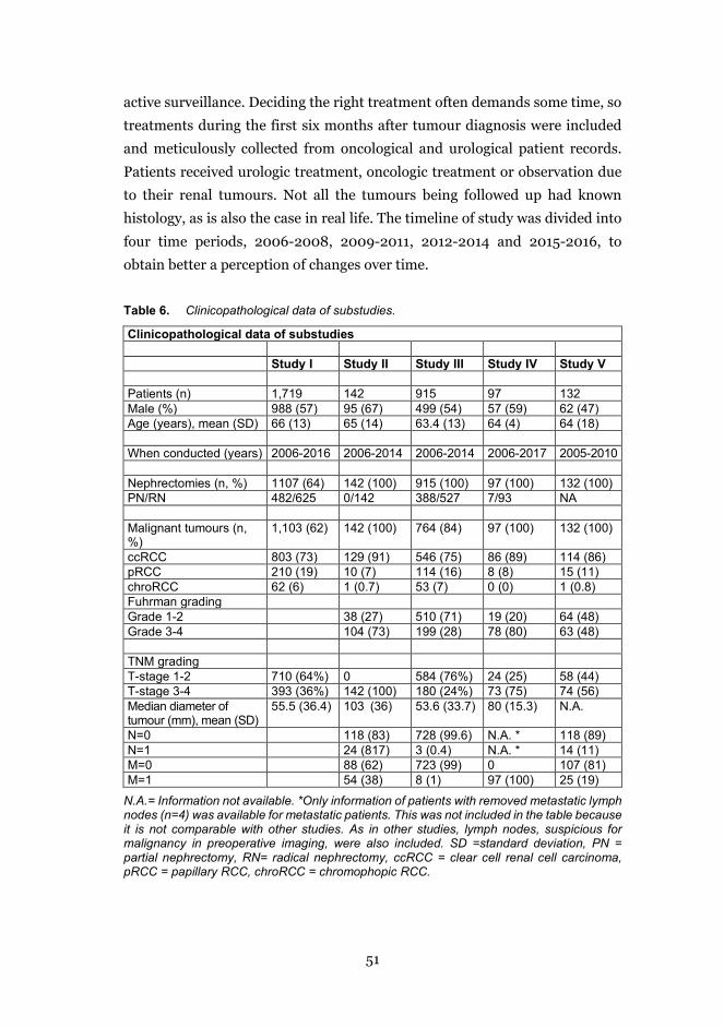
51
active surveillance. Deciding the right treatment often demands some time, so
treatments during the first six months after tumour diagnosis were included
and meticulously collected from oncological and urological patient records.
Patients received urologic treatment, oncologic treatment or observation due
to their renal tumours. Not all the tumours being followed up had known
histology, as is also the case in real life. The timeline of study was divided into
four time periods, 2006-2008, 2009-2011, 2012-2014 and 2015-2016, to
obtain better a perception of changes over time.
Table 6. Clinicopathological data of substudies.
Clinicopathological data of substudies Study I Study II Study III Study IV Study V Patients (n) 1,719 142 915 97 132 Male (%) 988 (57) 95 (67) 499 (54) 57 (59) 62 (47) Age (years), mean (SD) 66 (13) 65 (14) 63.4 (13) 64 (4) 64 (18) When conducted (years) 2006-2016 2006-2014 2006-2014 2006-2017 2005-2010 Nephrectomies (n, %) 1107 (64) 142 (100) 915 (100) 97 (100) 132 (100) PN/RN 482/625 0/142 388/527 7/93 NA Malignant tumours (n, %)
1,103 (62) 142 (100) 764 (84) 97 (100) 132 (100)
ccRCC 803 (73) 129 (91) 546 (75) 86 (89) 114 (86) pRCC 210 (19) 10 (7) 114 (16) 8 (8) 15 (11) chroRCC 62 (6) 1 (0.7) 53 (7) 0 (0) 1 (0.8) Fuhrman grading Grade 1-2 38 (27) 510 (71) 19 (20) 64 (48) Grade 3-4 104 (73) 199 (28) 78 (80) 63 (48) TNM grading T-stage 1-2 710 (64%) 0 584 (76%) 24 (25) 58 (44) T-stage 3-4 393 (36%) 142 (100) 180 (24%) 73 (75) 74 (56) Median diameter of tumour (mm), mean (SD)
55.5 (36.4) 103 (36) 53.6 (33.7) 80 (15.3) N.A.
N=0 118 (83) 728 (99.6) N.A. * 118 (89) N=1 24 (817) 3 (0.4) N.A. * 14 (11) M=0 88 (62) 723 (99) 0 107 (81) M=1 54 (38) 8 (1) 97 (100) 25 (19)
N.A.= Information not available. *Only information of patients with removed metastatic lymph nodes (n=4) was available for metastatic patients. This was not included in the table because it is not comparable with other studies. As in other studies, lymph nodes, suspicious for malignancy in preoperative imaging, were also included. SD =standard deviation, PN = partial nephrectomy, RN= radical nephrectomy, ccRCC = clear cell renal cell carcinoma, pRCC = papillary RCC, chroRCC = chromophopic RCC.
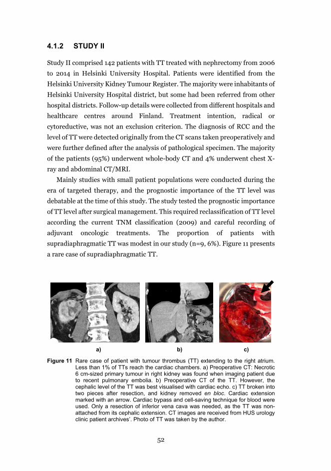
52
4.1.2 STUDY II
Study II comprised 142 patients with TT treated with nephrectomy from 2006
to 2014 in Helsinki University Hospital. Patients were identified from the
Helsinki University Kidney Tumour Register. The majority were inhabitants of
Helsinki University Hospital district, but some had been referred from other
hospital districts. Follow-up details were collected from different hospitals and
healthcare centres around Finland. Treatment intention, radical or
cytoreductive, was not an exclusion criterion. The diagnosis of RCC and the
level of TT were detected originally from the CT scans taken preoperatively and
were further defined after the analysis of pathological specimen. The majority
of the patients (95%) underwent whole-body CT and 4% underwent chest X-
ray and abdominal CT/MRI.
Mainly studies with small patient populations were conducted during the
era of targeted therapy, and the prognostic importance of the TT level was
debatable at the time of this study. The study tested the prognostic importance
of TT level after surgical management. This required reclassification of TT level
according the current TNM classification (2009) and careful recording of
adjuvant oncologic treatments. The proportion of patients with
supradiaphragmatic TT was modest in our study (n=9, 6%). Figure 11 presents
a rare case of supradiaphragmatic TT.
a) b) c)
Figure 11 Rare case of patient with tumour thrombus (TT) extending to the right atrium. Less than 1% of TTs reach the cardiac chambers. a) Preoperative CT: Necrotic 6 cm-sized primary tumour in right kidney was found when imaging patient due to recent pulmonary embolia. b) Preoperative CT of the TT. However, the cephalic level of the TT was best visualised with cardiac echo. c) TT broken into two pieces after resection, and kidney removed en bloc. Cardiac extension marked with an arrow. Cardiac bypass and cell-saving technique for blood were used. Only a resection of inferior vena cava was needed, as the TT was non-attached from its cephalic extension. CT images are received from HUS urology clinic patient archives’. Photo of TT was taken by the author.
53
4.1.3 STUDY III
Study III evaluated the performance of most fundamental tumour
characteristics, tumour diameter and parenchymal invasion depth, compared
with more compound classifications, such as PADUA classification and RTII,
in predicting the type of nephrectomy performed. Nephrectomy types
compared were PN (n=388, 42%) and RN (n=527, 58%). Patients underwent
operations at Helsinki University Hospital from 1.1.2006 to 31.12.2014 for
tumours suspicious for malignancy. The information was retrieved from
Helsinki University Kidney Tumour Register.
Twenty-six consultant urologists performed or assisted in these 1,284
operations. The type of nephrectomy was ultimately decided by the primary
surgeon based on CT or MRI imaging of the tumour and patient-derived
factors. No anatomic classifications were used as a decision aid at the time of
the study. For this study, tumour maximal diameter, the depth of parenchymal
invasion, PADUA classification and RTII were assessed by one urologist only
according to published methods (62, 68).
4.1.4 STUDY IV
Study IV was conducted to assess the safety and efficacy of metastasectomy for
RCC patients and to test the performance of the previously published LU
prognostic group model. The Helsinki University Kidney Tumour Register was
searched for patients with surgically treated metastases of RCC, and it
provided 97 patients with sporadic metastatic RCC treated from 2006 to 2017.
All patients included in the study also ultimately underwent surgical treatment
for the primary tumour. Medical oncologists planned oncological treatments
according to prevailing practise when no curable surgical treatment existed.
After nephrectomy and metastasectomy, patients were followed up with
imaging, laboratory tests and physical examinations according to risk of
recurrence. The LU prognostic groups comprised patients with 0 to 5 risk
factors, and the patients were divided to groups according the original
publication by Tosco et al. (205) (Figure 10).
54
4.1.5 STUDY V
Study V looked into the value of the serum marker TATI in predicting outcomes
after nephrectomy. Patients (n=173) who had undergone either PN or RN at
Helsinki University Hospital from 2005-2010 for any kind of renal tumour
were included in this study. Patients with benign histology or non-RCC cancer
as well as patients with severely reduced renal function were excluded from
this study (n=41). No patient had acute infection or pancreatitis at the time of
serum sample measurement. We used preoperative CT of the thorax, abdomen
and pelvis to determine the metastatic status, but the status was updated if
positive lymph nodes were seen in pathological examination.
Serum samples were taken preoperatively at a median of 1.0 day (range 0-
50 days) prior to the operation and a minimum of 3 weeks after nephrectomy
(median of 52 days, range 25-176 days), to avoid the effect of inflammatory
response caused by surgery. Serum samples were stored at -20oC before the
assay. S-TATI was determined by time-resolved immunofluorometry assay
(TR-IFMA) according to methods previously described.
4.2 STATISTICAL ANALYSIS
All statistical analyses were performed with IBM SPSS Statistics versions 22-
24 (IBM, Armonk, NY, USA). Statistical significance was set at P<0.05 in two
sided-tests.
In studies I and III, the tests used to assess comparisons amongst
clinicopathological factors were done with Student t-test and chi-squared test
for continuous and categorical variables, respectively. In study V, associations
between S-TATI and clinical variables were calculated using Spearman’s rho,
Mann-Whitney U test and Kruskall-Wallis test, when appropriate. The
Wilcoxon signed-rank test was used to analyse differences of medians of pre-
and postoperative S-TATI measurements.
In publications II, IV and V, the survival times (OS, CSS and RFS) were
defined as the time from nephrectomy to death from any cause, to death from
RCC or to first recurrence, respectively. In study IV, the OS and the RFS were
calculated from the first surgery for metastases. The Kaplan-Meier estimate
was used to analyse the median survival times with 95% CIs for all CSS, RFS
and OS, and statistical significance amongst groups was analysed using a log-
55
rank test. Effects of clinicopathological data and other factors studied were
assessed using uni- and multivariate Cox proportional hazard models. These
results are expressed as odds ratios (ORs) with 95% CI.
In study III, we calculated the receiver operating characteristic (ROC)
curves with areas under the curve (AUCs) to compare the effect of anatomical
parameters in predicting the type of nephrectomy. We performed
multivariable regression analyses for nephrectomy type using a backward
stepwise likelihood ratio test. The anatomic variables correlated strongly.
Therefore, we tested them separately and combined them with a constant
group of independent non-anatomic cofactors (hypertension, age and year of
surgery) aiming to recognise the real-life effect of a single anatomic factor. By
expressing the observed results as a percentage of the expected results, the
goodness of fit of the models was determined.
4.3 ETHICS
All data for studies I-IV were collected retrospectively from Helsinki University
Kidney Tumour Database and Helsinki University urological and oncological
patient registries, without requiring informed consent from patients. In study
V, patients had given informed consent at enrolment before the planned
nephrectomy. The Ethics Committee of the Helsinki and Uusimaa Hospital
District approved all studies.
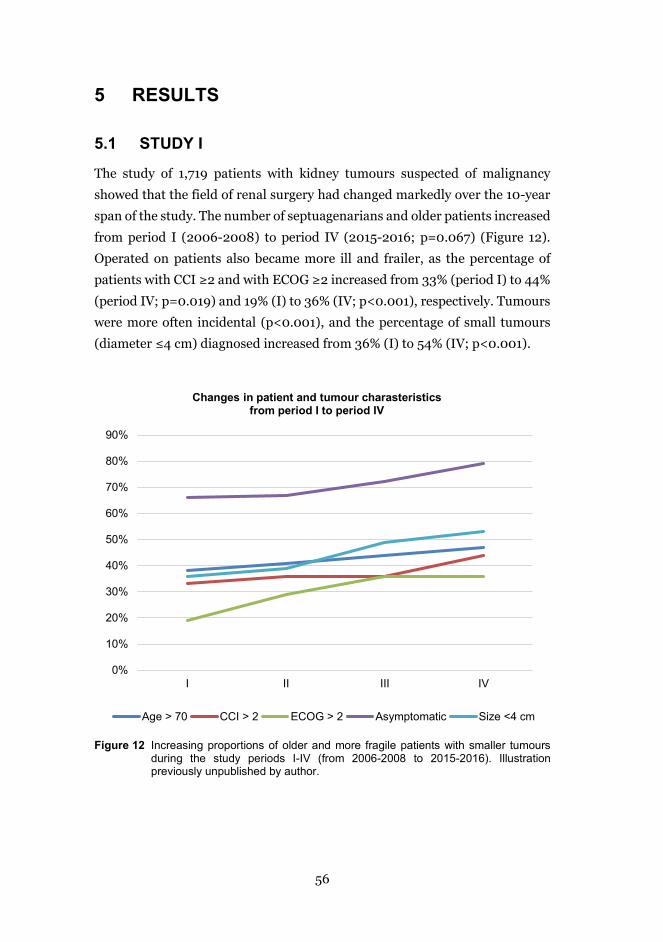
56
5 RESULTS
5.1 STUDY I
The study of 1,719 patients with kidney tumours suspected of malignancy
showed that the field of renal surgery had changed markedly over the 10-year
span of the study. The number of septuagenarians and older patients increased
from period I (2006-2008) to period IV (2015-2016; p=0.067) (Figure 12).
Operated on patients also became more ill and frailer, as the percentage of
patients with CCI ≥2 and with ECOG ≥2 increased from 33% (period I) to 44%
(period IV; p=0.019) and 19% (I) to 36% (IV; p<0.001), respectively. Tumours
were more often incidental (p<0.001), and the percentage of small tumours
(diameter ≤4 cm) diagnosed increased from 36% (I) to 54% (IV; p<0.001).
Figure 12 Increasing proportions of older and more fragile patients with smaller tumours
during the study periods I-IV (from 2006-2008 to 2015-2016). Illustration previously unpublished by author.
0%
10%
20%
30%
40%
50%
60%
70%
80%
90%
I II III IV
Changes in patient and tumour charasteristics from period I to period IV
Age > 70 CCI > 2 ECOG > 2 Asymptomatic Size <4 cm
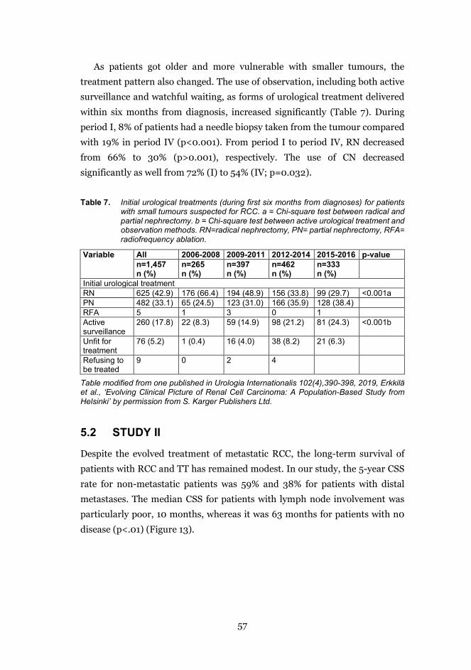
57
As patients got older and more vulnerable with smaller tumours, the
treatment pattern also changed. The use of observation, including both active
surveillance and watchful waiting, as forms of urological treatment delivered
within six months from diagnosis, increased significantly (Table 7). During
period I, 8% of patients had a needle biopsy taken from the tumour compared
with 19% in period IV (p<0.001). From period I to period IV, RN decreased
from 66% to 30% (p>0.001), respectively. The use of CN decreased
significantly as well from 72% (I) to 54% (IV; p=0.032).
Table 7. Initial urological treatments (during first six months from diagnoses) for patients with small tumours suspected for RCC. a = Chi-square test between radical and partial nephrectomy. b = Chi-square test between active urological treatment and observation methods. RN=radical nephrectomy, PN= partial nephrectomy, RFA= radiofrequency ablation.
Variable All 2006-2008 2009-2011 2012-2014 2015-2016 p-value n=1,457 n (%)
n=265 n (%)
n=397 n (%)
n=462 n (%)
n=333 n (%)
Initial urological treatment RN 625 (42.9) 176 (66.4) 194 (48.9) 156 (33.8) 99 (29.7) <0.001a PN 482 (33.1) 65 (24.5) 123 (31.0) 166 (35.9) 128 (38.4) RFA 5 1 3 0 1 Active surveillance
260 (17.8) 22 (8.3) 59 (14.9) 98 (21.2) 81 (24.3) <0.001b
Unfit for treatment
76 (5.2) 1 (0.4) 16 (4.0) 38 (8.2) 21 (6.3)
Refusing to be treated
9 0 2 4
Table modified from one published in Urologia Internationalis 102(4),390-398, 2019, Erkkilä et al., ‘Evolving Clinical Picture of Renal Cell Carcinoma: A Population-Based Study from Helsinki’ by permission from S. Karger Publishers Ltd.
5.2 STUDY II
Despite the evolved treatment of metastatic RCC, the long-term survival of
patients with RCC and TT has remained modest. In our study, the 5-year CSS
rate for non-metastatic patients was 59% and 38% for patients with distal
metastases. The median CSS for patients with lymph node involvement was
particularly poor, 10 months, whereas it was 63 months for patients with n0
disease (p<.01) (Figure 13).
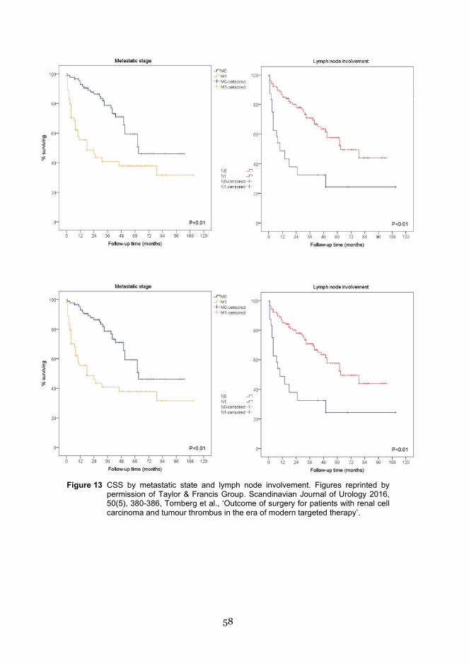
58
Figure 13 CSS by metastatic state and lymph node involvement. Figures reprinted by
permission of Taylor & Francis Group. Scandinavian Journal of Urology 2016, 50(5), 380-386, Tornberg et al., ‘Outcome of surgery for patients with renal cell carcinoma and tumour thrombus in the era of modern targeted therapy’.
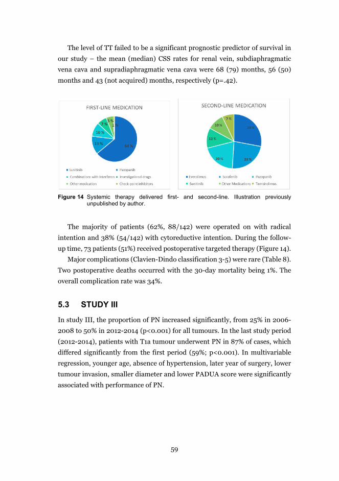
59
The level of TT failed to be a significant prognostic predictor of survival in
our study – the mean (median) CSS rates for renal vein, subdiaphragmatic
vena cava and supradiaphragmatic vena cava were 68 (79) months, 56 (50)
months and 43 (not acquired) months, respectively (p=.42).
Figure 14 Systemic therapy delivered first- and second-line. Illustration previously
unpublished by author.
The majority of patients (62%, 88/142) were operated on with radical
intention and 38% (54/142) with cytoreductive intention. During the follow-
up time, 73 patients (51%) received postoperative targeted therapy (Figure 14).
Major complications (Clavien-Dindo classification 3-5) were rare (Table 8).
Two postoperative deaths occurred with the 30-day mortality being 1%. The
overall complication rate was 34%.
5.3 STUDY III
In study III, the proportion of PN increased significantly, from 25% in 2006-
2008 to 50% in 2012-2014 (p<0.001) for all tumours. In the last study period
(2012-2014), patients with T1a tumour underwent PN in 87% of cases, which
differed significantly from the first period (59%; p<0.001). In multivariable
regression, younger age, absence of hypertension, later year of surgery, lower
tumour invasion, smaller diameter and lower PADUA score were significantly
associated with performance of PN.
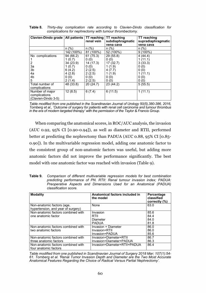
60
Table 8. Thirty-day complication rate according to Clavien-Dindo classification for complications for nephrectomy with tumour thrombectomy.
Clavien-Dindo grade All patients TT reaching renal vein
TT reaching subdiaphragmatic vena cava
TT reaching supradiaphragmatic vena cava
n (%) n (%) n (%) n (%) 142 (100%) 81 (100%) 52 (100%) 9 (100%) No complications 94 (66.2) 61 (75.3) 29 (55.8) 4 (44.4) 1 1 (0.7) 0 (0) 0 (0) 1 (11.1) 2 34 (23.9) 14 (17.3) 17 (32.7) 3 (33.3) 3a 1 (0.7) 0 (0) 1 (1.9) 0 (0) 3b 6 (4.2) 2 (2.5) 4 (7.7) 0 (0) 4a 4 (2.8) 2 (2.5) 1 (1.9) 1 (11.1) 4b 0 (0) 0 (0) 0 (0) 0 (0) 5 2 (1.4) 2 (2.5) 0 (0) 0 (0) Total number of complications
48 (33.8) 20 (24.7) 23 (44.2) 5 (55.5)
Number of major complications (Clavien-Dindo 3-5)
12 (8.5) 6 (7.4) 6 (11.5) 1 (11.1)
Table modified from one published in the Scandinavian Journal of Urology 50(5),380-386, 2016, Tornberg et al., ‘Outcome of surgery for patients with renal cell carcinoma and tumour thrombus in the era of modern targeted therapy’ with the permission of the Taylor & Francis Group.
When comparing the anatomical scores, in ROC/AUC analysis, the invasion
(AUC 0.92, 95% CI [0.90-0.94]), as well as diameter and RTII, performed
better at predicting the nephrectomy than PADUA (AUC 0.88, 95% CI [0.85-
0.90]). In the multivariable regression model, adding one anatomic factor to
the consistent group of non-anatomic factors was useful, but adding more
anatomic factors did not improve the performance significantly. The best
model with one anatomic factor was reached with invasion (Table 9).
Table 9. Comparison of different multivariable regression models for best combination predicting performance of PN. RTII: Renal tumour invasion index; PADUA: Preoperative Aspects and Dimensions Used for an Anatomical (PADUA) classification score.
Modality Anatomical factors included in the model
Percentage classified correctly (%)
Non-anatomic factors (age, hypertension, and year of surgery)
None 63.0
Non-anatomic factors combined with one anatomic factor
Invasion RTII Diameter PADUA
85.6 84.4 84.0 81.8
Non-anatomic factors combined with two anatomic factors
Invasion + Diameter Invasion+RTII Invasion+PADUA
86.0 86.0 85.6
Non-anatomic factors combined with three anatomic factors
Invasion+Diameter+RTII Invasion+Diameter+PADUA
86.7 86.3
Non-anatomic factors combined with four anatomic factors
Invasion+Diameter+RTII+PADUA 86.4
Table modified from one published in Scandinavian Journal of Surgery 2018 Mar; 107(1):54-61. Tornberg et al. ‘Renal Tumor Invasion Depth and Diameter are the Two Most Accurate Anatomical Features Regarding the Choice of Radical Versus Partial Nephrectomy’.
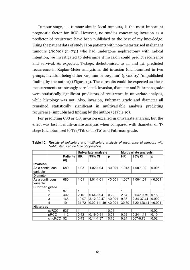
61
Tumour stage, i.e. tumour size in local tumours, is the most important
prognostic factor for RCC. However, no studies concerning invasion as a
predictor of recurrence have been published to the best of my knowledge.
Using the patient data of study II on patients with non-metastasised malignant
tumours (N0M0) (n=731) who had undergone nephrectomy with radical
intention, we investigated to determine if invasion could predict recurrence
and survival. As expected, T-stage, dichotomised to T1 and T2, predicted
recurrence in Kaplan-Meier analysis as did invasion (dichotomised in two
groups, invasion being either <25 mm or ≥25 mm) (p<0.005) (unpublished
finding by the author) (Figure 15). These results could be expected as these
measurements are strongly correlated. Invasion, diameter and Fuhrman grade
were statistically significant predictors of recurrence in univariate analysis,
while histology was not. Also, invasion, Fuhrman grade and diameter all
remained statistically significant in multivariable analysis predicting
recurrence (unpublished finding by the author) (Table 10).
For predicting CSS or OS, invasion excelled in univariate analysis, but the
effect was lost in multivariate analysis when compared with diameter or T-
stage (dichotomised to T1a/T1b or T1/T2) and Fuhrman grade.
Table 10. Results of univariate and multivariate analysis of recurrence of tumours with NoMo status at the time of operation.
Univariate analysis Multivariate analysis Patients
(n) HR 95% CI p HR 95% CI p
Invasion As a continuous variable
680 1.03 1.02-1.04 <0.001 1.013 1.00-1.02 0.005
Diameter As a continuous variable
680 1.01 1.01-1.01 <0.001 1.007 1.00-1.01 <0.001
Fuhrman grade 1 97 1 1 2 402 2.10 0.64-6.94 0.22 2.64 0.64-10.79 0.18 3 166 10.07 3.12-32.47 <0.001 9.36 2.34-37.44 0.002 4 19 31.72 9.02-111.49 <0.001 30.39 7.20-128.44 <0.001 Histology ccRCC 537 1 0.04 1 0.02 pRCC 112 0.42 0.19-0.91 0.03 0.52 0.24-1.13 0.10 chroRCC 52 0.43 0.14-1.37 0.16 0.24 007-0.78 0.02
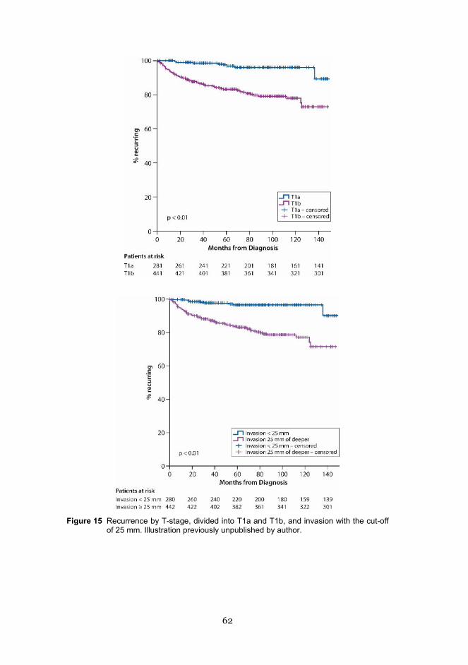
62
Figure 15 Recurrence by T-stage, divided into T1a and T1b, and invasion with the cut-off
of 25 mm. Illustration previously unpublished by author.
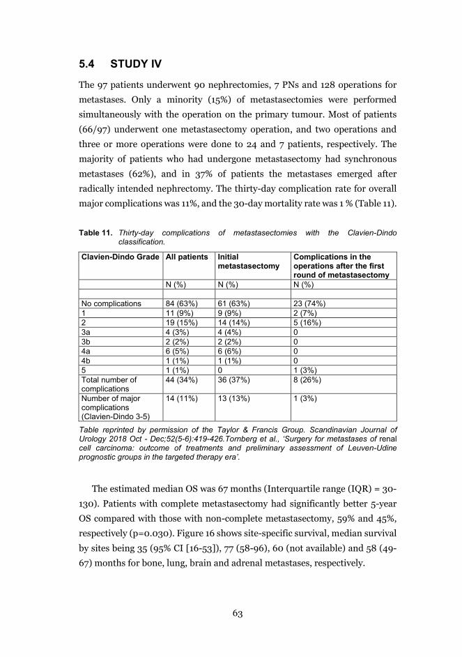
63
5.4 STUDY IV
The 97 patients underwent 90 nephrectomies, 7 PNs and 128 operations for
metastases. Only a minority (15%) of metastasectomies were performed
simultaneously with the operation on the primary tumour. Most of patients
(66/97) underwent one metastasectomy operation, and two operations and
three or more operations were done to 24 and 7 patients, respectively. The
majority of patients who had undergone metastasectomy had synchronous
metastases (62%), and in 37% of patients the metastases emerged after
radically intended nephrectomy. The thirty-day complication rate for overall
major complications was 11%, and the 30-day mortality rate was 1 % (Table 11).
Table 11. Thirty-day complications of metastasectomies with the Clavien-Dindo classification.
Clavien-Dindo Grade All patients Initial metastasectomy
Complications in the operations after the first round of metastasectomy
N (%) N (%) N (%) No complications 84 (63%) 61 (63%) 23 (74%) 1 11 (9%) 9 (9%) 2 (7%) 2 19 (15%) 14 (14%) 5 (16%) 3a 4 (3%) 4 (4%) 0 3b 2 (2%) 2 (2%) 0 4a 6 (5%) 6 (6%) 0 4b 1 (1%) 1 (1%) 0 5 1 (1%) 0 1 (3%) Total number of complications
44 (34%) 36 (37%) 8 (26%)
Number of major complications (Clavien-Dindo 3-5)
14 (11%) 13 (13%) 1 (3%)
Table reprinted by permission of the Taylor & Francis Group. Scandinavian Journal of Urology 2018 Oct - Dec;52(5-6):419-426.Tornberg et al., ‘Surgery for metastases of renal cell carcinoma: outcome of treatments and preliminary assessment of Leuven-Udine prognostic groups in the targeted therapy era’.
The estimated median OS was 67 months (Interquartile range (IQR) = 30-
130). Patients with complete metastasectomy had significantly better 5-year
OS compared with those with non-complete metastasectomy, 59% and 45%,
respectively (p=0.030). Figure 16 shows site-specific survival, median survival
by sites being 35 (95% CI [16-53]), 77 (58-96), 60 (not available) and 58 (49-
67) months for bone, lung, brain and adrenal metastases, respectively.
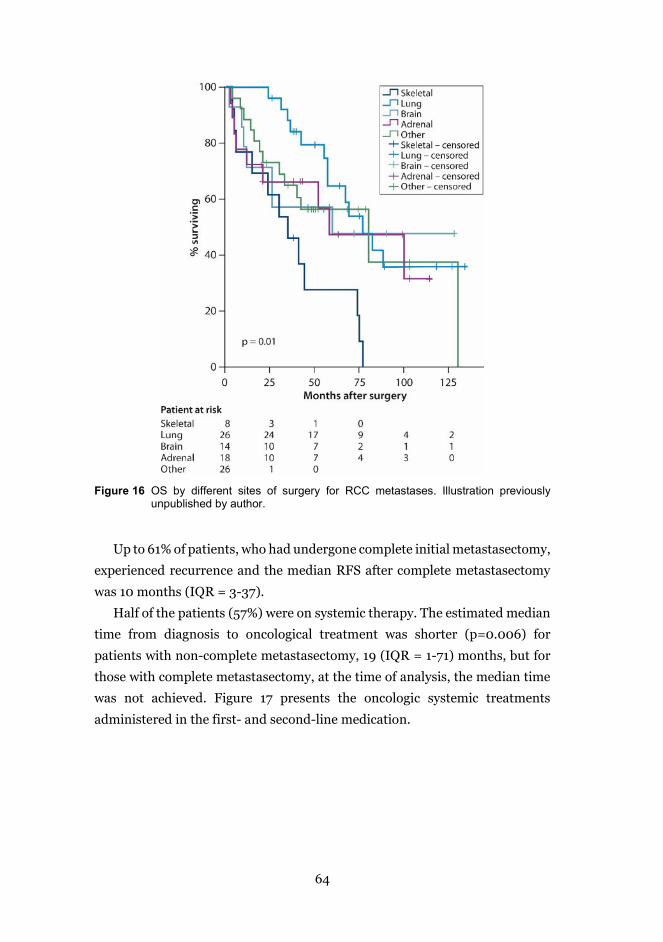
64
Figure 16 OS by different sites of surgery for RCC metastases. Illustration previously
unpublished by author.
Up to 61% of patients, who had undergone complete initial metastasectomy,
experienced recurrence and the median RFS after complete metastasectomy
was 10 months (IQR = 3-37).
Half of the patients (57%) were on systemic therapy. The estimated median
time from diagnosis to oncological treatment was shorter (p=0.006) for
patients with non-complete metastasectomy, 19 (IQR = 1-71) months, but for
those with complete metastasectomy, at the time of analysis, the median time
was not achieved. Figure 17 presents the oncologic systemic treatments
administered in the first- and second-line medication.
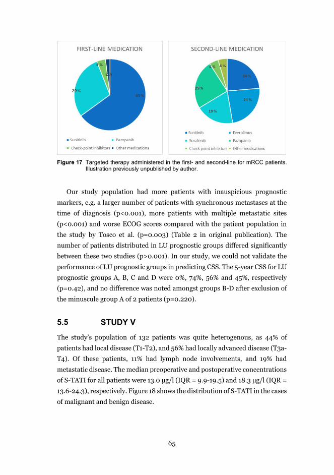
65
Figure 17 Targeted therapy administered in the first- and second-line for mRCC patients.
Illustration previously unpublished by author.
Our study population had more patients with inauspicious prognostic
markers, e.g. a larger number of patients with synchronous metastases at the
time of diagnosis (p<0.001), more patients with multiple metastatic sites
(p<0.001) and worse ECOG scores compared with the patient population in
the study by Tosco et al. (p=0.003) (Table 2 in original publication). The
number of patients distributed in LU prognostic groups differed significantly
between these two studies (p>0.001). In our study, we could not validate the
performance of LU prognostic groups in predicting CSS. The 5-year CSS for LU
prognostic groups A, B, C and D were 0%, 74%, 56% and 45%, respectively
(p=0.42), and no difference was noted amongst groups B-D after exclusion of
the minuscule group A of 2 patients (p=0.220).
5.5 STUDY V
The study’s population of 132 patients was quite heterogenous, as 44% of
patients had local disease (T1-T2), and 56% had locally advanced disease (T3a-
T4). Of these patients, 11% had lymph node involvements, and 19% had
metastatic disease. The median preoperative and postoperative concentrations
of S-TATI for all patients were 13.0 µg/l (IQR = 9.9-19.5) and 18.3 µg/l (IQR =
13.6-24.3), respectively. Figure 18 shows the distribution of S-TATI in the cases
of malignant and benign disease.
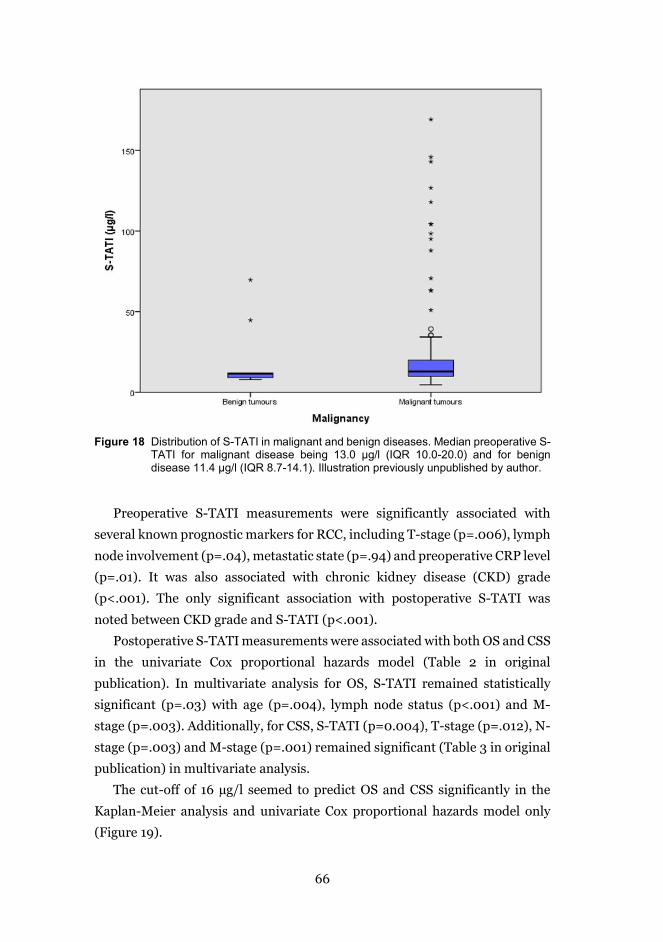
66
Figure 18 Distribution of S-TATI in malignant and benign diseases. Median preoperative S-
TATI for malignant disease being 13.0 µg/l (IQR 10.0-20.0) and for benign disease 11.4 µg/l (IQR 8.7-14.1). Illustration previously unpublished by author.
Preoperative S-TATI measurements were significantly associated with
several known prognostic markers for RCC, including T-stage (p=.006), lymph
node involvement (p=.04), metastatic state (p=.94) and preoperative CRP level
(p=.01). It was also associated with chronic kidney disease (CKD) grade
(p<.001). The only significant association with postoperative S-TATI was
noted between CKD grade and S-TATI (p<.001).
Postoperative S-TATI measurements were associated with both OS and CSS
in the univariate Cox proportional hazards model (Table 2 in original
publication). In multivariate analysis for OS, S-TATI remained statistically
significant (p=.03) with age (p=.004), lymph node status (p<.001) and M-
stage (p=.003). Additionally, for CSS, S-TATI (p=0.004), T-stage (p=.012), N-
stage (p=.003) and M-stage (p=.001) remained significant (Table 3 in original
publication) in multivariate analysis.
The cut-off of 16 µg/l seemed to predict OS and CSS significantly in the
Kaplan-Meier analysis and univariate Cox proportional hazards model only
(Figure 19).
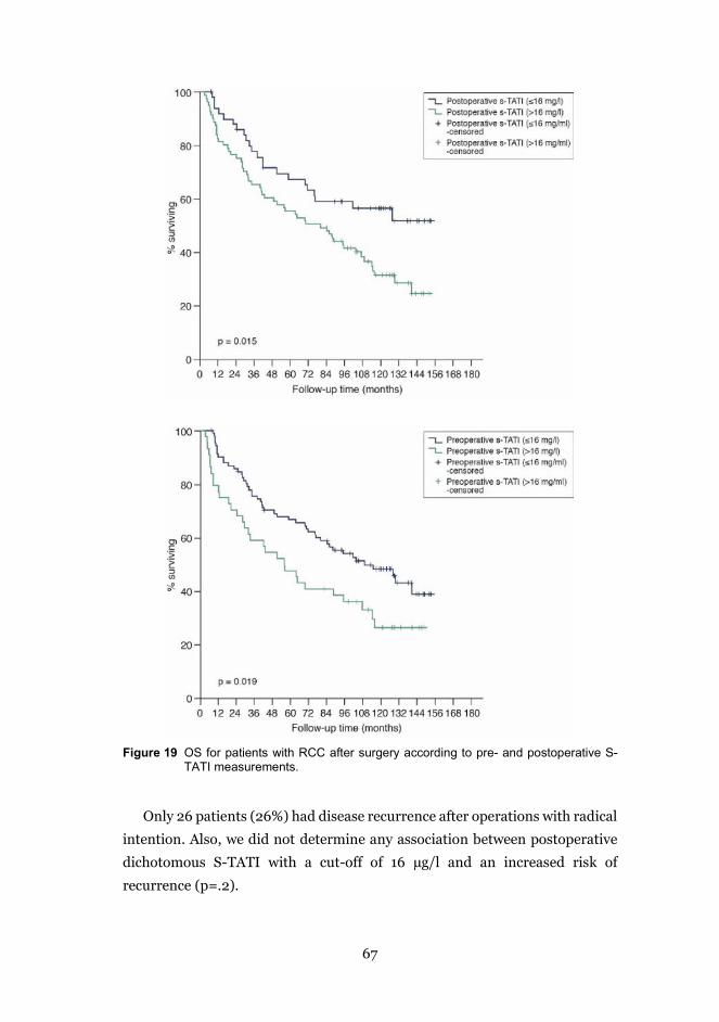
67
Figure 19 OS for patients with RCC after surgery according to pre- and postoperative S-
TATI measurements.
Only 26 patients (26%) had disease recurrence after operations with radical
intention. Also, we did not determine any association between postoperative
dichotomous S-TATI with a cut-off of 16 µg/l and an increased risk of
recurrence (p=.2).
68
6 DISCUSSION
The marked increase of incidentally found renal tumours has been estimated
to have reached a plateau or even started to decline, at least in Europe (18). As
the population ages, a larger proportion of renal tumours is found in elderly
and co-morbid people. Not all patients with localised RCC who are fit for
surgery undergo nephrectomy, but a growing proportion of patients are
actively surveilled. In addition, first-line options for mRCC and advanced RCC
are on the verge of a major change: the role of cytoreductive nephrectomy is
being questioned, and there is a variety of systemic therapies to choose from.
For finding the optimal follow-up and treatment protocol for each patient, the
search for an optimal biomarker is of greater interest than ever. The growing
understanding of immunogenic and molecular mechanisms of RCC has led us
to this moment when we finally might be able to find truly predictive
biomarkers or prognostic systems.
6.1 SUSPICIOUS MASS IN KIDNEY: ANY CHANGES IN TREATMENT PATTERNS?
The tumours suspected of being RCC are a major dilemma and pose serious
questions: when to follow-up, is biopsy needed, when to operate and will the
patient survive long enough to benefit from surgery. Our study (I) comprises
practically all tumours suspected of being RCC in the HUH region. By
restricting inclusion to tumours, not based on histology but suspicion of
malignancy, we wanted to address this common clinical challenge: how to
manage an unknown kidney mass identified radiologically. According to study
(I), the proportion of active surveillance increased during the study period,
although no commonly accepted protocol and admittance criteria exist. To
support the decision about surveillance or operation, the proportion of tumour
biopsies taken increased as well. During the study period, we aimed to
familiarise urologists, radiologists and pathologists with the method. However,
it must be remembered that RTB comes with morbidity. As it has been stated
by Uzosike et al., RTB is appropriate only if it will change the treatment (152).
And, due to tumour heterogeneity, Uzosike et al. does not recommend follow-
up based on biopsy findings in young and healthy patients fit enough to
undergo PN (152). Tumour seeding, especially when pRCC is concerned, is a
69
minor but existing risk, and it must be balanced against the usefulness of
information received by tumour biopsy.
We counted as initial or first-line the treatments delivered during the first
six months after the diagnosis because sometimes reaching the verdict takes
time. It is important to note that, based on large studies, it might not always be
wise to rush on selecting a definite treatment during the first six-month period
(152, 154) as, reportedly, brief follow-up is considered safe and is needed for
determining growth rate (152). Certainly, patients with an elevated growth rate
need to be considered for active treatment.
The portion of mRCC diagnosed did not significantly decrease during the
follow-up, although more incidental tumours were found. Our work, in line with
other studies, determined that metastatic disease is still diagnosed in up to 20%
of new RCC cases (2, 17). The rate of systemic treatments delivered have remained
at the same level, but the rate of CN has decreased steadily. In Finland and
Sweden, half of the mRCC patients currently receive CN. In the US, however,
merely 30% of mRCC patients undergo nephrectomy (96). The American authors
have speculated that the use of CN is lower in black, un-insured, unmarried
patients and in those treated outside academic centres (96, 216). However, the
role of CN might be changing. In light of the current data, CN is not being
recommended for high-risk patients by MKSCC criteria or for intermediate-risk
patients with progressing disease, despite oncological treatment (217).
6.2 TUMOUR SIZE, DOES IT MATTER?
At the time of the study III, anatomic classifications were widely studied and
complex classifications were invented to add more information about diameter
(T-stage). The efforts to develop a better nephrometry system are ongoing
(218). Although many parameters of tumour, such as diameter, depth of
tumour, location and proximity to hilar vessels and the urinary collecting
system, do guide the type of nephrectomy chosen, only diameter (T-stage)
exists in the guidelines (217). We wanted to see if simple invasion could
outperform the diameter and more complex classifications (PADUA and RTII).
Invasion, measured as the distance the tumour invades from the surface of
kidney cortex into the parenchyma, has been previously shown to be associated
with operation time (219), warm ischaemia time (219), postoperative renal
function (220) and postoperative complications (63). Our study investigated
70
whether invasion correlates with the type of nephrectomy performed. Invasion
was superior to other models tested in predicting the type of nephrectomy
chosen (III). Logically, the deeper the tumour invades, the more complicated
the surgery becomes.
To test usefulness of invasion further, we showed that tumour invasion has
the potential to predict recurrence (unpublished work by author). In the past,
PADUA score and RENAL nephrometry have been used to predict local
recurrence after ablative therapies (63, 221), and more complex tumours
associated with higher growth rate in active surveillance (222). However, we
found that patients with deeper invading tumours had recurrences more often
after both RN and PN. Invasion also remained a significant predictor when
combined with histology, diameter and Fuhrman grade. However, one might
argue that increased recurrence might be due to unsuccessful surgery, i.e.
positive surgical margins. As patients with deeper invasion more often undergo
RN, it might instead be the tumour amenities that cause the recurrence than
the tumour material left behind.
No previous studies concerning invasion as a predictor of recurrence exist
as far as we are aware of. Invasion may have been overlooked as it might seem
to be a composite of diameter. However, it remained significant in
multivariable analysis, suggesting that it could have some predictive value on
its own. It can be hypothesised that, the closer the tumour invades to the hilus
or pelvis, the more prone it might be to metastasise.
Another interesting question is whether increased recurrence will translate
to increased cancer mortality. In our data, there were no indications of this.
Invasion did lose its significance when combined with Fuhrman grade and
tumour diameter. According to the data, the majority of the recurrences occur
within the first three years, and greatest risk for recurrence is within five years.
Therefore, one could hypothesise that the median follow-up time of 5.6 years
(67 months) would be enough for showing the effect of invasion on recurrence,
but it would not be enough to show the true impact on CSS. A study with longer
follow-up times would be needed to answer this question.
6.3 S-TATI, SHOULD WE MEASURE IT?
The main finding in study V was that elevated S-TATI values, both
preoperatively and postoperatively measured, raised the risk of overall death
71
and death from RCC. Based on our data, postoperative S-TATI seemed to be a
better predictor of OS than of CSS. This is convincing, as OS is often seen as a
more significant and robust endpoint since it is not always clear when deciding
if RCC is the ultimate cause of death or not.
Against our first assumption, postoperative S-TATI did not predict the
recurrence significantly (V). There were only 26 N0M0 patients that did have
recurrence, so it is possible that association might become visible only with
larger patient populations.
S-TATI, or serine protease inhibitor 1 (SPINK1), acts as a protector of the
pancreas against premature activation of trypsin, as an acute phase reactant,
growth factor and inhibitor of apoptosis (223). In RCC, TATI is detected only in
cell lines but not in RCC tissue (123), leaving the source of TATI in RCC unclear.
In colorectal carcinoma, it is hypothesised that TATI has a role as an oncogene
as well (224, 225). With all these functions, S-TATI has the potential to measure
malignant behaviour of a tumour. However, having an acute phase reactant,
such as TATI, as a prognostic marker of cancer poses many questions that must
be addressed: how to exclude other acute infections, acute tissue damage caused
by recent operation or impact of renal dysfunction (226). In study (V), we tried
to address these points by excluding patients with severe renal dysfunction (CKD
grade 4 to 5) or acute infections and by measuring the S-TATI minimum at three
weeks after the operation based on previous publications (227).
S-TATI as a continuous variable seemed to predict mortality better than
dichotomized S-TATI. Often, a continuous variable is considered to be less
vulnerable to statistical errors than a dichotomised variable. However, being
able to give a cut-off value predicting malignancy would be of utmost interest
for clinicians. From the ROC analysis, the value of 16 µg/l seemed to be the
best cut-off for indicating malignancy (sensitivity being 62% and specificity
63%). The same cut-off figure was proposed earlier for RCC (228) and
colorectal carcinoma (229), and the reference interval of S-TATI in serum and
plasma was suggested to be 5-15 µg/l (226). However, S-TATI did not excel in
the ROC analysis, suggesting that using a biomarker with such a pleiotropic
role might be challenging or even suboptimal for recognising cancer.
Based on these findings, we could speculate that in the future postoperative
S-TATI measurements, in a combination with other markers, might be useful
when selecting the intensity of postoperative follow-up controls but even when
72
estimating if high-risk patients would benefit from adjuvant or neo-adjuvant
therapy. Räsänen et al. (15) have speculated that TATI might even have the
potential for becoming a target for cancer therapy in the near future.
S-TATI is readily available, reproducible and inexpensive, making it a
clinically applicable biomarker. To gain more information about the marker, it
might easily be added into preoperative and postoperative laboratory protocol
for patients undergoing nephrectomy. To properly examine the performance
of S-TATI as a prognostic biomarker, a large prospective study is needed, and
our study can be considered as a hypothesis-generating work.
6.4 LOCALLY ADVANCED, WHO SHOULD BE OPERATED?
We aimed to show that more extended TT would predict worse CSS after
nephrectomy and thrombectomy, but thrombus level did not reach statistical
significance when predicting survival (II). In our data, only 9 patients (6%) had
supradiaphragmatic venous thrombus, and such a limited sample is vulnerable to
results caused by pure chance. Before our data were gathered, the level of
thrombus as a prognostic factor had been controversial (172-174), and there were
speculations that the prognostic effect might become visible only in larger patient
data sets. Just recently, a large meta-analysis by Gu et al. showed that the level of
TT is a prognostic factor of CSS and OS for non-metastatic RCC with TT (171).
The main finding in study II, however, was that patients with RCC and venous
thrombus with lymph node metastasis had a poor prognosis after nephrectomy
and thrombectomy. Indeed, the median survival of N1 patients was shorter even
than that of patients with distant metastases, at 10 months and 18 months,
respectively. This finding concurs with that of other studies (170, 230). The
reasons behind this phenomenon are unknown, and possibly the speculated
hematogenous micrometastasis spread from TT is a less important factor than
spreading through lymphatic system. In the systematic review by Gu et al.,
positive lymph nodes predicted both inferior OS and CSS (171).
The question that remains is whether the prognosis would improve by
removing the pathological lymph nodes. A study by Tilki et al. showed superior
5-year CSS for N1 patients than our study did (5-year CSS 17.3% for Tilki and
0% for our population, respectively) (170). Our results might have been
affected by the low number of N1 patients (n=24). Another difference was that,
73
in their study, all of the N1 patients received lymphadenectomy, whereas in our
study II, positive lymph nodes might have been biopsied, resected with
lumbectomy or removed with thorough lymphadenectomy (170). A recent
study by Tilki et al. determined that up to 10% of clinically node-negative
patients revealed positive lymph nodes in pathological analysis (231).
Expectedly, clinically node-negative patients had better survival after
thrombectomy and lymphadenectomy in Tilki et al.’s study. The important
question is still unanswered as to whether node-negative patients benefit from
prophylactic lymphadenectomy in comparison with just being observed until
lymph nodes become visible in imaging studies.
Accurate and recent imaging before tumour thrombectomy is essential. Not
only the level of TT markedly affects the selection of the operating technique,
but also the personnel and equipment needed and the intensity and length of
postoperative care. The metastatic state and the lymph-node status as well as
other prognostic markers should be considered when deciding the treatment.
In our study, the survival of mRCC patients with TT was surprisingly good.
Compared with studies that had begun their patient gathering before the
targeted therapy era, there was a clear survival benefit (170). This could imply
that operations on mRCC with TT could be more advantageous when combined
with targeted therapy or novel immunotherapy. Based on our study II, we
would suggest considering operative treatment for mRCC patients with TT at
least in the presence of solitary and/or resectable metastases. However,
considering the poor prognosis of N1 patients, operative treatment as an
obvious first-line treatment might need to be reconsidered.
When it comes to non-metastasised RCC with TT, after complete removal
of tumour material, up to half of the patients experience recurrence in three
years (232). Unfortunately, adjuvant therapy with TKIs does not have much to
offer to this group of patients or N1 patients either, as adjuvant therapy does
not provide an OS benefit according to recent meta-analysis (187). This group
of patients showed a DFS benefit after adjuvant therapy however, but it is
unclear if this would translate to OS benefit in the future. The possible benefit
of adjuvant therapy must always be weighed against the risk of adverse effects.
The previously noted poor prognosis of patients with pRCC with TT
compared with ccRCC (172) also existed in our study II. Being clearly less
common than ccRCC, pRCC patients are often excluded from clinical trials for
74
cancer drugs (233, 234). Due to the lack of therapies targeted to pRCC, these
patients receive medication targeted for ccRCC, albeit pRCC does not respond
equally well (235). To improve the prognosis of locally advanced and
metastasised pRCC patients, we need medical trials targeted specifically for
pRCC patients.
6.5 METASTASISED RCC AND MODERN SYSTEMIC THERAPY, ANY POINT OF OPERATING?
Up to 200 retrospective studies about metastasectomy exist, but the patient
populations are often small, and these are mainly from the cytokine era. As a
handful of studies are from the targeted therapy era, there are arguments that
the effect of targeted therapy might diminish the shown effect of
metastasectomy on survival. Amongst these retrospective studies, our study IV
has large patient population and is from the targeted therapy era.
Concurrent with our work (IV), recent works by Sun et al. and Lyon et al.,
suggest that the survival benefit of metastasectomy is consistent also in the era
of targeted therapy (236, 237). A large database study by Sun et al. with 1,976
patients undergoing metastasectomy suggested that metastasectomy is
associated with 17% decline of all-cause mortality (236). This propensity score-
matched analysis showed that patients who underwent metastasectomy had
superior OS than patients who did not (median OS: 24.1 vs 18.9 months) (236).
A more recent study by Lyon et al. revealed all-cause mortality declined by
nearly 60% amongst patients undergoing complete metastasectomy (237). The
discrepancy in results might be explained by an inability to assess whether the
metastasectomy was complete or non-complete leading to all
metastasectomies being aggregated in a single group in the study by Sun (236).
Our study IV suggested a survival benefit for patients undergoing complete
metastasectomy vs patients with incomplete metastasectomy, which is in line
with other studies (195) (237). However, our survival rates were surprisingly
good, as the median overall survival was 77 months for complete and 57
months for incomplete metastasectomy. The increasing use of targeted therapy
might explain some of the survival benefit compared with that of the study of
Alt et al. with a median OS of 47 months and 15.6 months for complete and
incomplete metastasectomy, respectively (197). In Alt et al.’s study of patients
receiving systemic therapy, only 13% received targeted therapy, while 89%
75
(49/55) in our study received targeted therapy and one patient received novel
immunological treatment.
One might argue that the beneficial results from metastasectomy are due to
selection bias, i.e. only patients with favourable prognosis undergo an
operation. Although Sun et al. tried to eliminate the effects of potential bias by
propensity score adjustments (236), in the absence of RCTs, it is always
possible that confounding factors exist and affect the results.
When it comes to performance status, patients accepted for clinical medical
trials (e.g. CARMENA) have to have good performance status (ECOG
performance score of 0 or 1) (11). It is noteworthy that, in our study, 21% of
patients had low performance status (ECOG 2-5) (IV). This is markedly more
frequent than in the study of Lyon et al. (ECOG 2-5: 6%)(237) or the older one
by Vogl et al. (Karnofsky score ≤80%: 12.1%)(198). When compared with
patients in the study of Tosco et al. (205), our patients had poorer prognostic
markers in terms of a longer interval between nephrectomy and metastases,
solitary metastatic lesions and performance status (42).
Indeed, considering the poor prognostic features of our metastasectomy
population, the survival was surprisingly good. We do not have analogous OS
figures from the current era for comparison, however. Lyon et al. reported only
CSS figures with a shorter follow-up time, and in the study of Sun et al., median
OS of 24.1 months for metastasectomy and 18.9 months for no-
metastasectomy were affected by aggregation of complete and non-complete
metastasectomies. Even the median survival of non-complete resection in our
study surpassed those achieved with targeted therapy as the median OS of 26
months for sunitinib was noted, and for the nivolumab-plus-ipilimumab
group, it was not reached during the median follow-up of 25 months (211).
Poor prognostic features may have caused the relatively large number of
patients (71%) recurring soon (RFS ≤ 12 months) in our study IV. This
emphasises that patient selection for operative treatment is of utmost
importance. Maybe all recurring patients in our study did not personally
benefit from the surgery, although the survival of the whole population was
improved. Here, the patient’s preference also comes into question. To find the
patients who will benefit from metastasectomy, we need better prognostic
systems and predictive markers. Yet, our study failed to prove the accuracy of
LU prognostic groups as a tool for stratifying mRCC patients according to their
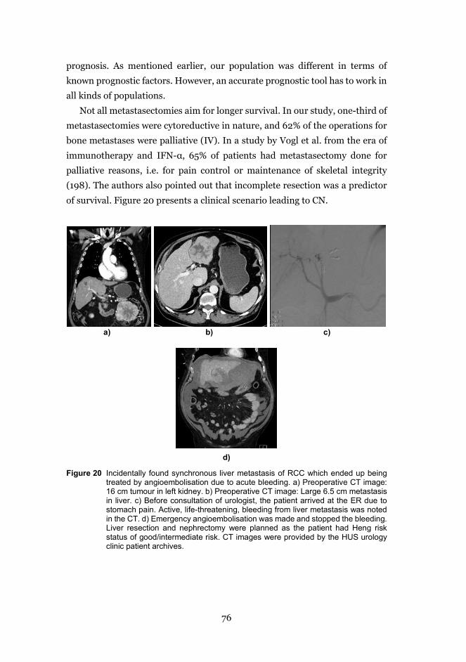
76
prognosis. As mentioned earlier, our population was different in terms of
known prognostic factors. However, an accurate prognostic tool has to work in
all kinds of populations.
Not all metastasectomies aim for longer survival. In our study, one-third of
metastasectomies were cytoreductive in nature, and 62% of the operations for
bone metastases were palliative (IV). In a study by Vogl et al. from the era of
immunotherapy and IFN-α, 65% of patients had metastasectomy done for
palliative reasons, i.e. for pain control or maintenance of skeletal integrity
(198). The authors also pointed out that incomplete resection was a predictor
of survival. Figure 20 presents a clinical scenario leading to CN.
a) b) c)
d)
Figure 20 Incidentally found synchronous liver metastasis of RCC which ended up being treated by angioembolisation due to acute bleeding. a) Preoperative CT image: 16 cm tumour in left kidney. b) Preoperative CT image: Large 6.5 cm metastasis in liver. c) Before consultation of urologist, the patient arrived at the ER due to stomach pain. Active, life-threatening, bleeding from liver metastasis was noted in the CT. d) Emergency angioembolisation was made and stopped the bleeding. Liver resection and nephrectomy were planned as the patient had Heng risk status of good/intermediate risk. CT images were provided by the HUS urology clinic patient archives.
77
Our data concurs with other available data suggesting that metastasectomy
still plays a part in this era of targeted therapy and check-point inhibitors.
Apart from palliation, only a subgroup of patients benefits from
metastasectomy. However, for some patients, metastasectomy offers clearly
prolonged survival and delays the initiation of systemic treatment. The value
of time without systemic therapy should not be underestimated as cancer drugs
come with side effects. Metastasectomy, having acceptable morbidity in
selected patients, might be gaining popularity as a part of a multimodality
setting, especially if on-going randomised trials prove its efficacy
(NCT00918775 and NCT01444807). Before that becomes a reality, however,
decisions to operate should be done in co-operation with oncologists, and
appropriate patient selection should be based on the prognostic factors, while
considering the risks of treatments remains of ultimate importance.
6.6 BIG OPERATIONS, BIG CENTRES AND BIG RISKS?
Although the data of studies II and IV included patients with locally advanced
or metastasized disease and technically challenged surgery, the complication
rate was not that high. We noted a 30-day complication rate of 34% for
operations on TT and 36% for initial metastasectomies, similar to the rates
documented in the literature, but with fewer major complications (169, 238).
The 30-day mortality was 1% for RCC with TT, being 0% for initial
metastasectomies, but 0.8% for all metastasectomies, which was in line with
the literature (169, 238). Patients undergoing CN have had more wound
complications and more delayed (>90 days) complications after initiation of
systemic therapy (239), but no differences in 30-day complications or severe
complications (Clavien-Dindo ≥3) were noted (239, 240). An association with
metastasectomy complications after commencement of targeted therapy has
not been ascertained in the era of targeted therapy (238).
Operating RCC with extensive TT is challenging and has a high risk of
vascular and major complications, and it is considered as safe when being
performed in high-volume centres with the help of vascular surgeons, thoracic
surgeons and anaesthesiologists. Interestingly, in a recent study of Meyer et
al., no difference in complications of metastasectomy was noted regardless of
the type of hospital or annual volume of cancer cases (238).
78
The team constellation, knowledgeability and volume of the centre may also
affect implementing new treatment recommendations into the clinical
practice. The 2001 EAU Guidelines on RCC stated the final evaluation of
oncologic efficacy to be uncertain for T1a tumours (241). However, by the time
the next fully updated version of the Guidelines was published in 2007, PN had
already become the treatment of choice for all T1a tumours (242). Accordingly,
the rate of PN increased markedly during the study period: in 2006-2008, only
57% of T1a patients underwent PN, but in 2012-2014, 87% did (III). The low
adherence to guidelines in the first years of study might seem disheartening in
a high-volume centre such as ours. At the time of the study, renal surgery was
seen as a common operation suitable for all urologists and residents, and in
study II, there were 26 urologist and/or residents who performed or assisted
in 915 operations. Since the days of the study, we have gradually centralised
the renal surgery to a limited number of urologists in our clinic. We assume
that not only the volume of the clinic matters; we presumably also need high-
volume urologists. In the future, we expect new treatment recommendations
to be promptly accepted and followed in our clinic.
6.7 LIMITATIONS
The limitations of these studies (I-IV) include the nature of retrospective,
single-centre study design, meaning that the results might not be generalizable
to other populations. Due to the centralised system, only sporadic patients are
missing from these patient cohorts gathered from the district of 1.67 million
people. Despite the comprehensive patient cohort, for rare diseases, the
volume of patients was too limited to offer sufficient distribution of patients
amongst different categories. The unequal distribution of patients between LU
prognostic groups (IV) and groups by level of TT involvement (II) may have
affected our results.
No re-evaluations were done concerning the pathological examinations. At
the beginning of these study periods (I-V), pathologists did not use any
structured report form, which might mean that some information about
necrosis, rhabdoid features, and sarcomatoid features might be missing from
reports. As a structured report is now commonly used, this format requires that
the pathologist always comment on these features. The ECOG performance
status, CCI and Clavien-Dindo complication score should be determined at the
79
time of diagnosis or complications, but in these studies, many of them were
assigned retrospectively by investigators. The limitations of these studies also
include the continually improving imaging accuracy, meaning that the quality
of imagines done at the beginning of study do not match the standards of today.
The main limitation of study V was minor and heterogenous patient data,
which might have lessened the value of S-TATI as a prognosticator. We had to
exclude many patients from the final analyses due to a disease of benign origin
or urothelial carcinoma.
Unfortunately, at the time of study planning, neutrophils, lymphocytes and
calcium were not seen as valuable prognostic markers for RCC in our clinic and
were not routinely measured. This precluded us from including the Heng score
or MKSCC nomogram in study IV. While in study V, comparing S-TATI with
other inflammation markers with proven prognostic significance, as the
neutrophil-lymphocyte ratio, would have been of significant interest. S-TATI
was not measured repeatedly, which would have been relevant, especially
considering the cases that recurred.
6.8 RCC, WHAT’S AHEAD?
Since the beginning of these studies, the treatment of RCC has changed
substantially as we are moving from targeted therapy to novel immunotherapy.
In addition, the substance of our work, the patient cohort, is changing. The
patient population with small renal tumours is getting older, frailer and more
prone to co-morbidities (I) (243, 244). Web-based, easy-to-use prediction
nomograms, similar to the ones for prostate cancer created by MSKCC (245)
to predict the risk of death and cancer-specific death of an individual patient,
are eagerly anticipated. One key issue for creating prediction tools is how to
define frailty. Co-morbidities or even performance status systems (i.e. ECOG)
do not paint the whole picture of the recovering ability of an elderly person,
while thorough geriatric questionnaires are often too detailed to use in daily
practice.
The increase in incidentally found small tumours has not led to a decrease
of newly diagnosed mRCC. Being the most lethal urological malignancy, but
curatively manageable when diagnosed early, RCC screening seems a credible
option. At the moment, no recommendations against or for RCC screening
exist. Since RCC has a relatively low incidence rate, screening subpopulations,
80
i.e. patients older than 70 years of age, might be more rewarding while they are
at an increased risk for high-stage and high-grade renal carcinoma (246)
The potential effect of screening is based on the assumption that early
treatment should yield better survival. Indeed, the increase of RCC mortality
has stabilised or even declined in recent years in many countries in
Scandinavia (16). However, treating small incidental tumours may not lower
overall mortality, which is more dependent on aging, frailty and co-morbidity.
Fenton et al. estimated a sojourn time for RCC (mean duration of the
detectable preclinical period) to be from 3.7 to 5.8 years, meaning that
screened RCC of people under 65 would usually progress to clinical diagnosis
during their life (247).
In one recent meta-analysis, which has presumably underestimated the
true prevalence of histologically proven RCC, Rossi et al. (248) speculated that
screening of RCC might even be as effective as the screening of abdominal
aortal aneurysm or colorectal cancer, both of which have established screening
programmes in UK. However, due to the different nature of diseases,
comparing diseases is spurious. While healthcare costs are continuously
increasing, screening cannot just be effective, but cost-effective as well. Rossi
et al. recently published a cost-effectiveness analysis, where ultrasound
screening seemed to be cost-efficient in 60-year old males (249). We must
remember that the true prevalence of RCC by age/sex is still unknown as
current figures may underestimate it due to uneven distribution of studies
worldwide. For screening purposes, there is a need for inexpensive but
accurate biomarkers, such as the ones measured from urine or blood.
Cancer treatment has made great progress. However, many of these
treatments, such as metastasectomy, CN and many systemic therapies are
effective only for some patients with good prognostic markers or for patients
expressing specific biomarkers predicting responsiveness to therapy. In the
future, the genetic and molecular analysis of tumour might be the base for
treatment. This author believes that nephrectomies and metastasectomies
might be needed in future not only for tumour histology but also for receiving
enough tissue material to test the effects of various drugs on heterogenous
tumour tissue (250).
In the future, treatment of RCC, especially when locally advanced or
metastasised, has to be customised concerning cancer amenities, patient
81
derived factors, possible side effects of treatments and treatment efficacy. To
offer the surgical and oncological treatments in the right sequence, we will
need tight collaboration between urologists and oncologists. When planning
the personalised treatment, we have to bear in mind the goal of each patient
whether it is the longest survival or maintaining best possible QOL. This means
that multidisciplinary teams should not just consider the tissue specimen but
also take into account the whole individual affected by the disease.
Multiple tissue-based predictive biomarkers for metastatic ccRCC are
emerging currently, but need to be validated, while non-ccRCC lacks
candidates for potential biomarkers. No other prognostic marker is superior to
tumour stage, despite the fervent search for a perfect prognostic marker. In the
future, all potential prognostic markers should be compared with the TNM
stage, the most substantial conventional prognostic marker, to prove their
accuracy. The anatomical, histological and clinical prognostic factors
combined have reached their ceiling in prediction accuracy (251). What we
should be looking for is a combination of clinical and histological markers
together with laboratory parameters, as genetic, molecular and tissue-based
markers, to obtain more precise prognosticators.
6.9 FUTURE SCIENTIFIC CONSIDERATIONS
Over the past few decades, several biomarkers have been studied to estimate
the prognosis of RCC. We are aiming to use the Helsinki Biobank RCC tissue
samples to test a variety of known histochemical and immunohistochemical
prognostic tissue markers in tumour microarrays (TMAs). To determine if any
markers seem to predict recurrence or death, we are comparing them with
prognostic data from the Helsinki University Kidney tumour database.
For this thesis, I ran further analyses to test the performance of tumour
invasion in predicting RCC recurrence. In these preliminary analyses, invasion
also seemed to be an independent prognostic marker when compared with
tumour diameter, histology and Fuhrman grade. This is an important result as
we can easily measure invasion using CT images. I intend to rerun the analyses,
with additional clinical parameters, e.g. tumour necrosis, to see if this effect
remains and to see if adding invasion to well-established prognostic scores, as
the Leibovich score, would improve the accuracy of prognostication. Indeed, it
82
would be valuable to see if the effect of invasion is lost when a tumour becomes
locally advanced, i.e. reaches the perirenal fat or pelvis.
One limitation of my studies (II, IV) was the small size of patient
subcohorts. Despite the fact that our hospital is amongst the largest RCC
centres in Northern European countries, when it comes to rare diseases as
supradiaphragmatic TT, we do not have enough patients to run reliable
analysis. After the initiation of my substudies, new collaboration groups for
RCC researchers have been founded. Our study group is active in FinnKidney,
a Finnish working group, and in NORENCA, a collaboration of RCC
researchers in Nordic countries. A multicentre approach allows us to enrol
more patients, making it possible to study rare diseases as well as to compare
differences amongst different centres or nationalities.
Preoperative and non-invasive prognostic prediction methods are urgently
needed to either replace or to be used with renal tumour biopsies. Today, the
prognostic nomograms rely heavily on histology, which is only gained by
invasive methods. As RCC patients get older and frailer, to operate or not to
operate becomes a fundamental question. Despite the small and heterogenous
patient number, TATI expression seems to be associated with adverse
prognosis. In the future, it would be of value to use serum and urine samples
of RCC patients from the Helsinki biobank to test the prognostic accuracy of
promising up-to-date biomarkers.
83
7 CONCLUSIONS
On the basis of the present studies, the following conclusions can be drawn
concerning the RCC patients treated surgically at Helsinki University Hospital.
1. RCC patients generally are getting older and more fragile. We are
finding smaller and smaller tumours and more asymptomatic
tumours. Mini-invasive treatments are becoming more popular, and
we are treating more patients with active surveillance and taking
biopsies of tumours more often. The proportion of newly diagnosed
M1 patients did not change during the surveillance period. A shift in
oncological treatments from targeted therapy to immunotherapy was
not yet clearly observed in these studies. The number of CNs has
declined.
2. The level of TT seems to be a prognostic marker only in large cohorts.
The improved survival and feasible complication rate of mRCC
patients after nephrectomy and thrombectomy makes operating on
mRCC patients with TT a reasonable choice in the era of targeted
therapy. The survival of the patients with pRCC and TT as well as N1
patients has been dismal.
3. Parenchymal invasion as well as diameter outperformed the more
complex classifications, PADUA and RTII, in predicting the type of
nephrectomy performed. Invasion seems to be an independent
prognostic marker of recurrence, also when compared with tumour
diameter.
4. Survival of all mRCC patients after metastasectomy was favourable,
although the patient cohort lacked prosperous features for survival
after metastasectomy. Assumedly, survival was superior for patients
with complete metastasectomy compared with incomplete surgery.
Complete metastasectomy led to longer systemic treatment-free time
as well. Previously published LU prognostic groups did not predict the
survival in this patient cohort.
5. S-TATI was able to predict CSS and OS as well as longer therapy-free
intervals. The earlier suggested cut-off point of 16 µg/l seemed to be a

84
sensible cut-off value for survival. To validate these results, S-TATI
should be investigated in large, prospective series, and the
confounding effects should be addressed more thoroughly.
85
8 ACKNOWLEDGEMENTS
This study was carried out at the Department of Urology at Helsinki University
Hospital from 2015-2020. Financial support from the Finnish Urology
Association and Helsinki University Hospital research funds (EVO) is
gratefully acknowledged.
I am grateful to Adjunct Professor Mika Matikainen, head of the
Department of Urology, for the opportunity to carry out this thesis. I am
grateful to him for providing the facilities and time off from clinical work as
well as serving as a member of my thesis committee. It is a privilege to have
such a flexible and supportive superior. I wish to express my gratitude to
Adjunct Professor Jukka Sairanen and Adjunct Professor Arto Mikkola for
allowing me to have time off from the Urology Department to immerse myself
in my research. I thank you both for being such good role models in managing
thousands of things brilliantly at the same time while never losing your
commitment.
I thank Professor Antti Rannikko for his contagious love of science and
research. Thank you, Antti, for always finding research facilities when they
were urgently needed. I am sure that you will lead our clinic further into the
world of science than we could ever dream of.
My heartfelt thanks go to my supervisors, the excellent Adjunct Professor
Harry Nisén and Professor Kimmo Taari, for guiding me throughout the whole
thesis project. Harry, you have always seen our goals as crystal clear despite
the minor obstacles on our journey. Your enthusiasm, passion and
determination have kept me going. I will be forever thankful to you for leading
me to the world of RCC both scientifically and clinically. I hope this love never
dies. Kimmo, you are so warm-hearted, you always found time to ask how I was
doing and kept the spirits up during this process. Thank you for being there for
me.
Petrus and Harri, ‘apuohjaajat’. It has been a pleasure working with you
both. You were very hands-on, always promptly replied even to my most basic
questions, and most of all, you were so supportive – without you, this thesis
would probably have never been finished. Riikka, thank you for backing me up
when needed. You are both a great co-researcher and a brilliant colleague. I
offer my sincere thanks to Tuomas, for your quick and precise comments and

86
your magical STATA skills. I am amazed. I offer my sincere thanks to all co-
writers, Kaisa Erkkilä, Ulf-Håkan Stenman, Petteri Hervonen, Tuomas Mirtti,
Petri Bono, Ilkka Kantonen and Jarmo Simpanen for supportive collaboration.
I acknowledge Tuomas Mirtti and Anni Virtanen for supplying example images
of histological RCC subtypes. I thank Merja Rignell for help in obtaining death
certificates and for coffee and empathy offered in the cellar of Meilahti Hospital
outside of office hours.
My warmest thanks go to my reviewers, Adjunct Professors Markku Vaarala
and Jukka Häkkinen, for kindly agreeing to examine my thesis. Your thorough
work and precise comments improved my thesis text significantly. I am obliged
to adjunct professor Martti Nurmi for accepting the role of the official
opponent in this thesis dissertation. I am grateful to our language consultant
for his expertise. I thank members of my thesis committee, Adjunct Professors
Ville Sallinen and Mika Matikainen for being there for me and for constructive
comments about study plans.
I thank all the colleagues in the HUH Urology Department. It has been a
pleasure working with you. You have always been willing to help, and I have
been privileged to be able to learn from you all. Working with you has been fun,
no matter what the circumstances have been. As the famous slogan says:
‘Urology is for cool kids’. Special thanks go to Heli and Robin for making the
time at work so enjoyable that it might be difficult to get things done.
I am deeply indebted to Vellu, Indrek and Sebastian, my dear colleagues in
Lappeenranta, from their guidance to the exciting world of urology and for
patiently teaching me the majority of my surgical skills. Thank you for
believing in me despite seeing my first fumbling operations.
I warmly thank all my friends along the way for being there for me. You
mean the world to me.
Anastasia, growing up together and sharing many ups and downs in life has
meant a great deal to me. I wish you would live closer.
My dear friends from the university, Eeva, Eliisa, Helka, Kanerva,
Marianna, Ninnu and Ulla, are still referred as ‘tytöt’ after so many cherished
years together. Thank you for your loving and continuous peer support in every
aspect of life and immediate whatsapp responses to all my questions about
family life, hairstyles, Netflix series, science, and to specific questions about
how to finish one’s thesis. I am especially thankful for Kanerva’s most recent

87
hands-on experience of this thesis struggle and for Eliisa’s holistic approach to
science.
My friends from university times. Maiju, Nora, Jaakko, Kökö and Timppa,
thank you for joyful moments at home and away. We have had great trips filled
with laughter. Good friends are the best distraction during the dull moments
of life.
To my friends met during the Lappeenranta years, Elisa, Emma-Lotta,
Maire, Minna and Mikko, I know that taking first steps in surgery together has
brought us close. Katja and Marianne, I could never imagine that true friends
could be found in a schoolyard when picking up children.
I thank my in-laws Jussi, Pirkko, Hilkka and Iina and my godmother Hiksu,
for their concrete contribution to my thesis work by babysitting my children.
My sister Iina, thank you for not only sharing experiences of coincidental
thesis projects but for being my loyal and loving friend since the day you were
born. Iina, Selene, Samuel and Sakke, it has been a true privilege to have
relatives living just next door. I hope that our children will be as close as we
are.
My deepest thanks go to my parents, Helena and Olavi, for always believing
in me. Knowing that I can always count on you, no matter whether it means
no-notice childcare or renovating a flat from floor to the ceiling, has given me
a solid foundation to concentrate on less practical matters, such as this project.
Thank you for being there for me.
Above all, I am ever grateful to my beloved husband, Juho-Antti for his
understanding and patience during this project. Having an absent and absent-
minded wife has not always been easy for you. However, I have always been
able to depend on you and your love. It means everything to me. I love you.
And your bad jokes.
My children, Isla, Okko and Pihla, I am very fortunate to have you. Your
existence has slowed this project considerably but filled my life with happiness,
joy and love beyond all expectations – what a great bargain! Thank you for
being you.
Helsinki, April 2020 Sara Tornberg
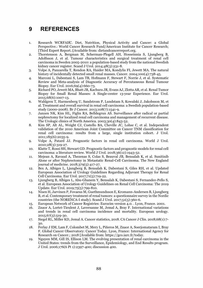
88
9 REFERENCES
1. Research WCRFAIfC. Diet, Nutrition, Physical Activity and Cancer: a Global Perspective.: World Cancer Research Fund/American Institute for Cancer Research; [Third Expert Report.:[Available from: dietandcancerreport.org.
2. Thorstenson A, Bergman M, Scherman-Plogell AH, Hosseinnia S, Ljungberg B, Adolfsson J, et al. Tumour characteristics and surgical treatment of renal cell carcinoma in Sweden 2005-2010: a population-based study from the national Swedish kidney cancer register. Scand J Urol. 2014;48(3):231-8.
3. Volpe A, Panzarella T, Rendon RA, Haider MA, Kondylis FI, Jewett MA. The natural history of incidentally detected small renal masses. Cancer. 2004;100(4):738-45.
4. Marconi L, Dabestani S, Lam TB, Hofmann F, Stewart F, Norrie J, et al. Systematic Review and Meta-analysis of Diagnostic Accuracy of Percutaneous Renal Tumour Biopsy. Eur Urol. 2016;69(4):660-73.
5. Richard PO, Jewett MA, Bhatt JR, Kachura JR, Evans AJ, Zlotta AR, et al. Renal Tumor Biopsy for Small Renal Masses: A Single-center 13-year Experience. Eur Urol. 2015;68(6):1007-13.
6. Wahlgren T, Harmenberg U, Sandstrom P, Lundstam S, Kowalski J, Jakobsson M, et al. Treatment and overall survival in renal cell carcinoma: a Swedish population-based study (2000-2008). Br J Cancer. 2013;108(7):1541-9.
7. Janzen NK, Kim HL, Figlin RA, Belldegrun AS. Surveillance after radical or partial nephrectomy for localized renal cell carcinoma and management of recurrent disease. The Urologic clinics of North America. 2003;30(4):843-52.
8. Kim SP, Alt AL, Weight CJ, Costello BA, Cheville JC, Lohse C, et al. Independent validation of the 2010 American Joint Committee on Cancer TNM classification for renal cell carcinoma: results from a large, single institution cohort. J Urol. 2011;185(6):2035-9.
9. Volpe A, Patard JJ. Prognostic factors in renal cell carcinoma. World J Urol. 2010;28(3):319-27.
10. Klatte T, Rossi SH, Stewart GD. Prognostic factors and prognostic models for renal cell carcinoma: a literature review. World J Urol. 2018;36(12):1943-52.
11. Mejean A, Ravaud A, Thezenas S, Colas S, Beauval JB, Bensalah K, et al. Sunitinib Alone or after Nephrectomy in Metastatic Renal-Cell Carcinoma. The New England journal of medicine. 2018;379(5):417-27.
12. Bex A, Albiges L, Ljungberg B, Bensalah K, Dabestani S, Giles RH, et al. Updated European Association of Urology Guidelines Regarding Adjuvant Therapy for Renal Cell Carcinoma. Eur Urol. 2017;71(5):719-22.
13. Ljungberg B, Albiges L, Abu-Ghanem Y, Bensalah K, Dabestani S, Fernandez-Pello S, et al. European Association of Urology Guidelines on Renal Cell Carcinoma: The 2019 Update. Eur Urol. 2019;75(5):799-810.
14. Nisen H, Jarvinen P, Fovaeus M, Guethmundsson E, Kromann-Andersen B, Ljungberg B, et al. Contemporary treatment of renal tumors: a questionnaire survey in the Nordic countries (the NORENCA-I study). Scand J Urol. 2017;51(5):360-6.
15. European Network of Cancer Registries: Eurocim version 4.0. . Lyon, France. 2001. 16. Znaor A, Lortet-Tieulent J, Laversanne M, Jemal A, Bray F. International variations
and trends in renal cell carcinoma incidence and mortality. European urology. 2015;67(3):519-30.
17. Siegel RL, Miller KD, Jemal A. Cancer statistics, 2018. CA Cancer J Clin. 2018;68(1):7-30.
18. Ferlay J EM, Lam F, Colombet M, Mery L, Piñeros M, Znaor A, Soerjomataram I, Bray F. Global Cancer Observatory: Cancer Today. Lyon, France: International Agency for Research on Cancer.; 2018 [Available from: https://gco.iarc.fr/today.
19. Nguyen MM, Gill IS, Ellison LM. The evolving presentation of renal carcinoma in the United States: trends from the Surveillance, Epidemiology, and End Results program. J Urol. 2006;176(6 Pt 1):2397-400; discussion 400.
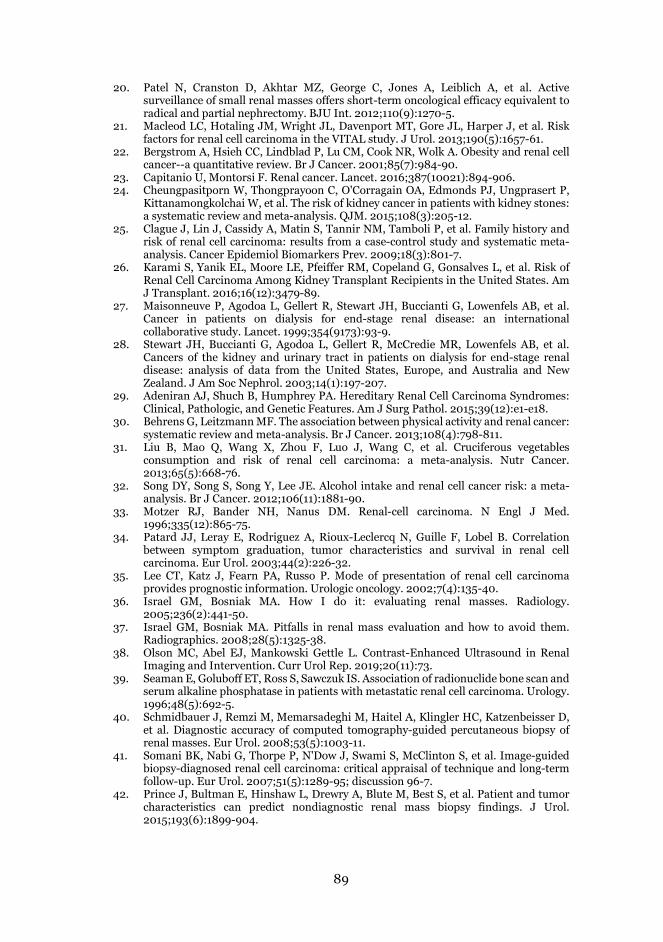
89
20. Patel N, Cranston D, Akhtar MZ, George C, Jones A, Leiblich A, et al. Active surveillance of small renal masses offers short-term oncological efficacy equivalent to radical and partial nephrectomy. BJU Int. 2012;110(9):1270-5.
21. Macleod LC, Hotaling JM, Wright JL, Davenport MT, Gore JL, Harper J, et al. Risk factors for renal cell carcinoma in the VITAL study. J Urol. 2013;190(5):1657-61.
22. Bergstrom A, Hsieh CC, Lindblad P, Lu CM, Cook NR, Wolk A. Obesity and renal cell cancer--a quantitative review. Br J Cancer. 2001;85(7):984-90.
23. Capitanio U, Montorsi F. Renal cancer. Lancet. 2016;387(10021):894-906. 24. Cheungpasitporn W, Thongprayoon C, O'Corragain OA, Edmonds PJ, Ungprasert P,
Kittanamongkolchai W, et al. The risk of kidney cancer in patients with kidney stones: a systematic review and meta-analysis. QJM. 2015;108(3):205-12.
25. Clague J, Lin J, Cassidy A, Matin S, Tannir NM, Tamboli P, et al. Family history and risk of renal cell carcinoma: results from a case-control study and systematic meta-analysis. Cancer Epidemiol Biomarkers Prev. 2009;18(3):801-7.
26. Karami S, Yanik EL, Moore LE, Pfeiffer RM, Copeland G, Gonsalves L, et al. Risk of Renal Cell Carcinoma Among Kidney Transplant Recipients in the United States. Am J Transplant. 2016;16(12):3479-89.
27. Maisonneuve P, Agodoa L, Gellert R, Stewart JH, Buccianti G, Lowenfels AB, et al. Cancer in patients on dialysis for end-stage renal disease: an international collaborative study. Lancet. 1999;354(9173):93-9.
28. Stewart JH, Buccianti G, Agodoa L, Gellert R, McCredie MR, Lowenfels AB, et al. Cancers of the kidney and urinary tract in patients on dialysis for end-stage renal disease: analysis of data from the United States, Europe, and Australia and New Zealand. J Am Soc Nephrol. 2003;14(1):197-207.
29. Adeniran AJ, Shuch B, Humphrey PA. Hereditary Renal Cell Carcinoma Syndromes: Clinical, Pathologic, and Genetic Features. Am J Surg Pathol. 2015;39(12):e1-e18.
30. Behrens G, Leitzmann MF. The association between physical activity and renal cancer: systematic review and meta-analysis. Br J Cancer. 2013;108(4):798-811.
31. Liu B, Mao Q, Wang X, Zhou F, Luo J, Wang C, et al. Cruciferous vegetables consumption and risk of renal cell carcinoma: a meta-analysis. Nutr Cancer. 2013;65(5):668-76.
32. Song DY, Song S, Song Y, Lee JE. Alcohol intake and renal cell cancer risk: a meta-analysis. Br J Cancer. 2012;106(11):1881-90.
33. Motzer RJ, Bander NH, Nanus DM. Renal-cell carcinoma. N Engl J Med. 1996;335(12):865-75.
34. Patard JJ, Leray E, Rodriguez A, Rioux-Leclercq N, Guille F, Lobel B. Correlation between symptom graduation, tumor characteristics and survival in renal cell carcinoma. Eur Urol. 2003;44(2):226-32.
35. Lee CT, Katz J, Fearn PA, Russo P. Mode of presentation of renal cell carcinoma provides prognostic information. Urologic oncology. 2002;7(4):135-40.
36. Israel GM, Bosniak MA. How I do it: evaluating renal masses. Radiology. 2005;236(2):441-50.
37. Israel GM, Bosniak MA. Pitfalls in renal mass evaluation and how to avoid them. Radiographics. 2008;28(5):1325-38.
38. Olson MC, Abel EJ, Mankowski Gettle L. Contrast-Enhanced Ultrasound in Renal Imaging and Intervention. Curr Urol Rep. 2019;20(11):73.
39. Seaman E, Goluboff ET, Ross S, Sawczuk IS. Association of radionuclide bone scan and serum alkaline phosphatase in patients with metastatic renal cell carcinoma. Urology. 1996;48(5):692-5.
40. Schmidbauer J, Remzi M, Memarsadeghi M, Haitel A, Klingler HC, Katzenbeisser D, et al. Diagnostic accuracy of computed tomography-guided percutaneous biopsy of renal masses. Eur Urol. 2008;53(5):1003-11.
41. Somani BK, Nabi G, Thorpe P, N'Dow J, Swami S, McClinton S, et al. Image-guided biopsy-diagnosed renal cell carcinoma: critical appraisal of technique and long-term follow-up. Eur Urol. 2007;51(5):1289-95; discussion 96-7.
42. Prince J, Bultman E, Hinshaw L, Drewry A, Blute M, Best S, et al. Patient and tumor characteristics can predict nondiagnostic renal mass biopsy findings. J Urol. 2015;193(6):1899-904.
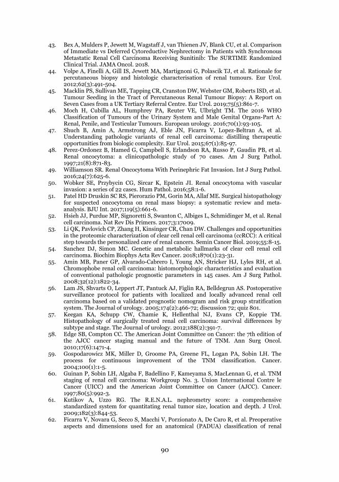
90
43. Bex A, Mulders P, Jewett M, Wagstaff J, van Thienen JV, Blank CU, et al. Comparison of Immediate vs Deferred Cytoreductive Nephrectomy in Patients with Synchronous Metastatic Renal Cell Carcinoma Receiving Sunitinib: The SURTIME Randomized Clinical Trial. JAMA Oncol. 2018.
44. Volpe A, Finelli A, Gill IS, Jewett MA, Martignoni G, Polascik TJ, et al. Rationale for percutaneous biopsy and histologic characterisation of renal tumours. Eur Urol. 2012;62(3):491-504.
45. Macklin PS, Sullivan ME, Tapping CR, Cranston DW, Webster GM, Roberts ISD, et al. Tumour Seeding in the Tract of Percutaneous Renal Tumour Biopsy: A Report on Seven Cases from a UK Tertiary Referral Centre. Eur Urol. 2019;75(5):861-7.
46. Moch H, Cubilla AL, Humphrey PA, Reuter VE, Ulbright TM. The 2016 WHO Classification of Tumours of the Urinary System and Male Genital Organs-Part A: Renal, Penile, and Testicular Tumours. European urology. 2016;70(1):93-105.
47. Shuch B, Amin A, Armstrong AJ, Eble JN, Ficarra V, Lopez-Beltran A, et al. Understanding pathologic variants of renal cell carcinoma: distilling therapeutic opportunities from biologic complexity. Eur Urol. 2015;67(1):85-97.
48. Perez-Ordonez B, Hamed G, Campbell S, Erlandson RA, Russo P, Gaudin PB, et al. Renal oncocytoma: a clinicopathologic study of 70 cases. Am J Surg Pathol. 1997;21(8):871-83.
49. Williamson SR. Renal Oncocytoma With Perinephric Fat Invasion. Int J Surg Pathol. 2016;24(7):625-6.
50. Wobker SE, Przybycin CG, Sircar K, Epstein JI. Renal oncocytoma with vascular invasion: a series of 22 cases. Hum Pathol. 2016;58:1-6.
51. Patel HD Druskin SC RS, Pierorazio PM, Gorin MA, Allaf ME. Surgical histopathology for suspected oncocytoma on renal mass biopsy: a systematic review and meta‐analysis. BJU Int. 2017;119(5):661-6.
52. Hsieh JJ, Purdue MP, Signoretti S, Swanton C, Albiges L, Schmidinger M, et al. Renal cell carcinoma. Nat Rev Dis Primers. 2017;3:17009.
53. Li QK, Pavlovich CP, Zhang H, Kinsinger CR, Chan DW. Challenges and opportunities in the proteomic characterization of clear cell renal cell carcinoma (ccRCC): A critical step towards the personalized care of renal cancers. Semin Cancer Biol. 2019;55:8-15.
54. Sanchez DJ, Simon MC. Genetic and metabolic hallmarks of clear cell renal cell carcinoma. Biochim Biophys Acta Rev Cancer. 2018;1870(1):23-31.
55. Amin MB, Paner GP, Alvarado-Cabrero I, Young AN, Stricker HJ, Lyles RH, et al. Chromophobe renal cell carcinoma: histomorphologic characteristics and evaluation of conventional pathologic prognostic parameters in 145 cases. Am J Surg Pathol. 2008;32(12):1822-34.
56. Lam JS, Shvarts O, Leppert JT, Pantuck AJ, Figlin RA, Belldegrun AS. Postoperative surveillance protocol for patients with localized and locally advanced renal cell carcinoma based on a validated prognostic nomogram and risk group stratification system. The Journal of urology. 2005;174(2):466-72; discussion 72; quiz 801.
57. Keegan KA, Schupp CW, Chamie K, Hellenthal NJ, Evans CP, Koppie TM. Histopathology of surgically treated renal cell carcinoma: survival differences by subtype and stage. The Journal of urology. 2012;188(2):391-7.
58. Edge SB, Compton CC. The American Joint Committee on Cancer: the 7th edition of the AJCC cancer staging manual and the future of TNM. Ann Surg Oncol. 2010;17(6):1471-4.
59. Gospodarowicz MK, Miller D, Groome PA, Greene FL, Logan PA, Sobin LH. The process for continuous improvement of the TNM classification. Cancer. 2004;100(1):1-5.
60. Guinan P, Sobin LH, Algaba F, Badellino F, Kameyama S, MacLennan G, et al. TNM staging of renal cell carcinoma: Workgroup No. 3. Union International Contre le Cancer (UICC) and the American Joint Committee on Cancer (AJCC). Cancer. 1997;80(5):992-3.
61. Kutikov A, Uzzo RG. The R.E.N.A.L. nephrometry score: a comprehensive standardized system for quantitating renal tumor size, location and depth. J Urol. 2009;182(3):844-53.
62. Ficarra V, Novara G, Secco S, Macchi V, Porzionato A, De Caro R, et al. Preoperative aspects and dimensions used for an anatomical (PADUA) classification of renal
91
tumours in patients who are candidates for nephron-sparing surgery. Eur Urol. 2009;56(5):786-93.
63. Waldert M, Waalkes S, Klatte T, Kuczyk MA, Weibl P, Schuller G, et al. External validation of the preoperative anatomical classification for prediction of complications related to nephron-sparing surgery. World journal of urology. 2010;28(4):531-5.
64. Alvim RG, Audenet F, Vertosick EA, Sjoberg DD, Touijer KA. Performance Prediction for Surgical Outcomes in Partial Nephrectomy Using Nephrometry Scores: A Comparison of Arterial Based Complexity (ABC), RENAL, and PADUA Systems. Eur Urol Oncol. 2018;1(5):428-34.
65. Hakky TS, Baumgarten AS, Allen B, Lin HY, Ercole CE, Sexton WJ, et al. Zonal NePhRO scoring system: a superior renal tumor complexity classification model. Clin Genitourin Cancer. 2014;12(1):e13-8.
66. Tannus M, Goldman SM, Andreoni C. Practical and intuitive surgical approach renal ranking to predict outcomes in the management of renal tumors: a novel score tool. J Endourol. 2014;28(4):487-92.
67. Simmons MN, Ching CB, Samplaski MK, Park CH, Gill IS. Kidney tumor location measurement using the C index method. J Urol. 2010;183(5):1708-13.
68. Nisen H, Ruutu M, Glucker E, Visapaa H, Taari K. Renal tumour invasion index as a novel anatomical classification predicting urological complications after partial nephrectomy. Scand J Urol. 2014;48(1):41-51.
69. Simmons MN, Hillyer SP, Lee BH, Fergany AF, Kaouk J, Campbell SC. Diameter-axial-polar nephrometry: integration and optimization of R.E.N.A.L. and centrality index scoring systems. J Urol. 2012;188(2):384-90.
70. Leslie S, Gill IS, de Castro Abreu AL, Rahmanuddin S, Gill KS, Nguyen M, et al. Renal tumor contact surface area: a novel parameter for predicting complexity and outcomes of partial nephrectomy. Eur Urol. 2014;66(5):884-93.
71. Shin TY, Komninos C, Kim DW, So KS, Bang KS, Jeong HJ, et al. A novel mathematical model to predict the severity of postoperative functional reduction before partial nephrectomy: the importance of calculating resected and ischemic volume. J Urol. 2015;193(2):423-9.
72. Moch H, Gasser T, Amin MB, Torhorst J, Sauter G, Mihatsch MJ. Prognostic utility of the recently recommended histologic classification and revised TNM staging system of renal cell carcinoma: a Swiss experience with 588 tumors. Cancer. 2000;89(3):604-14.
73. Patard JJ, Leray E, Rioux-Leclercq N, Cindolo L, Ficarra V, Zisman A, et al. Prognostic value of histologic subtypes in renal cell carcinoma: a multicenter experience. J Clin Oncol. 2005;23(12):2763-71.
74. Beuselinck B, Verbiest A, Couchy G, Job S, de Reynies A, Meiller C, et al. Pro-angiogenic gene expression is associated with better outcome on sunitinib in metastatic clear-cell renal cell carcinoma. Acta Oncol. 2018;57(4):498-508.
75. Delahunt B, Cheville JC, Martignoni G, Humphrey PA, Magi-Galluzzi C, McKenney J, et al. The International Society of Urological Pathology (ISUP) grading system for renal cell carcinoma and other prognostic parameters. Am J Surg Pathol. 2013;37(10):1490-504.
76. Delahunt B, Eble JN, McCredie MR, Bethwaite PB, Stewart JH, Bilous AM. Morphologic typing of papillary renal cell carcinoma: comparison of growth kinetics and patient survival in 66 cases. Hum Pathol. 2001;32(6):590-5.
77. Sui W, Matulay JT, Robins DJ, James MB, Onyeji IC, RoyChoudhury A, et al. Collecting duct carcinoma of the kidney: Disease characteristics and treatment outcomes from the National Cancer Database. Urol Oncol. 2017;35(9):540 e13- e18.
78. Katz MD, Serrano MF, Grubb RL, 3rd, Skolarus TA, Gao F, Humphrey PA, et al. Percent microscopic tumor necrosis and survival after curative surgery for renal cell carcinoma. J Urol. 2010;183(3):909-14.
79. Ro JY, Ayala AG, Sella A, Samuels ML, Swanson DA. Sarcomatoid renal cell carcinoma: clinicopathologic. A study of 42 cases. Cancer. 1987;59(3):516-26.
80. Kara O, Maurice MJ, Zargar H, Malkoc E, Akca O, Andrade HS, et al. Prognostic implications of sarcomatoid and rhabdoid differentiation in patients with grade 4 renal cell carcinoma. Int Urol Nephrol. 2016;48(8):1253-60.
92
81. Mian BM, Bhadkamkar N, Slaton JW, Pisters PW, Daliani D, Swanson DA, et al. Prognostic factors and survival of patients with sarcomatoid renal cell carcinoma. J Urol. 2002;167(1):65-70.
82. Connor Wells J, Donskov F, Fraccon AP, Pasini F, Bjarnason GA, Beuselinck B, et al. Characterizing the outcomes of metastatic papillary renal cell carcinoma. Cancer medicine. 2017;6(5):902-9.
83. Leroy X, Zini L, Buob D, Ballereau C, Villers A, Aubert S. Renal cell carcinoma with rhabdoid features: an aggressive neoplasm with overexpression of p53. Arch Pathol Lab Med. 2007;131(1):102-6.
84. Frank I, Blute ML, Cheville JC, Lohse CM, Weaver AL, Zincke H. An outcome prediction model for patients with clear cell renal cell carcinoma treated with radical nephrectomy based on tumor stage, size, grade and necrosis: the SSIGN score. J Urol. 2002;168(6):2395-400.
85. Lam JS, Shvarts O, Said JW, Pantuck AJ, Seligson DB, Aldridge ME, et al. Clinicopathologic and molecular correlations of necrosis in the primary tumor of patients with renal cell carcinoma. Cancer. 2005;103(12):2517-25.
86. Leibovich BC, Blute ML, Cheville JC, Lohse CM, Frank I, Kwon ED, et al. Prediction of progression after radical nephrectomy for patients with clear cell renal cell carcinoma: a stratification tool for prospective clinical trials. Cancer. 2003;97(7):1663-71.
87. Fuhrman SA, Lasky LC, Limas C. Prognostic significance of morphologic parameters in renal cell carcinoma. Am J Surg Pathol. 1982;6(7):655-63.
88. Srigley JR, Delahunt B, Eble JN, Egevad L, Epstein JI, Grignon D, et al. The International Society of Urological Pathology (ISUP) Vancouver Classification of Renal Neoplasia. Am J Surg Pathol. 2013;37(10):1469-89.
89. Cindolo L, de la Taille A, Messina G, Romis L, Abbou CC, Altieri V, et al. A preoperative clinical prognostic model for non-metastatic renal cell carcinoma. BJU Int. 2003;92(9):901-5.
90. Kim HL, Belldegrun AS, Freitas DG, Bui MH, Han KR, Dorey FJ, et al. Paraneoplastic signs and symptoms of renal cell carcinoma: implications for prognosis. J Urol. 2003;170(5):1742-6.
91. Scoll BJ, Wong YN, Egleston BL, Kunkle DA, Saad IR, Uzzo RG. Age, tumor size and relative survival of patients with localized renal cell carcinoma: a surveillance, epidemiology and end results analysis. J Urol. 2009;181(2):506-11.
92. Kristjansson SR, Nesbakken A, Jordhoy MS, Skovlund E, Audisio RA, Johannessen HO, et al. Comprehensive geriatric assessment can predict complications in elderly patients after elective surgery for colorectal cancer: a prospective observational cohort study. Crit Rev Oncol Hematol. 2010;76(3):208-17.
93. Ethun CG, Bilen MA, Jani AB, Maithel SK, Ogan K, Master VA. Frailty and cancer: Implications for oncology surgery, medical oncology, and radiation oncology. CA Cancer J Clin. 2017;67(5):362-77.
94. Chandrasekar T, Klaassen Z, Goldberg H, Kulkarni GS, Hamilton RJ, Fleshner NE. Metastatic renal cell carcinoma: Patterns and predictors of metastases-A contemporary population-based series. Urol Oncol. 2017;35(11):661 e7- e14.
95. Hellenthal NJ, Chamie K, Ramirez ML, deVere White RW. Sociodemographic factors associated with nephrectomy in patients with metastatic renal cell carcinoma. J Urol. 2009;181(3):1013-8; discussion 8-9.
96. Hanna N, Sun M, Meyer CP, Nguyen PL, Pal SK, Chang SL, et al. Survival Analyses of Patients With Metastatic Renal Cancer Treated With Targeted Therapy With or Without Cytoreductive Nephrectomy: A National Cancer Data Base Study. Journal of clinical oncology : official journal of the American Society of Clinical Oncology. 2016;34(27):3267-75.
97. Motzer RJ, Bacik J, Schwartz LH, Reuter V, Russo P, Marion S, et al. Prognostic factors for survival in previously treated patients with metastatic renal cell carcinoma. J Clin Oncol. 2004;22(3):454-63.
98. Rautiola J, Donskov F, Peltola K, Joensuu H, Bono P. Sunitinib-induced hypertension, neutropaenia and thrombocytopaenia as predictors of good prognosis in patients with metastatic renal cell carcinoma. BJU Int. 2016;117(1):110-7.
93
99. Penttila P, Bono P, Peltola K, Donskov F. Hyponatremia associates with poor outcome in metastatic renal cell carcinoma patients treated with everolimus: prognostic impact. Acta Oncol. 2018;57(11):1580-5.
100. Cancer Genome Atlas Research N. Comprehensive molecular characterization of clear cell renal cell carcinoma. Nature. 2013;499(7456):43-9.
101. Beroukhim R, Brunet JP, Di Napoli A, Mertz KD, Seeley A, Pires MM, et al. Patterns of gene expression and copy-number alterations in von-hippel lindau disease-associated and sporadic clear cell carcinoma of the kidney. Cancer Res. 2009;69(11):4674-81.
102. Patel PH, Chadalavada RS, Chaganti RS, Motzer RJ. Targeting von Hippel-Lindau pathway in renal cell carcinoma. Clin Cancer Res. 2006;12(24):7215-20.
103. Yao M, Yoshida M, Kishida T, Nakaigawa N, Baba M, Kobayashi K, et al. VHL tumor suppressor gene alterations associated with good prognosis in sporadic clear-cell renal carcinoma. J Natl Cancer Inst. 2002;94(20):1569-75.
104. Schraml P, Struckmann K, Hatz F, Sonnet S, Kully C, Gasser T, et al. VHL mutations and their correlation with tumour cell proliferation, microvessel density, and patient prognosis in clear cell renal cell carcinoma. J Pathol. 2002;196(2):186-93.
105. Bui MH, Visapaa H, Seligson D, Kim H, Han KR, Huang Y, et al. Prognostic value of carbonic anhydrase IX and KI67 as predictors of survival for renal clear cell carcinoma. J Urol. 2004;171(6 Pt 1):2461-6.
106. Shen H, Liu Q, Li M, Yang P. The prognostic value of vascular endothelial growth factor in patients with renal cell carcinoma: a systematic review of the literature and meta-analysis. Clin Invest Med. 2017;40(2):E40-E8.
107. Jilaveanu LB, Shuch B, Zito CR, Parisi F, Barr M, Kluger Y, et al. PD-L1 Expression in Clear Cell Renal Cell Carcinoma: An Analysis of Nephrectomy and Sites of Metastases. J Cancer. 2014;5(3):166-72.
108. Iacovelli R, Nole F, Verri E, Renne G, Paglino C, Santoni M, et al. Prognostic Role of PD-L1 Expression in Renal Cell Carcinoma. A Systematic Review and Meta-Analysis. Target Oncol. 2016;11(2):143-8.
109. Gandini S, Massi D, Mandala M. PD-L1 expression in cancer patients receiving anti PD-1/PD-L1 antibodies: A systematic review and meta-analysis. Crit Rev Oncol Hematol. 2016;100:88-98.
110. Giraldo NA, Becht E, Pages F, Skliris G, Verkarre V, Vano Y, et al. Orchestration and Prognostic Significance of Immune Checkpoints in the Microenvironment of Primary and Metastatic Renal Cell Cancer. Clin Cancer Res. 2015;21(13):3031-40.
111. Eichenauer T, Simmendinger L, Fraune C, Mandelkow T, Blessin NC, Kluth M, et al. High level of EZH2 expression is linked to high density of CD8-positive T-lymphocytes and an aggressive phenotype in renal cell carcinoma. World J Urol. 2020.
112. Erlmeier F, Weichert W, Autenrieth M, Wiedemann M, Schrader AJ, Hartmann A, et al. PD-L2: A prognostic marker in chromophobe renal cell carcinoma? Med Oncol. 2017;34(5):71.
113. Dvorak HF. Tumors: wounds that do not heal. Similarities between tumor stroma generation and wound healing. N Engl J Med. 1986;315(26):1650-9.
114. Hanahan D, Weinberg RA. Hallmarks of cancer: the next generation. Cell. 2011;144(5):646-74.
115. Qian BZ, Pollard JW. Macrophage diversity enhances tumor progression and metastasis. Cell. 2010;141(1):39-51.
116. de Visser KE, Eichten A, Coussens LM. Paradoxical roles of the immune system during cancer development. Nature reviews Cancer. 2006;6(1):24-37.
117. Hu Q, Gou Y, Sun C, Ding W, Xu K, Gu B, et al. The prognostic value of C-reactive protein in renal cell carcinoma: a systematic review and meta-analysis. Urol Oncol. 2014;32(1):50 e1-8.
118. Nunno VD, Mollica V, Gatto L, Santoni M, Cosmai L, Porta C, et al. Prognostic impact of neutrophil-to-lymphocyte ratio in renal cell carcinoma: a systematic review and meta-analysis. Immunotherapy. 2019;11(7):631-43.
119. Fox P, Hudson M, Brown C, Lord S, Gebski V, De Souza P, et al. Markers of systemic inflammation predict survival in patients with advanced renal cell cancer. British journal of cancer. 2013;109(1):147-53.
120. Zhou W, Zhang GL. C-reactive protein to albumin ratio predicts the outcome in renal cell carcinoma: A meta-analysis. PLoS One. 2019;14(10):e0224266.
94
121. Shen J, Chen Z, Zhuang Q, Fan M, Ding T, Lu H, et al. Prognostic Value of Serum Lactate Dehydrogenase in Renal Cell Carcinoma: A Systematic Review and Meta-Analysis. PLoS One. 2016;11(11):e0166482.
122. Itkonen O, Stenman UH. TATI as a biomarker. Clin Chim Acta. 2014;431:260-9. 123. Lukkonen A, Lintula S, von Boguslawski K, Carpen O, Ljungberg B, Landberg G, et al.
Tumor-associated trypsin inhibitor in normal and malignant renal tissue and in serum of renal-cell carcinoma patients. Int J Cancer. 1999;83(4):486-90.
124. Halila H, Huhtala ML, Schroder T, Kiviluoto T, Stenman UH. Pancreatic secretory trypsin inhibitor-like immunoreactivity in pancreatectomized patients. Clin Chim Acta. 1985;153(3):209-16.
125. Koskensalo S, Hagstrom J, Louhimo J, Stenman UH, Haglund C. Tumour-associated trypsin inhibitor TATI is a prognostic marker in colorectal cancer. Oncology. 2012;82(4):234-41.
126. Tramonti G, Ferdeghini M, Donadio C, Annichiarico C, Norpoth M, Bianchi R, et al. Serum levels of tumor associated trypsin inhibitor (TATI) and glomerular filtration rate. Ren Fail. 1998;20(2):295-302.
127. Hutterer GC, Posch F, Buser L, Zigeuner R, Morshauser L, Otto W, et al. BioScore (B7-H1, survivin, and Ki-67) does not predict cancer-specific mortality in surgically treated patients with renal cell carcinoma: An external validation study. Urol Oncol. 2019.
128. Gu LY, Li HZ, Chen LY, Ma X, Gao Y, Li XT, et al. MicroRNAs as prognostic molecular signatures in renal cell carcinoma: a systematic review and meta-analysis. Oncotarget. 2015;6(32):32545-60.
129. Steyerberg EW, Moons KG, van der Windt DA, Hayden JA, Perel P, Schroter S, et al. Prognosis Research Strategy (PROGRESS) 3: prognostic model research. PLoS Med. 2013;10(2):e1001381.
130. Kattan MW, Reuter V, Motzer RJ, Katz J, Russo P. A postoperative prognostic nomogram for renal cell carcinoma. J Urol. 2001;166(1):63-7.
131. Lindblad P. Epidemiology of renal cell carcinoma. Scand J Surg. 2004;93(2):88-96. 132. Karakiewicz PI, Briganti A, Chun FK, Trinh QD, Perrotte P, Ficarra V, et al. Multi-
institutional validation of a new renal cancer-specific survival nomogram. J Clin Oncol. 2007;25(11):1316-22.
133. Zigeuner R, Hutterer G, Chromecki T, Imamovic A, Kampel-Kettner K, Rehak P, et al. External validation of the Mayo Clinic stage, size, grade, and necrosis (SSIGN) score for clear-cell renal cell carcinoma in a single European centre applying routine pathology. Eur Urol. 2010;57(1):102-9.
134. Ficarra V, Martignoni G, Lohse C, Novara G, Pea M, Cavalleri S, et al. External validation of the Mayo Clinic Stage, Size, Grade and Necrosis (SSIGN) score to predict cancer specific survival using a European series of conventional renal cell carcinoma. J Urol. 2006;175(4):1235-9.
135. Kim HL, Seligson D, Liu X, Janzen N, Bui MH, Yu H, et al. Using protein expressions to predict survival in clear cell renal carcinoma. Clin Cancer Res. 2004;10(16):5464-71.
136. Zisman A, Pantuck AJ, Dorey F, Said JW, Shvarts O, Quintana D, et al. Improved prognostication of renal cell carcinoma using an integrated staging system. J Clin Oncol. 2001;19(6):1649-57.
137. Patard JJ, Kim HL, Lam JS, Dorey FJ, Pantuck AJ, Zisman A, et al. Use of the University of California Los Angeles integrated staging system to predict survival in renal cell carcinoma: an international multicenter study. J Clin Oncol. 2004;22(16):3316-22.
138. Dabestani S, Thorstenson A, Lindblad P, Harmenberg U, Ljungberg B, Lundstam S. Renal cell carcinoma recurrences and metastases in primary non-metastatic patients: a population-based study. World J Urol. 2016;34(8):1081-6.
139. Motzer RJ, Mazumdar M, Bacik J, Berg W, Amsterdam A, Ferrara J. Survival and prognostic stratification of 670 patients with advanced renal cell carcinoma. J Clin Oncol. 1999;17(8):2530-40.
140. Motzer RJ, Bacik J, Murphy BA, Russo P, Mazumdar M. Interferon-alfa as a comparative treatment for clinical trials of new therapies against advanced renal cell carcinoma. J Clin Oncol. 2002;20(1):289-96.
95
141. Choueiri TK, Garcia JA, Elson P, Khasawneh M, Usman S, Golshayan AR, et al. Clinical factors associated with outcome in patients with metastatic clear-cell renal cell carcinoma treated with vascular endothelial growth factor-targeted therapy. Cancer. 2007;110(3):543-50.
142. Heng DY, Xie W, Regan MM, Harshman LC, Bjarnason GA, Vaishampayan UN, et al. External validation and comparison with other models of the International Metastatic Renal-Cell Carcinoma Database Consortium prognostic model: a population-based study. Lancet Oncol. 2013;14(2):141-8.
143. Takagi T, Kondo T, Tachibana H, Iizuka J, Omae K, Yoshida K, et al. Comparison of Surgical Outcomes Between Resection and Enucleation in Robot-Assisted Laparoscopic Partial Nephrectomy for Renal Tumors According to the Surface-Intermediate-Base Margin Score: A Propensity Score-Matched Study. J Endourol. 2017;31(8):756-61.
144. Xu C, Lin C, Xu Z, Feng S, Zheng Y. Tumor Enucleation vs. Partial Nephrectomy for T1 Renal Cell Carcinoma: A Systematic Review and Meta-Analysis. Front Oncol. 2019;9:473.
145. Shah PH, Moreira DM, Okhunov Z, Patel VR, Chopra S, Razmaria AA, et al. Positive Surgical Margins Increase Risk of Recurrence after Partial Nephrectomy for High Risk Renal Tumors. J Urol. 2016;196(2):327-34.
146. Tellini R, Antonelli A, Tardanico R, Fisogni S, Veccia A, Furlan MC, et al. Positive Surgical Margins Predict Progression-free Survival After Nephron-sparing Surgery for Renal Cell Carcinoma: Results From a Single Center Cohort of 459 Cases With a Minimum Follow-up of 5 Years. Clin Genitourin Cancer. 2019;17(1):e26-e31.
147. Kutikov A, Fossett LK, Ramchandani P, Tomaszewski JE, Siegelman ES, Banner MP, et al. Incidence of benign pathologic findings at partial nephrectomy for solitary renal mass presumed to be renal cell carcinoma on preoperative imaging. Urology. 2006;68(4):737-40.
148. Patel HD, Riffon MF, Joice GA, Johnson MH, Chang P, Wagner AA, et al. A Prospective, Comparative Study of Quality of Life among Patients with Small Renal Masses Choosing Active Surveillance and Primary Intervention. J Urol. 2016;196(5):1356-62.
149. Clark PE, Schover LR, Uzzo RG, Hafez KS, Rybicki LA, Novick AC. Quality of life and psychological adaptation after surgical treatment for localized renal cell carcinoma: impact of the amount of remaining renal tissue. Urology. 2001;57(2):252-6.
150. Lesage K, Joniau S, Fransis K, Van Poppel H. Comparison between open partial and radical nephrectomy for renal tumours: perioperative outcome and health-related quality of life. Eur Urol. 2007;51(3):614-20.
151. Pierorazio P, McKiernan J, Allaf M. Quality of Life on Active Surveillance for a Small Renal Masses Versus Immediate Intervention: Interim Analysis of the Dissrm (Delayed Intervention and Surveillance for Small Renal Masses) Registry. J Urology. 2013;189(4):E259-E.
152. Uzosike AC, Patel HD, Alam R, Schwen ZR, Gupta M, Gorin MA, et al. Growth Kinetics of Small Renal Masses on Active Surveillance: Variability and Results from the DISSRM Registry. J Urol. 2018;199(3):641-8.
153. Gupta M, Alam R, Patel HD, Semerjian A, Gorin MA, Johnson MH, et al. Use of delayed intervention for small renal masses initially managed with active surveillance. Urol Oncol. 2019;37(1):18-25.
154. Smaldone MC, Kutikov A, Egleston BL, Canter DJ, Viterbo R, Chen DY, et al. Small renal masses progressing to metastases under active surveillance: a systematic review and pooled analysis. Cancer. 2012;118(4):997-1006.
155. Uzzo RG, Novick AC. Nephron sparing surgery for renal tumors: indications, techniques and outcomes. J Urol. 2001;166(1):6-18.
156. Van Poppel H, Da Pozzo L, Albrecht W, Matveev V, Bono A, Borkowski A, et al. A prospective, randomised EORTC intergroup phase 3 study comparing the oncologic outcome of elective nephron-sparing surgery and radical nephrectomy for low-stage renal cell carcinoma. Eur Urol. 2011;59(4):543-52.
157. Bhindi B, Wallis CJD, Boorjian SA, Thompson RH, Farrell A, Kim SP, et al. The role of lymph node dissection in the management of renal cell carcinoma: a systematic review and meta-analysis. BJU Int. 2018;121(5):684-98.
96
158. Blom JH, van Poppel H, Marechal JM, Jacqmin D, Schroder FH, de Prijck L, et al. Radical nephrectomy with and without lymph-node dissection: final results of European Organization for Research and Treatment of Cancer (EORTC) randomized phase 3 trial 30881. Eur Urol. 2009;55(1):28-34.
159. Sprenkle PC, Power N, Ghoneim T, Touijer KA, Dalbagni G, Russo P, et al. Comparison of open and minimally invasive partial nephrectomy for renal tumors 4-7 centimeters. Eur Urol. 2012;61(3):593-9.
160. Nadler RB, Loeb S, Clemens JQ, Batler RA, Gonzalez CM, Vardi IY. A prospective study of laparoscopic radical nephrectomy for T1 tumors--is transperitoneal, retroperitoneal or hand assisted the best approach? J Urol. 2006;175(4):1230-3; discussion 4.
161. Tugcu V, Bitkin A, Sonmezay E, Polat H, Ilbey YO, Tasci AI. Transperitoneal versus retroperitoneal laparoscopic partial nephrectomy: initial experience. Arch Ital Urol Androl. 2011;83(4):175-80.
162. Masson-Lecomte A, Yates DR, Hupertan V, Haertig A, Chartier-Kastler E, Bitker MO, et al. A prospective comparison of the pathologic and surgical outcomes obtained after elective treatment of renal cell carcinoma by open or robot-assisted partial nephrectomy. Urol Oncol. 2013;31(6):924-9.
163. Minervini A, Serni S, Tuccio A, Siena G, Vittori G, Masieri L, et al. Simple enucleation versus radical nephrectomy in the treatment of pT1a and pT1b renal cell carcinoma. Ann Surg Oncol. 2012;19(2):694-700.
164. Nisen H, Jarvinen P, Kilpelainen T, Jarvinen R, Visapaa H, Taari K. Hand-assisted laparoscopic versus open partial nephrectomy in patients with T1 renal tumor: Comparative perioperative, functional and oncological outcome. Scand J Urol. 2015;49(6):446-52.
165. Porpiglia F, Bertolo R, Amparore D, Fiori C. Margins, ischaemia and complications rate after laparoscopic partial nephrectomy: impact of learning curve and tumour anatomical characteristics. BJU Int. 2013;112(8):1125-32.
166. Moriarty MA, Nepple KG, Tracy CR, Strigenz ME, Lee DK, Brown JA. Impact of Robotic Fellowship Experience on Perioperative Outcomes of Robotic-Assisted Laparoscopic Partial Nephrectomy. Curr Urol. 2016;9(1):19-23.
167. Larcher A, Muttin F, Peyronnet B, De Naeyer G, Khene ZE, Dell'Oglio P, et al. The Learning Curve for Robot-assisted Partial Nephrectomy: Impact of Surgical Experience on Perioperative Outcomes. Eur Urol. 2019;75(2):253-6.
168. Rini BI, Campbell SC, Escudier B. Renal cell carcinoma. Lancet. 2009;373(9669):1119-32.
169. Martinez-Salamanca JI, Linares E, Gonzalez J, Bertini R, Carballido JA, Chromecki T, et al. Lessons learned from the International Renal Cell Carcinoma-Venous Thrombus Consortium (IRCC-VTC). Curr Urol Rep. 2014;15(5):404.
170. Tilki D, Hu B, Nguyen HG, Dall'Era MA, Bertini R, Carballido JA, et al. Impact of synchronous metastasis distribution on cancer specific survival in renal cell carcinoma after radical nephrectomy with tumor thrombectomy. The Journal of urology. 2015;193(2):436-42.
171. Gu L, Li H, Wang Z, Wang B, Huang Q, Lyu X, et al. A systematic review and meta-analysis of clinicopathologic factors linked to oncologic outcomes for renal cell carcinoma with tumor thrombus treated by radical nephrectomy with thrombectomy. Cancer Treat Rev. 2018;69:112-20.
172. Tilki D, Nguyen HG, Dall'Era MA, Bertini R, Carballido JA, Chromecki T, et al. Impact of histologic subtype on cancer-specific survival in patients with renal cell carcinoma and tumor thrombus. Eur Urol. 2014;66(3):577-83.
173. Martinez-Salamanca JI, Huang WC, Millan I, Bertini R, Bianco FJ, Carballido JA, et al. Prognostic impact of the 2009 UICC/AJCC TNM staging system for renal cell carcinoma with venous extension. Eur Urol. 2011;59(1):120-7.
174. Ciancio G, Manoharan M, Katkoori D, De Los Santos R, Soloway MS. Long-term survival in patients undergoing radical nephrectomy and inferior vena cava thrombectomy: single-center experience. Eur Urol. 2010;57(4):667-72.
175. Wagner B, Patard JJ, Mejean A, Bensalah K, Verhoest G, Zigeuner R, et al. Prognostic value of renal vein and inferior vena cava involvement in renal cell carcinoma. Eur Urol. 2009;55(2):452-9.
97
176. Field CA, Cotta BH, Jimenez J, Lane BR, Yim K, Lee HJ, et al. Neoadjuvant Sunitinib Decreases Inferior Vena Caval Thrombus Size and Is Associated With Improved Oncologic Outcomes: A Multicenter Comparative Analysis. Clin Genitourin Cancer. 2019;17(3):e505-e12.
177. Cost NG, Delacroix SE, Jr., Sleeper JP, Smith PJ, Youssef RF, Chapin BF, et al. The impact of targeted molecular therapies on the level of renal cell carcinoma vena caval tumor thrombus. Eur Urol. 2011;59(6):912-8.
178. Bigot P, Fardoun T, Bernhard JC, Xylinas E, Berger J, Roupret M, et al. Neoadjuvant targeted molecular therapies in patients undergoing nephrectomy and inferior vena cava thrombectomy: is it useful? World J Urol. 2014;32(1):109-14.
179. Bhindi B, Mason RJ, Haddad MM, Boorjian SA, Leibovich BC, Atwell TD, et al. Outcomes After Cryoablation Versus Partial Nephrectomy for Sporadic Renal Tumors in a Solitary Kidney: A Propensity Score Analysis. European urology. 2018;73(2):254-9.
180. Bertolo R, Garisto J, Armanyous S, Agudelo J, Lioudis M, Kaouk J. Perioperative, oncological and functional outcomes after robotic partial nephrectomy vs. cryoablation in the elderly: A propensity score matched analysis. Urol Oncol. 2019;37(4):294 e9- e15.
181. Rivero JR, De La Cerda J, 3rd, Wang H, Liss MA, Farrell AM, Rodriguez R, et al. Partial Nephrectomy versus Thermal Ablation for Clinical Stage T1 Renal Masses: Systematic Review and Meta-Analysis of More than 3,900 Patients. J Vasc Interv Radiol. 2018;29(1):18-29.
182. Schmidinger M, Szczylik C, Sternberg CN, Kania M, Kelly CS, Decker R, et al. Dose escalation and pharmacokinetics study of enzastaurin and sunitinib versus placebo and sunitinib in patients with metastatic renal cell carcinoma. Am J Clin Oncol. 2012;35(5):493-7.
183. Haas NB, Manola J, Dutcher JP, Flaherty KT, Uzzo RG, Atkins MB, et al. Adjuvant Treatment for High-Risk Clear Cell Renal Cancer: Updated Results of a High-Risk Subset of the ASSURE Randomized Trial. JAMA Oncol. 2017;3(9):1249-52.
184. Motzer RJ, Ravaud A, Patard JJ, Pandha HS, George DJ, Patel A, et al. Adjuvant Sunitinib for High-risk Renal Cell Carcinoma After Nephrectomy: Subgroup Analyses and Updated Overall Survival Results. European urology. 2018;73(1):62-8.
185. Motzer RJ, Haas NB, Donskov F, Gross-Goupil M, Varlamov S, Kopyltsov E, et al. Randomized Phase III Trial of Adjuvant Pazopanib Versus Placebo After Nephrectomy in Patients With Localized or Locally Advanced Renal Cell Carcinoma. Journal of clinical oncology : official journal of the American Society of Clinical Oncology. 2017;35(35):3916-23.
186. Gross-Goupil M, Kwon TG, Eto M, Ye D, Miyake H, Seo SI, et al. Axitinib versus placebo as an adjuvant treatment of renal cell carcinoma: results from the phase III, randomized ATLAS trial. Ann Oncol. 2018;29(12):2371-8.
187. Massari F, Di Nunno V, Mollica V, Graham J, Gatto L, Heng D. Adjuvant Tyrosine Kinase Inhibitors in Treatment of Renal Cell Carcinoma: A Meta-Analysis of Available Clinical Trials. Clin Genitourin Cancer. 2019.
188. Flanigan RC, Salmon SE, Blumenstein BA, Bearman SI, Roy V, McGrath PC, et al. Nephrectomy followed by interferon alfa-2b compared with interferon alfa-2b alone for metastatic renal-cell cancer. N Engl J Med. 2001;345(23):1655-9.
189. Mickisch GH, Garin A, van Poppel H, de Prijck L, Sylvester R, European Organisation for R, et al. Radical nephrectomy plus interferon-alfa-based immunotherapy compared with interferon alfa alone in metastatic renal-cell carcinoma: a randomised trial. Lancet. 2001;358(9286):966-70.
190. Heng DY, Wells JC, Rini BI, Beuselinck B, Lee JL, Knox JJ, et al. Cytoreductive nephrectomy in patients with synchronous metastases from renal cell carcinoma: results from the International Metastatic Renal Cell Carcinoma Database Consortium. Eur Urol. 2014;66(4):704-10.
191. Kassouf W, Sanchez-Ortiz R, Tamboli P, Tannir N, Jonasch E, Merchant MM, et al. Cytoreductive nephrectomy for metastatic renal cell carcinoma with nonclear cell histology. J Urol. 2007;178(5):1896-900.
192. Pierorazio PM, McKiernan JM, McCann TR, Mohile S, Petrylak D, Benson MC. Outcome after cytoreductive nephrectomy for metastatic renal cell carcinoma is
98
predicted by fractional percentage of tumour volume removed. BJU Int. 2007;100(4):755-9.
193. Abel EJ, Culp SH, Tannir NM, Tamboli P, Matin SF, Wood CG. Early primary tumor size reduction is an independent predictor of improved overall survival in metastatic renal cell carcinoma patients treated with sunitinib. Eur Urol. 2011;60(6):1273-9.
194. Lara PN, Jr., Evans CP. Cytoreductive Nephrectomy in Metastatic Renal Cell Cancer: Not All That It's Cut Out to Be. JAMA Oncol. 2018.
195. Dabestani S, Marconi L, Hofmann F, Stewart F, Lam TB, Canfield SE, et al. Local treatments for metastases of renal cell carcinoma: a systematic review. Lancet Oncol. 2014;15(12):e549-61.
196. Zaid HB, Parker WP, Safdar NS, Gershman B, Erwin PJ, Murad MH, et al. Outcomes Following Complete Surgical Metastasectomy for Patients with Metastatic Renal Cell Carcinoma: A Systematic Review and Meta-Analysis. J Urol. 2017;197(1):44-9.
197. Alt AL, Boorjian SA, Lohse CM, Costello BA, Leibovich BC, Blute ML. Survival after complete surgical resection of multiple metastases from renal cell carcinoma. Cancer. 2011;117(13):2873-82.
198. Vogl UM, Zehetgruber H, Dominkus M, Hejna M, Zielinski CC, Haitel A, et al. Prognostic factors in metastatic renal cell carcinoma: metastasectomy as independent prognostic variable. Br J Cancer. 2006;95(6):691-8.
199. Dabestani S, Marconi L, Bex A. Metastasis therapies for renal cancer. Curr Opin Urol. 2016;26(6):566-72.
200. Mitchell AP, Hirsch BR, Harrison MR, Abernethy AP, George DJ. Deferred systemic therapy in patients with metastatic renal cell carcinoma. Clin Genitourin Cancer. 2015;13(3):e159-66.
201. Bianchi M, Sun M, Jeldres C, Shariat SF, Trinh QD, Briganti A, et al. Distribution of metastatic sites in renal cell carcinoma: a population-based analysis. Ann Oncol. 2012;23(4):973-80.
202. Eggener SE, Yossepowitch O, Pettus JA, Snyder ME, Motzer RJ, Russo P. Renal cell carcinoma recurrence after nephrectomy for localized disease: predicting survival from time of recurrence. J Clin Oncol. 2006;24(19):3101-6.
203. Ouzaid I, Capitanio U, Staehler M, Wood CG, Leibovich BC, Ljungberg B, et al. Surgical Metastasectomy in Renal Cell Carcinoma: A Systematic Review. Eur Urol Oncol. 2019;2(2):141-9.
204. Zaorsky NG EJ, Kothari G, Louie AV, Siva S. Stereotactoc ablative radiation therapy for oligometastatic renal cell carcinom a(SABR ORCA): a meta-analysis of 28 studies. Eur Urol Oncol. 2019;2(5):515-23.
205. Tosco L, Van Poppel H, Frea B, Gregoraci G, Joniau S. Survival and impact of clinical prognostic factors in surgically treated metastatic renal cell carcinoma. Eur Urol. 2013;63(4):646-52.
206. Hutson TE. Targeted therapies for the treatment of metastatic renal cell carcinoma: clinical evidence. Oncologist. 2011;16 Suppl 2:14-22.
207. Motzer RJ, McCann L, Deen K. Pazopanib versus sunitinib in renal cancer. N Engl J Med. 2013;369(20):1970.
208. Motzer RJ, Michaelson MD, Rosenberg J, Bukowski RM, Curti BD, George DJ, et al. Sunitinib efficacy against advanced renal cell carcinoma. J Urol. 2007;178(5):1883-7.
209. Dunn GP, Old LJ, Schreiber RD. The three Es of cancer immunoediting. Annu Rev Immunol. 2004;22:329-60.
210. Utriainen M, Ramet M. [Immuno-oncology--silver bullets or mass destruction weapons?]. Duodecim. 2016;132(8):721-8.
211. Motzer RJ, Tannir NM, McDermott DF, Aren Frontera O, Melichar B, Choueiri TK, et al. Nivolumab plus Ipilimumab versus Sunitinib in Advanced Renal-Cell Carcinoma. N Engl J Med. 2018;378(14):1277-90.
212. Yates LR, Seoane J, Le Tourneau C, Siu LL, Marais R, Michiels S, et al. The European Society for Medical Oncology (ESMO) Precision Medicine Glossary. Ann Oncol. 2018;29(1):30-5.
213. Saeed K, Ojamies P, Pellinen T, Eldfors S, Turkki R, Lundin J, et al. Clonal heterogeneity influences drug responsiveness in renal cancer assessed by ex vivo drug testing of multiple patient-derived cancer cells. Int J Cancer. 2019;144(6):1356-66.
99
214. Amirouchene-Angelozzi N, Swanton C, Bardelli A. Tumor Evolution as a Therapeutic Target. Cancer Discovery. 2017;7(8):805-17.
215. Dindo D, Demartines N, Clavien PA. Classification of surgical complications: a new proposal with evaluation in a cohort of 6336 patients and results of a survey. Ann Surg. 2004;240(2):205-13.
216. Rosiello G, Knipper S, Palumbo C, Dzyuba-Negrean C, Pecoraro A, Mazzone E, et al. Unmarried status is a barrier for access to treatment in patients with metastatic renal cell carcinoma. Int Urol Nephrol. 2019.
217. Ljungberg B, Bensalah K, Canfield S, Dabestani S, Hofmann F, Hora M, et al. EAU guidelines on renal cell carcinoma: 2014 update. Eur Urol. 2015;67(5):913-24.
218. Ficarra V, Porpiglia F, Crestani A, Minervini A, Antonelli A, Longo N, et al. The Simplified PADUA REnal (SPARE) nephrometry system: a novel classification of parenchymal renal tumours suitable for partial nephrectomy. BJU Int. 2019.
219. Ellison JS, Montgomery JS, Hafez KS, Miller DC, He C, Wolf JS, Jr., et al. Association of RENAL nephrometry score with outcomes of minimally invasive partial nephrectomy. Int J Urol. 2013;20(6):564-70.
220. Bylund JR, Gayheart D, Fleming T, Venkatesh R, Preston DM, Strup SE, et al. Association of tumor size, location, R.E.N.A.L., PADUA and centrality index score with perioperative outcomes and postoperative renal function. J Urol. 2012;188(5):1684-9.
221. Camacho JC, Kokabi N, Xing M, Master VA, Pattaras JG, Mittal PK, et al. R.E.N.A.L. (Radius, exophytic/endophytic, nearness to collecting system or sinus, anterior/posterior, and location relative to polar lines) nephrometry score predicts early tumor recurrence and complications after percutaneous ablative therapies for renal cell carcinoma: a 5-year experience. J Vasc Interv Radiol. 2015;26(5):686-93.
222. Mehrazin R, Smaldone MC, Egleston B, Tomaszewski JJ, Concodora CW, Ito TK, et al. Is anatomic complexity associated with renal tumor growth kinetics under active surveillance? Urol Oncol. 2015;33(4):167 e7-12.
223. Rasanen K, Itkonen O, Koistinen H, Stenman UH. Emerging Roles of SPINK1 in Cancer. Clin Chem. 2016;62(3):449-57.
224. Tiwari R, Pandey SK, Goel S, Bhatia V, Shukla S, Jing X, et al. SPINK1 promotes colorectal cancer progression by downregulating Metallothioneins expression. Oncogenesis. 2015;4:e162.
225. Ida S, Ozaki N, Araki K, Hirashima K, Zaitsu Y, Taki K, et al. SPINK1 Status in Colorectal Cancer, Impact on Proliferation, and Role in Colitis-Associated Cancer. Mol Cancer Res. 2015;13(7):1130-8.
226. Janeiro E, Guimaraes J, Stenman UH, Catarino M, Itkonen O. Validation and comparison of tumor-associated trypsin inhibitor (TATI) immunoassays. Clin Chim Acta. 2012;413(15-16):1244-8.
227. Ogawa M. Pancreatic secretory trypsin inhibitor as an acute phase reactant. Clin Biochem. 1988;21(1):19-25.
228. Paju A, Jacobsen J, Rasmuson T, Stenman UH, Ljungberg B. Tumor associated trypsin inhibitor as a prognostic factor in renal cell carcinoma. J Urol. 2001;165(3):959-62.
229. Gaber A, Nodin B, Hotakainen K, Nilsson E, Stenman UH, Bjartell A, et al. Increased serum levels of tumour-associated trypsin inhibitor independently predict a poor prognosis in colorectal cancer patients. BMC Cancer. 2010;10:498.
230. Margulis V, Shariat SF, Rapoport Y, Rink M, Sjoberg DD, Tannir NM, et al. Development of accurate models for individualized prediction of survival after cytoreductive nephrectomy for metastatic renal cell carcinoma. Eur Urol. 2013;63(5):947-52.
231. Tilki D, Chandrasekar T, Capitanio U, Ciancio G, Daneshmand S, Gontero P, et al. Impact of lymph node dissection at the time of radical nephrectomy with tumor thrombectomy on oncological outcomes: Results from the International Renal Cell Carcinoma-Venous Thrombus Consortium (IRCC-VTC). Urol Oncol. 2018;36(2):79 e11-79 e17.
232. Kirkali Z, Van Poppel H. A critical analysis of surgery for kidney cancer with vena cava invasion. Eur Urol. 2007;52(3):658-62.
233. Motzer RJ, Hutson TE, Tomczak P, Michaelson MD, Bukowski RM, Rixe O, et al. Sunitinib versus interferon alfa in metastatic renal-cell carcinoma. N Engl J Med. 2007;356(2):115-24.
100
234. Motzer RJ, Hutson TE, Cella D, Reeves J, Hawkins R, Guo J, et al. Pazopanib versus sunitinib in metastatic renal-cell carcinoma. N Engl J Med. 2013;369(8):722-31.
235. Stenman M, Staehler M, Szabados B, Sandstrom P, Laurell A, Lindskog M, et al. Metastatic papillary renal cell carcinoma in the era of targeted therapy - a retrospective study from three European academic centres. Acta Oncol. 2019;58(3):306-12.
236. Sun M, Meyer CP, Karam JA, de Velasco G, Chang SL, Pal SK, et al. Predictors, utilization patterns, and overall survival of patients undergoing metastasectomy for metastatic renal cell carcinoma in the era of targeted therapy. Eur J Surg Oncol. 2018;44(9):1439-45.
237. Lyon TD, Thompson RH, Shah PH, Lohse CM, Boorjian SA, Costello BA, et al. Complete Surgical Metastasectomy for Renal Cell Carcinoma in the Post-cytokine Era. J Urol. 2019:101097JU0000000000000488.
238. Meyer CP, Sun M, Karam JA, Leow JJ, de Velasco G, Pal SK, et al. Complications After Metastasectomy for Renal Cell Carcinoma-A Population-based Assessment. Eur Urol. 2017;72(2):171-4.
239. Chapin BF, Delacroix SE, Jr., Culp SH, Nogueras Gonzalez GM, Tannir NM, Jonasch E, et al. Safety of presurgical targeted therapy in the setting of metastatic renal cell carcinoma. Eur Urol. 2011;60(5):964-71.
240. De Bruijn RE, Mulders P, Jewett MA, Wagstaff J, Van Thienen JV, Blank CU, et al. Surgical Safety of Cytoreductive Nephrectomy Following Sunitinib: Results from the Multicentre, Randomised Controlled Trial of Immediate Versus Deferred Nephrectomy (SURTIME). Eur Urol. 2019;76(4):437-40.
241. Mickisch G, Carballido J, Hellsten S, Schulze H, Mensink H, European Association of U. Guidelines on renal cell cancer. Eur Urol. 2001;40(3):252-5.
242. Ljungberg B, Cowan NC, Hanbury DC, Hora M, Kuczyk MA, Merseburger AS, et al. EAU guidelines on renal cell carcinoma: the 2010 update. Eur Urol. 2010;58(3):398-406.
243. Kutikov A, Egleston BL, Wong YN, Uzzo RG. Evaluating overall survival and competing risks of death in patients with localized renal cell carcinoma using a comprehensive nomogram. J Clin Oncol. 2010;28(2):311-7.
244. Sand KE, Hjelle KM, Rogde AJ, Gudbrandsdottir G, Bostad L, Beisland C. Incidentally detected renal cell carcinomas are highly associated with comorbidity and mortality unrelated to renal cell carcinoma. Scand J Urol. 2013;47(6):462-71.
245. Prostate Cancer Nomograms: Memorian Sloan Kettering Cancer Center; [cited 2019 30.9.2019]. Available from: http://www.mskcc.org/nomograms/prostate.
246. Diaz de Leon A, Pedrosa I. Imaging and Screening of Kidney Cancer. Radiol Clin North Am. 2017;55(6):1235-50.
247. Fenton JJ, Weiss NS. Screening computed tomography: will it result in overdiagnosis of renal carcinoma? Cancer. 2004;100(5):986-90.
248. Rossi SH, Hsu R, Blick C, Goh V, Nathan P, Nicol D, et al. Meta-analysis of the prevalence of renal cancer detected by abdominal ultrasonography. Br J Surg. 2017;104(6):648-59.
249. Rossi SH, Klatte T, Usher-Smith JA, Fife K, Welsh SJ, Dabestani S, et al. A Decision Analysis Evaluating Screening for Kidney Cancer Using Focused Renal Ultrasound. Eur Urol Focus. 2019.
250. Saeed K OP, Pellinen T, Eldfors S, Turkki R, Lundin J, Järvinen P, Nisen H, Taari K, Af Hällstrlm TM, Rannikko A, Mirtti T, Kallioniemi O, Östling P. Clonal heterogeneity influences drug responsiveness in renal cancer assessed by ex vivo drug testing of multiple patient-derived cancer cells. Int J Cancer. 2019;144(6):135-1366.
251. Galsky MD. A prognostic model for metastatic renal-cell carcinoma. Lancet Oncol. 2013;14(2):102-3.


SARA
TORN
BERG SU
RGICA
LLY TREATED REN
AL CELL CA
RCINO
MA
: PROG
NO
STIC FACTO
RS AN
D O
UTCO
MES O
F TREATMEN
T


