Embed Size (px)
Citation preview

Ferretti et al. AIDS Research and Therapy 2013, 10:22http://www.aidsrestherapy.com/content/10/1/22
CASE REPORT Open Access
Susac’s syndrome as HIV-associated immunereconstitution inflammatory syndromeFrancesca Ferretti1*, Simonetta Gerevini2, Bruno Colombo3, Manuela Testa1, Monica Guffanti1, Diego Franciotta4,Gaetano Bernardi5, Adriano Lazzarin1 and Paola Cinque1
Abstract
Susac’s Syndrome (SS) is an autoimmune endotheliopathy of cerebral, retinal and cochlear arterioles. We report ofan HIV-infected woman who developed a first SS episode following a spontaneous reduction of plasma viral loadand several relapses six years later, following initiation of combined antiretroviral therapy (cART). Corticosteroids andintravenous immunoglobulins alone did not control the disease, which improved after combined treatment withacyclovir and ganciclovir. SS onset in HIV infection and relapses during cART-induced immune reconstitution areconsistent with the dysimmune nature of the disease. The response to anti-herpes drugs suggests a viral contributein this case of SS.
IntroductionSusac’s syndrome (SS) is a rare autoimmune micro-angiopathic disorder, affecting precapillary arterioles of thebrain, retina and cochlea [1]. Until now, approximately 200cases have been reported [2], mainly in women. Headache,focal neurological signs, deafness and ocular symptoms arethe most common clinical manifestations, but do notnecessary occur simultaneously. SS is almost never fatal [3],however it is characterized by spontaneous remissions andrelapses that can only partially be controlled or delayed byimmunosuppressive drugs and often lead to irreversibleneurological sequelae and poor quality of life.We here describe a case of SS in an HIV-infected woman,
who developed a first episode following a spontaneousdecrease of plasma viral load, and several relapses 6years later, following introduction of combined anti-retroviral therapy (cART), as likely expression of animmune reconstitution inflammatory syndrome (IRIS).Notably, the neurological picture was not controlledby corticosteroids and intravenous immunoglobulinsalone, but only when acyclovir and ganciclovir wereadministered concomitantly, suggesting a possible role ofherpes viruses in SS pathogenesis in this case.
* Correspondence: [email protected] of Infectious Diseases, San Raffaele Scientific Institute, Milan,ItalyFull list of author information is available at the end of the article
© 2013 Ferretti et al.; licensee BioMed CentralCommons Attribution License (http://creativecreproduction in any medium, provided the or
Case reportIn September 2002, a 42 year-old woman with a 15-yearhistory of untreated HIV-1 and hepatitis C virus infectionwas admitted to our Infectious Diseases Department withheadache, facial paresthesias, amaurosis, hemianopsia, tin-nitus and vertigo (Table 1). Blood CD4+ cells were 355/μLand plasma HIV-RNA level had unexplainably droppedfrom 270,000 to 2000 copies/mL in the previous 9 months,in absence of ART. Brain magnetic resonance imaging(MRI) showed T2-hyperintense lesions in the basal ganglia,bilateral subcortical and deep cerebral white matter andmedium-posterior corpus callosum, some of which weregadolinium enhancing (images not available). Cerebro-spinal fluid (CSF) analysis showed only mild pleocytosisand protein increase, fundus oculi examination a retinalvascular occlusion in the superior temporal regions, audio-metric examination a neuro-sensorial left hypoacusia.The diagnosis was of cerebral vasculitis and suspectedcytomegalovirus (CMV) retinopathy. Intravenous (i.v.)methylprednisolone and gancyclovir were administered(Table 1), followed by clinical resolution.The patient remained asymptomatic for six years, with
MRI showing persistence of inactive brain lesions. InJanuary 2008 she started treatment with tenofovir,emtricitabine and unboosted atazanavir and in 4 weeksCD4+ cells increased from 202 to 260/μL and HIV-RNAdropped from 13,000 c/mL to undetectable (< 50 c/mL).In March 2008, after 6 weeks of cART, she presented
Ltd. This is an Open Access article distributed under the terms of the Creativeommons.org/licenses/by/2.0), which permits unrestricted use, distribution, andiginal work is properly cited.

Table 1 Clinical, laboratory, neuroradiological findings and therapies for each Susac Syndrome episode
Date New neurologicalsymptoms
Laboratoryexaminations
Brain MRI Other examinations Ongoing therapy(duration)
New therapy (duration) ClinicalOutcome
First episodeSeptember 2002
Headache, facialparesthesias,hemianopsia,amaurosis,tinnitus, vertigo
Blood T2-hyperintenseGd-enhancinglesions (brain)
FO and RFA: retinalbranch occlusion.Auditory examination:initial left neurosensorialhypoacusia. VEP, AEP: normal
None IV MEP 20 mg bid (3 days).IV GCV 5 mg/Kg bid (14 days)
Resolution
CD4+:355/μL
VL: 2000 c/mL
VDRL andTPHA neg
CSF
Cells: 5/mL
Proteins:89 gr/dL
Microbiology * neg
Viral genomes ** neg
VL<50 c/mL
First relapseMarch 2008
Headache, facial,lingual, oral andhand paresthesias
Blood Increased T2hyperintensity ofold lesions; new T2hyperintense nonGd-enhancing lesion(brain) (Figure 1a)
EEG: focal slow abnormalactivity in the left temporal region
cART: TDF, FTC,ATV (6 weeks)
Oral PDN 50 mg qd(5 days), then 25 mg qd(3 days). Stop cART
Worsening
CD4+: 260/μL
VL<50 c/mL
VDRL andTPHA neg
First relapse,follow-up(SS diagnosis)April 2008
Left hemiparesis,acute lefthypoacusia
CSF Further increasedT2 hyperintensity ofold lesions; new T2hyperintense nonGd-enhancing lesions(cerebellum) (Figure 1b)
Visual field: central scotoma of righteye, arcuate scotoma in the superiorand inferior field of left eye. FO: rightretinal vasculopathy. RFA: acute bilateralretinal vasculitis with reduced perfusion.VEP: absent response of right eye,reduced response of left eye; AEP:mixed bilateral hypoacusia.
None IV MEP 1 g qd (5 days),then oral PDN 50 mg qd(10 days). cART: TDF, FTC, ATV
Transientimprovement
Cells: 1/mL
Proteins:23 g/dL
Viral genomes*: neg
Oligoclonal bands: neg
IgG: 64 mg/dL
Albumin ratio: 4.52
IntrathecalHSV-1/2, VZV andCMV-specific IgGsynthesis: neg
Second relapseApril 2008
Blurred vision,hallucinations,gait and balancedeficit
Blood c-ANCA,p-ANCA, anticardiolipin, anti-beta2-gp, LA and ANA: neg
New T2 hyperintenselesions with mildGd-enhancement
cART: TDF, FTC,ATV (3 weeks)
IV MEP 1 g qd (6 days),then oral PDN 75 mg qd
Transientimprovement
Third relapseMay 2008
Worsening ofprevioussymptoms
Blood New Gd-enhancinglesions (brain andbrain stem) (Figure 1c)
cART: TDF, FTC,ATV (6 weeks).Oral PDN 75 mg qd(5 days)
IV Ig 15.5 g qd (5 days).IV MEP 40 mg bid (6 days)
No changes
CD4+: 113/μL
VL<50 c/mL
Ferrettietal.A
IDSResearch
andTherapy
2013,10:22Page
2of
7http://w
ww.aidsrestherapy.com
/content/10/1/22

Table 1 Clinical, laboratory, neuroradiological findings and therapies for each Susac Syndrome episode (Continued)
Third relapse,follow-upMay 2008
Persistence ofsymptoms
CSF cART: TDF, FTC,ATV (8 weeks).IV MEP 40 mgbid (6 days)
IV MEP 1 g qd (3 days),then oral PDN 150 mg qd,tapered to 5 mg in 16 weeks.IV GCV 5 mg/Kg bid (14 days),then oral V-GCV 450 mg qd(4 weeks). IV ACV 15 mg/Kgtid (14 days), then oral ACV800 mg q5h (3 weeks)
Improvement
Cells: N.A.
Proteins: 172 g/dL
Fourth relapseNovember 2008
Vertigo, visusdeficit
Blood New smallGd-enhancinglesions (cerebellum)(Figure 1d)
cART: TDF,3TC, ABV. OralPDN 5 mg
IV MEP 1 g qd (3 days).IV ACV 15 mg/Kg tid(14 days), then oralACV 400 mg bid (4 weeks)
Improvementand subsequentstabilizationCD4+: 536/μL
VL<50 c/mL
* Microbiology: microscopic and culture for bacteria, mycobacteria and fungi; ** Viral genomes: DNA of herpes simplex virus type 1 and 2 (HSV-1, HSV-2), varicella-zoster virus (VZV), cytomegalovirus (CMV), Epstein-Barrvirus, JC virus.VL HIV-RNA, VDRL Venereal disease research laboratory test, TPHA Treponema pallidum hemagglutination, neg Negative, CSF Cerebrospinal fluid, p-ANCA Perinuclear anti-neutrophil cytoplasmic antibodies, c-ANCACytoplasmic anti-neutrophil cytoplasmic antibodies, anti-beta 2-gp Anti beta-2 glycoprotein, LA Lupus anticoagulant antibodies, ANA Antinuclear antibodies, N.A. Not available, MRI Magnetic resonance imaging,Gd Gadolinium, FO Fundus oculi, RFA Retinal fluorangiography, VEP Visual evoked potentials, AEP Auditory evoked potential, EEG Electroencephalogram, cART Combination antiretroviral therapy, TDF Tenofovir,FTC Emtricitabine, ATV Atazanavir, 3TC Lamivudine, ABV Abacavir, IV Intravenous, MEP Methylprednisolone, PDN Prednisone, IV Ig Intravenous immunoglobulins, GCV Ganciclovir, V-GCV Valganciclovir, ACV Aciclovir.
Ferrettietal.A
IDSResearch
andTherapy
2013,10:22Page
3of
7http://w
ww.aidsrestherapy.com
/content/10/1/22

Figure 1 (See legend on next page.)
Ferretti et al. AIDS Research and Therapy 2013, 10:22 Page 4 of 7http://www.aidsrestherapy.com/content/10/1/22
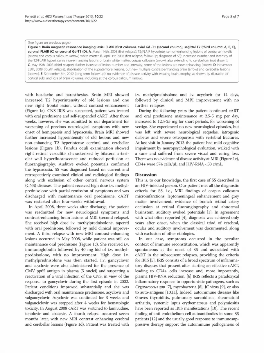
(See figure on previous page.)Figure 1 Brain magnetic resonance imaging: axial FLAIR (first column), axial Gd -T1 (second column), sagittal T2 (third column: A, B, E),coronal FLAIR (C) or coronal Gd-T1 (D). A. March 14th, 2008 (first relapse): T2/FLAIR hyperintense non-enhancing lesions of centra semiovalia(arrow) and corpus callosum (arrow) white matter. B. April 1st, 2008 (first relapse, follow-up; diagnosis of SS): increased number and intensity ofthe T2/FLAIR hyperintense non-enhancing lesions of brain white matter, corpus callosum (arrow), also extending to cerebellum (not shown).C. May 15th, 2008 (third relapse): further increase of lesion number and intensity, some of the lesions are now enhancing (arrow). D. November25th, 2008 (fourth relapse): stabilization of the supratentorial lesions, but new multiple contrast-enhancing brain (arrow) and cerebellar lesions(arrows). E. September 6th, 2012 (long-term follow-up): no evidence of disease activity with ensuing brain atrophy, as shown by dilatation ofcortical sulci and loss of brain volumes, including at the corpus callosum (arrow).
Ferretti et al. AIDS Research and Therapy 2013, 10:22 Page 5 of 7http://www.aidsrestherapy.com/content/10/1/22
with headache and paresthesias. Brain MRI showedincreased T2 hyperintensity of old lesions and onenew right frontal lesion, without contrast enhancement(Figure 1a). CNS-IRIS was suspected, patient was treatedwith oral prednisone and self-suspended cART. After threeweeks, however, she was admitted to our department forworsening of previous neurological symptoms with newonset of hemiparesis and hypoacusia. Brain MRI showedfurther increased hyperintensity of old lesions and newnon-enhancing T2 hyperintense cerebral and cerebellarlesions (Figure 1b). Fundus oculi examination showedright retinal vasculitis characterized by bilateral arteri-olar wall hyperfluorescence and reduced perfusion atfluorangiography. Auditive evoked potentials confirmedthe hypoacusia. SS was diagnosed based on current andretrospectively examined clinical and radiological findingsalong with exclusion of other central nervous system(CNS) diseases. The patient received high dose i.v. methyl-prednisolone with partial remission of symptoms and wasdischarged with maintenance oral prednisone. cARTwas restarted after four-weeks withdrawal.In April 2008, three weeks after discharge, the patient
was readmitted for new neurological symptoms andcontrast-enhancing brain lesions at MRI (second relapse).She received high dose i.v. methylprednisolone, taperedwith oral prednisone, followed by mild clinical improve-ment. A third relapse with new MRI contrast-enhancinglesions occurred in May 2008, while patient was still onmaintenance oral prednisone (Figure 1c). She received i.v.immunoglobulin followed by 40 mg bid of i.v. methyl-prednisolone, with no improvement. High dose i.v.methylprednisolone was then started. I.v. gancyclovirand acyclovir were also administered for the presence ofCMV pp65 antigen in plasma (5 nuclei) and suspecting areactivation of a viral infection of the CNS, in view of theresponse to gancyclovir during the first episode in 2002.Patient conditions improved substantially and she wasdischarged with oral maintenance prednisone, acyclovir andvalgancyclovir. Acyclovir was continued for 3 weeks andvalganciclovir was stopped after 4 weeks for hematologictoxicity. In August 2008 cART was switched to lamivudine,tenofovir and abacavir. A fourth relapse occurred sevenmonths later, with new MRI contrast enhancing cerebraland cerebellar lesions (Figure 1d). Patient was treated with
i.v. methylprednisolone and i.v. acyclovir for 14 days,followed by clinical and MRI improvement with nofurther relapses.During the following years the patient continued cART
and oral prednisone maintenance at 2.5-5 mg per day,increased to 12.5-25 mg for short periods, for worsening ofvertigo. She experienced no new neurological episodes, butwas left with severe neurological sequelae, iatrogenicdiabetes and severe osteoporosis with vertebral fractures.At last visit in January 2013 the patient had mild cognitiveimpairment by neuropsychological evaluation, walked witha cane and suffered from severe visual and earing loss.There was no evidence of disease activity at MRI (Figure 1e),CD4+ were 574 cells/μL and HIV-RNA <50 c/mL.
DiscussionThis is, to our knowledge, the first case of SS described inan HIV-infected person. Our patient met all the diagnosticcriteria for SS, i.e., MRI findings of corpus callosummicroinfarctions, leptomeningeal enhancement and greymatter involvement, evidence of branch retinal arteryocclusion at retinal fluorangiography and abnormalbrainstem auditory evoked potentials [1]. In agreementwith what often reported [4], diagnosis was achieved onlyyears after onset, when the classical triad of cerebral,ocular and auditory involvement was documented, alongwith exclusion of other etiologies.In our case, symptoms occurred in the peculiar
context of immune reconstitution, which was apparentlyspontaneous at the onset of SS and associated withcART in the subsequent relapses, providing the criteriafor IRIS [5]. IRIS consists of a broad spectrum of inflamma-tory diseases that present after starting an effective cART,leading to CD4+ cells increase and, more importantly,plasma HIV-RNA reduction. [6] IRIS reflects a paradoxycalinflammatory response to opportunistic pathogens, such asCryptococcus spp [7], mycobacteria [8], JC virus [9], or alsoto auto-antigens [10,11]. Indeed, autoimmune diseases likeGraves thyroiditis, pulmunary sarcoidosis, rheumatoidarthritis, systemic lupus erythematosus and polymiositishave been reported as IRIS manifestations [10]. The recentfinding of anti-endothelium cell autoantibodies in some SSpatients [12] and the usually good response to immunosup-pressive therapy support the autoimmune pathogenesis of

Ferretti et al. AIDS Research and Therapy 2013, 10:22 Page 6 of 7http://www.aidsrestherapy.com/content/10/1/22
SS. Thus our case suggests that SS might representan additional manifestation of “autoimmune IRIS”.Our patient did not show a long-lasting response to
high dose i.v. steroids or i.v. immunoglobulins alonein three attempts (first and second relapses), whereasa substantial and durable improvement was achievedboth times steroids were combined with antiviraldrugs, i.e., acyclovir, active on herpes simplex virustype 1 and type 2 and varicella-zoster virus (VZV),and gancyclovir, also active on CMV and Epstein-Barrvirus. It is known that the response to steroids in SScan not persist over time [13] and that SS can becharacterized by spontaneous attenuation of the immuno-pathological process [3]. However, the temporal relation-ship between antiviral treatment and clinical improvementwas strong and consistent in our case, suggesting thatreactivation of an herpesvirus in the CNS might havecontributed to disease pathogenesis.We did not find evidence of herpesviruses in CSF
(Table 1), but this does not exclude possible low levelreplication in CNS tissue [14]. We can speculate on apathogenic model by which an herpesvirus reactivatedin the context of immune reconstitution and, by rep-lication in vascular cells [15] and consequent expos-ure of either viral or auto-antigens, lead to immunemediated damage of cerebral vessels. According tothis model, antiviral therapy could have initially con-tributed to switch off the inflammatory stimulus bycontrol of viral replication. A potential candidate herpes-virus is VZV. VZV can cause direct damage of CNSvessels of all calibres and indirect damage of CNScells [16] and it is a well known cause of CNS vascu-litis and ocular infections in HIV-infected subjects[17,18]. In addition, herpes zoster, resulting from per-ipheral VZV reactivation, is a frequent IRIS manifest-ation and cases of VZV CNS-IRIS have recently beenreported [19,20].In conclusion, our case shows that SS may occur
within a dysimmune context that is possibly sustainedby HIV infection, cART-associated immune reconstitu-tion, or both. The clinical and neuroradiological remissionobserved when antiviral drugs were added to corticoste-roids support a potential pathogenetic role of herpes virusreactivations in SS in our case.
ConsentWritten informed consent was obtained from the patientfor publication of this Case report and any accompanyingimages. A copy of the written consent is available forreview by the Editor-in-Chief of this journal.
Competing interestsThe authors declare that they have no competing interests.
Authors’ contributionsFF, BC, MG, AL and PC contribuited to the care of the patient, SG toneuroradiologic examination, MT, DF and GB to laboratory analyses. FF drewup the first draft of the report, DF and PC made a substantial contribution todraft the manuscript and revised the draft. All authors read and approvedthe final version of the manuscript.
AcknowledgementsThe authors thank the medical and nursing staff at the Department ofInfectious Diseases of San Raffaele Hospital for their assistance in the care ofthis patient. We also wish to thank the patient and her family for theircollaboration.
Author details1Department of Infectious Diseases, San Raffaele Scientific Institute, Milan,Italy. 2Unit of Neuroradiology, San Raffaele Scientific Institute, Milan, Italy.3Department of Neurology, San Raffaele Scientific Institute, Milan, Italy.4Department of Neurology, IRCCS, C. Mondino National Institute ofNeurology, Pavia, Italy. 5Department of Neurology, IRCCS Neurologic InstituteCarlo Besta Institute, Milan, Italy.
Received: 17 June 2013 Accepted: 24 August 2013Published: 3 September 2013
References1. Susac JO, Egan RA, Rennebohm RM, Lubow M: Susac's syndrome:
1975–2005 microangiopathy/autoimmune endotheliopathy. J Neurol Sci2007, 257:270–272.
2. Rennebohm RM, Susac JO, Egan RA, Daroff RB: Susac's syndrome— update.J Neurol Sci 2010, 299:86–91.
3. Aubart-Cohen F, Klein I, Alexandra JF, et al: Long-term outcome in Susacsyndrome. Medicine (Baltimore) 2007, 86:93–102.
4. Kleffner I, Duning T, Lohmann H, et al: A brief review of Susac syndrome.J Neurol Sci 2012, 322:35–40.
5. French MA, Price P, Stone SF: Immune restoration disease afterantiretroviral therapy. AIDS 2004, 18:1615–1627.
6. Shelburne SA, Visnegarwala F, Darcourt J, et al: Incidence and risk factorsfor immune reconstitution inflammatory syndrome during highly activeantiretroviral therapy. AIDS 2005, 19:399–406.
7. Lortholary O, Fontanet A, Mémain N, et al: Incidence and risk factors ofimmune reconstitution inflammatory syndrome complicatingHIV-associated cryptococcosis in France. AIDS 2005, 19:1043–1049.
8. Meintjes G, Lawn SD, Scano F, et al: Tuberculosis-associated immunereconstitution inflammatory syndrome: case definitions for use inresource-limited settings. Lancet Infect Dis 2008, 8:516–523.
9. Cinque P, Koralnik IJ, Gerevini S, Miro JM, Price RW: Progressive multifocalleucoencephalopathy in HIV-1 infection. Lancet Infect Dis 2009, 9:625–636.
10. Maganti RM, Reveille JD, Williams FM: Therapy insight: the changingspectrum of rheumatic disease in HIV infection. Nat Clin Pract Rheumatol2008, 4:428–438.
11. Barber DL, Andrade BB, Sereti I, Sher A: Immune reconstitutioninflammatory syndrome: the trouble with immunity when you had none.Nat Rev Microbiol 2012, 10:150–156.
12. Magro CM, Poe JC, Lubow M, Susac JO: Susac syndrome: an organ-specificautoimmune endotheliopathy syndrome associated with anti-endothelialcell antibodies. Am J Clin Pathol 2011, 136:903–1012.
13. Rennebohm RM, Susac JO: Treatment of Susac's syndrome. J Neurol Sci2007, 257:215–220.
14. Nagel MA, Forghani B, Mahalingam R, et al: The value of detectinganti-VZV IgG antibody in CSF to diagnose VZV vasculopathy.Neurology 2007, 68:1069–1073.
15. Nagel MA, Traktinskiy I, Azarkh Y, et al: Varicella zoster virus vasculopathy:analysis of virus-infected arteries. Neurology 2011, 77:364–370.
16. Gnann JW Jr: Varicella-Zoster virus: atypical presentations and unusualcomplications. J Infect Dis 2002, 186:S91–S98.
17. Venkataramana A, Pardo CA, McArthur JC, et al: Immune reconstitutioninflammatory syndrome in the CNS of HIV-infected patients.Neurology 2006, 67:383–388.
18. Pagani JM, Duan JQ: Highly active antiretroviral therapy-induced immunerecovery in an HIV-positive patient with a history of herpes zosterophthalmicus. Optometry 2011, 82:77–82.

Ferretti et al. AIDS Research and Therapy 2013, 10:22 Page 7 of 7http://www.aidsrestherapy.com/content/10/1/22
19. Newsome SD, Nath A: Varicella-zoster virus vasculopathy and centralnervous system immune reconstitution inflammatory syndrome withhuman immunodeficiency virus infection treated with steroids.J Neurovirol 2009, 15:288–291.
20. Chang CC, McLean C, Vujovic O, et al: Fatal acute varicella-zoster virushemorrhagic meningomyelitis with necrotizing vasculitis in anHIV-infected patient. Clin Infect Dis 2009, 48:372–373.
doi:10.1186/1742-6405-10-22Cite this article as: Ferretti et al.: Susac’s syndrome as HIV-associatedimmune reconstitution inflammatory syndrome. AIDS Research andTherapy 2013 10:22.
Submit your next manuscript to BioMed Centraland take full advantage of:
• Convenient online submission
• Thorough peer review
• No space constraints or color figure charges
• Immediate publication on acceptance
• Inclusion in PubMed, CAS, Scopus and Google Scholar
• Research which is freely available for redistribution
Submit your manuscript at www.biomedcentral.com/submit


