Embed Size (px)
Citation preview
Carcinogenesisvol.20 no.5 pp.773–783, 1999
Asbestos induction of extended lifespan in normal humanmesothelial cells: interindividual susceptibility and SV40 T antigen
Lixin Xu, Barbara J.Flynn, Susan Ungar, Harvey I.Pass1,Kaija Linnainmaa 2, Karin Mattson 3 andBrenda I.Gerwin4
Laboratory of Human Carcinogenesis, National Cancer Institute, Bethesda,MD 20892,1Aerodigestive Program, Karmanos Cancer Institute, Detroit, MI48201, USA,2Institute of Occupational Health, Topeliuksenkatu, 41, aA00250, Helsinki and3Department of Pulmonary Medicine, University ofHelsinki, Haartmanikatu 4, 00290 Helsinki, Finland
4To whom correspondence should be addressedEmail: [email protected]
Normal human mesothelial cells from individual donorswere studied for susceptibility to asbestos-induction ofapoptosis and generation of an extended lifespan popula-tion. Such populations were generated after death of themajority of cells and arose from a subset of mesothelialcultures (4/16) whereas fibroblastic cells (5/5) did notdevelop extended lifespan populations after asbestos expo-sure. All mesothelial cultures were examined for thepresence of SV40 T antigen to obtain information on (i)the presence of SV40 T antigen expression in normalhuman mesothelial cells and (ii) the relationship betweengeneration of an extended lifespan population and expres-sion of SV40 T antigen. Immunostaining for SV40 T antigenwas positive in 2/38 normal human mesothelial cultures.These cultures also had elevated p53 expression. However,the two isolates expressing SV40 T antigen did not exhibitenhanced proliferative potential or develop an extendedlifespan population. Asbestos-generated extended lifespanpopulations were specifically resistant to asbestos-mediatedbut not to α-Fas-induced apoptosis. Deletion of p16Ink4a
was shown in 70% of tumor samples. All mesotheliomacell lines examined showed homozygous deletion of thislocus which extended to exon 1β. Extended lifespan cultureswere examined for expression of p16Ink4a to establishwhether deletion was an early response to asbestos expo-sure. During their rapid growth phase, extended lifespancultures showed decreased expression of p16Ink4a relativeto untreated cultures, but methylation was not observed,and p16Ink4a expression became elevated when cells enteredculture crisis. These data extend the earlier observationthat asbestos can generate extended lifespan populations,providing data on frequency and cell type specificity.In addition, this report shows that generation of suchpopulations does not require expression of SV40 T antigen.Extended lifespan cells could represent a populationexpressing early changes critical for mesothelioma develop-
Abbreviations: BSA, bovine serum albumin; DAPI, 49-6-diamidine-2-phenylindole; DTT, dithiothreitol; ECL, enhanced chemiluminescence; ELP,extended proliferative lifespan populations; EMEM, Eagle’s minimum essentialmedium; FITC, fluorescein isothiocyanate; LHC, Laboratory of HumanCarcinogenesis; MM, mesothelial medium; NAIMA, North American Insula-tion Manufacturers Association; PBS, phosphate-buffered saline; PI, propidiumiodide; PMSF, polymethylsulfonide fluoride; RB, retinoblastoma; TBST, Trisbuffered saline plus Tween20; UICC, Union Internationale contre le Cancer.
© Oxford University Press 773
ment. Further study of these populations could identifysuch changes.
Introduction
Mesothelioma is a form of cancer in which exposure toasbestos, a complete carcinogen, produces sufficient alterationsin normal mesothelial cells to generate a malignant population(1,2). These changes arise, presumably, both by direct inter-action with asbestos fibers (3–5) and by the generation, inresponse to asbestos, of an inflammatory environment con-taining strong proliferative signals as well as active oxygenspecies (6–8). Interestingly, mesothelioma cell lines and tumorshave been shown to contain few p53 mutations (9–11) and anormal retinoblastoma (RB) gene product (12–14). Since thefunctions of these tumor suppressor genes are inactivated inmost malignancies, mesothelioma is a tumor that is likely tocontain genetic or epigenetic alterations in molecules whichinterfere with p53 and/or RB function. Recent reports haveassociated the presence of SV40 T antigen sequences andprotein with mesothelioma tissue (15,16). Clearly, the presenceof a molecule such as T antigen, which could inactivate bothp53 and RB, would explain the lack of alterations of thesesuppressors.
Homozygous deletion of sequences on chromosome 9p21–22 is associated with mesothelioma (17–19) and loss orreduction of p16INK4a has been demonstrated in this tumor(20). Loss or inactivation of this gene has been shown todiminish RB cell cycle control, and loss of p16INK4a andalteration of RB have been shown to have a reciprocalrelationship (12,21,22). In addition, it has been shown that thealternate reading frame protein, p14ARF, interacts with MDM2(23,24) to affect the stability of the protein and is transcrip-tionally activated by E2F-1, linking release of this factor byRB phosphorylation to p53 stabilization (25). Thus, loss ofthe two functions of this locus in mesothelioma could reducethe activity of both p53 and RB.
Mesothelial cells exposed to asbestos show responsesranging from a proliferative response (26–29) to cell cyclearrest or apoptosis (30–32). These responses are likely to bemediated by altered gene expression following induction oftranscription factors and activation of intracellular signalingpathways (33–35). Interestingly, early studies ofin vitroamosite treatment of normal human mesothelial cells demon-strated the establishment of a population of cells with anextended lifespan and chromosomal alterations (36,37). Identi-fication of molecular alterations in such a population mightreveal changes that occur before immortalization, which arecritical in the process of tumorigenesis. Epigenetic down-regulation of p16INK4a has been shown in extended lifespanpopulations of mammary epithelial cells (38,39). Data fromstudies on bladder cancer specimens indicate that progressionto myoinvasive tumors correlates with alteration of RB orp16INK4a and the ability to bypass senescence in culture (40).
by guest on May 21, 2014
http://carcin.oxfordjournals.org/D
ownloaded from
L.Xu et al.
Thus, alteration of p16INK4a expression might contribute to theextended lifespan induced by asbestos exposure. In addition,expression of SV40 T antigen, if present in the normalpopulation, would be expected to increase the probability ofexpressing an extended lifespan (41,42). Therefore, measure-ment of SV40 T in the mesothelial isolates tested for lifespanextension should provide data concerning the biologicalrelevance of this gene to mesothelial carcinogenesis.
The data presented here confirm that directin vitro treatmentof normal human mesothelial cells with amosite asbestosinduces an acute apoptotic response, which is followed, insome cases, by generation of an extended lifespan population.While all mesothelial isolates showed an apoptotic response,only a subset of isolates generated extended lifespan popula-tions. These biologically altered populations were characterizedby a decreased sensitivity to asbestos-induced apoptosis butnot to apoptosis mediated byFas ligand-binding. SV40 Tantigen was expressed infrequently in normal human mesothel-ial cells and did not appear to contribute to generation of anextended lifespan. The expression level of p16INK4a wasreduced during the proliferative phase of extended lifespanpopulations but became elevated as in controls at senescence,indicating that the deletion of p16INK4a seen in tumors is notan early response to asbestos treatment. The reproduciblegeneration by asbestos of extended lifespan populations ofhuman mesothelial cells provides an important cellular modelfor analysis of critical steps in the progression to mesothelioma.
Materials and methodsCell culture and growth mediumCultures of mesothelial cells were initiated from pleural effusions obtainedfrom non-cancerous donors. The fluid was initially centrifuged at 125g for10 min. The pelleted cells were suspended in HEPES-buffered saline,washed by centrifugation, resuspended in growth medium, and inoculated intosurface coated 150 cm2 culture flasks at a ratio of 150 ml pleural fluid/flask.Semiconfluent cultures were dissociated by trypsin (43) and either expandedby inoculating 300 000 cells/75 cm2 flask or used according to experimentalprotocols.
Growth medium was LHC-MM (LHC-mesothelial medium) (Biofluids,Rockville, MD). Lux culture surfaces were coated with a mixture ofhuman fibronectin (10µg/ml), and crystallized bovine serum albumin (BSA)(10µg/ml) dissolved in LHC-Basal medium. The mixture was added to culturedishes at a ratio of 2 ml/100 cm2 of surface area. The plates were incubatedat 36.5°C for at least 10 min before the mixture was vacuum aspirated.
Normal human fibroblast cells obtained from Coriell Institute for MedicalResearch (Camden, NJ) were cultured with EMEM (Eagle’s minimum essentialmedium with Earle’s salts, Biofluids) with glutamine (2 mM), 10% fetalbovine serum (Biofluids) and gentamicin (50µg/ml) (Biofluids).
Asbestos and glass beadsTwo types of amosite fibers were obtained: UICC [Union Internationale contrele Cancer, a gift of Dr John Hoskins at the University of Leicester, meandiameter: 0.316 0.20µ; mean length: 2.46 1.8µ (44)] and NAIMA (NorthAmerican Insulation Manufacturers Association, provided by the association,diameter 70.6%5 0–0.5µ, 27.9%5 0.5–1.0µ, median5 0.38µ; and length26% 5 0–5µ, 25%5 5–10µ, 13%5 10–15µ, median5 9.3µ). Glass beadsfrom Particle Information Services were a gift from Dr Brooke Mossman(University of Vermont, Burlington, VT). Amosite fibers and glass beads weresuspended in distilled water at 1.0 mg/ml, heated at 100°C for 2 h, thentriturated eight times through a 22-gauge needle and autoclaved prior toaddition to culture medium at various concentrations.
CytotoxicitySix-well dishes (35 mm2) were inoculated with 25 000 cells per well. Twenty-four hours later, the growth media (LHC-MM or EMEM) were replaced withthe same media containing increasing concentrations of fibers. Each dose wasassayed in triplicate. After 3 days of exposure, attached cells were dissociatedand viable cell numbers counted.
Growth assaysA total of 33105 cells in each 75 cm2 flask were exposed to amosite asbestosby adding the fibers to LHC-MM or EMEM. After 3 days of incubation, the
774
medium was replaced with the same fresh media without fibers. When thecultures became confluent (~2 weeks), the cells were dissociated with trypsin,pooled and inoculated as before. The following day, the asbestos treatmentprotocol was repeated. A glass beads control and unexposed control cultureswere studied in parallel. In addition, normal human fibroblasts were treatedby the same protocol. After the second exposure, culture was continued andcells were counted at subculture to determine the population doublings.
Apoptosis measurement
About 25 000 cells per well were inoculated in six-well dishes. After asbestosor exposure to 200 ng/ml anti-Fas (45) (anti-Fas, clone CH-11, Medical andBiological Laboratories, Boston, MA), both detached and attached cells werecollected and concentrated by centrifugation. The medium was removed andthe cells were resuspended in 100µl of 13 binding buffer (1 ml of 1 MHEPES, pH 7.0 in 95 ml of distilled water, pH adjusted to 7.4, after which2.8 ml of 5 M NaCl, 0.037 g CaCl2 dihydrate and distilled water were addedto a total volume of 100 ml). Annexin V–FITC (final concentration 1 mg/ml)(Bio Whittaker, Walkersville, MD) and propidium iodide (PI, final concentra-tion 2.5 µg/ml) were then added. Following incubation at room temperaturein the dark for 5 min, 20µl of cell suspension was placed on a glass slideand a cover slip applied. These slides were evaluated by fluorescencemicroscopy. At least 200 cells were counted in each group. Cells with annexinV positive staining only were scored as early apoptosis. Cells with bothpositive staining of annexin V and PI and with condensed nuclei were countedas late apoptosis (32). Cells exhibiting PI staining without condensed nucleiwere not scored as apoptotic (Figure 2).
Immunofluorescent staining
Cells on glass slides were fixed with 4% paraformaldehyde [in phosphate-buffered saline (PBS)] for 10 min at room temperature followed by absolutemethanol for 20 min at room temperature. Slides were washed three timeswith PBS-plus solution (0.15 g glycine, 0.5% BSA in 100 ml PBS) beforeincubation with primary antibodies at 4°C overnight (16 h), then roomtemperature for 30 min. Primary antibodies were used at the followingconcentrations: p16Ink4a (Ab-1), 2.5 µg/ml; p21 (WAF1, Ab-1), 2.5µg/ml;p53 (Ab-6), 1.0µg/ml; and SV40 T-Ag (Ab-2, N-terminal), all from OncogeneResearch Products (Cambridge, MA), 0.2µg/ml; SV40 T-Ag (Pab101,C-terminal), Santa Cruz Biotechnology (Santa Cruz, CA), 0.2µg/ml. Afterfive washes with PBS, slides were incubated with goat anti-mouse-fluoresceinisothiocyanate (FITC) (1:300) (Vector Lab, Burlingame, CA) for 1 h atroomtemperature and washed again with PBS, five times. A drop of VectaShield,(Vector Lab), which contained 0.5µg/ml 4’-6-diamidine-2-phenylindole(DAPI), was added; cover slips were applied and results were evaluated byfluorescent microscopy. The percentage of positive cells was determinedby counting at least 200 cells. For determination of staining intensity, 100positive cells were scored using the segmentation program of the fluorescentimaging system (IPLab Spectrum™, Version 3.1, Scanalytics, Fairfax, VA).
PCR and methylation specific PCR
Genomic DNA from cultured cells was prepared using DNAzol (MolecularResearch Center, Inc., Cincinnati, OH) according to the manufacturer’sinstruction. For standard PCR amplification of p16Ink4a, the primers for exon2 were used: sense, 59-ATGGCGCGGACGTGGGTCCCGG-39 and antisense,59-TTATCTGTTCACTTGTGCCC-39. PCR reactions contained, in 50µl,genomic DNA 200 ng, 13PCR buffer, 1.1 mM Mg(OAc)2, 0.2 mM dNTP,15 pmol each primer and 1 U rTth DNA polymerase, XL (PE AppliedBiosystems, Norwalk, CT). Reaction conditions were 94°C, 4 min, then 94°Cfor 40 s, 60°C for 30 s, 68°C for 2 min 30 s, for 35 cycles followed by 68°Cfor 8 min. For methylation specific PCR, DNA (1µg) in a volume of 50µlwas denatured by NaOH (final concentration, 0.2 M) for 10 min at 37°C. Analiquot of 30µl of 10 mM hydroquinone (Sigma, St Louis, MO) and 520µlof 3 M sodium bisulfite (Sigma) at pH 5, both freshly prepared, were addedand mixed, and samples were incubated under mineral oil at 50°C for 16 h.Modified DNA was purified using the Wizard DNA purification resin accordingto the manufacturer (Promega, Madison, WI) and eluted into 50µl of water.Modification was completed by NaOH (final5 0.3 M) treatment for 5 minat room temperature, followed by ethanol precipitation. DNA was resuspendedin 50 µl water and used immediately or stored at –20°C (46). Primers usedfor a first amplification of p16 exon 1 outside the potentially methylatedregion were p16 wild-type, sense 59-CAGAGGGTGGGGCGGACCGC-39,antisense 59-CGGGCCGCGGCCGTGG-39 (size, 140 bp; annealing temper-ature 65°C; genomic position1171); p16 methylation specific, sense 59-TTATTAGAGGGTGGGGCGGATCGC-39, antisense 59-GACCCCGAACC-GCGACCGTAA-39 (size, 150 bp; annealing temperature 65°C; genomicposition1167); and p16 unmethylated specific, sense 59-TTATTAGAGGGT-GGGGTGGATTGT-39, antisense 59-CAACCCCAAACCACAACCATAA-39(size, 151 bp; annealing temperature 60°C; genomic position1167). ThePCR mixture contained 13PCR buffer (16.6 mM ammonium sulfate, 67 mM
by guest on May 21, 2014
http://carcin.oxfordjournals.org/D
ownloaded from
Extended lifespan in asbestos-treated human cells
Tris, pH 8.8, 6.7 mM MgCl2, 10 mM 2-mercaptoethanol), dNTP (each at1.25 mM), primers (300 ng each per reaction) and bisulfite-modified DNA(~50 ng) or unmodified DNA (50–100 ng) in a final volume of 50µl. PCRspecific for unmodified DNA also included 5% dimethyl sulfoxide. Reactionswere subjected to a hot start at 95°C for 5 min before the addition of 1.25 UTaq polymerase (BRL) followed by 35 cycles (30 s at 95°C, 30 s at theannealing temperature listed above, and 30 s at 72°C) and a final 4 minextension at 72°C. Products were analyzed on 2% agarose gels.
Southern blotDNA was extracted, digested withHindIII, probed with32P-labeled p16INK4a
and XPB probes and analyzed according to standard protocols (47). Ratios ofp16INK4a to XPB were determined for each sample and normal and tumorsamples were compared to determine the relative content of p16INK4a ineach sample.
Western blotWhole cell lysates were made by lysing cells in RIPA buffer [50 mM Tris–HCl, pH 7.4, 150 mM NaCl, 1% Triton X-100, 0.1% SDS and 1% deoxycholicacid, plus the following protease inhibitors added at time of use from thestock solutions: 10µl/ml of 10 mg/ml polymethylsulfonide fluoride (PMSF)in isopropanol, 30µl/ml of aprotinin (Sigma) and 10µl/ml of 100 mM sodiumorthovanadate]. Lysates were centrifuged at 40 000 r.p.m. for 35 min at 4°C(Sorvall RCM 120 ultracentrifuge in an RP100 AT-280 rotor). Aliquots of50 µg protein/sample were loaded on a pre-cast 16% Tris–glycine gel (Novex,San Diego, CA) run at 125 V for ~3 h until the running dye was eluted andthen transferred to an Immobilon-P membrane (Millipore, Bedford, MA).Membranes were blocked for 2 h at room temperature in TBST (10 mM Tris–HCl, pH 8.0, 150 mM NaCl, 0.05% TWEEN 20), which contained 5% non-fat milk and 0.5% BSA, incubated with anti-p16 antibody [polyclonalantiserum, cat. # 15126E, 1:1000 dilution in blocking solution, PharMingen(San Diego, CA)] at 4°C for 16 h; washed, and incubated for 1 h with a1:10 000 dilution of anti-rabbit horseradish peroxidase antibody (NA 934,Amersham, Arlington Heights, IL) in blocking solution at room temperature;washed and developed with enhanced chemiluminescence (ECL) solution(34080, Pierce, Rockford, IL) according to the manufacturer’s instructions.
Results
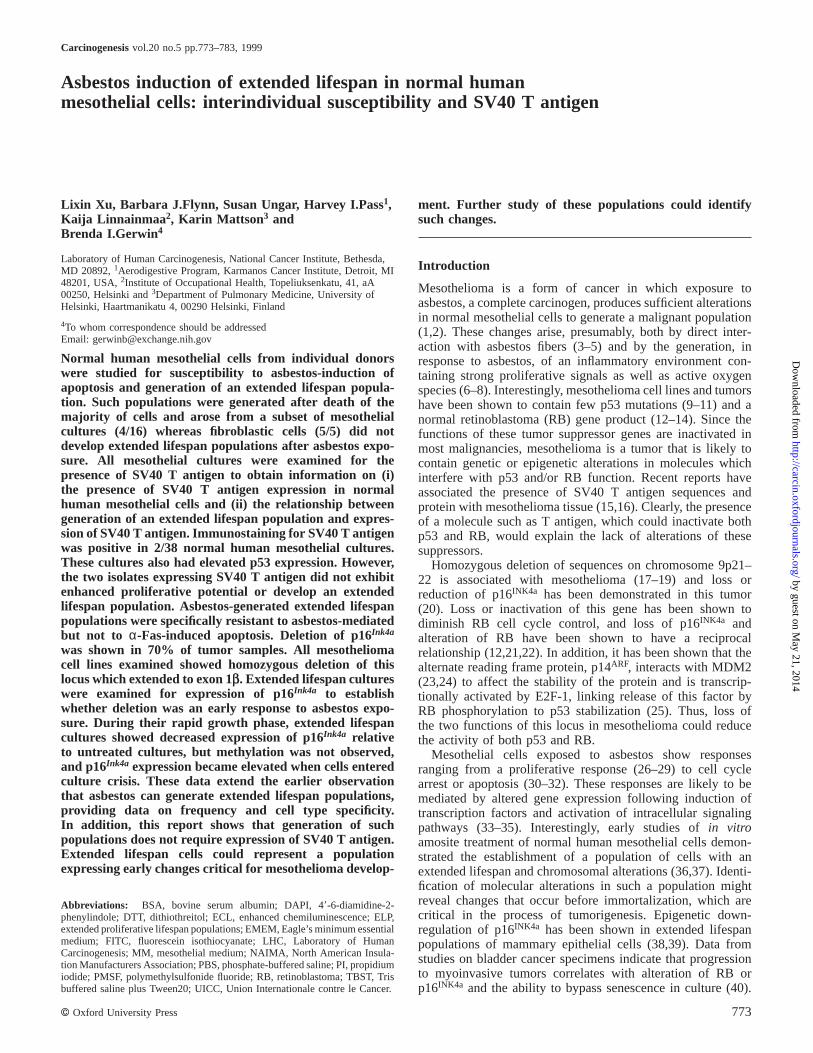
Comparative toxicity of amosite preparations for mesothelialcells and fibroblastsRecent reports show either apoptosis (31,32) or cell cyclearrest (30) induced by asbestos treatment of mesothelial cellsand suggest that these may have arisen because of differencesin fiber types used by different laboratories (30,48). Wecompared two types of amosite with different fiber dimensions,UICC and NAIMA (see Materials and methods) with respectto toxicity in normal human mesothelial cells in order toestablish the dose range for amosite treatment. Fibroblastswere also exposed to the NAIMA amosite to establish cell-type dose responsiveness. Figure 1 shows the results of a72 h treatment of either mesothelial cells or fibroblasts. Fourindividual cell strains of each cell type were tested. Interest-
Fig. 1. Cytotoxicity of amosite for human mesothelial cells and fibroblasts.Mesothelial cells or fibroblasts were grown in six-well dishes and exposedfor 72 h to various doses of UICC or NAIMA amosite asbestos as describedin Materials and methods. Each curve represents data from triplicate assaysof four individual cell strains. (.) Fibroblasts, NAIMA amosite; (m)mesothelial cells, UICC amosite; (n) mesothelial cells, NAIMA amosite.
775
ingly, the toxicity curves did not show significant inter-individual variation in contradistinction to the growth potentialvariation shown by mesothelial cells from different individuals(49). It is apparent, in confirmation of earlier reports usingcolony-forming assays (36), that human mesothelial cells showgreater sensitivity to asbestos toxicity than do fibroblasts. Thedata of Figure 1 indicate that the NAIMA amosite, selectedfor longer fibers is, as expected, more toxic than UICC amositewith an LD50 of 2.4 µg/cm2 for mesothelial cells as comparedwith 10.8 µg/cm2 for fibroblasts.
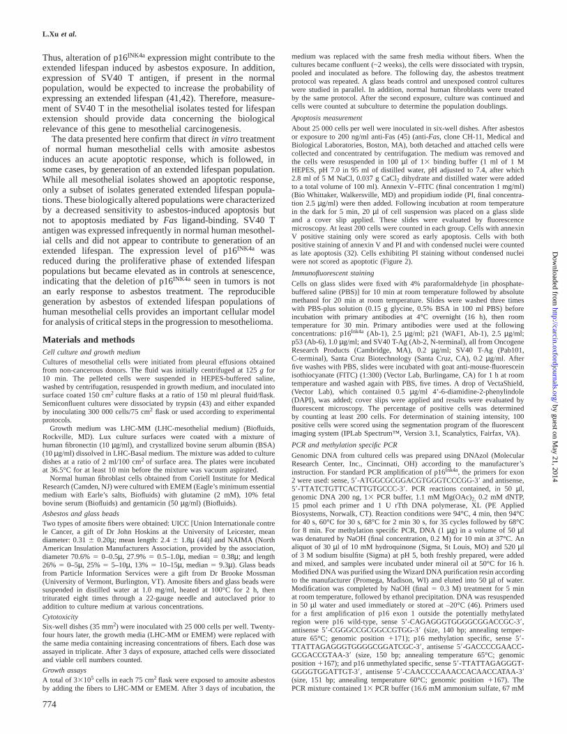
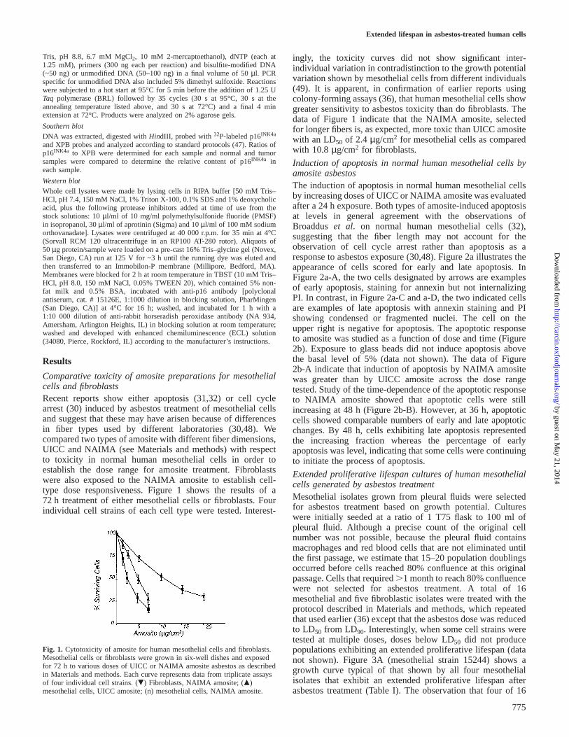
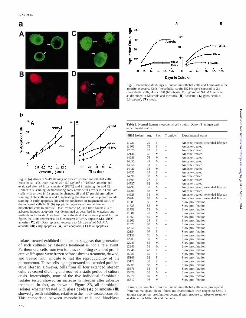
Induction of apoptosis in normal human mesothelial cells byamosite asbestosThe induction of apoptosis in normal human mesothelial cellsby increasing doses of UICC or NAIMA amosite was evaluatedafter a 24 h exposure. Both types of amosite-induced apoptosisat levels in general agreement with the observations ofBroaddus et al. on normal human mesothelial cells (32),suggesting that the fiber length may not account for theobservation of cell cycle arrest rather than apoptosis as aresponse to asbestos exposure (30,48). Figure 2a illustrates theappearance of cells scored for early and late apoptosis. InFigure 2a-A, the two cells designated by arrows are examplesof early apoptosis, staining for annexin but not internalizingPI. In contrast, in Figure 2a-C and a-D, the two indicated cellsare examples of late apoptosis with annexin staining and PIshowing condensed or fragmented nuclei. The cell on theupper right is negative for apoptosis. The apoptotic responseto amosite was studied as a function of dose and time (Figure2b). Exposure to glass beads did not induce apoptosis abovethe basal level of 5% (data not shown). The data of Figure2b-A indicate that induction of apoptosis by NAIMA amositewas greater than by UICC amosite across the dose rangetested. Study of the time-dependence of the apoptotic responseto NAIMA amosite showed that apoptotic cells were stillincreasing at 48 h (Figure 2b-B). However, at 36 h, apoptoticcells showed comparable numbers of early and late apoptoticchanges. By 48 h, cells exhibiting late apoptosis representedthe increasing fraction whereas the percentage of earlyapoptosis was level, indicating that some cells were continuingto initiate the process of apoptosis.
Extended proliferative lifespan cultures of human mesothelialcells generated by asbestos treatmentMesothelial isolates grown from pleural fluids were selectedfor asbestos treatment based on growth potential. Cultureswere initially seeded at a ratio of 1 T75 flask to 100 ml ofpleural fluid. Although a precise count of the original cellnumber was not possible, because the pleural fluid containsmacrophages and red blood cells that are not eliminated untilthe first passage, we estimate that 15–20 population doublingsoccurred before cells reached 80% confluence at this originalpassage. Cells that required.1 month to reach 80% confluencewere not selected for asbestos treatment. A total of 16mesothelial and five fibroblastic isolates were treated with theprotocol described in Materials and methods, which repeatedthat used earlier (36) except that the asbestos dose was reducedto LD50 from LD90. Interestingly, when some cell strains weretested at multiple doses, doses below LD50 did not producepopulations exhibiting an extended proliferative lifespan (datanot shown). Figure 3A (mesothelial strain 15244) shows agrowth curve typical of that shown by all four mesothelialisolates that exhibit an extended proliferative lifespan afterasbestos treatment (Table I). The observation that four of 16
by guest on May 21, 2014
http://carcin.oxfordjournals.org/D
ownloaded from
L.Xu et al.
Fig. 2. (a) Annexin V–PI staining of asbestos-treated mesothelial cells.Mesothelial cells were treated with 5.0µg/cm2 of NAIMA amosite andevaluated after 24 h for annexin V (FITC) and PI staining. (A and C)Annnexin V staining, demonstrating early (cells with arrows in A) and late(cells with arrows in C) apoptotic changes. (B and D) propidium iodidestaining of the cells in A and C indicating the absence of propidium iodidestaining in early apoptosis (B) and the condensed or fragmented DNA ofthe indicated cells in D. (b) Apoptotic response of normal humanmesothelial cells to amosite. Dose–response (A) and time-course (B) ofasbestos-induced apoptosis was determined as described in Materials andmethods in triplicate. Data from four individual donors were pooled for thisfigure. (A) Data represent a 24 h exposure; NAIMA amosite (m), UICCamosite (.); (B) Data represent exposure to 5.0µg/cm2 of NAIMAamosite; (j) early apoptosis, (m) late apoptosis, (.) total apoptosis.
isolates treated exhibited this pattern suggests that generationof such cultures by asbestos treatment is not a rare event.Furthermore, cells from two isolates exhibiting extended prolif-erative lifespans were frozen before asbestos treatment, thawed,and treated with amosite to test the reproducibility of thephenomenon. These cells again generated an extended prolifer-ative lifespan. However, cells from all four extended lifespancultures ceased dividing and reached a static period of culturecrisis. Interestingly, none of the five individual fibroblasticisolates tested showed an increase in lifespan after asbestostreatment. In fact, as shown in Figure 3B, all fibroblasticisolates whether treated with glass beads (m) or amosite (j)showed growth inhibition, relative to the mock-treated controls.This comparison between mesothelial cells and fibroblasts
776
Fig. 3. Population doublings of human mesothelial cells and fibroblasts afteramosite exposure. Cells (mesothelial strain 15244) were exposed to 2.4(mesothelial cells,A) or 10.8 (fibroblasts,B) µg/cm2 of NAIMA amositeas described in Materials and methods. (j) Amosite; (m) glass beads at5.0 µg/cm2; (.) mock.
Table I. Normal human mesothelial cell strains. Donor, T antigen andexperimental status
NHM isolate Age Sex T antigen Experimental status
11936 79 F – Amosite-treated; extended lifespan11963 75 F – Amosite-treated12075 73 F – Amosite-treated12144 66 M – Amosite-treated14286 76 M 1 Amosite-treated14355 49 M – Amosite-treated14356 51 F – Amosite-treated14421 63 M – Amosite-treated14535 35 F – Amosite-treated14598 83 M – Amosite-treated14599 76 F – Amosite-treated14737 62 M – Amosite-treated14792 57 M – Amosite-treated; extended lifespan14798 83 M – Amosite-treated14928 58 M – Amosite-treated; extended lifespan15244 88 M – Amosite-treated; extended lifespan11003 86 M – Slow proliferation11735 85 M – Slow proliferation11739 71 M – Slow proliferation11804 70 M – Slow proliferation11859 45 M – Slow proliferation11866 24 F – Slow proliferation11926 80 M – Slow proliferation12050 89 F – Slow proliferation12154 97 F – Slow proliferation12156 76 M – Slow proliferation12203 59 M – Slow proliferation12245 85 M – Slow proliferation12248 51 M – Slow proliferation15046 40 F – Slow proliferation15080 60 F – Slow proliferation15169 62 F – Slow proliferation15170 38 F – Slow proliferation15305 88 M – Slow proliferation15376 64 F – Slow proliferation15426 55 M – Slow proliferation15570 69 M 1 Slow proliferation15612 98 M – Slow proliferation
Consecutive samples of normal human mesothelial cells were propagatedfrom non-malignant pleural fluids and characterized with respect to SV40 Tantigen expression, proliferation potential and response to asbestos treatmentas detailed in Materials and methods.
by guest on May 21, 2014
http://carcin.oxfordjournals.org/D
ownloaded from
Extended lifespan in asbestos-treated human cells
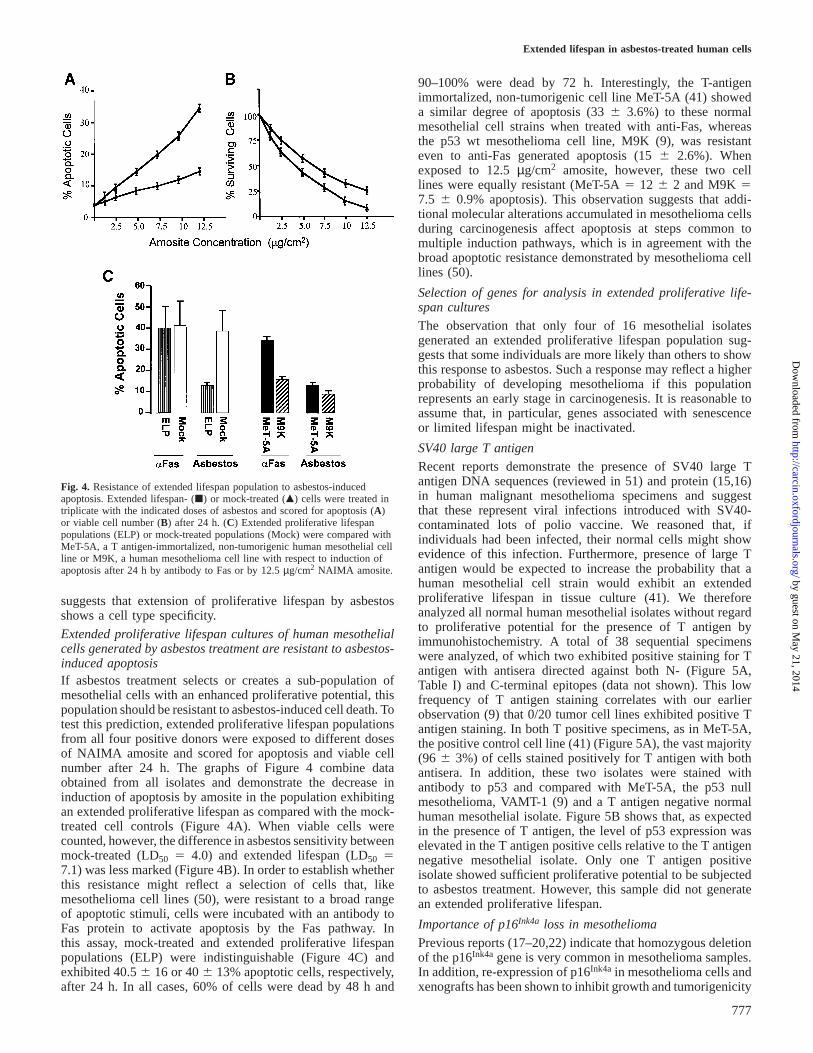
Fig. 4. Resistance of extended lifespan population to asbestos-inducedapoptosis. Extended lifespan- (j) or mock-treated (m) cells were treated intriplicate with the indicated doses of asbestos and scored for apoptosis (A)or viable cell number (B) after 24 h. (C) Extended proliferative lifespanpopulations (ELP) or mock-treated populations (Mock) were compared withMeT-5A, a T antigen-immortalized, non-tumorigenic human mesothelial cellline or M9K, a human mesothelioma cell line with respect to induction ofapoptosis after 24 h by antibody to Fas or by 12.5µg/cm2 NAIMA amosite.
suggests that extension of proliferative lifespan by asbestosshows a cell type specificity.
Extended proliferative lifespan cultures of human mesothelialcells generated by asbestos treatment are resistant to asbestos-induced apoptosisIf asbestos treatment selects or creates a sub-population ofmesothelial cells with an enhanced proliferative potential, thispopulation should be resistant to asbestos-induced cell death. Totest this prediction, extended proliferative lifespan populationsfrom all four positive donors were exposed to different dosesof NAIMA amosite and scored for apoptosis and viable cellnumber after 24 h. The graphs of Figure 4 combine dataobtained from all isolates and demonstrate the decrease ininduction of apoptosis by amosite in the population exhibitingan extended proliferative lifespan as compared with the mock-treated cell controls (Figure 4A). When viable cells werecounted, however, the difference in asbestos sensitivity betweenmock-treated (LD50 5 4.0) and extended lifespan (LD50 57.1) was less marked (Figure 4B). In order to establish whetherthis resistance might reflect a selection of cells that, likemesothelioma cell lines (50), were resistant to a broad rangeof apoptotic stimuli, cells were incubated with an antibody toFas protein to activate apoptosis by the Fas pathway. Inthis assay, mock-treated and extended proliferative lifespanpopulations (ELP) were indistinguishable (Figure 4C) andexhibited 40.56 16 or 406 13% apoptotic cells, respectively,after 24 h. In all cases, 60% of cells were dead by 48 h and
777
90–100% were dead by 72 h. Interestingly, the T-antigenimmortalized, non-tumorigenic cell line MeT-5A (41) showeda similar degree of apoptosis (336 3.6%) to these normalmesothelial cell strains when treated with anti-Fas, whereasthe p53 wt mesothelioma cell line, M9K (9), was resistanteven to anti-Fas generated apoptosis (156 2.6%). Whenexposed to 12.5µg/cm2 amosite, however, these two celllines were equally resistant (MeT-5A5 12 6 2 and M9K57.5 6 0.9% apoptosis). This observation suggests that addi-tional molecular alterations accumulated in mesothelioma cellsduring carcinogenesis affect apoptosis at steps common tomultiple induction pathways, which is in agreement with thebroad apoptotic resistance demonstrated by mesothelioma celllines (50).
Selection of genes for analysis in extended proliferative life-span cultures
The observation that only four of 16 mesothelial isolatesgenerated an extended proliferative lifespan population sug-gests that some individuals are more likely than others to showthis response to asbestos. Such a response may reflect a higherprobability of developing mesothelioma if this populationrepresents an early stage in carcinogenesis. It is reasonable toassume that, in particular, genes associated with senescenceor limited lifespan might be inactivated.
SV40 large T antigen
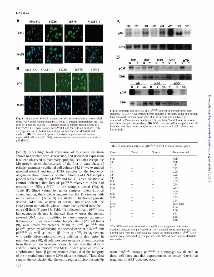
Recent reports demonstrate the presence of SV40 large Tantigen DNA sequences (reviewed in 51) and protein (15,16)in human malignant mesothelioma specimens and suggestthat these represent viral infections introduced with SV40-contaminated lots of polio vaccine. We reasoned that, ifindividuals had been infected, their normal cells might showevidence of this infection. Furthermore, presence of large Tantigen would be expected to increase the probability that ahuman mesothelial cell strain would exhibit an extendedproliferative lifespan in tissue culture (41). We thereforeanalyzed all normal human mesothelial isolates without regardto proliferative potential for the presence of T antigen byimmunohistochemistry. A total of 38 sequential specimenswere analyzed, of which two exhibited positive staining for Tantigen with antisera directed against both N- (Figure 5A,Table I) and C-terminal epitopes (data not shown). This lowfrequency of T antigen staining correlates with our earlierobservation (9) that 0/20 tumor cell lines exhibited positive Tantigen staining. In both T positive specimens, as in MeT-5A,the positive control cell line (41) (Figure 5A), the vast majority(96 6 3%) of cells stained positively for T antigen with bothantisera. In addition, these two isolates were stained withantibody to p53 and compared with MeT-5A, the p53 nullmesothelioma, VAMT-1 (9) and a T antigen negative normalhuman mesothelial isolate. Figure 5B shows that, as expectedin the presence of T antigen, the level of p53 expression waselevated in the T antigen positive cells relative to the T antigennegative mesothelial isolate. Only one T antigen positiveisolate showed sufficient proliferative potential to be subjectedto asbestos treatment. However, this sample did not generatean extended proliferative lifespan.
Importance of p16Ink4a loss in mesothelioma
Previous reports (17–20,22) indicate that homozygous deletionof the p16Ink4a gene is very common in mesothelioma samples.In addition, re-expression of p16Ink4a in mesothelioma cells andxenografts has been shown to inhibit growth and tumorigenicity
by guest on May 21, 2014
http://carcin.oxfordjournals.org/D
ownloaded from
L.Xu et al.
Fig. 5. Detection of SV40 T antigen and p53 in normal human mesothelialcells. (A) Normal human mesothelial cells, T antigen immortalized MeT-5Acells (41) and the p53 null, T antigen negative human mesothelioma cellline VAMT-1 (9) were stained for SV40 T antigen with an antibody (Pab416) specific for an N-terminal epitope as described in Materials andmethods. (B) Cells as in A, plus a T antigen negative normal humanmesothelial cell strain (#11804) were stained as above with an antibody top53 (DO-1).
(52,53). Since high level expression of this gene has beenshown to correlate with senescence, and decreased expressionhas been observed in mammary epithelial cells that escape theM0 growth arrest characteristic of the firstin vitro phase ofprimary mammary epithelial cell culture (34,38), we examinedmatched normal and tumor DNA samples for the frequencyof gene deletion in tumors. Southern blotting of DNA samplesprobed sequentially for p16Ink4a and for XPB as a constitutivecontrol indicated that loss of p16Ink4a relative to XPB hadoccurred in 75% (15/20) of the samples tested (Fig. 6,Table II). Since values for tumor samples reflect stromalcontamination, these values suggest that the 11 samples withratios below 0.5 (Table II) are likely to be homozygouslydeleted. Additional analysis of normal, tumor and cell lineDNAs from individuals whose tumors had yielded mesotheli-oma cell lines (Figure 6B, Table II) indicated that p16Ink4a washomozygously deleted in the cell lines whereas the tumorsshowed DNA loss. In addition to these samples, all meso-thelioma cell lines tested earlier for p53 status (20 cell lines)(9), were tested for presence of the p15Ink4b, p16Ink4a andp14ARF genes by amplifying the second exon of p15Ink4b andp16Ink4a as well as exon 1βfrom p14ARF. In agreementwith earlier observations showing deletion of this region inmesothelioma (18), all cell lines were negative for amplificationfrom these primers whereas normal human mesothelial cellsand the T antigen immortalized MeT-5A were positive. Positiveamplification fromβ-globin primers established the integrityof the mesothelioma sample DNA (data not shown). These datasupport the conclusion that the entire region of chromosome 9p
778
Fig. 6. Southern blot analysis of p16INK4a content of normal/tumor pairsamples. (A) DNA was extracted from samples of mesothelioma and normallung removed from the same individual at surgery and analyzed asdescribed in Materials and methods. The symbols N and T refer to normaland tumor samples, respectively. (B) DNA from normal/tumor pairs and celllines derived from tumor samples was analyzed as in A. CL refers to cellline sample.
Table II. Southern analysis of p16Ink4A content in tumor/normal pairs
Case Tumor Normal Tumor/normal
M33 – 1 Null14 – 1 Null9 1 1 0.1121 1 1 0.1310 1 1 0.2518 1 1 0.2519 1 1 0.25M9 1 1 0.33M91 1 1 0.34M65 1 1 0.341 1 1 0.34M92 1 1 0.52M99 1 1 0.533 1 1 0.54M10a 1 1 decreasedM79 1 1 1.044 1 1 1.085 1 1 1.117 1 1 1.158 1 1 1.16
aThe XPB band was abnormal so a qualitative judgement was made.Southern analysis was performed on DNA samples from mesothelioma andnormal lung from the same patients. Ratios of tumor/normal p16Ink4A DNAcontent were calculated by comparison with XPB as described in Materialsand methods.
from p15Ink4b through p16Ink4a is homozygously deleted inthese cell lines and that expression of an active N-terminalfragment of ARF does not occur.
by guest on May 21, 2014
http://carcin.oxfordjournals.org/D
ownloaded from
Extended lifespan in asbestos-treated human cells
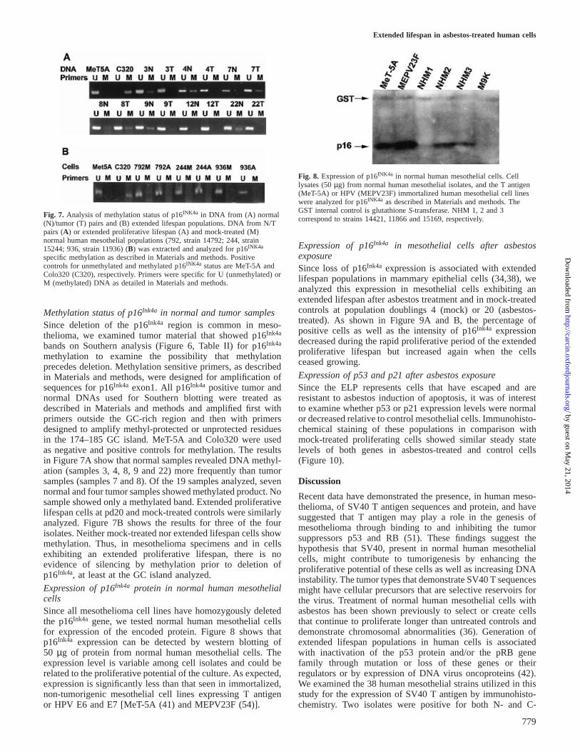
Fig. 7. Analysis of methylation status of p16INK4a in DNA from (A) normal(N)/tumor (T) pairs and (B) extended lifespan populations. DNA from N/Tpairs (A) or extended proliferative lifespan (A) and mock-treated (M)normal human mesothelial populations (792, strain 14792; 244, strain15244; 936, strain 11936) (B) was extracted and analyzed for p16INK4a
specific methylation as described in Materials and methods. Positivecontrols for unmethylated and methylated p16INK4a status are MeT-5A andColo320 (C320), respectively. Primers were specific for U (unmethylated) orM (methylated) DNA as detailed in Materials and methods.
Methylation status of p16Ink4a in normal and tumor samplesSince deletion of the p16Ink4a region is common in meso-thelioma, we examined tumor material that showed p16Ink4a
bands on Southern analysis (Figure 6, Table II) for p16Ink4a
methylation to examine the possibility that methylationprecedes deletion. Methylation sensitive primers, as describedin Materials and methods, were designed for amplification ofsequences for p16Ink4a exon1. All p16Ink4a positive tumor andnormal DNAs used for Southern blotting were treated asdescribed in Materials and methods and amplified first withprimers outside the GC-rich region and then with primersdesigned to amplify methyl-protected or unprotected residuesin the 174–185 GC island. MeT-5A and Colo320 were usedas negative and positive controls for methylation. The resultsin Figure 7A show that normal samples revealed DNA methyl-ation (samples 3, 4, 8, 9 and 22) more frequently than tumorsamples (samples 7 and 8). Of the 19 samples analyzed, sevennormal and four tumor samples showed methylated product. Nosample showed only a methylated band. Extended proliferativelifespan cells at pd20 and mock-treated controls were similarlyanalyzed. Figure 7B shows the results for three of the fourisolates. Neither mock-treated nor extended lifespan cells showmethylation. Thus, in mesothelioma specimens and in cellsexhibiting an extended proliferative lifespan, there is noevidence of silencing by methylation prior to deletion ofp16Ink4a, at least at the GC island analyzed.
Expression of p16Ink4a protein in normal human mesothelialcellsSince all mesothelioma cell lines have homozygously deletedthe p16Ink4a gene, we tested normal human mesothelial cellsfor expression of the encoded protein. Figure 8 shows thatp16Ink4a expression can be detected by western blotting of50 µg of protein from normal human mesothelial cells. Theexpression level is variable among cell isolates and could berelated to the proliferative potential of the culture. As expected,expression is significantly less than that seen in immortalized,non-tumorigenic mesothelial cell lines expressing T antigenor HPV E6 and E7 [MeT-5A (41) and MEPV23F (54)].
779
Fig. 8. Expression of p16INK4a in normal human mesothelial cells. Celllysates (50µg) from normal human mesothelial isolates, and the T antigen(MeT-5A) or HPV (MEPV23F) immortalized human mesothelial cell lineswere analyzed for p16INK4a as described in Materials and methods. TheGST internal control is glutathioneS-transferase. NHM 1, 2 and 3correspond to strains 14421, 11866 and 15169, respectively.
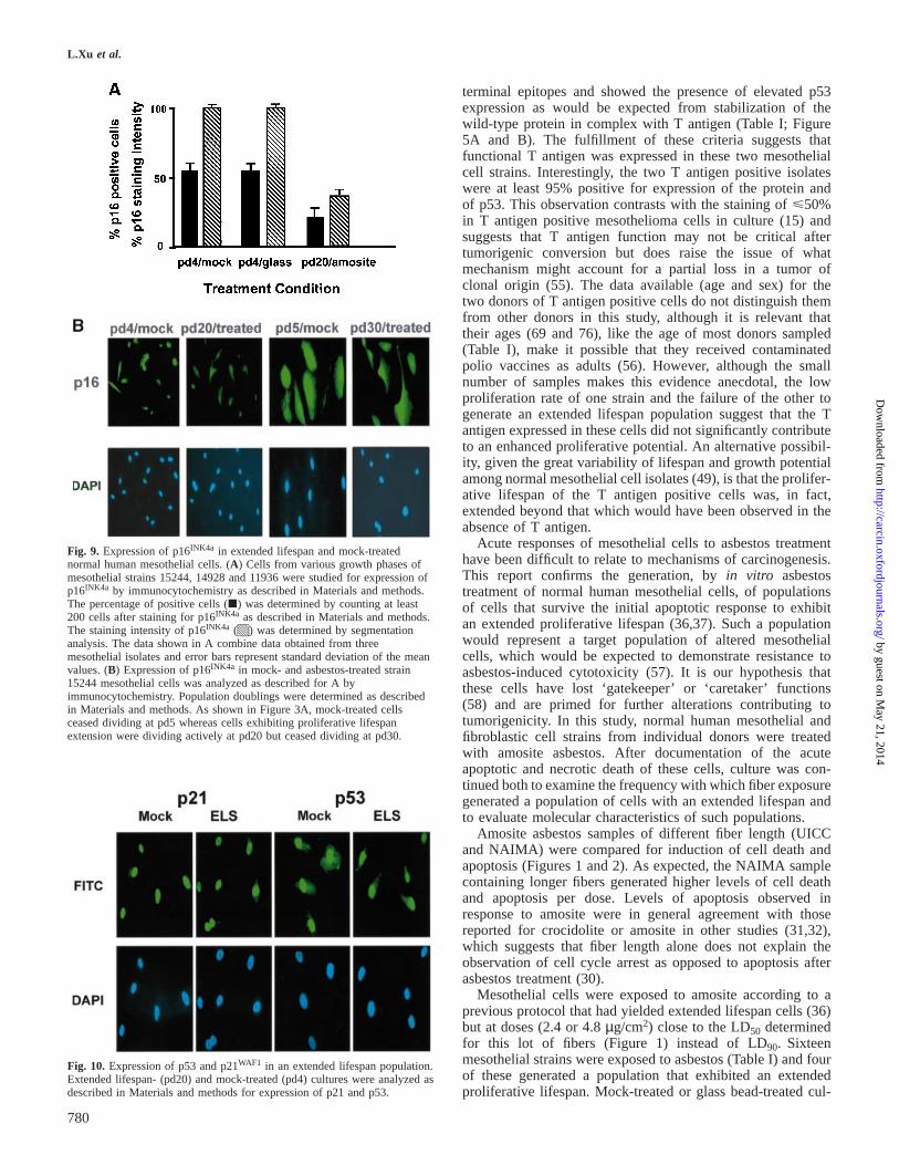
Expression of p16Ink4a in mesothelial cells after asbestosexposureSince loss of p16Ink4a expression is associated with extendedlifespan populations in mammary epithelial cells (34,38), weanalyzed this expression in mesothelial cells exhibiting anextended lifespan after asbestos treatment and in mock-treatedcontrols at population doublings 4 (mock) or 20 (asbestos-treated). As shown in Figure 9A and B, the percentage ofpositive cells as well as the intensity of p16Ink4a expressiondecreased during the rapid proliferative period of the extendedproliferative lifespan but increased again when the cellsceased growing.
Expression of p53 and p21 after asbestos exposureSince the ELP represents cells that have escaped and areresistant to asbestos induction of apoptosis, it was of interestto examine whether p53 or p21 expression levels were normalor decreased relative to control mesothelial cells. Immunohisto-chemical staining of these populations in comparison withmock-treated proliferating cells showed similar steady statelevels of both genes in asbestos-treated and control cells(Figure 10).
Discussion
Recent data have demonstrated the presence, in human meso-thelioma, of SV40 T antigen sequences and protein, and havesuggested that T antigen may play a role in the genesis ofmesothelioma through binding to and inhibiting the tumorsuppressors p53 and RB (51). These findings suggest thehypothesis that SV40, present in normal human mesothelialcells, might contribute to tumorigenesis by enhancing theproliferative potential of these cells as well as increasing DNAinstability. The tumor types that demonstrate SV40 T sequencesmight have cellular precursors that are selective reservoirs forthe virus. Treatment of normal human mesothelial cells withasbestos has been shown previously to select or create cellsthat continue to proliferate longer than untreated controls anddemonstrate chromosomal abnormalities (36). Generation ofextended lifespan populations in human cells is associatedwith inactivation of the p53 protein and/or the pRB genefamily through mutation or loss of these genes or theirregulators or by expression of DNA virus oncoproteins (42).We examined the 38 human mesothelial strains utilized in thisstudy for the expression of SV40 T antigen by immunohisto-chemistry. Two isolates were positive for both N- and C-
by guest on May 21, 2014
http://carcin.oxfordjournals.org/D
ownloaded from
L.Xu et al.
Fig. 9. Expression of p16INK4a in extended lifespan and mock-treatednormal human mesothelial cells. (A) Cells from various growth phases ofmesothelial strains 15244, 14928 and 11936 were studied for expression ofp16INK4a by immunocytochemistry as described in Materials and methods.The percentage of positive cells (j) was determined by counting at least200 cells after staining for p16INK4a as described in Materials and methods.The staining intensity of p16INK4a (q) was determined by segmentationanalysis. The data shown in A combine data obtained from threemesothelial isolates and error bars represent standard deviation of the meanvalues. (B) Expression of p16INK4a in mock- and asbestos-treated strain15244 mesothelial cells was analyzed as described for A byimmunocytochemistry. Population doublings were determined as describedin Materials and methods. As shown in Figure 3A, mock-treated cellsceased dividing at pd5 whereas cells exhibiting proliferative lifespanextension were dividing actively at pd20 but ceased dividing at pd30.
Fig. 10. Expression of p53 and p21WAF1 in an extended lifespan population.Extended lifespan- (pd20) and mock-treated (pd4) cultures were analyzed asdescribed in Materials and methods for expression of p21 and p53.
780
terminal epitopes and showed the presence of elevated p53expression as would be expected from stabilization of thewild-type protein in complex with T antigen (Table I; Figure5A and B). The fulfillment of these criteria suggests thatfunctional T antigen was expressed in these two mesothelialcell strains. Interestingly, the two T antigen positive isolateswere at least 95% positive for expression of the protein andof p53. This observation contrasts with the staining ofø50%in T antigen positive mesothelioma cells in culture (15) andsuggests that T antigen function may not be critical aftertumorigenic conversion but does raise the issue of whatmechanism might account for a partial loss in a tumor ofclonal origin (55). The data available (age and sex) for thetwo donors of T antigen positive cells do not distinguish themfrom other donors in this study, although it is relevant thattheir ages (69 and 76), like the age of most donors sampled(Table I), make it possible that they received contaminatedpolio vaccines as adults (56). However, although the smallnumber of samples makes this evidence anecdotal, the lowproliferation rate of one strain and the failure of the other togenerate an extended lifespan population suggest that the Tantigen expressed in these cells did not significantly contributeto an enhanced proliferative potential. An alternative possibil-ity, given the great variability of lifespan and growth potentialamong normal mesothelial cell isolates (49), is that the prolifer-ative lifespan of the T antigen positive cells was, in fact,extended beyond that which would have been observed in theabsence of T antigen.
Acute responses of mesothelial cells to asbestos treatmenthave been difficult to relate to mechanisms of carcinogenesis.This report confirms the generation, byin vitro asbestostreatment of normal human mesothelial cells, of populationsof cells that survive the initial apoptotic response to exhibitan extended proliferative lifespan (36,37). Such a populationwould represent a target population of altered mesothelialcells, which would be expected to demonstrate resistance toasbestos-induced cytotoxicity (57). It is our hypothesis thatthese cells have lost ‘gatekeeper’ or ‘caretaker’ functions(58) and are primed for further alterations contributing totumorigenicity. In this study, normal human mesothelial andfibroblastic cell strains from individual donors were treatedwith amosite asbestos. After documentation of the acuteapoptotic and necrotic death of these cells, culture was con-tinued both to examine the frequency with which fiber exposuregenerated a population of cells with an extended lifespan andto evaluate molecular characteristics of such populations.
Amosite asbestos samples of different fiber length (UICCand NAIMA) were compared for induction of cell death andapoptosis (Figures 1 and 2). As expected, the NAIMA samplecontaining longer fibers generated higher levels of cell deathand apoptosis per dose. Levels of apoptosis observed inresponse to amosite were in general agreement with thosereported for crocidolite or amosite in other studies (31,32),which suggests that fiber length alone does not explain theobservation of cell cycle arrest as opposed to apoptosis afterasbestos treatment (30).
Mesothelial cells were exposed to amosite according to aprevious protocol that had yielded extended lifespan cells (36)but at doses (2.4 or 4.8µg/cm2) close to the LD50 determinedfor this lot of fibers (Figure 1) instead of LD90. Sixteenmesothelial strains were exposed to asbestos (Table I) and fourof these generated a population that exhibited an extendedproliferative lifespan. Mock-treated or glass bead-treated cul-
by guest on May 21, 2014
http://carcin.oxfordjournals.org/D
ownloaded from
Extended lifespan in asbestos-treated human cells
tures of these strains exhibited lower proliferation rates andentered culture crisis within another five population doublings(Figure 3). These data indicate that asbestos treatment generatesor selects a subset of cells from a normal human mesothelialpopulation. The observation that only four of 16 isolatesgenerated such populations suggests the presence of inter-individual differences that predispose a cell strain to exhibitthis asbestos response. Differentiated characteristics may beimportant in this response since none of the five individualfibroblastic isolates treated developed an extended lifespan(Figure 3B). It is logical to predict that a population generatedor selected by asbestos exposure should be resistant to asbestos-generated cytotoxicity and apoptosis. This report demonstratesthat these cells, which are apoptosis competent when stimulatedby antibody to Fas (Figure 4C), are specifically resistant toinduction of apoptosis by asbestos exposure (Figure 4A). Thisresistant population expresses both p53 and p21 at the levelsseen in untreated cells (Figure 10). In addition, it has beenshown that many mesothelioma cell lines lacking p16INK4a
contain wild-type p53, which is elevated in response toγ-irradiation and can induce p21 transcription (59).
The data of Figure 4 show that resistance to death byapoptosis (Figure 4A) accounts for the resistance shown bythis population to asbestos cytotoxicity (Figure 4B). We haveshown earlier that human mesothelioma cell lines are resistantto apoptosis in response to asbestos and calcium ionophore.This resistance does not arise from high expression of Bcl-2,which was detected in only 3/14 mesothelioma lysates incontrast to the pro-apoptotic Bax which was present in allmesothelioma cell lines (50). In this study, we have shownthat the mesothelioma cell line, M9K, which expresses wild-type p53 (9), is resistant to both asbestos and Fas generatedapoptosis whereas the extended lifespan mesothelial cell popu-lation and the immortalized, non-tumorigenic MeT-5A cellsshow selective resistance to asbestos-generated apoptosis(Figure 4C), which suggests that additional changes accumu-lated during the generation of tumorigenicity in M9K arerequired for this broad resistance to apoptotic stimuli. Thus,additional changes that may alter the proteolytic cascadesrequired for apoptosis may contribute to the resistance totherapeutic regimens shown by mesothelioma (60,61).
Loss or inactivation of genes whose products contribute tothe normal functioning of p53 and pRB are expected tocontribute to tumorigenicity. Mutation and loss of p53 havebeen associated with immortalization of human fibroblasts(62,63). In the present study, normal and extended lifespancells were stained both for p53 and the downstream p21 whoseelevation is associated with senescence (64). Extended lifespancells did not show altered expression of these genes. Examina-tion of p16INK4a was suggested by the frequent loss of thechromosome 9p region encoding p16INK4a and p14ARF inprimary mesothelioma and in mesothelioma cell lines, as wellas the association of p16INK4a loss or methylation with extensionof lifespan in culture (34,38,65). The data presented here, inagreement with observations on mammary epithelial cells(38,39), show that p16INK4a protein expression is decreasedduring the rapid growth phase of lifespan extension. However,the gene is neither lost (Figure 9) nor detectably methylatedat the 174–185 GC island (Figure 7B). Furthermore, p16INK4a
protein is expressed at a high level when cells enter culturecrisis (Figure 9B), which indicates that epigenetic mechanismsor processes mediated at the level of transcription control thedown regulation.
781
The current data do not delineate a mechanism for generationof a population exhibiting an extended proliferative lifespan.However, it is clear that asbestos can induce growth factorexpression (7,66,67) and that human mesothelial cells areresponsive to such growth factors (68,69). Indeed, it has beenshown that there are great interindividual differences in growthfactor responsiveness of normal human mesothelial cells (49).Such differences could be critical in generation of populationsexhibiting extended proliferative lifespans and would beexpected to protect against apoptosis as well as stimulateproliferation.
In conclusion, we have demonstrated that a subset of normalhuman mesothelial cells from individual donors can developan extended proliferative lifespan in response to direct asbestostreatmentin vitro, although, for most cells, asbestos treatmentleads to cell death through apoptosis or cytotoxicity. Expressionof SV40 T antigen is not required for, nor was it shown to,facilitate development of an extended lifespan. The ELP isresistant to asbestos-induced but not Fas-induced apoptosis.This population exhibits decreased p16INK4a protein expressionduring its proliferative phase. Such a population may representcells that have already accumulated alterations of significanceon the pathway to mesothelioma. The increased breadth ofresistance to inducers of apoptosis shown by mesotheliomacell lines indicates that these alterations are specific to asbestos/mesothelial cell interactions. Future studies of such populationsmay provide valuable information concerning asbestos-inducedchanges that are significantly associated with carcinogenesis.
Acknowledgements
The encouragement and advice of Dr Curtis C.Harris and Dr John Lechnerhave been particularly helpful in the course of these experiments.
References
1.Barrett,J.C. (1994) Cellular and molecular mechanisms of asbestoscarcinogenicity: implications for biopersistence.Environ.Health Perspect.102 (suppl. 5), 19–23.
2.Mossman,B.T., Kamp,D.W. and Weitzman,S.A. (1996) Mechanisms ofcarcinogenesis and clinical features of asbestos-associated cancers.CancerInvest.,14, 466–480.
3.Wang,N.S., Jaurand,M.C., Magne,L., Kheuang,L., Pinchon,M.C. andBignon,J. (1987) The interactions between asbestos fibers and metaphasechromosomes of rat pleural mesothelial cells in culture. A scanning andtransmission electron microscopic study.Am.J. Pathol.,126, 343–349.
4.Jensen,C.G., Jensen,L.C., Rieder,C.L., Cole,R.W. and Ault,J.G. (1996)Long crocidolite asbestos fibers cause polyploidy by sterically blockingcytokinesis.Carcinogenesis,17, 2013–2021.
5.Yegles,M., Saint-Etienne,L., Renier,A., Janson,X. and Jaurand,M.C. (1993)Induction of metaphase and anaphase/telophase abnormalities by asbestosfibers in rat pleural mesothelial cellsin vitro. Am. J. Respir. Cell Mol.Biol., 9, 186–191.
6.Driscoll,K.E., Hassenbein,D.G., Carter,J.M., Kunkel,S.L., Quinlan,T.R. andMossman,B.T. (1995) TNF alpha and increased chemokine expression inrat lung after particle exposure.Toxicol.Lett., 82–83, 483–489.
7.Liu,J.Y., Morris,G.F., Lei,W.H., Hart,C.E., Lasky,J.A. and Brody,A.R.(1997) Rapid activation of PDGF-A and -B expression at sites of lunginjury in asbestos-exposed rats.Am.J.Respir. Cell Mol.Biol., 17, 129–140.
8.Dong,H., Buard,A., Renier,A., Levy,F., Saint-Etienne,L. and Jaurand,M.C.(1994) Role of oxygen derivatives in the cytotoxicity and DNA damageproduced by asbestos on rat pleural mesothelial cellsin vitro.Carcinogenesis,15, 1251–1255.
9.Metcalf,R.A., Welsh,J.A., Bennett,W.P.et al. (1992) p53 and Kirsten-rasmutations in human mesothelioma cell lines.Cancer Res., 52, 2610–2615.
10.Mor,O., Yaron,P., Huszar,M., Yellin,A., Jakobovitz,O., Brok-Simoni,F.,Rechavi,G. and Reichert,N. (1997) Absence of p53 mutations in malignantmesotheliomas.Am.J. Respir. Cell Mol. Biol., 16, 9–13.
by guest on May 21, 2014
http://carcin.oxfordjournals.org/D
ownloaded from
L.Xu et al.
11.Mayall,F.G., Goddard,H. and Gibbs,A.R. (1993) The frequency of p53immunostaining in asbestos-associated mesotheliomas and non-asbestos-associated mesotheliomas.Histopathology, 22, 383–386.
12.Otterson,G.A., Kratzke,R.A., Coxon,A., Kim,Y.W. and Kaye,F.J. (1994)Absence of p16INK4 protein is restricted to the subset of lung cancerlines that retains wild-type RB.Oncogene,9, 3375–3378.
13.Van der Meeren,A., Seddon,M.B., Kispert,J., Harris,C.C. and Gerwin,B.I.(1993) Lack of expression of the retinoblastoma gene is not frequentlyinvolved in the genesis of human mesothelioma.Eur. Respir. Rev., 3,177–179.
14.Ramael,M., Segers,K. and VanMarck,E. (1994) Differentialimmunohistochemical staining for retinoblastoma protein with theantibodies C15 and 1F8 in malignant mesothelioma.Pathol. Res.Pract.,190, 138–141.
15.Carbone,M., Rizzo,P., Grimley,P.M.et al. (1997) Simian virus-40 large-Tantigen binds p53 in human mesotheliomas [see comments].Nature Med.,3, 908–912.
16.De Luca,A., Baldi,A., Esposito,V.et al. (1997) The retinoblastoma genefamily pRb/p105, p107, pRb2/p130 and simian virus-40 large T-antigenin human mesotheliomas [see comments].Nature Med.,3, 913–916.
17.Cheng,J.Q., Jhanwar,S.C., Klein,W.M.et al. (1994) p16 alterations anddeletion mapping of 9p21–p22 in malignant mesothelioma.Cancer Res.,54, 5547–5551.
18.Xiao,S., Li,D., Vijg,J., Sugarbaker,D.J., Corson,J.M. and Fletcher,J.A.(1995) Codeletion of p15 and p16 in primary malignant mesothelioma.Oncogene,11, 511–515.
19.Prins,J.B., Williamson,K.A., Kamp,M.M., Van Hezik,E.J., van derKwast,T.H., Hagemeijer,A. and Versnel,M.A. (1998) The gene for thecyclin-dependent-kinase-4 inhibitor, CDKN2A, is preferentially deleted inmalignant mesothelioma.Int. J. Cancer, 75, 649–653.
20.Kratzke,R.A., Otterson,G.A., Lincoln,C.E., Ewing,S., Oie,H., Geradts,J.and Kaye,F.J. (1995) Immunohistochemical analysis of the p16INK4cyclin-dependent kinase inhibitor in malignant mesothelioma.J. NatlCancer Inst.,87, 1870–1875.
21.Aagaard,L., Lukas,J., Bartkova,J., Kjerulff,A.A., Strauss,M. and Bartek,J.(1995) Aberrations of p16Ink4 and retinoblastoma tumour-suppressorgenes occur in distinct sub-sets of human cancer cell lines.Int. J. Cancer,61, 115–120.
22.Okamoto,A., Demetrick,D.J., Spillare,E.A.et al. (1994) Mutations andaltered expression of genes regulating the cell cycle G1 checkpoint inhuman cancer.Proc. Natl Acad.Sci.USA,91, 11045–11049.
23.Zhang,Y., Xiong,Y. and Yarbrough,W.G. (1998) ARF promotes MDM2degradation and stabilizes p53: ARF-INK4a locus deletion impairs boththe Rb and p53 tumor suppression pathways.Cell, 92, 725–734.
24.Pomerantz,J., Schreiber-Agus,N., Liegeois,N.J.et al. (1998) The Ink4atumor suppressor gene product, p19Arf, interacts with MDM2 andneutralizes MDM29s inhibition of p53.Cell, 92, 713–723.
25.Bates,S., Philips,A.C., Clark,P.A., Stott,F., Peters,G., Ludwig,R.L. andVousden,K.H. (1998) p14ARF links the tumour suppressors RB and p53.Nature,395, 124–125.
26.Adamson,I.Y., Bakowska,J. and Bowden,D.H. (1994) Mesothelial cellproliferation: a nonspecific response to lung injury associated with fibrosis.Am.J. Respir. Cell Mol. Biol., 10, 253–258.
27.Moalli,P.A., MacDonald,J.L., Goodglick,L.A. and Kane,A.B. (1987) Acuteinjury and regeneration of the mesothelium in response to asbestos fibers.Am.J. Pathol.,128, 426–445.
28.BeruBe,K.A., Quinlan,T.R., Moulton,G., Hemenway,D., O’Shaughnessy,P.,Vacek,P. and Mossman,B.T. (1996) Comparative proliferative andhistopathologic changes in rat lungs after inhalation of chrysotile orcrocidolite asbestos.Toxicol.Appl. Pharmacol.,137, 67–74.
29.Goldberg,J.L., Zanella,C.L., Janssen,Y.M., Timblin,C.R., Jimenez,L.A.,Vacek,P., Taatjes,D.J. and Mossman,B.T. (1997) Novel cell imagingtechniques show induction of apoptosis and proliferation in mesothelialcells by asbestos.Am.J. Respir. Cell Mol. Biol., 17, 265–271.
30.Levresse,V., Renier,A., Fleury-Feith,J., Levy,F., Moritz,S., Vivo,C.,Pilatte,Y. and Jaurand,M.C. (1997) Analysis of cell cycle disruptions incultures of rat pleural mesothelial cells exposed to asbestos fibers [seecomments].Am.J. Respir. Cell Mol. Biol., 17, 660–671.
31.BeruBe,K.A., Quinlan,T.R., Fung,H., Magae,J., Vacek,P., Taatjes,D.J. andMossman,B.T. (1996) Apoptosis is observed in mesothelial cells afterexposure to crocidolite asbestos.Am. J. Respir. Cell Mol. Biol., 15,141–147.
32.Broaddus,V.C., Yang,L., Scavo,L.M., Ernst,J.D. and Boylan,A.M. (1996)Asbestos induces apoptosis of human and rabbit pleural mesothelial cellsvia reactive oxygen species.J. Clin. Invest.,98, 2050–2059.
782
33.Heintz,N.H., Janssen,Y.M. and Mossman,B.T. (1993) Persistent inductionof c-fosand c-junexpression by asbestos.Proc. Natl Acad.Sci.USA,90,3299–3303.
34.Zanella,C.L., Posada,J., Tritton,T.R. and Mossman,B.T. (1996) Asbestoscauses stimulation of the extracellular signal-regulated kinase 1 mitogen-activated protein kinase cascade after phosphorylation of the epidermalgrowth factor receptor.Cancer Res.,56, 5334–5338.
35.Pache,J.C., Janssen,Y.M., Walsh,E.S., Quinlan,T.R., Zanella,C.L.,Low,R.B., Taatjes,D.J. and Mossman,B.T. (1998) Increased epidermalgrowth factor-receptor protein in a human mesothelial cell line in responseto long asbestos fibers.Am.J. Pathol.,152, 333–340.
36.Lechner,J.F., Tokiwa,T., LaVeck,M.A., Benedict,W.F., Banks-Schlegel,S.P.,Yeager,H.Jr, Banerjee,A. and Harris,C.C. (1985) Asbestos-associatedchromosomal changes in human mesothelial cells.Proc. Natl Acad.Sci.USA,82, 3884–3888.
37.Linnainmaa,K., Gerwin,B.I., Pelin,K., Jantuene,K., LaVeck,M.A.,Lechner,J.F. and Harris,C.C. (1986) Asbestos-induced mesothelioma andchromosomal abnormalities in human mesothelial cellsin vitro. InAnonymous,The Changing Nature of Work and the Work Place. NIOSH,Cincinnati, OH, pp. 119–122.
38.Foster,S.A., Wong,D.J., Barrett,M.T. and Galloway,D.A. (1998)Inactivation of p16 in human mammary epithelial cells by CpG islandmethylation.Mol. Cell Biol., 18, 1793–1801.
39.Huschtscha,L.I., Noble,J.R., Neumann,A.A., Moy,E.L., Barry,P.,Melki,J.R., Clark,S.J. and Reddel,R.R. (1998) Loss of p16INK4 expressionby methylation is associated with lifespan extension of human mammaryepithelial cells.Cancer Res.,58, 3508–3512.
40.Yeager,T.R., DeVries,S., Jarrard,D.F.et al. (1998) Overcoming cellularsenescence in human cancer pathogenesis.Genes Dev., 12, 163–174.
41.Ke,Y., Reddel,R.R., Gerwin,B.I.et al. (1989) Establishment of a humanin vitro mesothelial cell model system for investigating mechanisms ofasbestos-induced mesothelioma.Am.J. Pathol.,134, 979–991.
42.Bryan,T.M. and Reddel,R.R. (1994) SV40-induced immortalization ofhuman cells.Crit. Rev. Oncog.,5, 331–357.
43.Lechner,J.F., Babcock,M.S., Marnell,M., Narayan,K.S. and Kaighn,M.E.(1980) Normal human prostate epithelial cell cultures.Methods Cell Biol.,21B, 195–225.
44.Yegles,M., Janson,X., Dong,H.Y., Renier,A. and Jaurand,M.C. (1995) Roleof fibre characteristics on cytotoxicity and induction of anaphase/telophaseaberrations in rat pleural mesothelial cellsin vitro: correlations within vivoanimal findings.Carcinogenesis,16, 2751–2758.
45. Itoh,N., Yonehara,S., Ishii,A., Yonehara,M., Mizushima,S., Sameshima,M.,Hase,A., Seto,Y. and Nagata,S. (1991) The polypeptide encoded by thecDNA for human cell surface antigen Fas can mediate apoptosis.Cell,66, 233–243.
46.Herman,J.G., Merlo,A., Mao,L., Lapidus,R.G., Issa,J.P., Davidson,N.E.,Sidransky,D. and Baylin,S.B. (1995) Inactivation of the CDKN2/p16/MTS1 gene is frequently associated with aberrant DNA methylation in allcommon human cancers.Cancer Res.,55, 4525–4530.
47.Maniatis,T., Fritsch,E.F. and Sambrook,J. (1982)Molecular Cloning: aLaboratory Manual. Cold Spring Harbor Press, Cold Spring Harbor, NY.
48.Broaddus,V.C. (1997) Asbestos, the mesothelial cell and malignancy: amatter of life or death [editorial; comment].Am.J. Respir. Cell Mol. Biol.,17, 657–659.
49.Lechner,J.F., LaVeck,M.A., Gerwin,B.I. and Matis,E.A. (1989) Differentialresponses to growth factors by normal human mesothelial cultures fromindividual donors.J. Cell. Physiol.,139, 295–300.
50.Narasimhan,S.R., Yang,L., Gerwin,B.I. and Broaddus,V.C. (1998)Resistance of pleural mesothelioma cell lines to apoptosis: relation toexpression of Bcl-2 and bax.Am.J. Physiol.,275, L165–L171.
51.Carbone,M., Rizzo,P. and Pass,H.I. (1997) Simian virus 40, poliovaccinesand human tumors: a review of recent developments.Oncogene,15,1877–1888.
52.Spillare,E.A., Okamoto,A., Hagiwara,K., Demetrick,D.J., Serrano,M.,Beach,D. and Harris,C.C. (1996) Suppression of growthin vitro andtumorigenicity in vivo of human carcinoma cell lines by transfectedp16INK4. Mol. Carcinogen.,16, 53–60.
53.Frizelle,S.P., Grim,J., Zhou,J., Gupta,P., Curiel,D.T., Geradts,J. andKratzke,R.A. (1998) Re-expression of p16INK4a in mesothelioma cellsresults in cell cycle arrest, cell death, tumor suppression and tumorregression.Oncogene,16, 3087–3095.
54.De Silva,R., Whitaker,N.J., Rogan,E.M. and Reddel,R.R. (1994) HPV-16E6 and E7 genes, like SV40 early region genes, are insufficient forimmortalization of human mesothelial and bronchial epithelial cells.Exp.Cell Res.,213, 418–427.
55.Pelin-Enlund,K., Husgafvel-Pursiainen,K., Tammilehto,L.et al. (1990)Asbestos-related malignant mesothelioma: growth, cytology,
by guest on May 21, 2014
http://carcin.oxfordjournals.org/D
ownloaded from

Extended lifespan in asbestos-treated human cells
tumourigenicity and consistent chromosome findings in cell lines fromfive patients.Carcinogenesis,11, 673–681.
56.Strickler,H.D., Rosenberg,P.S., Devesa,S.S., Hertel,J., Fraumeni,J.F.Jr andGoedert,J.J. (1998) Contamination of poliovirus vaccines with simian virus40 (1955–1963) and subsequent cancer rates.J. Am. Med. Assoc.,279,292–295.
57.Gabrielson,E.W., Van der Meeren,A., Reddel,R.R., Reddel,H., Gerwin,B.I.and Harris,C.C. (1992) Human mesothelioma cells and asbestos-exposedmesothelial cells are selectively resistant to amosite toxicity: a possiblemechanism for tumor promotion by asbestos.Carcinogenesis,13, 1359–1363.
58.Kinzler,K.W. and Vogelstein,B. (1998) Landscaping the cancer terrain[comment].Science,280, 1036–1037.
59.Ungar,S.L., Van der Meeren,A., Tammilehto,L., Linnainmaa,K., Mattson,K.and Gerwin,B.I. (1996) High levels of MDM2 are not correlated with thepresence of wild-type p53 in human malignant mesothelioma cell lines.Br. J. Cancer, 74, 1534–1540.
60.Bowman,R.V., Manning,L.S., Davis,M.R. and Robinson,B.W. (1991)Chemosensitivity and cytokine sensitivity of malignant mesothelioma.Cancer Chemother. Pharmacol.,28, 420–426.
61.Ong,S.T. and Vogelzang,N.J. (1996) Chemotherapy in malignant pleuralmesothelioma. A review.J. Clin. Oncol.,14, 1007–1017.
62.Bischoff,F.Z., Yim,S.O., Pathak,S., Grant,G., Siciliano,M.J.,Giovanella,B.C., Strong,L.C. and Tainsky,M.A. (1990) Spontaneousabnormalities in normal fibroblasts from patients with Li–Fraumeni cancersyndrome: aneuploidy and immortalization.Cancer Res., 50, 7979–7984.
783
63.Rogan,E.M., Bryan,T.M., Hukku,B.et al. (1995) Alterations in p53 andp16INK4 expression and telomere length during spontaneousimmortalization of Li–Fraumeni syndrome fibroblasts.Mol. Cell Biol., 15,4745–4753.
64.Noda,A., Ning,Y., Venable,S.F., Pereira-Smith,O.M. and Smith,J.R. (1994)Cloning of senescent cell-derived inhibitors of DNA synthesis using anexpression screen.Exp.Cell, Res.,211, 90–98.
65.Noble,J.R., Rogan,E.M., Neumann,A.A., Maclean,K., Bryan,T.M. andReddel,R.R. (1996) Association of extendedin vitro proliferative potentialwith loss of p16INK4 expression.Oncogene,13, 1259–1268.
66.Antony,V.B., Godbey,S.W., Sparks,J.A. and Hott,J.W. (1993) Pleuralmesothelial cells release a growth factor for fibroblasts.Eur. Resp.Rev.,3, 156–158.
67.Antony,V.B., Owen,C.L. and Hadley,K.J. (1989) Pleural mesothelial cellsstimulated by asbestos release chemotactic activity for neutrophilsin vitro.Am.Rev. Respir. Dis., 139, 199–206.
68.LaVeck,M.A., Somers,A.N.A., Moore,L.L., Gerwin,B.I. and Lechner,J.F.(1988) Dissimilar peptide growth factors can induce normal humanmesothelial cell multiplication.In Vitro, 24, 1077–1084.
69.Gabrielson,E.W., Gerwin,B.I., Harris,C.C., Roberts,A.B., Sporn,M.B. andLechner,J.F. (1988) Stimulation of DNA synthesis in cultured primaryhuman mesothelial cells by specific growth factors.FASEB J., 2, 2717–2722.
Received September 18, 1998; revised January 5, 1999;accepted January 25, 1999
by guest on May 21, 2014
http://carcin.oxfordjournals.org/D
ownloaded from


