Embed Size (px)
Citation preview
ISPH 2017 – Trenton, New Jersey 1
FOURTH INTERNATIONAL
SYMPOSIUM ON PALEOHISTOLOGY
ISPH 2017
Volume 4
EDITORS
Rodrigo A. Pellegrini
David C. Parris
ISPH 2017 – Trenton, New Jersey 1
FOURTH INTERNATIONAL SYMPOSIUM ON
PALEOHISTOLOGY – ISPH 2017 – Volume 4
©2017 New Jersey State Museum & ISPH 2017 Contributors
ISBN-10: 0-938766-10-4
ISBN-13: 978-0-938766-10-0
GRAPHIC DESIGN & LAYOUT:
Rodrigo Pellegrini (after Ethan Barshay, Christian Heck, Ellen Thérèse Lamm, Aurore
Canoville and Jessica Mitchell)
PHOTO CREDITS:
Cover/Back Graphic Design: R. Pellegrini
Front Cover Background Image:
Haversian canals in Hipparion,
© S. Gomez (used with permission)
Back Cover Images (clockwise from top right):
Mosasaurus maximus right pterygoid tooth dentine-osteocementum interface –
R. Pellegrini/New Jersey State Museum
Mosasaurus tooth Enamel-Dentine Junction © S. Gomez (used with permission)
Dentine and enamel in Globidens tooth R. Pellegrini/New Jersey State Museum
Transverse section, mid-shaft of a Tylosaurus tibia © R. Pellegrini
www.nj.gov/state/museum/index.html
www.nj.gov/state/museum/dos_museum_foundation.html
www.nj.gov/state/museum/dos_museum_isph.html
2 ISPH 2017 – Trenton, New Jersey
FOURTH INTERNATIONAL SYMPOSIUM ON
PALEOHISTOLOGY
July 10 - 12, 2017
NEW JERSEY STATE MUSEUM
Trenton, New Jersey USA
Symposium Convenors
David C. Parris
Rodrigo A. Pellegrini
Scientific Committee
Timothy G. Bromage
Barbara S. Grandstaff
Alexandra Houssaye
David C. Parris
Rodrigo A. Pellegrini
Allison Tumarkin-Deratzian
Organizing Committee
Sheila S. Blackwell
Wayne Callahan
Larry Conti
Robert Denton
Nicole Jannotte
Pamela Machold
Roland Machold
Margaret M. O'Reilly
Henry Sadowski
Lance Schnatterly
William J. Shankle
The 4th International Symposium on Paleohistology is organized by the
New Jersey State Museum, and supported in part by the New Jersey
State Museum Foundation.
ISPH 2017 – Trenton, New Jersey 3
Dedicated to Robert C. Ramsdell (1920-2014),
professor of Geology and Paleontology at Williams College,
Rutgers University and Montclair State University,
and long-time New Jersey State Museum volunteer.
The Professor was a devoted teacher, researcher, author, mentor and
counselor. But most of all, to so many of us who study the Earth,
he was a friend.
- Wayne Callahan
4 ISPH 2017 – Trenton, New Jersey
NEW JERSEY, WHERE NATURAL HISTORY INSPIRED
AMERICAN SCIENCES!
Welcome to the New Jersey State Museum, the proud host of the Fourth
International Symposium on Paleohistology! During this week, you will have ample
opportunities to learn about the Museum collections and research, and to experience
the traditions of paleontology in North America generally, as much of our science
began in this region. The host hotel is located in Princeton, where University
students Henry Fairfield Osborn and William Berryman Scott envisioned (and
fulfilled) a full academic program in paleontology. Guyot Hall, location of the
opening gathering, was one of the first university buildings in history to be designed
with a large museum hall as its centerpiece, and the stone carvings on the molding
around the building represent living animals at the Biology side of the building and
extinct animals on the Geology side. In addition to the elegance of the campus, the
town of Princeton has many tourist amenities, historic sites, shops, restaurants,
recreational facilities, and other points of interest.
Symposium sessions will be held at the New Jersey State Museum at the Cultural
Center in Trenton, practically within view of the classic gold-domed New Jersey
State Capitol. Trenton, the site of two of the most critical battles of the American
Revolution, once served as the capital of the United States. The State Museum,
chartered in 1895, grew from extensive natural history collections amassed prior to
that date by the antebellum field work of the New Jersey Geological Survey, which
remains one of the Museum’s most important cooperating institutions. The current
Museum campus, including the main building with its large Natural History Hall
and Planetarium, was erected in 1964.
It was no accident that so much of the work of early paleontologists took place in
this region. New Jersey is uncommonly rich in natural resources, having
physiographic diversity and geologic resources far exceeding what one might expect
for the size of the state. Although it is the most densely-populated of all the states in
the Union, New Jersey’s substantial wild and preserved tracts still provide
opportunities for field studies and collecting. The many opportunities for amateur
and avocational paleontology have provided the Museum with many volunteer
associates who are assisting with, and participating in, the Symposium. Among the
professional paleontologists who encouraged amateur scientists was Professor
Robert Ramsdell, a Trenton native, whom we honor at this Symposium.
Paleohistology has a long history at both Princeton University and the New Jersey
State Museum. Microscopy was applied in studies of the earliest collections,
although not formally reported in many cases. At Princeton, Professors Scott,
Sinclair, Jepsen, Dorf, Baird, and others applied new technologies to the study of
ISPH 2017 – Trenton, New Jersey 5
fossils, including X-ray views of specimens in matrix, thin-section studies, and
Scanning Electron Microscopy. The New Jersey State Museum continues these
traditions, as is evident in staff and associate presentations. The Museum welcomes
all participants to the state’s capital city, and looks forward to dynamic dialogue as
research is shared.
David C. Parris, Curator Rodrigo A. Pellegrini, Registrar
Bureau of Natural History, New Jersey State Museum
SYMPOSIUM CONVENORS
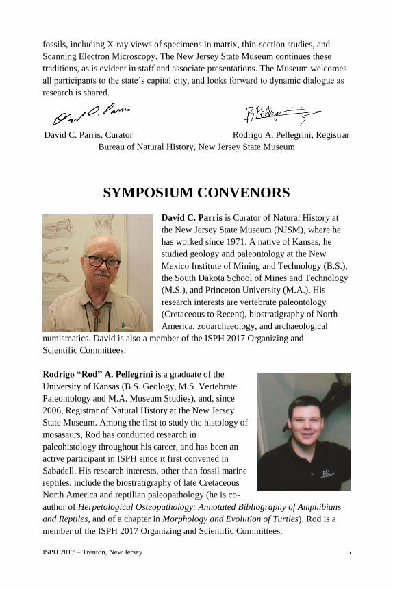
David C. Parris is Curator of Natural History at
the New Jersey State Museum (NJSM), where he
has worked since 1971. A native of Kansas, he
studied geology and paleontology at the New
Mexico Institute of Mining and Technology (B.S.),
the South Dakota School of Mines and Technology
(M.S.), and Princeton University (M.A.). His
research interests are vertebrate paleontology
(Cretaceous to Recent), biostratigraphy of North
America, zooarchaeology, and archaeological
numismatics. David is also a member of the ISPH 2017 Organizing and
Scientific Committees.
Rodrigo “Rod” A. Pellegrini is a graduate of the
University of Kansas (B.S. Geology, M.S. Vertebrate
Paleontology and M.A. Museum Studies), and, since
2006, Registrar of Natural History at the New Jersey
State Museum. Among the first to study the histology of
mosasaurs, Rod has conducted research in
paleohistology throughout his career, and has been an
active participant in ISPH since it first convened in
Sabadell. His research interests, other than fossil marine
reptiles, include the biostratigraphy of late Cretaceous
North America and reptilian paleopathology (he is co-
author of Herpetological Osteopathology: Annotated Bibliography of Amphibians
and Reptiles, and of a chapter in Morphology and Evolution of Turtles). Rod is a
member of the ISPH 2017 Organizing and Scientific Committees.
6 ISPH 2017 – Trenton, New Jersey
KEYNOTE SPEAKERS
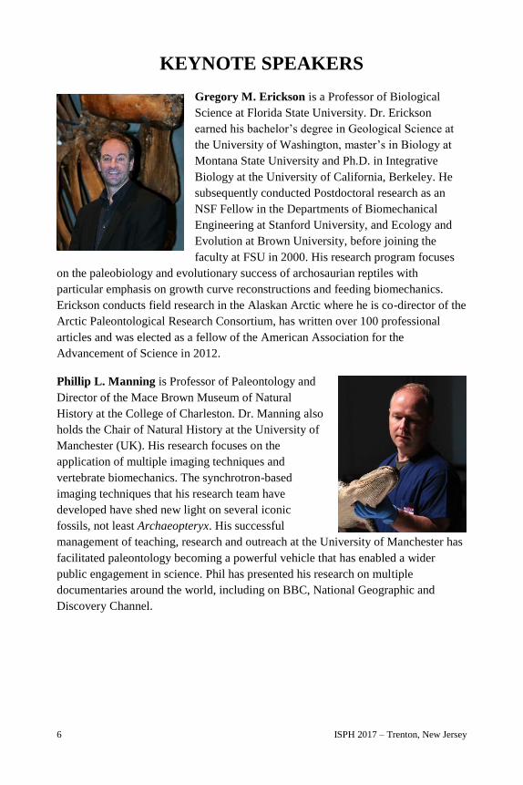
Gregory M. Erickson is a Professor of Biological
Science at Florida State University. Dr. Erickson
earned his bachelor’s degree in Geological Science at
the University of Washington, master’s in Biology at
Montana State University and Ph.D. in Integrative
Biology at the University of California, Berkeley. He
subsequently conducted Postdoctoral research as an
NSF Fellow in the Departments of Biomechanical
Engineering at Stanford University, and Ecology and
Evolution at Brown University, before joining the
faculty at FSU in 2000. His research program focuses
on the paleobiology and evolutionary success of archosaurian reptiles with
particular emphasis on growth curve reconstructions and feeding biomechanics.
Erickson conducts field research in the Alaskan Arctic where he is co-director of the
Arctic Paleontological Research Consortium, has written over 100 professional
articles and was elected as a fellow of the American Association for the
Advancement of Science in 2012.
Phillip L. Manning is Professor of Paleontology and
Director of the Mace Brown Museum of Natural
History at the College of Charleston. Dr. Manning also
holds the Chair of Natural History at the University of
Manchester (UK). His research focuses on the
application of multiple imaging techniques and
vertebrate biomechanics. The synchrotron-based
imaging techniques that his research team have
developed have shed new light on several iconic
fossils, not least Archaeopteryx. His successful
management of teaching, research and outreach at the University of Manchester has
facilitated paleontology becoming a powerful vehicle that has enabled a wider
public engagement in science. Phil has presented his research on multiple
documentaries around the world, including on BBC, National Geographic and
Discovery Channel.
ISPH 2017 – Trenton, New Jersey 7
Peter Dodson holds two degrees in geology (University
of Ottawa, 1968; University of Alberta, 1970). He
received his Ph.D. in paleontology from Yale University
in 1974. He is a professor of veterinary gross anatomy
and of dinosaur paleontology at the University of
Pennsylvania, where he has taught since 1974. His work
has taken him to India, Madagascar, Egypt, Argentina,
Mexico and China. He has supervised more than 20
Ph.Ds. and his graduates teach in China, Japan, and
Canada, as well as in the United States. With his students, he has described and
named six species of dinosaurs. He is the author of more than 100 scientific papers,
co-editor of The Dinosauria, (1990, 2004); author of The Horned Dinosaurs (1996),
and of several children’s books.
Allison R. Tumarkin-Deratzian received her B.S. in Geology and Environmental
Geosciences from Lafayette College (1997), and her Ph.D.
in Earth and Environmental Science from the University
of Pennsylvania (2003), where she also served as a
graduate instructor of Gross Anatomy in the School of
Veterinary Medicine. She was a Visiting Assistant
Professor in the Department of Earth Science and
Geography at Vassar College, from 2003-2006. Since
2006, she has been with the Department of Earth and
Environmental Science at Temple University, and
currently serves as Associate Professor of Instruction and
Undergraduate Chair for Geology and Environmental
Science. Although her formal degrees are in geology, her educational training and
research interests have always strayed to the biological side of the paleontology
spectrum. Her research primarily involves study of bone microstructure and growth
patterns of modern and fossil archosaurs, ontogeny and evolution of ornithischian
dinosaurs, and vertebrate taphonomy. Dr. Tumarkin-Deratzian is also a member of
the ISPH 2017 Scientific Committee.
8 ISPH 2017 – Trenton, New Jersey
WORKSHOP INSTRUCTORS
Timothy G. Bromage directs the Hard Tissue Research
Unit (HTRU), a mineralized tissue preparation and
imaging technology development laboratory of the
Department of Biomaterials and Biomimetics, New
York University College of Dentistry. The HTRU
emphasizes evolutionary research in environmental
context, employing a variety of 2D and 3D light and
electron microscopy techniques, in addition to mass
spectrometry-based approaches to the complete relevant
inorganic spectrum detectable in hard tissues. Professor
Bromage is recipient of the 2010 Max Planck Prize in
the Life Sciences (paleobiomics; emphasis in Human Evolution), is Honorary
Professor of La Salle University, Madrid, Spain, and an Honorary Research Fellow
of the Department of Paleoanthropology, Senckenberg Research Institute, Frankfurt,
Germany. He is also a member of the ISPH 2017 Scientific Committee.
Santiago Gomez is a professor of Pathology in the
Faculty of Medicine, University of Cadiz, Spain. He
has been a visiting scientist at University College
London, Università La Sapienza Roma, Cornell
University, and New York University. With an initial
interest in pathological calcification, in the last 30
years his research has focused on hard tissues with
special emphasis on their preparation, imaging, and
analysis. His excellence in technical work and
photomicrography has been recognized in winning
several Buehler and Nikon Small World awards.
Ellen-Thérèse Lamm is the Histology Lab Manager
at the Museum of the Rockies in Bozeman, Montana,
where she has worked with Jack Horner and
Montana State University students for over 26 years
producing a vast collection of thin-sections of over
2,500 specimens. She is an Adjunct Instructor at
MSU, teaching Paleohistology Techniques, and is the
co-editor of Bone Histology of Fossil Tetrapods.
Ellen-T. has worked with over 100 institutions
around the world thin-sectioning fossil material,
training visiting researchers, and presenting workshops at ISPH, SVP, and the
National Society of Histotechnology.
ISPH 2017 – Trenton, New Jersey 9
SCIENTIFIC COMMITTEE
Barbara Smith Grandstaff attended Millersville
State College (B.A.), Princeton University (M.A.)
and the University of Pennsylvania (Ph.D.). She
teaches gross anatomy, developmental biology, and
neuroscience at the School of Veterinary Medicine
of the University of Pennsylvania, and has also
taught geology and paleontology courses at Temple
University. Barbara’s research focuses on vertebrate
functional morphology, paleopathology, and late
Cretaceous communities.
Alexandra Houssaye received a Ph.D. from and works
at the Muséum National d’Histoire Naturelle of Paris,
France. She is a CNRS paleobiologist specializing in
functional morphology. Dr. Houssaye focuses her work
on the analysis of bone microanatomical adaptations
and their link with anatomical changes and
biomechanical constraints.
Other members of the Scientific Committee include Timothy G. Bromage, David C.
Parris, Rodrigo A. Pellegrini, and Allison Tumarkin-Deratzian, whose biographical
information is available earlier in this publication.
10 ISPH 2017 – Trenton, New Jersey
LOCAL ORGANIZING COMMITTEE
Sheila Scott Blackwell is a volunteer in the State Museum’s
Bureau of Natural History. She is a graduate of Trenton State
College, with a Bachelor of Science in Biology, and works for the
New Jersey State Department of Agriculture, Division of Plant
Industry, as a horticulturist.
Wayne Callahan is a volunteer in the Division of
Paleontology of the American Museum of Natural History, in
addition to volunteering with the Natural History Bureau of the
NJSM. Although his professional career took him into
manufacturing engineering and production management,
Wayne pursued his life-long interest in paleontology in the
graduate program in geoscience at Montclair State College. He has collected and
studied fossils, collaborated on fieldtrip guides, scientific papers and abstracts for
over 45 years. Wayne became a Research Associate in paleontology at the New
Jersey State Museum over 25 years ago, and is a member of the Society of
Vertebrate Paleontology.
Lawrence “Larry” G. Conti is a native Trentonian and a Board
Member of the New Jersey State Museum Foundation. At the
request of Uncle Sam, he spent a year and a half blowing up
balloons in the desert of New Mexico, and the next thirty-five years
as a bureaucrat for the State of New Jersey in Vocational
Rehabilitation. Larry volunteers at the NJSM, and has done some
paleontology work with the institution. He enjoys visiting his relatives in Rome and
Umbria, and has visited the K-Pg boundary in Gubbio, Italy.
Robert “Bob” Denton Jr. attended Richard Stockton State
University and graduated from Thomas Edison State University
with a B.A. in Natural Science. Bob has been a research
associate of the NJSM for nearly 40 years, and is the co-
discoverer of both the Ellisdale Fossil Site in New Jersey and
the Zuni Basin Dinosaur Site in New Mexico. He described two
new taxa of animals from Ellisdale, and is currently working on the description of a
new taxon from the Zuni Site. Bob is a Certified Professional Geologist and a
Licensed Professional Soil Scientist, and is the senior geologist at a geotechnical
consulting firm in Ashburn, VA.
ISPH 2017 – Trenton, New Jersey 11
Pamela Machold is a graduate of Sarah Lawrence College and has
a master’s degree in physical therapy from Acadia University. She
has served on Princeton’s Environmental and Shade Tree
Commissions, and as the President of the Marquand Park
Foundation. She and her mother founded the Princeton Child
Development Institute, which was the first school for autistic
children in New Jersey.
Roland Machold is a graduate of Yale and the Harvard Business
School. He worked in investment banking for a dozen years, and then
for twenty three years he served as the Director of the Division of
Investment for the State of New Jersey, with over $80 billion of
assets under his management when he retired in 1998. He came out
of retirement in 1999 and served as the State Treasurer until 2001.
He currently is on the Board of the Child Health Institute of New Jersey and is an
emeritus Trustee of both Bryn Mawr College and Columbia Teachers College. In
1989, he helped found “Dobro,” a Russian charity to help autistic children in
that country.
Margaret M. O’Reilly was named Executive Director of the
New Jersey State Museum in 2016. She began her career at the
State Museum in 1988 as the Publications/Graphics Art
Director. In 1997, she was named Assistant Curator, Fine Art
Collections and Exhibitions, and in 2008 became the Curator of
Fine Art. Ms. O’Reilly has been a panelist or lecturer at
museums, colleges, and professional conferences on topics including 20th century
American art, collection stewardship, museums and copyright, and careers in
museums, among others. Ms. O’Reilly currently serves on the Board of the Mid-
Atlantic Association of Museums and on the Visual Arts Advisory Committee of
Mercer County Community College.
Henry Sadowski is a retired attorney who received his law degree
at Rutgers Law School and served 35 years in the U.S. Department
of Justice. He has been a volunteer with the New Jersey State
Museum for four years, specializing in fossil preparation. He also
leads tours as a docent for the Museum.
12 ISPH 2017 – Trenton, New Jersey
Lance Schnatterly received his degree in Astronomy/Computer
Science from the Pennsylvania State University and is currently a
senior staff engineer at Lockheed Martin, where he has worked since
1981. Lance has been focusing his spare time on dinosaur
excavations in the Hell Creek and Lance Formations of Montana,
South Dakota and Wyoming. He is a member of the Society of
Vertebrate Paleontology and the Paleontological Society, and serves on the board of
the Delaware Valley Paleontological Society (DVPS).
The ISPH 2017 logo shows the silhouette of NJSM 11053, a nearly complete skull
of Mosasaurus maximus that has become emblematic of the natural history program
at New Jersey State Museum. The specimen has been used repeatedly for dental
histological studies by NJSM scientists for previous ISPH symposia, and is on
display in the Museum galleries. The silhouette is over a micrograph of Haversian
canals in Hipparion by Dr. Santiago Gomez, which serves not only as a
background, but as a colorful link to paleohistology.
ISPH 2017 – Trenton, New Jersey 13
MISSION
As a center of cultural, educational, and scientific engagement, the New Jersey State
Museum inspires innovation and lifelong learning through collections, research,
exhibitions and programs in science, history and art. The New Jersey State Museum
fosters state pride, serves as a cultivator of tomorrow's leaders and engages visitors
of all ages and diverse backgrounds in an exploration of New Jersey cultural and
natural history presented within a global context.
HISTORY
In 1895, the New Jersey State Legislature formally established the New Jersey State
Museum in the capital city of Trenton with a mission to collect and exhibit
specimens in natural history, archaeology, and industrial history. Original
collections included natural history specimens amassed in the 19th century by the
New Jersey Geological Survey. Between 1900 and 1912, the State Museum
strengthened its archaeology focus by initiating a field research program.
It adopted a statewide Education Extension Service in 1914 that lent models, lantern
slides, and specimens to schools throughout the state. In 1929, expanded space in
the newly-constructed State House Annex permitted extensive exhibitions of the
natural history and New Jersey prehistory collections. Decorative arts became a
third collection field at this time when the State Museum expanded its focus beyond
industrial history to include the manufacture of ceramics. The ethnographic
collection was initiated in 1932 when the State Museum purchased part of an
exhibition of North American Indian art. The collection was strengthened in the
1940s when anthropologist Frank Speck made field collections for the State
Museum among the Delaware Indians of Oklahoma and Canada. Exhibitions of
these artifacts attracted the interest of New Jersey residents whose donations from
private collections built the collection in the ensuing years. Growing interest in
education and culture in the late 1950s resulted in the construction of a modern
cultural complex near the State House that opened in 1965 and included a four-level
Museum building, an adjoining planetarium, and an adjacent auditorium. At that
time, fine art became the Museum's fourth collection area.
The State Museum was initially accredited by the American Alliance of Museums
in 1974. Since 1983 it has been a division of the New Jersey Department of State.
14 ISPH 2017 – Trenton, New Jersey
The New Jersey State Museum Foundation was founded in 1968 as a non-profit
501(c)(3) to support the Museum collections, exhibitions and programs through
fundraising, volunteerism, and advocacy. In recent years, the Foundation has
received generous support from the Hyde and Watson Foundation; the National
Endowment for the Arts; New Jersey Historical Commission; New Jersey State
Council on the Arts; NJM Insurance Group; and the PNC Foundation. In addition to
a robust membership program and special events, the Foundation operates a retail
shop featuring merchandise related to the Museum exhibitions and New Jersey
history and culture. Proceeds from the Foundation’s activities support the Museum
collections, exhibitions and programs.
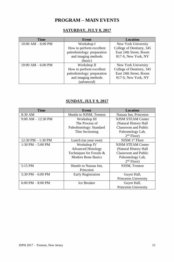
ISPH 2017 – Trenton, New Jersey 15
PROGRAM – MAIN EVENTS
SATURDAY, JULY 8, 2017
Time Event Location
10:00 AM – 6:00 PM Workshop I
How to perform excellent
paleohistology: preparation
and imaging methods
(basic)
New York University
College of Dentistry, 345
East 24th Street, Room
817-S, New York, NY
10:00 AM – 6:00 PM Workshop II
How to perform excellent
paleohistology: preparation
and imaging methods
(advanced)
New York University
College of Dentistry, 345
East 24th Street, Room
817-S, New York, NY
SUNDAY, JULY 9, 2017
Time Event Location
8:30 AM Shuttle to NJSM, Trenton Nassau Inn, Princeton
9:00 AM – 12:30 PM Workshop III
The Process of
Paleohistology: Standard
Thin Sectioning
NJSM STEAM Center
(Natural History Hall
Classroom and Public
Paleontology Lab,
2nd Floor)
12:30 PM – 1:30 PM Lunch (on your own) NJSM 1st Floor
1:30 PM – 5:00 PM Workshop IV
Advanced Histology
Techniques for Fossils &
Modern Bone Basics
NJSM STEAM Center
(Natural History Hall
Classroom and Public
Paleontology Lab,
2nd Floor)
5:15 PM Shuttle to Nassau Inn,
Princeton
NJSM, Trenton
5:30 PM – 6:00 PM Early Registration Guyot Hall,
Princeton University
6:00 PM – 8:00 PM Ice Breaker Guyot Hall,
Princeton University
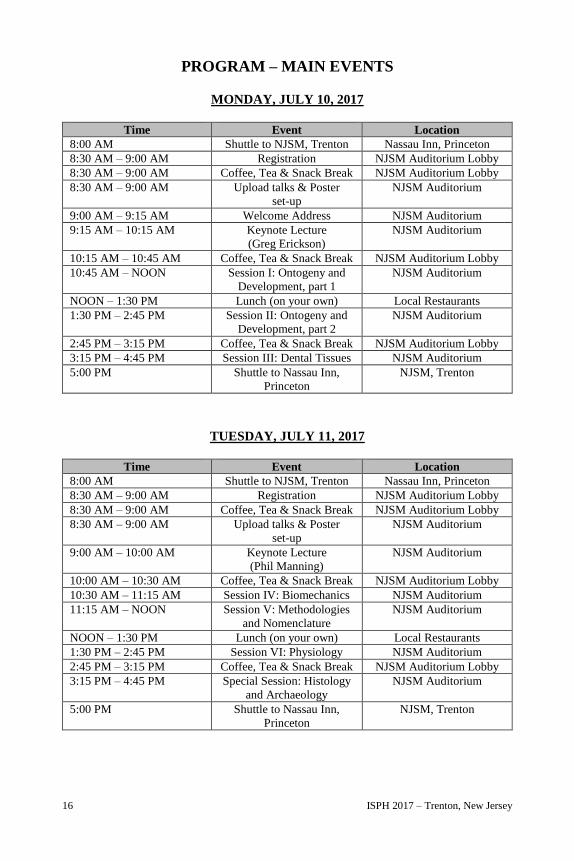
16 ISPH 2017 – Trenton, New Jersey
PROGRAM – MAIN EVENTS
MONDAY, JULY 10, 2017
Time Event Location
8:00 AM Shuttle to NJSM, Trenton Nassau Inn, Princeton
8:30 AM – 9:00 AM Registration NJSM Auditorium Lobby
8:30 AM – 9:00 AM Coffee, Tea & Snack Break NJSM Auditorium Lobby
8:30 AM – 9:00 AM Upload talks & Poster
set-up
NJSM Auditorium
9:00 AM – 9:15 AM Welcome Address NJSM Auditorium
9:15 AM – 10:15 AM Keynote Lecture
(Greg Erickson)
NJSM Auditorium
10:15 AM – 10:45 AM Coffee, Tea & Snack Break NJSM Auditorium Lobby
10:45 AM – NOON Session I: Ontogeny and
Development, part 1
NJSM Auditorium
NOON – 1:30 PM Lunch (on your own) Local Restaurants
1:30 PM – 2:45 PM Session II: Ontogeny and
Development, part 2
NJSM Auditorium
2:45 PM – 3:15 PM Coffee, Tea & Snack Break NJSM Auditorium Lobby
3:15 PM – 4:45 PM Session III: Dental Tissues NJSM Auditorium
5:00 PM Shuttle to Nassau Inn,
Princeton
NJSM, Trenton
TUESDAY, JULY 11, 2017
Time Event Location
8:00 AM Shuttle to NJSM, Trenton Nassau Inn, Princeton
8:30 AM – 9:00 AM Registration NJSM Auditorium Lobby
8:30 AM – 9:00 AM Coffee, Tea & Snack Break NJSM Auditorium Lobby
8:30 AM – 9:00 AM Upload talks & Poster
set-up
NJSM Auditorium
9:00 AM – 10:00 AM Keynote Lecture
(Phil Manning)
NJSM Auditorium
10:00 AM – 10:30 AM Coffee, Tea & Snack Break NJSM Auditorium Lobby
10:30 AM – 11:15 AM Session IV: Biomechanics NJSM Auditorium
11:15 AM – NOON Session V: Methodologies
and Nomenclature
NJSM Auditorium
NOON – 1:30 PM Lunch (on your own) Local Restaurants
1:30 PM – 2:45 PM Session VI: Physiology NJSM Auditorium
2:45 PM – 3:15 PM Coffee, Tea & Snack Break NJSM Auditorium Lobby
3:15 PM – 4:45 PM Special Session: Histology
and Archaeology
NJSM Auditorium
5:00 PM Shuttle to Nassau Inn,
Princeton
NJSM, Trenton
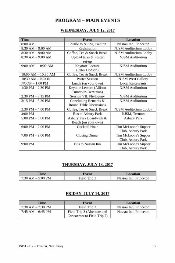
ISPH 2017 – Trenton, New Jersey 17
PROGRAM – MAIN EVENTS
WEDNESDAY, JULY 12, 2017
Time Event Location
8:00 AM Shuttle to NJSM, Trenton Nassau Inn, Princeton
8:30 AM – 9:00 AM Registration NJSM Auditorium Lobby
8:30 AM – 9:00 AM Coffee, Tea & Snack Break NJSM Auditorium Lobby
8:30 AM – 9:00 AM Upload talks & Poster
set-up
NJSM Auditorium
9:00 AM – 10:00 AM Keynote Lecture
(Peter Dodson)
NJSM Auditorium
10:00 AM – 10:30 AM Coffee, Tea & Snack Break NJSM Auditorium Lobby
10:30 AM – NOON Poster Session NJSM West Gallery
NOON – 1:00 PM Lunch (on your own) Local Restaurants
1:30 PM – 2:30 PM Keynote Lecture (Allison
Tumarkin-Deratzian)
NJSM Auditorium
2:30 PM – 3:15 PM Session VII: Phylogeny NJSM Auditorium
3:15 PM – 3:30 PM Concluding Remarks &
Round Table Discussions
NJSM Auditorium
3:30 PM – 4:00 PM Coffee, Tea & Snack Break NJSM Auditorium Lobby
4:00 PM Bus to Asbury Park NJSM, Trenton
5:00 PM – 6:00 PM Asbury Park Boardwalk &
Beach (on your own)
Asbury Park
6:00 PM – 7:00 PM Cocktail Hour Tim McLoone's Supper
Club, Asbury Park
7:00 PM – 9:00 PM Closing Dinner Tim McLoone's Supper
Club, Asbury Park
9:00 PM Bus to Nassau Inn Tim McLoone's Supper
Club, Asbury Park
THURSDAY, JULY 13, 2017
Time Event Location
7:30 AM – 5:00 PM Field Trip 1 Nassau Inn, Princeton
FRIDAY, JULY 14, 2017
Time Event Location
7:30 AM – 7:30 PM Field Trip 2 Nassau Inn, Princeton
7:45 AM – 6:45 PM Field Trip 3 (Alternate and
Concurrent to Field Trip 2)
Nassau Inn, Princeton
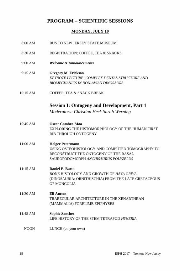
18 ISPH 2017 – Trenton, New Jersey
PROGRAM – SCIENTIFIC SESSIONS
MONDAY, JULY 10
8:00 AM BUS TO NEW JERSEY STATE MUSEUM
8:30 AM REGISTRATION; COFFEE, TEA & SNACKS
9:00 AM Welcome & Announcements
9:15 AM Gregory M. Erickson
KEYNOTE LECTURE: COMPLEX DENTAL STRUCTURE AND
BIOMECHANICS IN NON-AVIAN DINOSAURS
10:15 AM COFFEE, TEA & SNACK BREAK
Session I: Ontogeny and Development, Part 1
Moderators: Christian Heck Sarah Werning
10:45 AM Oscar Cambra-Moo
EXPLORING THE HISTOMORPHOLOGY OF THE HUMAN FIRST
RIB THROUGH ONTOGENY
11:00 AM Holger Petermann
USING OSTEOHISTOLOGY AND COMPUTED TOMOGRAPHY TO
RECONSTRUCT THE ONTOGENY OF THE BASAL
SAUROPODOMORPH ANCHISAURUS POLYZELUS
11:15 AM Daniel E. Barta
BONE HISTOLOGY AND GROWTH OF HAYA GRIVA
(DINOSAURIA: ORNITHISCHIA) FROM THE LATE CRETACEOUS
OF MONGOLIA
11:30 AM Eli Amson
TRABECULAR ARCHITECTURE IN THE XENARTHRAN
(MAMMALIA) FORELIMB EPIPHYSES
11:45 AM Sophie Sanchez
LIFE HISTORY OF THE STEM TETRAPOD HYNERIA
NOON LUNCH (on your own)
ISPH 2017 – Trenton, New Jersey 19
Session II: Ontogeny and Development, Part 2
Moderators: Justyna Miszkiewicz and Edina Prondvai
1:30 PM Megan R. Whitney
OSTEOHISTOLOGICAL SIGNATURES OF HATCHING IN
MODERN RATITES WITH IMPLICATIONS FOR
RECONSTRUCTING THE EARLY ONTOGENY OF DINOSAURS
1:45 PM Mateusz Wosik
DEFINING DINOSAUR NEONATAL BODY SIZE USING
OSTEOHISTOLOGICAL EVIDENCE
2:00 PM Edina Prondvai
ALLOMETRIC INTRASKELETAL GROWTH PATTERNS AND
THEIR FUNCTIONAL IMPLICATIONS IN DINOBIRDS
2:15 PM Jennifer Botha-Brink
OSTEOHISTOLOGY OF LATE TRIASSIC PROZOSTRODONTIAN
CYNODONTS FROM BRAZIL
2:30 PM Carmen Nacarino-Meneses
IDENTIFYING BIRTH FROM BONE HISTOLOGY: A STUDY IN
EXTANT EQUUS
2:45 PM COFFEE, TEA & SNACK BREAK
Session III: Dental Tissues
Moderators: Mateusz Wosik and Catherine Sartin
3:15 PM Julia Audije-Gil
AN APPROACH TO MICROPRESERVATION OF CROCODILIAN
FOSSIL TEETH THROUGH HISTOCHEMICAL DATA
3:30 PM Michael D. D’Emic
RAPID TOOTH REPLACEMENT RATES IN THE THEROPOD
DINOSAUR MAJUNGASAURUS FROM THE LATE CRETACEOUS
(MAASTRICHTIAN) OF MADAGASCAR
3:45 PM Yara Haridy
TOOTH REPLACEMENT AND MIGRATION IN THE EARLIEST
ACRODONT REPTILE, OPISTHODONTOSAURUS
20 ISPH 2017 – Trenton, New Jersey
4:00 PM Aaron R. H. LeBlanc
TOOTH ATTACHMENT IN MOSASAURIDS REVISITED: AN
ONTOGENETIC PERSPECTIVE TO SQUAMATE TOOTH
DEVELOPMENT AND EVOLUTION
4:15 PM Guillem Orlandi-Oliveras
FIRST APPROACH TO BONE AND DENTAL HISTOLOGY OF
GREEK HIPPARIONINES
4:30 PM Barbara S. Grandstaff
OVER A CENTURY OF THIN SECTION MICROSCOPY OF THE
FOSSIL FISH CYLINDRACANTHUS
5:00 PM BUS TO NASSAU INN
TUESDAY, JULY 11
8:00 AM BUS TO NEW JERSEY STATE MUSEUM
8:30 AM REGISTRATION; COFFEE, TEA & SNACKS
9:00 AM Phil Manning
KEYNOTE LECTURE: IMAGING FOSSILIZED BIOMATERIALS
10:00 AM COFFEE, TEA & SNACK BREAK
Session IV: Biomechanics
Moderators: David Parris and Rodrigo Pellegrini
10:30 AM Alida M. Bailleul
TYRANNOSAURUS REX SHOWS HISTOLOGICAL EVIDENCE FOR
AVIAN-STYLE CRANIAL KINESIS
10:45 AM Jordi Estefa
CAN THE SEMI-ERECT POSTURE OF MODERN ECHIDNA BE
CONSIDERED AS A PROXY FOR INTERPRETING THE POSTURE
OF EARLY TERRESTRIAL TETRAPODS?
11:00 AM Lucas Legendre
LONG BONE HISTOLOGY OF THE AARDVARK
(AFROTHERIA, TUBULIDENTATA)
ISPH 2017 – Trenton, New Jersey 21
Session V: Methodologies and Nomenclature
Moderators: David Parris and Rodrigo Pellegrini
11:15 AM Marilyn Fox
TREATMENT OF FOSSIL SPECIMENS BEFORE AND AFTER
DESTRUCTIVE SAMPLING
11:30 AM Alexandra Houssaye
QUANTITATIVE 3D ANALYSIS OF LONG BONE SHAFT
MICROANATOMICAL AND GEOMETRICAL FEATURES
AMONG MAMMALS
11:45 AM LUNCH (on your own)
Session VI: Physiology
Moderators: Alida Bailleul and Allison Tumarkin-Deratzian
1:30 PM Santiago Gomez
PRESERVED MINERALIZED FIBERS IN OSTEONS FROM A
SPINOSAURUS AEGYPTIACUS BONE
1:45 PM Maïtena Dumont
SYNCHROTRON INVESTIGATION OF THE 3D VASCULAR
SYSTEM OF SAUROPOD AND MAMMAL LONG BONES
2:00 PM Zachary M. Boles
SHELL BONE HISTOLOGY AND HABITAT PREFERENCE OF
TURTLES FROM THE K/PG HORNERSTOWN FORMATION,
NEW JERSEY (USA)
2:15 PM Rodrigo A. Pellegrini
SKELETOCHRONOLOGY, PALEOHISTOLOGY AND LIFESTYLE
OF HYPOSAURUS ROGERSII (CROCODYLIFORMES,
DYROSAURIDAE) FROM THE EARLY PALEOGENE OF NEW
JERSEY, USA
2:30 PM P. Martin Sander
UNIQUE CORTICAL HISTOLOGY OF GIANT LONG BONE
SHAFTS FROM EUROPEAN RHAETIAN (LATEST TRIASSIC)
BONEBEDS: THE ENIGMA CONTINUES
2:45 PM REGISTRATION; COFFEE, TEA & SNACK BREAK
22 ISPH 2017 – Trenton, New Jersey
Special Session
Microscopy of Prehistoric Bones and Teeth:
Interface between Histology and Archaeology
Moderators: Vijay Sathe and Jim Moss
3:15 PM Oscar Cambra-Moo
PALEOHISTOLOGY OF HUMAN CREMATED BONES: A CASE
FROM THE PREHISTORIC ARCHAEOLOGICAL SITE OF
LAGUNITA (SANTIAGO DE ALCÁNTARA, CÁCERES, SPAIN)
3:30 PM Orosia García-Gil
TAPHONOMICAL AND PALEOHISTOLOGICAL DATA FROM
HISPANO-MUSLIM MAQBARA OF SAN NICOLÁS
(MURCIA, SPAIN)
3:45 PM Rajeev Patnaik
PALEOHISTOLOGICAL STUDIES ON INCISOR ENAMEL OF
MODERN AND FOSSIL MURID RODENTS FROM INDIA:
IMPLICATIONS FOR TAXONOMY AND PALEOECOLOGY
4:00 PM Falguni Katkar
MICROBIAL INFESTATION ON ARCHAEOLOGICAL BONES: A
CASE STUDY OF BINJOR, RAJASTHAN
4:15 PM Justyna J. Miszkiewicz
CORTICAL HISTOMORPHOMETRY AND ROBUSTICITY IN
ANCIENT ADULT HUMAN FEMUR – BIOMECHANICAL AND
DIMENSIONAL RELATIONSHIPS?
4:30 PM Vijay Sathe
DENTAL HISTOLOGY AND CHEMISTRY OF PREHISTORIC
LARGE MAMMALIAN FAUNA FROM INDIA
5:00 PM BUS TO NASSAU INN
ISPH 2017 – Trenton, New Jersey 23
WEDNESDAY, JULY 12
8:00 AM BUS TO NEW JERSEY STATE MUSEUM
8:30 AM REGISTRATION; COFFEE, TEA & SNACKS
9:00 AM Peter Dodson
KEYNOTE LECTURE: FIVE CENTURIES OF LOOKING THROUGH
LENSES – A PALEONTOLOGIST’S PERSPECTIVE
10:00 AM COFFEE, TEA & SNACK BREAK
10:30 AM POSTER SESSION
NOON LUNCH (on your own)
1:30 PM Allison Tumarkin-Deratzian
KEYNOTE LECTURE: A REVIEW OF PALEOHISTOLOGY IN
PHILADELPHIA: 1990S THROUGH PRESENT
Session VII: Phylogeny
Moderators: Barbara Grandstaff and Alexandra Houssaye
2:30 PM John M. Rensberger
TOTAL BONE CELL CYTOPLASM AND SURFACE AREA IN
BIRDS, NON-AVIAN DINOSAURS, MAMMALS AND
OTHER TETRAPODS
2:45 PM Peter J. Makovicky
SYNCHROTRON SCANNING REVEALS EVOLUTION OF DIVING
IN HESPERORNITHIFORM BIRDS
3:00 PM Donald Davesne
THE EVOLUTION OF ACELLULAR BONE IN TELEOST FISHES
3:15 PM Concluding Remarks & Round Table Discussions
3:30 PM COFFEE, TEA & SNACK BREAK
4:00 PM BUS TO ASBURY PARK (CLOSING DINNER)
24 ISPH 2017 – Trenton, New Jersey
POSTERS
1. Eli Amson
LOCOMOTOR ADAPTATION IN THE FEMORAL HEAD TRABECULAR
ARCHITECTURE OF SCIUROMORPHS
2. Ashley W. Poust
HISTOLOGY OF THE MAMMALIAN BACULUM
3. Yumi Asakura
PALEOHISTOLOGY IN PAMPATHERE OSTEODERMS:
HOLMESINA PAULACOUTOI
4. Julia Audije-Gil
NEW OBSERVATIONS ON THE HISTOMORPHOLOGICAL VARIATIONS OF
THE HUMAN MIDSHAFT FEMUR
5. Vijay Sathe
OSTEON ARRANGEMENT AND TAXONOMY: SIGNIFICANCE OF BONE
HISTOLOGY IN ARCHAEOZOOLOGY
6. Orosia García-Gil
FIRST APPROACH TO THE TAPHONOMY AND PALEOHISTOLOGY OF THE
PREHISTORIC ARCHAEOLOGICAL SITE OF “CERRO DE LA ENCANTADA”
(II MILLENNIUM B.C., SPAIN)
7. Carmen Nacarino-Meneses
BONE HISTOLOGY OF EQUUS FROM STEINHEIM AN DER MURR
(MIDDLE PLEISTOCENE, GERMANY)
8. Guillem Orlandi-Oliveras
ENAMEL CYCLICAL MARKS OF UNKNOWN PERIODICITY IN EQUIDAE
9. Alexandra Houssaye
HISTOLOGICAL AND MICROANATOMICAL CONTRIBUTION TO
PALEOPHYSIOLOGICAL AND PALEOECOLOGICAL INFERENCES IN THE
DICYNODONT MOGHREBERIA NMACHOUENSIS
10. Yara Haridy
DENTICULATE CORONOIDS AND INTERNAL MANDIBULAR ANATOMY OF
THE PARAREPTILE DELORHYNCHUS
ISPH 2017 – Trenton, New Jersey 25
11. Christopher T. Griffin
PATHOLOGICAL BONE TISSUE IN A LATE TRIASSIC THEROPOD FIBULA,
WITH IMPLICATIONS FOR THE INTERPRETATION OF MEDULLARY BONE
12. Jingmai K. O’Connor
DEFINITIVE OCCURRENCE OF MEDULLARY BONE IN AN
ENANTIORNITHINE (AVES: ORNITHOTHORACES)
13. Rafael C.L.P. Andrade
OSTEOHISTOLOGY AS A PROXY FOR THE UNDERSTANDING OF GROWTH
STRATEGIES AND EVOLUTION IN EXTANT AND EXTINCT CAIMANINAE
(CROCODYLIA, ALLIGATORIDAE)
14. Sarah-Jane Strachan
TO WHAT EXTENT CAN PALEOHISTOLOGY HELP US UNDERSTAND
EXTINCT ANIMAL BEHAVIOR? A CONCEPTUAL FRAMEWORK APPROACH
15. Kevin Surya
PALEOHISTOLOGY TECHNIQUE OF SUB-FOSSILIZED BONE
16. Christian T. Heck
POLYESTER OR EPOXY: ASSESSING PRODUCT EFFICACY IN
PALEOHISTOLOGICAL METHODS
17. Rodrigo A. Pellegrini
PRESSURE VS. VACUUM IN THE RESIN IMPREGNATION OF
THIN-SECTION SAMPLES
26 ISPH 2017 – Trenton, New Jersey
In honor of the 2017 International Symposium on Paleohistology,
registrants receive 10% off their purchase in the Museum Shop.*
Proceeds from the Museum Shop support the collections, exhibitions,
programs and publications of the New Jersey State Museum.
*Some exclusions apply.
ISPH 2017 – Trenton, New Jersey 27
28 ISPH 2017 – Trenton, New Jersey
COMPLEX DENTAL STRUCTURE AND BIOMECHANICS IN
NON-AVIAN DINOSAURS
GREG OR Y M. ERICK SON
Department of Biological Science, Florida State University, Tallahassee, FL 32306, USA
Reptiles rarely approached the biomechanical sophistication for feeding or dietary diversity
seen in mammals. Their teeth are typically non-occluding, semi-conical structures with
simplistic parallel-crystallite enamel surrounding an orthodentine core. Conversely, most
mammals possess multi-cusped teeth that are drawn across one another during mastication
and self-wear to their functional morphology. The most complex dental architectures are seen
in herbivorous mammals. Their teeth are composed of up to four constituents (incl. prismatic
enamel) that strategically wear creating coarse grinding or slicing surfaces. These allow them
to comminute tough and/or abrasive plants and liberate nutrients inaccessible to other
animals. Non-avian dinosaurs stand out among reptiles in that precise, mammal-like dental
occlusion and self-wearing teeth evolved on at least four occasions. These topographies
enabled access to myriad floral types and facilitated their respective ecological
diversifications. My research group and colleagues showed that the teeth of these dinosaurs
are histologically more complex than formerly appreciated. Using cutting-edge material
science and tribological engineering indentation techniques we discovered that wear and
fracture relevant material properties are preserved in fossil dental tissues. This led to
development of the first 3D Archard’s wear model from which we determined how horse-like
occlusal surfaces for the grinding of plant matter occurred in hadrosaurids and fuller-like
slicing surfaces developed and functioned in ceratopsians. By incorporating micro-fracture
testing into our analyses we discovered how wavy enamel in grazing hadrosauroids, despite
lacking enamel prisms, served to stymie fracture during the ingestion of exogenous
inclusions. Notably our paleontologically-inspired wear model is seeing broad
industrial application.
ISPH 2017 – Trenton, New Jersey 29
EXPLORING THE HISTOMORPHOLOGY OF THE HUMAN
FIRST RIB THROUGH ONTOGENY
DAN IE L GARC ÍA-M ART ÍNE Z1 * , OSC AR CAM BR A-MOO
2 , 3 , ORO S IA GAR C ÍA G IL2 ,
MAR ÍA CAN ILL AS4 , M IGU E L ÁN GE L ROD R ÍG UEZ BARBE RO
4 , MARKU S BAST IR1 ,
AND ARM AND O GO NZ ÁLEZ MART ÍN2 , 3
1 Paleoanthropology Group, Museo Nacional de Ciencias Naturales, CSIC, Madrid, Spain 2 Laboratorio de Poblaciones del Pasado (LAPP). Departamento de Biología, Facultad de Ciencias,
Universidad Autónoma de Madrid, 28049 Madrid, Spain 3 Grupo de Investigación en Arqueología Antigua y Medieval, Universidad de Oviedo, 33011
Oviedo, Spain 4 Instituto de Cerámica y Vidrio (Consejo Superior de Investigaciones Científicas),
Madrid, Spain
Human first rib morphology provides important information concerning thorax anatomy from
both ontogenetic and phylogenetic perspectives. Luckily, they frequently appear well
preserved in the archaeological/fossil record and are easy to assess in commingled samples
because of their unique anatomy. Regarding first rib ontogeny, several studies have addressed
ontogenetic macro- and micro-anatomical changes separately, but no one has previously
combined both approaches.
We selected a sample of 14 first ribs from the ossuary of the Santa María de la Soledad
medieval church (Spain), from individuals ranging from perinatal to adult (aged through a
centroid size-based approach). We applied 3D morphometrics of sliding semi landmarks to
quantify rib curvature and mid-shaft cross-sectional outline, then histologically processed the
ribs to obtain mid-shaft thin sections. Macroscopically, the first rib changes from a low
curved configuration with a rounded cross-section (perinatal) to a highly curved morphology
with a “drop-shaped” section (adult). Microanatomically, we identified three patterns of
compartmentalization and histological configuration: 1) small rib, cortex poorly vascularized,
with a mineralized matrix configured as woven bone in which areas of paralleled-fibered
and/or lamellar bone could be distinguished (perinatal cortex); 2) a medium-size cross-
section in which a thicker cortex with a well-defined medullary cavity is easily identified, and
with a bone matrix mainly organized as lamellar bone (infant individuals); and 3) a large
cross-section, highly vascularized, with a thinner highly-remodeled cortex. These ontogenetic
changes are important not only to understand rib cage ontogeny but also thorax functional
anatomy and evolution.
30 ISPH 2017 – Trenton, New Jersey
USING OSTEOHISTOLOGY AND COMPUTED
TOMOGRAPHY TO RECONSTRUCT THE ONTOGENY OF
THE BASAL SAUROPODOMORPH
ANCHISAURUS POLYZELUS
HO LG ER PETE RM ANN1 *
AN D MATTEO FAB BR I1
1 Department of Geology and Geophysics, Yale University, New Haven, CT 06511, USA
Sauropod dinosaurs are the most gargantuan terrestrial organisms that ever lived on our
planet. However, even after decades of studies, the evolutionary history of sauropods is far
from being resolved. In particular, several questions regarding the origin of gigantism and
body size evolution, and the phylogenetic relationships characterizing the basal branches in
Sauropodomorpha, are still unanswered. Here, we present a new comprehensive study of the
Early Jurassic taxon Anchisaurus. Peculiarities regarding Anchisaurus are its body size and
its phylogenetic affinities. Anchisaurus is a slender and lightly built sauropodomorph
compared to its closest relatives (Sauropodiformes such as melanorosaurs) and immediate
ancestors (massospondylids). Moreover, Anchisaurus is usually recovered in different
positions within Sauropodomorpha, but is usually nested within the more derived basal
sauropodomorphs, close to Sauropoda proper. Given the combination of small body size in
comparison to the closest sister taxa and its derived phylogenetic position constantly
recovered in phylogenetic analyses, we hypothesize that Anchisaurus is a dwarfed basal
sauropodomorph, the first one recovered in the basal branches of Saurischia. We performed
osteohistological analyses on femora, humeri, and ribs of three specimens of Anchisaurus
housed at the Yale Peabody Museum. We found confirmation of dwarfism in this taxon.
Using microCT scan data, we were able to reconstruct the skull morphology of the youngest
and oldest individuals, allowing for reinterpretation of the diagnosis of this taxon and to test,
through application of morphometrics, the heterochronic event that led to dwarfism in
Anchisaurus and shaped the general skull morphology found in Sauropoda.
ISPH 2017 – Trenton, New Jersey 31
BONE HISTOLOGY AND GROWTH OF HAYA GRIVA
(DINOSAURIA: ORNITHISCHIA) FROM THE LATE
CRETACEOUS OF MONGOLIA
DAN IE L E. BART A1 , 2 *
AND MAR K A. NOR E LL1 , 2
1 Richard Gilder Graduate School, American Museum of Natural History, New York, NY, 10024, USA 2 Division of Paleontology, American Museum of Natural History, New York, NY, 10024, USA
Haya griva is a basal neornithischian (or “hypsilophodontid”) dinosaur known from dozens
of specimens, including a partial growth series, from the Upper Cretaceous Javkhlant
Formation of southeastern Mongolia. Previous studies suggest skeletally mature specimens of
basal neornithischian dinosaurs are rare. Given the wide size range of Haya griva femora in
the collection, we examined growth patterns to ascertain whether any of the individuals had
reached somatic maturity. These data additionally inform our work on morphological
variation and the systematics of Haya. To investigate the maturity of individual specimens,
we sampled three femora, representing the longest (162 mm) and shortest (~64 mm) presently
available, as well as one intermediate in length (129 mm). Transverse sections of these bones
were embedded in epoxy, mounted on glass slides, and ground and polished until transparent.
Photomicrographs reveal predominantly parallel-fibered bone in the smaller two femora, and
fibrolamellar bone in the largest. The smallest femur lacks growth lines (=LAGs, annuli).
Growth lines are difficult to discern in the medium-sized femur because of poor preservation.
The largest femur contains at least four growth lines, but lacks an external fundamental
system, indicating it had not slowed growth asymptotically at the time of death. Body masses
estimated from the femoral circumferences of the sectioned individuals are 0.95, 11, and 30
kg. We conclude that, as for closely related taxa, all Haya griva specimens discovered so far
are probably skeletally immature and that the upper limit of body size for this taxon
remains unknown.
32 ISPH 2017 – Trenton, New Jersey
TRABECULAR ARCHITECTURE IN THE XENARTHRAN
(MAMMALIA) FORELIMB EPIPHYSES
ELI AM SON1 * , PAT R IC K AR NO LD
2 , ANN EKE H. V AN HET ERE N3 , AURO RE CAN OV ILLE
4 ,
AND JO HN A. NY AK AT UR A1
1 AG Morphologie und Formengeschichte, Bild Wissen Gestaltung – ein interdisziplinäres Labor &
Institut für Biologie, Humboldt-Universität zu Berlin, Unter den Linden 6, Berlin 10117, Germany 2 Institut für Spezielle Zoologie und Evolutionsbiologie mit Phyletischem Museum, Friedrich-Schiller-
Universität Jena, 07743, Germany 3 Sektion Mammalogie, Zoologische Staatssammlung München – Staatliche Naturkundliche Sammlungen
Bayerns, München, 81247, Germany 4 Department of Biological Sciences, North Carolina State University, Raleigh, NC 27695, USA
Trabecular architecture (i.e., relative number of trabeculae, their main orientation, mean
thickness, spacing, etc.) has been shown experimentally to adapt with extreme accuracy and
sensitivity to the loadings applied to the bone during life. However, the functional signal in
trabecular parameters has only been superficially studied, as present studies are mostly
limited to primates. Here we use high-resolution computed tomography to analyze the 3D
architecture in the epiphyses of the forelimb of xenarthrans, i.e., sloths, anteaters, and
armadillos, and their extinct relatives. Xenarthrans form a major clade of placental mammals,
with modern taxa specialized in several locomotor styles and utilizing diverse hand postures:
unguligrade-subunguligrade (armadillos), inverted hand (small and medium-sized anteaters),
‘knuckle-walking’ (giant anteater), and suspensory (extant sloths). They are also
characterized by various degrees of digging ability. Extinct xenarthrans, “ground sloths” in
particular, were also reconstructed as practicing several peculiar stances, mostly based on the
gross morphology of their postcrania. Xenarthrans hence offer a suitable framework to study
the response of trabecular bone to different loading regimes within a well-defined clade. Our
analyses involve 3D trabecular parameters deriving from the selection of regions of interest
in the epiphyses of the forelimb. We found significant differences among trabecular
parameters of the extant xenarthran clades (e.g., higher degree of anisotropy in the armadillo
glenoid cavity, even after size correction). These differences have the potential to help
elaborate paleobiological inferences.
ISPH 2017 – Trenton, New Jersey 33
LIFE HISTORY OF THE STEM TETRAPOD HYNERIA
V IKT OR IIA KAM SK A1 , ED W AR D B. DAE SC H LE R
2 , JASON P. DOW N S2 , PE R E. AH LBER G
1 ,
PAU L TAFF ORE AU3
AND SOPH IE SANC HEZ1 , 3 *
1 Science for Life Laboratory and Uppsala University, Department of Organismal Biology, Evolution and
Development, Uppsala University, Uppsala, 75236, Sweden 2 Academy of Natural Sciences of Philadelphia, Philadelphia, PA 19103, USA 3 European Synchrotron Radiation Facility, Grenoble, 38043, France
In order to understand the ecological dimension of the fish-tetrapod transition, which
occurred within the tetrapod stem group during the Devonian Period (419-359 million years
ago), we need life-history data from transitional forms. Only recently have serious attempts
begun to utilize limb-bone histology as a source of such data. Here we present histological
life history data from a humerus (ANSP 21483) of Hyneria lindae obtained by propagation
phase contrast synchrotron microtomography (ESRF, France). Hyneria, a fish member of the
tetrapod stem group from the Late Devonian Catskill Formation (Pennsylvania, USA), is
closely related to the better known Eusthenopteron but much larger (body length at least two
meters). The internal structure of its humerus is similar to those of Eusthenopteron and the
limbed stem tetrapod Acanthostega. The spongiosa contains primitive bone marrow
processes, indicating that bone marrow was already intimately associated with long-bone
elongation and endochondral ossification before the transition from fins to limbs. The
humerus presents a mixture of ‘juvenile’ and ‘adult’ histological features, suggesting either
that it represents a subadult individual or that Hyneria was in some respects paedomorphic.
The latter hypothesis is supported by the unossified condition of the endocranium even in
large individuals. The thin humeral cortex exhibits a relatively slow bone growth rate.
Interestingly, Eusthenopteron and Acanthostega also show slow growth and late (> 6 years)
sexual maturity. A broad-sample investigation of additional taxa will be needed to assess the
generality of this pattern and map the reproductive trends during the early evolution
of tetrapods.
34 ISPH 2017 – Trenton, New Jersey
OSTEOHISTOLOGICAL SIGNATURES OF HATCHING IN
MODERN RATITES WITH IMPLICATIONS FOR
RECONSTRUCTING THE EARLY ONTOGENY
OF DINOSAURS
MEG AN R. WH IT NEY1 , MATEU S Z WO SIK
2 , 3 , LI SA DO R AN1 , KR IST IN A CURR Y ROGE R S
4 ,
HO LLY WO ODW ARD5 ,
AND DAV ID C. EV AN S2 , 3
1 Department of Biology, University of Washington, Seattle, WA 98195, USA 2 Department of Ecology and Evolutionary Biology, University of Toronto, Toronto, ON
M5S 2C6, Canada 3 Department of Natural History, Royal Ontario Museum, Toronto, ON M5S 2C6, Canada 4 Biology and Geology Departments, Macalester College, St. Paul, MN 55105, USA 5 Center for Health Sciences, Oklahoma State University, Tulsa, OK 74107, USA
Circumferential zones of reduced vascularity have been proposed to represent an
osteohistological indicator of hatching in a perinatal sauropod and have been used to infer an
approximate hatchling body size. However, a lack of data on neonatal signals in long bone
histology among extant vertebrates limits paleohistological interpretations. Here we report on
the long bone histology of pre- and post-hatching ratites to detail the features of this
important transitionary period between embryo and neonate. Femora and tibiotarsi from a
constrained ontogenetic sample of embryonic and perinatal Dromaius novaehollandiae (emu)
and Struthio camelus (ostrich) individuals (n=13) were transversely thin-sectioned at the
minimum diaphyseal circumference.
All specimens display highly vascularized fibrolamellar and/or woven tissue with
longitudinal primary osteon orientation in both the femora and tibiotarsi. Pre-hatching
individuals show relatively uniform vascular porosity (~30%) throughout the entire cortex,
with the exception of a single specimen that exhibits slightly narrower canal spaces towards
the periosteal surface. Post-hatching individuals consistently have a circumferential zone
characterized by a reduction in osteon diameter where the percentage of open vascular spaces
within the mid-cortex is significantly smaller (~9.8%) than towards the endosteal (~18.20%)
or periosteal (~28.06%) surfaces. This temporary reduction in vascular space is a result
mostly of increased osteonal deposition around the canals within this zone and is similar to
what has been observed in a perinatal sauropod dinosaur. Continued investigation of a
neonatal signal in extant vertebrates will refine the definition of this osteohistological
signature and its implications in understanding early dinosaur ontogeny.
ISPH 2017 – Trenton, New Jersey 35
DEFINING DINOSAUR NEONATAL BODY SIZE USING
OSTEOHISTOLOGICAL EVIDENCE
MAT EU SZ WO SIK1 , 2 * , MEG AN WH IT NEY
3 , KR IST IN A CUR RY ROGER S4 , HO LLY
WOOD W AR D5 , AN D DAV ID C. EV AN S
1 , 2
1 Department of Ecology and Evolutionary Biology, University of Toronto, Toronto,
ON M5S 2C6, Canada 2 Department of Natural History, Royal Ontario Museum, Toronto, ON M5S 2C6, Canada 3 Department of Biology, University of Washington, Seattle, WA 98195, USA 4 Departments of Biology and Geology, Macalester College, St. Paul, MN 55105, USA 5 Center for Health Sciences, Oklahoma State University, Tulsa, OK 74107, USA
The earliest life histories of extant vertebrates are punctuated by a transition in bone
deposition immediately after birth/hatching and recorded in teeth and bones as a ‘neonatal’ or
‘hatching’ line. Recent studies have noted comparable features in perinatal dinosaurs,
allowing approximation of hatchling size. Here we investigate embryonic and perinatal bone
histology in extant ratites (emu, ostrich) and non-avian dinosaurs (hadrosaurid,
hypsilophodontid, sauropod) in order to qualitatively compare the osteohistological signal
of hatching.
Ratite femora and tibiotarsi were thin-sectioned at mid-diaphysis from a series of 13
individuals with known age bracketing the hatching period. Dinosaur stylopodial and
zeugopodial elements for 17 individuals were similarly thin-sectioned and categorized by size
and association with eggshell material. Regardless of taxonomic identity, embryos exhibit
highly cancellous and disorganized woven-fibered bone whereas perinates have fibro-
lamellar bone with longitudinally or radially orientated vascular canals. Perinates preserve a
narrow zone of reduced vascularity typically coinciding with a darkened band. This
circumferentially oriented zone is intraspecifically consistent with known hatchling sizes for
ratites and partitions the embryonic and perinatal bone regions, although with some degree of
circumferential variation. Therefore, we suggest this is an osteohistological indicator for
hatching and define it as the neonatal signal.
Clarification of the biological meaning of the neonatal signal is important because it provides
an accurate neonatal size for growth rate analyses and may convey significant insight for
osteohistological cues related to precocity. Further investigation is required to outline the
relationships of the neonatal signal to biomechanical, nutritional, environmental, and
phylogenetic effects.
36 ISPH 2017 – Trenton, New Jersey
ALLOMETRIC INTRASKELETAL GROWTH PATTERNS AND
THEIR FUNCTIONAL IMPLICATIONS IN DINOBIRDS
ED IN A PRO NDV AI1 * , DONG -YU HU
2 , DO M IN IQU E AD R IAEN S1 AND PASC AL GO DEF RO IT
3
1 Department of Biology, Evolutionary Morphology of Vertebrates, Ghent University,
Gent, 9000, Belgium 2 Paleontological Institute, Shenyang Normal University, Shenyang, 110000, China 3 Directorate ‘Earth and History of Life’, Royal Belgian Institute of Natural Sciences,
Brussels, 1000, Belgium
Paravian dinosaurs are usually characterized by disproportionately long and robust arms, the
size of which often even exceeds that of the hind limbs. This allometry can be achieved by
differential growth rates of limb elements in different phases of ontogeny. To get insight into
intraskeletal growth dynamics, we studied the osteohistology of limb bones in five paravian
dinosaurs from the Middle/Late Jurassic and Early Cretaceous of China, Anchiornis,
Aurornis, Eosinopteryx, Jeholornis and a yet unnamed taxon, with qualitative and various
quantitative methods. Our results show that despite their largely uniform ‘dinobird’ bauplan,
different bones reveal diverse growth dynamics with no element-specific consistent pattern
across taxa. Furthermore, ontogenetic stages combined with intraskeletal growth dynamics
explain osteohistological diversity patterns better than pure taxonomic status in these
paravian dinosaurs. The only possible exception is the avian Jeholornis which, besides being
larger, had superior aerial skills compared to the other studied non-avian taxa. However,
locomotion-related and phylogenetic influences resulting in the distinct histology of this
taxon cannot currently be separated. Histology also provided independent evidence of
restored hand bones in this Jeholornis specimen. Nevertheless, ≥50% of variance remains
consistently unexplained, which implies the importance of unexplored factors, including
possibly locomotor adaptations, in shaping the investigated histological characters. As
allometric intraskeletal development in these paravian taxa involves significant and diverse
proportional changes in limb bones during ontogeny, our study reveals the importance of
exploring ontogenetic stage and postnatal intraskeletal growth dynamics when studying
aerodynamic performance of dinosaur-bird transitional forms to reconstruct the evolution of
avian flight.
ISPH 2017 – Trenton, New Jersey 37
OSTEOHISTOLOGY OF LATE TRIASSIC
PROZOSTRODONTIAN CYNODONTS FROM BRAZIL
JENN IFE R BOTH A-BR INK1 , 2 * , MAR IN A BENT O SO ARE S
3 ,
AND AGU ST IN GU ILLE RMO MART IN E LLI3
1 Department of Karoo Palaeontology, National Museum, Bloemfontein, 9300, South Africa 2 Department of Zoology and Entomology, University of the Free State, Bloemfontein, 9300, South Africa 3 Departamento de Paleontologia e Estratigrafia, Instituto de Geociências, Porto Alegre,
91501-970, Brazil
The Prozostrodontia was a group of Triassic eucynodonts, the derived members of which
gave rise to the Mammaliaformes, in which Mammalia is nested. Analyzing their growth
patterns is thus important for understanding the evolution of mammalian life histories.
Obtaining material for osteohistological analysis is difficult due to the rare and delicate
nature of most of the prozostrodontian taxa much of which comprises only crania or
sometimes even only teeth. Here we present a rare opportunity to observe the osteohistology
of several postcranial elements of the basal prozostrodontid Prozostrodon brasiliensis, the
tritheledontid Irajatherium hernandezi, and the brasilodontids Brasilodon quadrangularis
and Brasilitherium riograndensis from the Late Triassic of Brazil (Santa Maria
Supersequence). Prozostrodon and Irajatherium reveal similar growth patterns of rapid early
growth with annual interruptions later in ontogeny. These interruptions are associated with
wide zones of slow growing bone tissue. Brasilodon and Brasilitherium exhibit a mixture of
woven-fibered bone tissue and slower growing lamellar bone. The slower growing bone
tissues are present even during early ontogeny. The relatively slower growth in Brasilodon
and Brasilitherium may be related to their small body size compared to Prozostrodon and
Irajatherium. These brasilodontids also exhibit osteohistological similarities with the
mammaliaform Morganucodon. This may be due to similar small body sizes, but may also
reflect their close phylogenetic affinities as Brasilodon and Brasilitherium are the closest
relatives to the Mammaliaformes.
38 ISPH 2017 – Trenton, New Jersey
IDENTIFYING BIRTH FROM BONE HISTOLOGY: A STUDY
IN EXTANT EQUUS
CAR ME N NAC AR INO -MENE SE S1 *
AND ME IK E KÖ H LE R1 , 2 , 3
1 Evolutionary Paleobiology, Institut Català de Paleontologia Miquel Crusafont, Cerdanyola del Vallès,
08193, Spain 2 Department of Animal Biology, Plant Biology and Ecology (BABVE), Universitat Autònoma de
Barcelona, Cerdanyola del Vallès, 08193, Spain 3 Catalan Institution for Research and Advanced Studies (ICREA), Barcelona, 08010, Spain
The study of bone microstructure is known to provide key insights about the growth and life
history strategy of extant and extinct vertebrates. Because bone growth marks usually register
annual cycles of growth, they have been used to reconstruct important life history traits of the
species such as longevity or age at maturity. However, non-cyclical bone growth marks can
also be identified in the bone cortex. Although the ultimate causes of deposition of this kind
of features are poorly known, they are supposed to record moments of physiological stress in
the organism.
Here, we aim to investigate the relationship between non-cyclical bone growth marks and
stressful biological events. To achieve that objective, histological slices of femora, tibiae and
metapodial bones have been prepared from individuals of known age and sex of Equus
hemionus, E. quagga and E. grevyi. Several of the specimens lived captive in the Hagenbeck
Zoo (Hamburg, Germany), while others lived semi-captive in the Réserve Africaine de
Sigean (Sigean, France). The results obtained reveal the presence of a non-cyclical bone
growth mark that is deposited around birth, analogous to the neonatal line described for teeth.
This neonatal line in bones, which is accompanied by other histological changes in femur and
tibia, has been identified in various bones of all age groups. Our findings of an important
disruption in the deposition of perinatal bone tissue and an associated change in tissue type,
are essential for future skeletochronological studies in extant and extinct equids.
ISPH 2017 – Trenton, New Jersey 39
AN APPROACH TO MICROPRESERVATION OF
CROCODILIAN FOSSIL TEETH THROUGH
HISTOCHEMICAL DATA
JU LIA AU D IJE -G IL1 , 2 * , MAR ÍA CAN ILL AS PÉRE Z
3 , MÉ LAN I BE RRO C AL-CASE RO1 , 4 ,
FERN AN DO BAR RO SO-BARCE N ILLA1 , 4 , OSC AR CAM BR A-MOO
2
AND M IGU E L ÁNGE L RODR ÍGU EZ-BARBE RO3
1 Grupo de Investigación IberCreta, Departamento de Geología, Geografía y Medio Ambiente,
Universidad de Alcalá, Alcalá de Henares, 28871, Spain 2 Laboratorio de Poblaciones del Pasado (LAPP), Departamento de Biología, Universidad Autónoma de
Madrid, Madrid, 28049, Spain 3 Instituto de Cerámica y Vidrio, Consejo Superior de Investigaciones Científicas (CSIC), Madrid, Spain 4 Grupo de Investigación Procesos Bióticos Mesozoicos, Departamento de Paleontología, Universidad
Complutense de Madrid, 28040 Madrid, Spain
The fossil site of “Lo Hueco” is an Upper Cretaceous Konzentrat-Lagerstätte located in
Cuenca (Spain) in which microstructure of bones and teeth is exceptionally preserved due to
possible differential micropreservation events. In order to enrich fossilization and
paleobiological knowledge, paleohistological and physicochemical analyses of several teeth
from this outcrop were performed. First, after preparing thin sections of two Eusuchian
crocodyliform teeth from two marly mudstone levels of the site, their structure was observed
and photographed, using polarized light microscopy. Second, we contrasted the information
of the composition and structure from seven points of the complete tooth length by two
means: Fourier Transform Infrared Spectroscopy (FTIR) and Scanning Electron Microscope
(SEM). As previously described in the literature, two sort of periodic layers, representing
long and short depositional periods, have been identified in the dentine microstructure. The
analysis of the nanometric texture of the tooth reveals also two rugosity levels and surface
unevenness on the mineral phase. Concerning the chemical analysis, an alteration of the
original bioapatite of the tooth was observed and photographed, but no important variations
on the composition of each layer (no significant changes in the Ca/P ratio along the tooth)
were observed. Finally, Fe was detected between some layers, as well as a slight degree of
sample carbonation. These biomineralized tissue studies, through the combination of
microstructure data and geochemical analysis, offer accurate insights to the preservation
processes and paleobiological interpretations of the fossil biotas.
40 ISPH 2017 – Trenton, New Jersey
RAPID TOOTH REPLACEMENT RATES IN THE THEROPOD
DINOSAUR MAJUNGASAURUS FROM THE LATE
CRETACEOUS (MAASTRICHTIAN) OF MADAGASCAR
M IC H AE L D. D’EM IC1 , 2 , 3 * , PAT R ICK M. O’CO NN OR
4 , 5 , ELIZ AB ETH M AR D AK H AY AV A1 ,
AND ER IC LUN D5
1 Department of Biology, Adelphi University, Garden City, NY 11530, USA 2 Department of Anatomical Sciences, Stony Brook University, Stony Brook, NY 11217, USA 3 Burpee Museum of Natural History, Rockford, IL 61103, USA 4 Department of Anatomy, Ohio University, Athens, OH 45701, USA 5 Department of Neuroscience, Ohio University, Athens, OH 45701, USA
Majungasaurus crenatissimus is an abelisaurid theropod dinosaur known from abundant
material from the latest Cretaceous (Maastrichtian) of Madagascar. The species was
cannibalistic; lived in a harsh, seasonal paleoenvironment; and underwent unusually slow
somatic growth to achieve a modest body size. Like other abelisaurids, Majungasaurus had
smaller teeth and a dorsoventrally tall, anteroposteriorly short skull relative to other
theropods. In order to investigate the unusual anatomy of Majungasaurus, we thin-sectioned
a sample of over 20 isolated teeth and CT scanned over a dozen dentigerous elements
collected across the large exposures of the Maevarano Formation in northwest Madagascar.
We contextualize our findings with novel CT and histological data from Allosaurus and
Ceratosaurus. Daily-deposited incremental lines of von Ebner in Majungasaurus have
thicknesses in the range seen in other dinosaurs. Owing to their small size Majungasaurus
teeth formed relatively quickly. CT scans reveal multiple generations of replacement teeth in
each alveolus. There is only a small size discrepancy between successive replacement teeth,
indicating rapid tooth replacement rates relative to other theropods. This is perhaps related to
a specialized diet and/or feeding style in Majungasaurus.
ISPH 2017 – Trenton, New Jersey 41
TOOTH REPLACEMENT AND MIGRATION IN THE
EARLIEST ACRODONT REPTILE, OPISTHODONTOSAURUS
YAR A HAR IDY1 * , AARON R. H. LEBLAN C
2 AND RO BER T R. REISZ
1
1 Department of Biology, University of Toronto Mississauga, Mississauga, Ontario, L5L 1C6, Canada 2 Department of Biological Sciences, University of Alberta, Edmonton, Alberta, T6G 2E9, Canada
Almost all extant squamates possessing acrodont dentition are grouped within the clade
Acrodonta. Extant members of Acrodonta do not replace their teeth (monophyodonty) and
other acrodont lepidosaurs exhibit reduced tooth replacement; this shared reduction of
replacement within the clade has resulted in oligophyodonty becoming associated with the
evolution of acrodont implantation. In this study we present new histological data and an
ontogenetic series of the earliest acrodont amniote, Opisthodontosaurus, from the early
Permian. Opisthodontosaurus is unusual in exhibiting continuous tooth replacement and
migration of tooth positions through ontogeny. This suggests that acrodonty is not universally
associated with reduced tooth replacement, but rather that acrodonty predates the evolution of
oligophyodonty. Moreover, multiple superimposed generations of alveolar bone and dentine
are present at each tooth position in thin section. These layered remnants of teeth track the
migration of tooth positions through ontogeny. The vasculature and tooth remnants show an
incremental migration posteriorly. This is likely caused by allometric growth of the jawbone
that resulted in migration of the tooth positions, whereas the corresponding dental lamina
remained in place within the soft tissue. In many other reptiles, this growth is correlated with
an increase in tooth count; however in Opisthodontosaurus, there is a reduction in tooth count
through ontogeny that is likely caused by allometry in tooth size. With this new evidence we
offer insights into the supposed link between acrodonty and oligophyodonty, and use
Opisthodontosaurus as a model organism to describe the mechanisms underlying tooth
migration in non-mammalian amniotes.
42 ISPH 2017 – Trenton, New Jersey
TOOTH ATTACHMENT IN MOSASAURIDS REVISITED: AN
ONTOGENETIC PERSPECTIVE TO SQUAMATE TOOTH
DEVELOPMENT AND EVOLUTION
AAR ON R. H. LEBLAN C1 * , AN D M IC H AE L W. CALDW E LL
1
1 Department of Biological Sciences, University of Alberta, Edmonton, T6G 2E9, Canada
Mosasaurids were giant marine squamates that roamed the world’s oceans during the Late
Cretaceous. Their tooth attachment and implantation have been studied extensively in order
to determine their affinities within Squamata as well as to compare squamate tooth
attachment to that of other amniotes. Mosasaurids provided the first evidence of “mammal-
like” tooth attachment in a lizard and have been the focus of renewed interest in the evolution
and development of teeth. What remains to be determined, however, is if a lizard is capable
of attaching its teeth to the jaws by way of a ligament, a condition elsewhere only seen in
mammals and archosaurs. We re-examine tooth attachment tissue histology and development
in mosasaurids and show that their teeth were initially held in place by a network of collagen
fiber bundles forming a periodontal ligament. Fiber bundles extend through the entire length
of the osteocementum of the tooth root. A ligament therefore forms the initial attachment of
the tooth to the alveolus and calcifies from the root surface outwards before fusing the tooth
in place. The presence of a periodontal ligament in a squamate and its capacity to fully
calcify provide new insights into the evolution of tooth attachment in amniotes. What were
previously thought to be different tooth attachment tissues in squamates, mammals, and
archosaurs are confirmed to be homologous tissues that differ in the extent and timing of the
mineralization of the soft tissues supporting the tooth.
ISPH 2017 – Trenton, New Jersey 43
FIRST APPROACH TO BONE AND DENTAL HISTOLOGY OF
GREEK HIPPARIONINES
GU ILLEM OR LAN D I-OLIVER AS1 * , CAR MEN NAC AR INO-ME NE SE S
1 ,
D IM ITR IS S. KO STO PO U LO S2 , GEORG E D. KO UF O S
2 AN D ME IKE KÖH LER
1 , 3 , 4
1 Evolutionary Paleobiology, Institut Català de Paleontologia Miquel Crusafont, Cerdanyola del Vallès,
08193, Spain 2 Department of Geology, Aristotle University of Thessaloniki, 54124, Greece 3 Department of Animal Biology, Plant Biology and Ecology (BABVE), Universitat Autònoma de
Barcelona, Cerdanyola del Vallès, 08193, Spain 4 Catalan Institution for Research and Advanced Studies (ICREA), Barcelona, 08010, Spain
The rich late Miocene fossil assemblages of Greece have provided numerous hipparionine
remains that have been the object of several studies. The baseline research on the systematics
of these hipparionines, has contributed to set a perfect background for conducting
paleohistological analyses on an evolutionary framework. Considering the taxonomic and
body size diversity of Greek hipparionines from the late Vallesian (MN10) to the late
Turolian (MN13), we aim to characterize their bone and dental histology to infer about their
life history strategies.
Metapodials and inferior molars of four distinct species (Hipparion cf. H. sebastopolitanum,
Hipparion macedonicum, Hipparion sithonis, and Hipparion philipus) from five fossil sites
were analyzed. From each tooth, we estimated growth patterns and parameters like daily
secretion rate (DSR), enamel extension rate (EER) and total crown formation time (CFT).
Growth marks and tissue types were used to describe and reconstruct the bone growth of the
metapodial bones.
Primary bone tissue types of the hipparionines studied are similar to those observed in extant
equids, mainly characterized by a fibrolamellar complex (FLC) with longitudinal primary
osteons oriented in circular rows. Regarding dental histology, differences between species
were found in several of the parameters studied. Both bone growth marks and dental growth
patterns allowed the reconstruction of growth curves for each taxon. The differences
observed should be set on an evolutionary, stratigraphic and environmental context.
44 ISPH 2017 – Trenton, New Jersey
OVER A CENTURY OF THIN SECTION MICROSCOPY OF
THE FOSSIL FISH CYLINDRACANTHUS
BAR B AR A S. GR AN D ST AF F1 * , RODR IGO A. PE LLEG R IN I
2 , KEN N ETH A. MON SCH3 ,
DAV ID C. PARR IS2
AND DON AL D CLEME NT S2
1 School of Veterinary Medicine, University of PA, Philadelphia, PA 19104-6045, USA 2 New Jersey State Museum, Trenton, NJ 08625-0530, USA 3 Department of Taxonomy and Systematics Naturalis Biodiversity Center, Leiden,
The Netherlands
Study of the microscopic anatomy of Cylindracanthus dates to the nineteenth century. This
was continued in the early twentieth century using thin sections of Nigerian specimens. This
remarkable genus, known only from its distinctive rostral spines, remains enigmatic. We have
reviewed the historical slides of Nigerian specimens in the collections of the British Museum
of Natural History and expanded the histologic study of Cylindracanthus to include the first
detailed description of the tooth bases, examination of possible lesions and healed tissue,
petrography of the fossil, and probable functional anatomy. We have repeated the historical
comparative studies between Cylindracanthus and modern billfish. Billfish rostral structure
has no real resemblance (structural or mineralogical) to Cylindracanthus. Tooth base
attachments in billfish are subdermal. The tooth base structures, lesions, and damaged
surfaces in Cylindracanthus give no evidence of having been subdermal. We have expanded
comparative histological research to include comparisons between Cylindracanthus and
Polyodon (paddlefishes) and Acipenser (sturgeons).
Although fish teeth are typically acrodont (i.e., fused to the oral surfaces of the jaw and
palatal elements), the teeth of Cylindracanthus are functionally pleurodont. They are
attached along (in fact, within) a groove, from which they protrude only slightly. The teeth
point backwards toward the mouth (away from the tapered end). Hypothetically, the teeth
could function in predation by acting as barbs that could cause damage during a stab-and-
withdraw motion of the rostral spine; they would be useless in a lateral striking and/or
slapping motion. They could have been highly effective during head-on attacks on shelled
cephalopods such as ammonites. This would have been advantageous during the Cretaceous
Period but of little value during the Eocene, after ammonites had become extinct. We
conjecture that the teeth became vestigial after the K-Pg event, as they are unknown in
Cenozoic specimens.
ISPH 2017 – Trenton, New Jersey 45
IMAGING FOSSILIZED BIOMATERIALS
PH ILLIP L. M ANN ING1 , 2 * , JENN I FER E. AN NÈ
2 , ROY A. WOG E LIU S2 ,
N ICH O LAS P. EDW AR D S2 , 3 , V IC TOR IA E. EGER TON
1 , 2 , AND UW E BERGM ANN3
1 Department of Geology and Environmental Geosciences, College of Charleston, Charleston,
SC 29424, USA 2 Department of Earth and Environmental Geosciences, University of Manchester, Manchester,
M13 9PL, UK 3 Stanford Synchrotron Radiation Lightsource, University of Stanford, Menlo Park, CA 94025, USA
Quantitative chemical analyses are rarely undertaken on fossils, despite the potentially
valuable information. Trace metal biomarkers have shown that discrete elemental inventories
can be identified and mapped in fossil organisms that correlate to specific anatomical
structures. Infra-red mapping (FTIR) of organic functional groups also helps in the
identification of organometallic compounds that map within discrete biological structures.
Additionally, Pyrolysis Gas Chromatography Mass Spectrometry (Py-GCMS) and Matrix
Assisted Laser Desorption/Ionization (MALDI) mass spectrometry further constrain the
macromolecular composition and endogeneity of samples being mapped. Such elemental
inventories and macromolecular data provide insight to specific biosynthetic pathways.
Ideally such analyses measure and map the chemistry of bone, soft tissue structures and
embedding rock matrix. Mapping fossils in situ helps place constraints on mass transfer
between the embedding matrix and fossil, aiding in distinguishing taphonomic processes
from original chemical zonation remnant from the fossil itself. Conventional non-destructive
analytical methods face serious problems in this case and most recent technological advances
have been targeted at developing nanometer-scale rather than decimeter-scale capabilities.
However, the development of Synchrotron Rapid Scanning X-ray Fluorescence (SRS-XRF)
at the Stanford Synchrotron permits large specimens to be non-destructively analyzed (1-100
µm resolution) and imaged using major, minor and trace element concentrations. Fossil and
extant samples are mounted in a purpose-built sample chamber held on a computerized x-y-z
translational stage. For light-element XRF imaging, an X-ray-transparent ∼30-μm thick
polyethylene film was placed on the sample chamber, and purged of air with He to minimize
scattering and increase signal to the detector. X-ray Absorption Spectroscopy (XAS)
constrains oxidation state and coordination chemistry of key elements (S, Ca, Cu, Zn, Sr,
etc.), providing key insight to the endogeneity of chemistry mapped to PPM sensitivity. The
combination of precise, repeatable and quantitative techniques build upon decades of research
at synchrotron and lab-based facilities, shedding new light on the chemistry and evolution
of life.
46 ISPH 2017 – Trenton, New Jersey
TYRANNOSAURUS REX SHOWS HISTOLOGICAL
EVIDENCE FOR AVIAN-STYLE CRANIAL KINESIS
ALID A M. BAILLE U L1 * , JO HN R. HORN ER
2 , JO HN SC AN NE LLA3 ,
AND CASEY M. HO LLID AY1
1 Department of Pathology and Anatomical Sciences, University of Missouri, Columbia, MO 65211, USA 2 Chapman University, Orange, CA 92866, USA 3 Museum of the Rockies, Bozeman, MT 59717, USA
Avian cranial kinesis played a significant role in the adaptive radiation of birds, as they
evolved from less kinetic, non-avian theropod ancestors. This phenomenon is mediated by
synovial joints, which differ from those of other extant sauropsids by forming secondary
articular cartilage on membranous elements. This tissue is systematically associated with a
synovial cavity, hence kinesis, within the heads of modern birds, and therefore can serve as a
diagnostic character for cranial kinesis in non-avian theropod joints.
For the first time, we investigate the presence of secondary articular cartilage in a non-avian
theropod dinosaur, Tyrannosaurus rex (MOR 1125). We compare the jaw joint and the otic
joint, which are responsible for streptostyly in modern birds. Five extracted fragments of
articular surfaces from the surangular, quadrate and squamosal were microCT-scanned and
serially sectioned via paleohistology. Complementary data from two extant archosaurs (ducks
and American alligators) were also examined.
The squamosal and surangular of T. rex possess a layer of secondary articular cartilage above
their subchondral bone, and the quadrate fragments possess primary articular cartilage. These
results suggest that the jaw and otic joints of T. rex were both synovial, and identical in
structure to those of modern birds, but substantively different from those of alligators. This
study provides for the first time clear paleohistological evidence for a synovial otic joint,
which bolsters hypotheses of avian-style streptostyly in a theropod dinosaur. It also suggests
that birds inherited secondary cartilage from their dinosaurian ancestors, and improves our
understanding of dinosaurian cranial kinesis.
ISPH 2017 – Trenton, New Jersey 47
CAN THE SEMI-ERECT POSTURE OF MODERN ECHIDNA
BE CONSIDERED AS A PROXY FOR INTERPRETING THE
POSTURE OF EARLY TERRESTRIAL TETRAPODS?
JORD I ESTE F A1 * , STEP HE N WRO E
3 , PAU L TAFF OR E AU2 , JOZE F KLEMB AR A
4 , JOC E LY N
FALC ON NET5 , DAM IEN GER M AIN
3 , PER E. AH LB ERG1
AND SOP H IE SANC HEZ1
1 Department of Organismal Biology, Evolution and Development, Uppsala University,
Uppsala, 75236, Sweden. 2 European Synchrotron Radiation Facility, Grenoble, 38043, France. 3 Department of Zoology, School of Environmental and Rural Sciences, University of New England,
Armidale, NSW 2351, Australia. 4 Comenius University in Bratislava, Mlynská dolina, Bratislava, 842 15, Slovakia. 5 Muséum national d'Histoire naturelle, Paris, 75005, France.
Since terrestrial tetrapods’ appearance during the Paleozoic, their posture and gait have
diversified in response to biomechanical constraints and other evolutionary pressures. The
close relationship between locomotion and limbs has made possible the use of limb bone
morphology and microanatomy to infer lifestyle, posture, gait, ecology, and other traits of
fossil species. The sprawling posture and gait of modern salamanders has commonly been
used as a proxy to infer the posture of early terrestrial tetrapods although the gait of early
amniotes evolved similarly to that of modern lizards, and diversified later on within the
mammalian lineage. Stem mammals present humeri with wide metaphyses and a spongy
diaphysis. From an extensive review of 250 mid-shaft thin sections from the literature, the
only humeral compactness profile resembling that of Seymouria is the profile of the short-
beaked echidna. The humeral morphology of the echidna exhibits broad metaphyses and
differs substantially from the tubular shape of the humerus of modern salamanders,
suggesting different locomotion types. We performed Finite Element Analyses (FEA) on 3D
models from propagation phase-contrast synchrotron X-ray microtomographic data of the
humeri of the Palaeozoic tetrapod Seymouria sanjuanensis, the modern short-beaked echidna
and the Japanese giant salamander. Their comparisons suggest that the humeral morphology
and microanatomy of the modern echidna would be a better proxy than modern salamanders
for studying the posture and gait of Paleozoic terrestrial tetrapods.
48 ISPH 2017 – Trenton, New Jersey
LONG BONE HISTOLOGY OF THE AARDVARK
(AFROTHERIA, TUBULIDENTATA)
LU C AS LE GE NDR E1 *
AN D JE NN IFER BOT H A-BR IN K1 , 2
1 Department of Karoo Palaeontology, National Museum, P. O. Box 266, Bloemfontein, 9300,
South Africa 2 Department of Zoology and Entomology, University of the Free State, Bloemfontein, 9300, South Africa
The aardvark (Orycteropus afer) is a large mammal endemic to sub-Saharan Africa. As an
insectivore living in a semi-arid environment, it has developed a series of unique abilities,
including a highly specialized fossorial lifestyle. Its anatomical and physiological adaptations
linked with fossoriality, including the role of its limb bones, have been extensively described
in many comparative studies. The bone microstructure of the aardvark limb bones, however,
has never been studied, despite an increasing number of publications on the bone histology of
mammals in recent years.
Here we describe the histology of all six limb bones in the aardvark, from transverse and
longitudinal sections, with a focus on their functional role in fossorial activity for each of
them. All bones show extensive remodeling during the last stages of bone growth, and
display histological profiles compatible with a strong resistance to bending torsion. Most
bones also present a high number of Sharpey’s fibers, corresponding to specific muscular
insertions, showing a strong functional link between myology and bone microstructure.
The bone histology of the aardvark thus reflects a highly derived burrowing strategy, unique
to Tubulidentata. The arrangement of secondary osteons in the outer region of the cortex
differs completely from that of all other burrowing mammals, and likely reflects structural
constraints linked with large body size. These preliminary results show that bone histology
can reflect adaptations to fossorial lifestyle in mammals, and could potentially be used in
future studies to infer burrowing strategies in fossil synapsids.
ISPH 2017 – Trenton, New Jersey 49
TREATMENT OF FOSSIL SPECIMENS BEFORE AND AFTER
DESTRUCTIVE SAMPLING
MAR ILY N FO X
Division of Vertebrate Paleontology, Yale Peabody Museum of Natural History, Yale University,
New Haven, CT 06511, USA
Best practices call for sampling a fossil specimen while causing the least amount of damage
to rare and irreplaceable resources. Thought should always be given to future use of the
specimen and whether current treatments will interfere with future studies, such as isotope or
surface studies. Common practices include consolidation, molding and casting. This requires
a familiarity with adhesives and molding materials and with effective techniques for their use.
Molding is done to retain the morphological information in the original specimen. Molding
protocols call for a thin, conservationally sound, barrier layer to protect the bone from rubber
oils, filling holes with an easily removable material, such as Carbowax, and careful mold set-
up and molding that accurately captures all morphological details. If the specimen is covered
with a heavy consolidant or molding is not done with skill, critical information can be forever
lost - there may be no record of the original anatomy if sampling damages or destroys those
features. After sampling, when reconstructing the element, it is important to show the
reconstructed area. Inpainting to conceal the damage only confuses future researchers. It is
essential that all materials that are now an irremovable part of the specimen be documented,
including specific brand and grade names. Rather than viewing the histological study in
isolation, researchers must understand that everything that happens to the specimen during
the process is a permanent part of the history of that specimen. Following best practices in
treatment will maintain the specimen for future research.
50 ISPH 2017 – Trenton, New Jersey
QUANTITATIVE 3D ANALYSIS OF LONG BONE SHAFT
MICROANATOMICAL AND GEOMETRICAL FEATURES
AMONG MAMMALS
ALEX AN DR A HOU SSAY E1 * , MAX IME TAVER NE
1 AND RAP H AË L CORNE TTE
2
1 UMR 7179 CNRS/Muséum National d'Histoire Naturelle, Département Adaptations du Vivant, 57 rue
Cuvier CP-55, 75000 Paris, France 2 UMR CNRS/MNHN/UPMC/EPHE 7205, Institut de Systématique, Evolution, Biodiversité (ISYEB),
Muséum National d'Histoire Naturelle, 45 rue Buffon, 75005 Paris, France
Long bone inner structure and cross sectional geometry have been assumed to display a
strong functional signal, leading to convergences, and are widely analyzed in comparative
anatomy at small and large taxonomic scales. Long bone microanatomical studies have been
conducted essentially on transverse sections but also on a few longitudinal ones. Recent
studies highlighted the interest of analyzing variations of the inner structure all along the
diaphysis and with a qualitative but also quantitative approach. The development of
microtomography enables to non-destructively access bone inner structure and to develop
three-dimensional virtual bone microanatomy, which offers new types of investigations. We
analyzed the diaphysis of humeri and femora representing a large ecological and systematic
diversity among mammals, resorting to X-ray microtomography and image processing. We
selected the quantitative parameters we can use to describe at best the shape changes and
distribution of the osseous tissue all along the diaphysis. We notably discussed if some
parameters co-vary and their historical (phylogenetic), structural (notably linked to size and
allometry), and functional (linked to animal’s ecology) signals. This enabled us to select the
relevant microanatomical and geometrical parameters to use in a comparative analysis of
bone diaphyseal structure in three dimensions.
ISPH 2017 – Trenton, New Jersey 51
PRESERVED MINERALIZED FIBERS IN OSTEONS FROM A
SPINOSAURUS AEGYPTIACUS BONE
SANT IAG O GO MEZ1 , B IN HU
2 AND T IM OTH Y G.BRO M AG E
2 *
1 Department of Pathological Anatomy, University of Cadiz, Fragela 9, 11003 Cadiz, Spain 2 Department of Biomaterials & Biomimetics, New York University College of Dentistry, New York, NY
10010-4086, USA
Types of bone tissue in fossilized dinosaur bone are in general well characterized
microscopically, whereas the mineralized collagen fiber organization is not often identified.
In the present investigation we imaged, at the micron scale, the fossil traces of mineralized
fibers preserved in the osteons of a dinosaur bone and compared these traces with fibers
observed in ostrich bone.
The specimen examined was a portion of a limb bone of Spinosaurus aegyptiacus from Kem
Kem beds, Morocco. The fossil bone was cut into several blocks which were embedded in
poly-methyl methacrylate. Polished block surfaces were analyzed using backscattered
electron microscopy and energy dispersive X-ray microanalysis in the SEM. Thin ground
sections were examined by polarization microscopy and by green polarization microscopy, a
method which uses green interference color to form a high resolution monochromatic image.
The Spinosaurus specimen consisted mostly of secondary osteonal bone. Osteons varied in
size (320 ± 127 μm in diameter) and were mainly composed of apatite mineral, confirmed by
a predominance of phosphorous and calcium and the polarization color when using a 1 λ-
plate. Examination of the thinnest sections with green polarization microscopy revealed a
mesh of mineralized fibers, stained by green color, against a black background. The
mineralized fiber traces had a diameter of about 0.7 μm and formed a rather tight fabric. Size
and organization of these fiber traces were similar to those found in ostrich osteons.
Interestingly, when Spinosaurus sections were decalcified by acid solutions, permineralized
fiber traces remained visible.
52 ISPH 2017 – Trenton, New Jersey
SYNCHROTRON INVESTIGATION OF THE 3D VASCULAR
SYSTEM OF SAUROPOD AND MAMMAL LONG BONES
MAÏTEN A DU MON T1 , 2 * , ANTH O NY HE RRE L
2 PAU L TAFFO RE AU
3
AND SOPH IE SANC HEZ1 , 3 , 4
1 Department of Organismal Biology, EBC Uppsala University, Uppsala, 75236, Sweden 2 Department of Mecadev, Muséum National d´Histoire Naturelle UMR 7179, Paris, 75231, France 3 European Synchrotron Radiation Facility, Grenoble, 38043, France 4 Science for Life Laboratory, 75236, Uppsala, Sweden
* Correspondence [email protected]
Bone is, in general, a vascularized hard tissue, for which the organization is used in histology
to classify the different bone tissue types. The interconnected network formed by the canals is
an integral component of its microstructure and undergoes continual change throughout life.
Its development in the sauropod bones is of particular interest to understand the establishment
of their heavy body mass and late ontogenetic remodeling. We used propagation phase-
contrast synchrotron microtomography to characterize and quantify the vascular cortex
system of an ontogenetic series of sauropod long bones in 3D and compare it to those
obtained for large mammals. The vascular volume, orientation and connectivity permit
understanding of the impact of growth and bone remodeling on bone biomechanics.
Sauropods present in their early stages of development, a laminar vascular organization made
of circumferential plates, similar to those observed in juvenile mammals. This organization
rapidly fades in mammal bone, and is replaced by a secondary Haversian architecture. The
late bone remodeling in sauropod bones has the consequence of keeping the laminar
organization at the cortex periphery at the adult stage with a progressive narrowing of the
plates and a consecutive enlargement of the radial canals. The bone remodeling results
subsequently in both groups in an obvious change of the microstructure with a general
reduction of the canal size, a different vascular connectivity, and a more tubular longitudinal
orientation. Therefore the specific vascular arrangement observed in adults seems to be
highly related to the bone biomechanics to support the heavy mass of giant dinosaurs.
ISPH 2017 – Trenton, New Jersey 53
SHELL BONE HISTOLOGY AND HABITAT PREFERENCE OF
TURTLES FROM THE K/PG HORNERSTOWN FORMATION,
NEW JERSEY (USA)
ZACH ARY M . BO LE S1 * , KENN ET H J. LACO V AR A
1 AN D ALLISO N TUM ARK IN -DER AT Z IAN
2
1 Department of Geology, Rowan University, Glassboro, 08028, USA 2 Department of Earth & Environmental Science, Temple University, Philadelphia, 19122, USA
Study and description of the bone microstructure of turtle shells has become common in the
past decade, as these details can provide important phylogenetic and ecological insights. At
least eight turtle species exhibiting various shell morphologies and sizes are known from the
Main Fossiliferous Layer (MFL) of the Hornerstown Formation in New Jersey. This diversity
may reflect varying niches or degrees of aquatic adaptation among the turtle taxa. As part of a
taphonomic and paleoecological study of the MFL, we studied the shell bone microstructure
of six of the eight taxa to assess their degree of aquatic adaptation and to infer their habitat
preference. Comparisons were made with published descriptions of the seventh taxon
(Osteopygis emarginatus); the eighth taxon (Euclastes wielandi) could not be included
because it is only known from cranial material. All specimens displayed the plesiomorphic
diploe structure. Primary differences in bone structure among the examined turtles related to
thickness of the cortices and the degree of remodeling and expansion of the cancellous
region. Adocus beatus and Agomphus pectoralis are here regarded as semi-aquatic turtles that
likely inhabited onshore freshwater environments. Taphrosphys sulcatus, Bothremys sp., and
Peritresius ornatus were found to be fully aquatic turtles, with P. ornatus exhibiting a shell
morphology similar to more pelagic marine forms. The shell bones of Catapleura repanda
have extremely thin cortices and expansive cancellous bone regions. Combined with its
overall shell morphology, this taxon was regarded as well adapted to a pelagic lifestyle.
54 ISPH 2017 – Trenton, New Jersey
SKELETOCHRONOLOGY, PALEOHISTOLOGY AND
LIFESTYLE OF HYPOSAURUS ROGERSII
(CROCODYLIFORMES, DYROSAURIDAE) FROM THE
EARLY PALEOGENE OF NEW JERSEY, USA
RODR IGO A. PE LLE GR IN I1 * , WA YNE R. CALLAH AN
1 , ALE X ANDE R K. HAST IN G S2 ,
DAV ID C. PARR IS1 , AND JO HN D. MCCAU LE Y
1
1 New Jersey State Museum, Trenton, NJ 08625-0530, USA 2 Virginia Museum of Natural History, Martinsville, VA 24112, USA
Although dyrosaurids are common in the fossil record, and are among the few lineages of
crocodylomophs that survived the K-Pg extinction, their bone histology is only known from a
small sample. Previous workers have made lifestyle inferences for the group from sections of
the snout, vertebrae, partial femur, and tibia. To test and improve previous conclusions, we
conducted a skeletochronological and paleohistological study of midshaft cross sections of
both femora and humeri of NJSM 23368, a nearly complete Hyposaurus rogersii skeleton
recovered from just above the K-Pg boundary in mid-Danian (early Paleocene) strata of
New Jersey.
Histology revealed many closely-spaced LAGs in lamellar-zonal bone that underwent a good
measure of remodeling, as evidenced by abundant secondary osteons and small resorption
cavities. The completely open medullary cavity is slightly smaller than an extant alligator’s.
No trabeculae exist, and endosteal deposition appears limited to a very thin lamellar layer that
varies in thickness around the medullary cavity. Periosteally, a dark band obscures the bone
surface and makes an EFS difficult to confirm. However, at least 4 closely spaced LAGs are
visible within it, supporting its presence.
Whether the closely spaced LAGs are annual or double LAGs, the histology proves NJSM
23368 was a fully grown adult. Absence of evidence for osteosclerosis, osteoporosis, and
pachyostosis indicates H. rogersii was not a fast swimmer in the open ocean, and supports the
traditional interpretation: a near-shore marine predator that swims in the axial manner of
extant crocodylians, aided by a more powerful undulatory tail.
ISPH 2017 – Trenton, New Jersey 55
UNIQUE CORTICAL HISTOLOGY OF GIANT LONG BONE
SHAFTS FROM EUROPEAN RHAETIAN (LATEST TRIASSIC)
BONEBEDS: THE ENIGMA CONTINUES
P. MAR T IN SAN DER1 *
AND TAN J A W IN TR ICH1
1 Steinmann Institut für Geologie, Mineralogie und Paläontologie, Rheinischen Friedrich-Wilhelms-
Universität Bonn, Nussallee 8, 53115 Bonn, Germany
The Rhaetian bonebed of Aust Cliff, U.K., is a classical source of a wide variety of latest
Triassic vertebrate fossils. The largest bones from this bonebed are shafts of presumable
femora. The straightness and great diameter (>16 cm) of the shaft suggest a total length of
these bones of >1 m. Such femora or other large, straight long bones can reasonably be
attributed only to Archosauriformes, pertaining either to giant rauisuchian Crurotarsi or to
dinosaurs (Sauropoda, Stegosauria, and Ankylosauria - although the latter two taxa do not
appear in the fossil record before the Middle Jurassic). Cortical histology of the Aust Cliff
bones is dominated by longitudinal vascular canals that are primary in origin, but with
secondary osteons deeper in the cortex. In addition the bone matrix shows an abundance of
randomly-organized structural fibers (not to be confused with woven bone) and closely-
spaced growth marks. Over the past decades, bone histology of Archosauriformes has been
comprehensively sampled, and has been shown to be clade-specific. The histology of the
Aust Cliff bones does not match any known histology. While size would favor sauropod
affinities, histology of these bones shows the least resemblance to the laminar fibrolamellar
bone of this clade. Cortical fragments of very large bones are common in a newly discovered
Rhaetian bonebed in Germany, and these show the same histology as the Aust Cliff shafts.
The unique histology of giant bone shafts from European Rhaetian bonebeds hints at a
previously unrecognized experiment in gigantism in Archosauriformes of the latest Triassic.
56 ISPH 2017 – Trenton, New Jersey
PALEOHISTOLOGY OF HUMAN CREMATED BONES: A
CASE FROM THE PREHISTORIC ARCHAEOLOGICAL SITE
OF LAGUNITA (SANTIAGO DE ALCÁNTARA,
CÁCERES, SPAIN)
OSC AR CAM BR A-MOO1 , 2 * , RO SA BARRO SO BER MEJ O
3 , OR O SIA GARC ÍA G IL1 ,
PR IM IT IV A BUE NO RAM ÍRE Z3 , JO SEF IN A RASC ÓN PÉ REZ
1 ,
ARM AND O GO NZ ÁLEZ MAR T ÍN1 , 2 , AND ROD R IG O DE BALB ÍN BEHR M AN N
3
1 Laboratorio de Poblaciones del Pasado (LAPP). Departamento de Biología, Facultad de Ciencias,
Universidad Autónoma de Madrid, 28049 Madrid, Spain. 2 Grupo de Investigación en Arqueología Antigua y Medieval, Universidad de Oviedo, 33011
Oviedo, Spain. 3 Área de Prehistoria, Universidad de Alcalá, Alcalá de Henares, 28801 Madrid, Spain.
Burnt bones are typically found in secondary deposits in which remains appear most times
partially preserved or fragmented. Complete or articulated bones are quite unusual. Despite
many researchers having attempted the analysis of burnt skeletal remains to infer cremation
effects on bones, depositional and post-depositional taphonomical events introduce a huge
variety of alterations that are difficult to discriminate from cremation effects. Most times
anatomical description is not possible due to the alterations, and multidisciplinary approaches
become crucial to correctly interpreting changes in volume (shrinkage), weight, coloration, or
even microstructure and crystallinity. In this sense, paleohistological approaches have
contributed interesting data which assists investigation of the possible pyre conditions.
In the present report we present the results of the histomorphological investigation of burnt
bones from the prehistoric archaeological site of Lagunita I (Cáceres, Spain). We analyzed
two humeral fragments that were meticulously excavated from a funerary urn and
anatomically attributed to a young-adult individual. The two fragments were analyzed
histomorphologically, permitting not only description of their stage of preservation but also
of their microanatomical characteristics. The humerus metaphysis fragment, in which the
cortical bone was thinner, presented white color and heavily altered microstructure. By
contrast, the humerus mid-shaft fragment preserved some microanatomical details (the bone
matrix was still identifiable). Both fragments confirmed high temperature for pyre conditions.
Analysis of tissue compartmentalization in the mid-shaft fragment permitted us to discuss the
possibility that the cremated individual was younger that inferred from macroscopic analysis.
ISPH 2017 – Trenton, New Jersey 57
TAPHONOMICAL AND PALEOHISTOLOGICAL DATA
FROM A HISPANO-MUSLIM MAQBARA OF SAN NICOLÁS
(MURCIA, SPAIN)
ORO SIA GAR C ÍA-G IL1 * , JU LIA AUD IJE -G IL
1 , 2 , OSC AR CAMB R A-MOO1 , 3 ,
IR E NE OLMO LI ANE S1 , MAR ÍA MOLIN A MOR ENO
1 , MAR ÍA CAN ILLA S4 ,
AND ARM AND O GO NZ ÁLEZ MART ÍN1 , 3
1 Laboratorio de Poblaciones del Pasado (LAPP), Departamento de Biología, Universidad Autónoma de
Madrid, Madrid, 28049, Spain 2 IberCreta, Departamento de Geología, Geografía y Medio Ambiente, Universidad de Alcalá, Alcalá de
Henares, 28871, Spain 3 Grupo de Investigación en Arqueología Antigua y Medieval, Universidad de Oviedo, Oviedo,
33011, Spain 4 Instituto de Cerámica y Vidrio (ICV), Consejo Superior de Investigaciones Científicas (CSIC), Madrid,
28049, Spain
The Hispano-Muslim archaeological site of San Nicolás (Murcia, Spain) was used as a
cemetery (maqbara) during the Islamic occupation of the Iberian Peninsula (11th to 13th
centuries). Following Muslim funerary rite, all burials were simple, with the body deposited
on its right lateral side, in contact with the sediment and oriented towards the Qibla (facing
the Kaaba in Mecca). The site excavation exposed 823 individuals of which 233 were non-
adults. Preliminary evaluation of the macro-preservational patterns shows that human
remains were in general well preserved, but highly affected by body position (skeletal
completeness varied considerably from right to left side, with the left more poorly preserved).
We performed a paleohistological analysis to evaluate bone tissue quality or micro-
preservation. Thin sections of left humeri at mid-shaft, and three cranial bones (frontal,
parietal, occipital) of seven individuals, ranging from infant to young-adult, were
histologically analyzed.
Our results show that the microstructure of bone tissues was generally poorly preserved
(there is extensive alteration from microorganisms). Nevertheless, we observed that bone
tissue preservation varies between bones of the same individual and this does not correlate
with macroscopic completeness or with individual age. We also observed that cortical bone
section compartmentalization proportions remained similar to those described in previous
research on bones of similar individuals from an archaeological site of different age. Thus,
quantitative analyses can be used to extract biological data despite bad preservation. Further
analysis is necessary to more deeply understand micro-preservational patterns and the manner
they can relate to biological characterization of past populations.
58 ISPH 2017 – Trenton, New Jersey
PALEOHISTOLOGICAL STUDIES ON INCISOR ENAMEL OF
MODERN AND FOSSIL MURID RODENTS FROM INDIA:
IMPLICATIONS FOR TAXONOMY AND PALEOECOLOGY
RAJ EEV PAT N AIK1 *
AN D AN K IT A S IN G LA1
1 CAS in Geology, Panjab University, Chandigarh 160014, India
Rodents in archaeological and paleontological sites are primarily identified on the basis of
their molar morphology, cranial and post-cranial characteristics. However, molars are less
abundant in the fossil record compared to the ever-growing incisors. Rodent incisors exhibit
all the stages of tooth formation simultaneously, and this characteristic makes them an ideal
subject for the study of enamel formation and taxonomy.
We carried out preliminary paleohistological studies on incisors of fossil murid rodents
collected from Siwaliks (ranging in age from ~14 to 1 Ma), Kutch (~11 Ma), Narmada Valley
(~ 150 Ka) and Kurnool Caves (~ 20 Ka). The teeth were compared with those of modern
murid rodents from India for taxonomic identification and to understand their biomechanical
properties. The results obtained so far indicate that these taxa can be identified in the fossil
assemblages at least up to the genus level. The decussation angles observed among rats are
larger than those of mice. The incisal inclination of prisms in lower incisors of rats is less
than that in mice. Upper incisors have thicker overall enamel and outer enamel compared to
lower incisors. Fossil and field mice and rats show slightly thicker outer enamel, compared to
the teeth of their extant and domestic counterparts. Those with fossorial adaptation also show
thicker outer enamel in their incisors.
ISPH 2017 – Trenton, New Jersey 59
MICROBIAL INFESTATION ON ARCHAEOLOGICAL
BONES: A CASE STUDY OF BINJOR, RAJASTHAN
FALG UN I KAT K AR1 * , V IJ AY SAT HE
1 , 2 VAR SH A KE LK AR M AN E
1 ,
SANJ AY KUM AR MANJ U L2 ,
AN D ARV IN MANJ U L3
1 Department of AIHC and Archaeology, Deccan College Post graduate and Research Institute, Pune,
411006, India 2 Institute of Archaeology, New Delhi, 1 10011, India 3 Excavation Branch, Archaeological Survey of India, New Delhi, 110003, India
Survival of bones in archaeozoological deposits and change in their histology is primarily
dependent on pre and post burial conditions. The process of diagenesis takes place through
chemical deterioration of collagen (the organic phase), hydroxyapatite (the mineral phase),
and microbial attack on the composite of both. Physical factors like temperature, pH, and
active hydrology also govern the process of diagenesis. This study focuses on the microbial
alterations on mammalian bones from the proto historic site 4MSR- Binjor in Rajasthan,
India. The site is dated to approximately 2200 B.C. Bones of large mammals, fish and reptiles
are abundantly found in the habitation area.
This comparative study of sections of archaeological samples and fresh bones infested with
pure cultures of bacteria and fungi, sheds light on the process of bone penetration and
exploitation of organic contents of bone tissue. Fresh bones of chicken, goat and kingfish
were infested with pure cultures of Bacillus sp. and Cladosporium sp. Pure culture
microorganisms, as well as those inherent with the bone, started scavenging the nutrients
from the bone upon consumption of organic nutrients from supporting media, thus changing
its physical appearance. Microscopic fractures, changes in porosity and surface pigmentation
might indicate microbial interactions, and could be used for visual identification in
archaeozoological deposits.
60 ISPH 2017 – Trenton, New Jersey
CORTICAL HISTOMORPHOMETRY AND ROBUSTICITY IN
ANCIENT ADULT HUMAN FEMUR – BIOMECHANICAL
AND DIMENSIONAL RELATIONSHIPS?
JU ST YN A J. M ISZK IEW ICZ1 , 2 * , AND PATR ICK MAHO NE Y
2
1 Skeletal Biology and Forensic Anthropology Research Group, School of Archaeology and
Anthropology, Australian National University, Canberra, 2601 ACT, Australia 2 Human Osteology Research Lab, Skeletal Biology Research Centre, School of Anthropology and
Conservation, University of Kent, Canterbury, CT2 7NR, United Kingdom
Limb bone paleohistology is an insightful tool when reconstructing ancient human behavior.
While it is known that biological relationships exist between a range of cortical histology
parameters, their association with gross bone anatomy remains largely unexplored. Using
quantitative data from English Medieval adult human midshaft femora, we assess how micro-
and macroscopic bone structures affect one another from the perspective of tissue form and
function. Parameters that included geometry and density of secondary osteons, osteocyte
lacunae, and Haversian canals recorded in thin sections from posterior midshaft femur were
correlated with the corresponding width of cortex and an estimated bone robusticity in young
(n = 49) and middle-aged males (n = 180), and young (n = 77) and middle-aged females (n =
139). A series of statistically significant relationships accounting for moderate portions of
data variation were observed. There was an overall positive trend of an increase in cortical
width and robusticity with osteon and osteocyte lacunae densities. Diameter and area of
osteons and Haversian canals decreased as the width of cortex and robusticity increased.
However, these patterns were not consistent across the whole sample. Data indicate that
thicker cortex and higher femur robusticity present corresponding variation in histological
products of bone remodeling, in agreement with biomechanical principles. However, we do
not rule out a bone anatomy dimensional macro-microscopic relationship. Future
paleohistology studies utilizing ancient human femora may benefit from undertaking
complementary histology and macrostructural analyses.
ISPH 2017 – Trenton, New Jersey 61
DENTAL HISTOLOGY AND CHEMISTRY OF PREHISTORIC
LARGE MAMMALIAN FAUNA FROM INDIA
V IJ AY SATHE
Department of AIHC & Archaeology, Deccan College Postgraduate & Research Institute, Deemed
University, Pune 411006, India
Until recently not much has been explained about proxies of climate change based on
histological and chemical analyses of calcified tissues, especially with regard to the Indian
Pleistocene. The significant increase in Hunter-Schreger Bands in the enamel of large bovine
teeth, suggesting possible shifts in dietary regime of initial early Holocene domesticates, and
higher saline levels suggested in the habitats of hippos, are some of the few attempts
undertaken so far. The paucity of substantial data with regard to climate change and
megafaunal extinction in Indian subcontinent has prompted a fresh enquiry into tooth enamel
microstructure and into elemental analyses of calcified tissues of large mammals like rhinos,
elephants, large bovines etc. - including climatically sensitive hippopotami, which are known
to have perished towards the end of the Pleistocene. The fossil materials in the present study
come from three Late-middle to Terminal Pleistocene localities in Peninsular India whose
association with prehistoric stone tools are well documented. Age of the fauna ranges
between ~150k to ~18k before the present.
The paper is structured in two parts, a) Tooth enamel microstructure, which investigates
functional and evolutionary trends, and b) Elemental profiles of the bioapatite, for habitat and
environmental interpretations. The results offer fresh information regarding the distribution of
different enamel types and an array of elements in dental tissues for a better interpretation of
climatic conditions. This method has been employed on large mammals in Europe, Africa and
the Americas. The diverse faunal population in the Indian Subcontinent offers great potential
for testing the same hypothesis, but in the Indian context which has not hitherto been done.
62 ISPH 2017 – Trenton, New Jersey
FIVE CENTURIES OF LOOKING THROUGH LENSES – A
PALEONTOLOGIST’S PERSPECTIVE
PETE R DO D SO N
School of Veterinary Medicine and Dept. of Earth and Environmental Science, University of
Pennsylvania, Philadelphia, PA 19104, USA
Microscopic studies of fossil bone are a commonplace today, a standard practice. Indeed,
Horner’s dictum succinctly states "Cut absolutely everything." As recently as 25 years ago
this was not true. We can recognize modern innovators who explored dinosaur bone histology
in the 1960s, such as Armand de Ricqlès who was a lonely pioneer for many years. It is
surprising perhaps to learn that studies of fossil bone have a deep history especially in the
19th century. Richard Owen’s monograph on Comparative Odontology of 1846 stands out as
a beacon. In this study we review seminal studies of the past and document the current
bibliographic explosion.
ISPH 2017 – Trenton, New Jersey 63
A REVIEW OF PALEOHISTOLOGY IN PHILADELPHIA:
1990S THROUGH PRESENT
ALLI SON R. TUM AR K IN -DE R AT Z IAN
Department of Earth & Environmental Science, Temple University, Philadelphia, 19122, USA
Philadelphia is widely known as a historically significant contributor to North American
paleontology, with the continent’s oldest natural history museum, the Academy of Natural
Sciences, housing the first named North American dinosaur specimens (named by Joseph
Leidy in 1856) and the first reasonably complete dinosaur skeleton, Hadrosaurus foulkii
(named and described by Leidy in 1858). Philadelphia’s impact on the field of paleohistology
is perhaps less well known, as its contributions have originated not from a single institution,
but from a web of interactions including research groups at the University of Pennsylvania,
Temple University, and Drexel University. Faculty and student collaboration across
institutions, and students and post-docs retaining strong ties to their former research groups,
has resulted in a sizable volume of significant contributions over the last three decades. Here
I review and highlight the notable output of paleohistological research in Philadelphia since
the early 1990s, emphasizing the interconnected and interdisciplinary nature of the city’s
former and current research community. Research foci have concentrated in four areas: (1)
ontogeny and growth dynamics, primarily of crocodilians, non-avian dinosaurs, and birds; (2)
paleopathology, primarily of non-avian dinosaurs and birds; (3) paleoecology; primarily of
non-avian dinosaurs and turtles; and (4) effects of bone microstructure on geochemistry of
fossilization, in diverse taxa from the Cretaceous to Neogene. Together, these three
institutions, with additional connections to the Academy of Natural Sciences of Drexel
University and the New Jersey State Museum, have created a vibrant local academic
community for paleohistological research, now and for the foreseeable future.
64 ISPH 2017 – Trenton, New Jersey
TOTAL BONE CELL CYTOPLASM AND SURFACE AREA IN
BIRDS, NON-AVIAN DINOSAURS, MAMMALS AND
OTHER TETRAPODS
JOHN M. REN SBER GER
Department of Earth and Space Sciences, University of Washington, Seattle, Washington, USA
Dinosaurs contain taxa with the largest body sizes known among terrestrial tetrapods and
their modern descendants, birds, have the highest postnatal growth rates among extant
terrestrial tetrapods. Recent measurement of the spaces within bone tissue (osteocyte lacunae
and canaliculi) in diverse tetrapods indicated that a denser organization of the cell cytoplasm
occurs in birds and their saurischian dinosaur relatives than in other groups. Because
osteocyte cell bodies are smaller in the saurischians, this raises the question whether there
may be a constraint on the total mass of cytoplasm and that this is what causes the cell bodies
to be smaller in taxa with increased density of osteocyte extensions. To assess this question,
the total area of cytoplasm in the cell bodies and their extensions in taxa with denser
canaliculi and small osteocyte cell bodies are compared with that in taxa having larger
osteocyte cell bodies. The results indicate that the total cytoplasmic area in thin-sections is
relatively constant among major groups, supporting the existence of a constraint on the total
cytoplasmic mass.
ISPH 2017 – Trenton, New Jersey 65
SYNCHOTRON SCANNING REVEALS EVOLUTION OF
DIVING IN HESPERORNITHIFORM BIRDS
PETE R J. MAKOV ICK Y1 * , N. AD AM SM ITH
2 , CARME N SO R IANO-HOY UE LO S3 ,
AND KATE R IN A HO SKO V A4
1 Section of Earth Sciences, Field Museum of Natural History, Chicago IL 60605, USA 2 Bob Campbell Geology Museum, Clemson University, Clemson SC 29634, USA 3 Advanced Photon Source, Argonne National Laboratories, Argonne IL 60439, USA 4 University of Chicago, Chicago IL, 60637, USA
Hesperornithiformes are a clade of extinct stem-group birds from the Cretaceous of Laurasia.
The anatomy and skeletal pachyostosis of derived hesperornithiformes indicate they were
flightless, foot-propelled divers. However, recent discoveries from Asia and North America
led to the recognition of a new sub-clade, Brodavidae, whose members were thought to lack
pachyostosis, and therefore may have been volant. To infer the ecological history of the
clade, we conducted a virtual histological study on hindlimb bones of hesperornithiform taxa,
including recently identified brodavid specimens, and a comparative sample of fossil and
extant bird species at the Advanced Photon Source synchotron at Argonne National
Laboratory. Three parameters for bone wall thickness and density was quantified in
Boneprofiler and optimized on a consensus phylogeny of birds. Brodavid femoral and
tarsometatarsal bones are pachyostotic with bone thickness ratios comparable to other
hesperornithiformes, but significantly greater than in non-diving extant and fossil outgroups.
These results support phylogenetic studies positing that diving habits, and likely flight loss,
evolved early in the history of the clade, and that these ecological traits were not secondarily
reversed in brodavids.
66 ISPH 2017 – Trenton, New Jersey
THE EVOLUTION OF ACELLULAR BONE IN
TELEOST FISHES
DON ALD DAVE SN E1 * , ROGE R B. J. BEN SON
1 , M AT T FR IED M AN2 , FR AN ÇO IS J. MEUN IER
3
AND OLG A OTE RO4
1 Department of Earth Sciences, University of Oxford, Oxford, OX1 3AN, United Kingdom 2 Museum of Paleontology & Department of Earth and Environmental Sciences, University of Michigan,
Ann Arbor, MI 48108-1079, USA 3 Biologie des Organismes et Ecosystèmes Aquatiques (UMR 7208), Muséum national d'Histoire
naturelle, Paris, 75005, France 4 Institut de Paléoprimatologie, Paléontologie Humaine: Evolution et Paléoenvironnements (UMR
7262), Université de Poitiers, Poitiers, 86000, France
Acellular bone – bone lacking enclosed osteocytes – is widespread in teleosts, a clade of ray-
finned fishes (Actinopterygii) that accounts for roughly half of extant vertebrate diversity.
This feature has been recognized since the mid-19th century in a variety of taxa across teleost
diversity. Distribution of acellular bone is tightly linked with phylogeny: it is almost always
found in Neoteleostei, a clade including the highly diverse spiny-rayed fishes
(Acanthomorpha). However, acellular bone is also found in other teleosts, such as pikes
(Esocidae) and smelts (Osmeridae), implying a more complex evolutionary history. Past
studies on the distribution of acellular bone largely lack a formal phylogenetic framework.
Here, we use a dated super-tree built from recent molecular topologies to reconstruct the
history of acellular bone in teleosts. The inclusion of key fossil taxa from different extant
lineages increases our understanding of the timing and distribution of this character in the
phylogenetic tree. Our results suggest that acellular bone first appeared in a larger clade,
Euteleostei. However, this clade also includes taxa with cellular bone, such as salmons and
trouts (Salmonidae) and marine smelts (Argentinidae). This fact, combined with persistent
uncertainty in the phylogenetic relationships at the base of the euteleost tree, makes
interpretation of the results ambiguous. A more systematic survey of the distribution of
acellular bone in non-acanthomorph euteleost groups, along with a consolidation of
topologies near the base of the teleost tree, are necessary in order to constrain the evolution of
this peculiar histological character.
ISPH 2017 – Trenton, New Jersey 67
LOCOMOTOR ADAPTATION IN THE FEMORAL HEAD
TRABECULAR ARCHITECTURE OF SCIUROMORPHS
MAJ A M IE LK E1 , ELI AM SO N
1 * , PATR ICK ARN O LD2 , JAN WÖ LF E R
1 ,
ANNE KE H. V AN HETE RE N3
AN D JOHN A. NY AK ATUR A1
1 AG Morphologie und Formengeschichte, Bild Wissen Gestaltung. Ein interdisziplinäres Labor und
Institut für Biologie, Humboldt-Universität zu Berlin, Unter den Linden 6, Berlin 10117, Germany 2 Institut für Spezielle Zoologie und Evolutionsbiologie mit Phyletischem Museum, Friedrich-Schiller-
Universität Jena, Erbert-Straße 1, Jena 07743, Germany 3 Sektion Mammalogie, Zoologische Staatssammlung München – Staatliche Naturkundliche Sammlungen
Bayerns, München, 81247, Germany
Trabeculae, as part of bone inner structure, build a network of thin bony struts that is
constantly remodeled and therefore reflects changes in loading during life (Wolff's law). This
adjustment affects both the orientation and the dimensions of the trabeculae. In order to
investigate how trabecular architecture reflects different locomotor types, we analyzed the 3D
inner bone microstructure of femoral heads in Sciuromorpha. This taxon is diverse in terms
of body size and adopted different lifestyles and locomotor types (aerial, arboreal, fossorial).
In this study we investigate the effect of locomotor type on trabecular structure while taking
body size into account. Analysis was done on high resolution computed tomography scans. A
cubic volume of interest was selected in the center of each femoral head and analyzed by
extraction of various parameters that characterize trabecular architecture. Some parameters
(bone surface density (BS/BV), connectivity density (ConnD), trabecular thickness (TbTh)
and trabecular separation (TbSp)) are subject to strong allometry. Degree of anisotropy (DA)
and bone volume fraction (BV/TV) show weak allometry. For all parameters except BS/BV
and TbTh the size-corrected data show to some extent adaptive signal to the different
locomotor types. The methodology of this study can be applied in a paleobiological
framework and enable us to gain information about the lifestyle of extinct sciuromorph taxa.
68 ISPH 2017 – Trenton, New Jersey
HISTOLOGY OF THE MAMMALIAN BACULUM
ASH LEY W. POU ST
University of California Museum of Paleontology and Department of Integrative Biology, University of
California, Berkeley, CA 94720-4780, USA
The baculum is a bone with high morphological variation found in the penis of some
placental mammals. It is widely and unevenly distributed across the placental tree, originating
multiple times. The utility of bacular gross morphology for relative age assessment has been
demonstrated in some taxa, but histological studies remain rare.
In this pilot study, I sample baculum mid-shafts in five taxa from two orders (Carnivora and
Rodentia) using standard histological methods. Histovariability among these taxa is high:
bacula vary in presence of a medullary cavity, distribution of endosteal lamellae, vascularity,
and abundance of secondary osteons. Periosteal erosion and remodeling is suggested by
cementing lines between dense Haversian tissue and outer lamellar bone in Castor
canadensis (beaver), Neovison vison (mink), and Canis latrans (coyote). From Lontra
canadensis (otter), I report the first lines of arrested growth (LAGs) in a baculum. The three
LAGs are more closely spaced near the urethra showing that growth is primarily dorsal.
Similar differential dorsal growth in Castor is instead achieved by resorption and addition of
avascular lamellar bone suggesting convergent processes.
The variation present suggests that an expanded dataset could reveal correlations with
functional and life-history traits. Bacula are often kept with harvested furs and are present in
taxa where other bones (i.e. femora) may be influenced by unique functional factors. Bacular
histological structure and variation may elucidate heterochronic patterns underlying
independent origins of the baculum and its variable morphology, and may provide a novel
skeletochronologic tool in animals where other methods are problematic or unavailable.
ISPH 2017 – Trenton, New Jersey 69
PALEOHISTOLOGY IN PAMPATHERE OSTEODERMS:
HOLMESINA PAULACOUTOI
YUM I ASAK U R A1 *
AND ED ISO N V ICE NTE OLIVE IR A¹
1Laboratório de Paleontologia, Departamento de Geologia, Centro de Tecnologia e Geociências,
Universidade Federal de Pernambuco, Av. Prof. Moraes Rego, 1235, Recife, Pernambuco, Brazil
Xenarthran osteoderms are integumentary bones with high fossilization potential presenting a
high degree of morphological and histological diversity. Pampatheres are extinct, large-
bodied cingulates that share morphological characters with both armadillos and glyptodonts,
but are more closely related to the latter. Here, we present for the first time the histological
description of an isolated buckler osteoderm from the South American pampatheriid
Holmesina paulacoutoi (DGEO-UFPE 5964). The osteoderm presents the classic diploe
conformation, but unlike the heavily remodeled osteonal tissues in the osteoderms of large
glyptodonts, H. paulacoutoi presents a mainly primary compact bone in both the superficial
and deep zones. Osteocyte lacunae appear rounded, and sometimes flattened and elongate.
They are irregularly and loosely distributed in the deeper zone, but superficially become
increasingly densely spaced and arranged in surface-parallel layers. Vascularization is
generally low within the superficial and deep zones, consisting of a few primary osteons and
some rare secondary osteons. Orientation of the collagen fiber bundles ranges from radial to,
to parallel with, the osteoderm surface. The compact bone at the osteoderm margins is
marked by abundant Sharpey’s fibers arranged in thick but rather loose bundles, which has
been interpreted as a synapomorphy of pampatheres. The central trabecular zone exhibits
extensive remodeling characterized by numerous large resorption areas, surrounded by
concentric bone lamellae with abundant and evenly spaced osteocyte lacunae. The
histological pattern described here corroborates the idea of a highly uniform histological
organization for osteoderms of pampatheres already described in the literature.
70 ISPH 2017 – Trenton, New Jersey
NEW OBSERVATIONS ON THE HISTOMORPHOLOGICAL
VARIATIONS OF THE HUMAN MIDSHAFT FEMUR
JU LIA AU D IJE -G IL1 , 2 * , ORO SIA GAR C ÍA-G IL
2 , OSC AR CAMBR A-MOO2 ,
ARM AND O GO NZ ÁLEZ2 , M IG UE L ÁNG E L ROD R ÍG UEZ -BAR BER O ,
CHAR LOTT E R ITTE M ARD4 AND HÉ LÈ NE COQ UEU GN IOT
4
1 IberCreta, Departamento de Geología, Geografía y Medio Ambiente, Universidad de Alcalá, Alcalá de
Henares, 28871, Spain 2 Laboratorio de Poblaciones del Pasado (LAPP), Departamento de Biología, Universidad Autónoma de
Madrid, Madrid, 28049, Spain 3 Instituto de Cerámica y Vidrio, Consejo Superior de Investigaciones Científicas (CSIC), Madrid, Spain 4 Laboratoires PACEA, CNRS UMR 5199, Université Bordeaux, Pessac, 33615, France
Bone undergoes changes in shape, size and tissue typologies through ontogeny. The mapping
of bone compartmentalization allows for quantification of these changes in order to
understand bone microstructure and growth process. In this work we integrate data
concerning the histological description and compartmentalization. We analyzed the
microstructure of 6 human mid-shaft femora of different age stages. These femurs came from
an archaeological context and were classified in four age groups: 2 perinatals (P1, P2; <1
year), 1 early childhood (I; 2-3 years), 1 juvenile (J; 14-15 years) and 2 adults (A1, A2; >20
years). Thin sections were observed and photographed using brightfield and linearly
polarized light microscopy. Histology was described and photomontages were mapped to
study the spatial distribution of the bone compartments. We implemented a methodology
based on geographic information systems to map the extent of the mineralized area,
medullary cavity and vascularization. In relation to histological description, our results are in
concordance with the idea of a faster bone growth during early age stages. We have identified
different typologies of bone tissue, previously described in the literature, for these stages.
Concerning the compartmentalization results, we observe a decrease in the extent of the
mineralized area during the early stages (P1, P2, I), while the medullary cavity and
vascularization increase. In contrast, juveniles and adults present the opposite pattern, giving
evidence of a change during late childhood. Our research provides new insights into human
limb bone histomorphology which could contribute to the knowledge of normal growth and
bone complexity.
ISPH 2017 – Trenton, New Jersey 71
OSTEON ARRANGEMENT AND TAXONOMY:
SIGNIFICANCE OF BONE HISTOLOGY
IN ARCHAEOZOOLOGY
V IJ AY SATHE1 * , MAR CO ZE DD A
2 , V ITT OR IO FAR IN A2
AND PR ATEEK CH AKR AB ORT Y1
1 Department of AIHC & Archaeology, Deccan College Postgraduate and Research Institute, Deemed
University, Pune 411006. India 2 Department of Veterinary Medicine, University of Sassari, Italy
Bones and teeth are generally found in abundance at an archaeological site and are a rich
source of information for animal-based subsistence, ecology and socio-economic and
religious history of ancient civilizations. However, inherent taphonomic bias leaves a series
of questions like the precise identification of animal species, provenance, and diagenetic
record that are frequently confronted in archaeo-faunal studies. In recent years the focus of
osteology has extended beyond the macroscopic observations into the world of microanatomy
of bones and teeth.
The present paper describes bone histology with special reference to the Holocene faunal
record, taxonomy and biomechanics. This plays a significant role in identification of bones
from archaeological sites that may represent part of the food refuse, or that may have been
raw materials intended for shaping into different tool types or objects of adornment.
The micromorphological and morphometrical features of bone tissue, focusing upon various
issues of osteons among horses, cows, sheep, goats, cats, and dogs are discussed. A
relationship has been observed between osteon density and different bone areas subjected to
different mechanical forces. Osteon density is higher in medial and caudal areas, where
forces of compression prevail, whereas it is lower in cranial and lateral areas, where forces of
extension are dominant. Most osteons are elliptical in shape, rather than circular, with their
major axis oriented towards the directions of prevailing forces. Biomechanical properties
offer useful insights into the issues of taxonomy, as they offer an alternative method to
identification of bones through osteon morphology.
72 ISPH 2017 – Trenton, New Jersey
FIRST APPROACH TO THE TAPHONOMY AND
PALEOHISTOLOGY OF THE PREHISTORIC
ARCHAEOLOGICAL SITE OF “CERRO DE LA ENCANTADA”
(II MILLENNIUM B.C., SPAIN)
MAR ÍA MO LIN A MORE NO1 , ORO SIA GARC ÍA-G I L
1 * , OSC AR CAM BR A-MOO1 , 2 ,
JO SE F IN A RASC ÓN PÉRE Z1 , DAN IE L GAR C ÍA-MART ÍNE Z
3 , JU LI A AUD IJE -G IL1 , 4 ,
AND ARM AND O GO NZ ÁLEZ MART ÍN1 , 2
1 Laboratorio de Poblaciones del Pasado (LAPP). Departamento de Biología, Facultad de Ciencias,
Universidad Autónoma de Madrid, Madrid, 28049, Spain 2 Grupo de Investigación en Arqueología Antigua y Medieval, Universidad de Oviedo, Oviedo,
33011, Spain 3 Paleoanthropology Group, Museo Nacional de Ciencias Naturales, CSIC, Madrid, 28006, Spain 4 IberCreta, Departamento de Geología, Geografía y Medio Ambiente, Universidad de Alcalá, Alcalá de
Henares, 28871, Spain
Preserved bones retain in their microanatomy not only paleobiological reminiscences but also
information concerning taphonomic modifications during postdepositional processing.
Previous studies have shown the importance of paleohistology in the interpretation of
preservational patterns in archaeological human bones. The aim of this study is to use the
paleohistological approach to enrich the investigation of bone remains from prehistoric sites
with different funerary practices. For that purpose, two adult skeletons with similar age at
death and recovered from different types of burials in the “Cerro de La Encantada”
archaeological site (II millennium B.C., Granátula de Calatrava, Ciudad Real, Spain), have
been taphonomically and paleohistologically analyzed.
Taphonomic analysis of the skeletal remains shows that macro-preservation patterns could be
influenced by the different funerary practices described at the archaeological site. In this
sense, a better quality of preservation has been described for the individual buried in a
masonry tomb than for the one buried in a slab tomb. In contrast, the paleohistological
analysis, carried out through thin sections of rib fragments of both individuals, shows that the
microstructure of bone tissues of both individuals was well preserved, and no significant
differences were observable between them. Bone matrix was easily observable in both thin
sections and typical adults’ microanatomical configuration previously described in the
literature was identifiable. Further analyses will be needed to explore in detail how
taphonomic processes act over the macro- and microstructure of individuals recovered from
the same archaeological site but with different biological characteristics (age at death
and/or sex).
ISPH 2017 – Trenton, New Jersey 73
BONE HISTOLOGY OF EQUUS FROM STEINHEIM AN DER
MURR (MIDDLE PLEISTOCENE, GERMANY)
CAR ME N NAC AR INO -MENE SE S1 * , GU ILLEM OR LAND I-OLIV ER A S
1
AND ME IK E KÖ H LE R1 , 2 , 3
1 Evolutionary Paleobiology, Institut Català de Paleontologia Miquel Crusafont, Cerdanyola del Vallès,
08193, Spain 2 Department of Animal Biology, Plant Biology and Ecology (BABVE), Universitat Autònoma de
Barcelona, Cerdanyola del Vallès, 08193, Spain 3 Catalan Institution for Research and Advanced Studies (ICREA), Barcelona, 08010, Spain
The family Equidae has been extensively studied because it offers important insights into the
mechanisms that drive evolution. A large-scale trend towards ever-increasing body size is
observed during the evolutionary history of the group, interrupted by repeated size decreases,
particularly during the European Pleistocene. Body size is an important life history (LH) trait
that not only depends on the ecological conditions of the ecosystem but also correlates with
other biological traits such as longevity and growth rate. Over the last few years,
paleohistology has been shown to be a key tool to infer these traits in fossil taxa. However,
little has been done yet to understand the relation between body size and other LH traits in
fossil equids.
Here, paleohistology and body sizes of Equus cf. steinheimensis from Steinhem an der Murr
(Baden-Württenberg, Germany) are studied to settle certain LH traits of the species in
relation to its body size. Body mass was calculated from measurements in dental and
postcranial elements while histological analyses were performed using metapodials. An
estimated weight of about 470 kg was obtained for this fossil species. Bone histology reveals
similar bone tissue types as in extant equids, although the deposition of the external
fundamental system is delayed in the metacarpals of E. cf. steinheimensis. Our analysis of
cyclical bone growth marks suggests a growth pattern like that of extant E. hemionus for E.
cf. steinheimensis. Equus. cf. steinheimensis hence had a slow LH in relation to body mass,
comparable to that of extant asses.
74 ISPH 2017 – Trenton, New Jersey
CYCLICAL ENAMEL MARKS OF UNKNOWN PERIODICITY
IN EQUIDAE
GU ILLEM OR LAN D I-OLIVER AS1 * , CAR MEN NAC AR INO-ME NE SE S
1 ,
D IM ITR IS S. KO STO PO U LO S2 , GEORG E D. KO UF O S
2 AN D ME IKE KÖH LER
1 , 3 , 4
1 Evolutionary Paleobiology, Institut Català de Paleontologia Miquel Crusafont, Cerdanyola del Vallès,
08193, Spain 2 Department of Geology, Aristotle University of Thessaloniki, 54124, Greece 3 Department of Animal Biology, Plant Biology and Ecology (BABVE), Universitat Autònoma de
Barcelona, Cerdanyola del Vallès, 08193, Spain 4 Catalan Institution for Research and Advanced Studies (ICREA), Barcelona, 08010, Spain
Dental histology has predominantly been performed in primate teeth; generally focused on
the description and analysis of the main enamel incremental features (circum-weekly striae of
Retzius and circadian cross-striations). While striae of Retzius have been observed in all
mammalian groups, recent studies on ungulate teeth show that the most conspicuous daily
incremental patterns are not cross-striations, as in primates, but laminations.
Here, we describe the presence of a set of enamel marks that do not seem to correspond with
any of the aforementioned periodicities. We identified these lines in a first-second lower
molar of Hipparion macedonicum from the Greek fossil site of Ravin de la Pluie (MN10). In
this tooth, the obtained distance between laminations is about 15 µm (Daily Secretion Rate),
whereas the Retzius lines are spaced approximately 75 µm. In addition, the unknown
increments are spaced around 30 µm.
In extant Equus, marks of similar periodicity have also been observed. However, in the
studied molar, these lines are grouped in a continuous and localized set that can be followed
from the outer to the inner enamel. Different hypotheses are raised to explain the presence of
these marks of unknown periodicity. On the one hand, the occurrence of these lines could be
explained as an optical phenomenon caused by the overlap of laminations and influenced by
the sectioning plane. On the other hand, they could be the consequence of an alteration of the
enamel secretion rate due to intrinsic or extrinsic factors of the organism.
ISPH 2017 – Trenton, New Jersey 75
HISTOLOGICAL AND MICROANATOMICAL
CONTRIBUTION TO PALEOPHYSIOLOGICAL AND
PALEOECOLOGICAL INFERENCES IN THE DICYNODONT
MOGHREBERIA NMACHOUENSIS
CHLOÉ OLIV IER1 , 2 , ALEX ANDR A HOU SSAY E
3 * , NOU R-ED D IN E JALIL2 AN D JOR GE CUB O
1
1 Institut des Sciences de la Terre Paris, Sorbonne Universités, UPMC, CNRS, Paris, 75005, France 2 Sorbonne Universités-CR2P MNHN, CNRS, UPMC- Paris6, Muséum national d’Histoire naturelle, 57
rue Cuvier, CP38 F-75005, France 3 UMR 7179 CNRS/MNHN, Département Adaptations du Vivant, Paris, 75005, France
The structural bone organization of extinct organisms can help to reveal their growth rate,
metabolic rate, the biomechanical constraints they underwent, and their lifestyle. Here we
report on stylopod bones of the Upper Triassic (Carnian) kannemeyeriiform Moghreberia
nmachouensis from Morocco. First, the histological analysis of its bones shows well-
vascularized incipient fibrolamellar bone evidencing a moderately high growth rate. This
particular bone organization, consisting in an alternation of zones of woven bone and annuli of
parallel-fibered bone, characterizes cyclical growth. Parallel-fibered bone deposits in the
periphery indicate a growth decrease and thus suggest the onset of sexual maturity. Remodeling
is high and differs between the femur and the humerus, suggesting different biomechanical
constraints perhaps linked with the bimodal posture hypothesized in some dicynodonts. The
high remodeling activity and the growth pattern suggest that the individuals were at least
subadults. Qualitative histology being insufficient to rather precisely infer resting metabolic
rates or physiological thermoregulation, we performed the first quantitative estimation of
metabolic rate on fossil synapsids (Moghreberia, and the dicynodonts Oudenodon and
Lystrosaurus for comparative purpose) using bone histological features and phylogenetic
eigenvector maps. The results infer endothermy in the three dicynodonts analyzed. The
acquisition of mammalian endothermy thus probably occurred at the Neotherapsida node, more
than 260 Myr ago. Concerning the bone microanatomical structure, it is very compact with a
trabeculae-filled medullary cavity. By comparing with other dicynodonts, extant organisms and
paleoenvironmental data, we infer a probably semi-aquatic lifestyle for Moghreberia.
Moreover, based on the microanatomy, anatomy, and size of this dicynodont, posture of the
hindlimbs would have been graviportal.
76 ISPH 2017 – Trenton, New Jersey
DENTICULATE CORONOIDS AND INTERNAL
MANDIBULAR ANATOMY OF THE
PARAREPTILE DELORHYNCHUS
YAR A HAR IDY1 * , MARK J. MAC DOUG AL L
1 AN D ROBE RT R. REISZ
1
1 Department of Biology, University of Toronto Mississauga, Mississauga, Ontario L5L 1C6, Canada
The Richards Spur locality has yielded an abundance of pristinely preserved specimens that
allow us to recognize the presence of the two-coronoid ossification in a Paleozoic parareptile,
Delorhynchus. Additionally, both coronoids bear numerous small teeth, a feature commonly
found in anamniotes, but rare in amniotes. These coronoid teeth are normally described as
‘denticles’, which is a problematic term often used to describe scales, serrations and other
features. With new histological data, a more accurate identification of the dental tissues can
be recognized, which reveals that the coronoid dentition is both morphologically and
histologically indistinguishable from the marginal dentition. Additionally, through serial
sectioning of the mandibular ramus, we can further understand the internal morphology of the
parareptile mandible, and the biomechanical interactions between the various elements.
Remarkably, we can see that the denticulate coronoids are well reinforced internally via a
unique anterior extension of the prearticular and thickening of the dentary; this is presumably
favorable for force distribution during feeding. The histological sampling of this parareptilian
mandible provides a novel basis for comparison with that of the co-occurring eureptile
Captorhinus, the only other histologically sampled reptilian lower jaw from this period.
Captorhinus and Delorhynchus were likely insectivorous, but they feature different
adaptations for increasing the prey-capture surface area. While captorhinids utilize multiple
tooth rows effectively increasing their dentulous surface area, Delorhynchus and other
lanthanosuchoids have integrated coronoid dentition into their feeding strategy. This
difference in feeding adaptation is reflected in the internal morphology of the mandibles.
ISPH 2017 – Trenton, New Jersey 77
PATHOLOGICAL BONE TISSUE IN A LATE TRIASSIC
THEROPOD FIBULA, WITH IMPLICATIONS FOR THE
INTERPRETATION OF MEDULLARY BONE
CHR IST OP HER T. GR IF F IN
Department of Geosciences, Virginia Polytechnic Institute and State University, Blacksburg,
VA 24061, USA
Typical archosaurian bone tissues and the broad biological features they indicate (e.g., growth
rate, basic metabolic strategy) are now well understood from both paleontological and
neontological studies. However, definitions, diagnoses, and the paleobiological implications
of uncommon osteohistological features, such as medullary bone (MB; indicative of female
reproductive activity) or pathological tissues, remain contentious. Here, I describe unusual
bone tissue from the shaft of a theropod fibula from the Upper Triassic of New Mexico. Most
of the interior portion of the cortex of this element consists of normal primary woven bone.
However, nearly the entire outer portion of the cortex is composed of a woven network of
spicules, with large longitudinally- and radially-oriented vascular canals and abundant
osteocyte lacunae. This unusual tissue is separated from the normal tissue by an annulus. In
two places the unusual bone “invades” the inner cortex and extends to the endosteal surface.
Tissue stratification indicates that this invading bone was deposited after the normal primary
bone. The unusual bone was deposited within the medullary cavity internal to the endosteal
lamellae in several regions, and a second endosteal lamella is present in these regions.
Surprisingly, the unusual bone grades into normal woven primary bone ~1 mm beneath the
subperiosteal surface, and the bone remains vascularized to this surface. I interpret this
unusual tissue as pathological. Because the internal portions of this tissue also fulfill
diagnostic criteria of MB (i.e., tissue type, position, and endosteal origin) care should be
taken when interpreting similar tissues.
78 ISPH 2017 – Trenton, New Jersey
DEFINITIVE OCCURRENCE OF MEDULLARY BONE IN AN
ENANTIORNITHINE (AVES: ORNITHOTHORACES)
J ING M AI K. O’CO NNO R1 * , GR E G ER IC K SO N
2 , MARK NORE LL3 , HAN HU
1
AND ZH ONG HE ZH OU1
1 Key Laboratory of Vertebrate Evolution and Human Origins of Chinese Academy of Sciences, Institute
of Vertebrate Paleontology and Paleoanthropology, Chinese Academy of Sciences, Beijing, China 2 Department of Biological Science, Florida State University, Tallahassee, FL, USA 3 Division of Paleontology, American Museum of Natural History, New York, NY.
Medullary bone (MB) is an ephemeral type of bone tissue, found today only in reproductively
active female birds, that provides a calcium reservoir for eggshell formation. The
identification of MB in non-avian theropod dinosaurs provided biological evidence that
further supported an ancestral link between birds and dinosaurs. However, the discovery of
similar tissue in dinosaurs only distantly related to birds and in pterosaurs, as well as studies
on bone pathologies in extant birds, has led to the reinterpretation of these tissues as
pathological tissues not associated with reproductive activity. This lead to the hypothesis that
MB evolved with the rapid growth strategy present in derived members of Ornithuromorpha,
the clade that includes all living birds nested within it. Here we describe the histology of a
previously unreported pengornithid enantiornithine (Aves: Ornithothoraces) IVPP V15576.
Although incomplete – the specimen consists only of an isolated left hindlimb - its excellent
three dimensional preservation contrasts with the typically crushed Jehol specimens.
Histology suggests that this unusual preservation is due to the presence of a thick layer of
MB filling the bone cavity. Unlike previous reports of medullary bone in Cretaceous fossils,
the morphology of the endosteal tissue is nearly identical to observations from neornithine
birds. This represents the first definitive record of medullary bone in a non-neornithine avian
and shows that this feature was present in birds with more prolonged growth strategies.
ISPH 2017 – Trenton, New Jersey 79
OSTEOHISTOLOGY AS A PROXY FOR THE
UNDERSTANDING OF GROWTH STRATEGIES AND
EVOLUTION IN EXTANT AND EXTINCT CAIMANINAE
(CROCODYLIA, ALLIGATORIDAE)
RAF AE L C.L.P. ANDR ADE1 * , MAR IAN A V.A. SEN A
1 , ESAÚ V. AR AÚJO2 ,
REN AN A.M. BANT IM2 , DOUG L AS R IFF
3 , SAR AH WE RN ING4
AN D JU LIAN A M.SAY ÃO2
1 Programa de Pós Graduação em Geociências, Universidade Federal de Pernambuco, Recife, Brazil 2 Laboratório de Biodiversidade do Nordeste, Núcleo de Biologia, Centro Acadêmico de Vitória,
Universidade Federal de Pernambuco, Vitória de Santo Antão, Brazil 3 Laboratório de Paleontologia, Instituto de Biologia, Universidade Federal de Uberlândia, Brazil 4 Department of Anatomy, Des Moines University, Des Moines, IA 50312 USA
Caimaninae are a South American lineage of crocodylians well represented in Brazil. They
are composed of six species within three genera: Caiman, Melanosuchus and Paleosuchus.
Many points about their growth strategies have been questioned, as if they present different
growth rates and skeletal specializations for a certain ecology. Previews osteohistological
studies in extant crocodylians were conducted in long bones and osteoderms of Alligator
mississippiensis, Caiman crocodilus and Crocodylus niloticus and provide substantial data
about microstructure and physiology regarding Alligatoridae. Herein, we gather
osteohistological description among long bones of a fossil Caiman sp. from Solimões
Formation (Acre Basin) and in extant species of Caiman yacare and make comparisons, to
the data already published. The thin sections revealed three types of primary cortex,
confirming that the Alligatoroidea have distinct growth rates caused by endogenous rhythms
since the Miocene. The presence of cyclical growth marks and some microstructures
indicates the maintenance of the inner cortex that could be related to buoyancy control. The
skeletal variability found in Caiman yacare reinforces that humerus, femur and tibia are a
good source to investigate the growth rates, and the radius is better for skeletochronology.
The clade Crocodylia seems to retain the plesiomorphic characteristic observed in basal
archosaurs, forming fast-growing tissue. This goes against the hypothesis that crocodiles lost
the capacity of high growth rates in relation to some Triassic Crurotarsi.
80 ISPH 2017 – Trenton, New Jersey
TO WHAT EXTENT CAN PALEOHISTOLOGY HELP US
UNDERSTAND EXTINCT ANIMAL BEHAVIOR?
A CONCEPTUAL FRAMEWORK APPROACH
SAR AH-JAN E STR AC H AN*
Department of Earth Sciences, University College London, Gower Street, London, WC1E 6BT, UK
Many authors have cited the synergistic nature of development and evolution of animal
behavior and morphology, and the benefits (and indeed necessity) of considering form and
function at different levels of the biological hierarchy. The use of histology to understand
extinct animal behavior transcends this hierarchy but faces particular challenges when
drawing conclusions regarding certain types of behavior. To consider this issue, a conceptual
framework was constructed to visually illustrate the relationship between form, function,
behavior and morphology. The historical, functional, and structural components of
paleohistology were then mapped onto this framework.
The framework is based on several basic premises. Morphology (including histological
structures) enables behavior, and the same morphology may be ‘co-opted’ for different
behaviors. Form (encompassing both behavior and morphology) achieves a particular
function, altering plastically in response to internal influences (such as ontogeny) and
external influences (such as the environment) within historical constraints. Of the many
definitions of behavior, the distinction between simple, often instinctive behaviors
(locomotion, ingestion, reproduction, elimination) and complex behaviors (requiring a
functional interpretation) proved critical. Paleohistology offers important insights into
simple behaviors, often connected with locomotory style and mechanics, but in general
comments regarding complex behaviors are limited due to the temporal and spatially defined
nature of these behaviors.
It is hoped that this pluralistic approach will provide an initial reference point to both better
understand and position paleohistology research within the key themes of evolutionary
biology, and prompt further discussion on the ability of paleohistology to inform on extinct
animal behavior.
ISPH 2017 – Trenton, New Jersey 81
PALEOHISTOLOGY TECHNIQUE FOR
SUB-FOSSILIZED BONE
KEV IN SU RY A1 * , LÁZ ARO W. V IÑO LA-LÓ PEZ
1 AND ELLEN -TH É RÈ SE LAMM
1 , 2
1 Department of Earth Sciences, Montana State University, Bozeman, MT 59717, USA 2 Department of Paleontology, Museum of the Rockies, Bozeman, MT 59717, USA
Bone histology techniques for fully fossilized and modern bones are well established.
However, there is to date no specific method for use on sub-fossilized bones. Here, we
subject four sub-fossilized humeri of Quaternary iguanid from Cuba (Cyclura nubila and
Cyclura sp.) to a protocol suited for fossilized bone and another two to a protocol for modern
bone. Mid-shaft portions of all humeri were cut after the bones had been stabilized with a
cyanoacrylate penetrant stabilizer. In the fossilized bone protocol, the cut sections were
embedded in polyester resin and sectioned on a wet-saw with a diamond-edged wheel. In the
modern bone protocol, the humeri were first fixed with 10% neutral buffered formalin,
dehydrated using ethanol with increasing concentrations (70%, 85%, 95%, and 100%),
stabilized with a polyvinylalcohol-vinylacetate stabilizer during dehydration to mitigate crack
expansions, and cleared using citric acid. Then, the mid-shaft portions were embedded in
low-viscosity epoxy resin and sectioned in the same way as in the previous protocol.
Specimens from both protocols were mounted on either pre-ground plastic or glass slide
using superglue or epoxy glue respectively. Lastly, all sections underwent toluidine blue
staining. Besides observed cracks in the sections that were produced from the modern
protocol dehydration phase, we found no other differences in: how effectively the bones
embedded; section adherence to slide; histological tissue clarity, and staining signal.
Therefore, the fossilized bone protocol is the more appropriate choice for sub-fossilized bone
since this method prevents cracks, requires less time for processing, and less expense
for materials.
82 ISPH 2017 – Trenton, New Jersey
POLYESTER OR EPOXY: ASSESSING PRODUCT EFFICACY
IN PALEOHISTOLOGICAL METHODS
CHR IST IAN T. HECK1 *
AN D GW YNET H VO LKM AN N1
1 Department of Biomedical Sciences, Oklahoma State University Center for Health Sciences, Tulsa,
74107, USA
Examination of bone microstructure provides insights into the physiology of vertebrates.
Standard fossil material embedding procedure involves inexpensive polyester resin while
small fossils and modern bones are embedded in higher priced epoxy resin. Modern bone and
small fossils often require polishing near or below 100 micrometers in thickness. Anecdotal
evidence suggests polyester resin polished thinner than 100 micrometers increases likelihood
of sample peeling, material loss, and is unsuitable for modern bone and small fossil
embedding. To test this assumption, three fossil bones and two modern bones were embedded
in epoxy resin, while five fossil bones and four modern bones were embedded in polyester.
Multiple sections from embedded specimens were mounted and polished following standard
protocol. Additionally, two slides, one with a polyester embedded specimen and one with an
epoxy embedded specimen, were ground until peeling occurred. Slide thickness at point of
peeling was measured for comparison of resins and timing of specimen loss. Finished slide
thickness ranged from 23-230 micrometers. We found no appreciable difference in
microstructure visibility between resin types, and none of the 35 slides exhibited signs of
peeling. For the two additional slides ground until peeling occurred, there was no observable
difference in timing of specimen peeling. The specimen embedded in epoxy peeled at 77
microns while the specimen in polyester peeled at 55 microns. Our results suggest that epoxy
resins can be replaced by polyester in histological preparation of modern bone and small
fossil material, but long-term integrity needs to be addressed in future studies.
ISPH 2017 – Trenton, New Jersey 83
PRESSURE VS. VACUUM IN THE RESIN IMPREGNATION
OF THIN-SECTION SAMPLES
RODR IGO A. PE LLE GR IN I
New Jersey State Museum, Trenton, NJ 08625-0530, USA
Sample impregnation in resin under vacuum is not 100% effective at displacing air in the
mixture when using the lower technology, low-cost method of repeatedly applying a vacuum
while tapping the bottle containing the sample. Molding and casting techniques historically
used a vacuum to diminish the size and number of bubbles until pressure experiments yielded
better results. A literature search found no references for the use of pressure in the
impregnation of thin-section samples, prompting this experiment.
Two fish coprolites from the Hornerstown formation of central New Jersey (NJSM 11266)
were selected as subject samples due to their porosity and consistent composition. Each
sample was cut into four sections, and then two of each four were impregnated with resin
under vacuum, and two under pressure (30 PSI). Thin sections were prepared and studied,
comparing the occurrence and size of bubbles in each pressure section to its mirror vacuum
section. Unexpectedly, the resulting sections show very few bubbles within the resin in both
vacuum and pressure-impregnated sections. Both types are comparable in area and
occurrence of bubbles to the point of indifference. Further experimentation under higher
pressure (65 PSI) is expected to produce more distinct results.
84 ISPH 2017 – Trenton, New Jersey
PARTICIPANT LIST AND AUTHOR INDEX
Participants are listed alphabetically by surname. Presenters are listed with bold page
numbers, junior authors who are present (but not speaking) are listed with non-bold page numbers. All other attendees are listed with a dash instead of a page number.
Name Email Page Number
A
Eli Amson [email protected] 33, 68
Rafael C.L.P. Andrade [email protected] 80
Yumi Asakura [email protected] 70
Julia Audije-Gil [email protected] 40, 71
B
Alida M. Bailleul [email protected] 47
Holly Woodward Ballard [email protected] 35, 36
Daniel E. Barta [email protected] 32
Zachary M. Boles [email protected] 54
Jennifer Botha-Brink [email protected] 38
Timothy Bromage [email protected] 52
C
Oscar Cambra-Moo [email protected] 30, 57
Rocco Carsia [email protected] -
D
Donald Davesne [email protected] 67
Michael D'Emic [email protected] 41
Allison Tumarkin-Deratzian [email protected] 64
Peter Dodson [email protected] 63
Maïtena Dumont [email protected] 53
E
Gregory M. Erickson [email protected] 29
Jordi Estefa [email protected] 48
F
Marilyn Fox [email protected] 50
G
Orosia García-Gil [email protected] 58, 73
Erika Goldsmith [email protected] -
Santiago Gomez [email protected] 52
Barbara Smith Grandstaff [email protected] 45
Christopher Griffin [email protected] 78
ISPH 2017 – Trenton, New Jersey 85
PARTICIPANT LIST AND AUTHOR INDEX
Name Email Page Number
H
Yara Haridy [email protected] 42, 77
Christian T. Heck [email protected] 83
Alexandra Houssaye [email protected] 51, 76
Anderson Howington [email protected] -
I
Claire Ingles [email protected] -
K
Falguni Katkar [email protected] 60
L
Aaron R. H. LeBlanc [email protected] 43
Lucas Legendre [email protected] 49
M
Peter J. Makovicky [email protected] 66
Phil Manning [email protected] 46
Justyna Miszkiewicz [email protected] 61
N
Carmen Nacarino-Meneses [email protected] 39, 74
O
Jingmai O'Connor [email protected] 79
Guillem Orlandi-Oliveras [email protected] 44, 75
P
David C. Parris [email protected] 45
Rajeev Patnaik [email protected] 59
Rodrigo A. Pellegrini [email protected] 55, 84
Holger Petermann [email protected] 31
Ashley Poust [email protected] 69
Edina Prondvai [email protected] 37
R
John M. Rensberger [email protected] 65
86 ISPH 2017 – Trenton, New Jersey
PARTICIPANT LIST AND AUTHOR INDEX
Name Email Page Number
S
Sophie Sanchez [email protected] 34
P. Martin Sander [email protected] 56
Catherine Sartin [email protected] -
Vijay Sathe [email protected] 62, 72
Kevin Sievers [email protected] -
Sarah-Jane Strachan [email protected] 81
Kevin Surya [email protected] 82
Benjamin Sweeney [email protected] -
W
Megan R. Whitney [email protected] 35
Mateusz Wosik [email protected] 36

ISPH 2017 – Trenton, New Jersey 87
NOTES
88 ISPH 2017 – Trenton, New Jersey


![41 \]'[;plouytdsaz 5 - NJ.gov](https://img.pdfslide.net/doc/110x75/631bc6d87051d371800f2c5b/41-plouytdsaz-5-njgov.jpg)


