Embed Size (px)
Citation preview
699RESEARCH ARTICLE
INTRODUCTIONCell migration is a fundamental process during embryonicdevelopment that involves interplay between extracellular signalingmolecules, cell surface receptors and intracellular signaltransduction pathways (reviewed by Aman and Piotrowski, 2010;Keller, 2005; Kunwar et al., 2006; Ridley et al., 2003). Movementof cells is often directional, with cells sensing the appropriatedirection of migration based on recognition of region-specific cues(Parent and Devreotes, 1999; Rorth, 2011). Collective cellmigration must be regulated temporally and spatially for organismsto develop properly, and can play an important role in homeostaticprocesses such as the immune response and the repair of injuredtissues (Friedl and Gilmour, 2009; Montell, 2006).
In Drosophila, caudal visceral mesoderm (CVM) cells, thefounders of longitudinal visceral muscles, originate from theposterior end of the embryonic mesoderm and subsequently undergothe longest cell migration of Drosophila embryogenesis (Fig. 1A-D)(Georgias et al., 1997; Kusch and Reuter, 1999). Migration is anecessary step for their specification into longitudinal muscle fibers.Little is known about the molecular guidance cues that support thismigration process. However, in mutants that eliminate function ofthe fibroblast growth factor receptor (FGFR) Heartless (Htl), it wasfound that longitudinal visceral muscle fibers are absent, and oneproposed explanation was that Htl may play a role in supportingCVM cell survival (Mandal et al., 2004).
In Drosophila, FGF signaling through the Htl FGFR playsmultiple roles in mesoderm development (Beiman et al., 1996;Gisselbrecht et al., 1996; Michelson et al., 1998). Duringgastrulation, Htl FGFR-activation by either FGF ligands Pyramus(Pyr) or Thisbe (Ths) supports distinct as well as overlappingactivities: Ths controls collapse of the invaginated mesodermaltube; both ligands are required to form a cell monolayer at theculmination of mesoderm spreading; and, following mesodermspreading, Pyr predominantly supports differentiation of dorsalmesoderm lineages (Klingseisen et al., 2009; McMahon et al.,2010; Michelson et al., 1998). It does not appear that dedicatedfunctions can be ascribed to a ligand for the course of development.For example, Thisbe supports mesoderm cell movement in theearly embryo (Klingseisen et al., 2009; McMahon et al., 2010), butsupports cell differentiation in the eye (Franzdottir et al., 2009).Therefore, ligand functions are probably context dependent.
It is also not known whether Pyr and Ths ligands activate the HtlFGFR individually as homodimers or coordinately as heterodimers,and this is also an unresolved issue in the FGF field. Althoughstudies in vertebrate systems postulate that heterodimeric FGFligand combinations do activate FGFRs and structural studies ofFGF-FGFR interactions support this view (Plotnikov et al., 2000;Zhang et al., 2006), to our knowledge, no definitive experimentalevidence exists. Over 120 FGF-FGFR interactions are presumed tofunction in vertebrates (Zhang et al., 2006), whereas evidence foractivity of three combinations has been presented in Drosophila(Kadam et al., 2009; Tulin and Stathopoulos, 2010). Here, takingadvantage of this simplified receptor-ligand system in Drosophila,we studied the differential effects of multiple FGF ligands inactivating the same FGFR receptor and how this contributes tosymmetric and synchronous migration of CVM cells.
Development 139, 699-708 (2012) doi:10.1242/dev.068791© 2012. Published by The Company of Biologists Ltd
Division of Biology, California Institute of Technology, 1200 East CaliforniaBoulevard, MC114-96, Pasadena, CA 91125, USA.
*Author for correspondence ([email protected])
Accepted 30 November 2011
SUMMARYCaudal visceral mesoderm (CVM) cells migrate synchronously towards the anterior of the Drosophila embryo as two distinctgroups located on each side of the body, in order to specify longitudinal muscles that ensheath the gut. Little is known about themolecular cues that guide cells along this path, the longest migration of embryogenesis, except that they closely associate withtrunk visceral mesoderm (TVM). The expression of the fibroblast growth factor receptor (FGFR) heartless and its ligands, pyramus(pyr) and thisbe (ths), within CVM and TVM cells, respectively, suggested FGF signaling may influence CVM cell guidance. In FGFmutants, CVM cells die before reaching the anterior region of the TVM. However, an earlier phenotype observed was that thetwo cell clusters lose direction and converge at the midline. Live in vivo imaging and tracking analyses identified that themovements of CVM cells were slower and no longer synchronous. Moreover, CVM cells were found to cross over from one groupto the other, disrupting bilateral symmetry, whereas such mixing was never observed in wild-type embryos. Ectopic expression ofeither Pyr or Ths was sufficient to redirect CVM cell movement, but only when the endogenous source of these ligands wasabsent. Collectively, our results show that FGF signaling regulates directional movement of CVM cells and that native presentationof both FGF ligands together is most effective at attracting cells. This study also has general implications, as it suggests that theactivity supported by two FGF ligands in concert differs from their activities in isolation.
KEY WORDS: Cell migration, FGF signaling, Longitudinal visceral mesoderm precursor cells, Caudal visceral mesoderm, Drosophilaembryogenesis
Synchronous and symmetric migration of Drosophila caudalvisceral mesoderm cells requires dual input by two FGFligandsSnehalata Kadam, Srimoyee Ghosh and Angelike Stathopoulos*
DEVELO
PMENT
DEVELO
PMENT
700 RESEARCH ARTICLE Development 139 (4)
MATERIALS AND METHODSDrosophila fly stocks and geneticsAll crosses and strains were maintained at 25°C. The following lines wereused: yw, croc-lacZ (Hacker et al., 1995) and UAS-CD2 G447.Gal4(Georgias et al., 1997) for wild type; DfBSC25/CyO wg-lacZ (CWLZ)(Stathopoulos et al., 2004), Df(2R)ths238/CWLZ (Kadam et al., 2009),Df(2R)pyr36/CWLZ (Kadam et al., 2009) and htlAB42/TM3 ftz-lacZ (TFLZ)(Gisselbrecht et al., 1996).
The Gal4 driver sim.Gal4 was obtained from Stephen Crews (Universityof North Carolina, Chapel Hill, NC, USA) (Xiao et al., 1996), bap.Gal4from Manfred Frasch (University of Erlangen-Nuremberg), fkh.Gal4 fromDeborah Andrew (Johns Hopkins University School of Medicine,Baltimore, MD, USA) (Henderson and Andrew, 2000), UAS.p35 from theBloomington Stock Center, UAS.DN-Htl from Alan Michelson (HarvardMedical School, Boston, Massachusetts), UAS.htl-RNAi (40627) fromVDRS stock center (Vienna, Austria) and ushHOA27/CyO from Rolf Reuter(University of Tuebingen, Germany). UAS.pyr (AMS330-3) and UAS.ths(AMS289-22) stocks, described previously (Kadam et al., 2009), wererecombined with the bHLH54F-H2A.mCherry reporter generated in thisstudy.
The following fly stocks were created using standard genetic crosses:(1) DfBSC25/CWLZ; UAS.p35, (2) bap.Gal4; DfBSC25/CWLZ, (3)
DfBSC25/CWLZ; fkh.Gal4/TFLZ, (4) DfBSC25 UAS.CD2G447.Gal4/CWLZ, (5) DfBSC25 sim.Gal4/CWLZ, (6) DfBSC25sim.Gal4/CWLZ; UAS.ths, (7) DfBSC25 sim.Gal4/CWLZ;UAS.pyr/TFLZ, (8) DfBSC25 croc.lacZ/CWLZ; UAS.ths and (9)DfBSC25 croc.lacZ/CWLZ; UAS.pyr.
CyO wg.lacZ (CWLZ) balancers were used in staining experiments andCyO dfd-GMR-Venus (CDV) (Lee et al., 2006) were used for imagingstudies, whenever possible.
Fixation, immunohistochemistry and in situ hybridizationEmbryos were fixed and stained using in situ, antibody, or combinedantibody and in situ protocols as previously described (Jiang et al., 1991;Kosman et al., 2004). The following antibodies were used in the study:rabbit anti-b-gal antibody (1:400; Molecular Probes), mouse anti-Fas IIIantibody (1:10; Developmental Studies Hybridoma Bank), mouse anti-CD2antibody (1:300; Serotec), mouse anti-bio (1:500; Roche) and sheep anti-dig (1:500; Roche). Embryos were mounted in Permount (FischerScientific) for whole-mount studies or embedded in acetone-araldite(Electron Microscopy Sciences) and cut with a microtome (LKB Bromna)to create 10 mm sections. Fluorescently labeled embryos were mounted inVecta-shield mounting medium (Vector Laboratories) and images wereobtained with a Pascal confocal microscope (Carl Zeiss).
TUNEL assays were carried out using the Millipore Apoptag PeroxidaseInSitu Apoptosis Detection Kit as previously described (Reim et al., 2003)with the modification of Proteinase K treatment for 1 minute.
Construction of fluorescent reporter for live in vivo imaging ofCVM migrationTo construct a fluorescent reporter, we used a CVM-specific enhancerassociated with the bHLH54F gene (Ismat et al., 2010) to supportexpression of nuclearly localized mCherry fluorescent protein within theCVM cells. DNA (1.5 kb) was isolated from the entire second intron of thebHLH54F gene using PCR, based on ChIP-chip binding data in this regionfor the visceral mesoderm-associated transcription factor Biniou (Jakobsenet al., 2007). This sequence was placed upstream of the even-skippedminimal promoter driving expression of a reporter gene, Histone2A (H2A)fused to mCherry (bHLH.H2A-mCherry). Transgenic stocks were isolatedusing standard P-element transgenesis.
Genetic backgrounds of embryos used for live imagingFor rescue experiment movies: DfBSC25 sim.Gal4/CDV was crossed withDfBSC25/CDV; UASths bHLH.H2A-mCherry to obtain 1�ths rescuemovies. For 2�ths movies, DfBSC25 sim.Gal4/CWLZ; UAS.ths/UAS.ths(chr III homozygous viable) stock was crossed with DfBSC25/CDV;UAS.ths bHLH.H2A-mCherry. For 1�pyr movies, DfBSC25sim.Gal4/CDV was crossed with DfBSC25/CDV; UAS.pyr bHLH.H2A-mCherry. For 2�pyr movies, DfBSC25, sim.Gal4/CWLZ; UAS.pyr/TFLZ
(chromosome III homozygous lethal) was crossed to DfBSC25/CDV;UAS.pyr bHLH.H2A-mCherry. For 1�pyr+1�ths movies, DfBSC25sim.Gal4/CWLZ; UAS.ths was crossed to DfBSC25/CDV; UAS.pyrbHLH.H2A-mCherry.
Stocks DfBSC25 sim.Gal4/CWLZ; UAS.ths and DfBSC25sim.Gal4/CWLZ; UAS.pyr/TFLZ could not be rebalanced using drd-Venusbalancers because such lines were not viable; presumably, geneticinteractions prevented their generation. Therefore, for isolation of 2�thsand 2�pyr rescue experiments in the DfBSC25 background, we relied onviability assay to distinguish homozygotes from heterozygotes (i.e.DfBSC25 homozygous mutants died by stage 16-17, whereas heterozygoteswere viable). Head malformation defects associated with the DfBSC25homozygous mutant background served as secondary confirmation ofgenotype. In addition, for 2�pyr, we could not homozygose UAS.pyr onchromosome III as it was lethal in combination with sim.Gal4. Therefore,we imaged nine DfBSC25 homozygous embryos, of which half wereexpected to contain 2�pyr.
As controls, DfBSC25 sim.Gal4/CWLZ; UAS.ths and DfBSC25sim.Gal4/CDV lines were crossed with UAS.pyr bHLH.H2A-mCherry(chromosome III homozygous viable in a wild-type background). Allembryos ectopically expressed pyr at the midline and yet no CVM cellmigration phenotypes were observed. Furthermore, all imaged embryoswere viable (n5).
Live imaging and image analysisFor live imaging, staged embryos were dechorionated using 50% bleach(Sigma) for 30 seconds. Stage 10 embryos were manually picked from adark background agar plate using a light microscope. The selected dorsallyor laterally oriented embryos were mounted on a heptane glue slide, and adrop of water was placed on the embryos to maintain their survival duringimaging.
Embryos were imaged using a Zeiss Pascal confocal microscope at 543 nm wavelength with a 40� water lens. The specifications for liveimaging are as follows: laser power, 25% or 0.25 mW; pixel time, 12.69mseconds; pinhole, 8.77 AU; total time interval between start of one scanto another, 3.3 minutes; z-stack size, 57.28 mm; z-stack overlap, 9.55 mm.The specifications for movies that were used for tracking are as follows:laser power, 30% or 0.3 mW; pixel time, 3.20 mseconds; pinhole, 7.43 AU;time interval, 2 minutes and 34 seconds; z-stack size, 60.06 mm; z-stackoverlap, 7.2 mm.
For the cell-tracking study, we analyzed the following movies: threeembryos of wild-type bHLH.H2A-mCherry and Df(2R)BSC25/CDV;bHLH.H2A-mCherry backgrounds; two embryos of DfBSC25G447.Gal4/CWLZ � DfBSC25/CWLZ;UAS.pyr bHLH.H2A-mCherrybackground; and one embryo from the htlAB42 bHLH.H2A-mCherry/TM3-dfd-GMR-Venus YFP (TDV) background. All the embryos used formaking of movies were followed until hatching to confirm viability in caseof heterozygotes and lethality in the case of homozygotes. Imaris software(Bitplane) was used to perform tracking on imaged data. Individual cellswere manually tracked by following their nuclei throughout the course of3.5 hours. Time ‘0’ corresponds roughly to the separation of CVM cellsinto two cell groups on either side of the embryo (i.e. left and right), unlessotherwise noted.
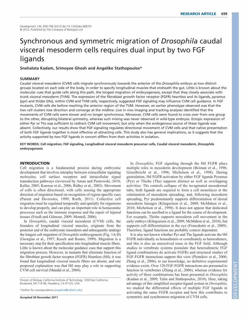
RESULTSCVM cell movement involves active migration ofcellsWe used a previously characterized croc-lacZ reporter gene tovisualize CVM cells throughout the course of their migration (seeFig. 1E-H compare with 1A-D) (Hacker et al., 1995). Twobilaterally symmetric clusters of cells form at stage 10 and migratetoward the anterior in a synchronous fashion (supplementarymaterial Fig. S1D-F). The extent of CVM cell migration correlatesprecisely with developmental stage (supplementary material Fig.S1A-C). At the end of the migration, CVM cells specifylongitudinal muscles that ensheath the gut. D
EVELO
PMENT
DEVELO
PMENT
As germ band retraction (GBR) occurs at stage 11 after CVMcells have initiated migration but before its completion(supplementary material Fig. S1F), we investigated whether anypart of CVM cell migration might passively reflect movement ofthese cells resulting from GBR. We assayed CVM migration withinu-shaped (ush) mutants, in which GBR does not occur (Goldman-Levi et al., 1996), and found that CVM cells continue to migratetoward the anterior (supplementary material Fig. S1G). This resultsuggested that active migration is likely to be required for cells toreach the anterior.
CVM migration is aberrant in the absence ofeither the Htl FGFR or Pyr and Ths FGF ligandsAs the CVM cells migrate from the posterior of the embryo to theanterior, each group of migrating CVM cells remains closelyassociated with one of the two bands of TVM tissue present oneither side of the embryonic body, especially from stage 11onwards (Fig. 2A-C, left side view). This observation suggested tous that the TVM might support guidance of CVM cell migration,and our previous study had identified that the FGF ligand thisbe isexpressed within the TVM (Stathopoulos et al., 2004).
We examined further the expression of FGF signalingcomponents, and found that genes encoding both ligands for Htl,ths and pyr, are expressed in the TVM (Fig. 1J-J�). Ligandexpression appears to be differentially regulated within the TVM,as pyr is only expressed early at stage 10 and 11, whereas thsexpression spans stages 10-13 (supplementary material Fig. S2).By contrast, the htl gene encoding the FGFR is expressed in CVM
701RESEARCH ARTICLEMigration regulated by two FGFs
cells (Fig. 1I) (Mandal et al., 2004) and we noticed its expressionis also temporally regulated, as expression is not present until afterCVM cells have initiated their migration (Fig. 1I; data not shown).
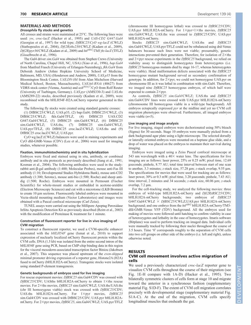
To investigate a role for this signaling pathway in regulatingCVM cell migration, we assayed mutants that affect FGF signalingfor effects on cell migration. Our results showed that CVMdevelopment is severely defective in the absence of FGF signaling(Fig. 2D-I). In the Df(2R)BSC25 mutant, in which the linked pyrand ths genes are removed (Stathopoulos et al., 2004), CVM cellsreach only half-way along their course when compared with theposition obtained by wild-type CVM cells (compare Fig. 2H). Thisphenotype was comparable with that observed in htl mutants (Fig.2E) (Mandal et al., 2004). Furthermore, the CVM cells in thesemutants lost their close association with the TVM and exhibited anaberrant cell shape, as cells lost their ellipsoidal characteristics andnuclei were small and circular. In either pyr or ths single mutantembryos [Df(2R)pyr36 and Df(2R)ths238, respectively (Kadam etal., 2009)], CVM phenotypes were also present but appeared lesssevere, with fewer cells exhibiting the altered morphologycharacteristic of both htl and Df(2R)BSC25 mutants (Fig. 2F,Icompare with 2E,H). Longitudinal muscles were partially formedin the single mutants, whereas they were completely absent in htlmutants (Mandal et al., 2004) or Df(2R)BSC25 mutants (data notshown). These results suggested that both ligands contribute toCVM cell migration.
Our next goal was to determine whether FGF signaling acts toinfluence CVM cells directly, possibly acting as a guidance factor,or instead might influence CVM cell migration indirectly through
Fig. 1. FGF ligands pyramus and thisbe are expressed in the TVM, and the FGF receptor heartless is expressed in migrating CVM cells.Embryos are oriented with anterior towards the left and dorsal upwards. (A-D)Schematic representation of visceral mesoderm and endodermdevelopment. (A)At stage 10, CVM cells (red) originate as a single group of cells arising from the posterior tip of the mesoderm anlage. (B)At stage11, CVM cells arrange themselves in two rows on either side of the midgut primordium and subsequently migrate anteriorly along the TVM.(C,D)At stage 13, the CVM cells reach their final destination along the TVM (C) and adopt the elongated morphology characteristic of longitudinalmuscle fibers (D). Adapted from an image kindly provided by R. Reuter (University of Tuebingen, Germany). (E-H)Anti-bgal antibody staining ofwild-type embryos containing the croc-lacZ reporter gene depicting the steps of migratory CVM cell development at stages equivalent toschematics in A-D. (I-J�) Embryos expressing croc.lacZ stained using riboprobes to htl, pyr, ths and/or lacZ by multiplex fluorescent in situhybridization. (I-I�) htl (green) and lacZ (red) colocalize within migrating CVM cells. (J-J�) ths (red) and pyr (blue) expression detected within the TVM,and lacZ (green) expression detected within migrating CVM cells.
DEVELO
PMENT
DEVELO
PMENT
702
effect on another tissue (e.g. specification of TVM). FGF signalingis active pervasively throughout development in a number ofdifferent cell types, and, in particular, controls mesoderm spreadingduring gastrulation required for TVM specification (Frasch, 1995;Staehling-Hampton et al., 1994).
Gaps were identified at random positions within the TVM in theFGF mutants. However, we found that CVM cells were able tomigrate past such gaps. At least at early stages in the migrationprocess, an intact TVM is not required to support migration (Fig.2D,G). Although anteriorly localized gaps sometimes correlated inposition with cessation of migration (e.g. Fig. 2E), this defect wasprobably not causative as migration ceases at this point in mutantsregardless (i.e. even when the TVM is intact; Fig. 2H).
Next, we specifically knocked down FGF signaling in the CVMand examined the effect on the migration of cells. To achieve this,we expressed a dominant-negative Htl receptor (UAS.DN-Htl) ordsRNA hairpin construct targeting htl transcripts (UAS.htl-RNAi)using the G447.Gal4 driver, which supports expression of UAS-containing transgenes in CVM cells as well as an adjacent relatedpopulation of cells, malphigian tubule (MT) precursors (Denholmet al., 2003; Georgias et al., 1997; Phelps and Brand, 1998). In bothcases, CVM cell migration was clearly aberrant, with cells failingto reach the anterior section of the TVM and cell number appearingreduced, possibly owing to increased cell death (supplementarymaterial Fig. S3). The phenotype was observed in 30-40% ofembryos; such partial penetrance using transgene-mediate ectopicexpression to reduce gene function is common (Dietzl et al., 2007).This result provided evidence that FGF signaling is required withinmigrating CVM cells directly, at least in part, to support theirmigration. Therefore, we subsequently conducted a directed set ofexperiments aimed at deciphering the role of FGF signaling withinthese cells.
FGF signaling supports CVM cell survivalAs we had observed that CVM cell numbers are decreased and thatcell nuclei appeared smaller and more circular from late stage 12onwards in FGF mutants, we investigated whether cell death resultedin the absence of signaling. Using a standard TUNEL assay (e.g.
RESEARCH ARTICLE Development 139 (4)
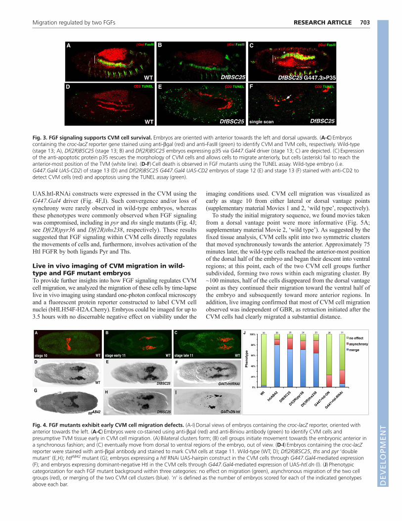
Reim et al., 2003), we found that CVM cells undergo increased celldeath in the Df(2R)BSC25 mutant embryos as TUNEL-positive cellsare identified by stage 13 (Fig. 3B,F) but not at earlier stages (Fig.3E). By contrast, little to no cell death is associated with CVM cellspresent in wild-type embryos at any stage (Fig. 3A,D).
To test the idea that FGF signaling supports cell survival duringCVM migration, we blocked CVM cell death in Df(2R)BSC25mutants by expressing the baculovirus anti-apoptotic p35 protein, apotent inhibitor of programmed cell death that inhibits Drosophilacaspases (Huh et al., 2004). p35 was expressed within CVM cells ofDf(2R)BSC25 mutants using the G447.Gal4 driver (Denholm et al.,2003; Georgias et al., 1997). Expression of p35 within CVM cells ofDf(2R)BSC25 mutant embryos kept cells alive. Specifically, cellnumber was similar to wild type, cell nuclei were of normal size andcells were once again closely associated with the TVM. However,CVM migration was not completely rescued in this background, forcells did not reach the anterior TVM position normally attainable bywild-type cells (Fig. 3C, compare with 3A). This result suggestedthat FGF signaling not only supports cell survival, as had beenproposed previously (Mandal et al., 2004), but also likely regulateseffective movement of CVM cells, allowing them to reach the mostanterior position of the TVM.
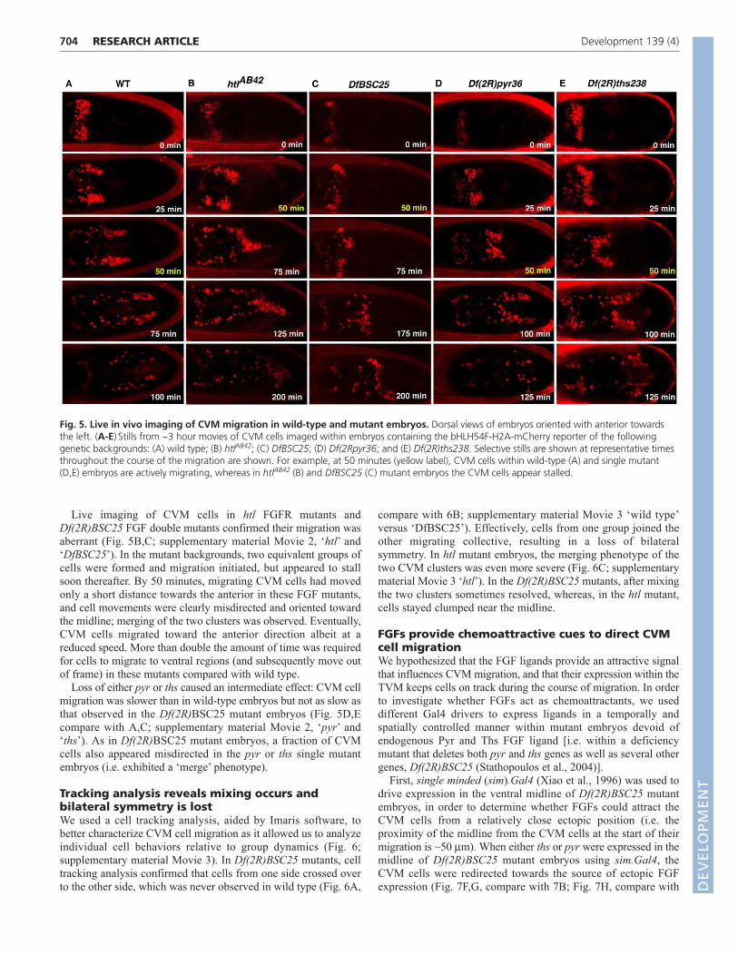
FGFs regulate migration of two bilaterallysymmetric clusters of CVM cells at early stagesIn wild-type embryos, CVM cells originate as a single cluster andsubsequently split into two bilaterally symmetric groups of ~30cells on either side of the dorsal midline (Fig. 4A). We inferredfrom the examination of fixed sample that these two cell groupsappeared to move in sync, with the leading cells of each grouptaking up equivalent positions along the length of the embryo ateach timepoint examined (Fig. 4B,C).
By contrast, htl and Df(2R)BSC25 mutants exhibited severeCVM cell migration defects affecting cell organization during theearly stage of the migration process (Fig. 4G,E,H compare with4D). Cells merged at the midline (‘merge’; Fig. 4E,G), and the twocell groups also moved out of sync (‘asynchrony’; Fig. 4H).Similar phenotypes were observed when either the UAS.DN-Htl or
Fig. 2. CVM cell migration is aberrant in FGF mutants. Embryos are oriented with anterior towards the left and dorsal upwards. (A-I)Embryosof the following genotypes containing croc-lacZ were stained using anti-bgal antibody (red) to mark CVM and anti-FasIII antibody (green) to markTVM: (A-C) wild type, (D,E) htlAB42 mutant embryos, (F) Df(2R)pyr36 (pyr single mutant), (G,H) Df(2R)BSC25 (deficiency chromosome removing pyrand ths) and (I) Df(2R)ths238 (ths single mutant). Embryos at stage 12 are depicted in A,D,G; embryos at stage 13 are depicted in B,C,E,F,H,I.
DEVELO
PMENT
DEVELO
PMENT
UAS.htl-RNAi constructs were expressed in the CVM using theG447.Gal4 driver (Fig. 4F,I). Such convergence and/or loss ofsynchrony were rarely observed in wild-type embryos, whereasthese phenotypes were commonly observed when FGF signalingwas compromised, including in pyr and ths single mutants (Fig. 4J;see Df(2R)pyr36 and Df(2R)ths238, respectively). These resultssuggested that FGF signaling within CVM cells directly regulatesthe movements of cells and, furthermore, involves activation of theHtl FGFR by both ligands Pyr and Ths.
Live in vivo imaging of CVM migration in wild-type and FGF mutant embryosTo provide further insights into how FGF signaling regulates CVMcell migration, we analyzed the migration of these cells by time-lapselive in vivo imaging using standard one-photon confocal microscopyand a fluorescent protein reporter constructed to label CVM cellnuclei (bHLH54F-H2A.Cherry). Embryos could be imaged for up to3.5 hours with no discernable negative effect on viability under the
703RESEARCH ARTICLEMigration regulated by two FGFs
imaging conditions used. CVM cell migration was visualized asearly as stage 10 from either lateral or dorsal vantage points(supplementary material Movies 1 and 2, ‘wild type’, respectively).
To study the initial migratory sequence, we found movies takenfrom a dorsal vantage point were more informative (Fig. 5A;supplementary material Movie 2, ‘wild type’). As suggested by thefixed tissue analysis, CVM cells split into two symmetric clustersthat moved synchronously towards the anterior. Approximately 75minutes later, the wild-type cells reached the anterior-most positionof the dorsal half of the embryo and began their descent into ventralregions; at this point, each of the two CVM cell groups furthersubdivided, forming two rows within each migrating cluster. By~100 minutes, half of the cells disappeared from the dorsal vantagepoint as they continued their migration toward the ventral half ofthe embryo and subsequently toward more anterior regions. Inaddition, live imaging confirmed that most of CVM cell migrationobserved was independent of GBR, as retraction initiated after theCVM cells had clearly migrated a substantial distance.
Fig. 3. FGF signaling supports CVM cell survival. Embryos are oriented with anterior towards the left and dorsal upwards. (A-C)Embryoscontaining the croc-lacZ reporter gene stained using anti-bgal (red) and anti-FasIII (green) to identify CVM and TVM cells, respectively. Wild-type(stage 13; A), Df(2R)BSC25 (stage 13; B) and Df(2R)BSC25 embryos expressing p35 via G447.Gal4 driver (stage 13; C) are depicted. (C)Expressionof the anti-apoptotic protein p35 rescues the morphology of CVM cells and allows cells to migrate anteriorly, but cells (asterisk) fail to reach theanterior-most position of the TVM (white line). (D-F)Cell death is observed in FGF mutants using the TUNEL assay. Wild-type embryo (i.e.G447.Gal4 UAS-CD2) of stage 13 (D) and Df(2R)BSC25 G447.Gal4 UAS-CD2 embryos of stage 12 (E) and stage 13 (F) stained with anti-CD2 todetect CVM cells (red) and apoptosis using the TUNEL assay (green).
Fig. 4. FGF mutants exhibit early CVM cell migration defects. (A-I)Dorsal views of embryos containing the croc-lacZ reporter, oriented withanterior towards the left. (A-C)Embryos were co-stained using anti-bgal (red) and anti-Biniou antibody (green) to identify CVM cells andpresumptive TVM tissue early in CVM cell migration. (A)Bilateral clusters form; (B) cell groups initiate movement towards the embryonic anterior ina synchronous fashion; and (C) eventually move from dorsal to ventral regions of the embryo, out of view. (D-I)Embryos containing the croc-lacZreporter were stained with anti-bgal antibody and stained to mark CVM cells at stage 11. Wild-type (WT; D); Df(2R)BSC25, ths and pyr ‘doublemutant’ (E,H); htlAB42 mutant (G); embryos expressing a htl RNAi UAS-hairpin construct in the CVM cells through G447.Gal4-mediated expression(F); and embryos expressing dominant-negative Htl in the CVM cells through G447.Gal4-mediated expression of UAS-htl.dn (I). (J)Phenotypiccategorization for each FGF mutant background within three categories: no effect on migration (green), asynchronous migration of the two cellgroups (red), or merging of the two CVM cell clusters (blue). ‘n’ is defined as the number of embryos scored for each of the indicated genotypesabove each bar. D
EVELO
PMENT
DEVELO
PMENT
704
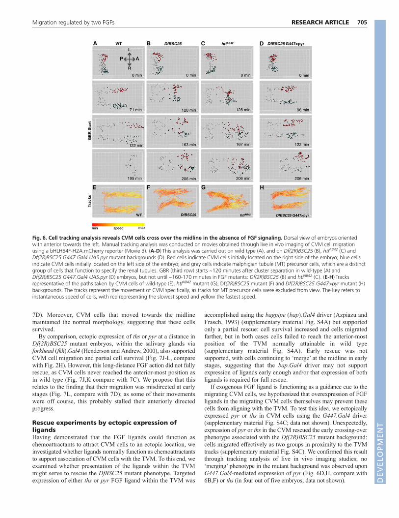
Live imaging of CVM cells in htl FGFR mutants andDf(2R)BSC25 FGF double mutants confirmed their migration wasaberrant (Fig. 5B,C; supplementary material Movie 2, ‘htl’ and‘DfBSC25’). In the mutant backgrounds, two equivalent groups ofcells were formed and migration initiated, but appeared to stallsoon thereafter. By 50 minutes, migrating CVM cells had movedonly a short distance towards the anterior in these FGF mutants,and cell movements were clearly misdirected and oriented towardthe midline; merging of the two clusters was observed. Eventually,CVM cells migrated toward the anterior direction albeit at areduced speed. More than double the amount of time was requiredfor cells to migrate to ventral regions (and subsequently move outof frame) in these mutants compared with wild type.
Loss of either pyr or ths caused an intermediate effect: CVM cellmigration was slower than in wild-type embryos but not as slow asthat observed in the Df(2R)BSC25 mutant embryos (Fig. 5D,Ecompare with A,C; supplementary material Movie 2, ‘pyr’ and‘ths’). As in Df(2R)BSC25 mutant embryos, a fraction of CVMcells also appeared misdirected in the pyr or ths single mutantembryos (i.e. exhibited a ‘merge’ phenotype).
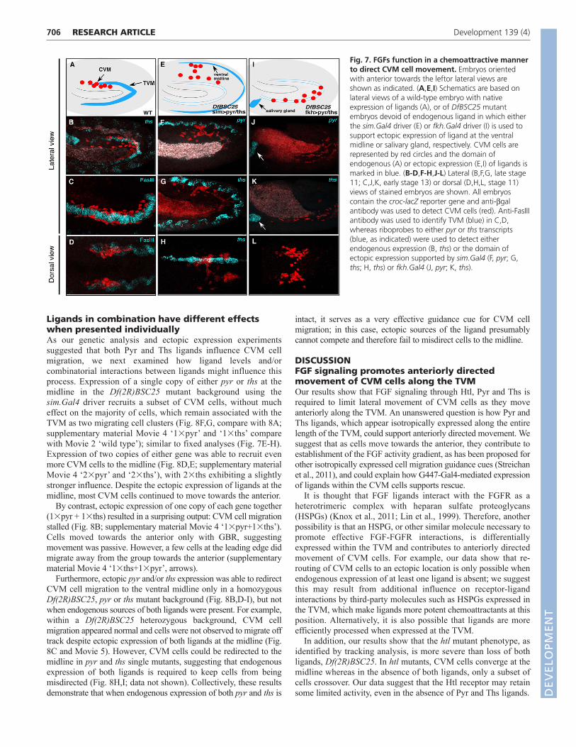
Tracking analysis reveals mixing occurs andbilateral symmetry is lostWe used a cell tracking analysis, aided by Imaris software, tobetter characterize CVM cell migration as it allowed us to analyzeindividual cell behaviors relative to group dynamics (Fig. 6;supplementary material Movie 3). In Df(2R)BSC25 mutants, celltracking analysis confirmed that cells from one side crossed overto the other side, which was never observed in wild type (Fig. 6A,
RESEARCH ARTICLE Development 139 (4)
compare with 6B; supplementary material Movie 3 ‘wild type’versus ‘DfBSC25’). Effectively, cells from one group joined theother migrating collective, resulting in a loss of bilateralsymmetry. In htl mutant embryos, the merging phenotype of thetwo CVM clusters was even more severe (Fig. 6C; supplementarymaterial Movie 3 ‘htl’). In the Df(2R)BSC25 mutants, after mixingthe two clusters sometimes resolved, whereas, in the htl mutant,cells stayed clumped near the midline.
FGFs provide chemoattractive cues to direct CVMcell migrationWe hypothesized that the FGF ligands provide an attractive signalthat influences CVM migration, and that their expression within theTVM keeps cells on track during the course of migration. In orderto investigate whether FGFs act as chemoattractants, we useddifferent Gal4 drivers to express ligands in a temporally andspatially controlled manner within mutant embryos devoid ofendogenous Pyr and Ths FGF ligand [i.e. within a deficiencymutant that deletes both pyr and ths genes as well as several othergenes, Df(2R)BSC25 (Stathopoulos et al., 2004)].
First, single minded (sim).Gal4 (Xiao et al., 1996) was used todrive expression in the ventral midline of Df(2R)BSC25 mutantembryos, in order to determine whether FGFs could attract theCVM cells from a relatively close ectopic position (i.e. theproximity of the midline from the CVM cells at the start of theirmigration is ~50 mm). When either ths or pyr were expressed in themidline of Df(2R)BSC25 mutant embryos using sim.Gal4, theCVM cells were redirected towards the source of ectopic FGFexpression (Fig. 7F,G, compare with 7B; Fig. 7H, compare with
Fig. 5. Live in vivo imaging of CVM migration in wild-type and mutant embryos. Dorsal views of embryos oriented with anterior towardsthe left. (A-E)Stills from ~3 hour movies of CVM cells imaged within embryos containing the bHLH54F-H2A-mCherry reporter of the followinggenetic backgrounds: (A) wild type; (B) htlAB42; (C) DfBSC25; (D) Df(2Rpyr36; and (E) Df(2R)ths238. Selective stills are shown at representative timesthroughout the course of the migration are shown. For example, at 50 minutes (yellow label), CVM cells within wild-type (A) and single mutant(D,E) embryos are actively migrating, whereas in htlAB42 (B) and DfBSC25 (C) mutant embryos the CVM cells appear stalled.
DEVELO
PMENT
DEVELO
PMENT
7D). Moreover, CVM cells that moved towards the midlinemaintained the normal morphology, suggesting that these cellssurvived.
By comparison, ectopic expression of ths or pyr at a distance inDf(2R)BSC25 mutant embryos, within the salivary glands viaforkhead (fkh).Gal4 (Henderson and Andrew, 2000), also supportedCVM cell migration and partial cell survival (Fig. 7J-L, comparewith Fig. 2H). However, this long-distance FGF action did not fullyrescue, as CVM cells never reached the anterior-most position asin wild type (Fig. 7J,K compare with 7C). We propose that thisrelates to the finding that their migration was misdirected at earlystages (Fig. 7L, compare with 7D); as some of their movementswere off course, this probably stalled their anteriorly directedprogress.
Rescue experiments by ectopic expression ofligandsHaving demonstrated that the FGF ligands could function aschemoattractants to attract CVM cells to an ectopic location, weinvestigated whether ligands normally function as chemoattractantsto support association of CVM cells with the TVM. To this end, weexamined whether presentation of the ligands within the TVMmight serve to rescue the DfBSC25 mutant phenotype. Targetedexpression of either ths or pyr FGF ligand within the TVM was
705RESEARCH ARTICLEMigration regulated by two FGFs
accomplished using the bagpipe (bap).Gal4 driver (Azpiazu andFrasch, 1993) (supplementary material Fig. S4A) but supportedonly a partial rescue: cell survival increased and cells migratedfarther, but in both cases cells failed to reach the anterior-mostposition of the TVM normally attainable in wild type(supplementary material Fig. S4A). Early rescue was notsupported, with cells continuing to ‘merge’ at the midline in earlystages, suggesting that the bap.Gal4 driver may not supportexpression of ligands early enough and/or that expression of bothligands is required for full rescue.
If exogenous FGF ligand is functioning as a guidance cue to themigrating CVM cells, we hypothesized that overexpression of FGFligands in the migrating CVM cells themselves may prevent thesecells from aligning with the TVM. To test this idea, we ectopicallyexpressed pyr or ths in CVM cells using the G447.Gal4 driver(supplementary material Fig. S4C; data not shown). Unexpectedly,expression of pyr or ths in the CVM rescued the early crossing-overphenotype associated with the Df(2R)BSC25 mutant background:cells migrated effectively as two groups in proximity to the TVMtracks (supplementary material Fig. S4C). We confirmed this resultthrough tracking analysis of live in vivo imaging studies; no‘merging’ phenotype in the mutant background was observed uponG447.Gal4-mediated expression of pyr (Fig. 6D,H, compare with6B,F) or ths (in four out of five embryos; data not shown).
Fig. 6. Cell tracking analysis reveals CVM cells cross over the midline in the absence of FGF signaling. Dorsal view of embryos orientedwith anterior towards the left. Manual tracking analysis was conducted on movies obtained through live in vivo imaging of CVM cell migrationusing a bHLH54F-H2A.mCherry reporter (Movie 3). (A-D)This analysis was carried out on wild type (A), and on Df(2R)BSC25 (B), htlAB42 (C) andDf(2R)BSC25 G447.Gal4 UAS.pyr mutant backgrounds (D). Red cells indicate CVM cells initially located on the right side of the embryo; blue cellsindicate CVM cells initially located on the left side of the embryo; and gray cells indicate malphigian tubule (MT) precursor cells, which are a distinctgroup of cells that function to specify the renal tubules. GBR (third row) starts ~120 minutes after cluster separation in wild-type (A) andDf(2R)BSC25 G447.Gal4 UAS.pyr (D) embryos, but not until ~160-170 minutes in FGF mutants: Df(2R)BSC25 (B) and htlAB42 (C). (E-H)Tracksrepresentative of the paths taken by CVM cells of wild-type (E), htlAB42 mutant (G), Df(2R)BSC25 mutant (F) and Df(2R)BSC25 G447>pyr mutant (H)backgrounds. The tracks represent the movement of CVM specifically, as tracks for MT precursor cells were excluded from view. The key refers toinstantaneous speed of cells, with red representing the slowest speed and yellow the fastest speed.
DEVELO
PMENT
DEVELO
PMENT
706
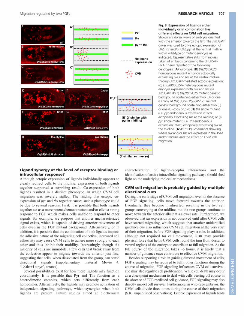
Ligands in combination have different effectswhen presented individuallyAs our genetic analysis and ectopic expression experimentssuggested that both Pyr and Ths ligands influence CVM cellmigration, we next examined how ligand levels and/orcombinatorial interactions between ligands might influence thisprocess. Expression of a single copy of either pyr or ths at themidline in the Df(2R)BSC25 mutant background using thesim.Gal4 driver recruits a subset of CVM cells, without mucheffect on the majority of cells, which remain associated with theTVM as two migrating cell clusters (Fig. 8F,G, compare with 8A;supplementary material Movie 4 ‘1�pyr’ and ‘1�ths’ comparewith Movie 2 ‘wild type’); similar to fixed analyses (Fig. 7E-H).Expression of two copies of either gene was able to recruit evenmore CVM cells to the midline (Fig. 8D,E; supplementary materialMovie 4 ‘2�pyr’ and ‘2�ths’), with 2�ths exhibiting a slightlystronger influence. Despite the ectopic expression of ligands at themidline, most CVM cells continued to move towards the anterior.
By contrast, ectopic expression of one copy of each gene together(1�pyr + 1�ths) resulted in a surprising output: CVM cell migrationstalled (Fig. 8B; supplementary material Movie 4 ‘1�pyr+1�ths’).Cells moved towards the anterior only with GBR, suggestingmovement was passive. However, a few cells at the leading edge didmigrate away from the group towards the anterior (supplementarymaterial Movie 4 ‘1�ths+1�pyr’, arrows).
Furthermore, ectopic pyr and/or ths expression was able to redirectCVM cell migration to the ventral midline only in a homozygousDf(2R)BSC25, pyr or ths mutant background (Fig. 8B,D-I), but notwhen endogenous sources of both ligands were present. For example,within a Df(2R)BSC25 heterozygous background, CVM cellmigration appeared normal and cells were not observed to migrate offtrack despite ectopic expression of both ligands at the midline (Fig.8C and Movie 5). However, CVM cells could be redirected to themidline in pyr and ths single mutants, suggesting that endogenousexpression of both ligands is required to keep cells from beingmisdirected (Fig. 8H,I; data not shown). Collectively, these resultsdemonstrate that when endogenous expression of both pyr and ths is
RESEARCH ARTICLE Development 139 (4)
intact, it serves as a very effective guidance cue for CVM cellmigration; in this case, ectopic sources of the ligand presumablycannot compete and therefore fail to misdirect cells to the midline.
DISCUSSIONFGF signaling promotes anteriorly directedmovement of CVM cells along the TVMOur results show that FGF signaling through Htl, Pyr and Ths isrequired to limit lateral movement of CVM cells as they moveanteriorly along the TVM. An unanswered question is how Pyr andThs ligands, which appear isotropically expressed along the entirelength of the TVM, could support anteriorly directed movement. Wesuggest that as cells move towards the anterior, they contribute toestablishment of the FGF activity gradient, as has been proposed forother isotropically expressed cell migration guidance cues (Streichanet al., 2011), and could explain how G447-Gal4-mediated expressionof ligands within the CVM cells supports rescue.
It is thought that FGF ligands interact with the FGFR as aheterotrimeric complex with heparan sulfate proteoglycans(HSPGs) (Knox et al., 2011; Lin et al., 1999). Therefore, anotherpossibility is that an HSPG, or other similar molecule necessary topromote effective FGF-FGFR interactions, is differentiallyexpressed within the TVM and contributes to anteriorly directedmovement of CVM cells. For example, our data show that re-routing of CVM cells to an ectopic location is only possible whenendogenous expression of at least one ligand is absent; we suggestthis may result from additional influence on receptor-ligandinteractions by third-party molecules such as HSPGs expressed inthe TVM, which make ligands more potent chemoattractants at thisposition. Alternatively, it is also possible that ligands are moreefficiently processed when expressed at the TVM.
In addition, our results show that the htl mutant phenotype, asidentified by tracking analysis, is more severe than loss of bothligands, Df(2R)BSC25. In htl mutants, CVM cells converge at themidline whereas in the absence of both ligands, only a subset ofcells crossover. Our data suggest that the Htl receptor may retainsome limited activity, even in the absence of Pyr and Ths ligands.
Fig. 7. FGFs function in a chemoattractive mannerto direct CVM cell movement. Embryos orientedwith anterior towards the leftor lateral views areshown as indicated. (A,E,I) Schematics are based onlateral views of a wild-type embryo with nativeexpression of ligands (A), or of DfBSC25 mutantembryos devoid of endogenous ligand in which eitherthe sim.Gal4 driver (E) or fkh.Gal4 driver (I) is used tosupport ectopic expression of ligand at the ventralmidline or salivary gland, respectively. CVM cells arerepresented by red circles and the domain ofendogenous (A) or ectopic expression (E,I) of ligands ismarked in blue. (B-D,F-H,J-L) Lateral (B,F,G, late stage11; C,J,K, early stage 13) or dorsal (D,H,L, stage 11)views of stained embryos are shown. All embryoscontain the croc-lacZ reporter gene and anti-bgalantibody was used to detect CVM cells (red). Anti-FasIIIantibody was used to identify TVM (blue) in C,D,whereas riboprobes to either pyr or ths transcripts(blue, as indicated) were used to detect eitherendogenous expression (B, ths) or the domain ofectopic expression supported by sim.Gal4 (F, pyr; G,ths; H, ths) or fkh.Gal4 (J, pyr; K, ths).
DEVELO
PMENT
DEVELO
PMENT
Ligand synergy at the level of receptor binding orintracellular response?Although ectopic expression of ligands individually appears toclearly redirect cells to the midline, expression of both ligandstogether supported a surprising result. Co-expression of bothligands resulted in a distinct phenotype, in which CVM cellmigration was severely stalled. The finding that ectopic co-expression of pyr and ths together causes such a phenotype couldbe due to several reasons. First, it is possible that both ligandstogether act as a more potent chemoattractant and/or elicit a strongresponse to FGF, which makes cells unable to respond to othersignals; for example, we propose that another uncharacterizedsignal exists, which is capable of driving anterior movement ofcells even in the FGF mutant background. Alternatively, or inaddition, it is possible that the combination of both ligands impactsthe adhesive nature of the migrating cell collective; increased celladhesivity may cause CVM cells to adhere more strongly to eachother and thus inhibit their mobility. Interestingly, though themajority of cells are immobile, a few cells that break away fromthe collective appear to migrate towards the anterior just fine,suggesting that cells, when dissociated from the group, can sensedirectional signals (supplementary material Movie 4,‘1�ths+1�pyr’, arrows).
Several possibilities exist for how these ligands may functioncoordinately. It is possible that Pyr and Ths function as aheterodimeric complex, which acts differently from eachhomodimer. Alternatively, the ligands may promote activation ofindependent signaling pathways, which synergize when bothligands are present. Future studies aimed at biochemical
707RESEARCH ARTICLEMigration regulated by two FGFs
characterization of ligand-receptor interactions and theidentification of active intracellular signaling pathways should shedlight on the underlying molecular mechanisms.
CVM cell migration is probably guided by multipledirectional cuesDuring the early stage of CVM cell migration, even in the absenceof FGF signaling, cells move forward towards the anterior.Eventually, they become misdirected, resulting in the two cellgroups converging at the midline, but the two groups continue tomove towards the anterior albeit at a slower rate. Furthermore, weobserved that htl expression is not observed until after CVM cellshave started migrating, which suggests that another unidentifiedguidance cue also influences CVM cell migration at the very startof their migration, before FGF signaling plays a role. In addition,although not required for cell movement, GBR may providephysical force that helps CVM cells round the turn from dorsal toventral regions of the embryo to contribute to full migration. As thefull course of the migration takes ~6 hours, it is likely that anumber of guidance cues contribute to effective CVM migration.
Besides supporting a role in guiding directed movement of cells,FGF signaling may be required to fulfil other functions during thecourse of migration. FGF signaling influences CVM cell survival,and may also regulate cell proliferation. While cell death may occuras a checkpoint mechanism to deal with cells veering off course inthe absence of FGF-mediated cell guidance, FGF signaling may alsodirectly impact cell survival. Furthermore, in wild-type embryos, theCVM cells divide three times during the course of their migration(S.K., unpublished observations). Ectopic expression of ligands leads
Fig. 8. Expression of ligands eitherindividually or in combination hasdifferent effects on CVM cell migration.Shown are dorsal views of embryos orientedwith the anterior towards the left. The sim.Gal4driver was used to drive ectopic expression ofUAS.ths and/or UAS.pyr at the ventral midlinewithin wild-type or mutant embryos asindicated. Representative stills from moviestaken of embryos containing the bHLH54F-H2A.Cherry reporter of the followinggenotypes: (A) wild-type; (B) Df(2R)BSC25homozygous mutant embryos ectopicallyexpressing pyr and ths at the ventral midlinethrough sim.Gal4-mediated ectopic expression;(C) Df(2R)BSC25/+ heterozygous mutantembryos expressing both pyr and ths viasim.Gal4; (D,F) Df(2R)BSC25 mutant geneticbackground containing either two (D) or one(F) copy of ths; (E,G) Df(2R)BSC25 mutantgenetic background containing either two (E)or one (G) copy of pyr; (H) ths single mutant(i.e. pyr endogenous expression intact)ectopically expressing ths at the midline; or (I)pyr single mutant (i.e. ths endogenousexpression intact) ectopically expressing pyr atthe midline. (A�-D�,�,H�) Schematics showingwhere pyr and/or ths are expressed in the TVMand/or midline and the effect on CVM cellmigration.
DEVELO
PMENT
DEVELO
PMENT
708 RESEARCH ARTICLE Development 139 (4)
to an increase in the number of cells present in dorsal regions, butwhether this relates to an increase in proliferation or occurs becausethe movement of cells is slowed remains to be determined. Futurestudies aimed at analysis of the full course of the migration willprovide additional answers and, although these experiments are notyet technically possible, advances in microscopy make this apromising avenue for future research.
AcknowledgementsWe thank Leslie Dunipace, Alphan Altinok and Sarah Wadsworth for excellenttechnical assistance; Manfred Frasch and Mayra Garcia for helpful discussions;and Marianne Bronner and members of the Stathopoulos laboratory, especiallyYoung Bae, for comments on the manuscript. In addition, we are grateful toRolf Reuter for sharing fly strains, providing the schematic shown in Fig. 1 andsupporting S.K.’s preliminary studies of CVM cell migration (parts ofsupplementary material Fig. S1 and Fig. S2).
FundingThis work was funded by a grant from the National Institute of GeneralMedical Sciences (NIGMS) [R01GM078542 to A.S.]. Deposited in PMC forrelease after 12 months.
Competing interests statementThe authors declare no competing financial interests.
Author contributionsS.K. and A.S. designed the experiments; S.K. conducted all the experimentsexcept the manual tracking in Fig. 6, which was carried out by S.G.; S.K. andA.S. analyzed the data and wrote the manuscript.
Supplementary materialSupplementary material available online athttp://dev.biologists.org/lookup/suppl/doi:10.1242/dev.068791/-/DC1
ReferencesAman, A. and Piotrowski, T. (2010). Cell migration during morphogenesis. Dev.
Biol. 341, 20-33.Azpiazu, N. and Frasch, M. (1993). tinman and bagpipe: two homeo box genes
that determine cell fates in the dorsal mesoderm of Drosophila. Genes Dev. 7,1325-1340.
Beiman, M., Shilo, B. Z. and Volk, T. (1996). Heartless, a Drosophila FGF receptorhomolog, is essential for cell migration and establishment of several mesodermallineages. Genes Dev. 10, 2993-3002.
Denholm, B., Sudarsan, V., Pasalodos-Sanchez, S., Artero, R., Lawrence, P.,Maddrell, S., Baylies, M. and Skaer, H. (2003). Dual origin of the renal tubulesin Drosophila: mesodermal cells integrate and polarize to establish secretoryfunction. Curr. Biol. 13, 1052-1057.
Dietzl, G., Chen, D., Schnorrer, F., Su, K. C., Barinova, Y., Fellner, M., Gasser, B.,Kinsey, K., Oppel, S., Scheiblauer, S. et al. (2007). A genome-wide transgenicRNAi library for conditional gene inactivation in Drosophila. Nature 448, 151-156.
Franzdottir, S. R., Engelen, D., Yuva-Aydemir, Y., Schmidt, I., Aho, A. andKlambt, C. (2009). Switch in FGF signalling initiates glial differentiation in theDrosophila eye. Nature 460, 758-761.
Frasch, M. (1995). Induction of visceral and cardiac mesoderm by ectodermal Dpp inthe early Drosophila embryo. Nature 374, 464-467.
Friedl, P. and Gilmour, D. (2009). Collective cell migration in morphogenesis,regeneration and cancer. Nat. Rev. Mol. Cell. Biol. 10, 445-457.
Georgias, C., Wasser, M. and Hinz, U. (1997). A basic-helix-loop-helix proteinexpressed in precursors of Drosophila longitudinal visceral muscles. Mech. Dev. 69,115-124.
Gisselbrecht, S., Skeath, J. B., Doe, C. Q. and Michelson, A. M. (1996). heartlessencodes a fibroblast growth factor receptor (DFR1/DFGF-R2) involved in thedirectional migration of early mesodermal cells in the Drosophila embryo. GenesDev. 10, 3003-3017.
Goldman-Levi, R., Miller, C., Greenberg, G., Gabai, E. and Zak, N. B. (1996).Cellular pathways acting along the germband and in the amnioserosa mayparticipate in germband retraction of the Drosophila melanogaster embryo. Int. J.Dev. Biol. 40, 1043-1051.
Hacker, U., Kaufmann, E., Hartmann, C., Jurgens, G., Knochel, W. and Jackle,H. (1995). The Drosophila fork head domain protein crocodile is required for theestablishment of head structures. EMBO J. 14, 5306-5317.
Henderson, K. D. and Andrew, D. J. (2000). Regulation and function of Scr, exd,and hth in the Drosophila salivary gland. Dev. Biol. 362-374.
Huh, J. R., Guo, M. and Hay, B. A. (2004). Compensatory proliferation induced bycell death in the Drosophila wing disc requires activity of the apical cell deathcaspase Dronc in a nonapoptotic role. Curr. Biol. 14, 1262-1266.
Ismat, A., Schaub, C., Reim, I., Kirchner, K., Schultheis, D. and Frasch, M.(2010). HLH54F is required for the specification and migration of longitudinalgut muscle founders from the caudal mesoderm of Drosophila. Development137, 3107-3117.
Jakobsen, J. S., Braun, M., Astorga, J., Gustafson, E. H., Sandmann, T.,Karzynski, M., Carlsson, P. and Furlong, E. E. (2007). Temporal ChIP-on-chipreveals Biniou as a universal regulator of the visceral muscle transcriptionalnetwork. Genes Dev. 21, 2448-2460.
Jiang, J., Kosman, D., Ip, Y. T. and Levine, M. (1991). The dorsal morphogengradient regulates the mesoderm determinant twist in early Drosophila embryos.Genes Dev. 5, 1881-1891.
Kadam, S., McMahon, A., Tzou, P. and Stathopoulos, A. (2009). FGF ligands inDrosophila have distinct activities required to support cell migration anddifferentiation. Development 136, 739-747.
Keller, R. (2005). Cell migration during gastrulation. Curr. Opin. Cell Biol. 17, 533-541.
Klingseisen, A., Clark, I. B., Gryzik, T. and Muller, H. A. (2009). Differential andoverlapping functions of two closely related Drosophila FGF8-like growth factors inmesoderm development. Development 136, 2393-2402.
Knox, J., Moyer, K., Yacoub, N., Soldaat, C., Komosa, M., Vassilieva, K., Wilk,R., Hu, J., Vasquez Pas, L. D., Syed, Q. et al. (2011). Syndecan contributes toheart cell specification and lumen formation during Drosophila cardiogenesis. Dev.Biol. 356, 279-290.
Kosman, D., Mizutani, C. M., Lemons, D., Cox, W. G., McGinnis, W. and Bier, E.(2004). Multiplex detection of RNA expression in Drosophila embryos. Science305, 846.
Kunwar, P. S., Siekhaus, D. E. and Lehmann, R. (2006). In vivo migration: a germcell perspective. Annu. Rev. Cell Dev. Biol. 22, 237-265.
Kusch, T. and Reuter, R. (1999). Functions for Drosophila brachyenteron andforkhead in mesoderm specification and cell signalling. Development 126, 3991-4003.
Le T., Liang, Z., Patel, H., Yu, M. H., Sivasubramaniam, G., Slovitt, M.,Tanentzapf, G., Mohanty, N., Paul, S. M., Wu, V. M. and Beitel, G. J.(2006). A new family of Drosophila balancer chromosomes with a w- dfd-GMRyellow fluorescent protein marker. Genetics 174, 2255-2257.
Lin, X., Buff, E. M., Perrimon, N. and Michelson, A. M. (1999). Heparan sulfateproteoglycans are essential for FGF receptor signaling during Drosophila embryonicdevelopment. Development 126, 3715-3723.
Mandal, L., Dumstrei, K. and Hartenstein, V. (2004). Role of FGFR signaling in themorphogenesis of the Drosophila visceral musculature. Dev. Dyn. 231, 342-348.
McMahon, A., Reeves, G. T., Supatto, W. and Stathopoulos, A. (2010).Mesoderm migration in Drosophila is a multi-step process requiring FGF signalingand integrin activity. Development 137, 2167-2175.
Michelson, A. M., Gisselbrecht, S., Zhou, Y., Baek, K. H. and Buff, E. M. (1998).Dual functions of the heartless fibroblast growth factor receptor in development ofthe Drosophila embryonic mesoderm. Dev. Genet. 22, 212-229.
Montell, D. J. (2006). The social lives of migrating cells in Drosophila. Curr. Opin.Genet. Dev. 16, 374-383.
Parent, C. A. and Devreotes, P. N. (1999). A cell’s sense of direction. Science 284,765-770.
Phelps, C. B. and Brand, A. H. (1998). Ectopic gene expression in Drosophila usingGAL4 system. Methods 14, 367-379.
Plotnikov, A. N., Hubbard, S. R., Schlessinger, J. and Mohammadi, M. (2000).Crystal structures of two FGF-FGFR complexes reveal the determinants of ligand-receptor specificity. Cell 101, 413-424.
Reim, I., Lee, H. H. and Frasch, M. (2003). The T-box-encoding Dorsocross genesfunction in amnioserosa development and the patterning of the dorsolateral germband downstream of Dpp. Development 130, 3187-3204.
Ridley, A. J., Schwartz, M. A., Burridge, K., Firtel, R. A., Ginsberg, M. H.,Borisy, G., Parsons, J. T. and Horwitz, A. R. (2003). Cell migration: integratingsignals from front to back. Science 302, 1704-1709.
Rorth, P. (2011). Whence directionality: guidance mechanisms in solitary andcollective cell migration. Dev. Cell 20, 9-18.
Staehling-Hampton, K., Hoffmann, F. M., Baylies, M. K., Rushton, E. and Bate,M. (1994). dpp induces mesodermal gene expression in Drosophila. Nature 372,783-786.
Stathopoulos, A., Tam, B., Ronshaugen, M., Frasch, M. and Levine, M. (2004).pyramus and thisbe: FGF genes that pattern the mesoderm of Drosophilaembryos. Genes Dev. 18, 687-699.
Streichan, S. J., Valentin, G., Gilmour, D. and Hufnagel, L. (2011). Collective cellmigration guided by dynamically maintained gradients. Phys. Biol. 8, 045004.
Tulin, S. and Stathopoulos, A. (2010). Extending the family table: Insights frombeyond vertebrates into the regulation of embryonic development by FGFs. BirthDefects Res. C Embryo Today 90, 214-227.
Xiao, H., Hrdlicka, L. A. and Nambu, J. R. (1996). Alternate functions of thesingle-minded and rhomboid genes in development of the Drosophila ventralneuroectoderm. Mech. Dev. 58, 65-74.
Zhang, X., Ibrahimi, O. A., Olsen, S. K., Umemori, H., Mohammadi, M. andOrnitz, D. M. (2006). Receptor specificity of the fibroblast growth factor family.The complete mammalian FGF family. J. Biol. Chem. 281, 15694-15700. D
EVELO
PMENT
DEVELO
PMENT


