Embed Size (px)
Citation preview
Synthesis and characterization of up-conversion emission on
lanthanides doped ZrO2 nanocrystals coated with SiO2 for
biological applications. Tzarara López-Lukea, Elder de la Rosaa, Ana Lilia González-Yebrab, Beatriz González-
Yebrac, Carlos Ángeles-Chávezd, David Solísa, Pedro Salase, Carlos Saldañaf and
Octavio Mezaa aCentro de Investigaciones en Óptica, A. P. 1-948, León, Gto., 37160, México
bDepartamento de Ciencias Aplicadas al Trabajo Universidad de Guanajuato, Ave.
Eugenio Garza Sada #572, Lomas del Campestre Secc. II, León, Gto., 37150, México cDepartamento de Medicina, Universidad de Guanajuato, 20 de enero #929, Centro,
León, Gto., 37320, México dInstituto Mexicano del Petróleo, Programa de Ingeniería Molecular, A.P.11-848,
México, D.F. 07730, México eCentro de Física Aplicada y Tecnología Avanzada, Universidad Nacional Autónoma de
México, A.P. 1-1010, Querétaro, Qro. 76000, México. f Departamento de biomedicina, Facultad de medicina, Universidad Autonoma de Querétaro, QRO, México, A. P. 76176.
Correspondig author: [email protected] Abstract Er doped and Yb-Er-Tm codoped ZrO2 nanocrystals of average 80 nm in size were prepared by a sol-gel
process with the presence of nonionic (PLURONIC F-127) surfactant, and the up-conversion emission
was characterized under IR (980 nm) excitation. The effect of the codoped conditions on the crystalline
structure and photoluminescence properties were studied. A strong green emission was produced with 5
mol %, 0.2 mol %, 0.01 mol % of Yb3+-Er3+-Tm3+ codoped ZrO2 respectively. It was prepared Er doped
ZrO2 –SiO2 core-shell and SiO2 coated Er doped ZrO2 in 2-propanol and water, respectively. The presence
of the silica shell of average of 15 nm in thickness has been confirmed by transmition electron
microscopy. Photolumineiscence studies show that the silica shell does not affect the emission when the
nanoparticles are excited with 980 nm. The up-converting Yb3+-Er3+-Tm3+ codoped ZrO2 nanocrystal has
showed to be a powerful tool to future detection techniques. The viability of the nanoparticles of codoped
ZrO2 for biological imaging was confirmed by multiphotonic microscope imaging of cervix tissue with
(a)
Reporters, Markers, Dyes, Nanoparticles, and Molecular Probes for Biomedical Applications II,edited by Samuel Achilefu, Ramesh Raghavachari, Proc. of SPIE Vol. 7576, 75760T
© 2010 SPIE · CCC code: 1605-7422/10/$18 · doi: 10.1117/12.842993
Proc. of SPIE Vol. 7576 75760T-1
Downloaded From: http://proceedings.spiedigitallibrary.org/ on 11/27/2013 Terms of Use: http://spiedl.org/terms
inserted codoped ZrO2 nanoparticles. The cervix tissue has a moderate dysplasia. The nanoparticles were
introduced at 80 % of the tissue depth (5 μm) without being functionalized.
Keywords: up-conversion, nanocrystals, core-shell, coating and photoluminescence
1. INTRODUCTION
Rare-earth-doped nanophosphors emitting in the visible range by the upconversion process (UPC) have
recently received special attention given their great potential in human diagnostic applications [1].
Upconversion is the generation of visible or UV light by excitation with larger wavelengths, usually near
infrared (NIR), of trivalent rare-earth ions supported into a solid-state host. Several features separate UPC
from other fluorescent techniques as markers. First, infrared (IR) up-conversion is a unique process,
which does not occur in nature. The inherent autofluorescence associated with most fluorescence-based
methods is completely absent in up-converting phosphor assays. Second, the demand for extended
product stability supports UPC reporters as a superior choice because they do not bleach or fade, they can
be stored indefinitely without a decrease in light emitting efficiency and thus allow repetitive re-analysis.
Zirconium dioxide (ZrO2) has a low phonon energy of about 470 cm-1, which is very small
compared to that of other hosts such as YAG (850cm-1) or Y2O3 (597cm-1) [2,3]. This low phonon energy
opens up the possibility of higher efficient luminescence of active ions incorporated into the host [4].
Thus, combining the properties of rare-earth ions, nanosize effect and good qualities of ZrO2, the study of
luminescence properties in zirconium oxide nanocrystals is worthy of attention. Co-doping rare-earth ions
with Yb increases the efficiency of the UPC process and in some cases can induce upconversion whereas
it is not possible in the single ion. In this case, the emission is the result of energy transfer from donors to
acceptors such as Tm, Ho and Er [5,6].
The introduction of an inert crystalline shell of an undoped material around each doped
nanocrystal provides an effective way to improve the UPC efficiency of lanthanide-doped nanocrystals
[6]. Amorphous silica shells have been shown to provide some prime advantages necessary for
bioconjugation of phosphors, semiconductors and metals. First, the silica shell prevents flocculation of
particles, prevents species from adsorbing onto the surface, helps to maintain the photoluminescence and
often helps to increase luminescence [7]. Second, the silica shell also provides a potential platform for
attaching biological macromolecules for various biomedical applications. It is possible to decorate of the
silica shell with any number of functional groups including thiol, amine, phosphate, carboxylate, and
poly(ethylene glycol) (PEG) groups allow for greater control in conjugation protocols.
Proc. of SPIE Vol. 7576 75760T-2
Downloaded From: http://proceedings.spiedigitallibrary.org/ on 11/27/2013 Terms of Use: http://spiedl.org/terms
2. EXPERIMENTAL SECTION.
2.1 Sample preparation.
Regarding the nanoparticles of lantanides doped ZrO2 by sol-gel process, all chemicals used were of
reactant grade and were supplied by Aldrich, Inc. Nanoparticles of Er doped ZrO2 with a molar
composition of 0.2 mol % Er2O3 and Yb-Er-Tm codoped ZrO2 with a molar composition of 5 mol % of
Y2O3, 0.2 mol % of Er2O3 and 0.01 mol % of Tm2O3 respectively, were obtained by mixing zirconium n-
propoxide, ytterbium chloride, erbium nitrate and thulium chloride. Care was taken in the addition of ions
to guarantee the same concentration in all prepared samples. In a typical preparation of Er doped ZrO2,
0.043 g of erbium nitrate was dissolved in 57 mL of ethanol and 10.9 mL of zirconium n-propoxide. After
complete dilution, 1.5 mL of nitric acid, 0.6 mL of hydrochloric acid, and 1.7 mL of distilled water were
added. The surfactant, PLURONIC P-127, was added 10 min later under strong stirring conditions. The
former is a well-known PLURONIC; a nonionic surfactant with two hydrophilic terminations. Surfactant
was added at molar ratio Mrp = PLURONIC/ZrO2 = 0.0082. The mixed solution was stirred for 10 min,
and the resulting suspension was transferred into sealed autoclaves. Hydrothermal treatment was carried
at 80 °C for 24 h. After that, the autoclave was allowed to cool at room temperature and the resulting gel
was washed twice with absolute ethanol. All samples were annealed at 1000 °C and removed from the
furnace after 5 h. The heating rate was 5 °C/min and stayed at 300 and 500 °C for 2 h.
ZrO2 - SiO2 core-shell.
15 mg of ZrO2:Er powder in 100 ml of 2 – propanol were sonicated around 30 minutes. After this 6 gr. of
Polyethylene glycol (FW = 10,000) were added and dissolved in 12 ml of distilled water. 5ml of
ammonium hydroxide was added and stirred during 10 min. Finally 0.1 ml of tetraethoxysilane (TEOS)
was added. The sample was stirred during 24 h.
SiO2 coated ZrO2.
15 mg of ZrO2:Er powder were pour into 100 ml of distilled water and sonicated for around 30 minutes.
After this 6 gr. of Polyethylene glycol (FW = 10,000) was added, dissolved in 12 ml of distilled water.
The liquid was removed for 1 h. 7.5 ml of ammonium hydroxide and 5 ml of ETOH were dissolved in 11
ml of distilled water (PH = 12 ) and added to the doped ZrO2 nanoparticles flask and stirred during 30
min. Finally 0.1 ml of tetraethoxysilane (TEOS) was added. The sample was stirred during 24 h.
Insertion of the nanoparticles into cervix tissue.
Proc. of SPIE Vol. 7576 75760T-3
Downloaded From: http://proceedings.spiedigitallibrary.org/ on 11/27/2013 Terms of Use: http://spiedl.org/terms
The cervix tissue with moderated dysplasia embedded in paraffin was put in a muffle at 60 oC during 1h.
After this, the paraffin was removed with xilol, absolute ethanol at 100, 90 and 70 % of volume and
phosphate buffered saline (PBS) baths. Each bath was carried out during 15 min. 0.02 gr of codoped
ZrO2 nanoparticles in 2 ml of water were sonicated during 1h. After this 100 ml of the suspended
nanoparticles were poured on the tissue on the slide, covering the complete tissue. The sourrounding
tissue was previously marked with a hydrophobic pencil to control the spill. The tissue was in turn
incubated at 35 oC during 2 h. The tissue thickness was 5 μm.
2.2 Structural and Morphology Characterization.
X-ray diffraction (XRD) patterns were obtained using a SIEMENS D-5005 equipment provided with a
Cu tube with Kr radiation at 1.5405 Å, scanning in the 15°-80° 2θ range with increments of 0.08° and a
sweep time of 2 s. Scanning transmission electron microscopy (STEM) analyses were performed in a
microscope JEM-2200FS with an accelerating voltage of 200 kV. Images were acquired by a high angle
annular dark field (HAADF) detector. The samples were ground, suspended in isopropanol at room
temperature, and dispersed with ultrasonic agitation; then a drop of the solution was placed on a 3 mm
diameter carbon film supported on a holey copper grid. Chemical analysis was performed by X-ray
energy dispersive spectroscopy (EDAX). Chemical mapping was obtained combining scanning
transmission electron microscopy and X-ray energy dispersive spectroscopy techniques. Samples were
suspended in isopropyl alcohol at room temperature and dispersed with ultrasonic and then aliquots of
this solution were dropped on a 3 mm diameter lacey carbon copper grids.
2.3 Photoluminescence Characterization.
The photoluminescence (PL) characterization was performed using a CW semiconductor laser diode with
a 350 mW pumping source centered at 980 nm. The fluorescence emission was analyzed with an
Spectrograph Spectra Pro 2300i and a R955 Hamamatsu photomultiplier tube. All measurements were
done at room temperature. Nanopowder was supported in a capillary tube with a diameter of 1 mm in
order to guarantee the same quantity of excited material. Special care was taken to maintain the alignment
of the setup in order to compare the intensities of the up-converted signal between different characterized
samples. The reproducibility was proven, and based on this an uncertainty of 7% was estimated in the
measurement of the emitted signal emitted.
3. RESULTS AND DISCUSSION
3.1 Structural and Morphology Characterization.
The XRD patterns of ZrO2:Er 3+ (0.2 mol %) and ZrO2:Yb3+–Er 3+–Tm 3+ (5 - 0.2 - 0.01 mol %)
nanocrystals annealed at 1000 oC are shown in Figure 1. The phase composition was estimated using the
Proc. of SPIE Vol. 7576 75760T-4
Downloaded From: http://proceedings.spiedigitallibrary.org/ on 11/27/2013 Terms of Use: http://spiedl.org/terms
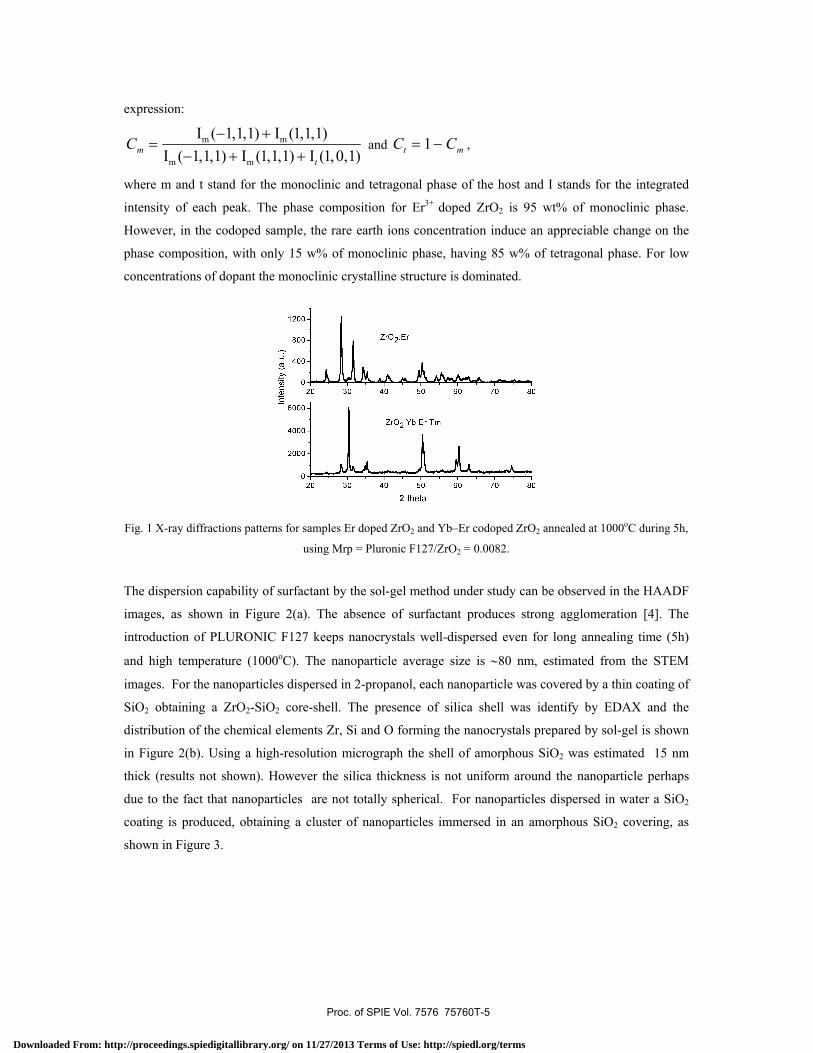
expression:
Cm =Im (−1,1,1) + Im (1,1,1)
Im (−1,1,1) + Im (1,1,1) + It (1,0,1) and Ct = 1− Cm ,
where m and t stand for the monoclinic and tetragonal phase of the host and I stands for the integrated
intensity of each peak. The phase composition for Er3+ doped ZrO2 is 95 wt% of monoclinic phase.
However, in the codoped sample, the rare earth ions concentration induce an appreciable change on the
phase composition, with only 15 w% of monoclinic phase, having 85 w% of tetragonal phase. For low
concentrations of dopant the monoclinic crystalline structure is dominated.
Fig. 1 X-ray diffractions patterns for samples Er doped ZrO2 and Yb–Er codoped ZrO2 annealed at 1000oC during 5h,
using Mrp = Pluronic F127/ZrO2 = 0.0082.
The dispersion capability of surfactant by the sol-gel method under study can be observed in the HAADF
images, as shown in Figure 2(a). The absence of surfactant produces strong agglomeration [4]. The
introduction of PLURONIC F127 keeps nanocrystals well-dispersed even for long annealing time (5h)
and high temperature (1000oC). The nanoparticle average size is ∼80 nm, estimated from the STEM
images. For the nanoparticles dispersed in 2-propanol, each nanoparticle was covered by a thin coating of
SiO2 obtaining a ZrO2-SiO2 core-shell. The presence of silica shell was identify by EDAX and the
distribution of the chemical elements Zr, Si and O forming the nanocrystals prepared by sol-gel is shown
in Figure 2(b). Using a high-resolution micrograph the shell of amorphous SiO2 was estimated 15 nm
thick (results not shown). However the silica thickness is not uniform around the nanoparticle perhaps
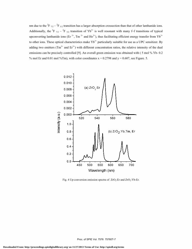
due to the fact that nanoparticles are not totally spherical. For nanoparticles dispersed in water a SiO2
coating is produced, obtaining a cluster of nanoparticles immersed in an amorphous SiO2 covering, as
shown in Figure 3.
(a) Proc. of SPIE Vol. 7576 75760T-5
Downloaded From: http://proceedings.spiedigitallibrary.org/ on 11/27/2013 Terms of Use: http://spiedl.org/terms
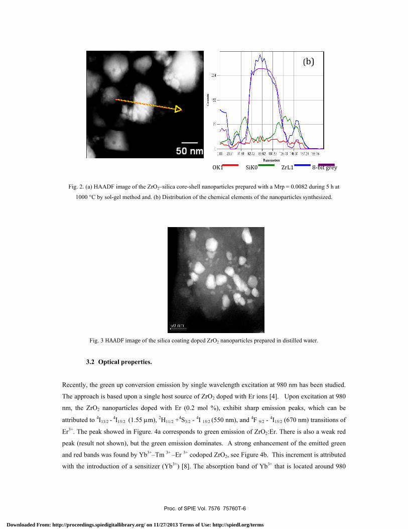
Fig. 2. (a) HAADF image of the ZrO2–silica core-shell nanoparticles prepared with a Mrp = 0.0082 during 5 h at
1000 °C by sol-gel method and. (b) Distribution of the chemical elements of the nanoparticles synthesized.
Fig. 3 HAADF image of the silica coating doped ZrO2 nanoparticles prepared in distilled water.
3.2 Optical properties.
Recently, the green up conversion emission by single wavelength excitation at 980 nm has been studied.
The approach is based upon a single host source of ZrO2 doped with Er ions [4]. Upon excitation at 980
nm, the ZrO2 nanoparticles doped with Er (0.2 mol %), exhibit sharp emission peaks, which can be
attributed to 4I13/2 - 4I15/2 (1.55 μm), 2H11/2 +4S3/2 - 4I 15/2 (550 nm), and 4F 9/2 - 4I15/2 (670 nm) transitions of
Er3+. The peak showed in Figure. 4a corresponds to green emission of ZrO2:Er. There is also a weak red
peak (result not shown), but the green emission dominates. A strong enhancement of the emitted green
and red bands was found by Yb3+–Tm 3+ –Er 3+ codoped ZrO2, see Figure 4b. This increment is attributed
with the introduction of a sensitizer (Yb3+) [8]. The absorption band of Yb3+ that is located around 980
OK1 SiK0 ZrL1 8-bit grey
(b) (b)
Proc. of SPIE Vol. 7576 75760T-6
Downloaded From: http://proceedings.spiedigitallibrary.org/ on 11/27/2013 Terms of Use: http://spiedl.org/terms
nm due to the 2F 7/2 – 2F 5/2 transition has a larger absorption crossection than that of other lanthanide ions.
Additionally, the 2F 7/2 – 2F 5/2 transition of Yb3+ is well resonant with many f–f transitions of typical
upconverting lanthanide ions (Er 3+, Tm 3+ and Ho3+), thus facilitating efficient energy transfer from Yb3+
to other ions. These optical characteristics make Yb3+ particularly suitable for use as a UPC sensitizer. By
adding two emitters (Tm3+ and Er3+) with different concentration ratios, the relative intensity of the dual
emissions can be precisely controlled [9]. An overall green emission was obtained with ( 5 mol % Yb- 0.2
% mol Er and 0.01 mol %Tm), with color coordinates x = 0.2798 and y = 0.607, see Figure. 5.
Fig. 4 Up-conversion emission spectra of ZrO2:Er and ZrO2:Yb-Er.
Proc. of SPIE Vol. 7576 75760T-7
Downloaded From: http://proceedings.spiedigitallibrary.org/ on 11/27/2013 Terms of Use: http://spiedl.org/terms
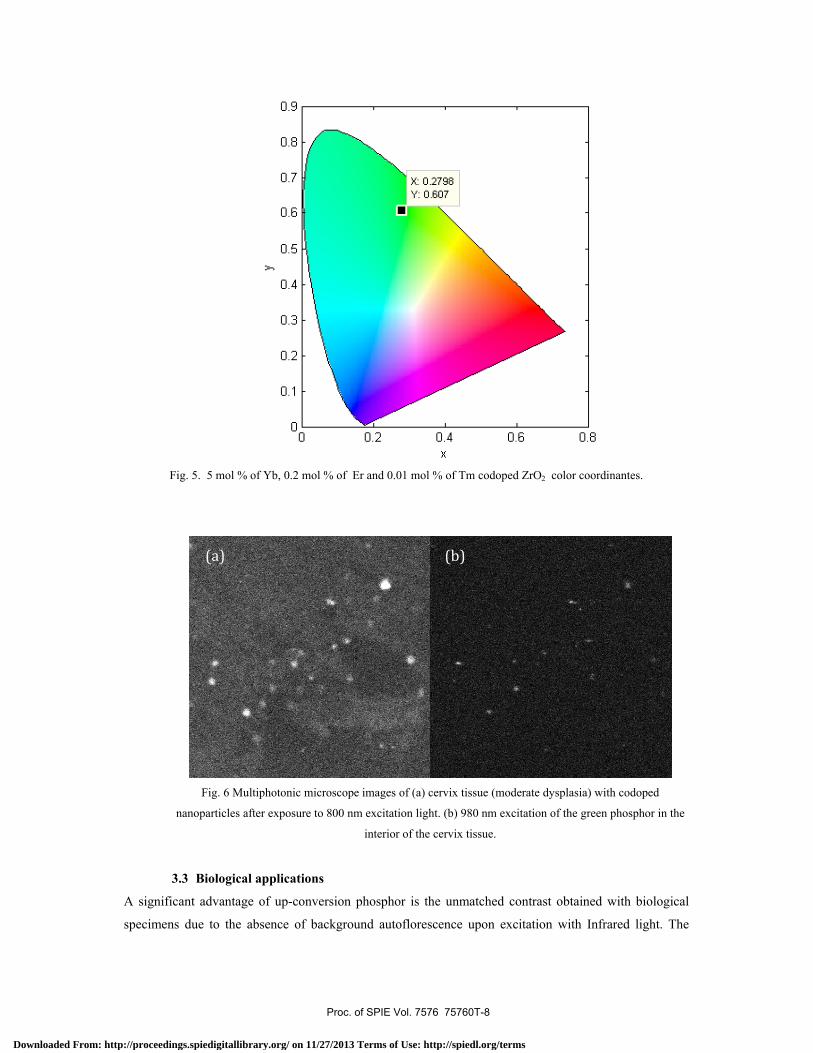
Fig. 5. 5 mol % of Yb, 0.2 mol % of Er and 0.01 mol % of Tm codoped ZrO2 color coordinantes.
Fig. 6 Multiphotonic microscope images of (a) cervix tissue (moderate dysplasia) with codoped
nanoparticles after exposure to 800 nm excitation light. (b) 980 nm excitation of the green phosphor in the
interior of the cervix tissue.
3.3 Biological applications
A significant advantage of up-conversion phosphor is the unmatched contrast obtained with biological
specimens due to the absence of background autoflorescence upon excitation with Infrared light. The
(a) (b)
Proc. of SPIE Vol. 7576 75760T-8
Downloaded From: http://proceedings.spiedigitallibrary.org/ on 11/27/2013 Terms of Use: http://spiedl.org/terms
images of confocal microscopy in Figure 6 demonstrate that codoped ZrO2 nanoparticles are inside of the
cervix tissue with moderate cancer until 80 % of depth. The Figure 6(a) shows the projection image of the
of cervix tissue with nanoparticles in the interior (depth ∼ 4 μm). It is possible to visualize the tissue with
the nanoparticles due to the light scattering on these, which is produced by the high power of the incident
signal at 800 nm. In contrast, Figure 6(b) shows only the visible light produced by the nanoparticles
inside of the tissue after be excited at 980 nm. It has been shown that tissue autofluorescence signal
overlaps with the specific blue phosphor signal [1]. In contrast phosphors excited with an IR signal are
distinctively visible. This is a consequence of the higher energy of blue or ultraviolet light required for
excitation of commonly used fluorescent dyes and due to the fact that many biological materials also
fluoresce.
4. CONCLUSSIONS
We have synthesized Er doped ZrO2:Er 3+ and Yb3+-Er3+-Tm3+ codoped ZrO2 nanocrystals by a sol-gel method
and performed a systematic characterization of both structural and luminescence properties as functions of ion
concentrations. The presence of the silica shell of average of 15 nm in thickness has been confirmed by
transmtion electron microscopy, providing a protective layer for biocompatibility. Also silica coated
doped ZrO2 nanoparticles were prepared. The nanoparticles are ready to be functionalized with antibody
proteins for standard bioconjugation. The viability of the nanoparticles for biological imaging was
confirmed by imaging of cervix tissue with nanoparticles inserted, observed with multiphotonic confocal
microscope. Compared with convencional fluorescent methods, UPT particles offer significant
advantages including lack of complicated background autofluorescence and no photobleaching. We
consider that our nanoparticles are ready to be functionalized with antibody proteins, for standard
bioconjugation, and test their use in early detection of cervical cancer.
Acknowledgment
We thank Nydia Hernández-Rios from Neurology Institute, UNAM-Querétaro for operating the
multiphotonic microscope.
REFERENCES
[1] Corstjens, P. L. A. M., S., Li, M., Zuiderwijk, K., Kardos, W. R., Abrams, R.S., Niedbala, and
Tanke, H.J., "Infrared up-converting phosphors for bioassays", IEE Proc.-Nanobiotech. Vol.
152, No. 2, 64-72 (2005).
[2] Qin, G., J., Lu, J. F., Bisson, Y., Feng, K.-i., Ueda, H., Yagi, and Yanagitani, T., "Upconversion
luminescence of Er3+ in highly transparent YAG ceramics," Solid State Communications 132(2),
103-106 (2004).
Proc. of SPIE Vol. 7576 75760T-9
Downloaded From: http://proceedings.spiedigitallibrary.org/ on 11/27/2013 Terms of Use: http://spiedl.org/terms
[3] De, G., W., Qin, J., Zhang, J., Zhang, Y., Wang, C., Cao, and Cui, Y., "Upconversion
luminescence properties of Y2O3:Yb3+, Er3+ nanostructures," J. Luminesc. 119-120, 258-263
(2006).
[4] Lopez-Luke, T., E., De la Rosa, P., Salas, C., Angeles-Chavez, L. A., Diaz-Torres, and
Bribiesca, S., "Enhancing the up-conversion emission of ZrO2 : Er3+ nanocrystals prepared by a
micelle process," J. Physic. Chem. C 111(45), 17110-17117 (2007).
[5] Patra, A., S., Saha, M., Alencar, N., Rakov and Maciel, G. S.,"Blue upconversion emission of
Tm3+-Yb3+ in ZrO2 nanocrystals: Role of Yb3+ ions," Chem. Phys. Lett. 407(4-6), 477-481
(2005).
[6] Yi, G.-S., and Chow, G.-M.,"Water-Soluble NaYF4:Yb,Er(Tm)/NaYF4/Polymer
Core/Shell/Shell Nanoparticles with Significant Enhancement of Upconversion Fluorescence,"
Chemistry of Materials 19(3), 341-343 (2007).
[7] Lue, Q., F., Guo, L., L., Sun A., and Zhao, L. J., Appl. Phys 103, 123533 (2008).
[8] Solis, D., T., Lopez-Luke, E., De la Rosa, P., Salas, and Angeles-Chavez, C., "Surfactant effect
on the upconversion emission and decay time of ZrO2:Yb-Er nanocrystals," J. Luminesc.
129(5), 449-455 (2009).
[9] Wang, F. and Liu, X.,"Recent advances in the chemistry of lanthanide-doped upconversion
nanocrystals," Chem. Soc. Rev. 38, 976-989 (2009).
Proc. of SPIE Vol. 7576 75760T-10
Downloaded From: http://proceedings.spiedigitallibrary.org/ on 11/27/2013 Terms of Use: http://spiedl.org/terms


