Embed Size (px)
Citation preview

Synthesis, characterization, biodistribution and scintigraphyof 99mTc-paclitaxel: a potential tracer of paclitaxel
Indranil Banerjee • Ashok Behera • Kakali De •
Sankha Chattopadhyay • Satbir Singh Sachdev •
Bharat Sarkar • Santanu Ganguly • Mridula Misra
Received: 8 August 2014
� Akademiai Kiado, Budapest, Hungary 2014
Abstract 99mTc-paclitaxel was synthesized by using
sodium borohydride as a reducing agent. Greater than 95 %
labelling efficiency was achieved. Radiochemical purity of
the synthesized 99mTc-paclitaxel was validated by thin
layer chromatography (TLC) scanner and high perfor-
mance liquid chromatography (HPLC). 99mTc-paclitaxel
passed in vitro stability tests. Biodistribution and scintig-
raphy studies were performed in Sprague–Dawley rats. The
biodistribution study results of 99mTc-paclitaxel were
related mainly to the metabolism and excretion routes
followed by the parental drug, paclitaxel. Apart from that,
biodistribution of 99mTc-paclitaxel was altered after pre-
treatment with cold paclitaxel. Hence, 99mTc-paclitaxel
may be used as a tracer for paclitaxel.
Keywords 99mTc-paclitaxel � Sodium borohydride �Quantitative biodistribution � Scintigraphy
Introduction
In recent years, the use of organometallic radioactive bio-
molecules for diagnostic and therapeutic purpose has made
a tremendous progress. In conjugation with that molecular
imaging has achieved a pivotal role in the drug develop-
ment process. It can provide an essential link between
in vitro studies and those performed in vivo [1]. Currently
the molecular imaging techniques that employ radiotracers
are one of the most sensitive techniques. For drug devel-
opment studies, radionuclide imaging has received the
most attention. Radionuclide imaging can be performed
with single photon emission computed tomography
(SPECT) or positron emission tomography (PET). SPECT
and PET are based on the molecular tracer principle and
they are established tools in non-invasive imaging of
radiolabelled drugs administered in the nano or picomolar
range [2–4]. Drugs administrated at these low amounts do
not produce any pharmacological effects which reduce the
risk of serious adverse effects in human volunteers or
patients. Depending on the ligands and radionuclides used,
it is possible to generate pharmacokinetic data of radiola-
belled drug molecules in humans using a microdose
imaging approach [4]. In this report, paclitaxel was used as
a model drug, and 99mTc was used as a c-emitting
radionuclide.
Paclitaxel (C47H51NO14) is a pseudoalkaloid. It acts as
an antineoplastic agent due to its inhibitory effect of
cellular growth by stabilising the microtubule assembly
and, thus, blocking the cell replication in the late G2
mitotic phase of the cell cycle [5]. Paclitaxel is
I. Banerjee � A. Behera � K. De � M. Misra (&)
Department of Infectious Diseases and Immunology (Nuclear
Medicine Division), Council of Scientific and Industrial
Research (CSIR)–Indian Institute of Chemical Biology (CSIR-
IICB), 4 Raja S C Mullick Road, Kolkata 700032, West Bengal,
India
e-mail: [email protected]
I. Banerjee
e-mail: [email protected]
S. Chattopadhyay
Radiopharmaceuticals Laboratory, Regional Centre, Board of
Radiation and Isotope Technology, Variable Energy Cyclotron
Centre, 1/AF, Bidhan Nagar, Kolkata 700064, India
e-mail: [email protected]
S. S. Sachdev
Board of Radiation and Isotope Technology, Vashi Complex,
Sector 20, Vashi, Navi Mumbai 400705, India
B. Sarkar � S. Ganguly
Regional Radiation Medicine Centre, Thakurpukur Cancer
Research Centre, Kolkata 700063, India
123
J Radioanal Nucl Chem
DOI 10.1007/s10967-014-3825-3

prescribed mainly to treat breast and ovarian cancers, but
it is known that various cancer cells can be killed
effectively by this drug [6]. A number of clinical trials
are going on with paclitaxel to explore its potential for
the treatment of various cancers [7]. If we can determine
the biodistribution of paclitaxel at microdose level, it will
be helpful to monitor drug targets as well as possible
reaction of taking new medications in heterogeneous
patient populations.
There are different methods to determine biodistribution
of drugs such as fluorescence imaging, radiotracing and
mass spectrometry. Among these methods, radiolabelling is
one of the most suitable methods to determine quantitative
biodistribution [8]. To determine biodistribution of drugs,
radionuclide 99mTc is used extensively in nuclear medicine
for economic reasons, as well as favourable imaging
characteristics of technetium (c energy of 140 keV and 6 h
half-life). Stannous salts are used for reduction of sodium
pertechnetate (Na99mTcO4) to obtain lower valency state of99mTc but during labelling by this method radiocolloids are
generated [9, 10]. The biodistribution of desired molecule
is affected by these radiocolloids. We have to optimize the
amount of stannous salts and pH of the reaction for getting
maximum labelling efficiency. Some time we have to pass
the mixture of compounds through a column to obtain ra-
diocolloids free labelled compound [11]. These processes
are time consuming.
In this article, we report a novel, rapid and effective
method for radiolabelling of paclitaxel by 99mTc using
sodium borohydride as a reducing agent. Sodium borohy-
dride was used for reduction of 99mTc (VII) ions, and
subsequent complexation was done with paclitaxel mole-
cules. Synthesized 99mTc-paclitaxel has been undergone for
quality control, characterization, biodistribution and scin-
tigraphy studies.
Materials and methods
Paclitaxel was obtained as a gift sample from Fresenius
Kabi Oncology Ltd., Kalyani, India. Diethylene-
triaminepentaacetic acid (DTPA), and Cremophor EL were
purchased from Sigma-Aldrich, St. Louis, USA. Sodium
borohydride was purchased from Merck, Hohenbrunn,
Germany. 99Mo/99mTc kits were obtained from Board of
Radiation and Isotope Technology (BRIT, Mumbai, India).99mTc extraction was performed in Council of Scientific
and Industrial Research-Indian Institute of Chemical
Biology (CSIR-IICB), Kolkata, India. All other reagents
and solvents were obtained from Merck, Hohenbrunn,
Germany or SRL, Mumbai, India, and they are either
HPLC or analytical grade. All chemicals were used without
further purification. Animal experiments were performed in
compliance with the regulations of Institutional Animal
Ethics Committee, CSIR-IICB, Kolkata, India. In-house
Sprague–Dawley rats (weighing approximately 200–250 g)
were used for quantitative biodistribution as well as scin-
tigraphy studies.
Synthesis of 99mTc-paclitaxel
Radiolabelling of paclitaxel was done with previously
reported method with modifications [12, 13]. Nitrogen
purging, prior to mixing was carried out to degas all
solutions. To 100 ll of 99mTc (111 MBq) in saline, 5 mg
of solid sodium borohydride was added directly with
continuous stirring followed by immediate addition of
500 ll of the paclitaxel solution (1 mg/ml, paclitaxel was
dissolved in a mixture of ethanol and Tris–HCl buffer, pH
7.4; ethanol: Tris–HCl buffer = 2:1). The solution was
stirred for 45 min at room temperature (25 ± 2 �C). The
contents were filtered using 0.22-micron filter (Millipore
Corporation, Carrigtwohill, Ireland) and transferred into
an evacuated sterile sealed vial and used for further
experiment.
Quality control and stability study
Quality control was performed by following the method
described earlier [14]. The labelling efficiency of 99mTc to
paclitaxel was assessed by ascending instant thin layer
chromatography using silica gel plates (ITLC-SG). The
ITLC-SG was performed using acetone as the mobile
phase. Approximately, 2–3 ll of the radiolabelled complex
was applied at the bottom point, 1.0 cm from the end of an
ITLC strip. The strip was developed until solvent front
reached 8.5 cm from the origin. The labelling efficiency
was estimated after dividing the ITLC sheets into two equal
halves and counting radioactivity of each segment using
gamma ray spectrometer: GRS 23C, ECIL, Mumbai, India.
The stability of 99mTc-paclitaxel was checked for 24 h at
room temperature. Labelling efficiency was calculated
using the following equation.
(i) Labelling Efficiency (%) = [(Total counts - counts
of free pertechnetate)/Total counts] 9 100 %
Radiochemical purity of 99mTc-paclitaxel
and Na99mTcO4
One to two ll of synthesized 99mTc-paclitaxel was applied
at the bottom point, 1.0 cm from the end of an ITLC strip
(6 cm long). The chromatogram was developed in acetone
solution. After the run, the strips were dried and scanned
under a mini Gita TLC scanner (Ray test, Straubenhardt,
Germany). Similarly, an ITLC of Na99mTcO4 used for the
J Radioanal Nucl Chem
123

synthesis of 99mTc-paclitaxel was developed in acetone and
scanned under the mini Gita TLC scanner.
HPLC analysis of 99mTc-paclitaxel
For HPLC analysis of 99mTc-paclitaxel, reversed phase
C-18 column (3.9 mm 9 300 mm, l bondapak column
from Waters, Bangalore, India) suitable for radiolabelled
molecule detection was used. The mobile phase used in
RP-HPLC for gradient system (Gradient I) consisted of
water (solvent A) and acetonitrile (solvent B). Gradient I:
0 min 100 % A (0 % B), 2 min 90 % A (10 % B), 6 min
80 % A (20 % B), 12 min 50 % A (50 % B), 25 min 10 %
A (90 % B), 30 min 0 % A (100 % B), 35 min 100 % A
(0 % B). To obtain chromatogram, 10 ll of the complex
was injected into the HPLC system fitted with radioactive
detector. Here, flow rate was 1 ml/min.
In vitro stability tests
In vitro stability of the 99mTc-paclitaxel complex was
determined in phosphate buffer saline (PBS) [pH 7.4] and
in rat serum separately at different time points, at room
temperature [15]. The labelled complex (0.1 ml) was
incubated with 0.4 ml of PBS or freshly collected rat
serum. The stability tests were performed by determining
the changes in labelling efficiency. The samples were
analyzed by using ITLC at regular intervals up to 24 h.
Chromatograms were obtained in gamma ray spectrometer.
DTPA challenge test
In order to check the strength of binding of 99mTc with pac-
litaxel, 0.1 ml of the labelled preparation was challenged
against various concentrations (10, 30 and 50 mM) of DTPA
and incubated for 1 h at 37 �C [16]. The effect of DTPA on
labelling efficiency was measured on ITLC-SG using acetone
as the mobile phase, which allowed the separation of free
pertechnetate (Rf = 0.9–1.0) and DTPA-complex
(Rf = 0.8–0.9) from the 99mTc-paclitaxel, which remained at
the point of application (Rf = 0). ITLC was carried out in
11 cm long ITLC strip. Rf values of free pertechnetate and99mTc-DTPA were validated by using standard solutions of
them, respectively. Standard 99mTc-DTPA was prepared as
described previously [17], and standard pertechnetate was
prepared by adding saline in extracted 99mTc. Percentage
Transchelation was calculated by the following formula.
(ii) % Transchelation = [decrease in labelling effi-
ciency of 99mTc-paclitaxel after 1 h incubation
with DTPA/labelling efficiency of 99mTc-paclitaxel
at 1 h in room temperature] 9 100 %
Lipophilicity test of 99mTc-paclitaxel
The octanol/buffer partition coefficient was measured
using a standard protocol [18]. Two ml of 1-octanol and
two ml of PBS (pH 7.4) were mixed in a centrifuge tube.
One hundred ll of 99mTc-paclitaxel was added to the sys-
tem, and the mixture was vortexed at room temperature for
1 min and then centrifuged at 5,000 rpm (1,3989g) for
5 min. From each phase, 0.1 ml of the aliquot was pipetted
and counted in gamma spectrometer. Care was taken to
avoid cross contamination between the two phases. The
measurement was done in triplicate. The partition coeffi-
cient was calculated by using the following equation.
(iii) Log P = Log [(cpm in octanol - cpm in back-
ground)/(cpm in buffer - cpm in background)]
Blood clearance and total plasma protein binding study
Blood clearance and plasma protein binding [10] of 99mTc-
paclitaxel was studied in rats (Sprague–Dawley) injecting
18.5 MBq (500 lCi) of the radiopharmaceutical through the
femoral vein. Blood (250 ll) was collected by puncturing
heart of rat. The radioactivity of the withdrawn blood at
different time intervals (5 min, 30 min, 1 h, 2 h, 6 h and
24 h) was measured by using gamma ray spectrometer to
obtain the blood clearance curve of the complex.
The total plasma protein binding of 99mTc-paclitaxel was
determined by 10 % trichloroacetic acid (TCA) method from
heparinized blood. Blood cells and plasma were separated by
precipitation at 10,000 rpm for 6 min. Plasma proteins were
precipitated and isolated by centrifugation for 6 min at
10,000 rpm following the addition of an equal volume of
10 % TCA in the plasma. The supernatant was decanted, and
the pellet was re-suspended in 1 ml TCA (10 % v/v). Again
centrifugation was done, and the supernatant was decanted.
Radioactivity in both the supernatants was measured. This
count was expressed as a percentage of count obtained with
the same volume of unprocessed plasma.
(iv) % Protein bound = [(counts in plasma - counts in
both the supernatants)/counts in plasma] 9 100
Quantitative biodistribution study
Biodistribution studies were performed in rats (Sprague–
Dawley rats weighing approximately 200–250 g) [13].99mTc-paclitaxel complex solution (approximately 500 lCi,
200 ll) was administered through the femoral vein of
anaesthetized rats with 0.5 mm polyethylene (PE) catheter.
At different time intervals (5 min, 30 min, 1 h, 2 h, 6 h and
24 h), the animals were sacrificed by intravenous injection of
air. Urine samples were obtained by puncturing urinary
J Radioanal Nucl Chem
123
bladder. Other organs (brain, heart, liver, lungs, spleen,
muscle, kidney, intestines, stomach and pancreas) were
removed, rinsed with saline and blotted to remove any
adherent material. The organs were collected in pre-weighed
scintillation tubes after drying. Then organs were weighed
and radioactivity was counted in a well-type counter (gamma
ray spectrometer) with respect to suitably diluted aliquots of
the injected solution as standard. The biodistribution of99mTc labelled paclitaxel in each organ was calculated as a
percentage of the injected dose per gram of the tissue (%ID/
g).
(v) %ID/g of Tissue = [(counts in sample)/(weight of
sample X total counts injected after decay
correction)] 9 100 %
Quantitative biodistribution study after pre-treatment
with cold paclitaxel
This study was done according to previously reported
method [19] with modifications. Briefly, 20 mg/kg of
paclitaxel was injected intravenously into the Sprague–
Dawley rats weighing approximately 200–250 g at day 1.
After that, 99mTc-paclitaxel was injected in those rats at
day 5 (approximately 500 lCi, 200 ll), and biodistribution
of 99mTc-paclitaxel was determined at 30 min, 1 h, 2 h, 6 h
and 24 h by following the method indicated in ‘‘Quanti-
tative biodistribution study’’ section.
99mTc-paclitaxel scintigraphy
Imaging studies were performed on normal Sprague–
Dawley rat, 200–250 g at Thakurpukur Cancer Research
Centre (Regional Radiation Monitoring Centre, Kolkata,
India) under dual-head gamma camera (GE Hawkeyes,
Pittsburgh, USA). The urethane-anaesthetised rat was
injected via the femoral vein with a bolus containing99mTc-paclitaxel (approximately 500 lCi, 200 ll). Rat was
placed in a typical position for planar imaging under a
small field of view experimental gamma camera, suitable
for both planar and tomographic imaging. Whole body
image acquisition study was done at 5 and 90 min after
injection, and scan time was 1 min. Image data were
obtained and analyzed using a gamma camera (GE
Hawkeyes) fitted with a low energy high-resolution all
purpose collimator using static procedure of the Xeleris
(Functional Imaging) Workstation system.
Statistical analysis
The calculations of mean and standard deviation (SD) were
performed on Microsoft Excel. Statistical significance was
considered when p \ 0.05 and calculations were done
using GraphPad InStat (GraphPad Software Inc., San
Diego, USA) Software.
Results
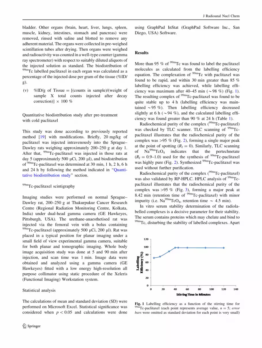
More than 95 % of 99mTc was found to label the paclitaxel
molecules as calculated from the labelling efficiency
equation. The complexation of 99mTc with paclitaxel was
found to be rapid, and within 30 min greater than 85 %
labelling efficiency was achieved, while labelling effi-
ciency was maximum after 40–45 min (*98 %) (Fig. 1).
The resulting complex of 99mTc-paclitaxel was found to be
quite stable up to 4 h (labelling efficiency was main-
tained *95 %). Then labelling efficiency decreased
slightly at 6 h (*94 %), and the calculated labelling effi-
ciency was found greater than 90 % at 24 h (Table 1).
Radiochemical purity of the complex (99mTc-paclitaxel)
was checked by TLC scanner. TLC scanning of 99mTc-
paclitaxel illustrates that the radiochemical purity of the
complex was [95 % (Fig. 2), forming a single major peak
at the point of spotting (Rf = 0). Similarly, TLC scanning
of Na99mTcO4 indicates that the pertechnetate
(Rf = 0.9–1.0) used for the synthesis of 99mTc-paclitaxel
was highly pure (Fig. 2). Synthesized 99mTc-paclitaxel was
used without further purification.
Radiochemical purity of the complex (99mTc-paclitaxel)
was also validated by RP-HPLC. HPLC analysis of 99mTc-
paclitaxel illustrates that the radiochemical purity of the
complex was [95 % (Fig. 3), forming a major peak at
8.42 min (retention time of 99mTc-paclitaxel) with minor
impurity (i.e. Na99mTcO4, retention time * 4.5 min).
In vitro serum stability determination of the radiola-
belled complexes is a decisive parameter for their stability.
The serum contains proteins which may chelate and bind to99mTc, disturbing the stability of labelled complexes. Apart
Fig. 1 Labelling efficiency as a function of the stirring time for99mTc-paclitaxel (each point represents average value, n = 5; error
bars were omitted as standard deviation for each point is very small)
J Radioanal Nucl Chem
123

from that, physiological pH (i.e. 7.4) may also affect the
stability of the complex. It was found that 99mTc-paclitaxel
complex was stable in PBS and rat serum (Table 2) for
24 h post labelling. The stability of the labelled complex in
PBS and serum supports its stability in biological envi-
ronment upon administration into the body.
DTPA challenge was performed to get information on
the transchelation (a measure of strength of binding) of the
synthesized 99mTc-paclitaxel. Transchelation can be
defined as a form of chelation in which one chelate group
replaces another. When 99mTc-paclitaxel was incubated
with DTPA, a small amount of DTPA complex was
formed. The formed complex was 99mTc-DTPA as sug-
gested by our TLC studies using standard 99mTc-DTPA.
So, transchelation occurred as some amount of 99mTc-
paclitaxel was replaced by 99mTc-DTPA. Challenge studies
demonstrated that the labelling efficiency of 99mTc-paclit-
axel did not alter much in the presence of DTPA (Table 3).
At 50 mM concentration of DTPA, the transchelation was
found to be 4.76 % for 99mTc-paclitaxel which confirms the
in vitro stability of the radiolabelled complex [20].
Log P was calculated from the octanol/buffer (pH 7.4)
partition coefficient method. The partition coefficient of the
99mTc-paclitaxel complex was –1.46 ± 0.03 (n = 3). It
clearly indicates that the complex was hydrophilic in nature.
The blood clearance curve of 99mTc-paclitaxel (Fig. 4)
showed that the initial accumulation of radioactivity in the
blood of animal increased up to 1 h, then decreased rapidly
Fig. 2 A typical thin layer chromatogram for 99mTc-paclitaxel and
Na99mTcO4
Fig. 3 HPLC peak for 99mTc-paclitaxel
Table 2 Stability of 99mTc-paclitaxel in PBS and rat serum, values
represent mean ± SD (n = 3)
Time Labelling efficiency in PBS
(%)
Labelling efficiency in serum
(%)
5 min 95.32 ± 0.17 93.58 ± 1.28
30 min 94.88 ± 1.25 92.13 ± 1.47
1 h 92.81 ± 1.92 91.33 ± 0.57
2 h 92.35 ± 1.87 90.59 ± 0.96
6 h 90.13 ± 1.26 89.18 ± 1.81
24 h 88.62 ± 1.91 86.24 ± 1.09
Table 3 In vitro stability of 99mTc-paclitaxel in presence of DTPA,
values represent mean ± SD (n = 3)
Concentration of DTPA (mM) % Transchelation
10 0.82 ± 0.26
30 1.94 ± 1.18
50 4.76 ± 0.36
Table 1 Stability of 99mTc-
paclitaxel at room temperature
at different time points, values
represent mean ± standard
deviation (SD) (n = 3)
Time
(h)
Labelling efficiency
(%)
1 97.39 ± 0.42
2 97.14 ± 0.36
4 95.11 ± 1.21
6 94.36 ± 0.83
24 90.66 ± 1.63
J Radioanal Nucl Chem
123
in radioactivity up to 6 h. The complex showed a decrease
in the clearance rate after 6 h. This indicates a triphasic
clearance pattern of the complex from the system.
Approximately, 75 % of the complex was removed from
the blood after 6 h.
The extent of free 99mTc-paclitaxel in rat plasma was
calculated by trichloroacetic acid (TCA) method. TCA
method provided the total plasma protein binding level of
the complex. The percentage of protein bound drug was
found to be 26.13 ± 2.59 (n = 3), indicating that signifi-
cant amount of the drug was free in rat plasma.
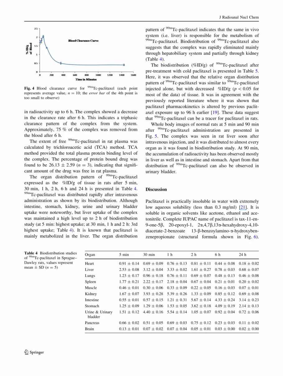
The organ distribution pattern of 99mTc-paclitaxel
expressed as the %ID/g of tissue in rats after 5 min,
30 min, 1 h, 2 h, 6 h and 24 h is presented in Table 4.99mTc-paclitaxel was distributed rapidly after intravenous
administration as shown by its biodistribution. Although
intestine, stomach, kidney, urine and urinary bladder
uptake were noteworthy, but liver uptake of the complex
was maintained a high level up to 2 h of biodistribution
study (at 5 min: highest uptake; at 30 min, 1 h and 2 h: 3rd
highest uptake; Table 4). It is known that paclitaxel is
mainly metabolized in the liver. The organ distribution
pattern of 99mTc-paclitaxel indicates that the same in vivo
system (i.e. liver) is responsible for the metabolism of99mTc-paclitaxel. Biodistribution of 99mTc-paclitaxel also
suggests that the complex was rapidly eliminated mainly
through hepatobiliary system and partially through kidney
(Table 4).
The biodistribution (%ID/g) of 99mTc-paclitaxel after
pre-treatment with cold paclitaxel is presented in Table 5.
Here, it was observed that the relative organ distribution
pattern of 99mTc-paclitaxel was similar to 99mTc-paclitaxel
injected alone, but with decreased %ID/g (p \ 0.05 for
most of the data) of tissue. It was in agreement with the
previously reported literature where it was shown that
paclitaxel pharmacokinetics is altered by previous paclit-
axel exposure up to 96 h earlier [19]. These data suggest
that 99mTc-paclitaxel can be a tracer for paclitaxel in rats.
Whole body images of normal rats at 5 min and 90 min
after 99mTc-paclitaxel administration are presented in
Fig. 5. The complex was seen in rat liver soon after
intravenous injection, and it was distributed to almost every
organ as it was found in biodistribution study. At 90 min,
the accumulation of radioactivity has been observed mostly
in liver as well as in intestine and stomach. Apart from that
distribution of 99mTc-paclitaxel can also be observed in
urinary bladder.
Discussion
Paclitaxel is practically insoluble in water with extremely
low aqueous solubility (less than 0.3 mg/ml) [21]. It is
soluble in organic solvents like acetone, ethanol and ace-
tonitrile. Complete IUPAC name of paclitaxel is tax-11-en-
9-one-5b, 20-epoxyl-1, 2a,4,7b,13a-hexahydroxy-4,10-
diacetate-2-benzoate 13-b-benzoylamino-a-hydroxyben-
zenepropionate (structural formula shown in Fig. 6).
Fig. 4 Blood clearance curve for 99mTc-paclitaxel (each point
represents average value, n = 10; the error bar of the 4th point is
too small to observe)
Table 4 Biodistribution studies
of 99mTc-paclitaxel in Sprague–
Dawley rats, values represent
mean ± SD (n = 5)
Organ 5 min 30 min 1 h 2 h 6 h 24 h
Heart 0.91 ± 0.14 0.69 ± 0.09 0.76 ± 0.13 0.81 ± 0.11 0.44 ± 0.08 0.18 ± 0.02
Liver 2.53 ± 0.08 3.12 ± 0.04 3.33 ± 0.02 1.61 ± 0.27 0.78 ± 0.03 0.68 ± 0.07
Lungs 1.23 ± 0.17 0.96 ± 0.18 0.76 ± 0.11 0.69 ± 0.07 0.48 ± 0.13 0.46 ± 0.08
Spleen 1.77 ± 0.21 2.22 ± 0.17 2.18 ± 0.04 0.67 ± 0.04 0.21 ± 0.01 0.20 ± 0.02
Muscle 0.46 ± 0.01 0.30 ± 0.06 0.33 ± 0.09 0.22 ± 0.05 0.16 ± 0.03 0.07 ± 0.01
Kidney 1.67 ± 0.07 3.93 ± 0.28 5.39 ± 0.26 1.33 ± 0.09 0.85 ± 0.12 0.69 ± 0.08
Intestine 0.55 ± 0.01 0.57 ± 0.15 1.21 ± 0.31 5.67 ± 0.14 4.33 ± 0.24 3.14 ± 0.23
Stomach 1.25 ± 0.09 1.29 ± 0.06 1.53 ± 0.05 3.62 ± 0.18 4.09 ± 0.19 2.14 ± 0.13
Urine & Urinary
bladder
1.51 ± 0.12 4.40 ± 0.16 5.54 ± 0.14 1.05 ± 0.07 0.92 ± 0.04 0.72 ± 0.06
Pancreas 0.66 ± 0.02 0.51 ± 0.05 0.69 ± 0.03 0.75 ± 0.12 0.23 ± 0.03 0.11 ± 0.02
Brain 0.13 ± 0.01 0.07 ± 0.02 0.07 ± 0.04 0.05 ± 0.01 0.03 ± 0.00 0.02 ± 0.00
J Radioanal Nucl Chem
123

Synthesized 99mTc-paclitaxel was characterized radio-
chemically. Single peak in TLC scanner confirms the
radiochemical purity of the synthesized 99mTc-paclitaxel. It
also validates the identity of 99mTc-paclitaxel with respect
to Na99mTcO4. HPLC analysis of 99mTc-paclitaxel also
supports our observation in ITLC. We found that the
complex was fairly stable in room temperature. Paclitaxel
possesses four main binding sites (=O, -OH, [ NH,
and = O at 10, 20, 30 and 40 position of paclitaxel molecule,
respectively; Fig. 6) which can participate to form stable
complex with 99mTc. Probably, two molecules of paclitaxel
bind with 99mTc (Fig. 7), which can explain the stability of
the formed complex. We expected the structure of 99mTc-
paclitaxel from literature [22] and MM2 energy minimized
structure by energy minimization computation of all
possible structures using Chem-3D ultra software (Fig. 8)
[23]. It has been reported in the literature that 4, 5 or 6
membered ring containing 99mTc can form stable complex
[22].
We have also evaluated 99mTc-paclitaxel biologically.
Evaluation of radiolabelled paclitaxel in animals is
important to the study of its possible use for the prediction
of chemotherapeutic efficacy with paclitaxel in cancer
patients. Rapid clearance (as well as clearance pattern) of99mTc-paclitaxel from the system, its liver accumulation as
well as elimination pattern, and organ biodistribution data
after pre-treatment with cold paclitaxel suggest that syn-
thesized complex has the potential to be used as a tracer for
paclitaxel in vivo. The organ distribution of 99mTc-paclit-
axel was related mainly to the metabolism and excretion
Table 5 Biodistribution studies
of 99mTc-paclitaxel after pre-
treatment with cold paclitaxel in
Sprague–Dawley rats, values
represent mean ± SD (n = 5)
* Indicates significant
(p \ 0.05)
Organ 30 min 1 h 2 h 6 h 24 h
Heart 0.42 ± 0.02* 0.56 ± 0.08* 0.62 ± 0.04* 0.24 ± 0.03* 0.09 ± 0.01*
Liver 2.07 ± 0.09* 2.29 ± 0.05* 1.01 ± 0.12* 0.45 ± 0.08* 0.47 ± 0.09*
Lungs 0.68 ± 0.08* 0.51 ± 0.06* 0.49 ± 0.05* 0.38 ± 0.06* 0.21 ± 0.03*
Spleen 1.62 ± 0.07* 1.49 ± 0.13* 0.37 ± 0.03* 0.12 ± 0.02* 0.15 ± 0.02*
Muscle 0.15 ± 0.01* 0.15 ± 0.01* 0.11 ± 0.03* 0.09 ± 0.01* 0.04 ± 0.00
Kidney 2.48 ± 0.17* 3.69 ± 0.16* 0.74 ± 0.09* 0.46 ± 0.06* 0.49 ± 0.04*
Intestine 0.31 ± 0.03* 0.81 ± 0.05* 3.02 ± 0.18* 2.91 ± 0.14* 1.53 ± 0.18*
Stomach 0.76 ± 0.06* 1.09 ± 0.02* 2.17 ± 0.11* 2.35 ± 0.07* 1.06 ± 0.11*
Urine & Urinary bladder 2.38 ± 0.09* 3.39 ± 0.08* 0.65 ± 0.06* 0.49 ± 0.05* 0.52 ± 0.08*
Pancreas 0.33 ± 0.04* 0.41 ± 0.03* 0.47 ± 0.06* 0.14 ± 0.04* 0.04 ± 0.01*
Brain 0.03 ± 0.01* 0.02 ± 0.01* 0.02 ± 0.00 0.00 ± 0.00 0.00 ± 0.00
Fig. 5 Scintigraphic image of99mTc-paclitaxel in rat at 5 and
90 min
J Radioanal Nucl Chem
123

routes reported for the parental drug, paclitaxel [24]. This
study confirms that 99mTc-paclitaxel biodistribution pro-
vides a good estimate of paclitaxel biodistribution. There-
fore, 99mTc-paclitaxel may be able to predict the uptake of
paclitaxel in solid tumors. Apart from that, paclitaxel is a
known substrate for P-glycoprotein (Pgp). Pgp is a mem-
brane pump whose overexpression has been shown to result
in multidrug resistance (MDR) [25]. For this reason, it may
Fig. 6 Structure of paclitaxel
Fig. 7 Radiolabelled complex
of 99mTc-paclitaxel (probable
structure)
J Radioanal Nucl Chem
123

also be assumed that 99mTc-paclitaxel may have role to
image MDR, however, comparison with 99mTc-sestamibi
and other related MDR imaging radiotracers is mandatory.99mTc radiopharmaceutical imaging is used in approxi-
mately 85 % of nuclear medicine procedures [26], and it is
well known that cost effectiveness of SPECT imaging is
higher than that of PET imaging. Previously, a number of
articles described the radiolabelling of paclitaxel with
different radioisotopes [11, 27, 28] for SPECT imaging.99mTc-paclitaxel was synthesized by stannous chloride
method [11], but its potential as a tracer for paclitaxel was
not evaluated. Indium-111-DTPA-paclitaxel was synthe-
sized and biologically evaluated [27]. Limitation with
indium-111 is that its high affinity for transferrin receptors.
This leads to excessive radiation burden to the bone mar-
row of patients treated with indium-111 labelled product.
I123 was also used to radiolabel paclitaxel, but this complex
was not biologically evaluated [28].
In this work, we radiolabelled paclitaxel with 99mTc by
using sodium borohydride as a reducing agent. Sodium
borohydride has long been used in radiolabelling purpose
[12, 13, 29]. It is worth noting that radiolabelling by tri-
carbonyl method also uses sodium borohydride in labelling
procedure [30]. Boric acid is generated as a by-product
when sodium borohydride is used in radiolabelling purpose
[29]. It has been reported in the literature that up to 611 mg
of boric acid was administered by the intravenous route in
human being with no signs of toxicity [31]. It has also been
reported that 350 parts per million (ppm) of boric acid had
no adverse effects on fertility, lactation, weight or
appearance of rats [32]. The amount of boric acid gener-
ated by the reaction described in this work was less than
10 mg, and in agreement with the literature report we
haven’t seen loss of weight or change of appearance in
Sprague–Dawley rats during 24 h biodistribution study.
Anaphylaxis and severe hypersensitivity reactions
characterized by dyspnea and hypotension requiring treat-
ment, angioedema, and generalized urticaria have occurred
in some patients receiving paclitaxel (Taxol) in different
clinical trials. Fatal reactions occurred in patients despite
premedication [33]. Taxol formulation contains paclitaxel
dissolved in a 50:50 (v/v) mixture of the surfactant poly-
oxyethylated castor oil (Cremophor EL) and dehydrated
ethanol. Cremophor EL has been shown to cause those
acute hypersensitivity reactions, one of the most severe
side effects associated with discontinuation of paclitaxel
therapy [34]. We radiolabelled paclitaxel in the presence of
Cremophor EL (composition similar to Taxol formulation
and amount of paclitaxel 1 mg/ml) by the method descri-
bed in this article. We have found similar radiochemical
purity and stability data, but we did observe altered total
plasma protein binding (71.54 ± 2.06, n = 3) and biodis-
tribution pattern [13] in the presence of Cremophor EL as
indicated by others also [35–37]. It has been demonstrated
that Cremophor EL induced the appearance of a lipoprotein
dissociation product for which paclitaxel has a high affinity
[35], and Cremophor EL can change the biodistribution of
drugs [38].
Dose, regimen and effect of combination of paclitaxel
with other anticancer drugs are still evolving [7]. Further,
low-dose paclitaxel has shown antiangiogenic activity
in vivo and this antiangiogenic activity of paclitaxel was
not linked with its cytotoxicity [39]. It is worth noting that
chemotherapy doses of paclitaxel (minimum dose
180 mg/m2) are quite high compared to 200 lg dose of99mTc-paclitaxel used for imaging purpose. A maximum
activity of 740 MBq (20 mCi) was used to radiolabel
80 lg of paclitaxel and no effect on stability and radio-
chemical purity was noticed as such when compared to
111 MBq (3 mCi) 99mTc-paclitaxel. Multiple scan of a
patient may be performed during the course of chemo-
therapy by using 20 mCi 99mTc-paclitaxel [18]. Hence99mTc-paclitaxel may also be used as a tracer for paclit-
axel in human being. Possible reaction of taking new
medications and treatment modalities can be determined
based on the test results obtained from administration of99mTc-paclitaxel.
Fig. 8 MM2 energy minimized structure of 99mTc-paclitaxel
J Radioanal Nucl Chem
123

Conclusion
Radiosynthesis of 99mTc-paclitaxel was done successfully by
using sodium borohydride as a reducing agent. No such
interference of colloid was observed by this labelling pro-
cedure as both the radiolabelling efficiency (97.79 ± 0.42,
n = 3) and radiochemical purity (96.77 ± 0.31, n = 3) of
the complex was found to be [95 %. Labelling of paclitaxel
by sodium borohydride was found to be easy and effective.
The complex was hydrophilic in nature, and the majority of
this complex (without Cremophor EL) remains free in
plasma. Cremophor EL has a profound effect on biodistri-
bution and plasma protein binding of 99mTc-paclitaxel.
Scintigraphy and biodistribution indicate that significant
accumulation site for 99mTc-paclitaxel is liver for up to 2 h.
The doses of 99mTc-paclitaxel can be reduced by using
higher activities of 99mTc in labelling procedure. Pre-treat-
ment with cold paclitaxel altered the biodistribution of99mTc-paclitaxel in rats. These studies endorse that 99mTc-
paclitaxel has the potential to be used as a tracer during the
course of chemotherapy, however further evaluation is
mandatory.
Acknowledgments The authors gratefully acknowledge Prof. Sid-
dhartha Roy (Director), CSIR- Indian Institute of Chemical Biology
(CSIR-IICB), Kolkata, India for his support. We are also thankful to
Board of Research in Nuclear Sciences (BRNS), Department of
Atomic Energy (DAE) and CSIR for financial support.
References
1. Pomper MG, Lee JS (2005) Small animal imaging in drug
development. Curr Pharm Des 11:3247–3272
2. Franc BL, Acton PD, Mari C, Hasegawa BH (2008) Small-animal
SPECT and SPECT/CT: important tools for preclinical investi-
gation. J Nucl Med 49:1651–1663
3. Golestani R, Wu C, Tio RA, Zeebregts CJ, Petrov AD, Beekman
FJ, Dierckx RA, Boersma HH, Slart RH (2010) Small-animal
SPECT and SPECT/CT: application in cardiovascular research.
Eur J Nucl Med Mol Imaging 37:1766–1777
4. Gomes CM, Abrunhosa AJ, Ramos P, Pauwels EK (2011)
Molecular imaging with SPECT as a tool for drug development.
Adv Drug Deliv Rev 63:547–554
5. Horwitz SB (1992) Mechanism of action of taxol. Trends Phar-
macol Sci 13:134–136
6. Lin HL, Liu TY, Chau GY, Lui WY, Chi CW (2000) Comparison
of 2-methoxyestradiol-induced, docetaxel-induced, and paclit-
axel-induced apoptosis in hepatoma cells and its correlation with
reactive oxygen species. Cancer 89:983–994
7. http://www.clinicaltrials.gov/. Accessed 31 July 2014
8. Liu Y, Tseng YC, Huang L (2012) Biodistribution studies of
nanoparticles using fluorescence imaging: a qualitative or quan-
titative method? Pharm Res 29:3273–3277
9. Behera A, Banerjee I, De K, Munda RN, Chattopadhayay S,
Samanta A, Sarkar B, Ganguly S, Misra M (2012) Synthesis,
characterization, conformational analysis of a cyclic conjugated
octreotate peptide and biological evaluation of (99 m)Tc-
HYNIC-His (3)-Octreotate as novel tracer for the imaging of
somatostatin receptor-positive tumors. Amino Acids 44:933–946
10. De K, Bhowmik A, Behera A, Banerjee I, Ghosh MK, Misra M
(2012) Synthesis, radiolabeling, and preclinical evaluation of a
new octreotide analog for somatostatin receptor-positive tumor
scintigraphy. J Pept Sci. doi:10.1002/psc.2458
11. Jain V, Swarnakar NK, Mishra PR, Verma A, Kaul A, Mishra
AK, Jain NK (2012) Paclitaxel loaded PEGylated gleceryl mo-
nooleate based nanoparticulate carriers in chemotherapy. Bio-
materials 33:7206–7220
12. Banerjee T, Mitra S, Kumar Singh A, Kumar Sharma R, Maitra A
(2002) Preparation, characterization and biodistribution of ultra-
fine chitosan nanoparticles. Int J Pharm 243:93–105
13. Banerjee I, De K, Chattopadhyay S, Bandyopadhyay AK, Misra
M (2014) An easy and effective method for radiolabelling of
solid lipid nanoparticles. J Radioanal Nucl Chem. doi: 10.1007/
s10967-014-3258-z
14. Theobald AE (1990) Textbook of radiopharmacy: theory and
practice. Gorden and Breach, New York
15. Reddy LH, Sharma RK, Murthy RS (2004) Enhanced tumour
uptake of doxorubicin loaded poly (butyl cyanoacrylate) nano-
particles in mice bearing Dalton’s lymphoma tumour. J Drug
Target 12:443–451
16. Mishra AK, Iznaga-Escobar N, Figueredo R, Jain VK, Dwarak-
anath BS, Perez-Rodrıguez R, Sharma RK, Mathew TL (2002)
Preparation and comparative evaluation of 99 mTc-labeled
2-Iminothiolane modified antibodies and CITC-DTPA immuno-
conjugates of anti-EGF-receptor antibodies. Methods Find Exp
Clin Pharmacol 24:653–660
17. http://www.britatom.gov.in/docs/rph_ria_cat/RPH_COLD/RPH_
TCK7.pdf. Accessed 17 Oct 2014
18. Faheem AR, Bokhari TH, Roohi S, Mushtaq A, Sohaib M (2013)99mTc-Daunorubicin a potential brain imaging and theranostic
agent: synthesis, quality control, characterization, biodistribution
and scintigraphy. Nucl Med Biol 40:148–152
19. Gustafson DL, Long ME, Bradshaw EL, Merz AL, Kerzic PJ
(2005) P450 induction alters paclitaxel pharmacokinetics and
tissue distribution with multiple dosing. Cancer Chemother
Pharmacol 56:248–254
20. Harivardhan Reddy L, Sharma RK, Chuttani K, Mishra AK,
Murthy RS (2005) Influence of administration route on tumor
uptake and biodistribution of etoposide loaded solid lipid nano-
particles in Dalton’s lymphoma tumor bearing mice. J Control
Release 105:185–198
21. Wenk MR, Fahr A, Reszka R, Seelig J (1996) Paclitaxel parti-
tioning into lipid bilayers. J Pharm Sci 85:228–231
22. Zolle I (2007) Technetium-99 m pharmaceuticals: preparation
and quality control in nuclear medicine. Springer, Berlin
23. De K, Behera A, Banerjee I, Sarkar B, Ganguly S, Misra M
(2014) Radiolabeled novel peptide for imaging somatostatin-
receptor expressing tumor: synthesis and radiobiological evalu-
ation. J Radioanal Nucl Chem. doi:10.1007/s10967-014-3199-6
24. Sparreboom A, van Tellingen O, Nooijen WJ, Beijnen JH (1998)
Preclinical pharmacokinetics of paclitaxel and docetaxel. Anti-
cancer Drugs 9:1–17
25. Kurdziel KA, Kalen JD, Hirsch JI, Wilson JD, Agarwal R, Barrett
D, Bear HD, McCumiskey JF (2007) Imaging multidrug resis-
tance with 4-[18F]fluoropaclitaxel. Nucl Med Biol 34:823–831
26. Eckelman WC (2009) Unparalleled contribution of Technetium-
99m to medicine over 5 decades. JACC Cardiovasc Imaging
2:364–368
27. Li C, Yu DF, Inoue T, Yang DJ, Tansey W, Liu CW, Milas L,
Hunter NR, Kim EE, Wallace S (1997) Synthesis, biodistribution
and imaging properties of indium-111-DTPA-paclitaxel in mice
bearing mammary tumors. J Nucl Med 38:1042–1047
J Radioanal Nucl Chem
123

28. Roh EJ, Park YH, Song CE, Oh SJ, Choe YS, Kim BT, Chi DY,
Kim D (2000) Radiolabeling of paclitaxel with electrophilic 123I.
Bioorg Med Chem 8:65–68
29. Banerjee T, Singh AK, Sharma RK, Maitra AN (2005) Labeling
efficiency and biodistribution of Technetium-99m labeled nano-
particles: interference by colloidal tin oxide particles. Int J Pharm
289:189–195
30. Halder KK, Nayak DK, Baishya R, Sarkar BR, Sinha S, Ganguly
S, Debnath MC (2011) (99m)Tc-labeling of ciprofloxacin and
nitrofuryl thiosemicarbazone using fac-[(99m)Tc(CO)3(H2O)3]
core: evaluation of their efficacy as infection imaging agents.
Metallomics 3:1041–1048
31. Jansen JA, Andersen J, Schou JS (1984) Boric acid single dose
pharmacokinetics after intravenous administration to man. Arch
Toxicol 55:64–67
32. http://www.ema.europa.eu/docs/en_GB/document_library/Max
imum_Residue_Limits_-_Report/2009/11/WC500011109.pdf.
Accessed 31 July 2014
33. http://packageinserts.bms.com/pi/pi_taxol.pdf. Accessed 31 July
2014
34. Koziara JM, Lockman PR, Allen DD, Mumper RJ (2004) Pac-
litaxel nanoparticles for the potential treatment of brain tumors.
J Control Release 99:259–269
35. Sykes E, Woodburn K, Decker D, Kessel D (1994) Effects of
Cremophor EL on the distribution of taxol to serum lipoproteins.
Br J Cancer 70:401–404
36. Sparreboom A, van Tellingen O, Nooijen WJ, Beijnen JH (1996)
Nonlinear pharmacokinetics of paclitaxel in mice results from the
pharmaceutical vehicle Cremophor EL. Cancer Res 56:2112–2115
37. Scripture CD, Figg WD, Sparreboom A (2005) Paclitaxel che-
motherapy: from empiricism to a mechanism-based formulation
strategy. Ther Clin Risk Manag 1:107–114
38. Woodburn K, Chang CK, Lee S, Henderson B, Kessel D (1994)
Biodistribution and PDT efficacy of a ketochlorin photosensitizer
as a function of the delivery vehicle. Photochem Photobiol
60:154–159
39. Belotti D, Vergani V, Drudis T, Borsotti P, Pitelli MR, Viale G,
Giavazzi R, Taraboletti G (1996) The microtubule-affecting drug
paclitaxel has antiangiogenic activity. Clin Cancer Res 2:1843–1849
J Radioanal Nucl Chem
123


