Embed Size (px)
Citation preview

lable at ScienceDirect
LWT - Food Science and Technology 59 (2014) 1093e1099
Contents lists avai
LWT - Food Science and Technology
journal homepage: www.elsevier .com/locate/ lwt
Synthesis, characterization of nisin loadedalginateechitosanepluronic composite nanoparticlesand evaluation against microbes
Manju Bernela, Pawan Kaur, Meenu Chopra, Rajesh Thakur*
Department of Bio and Nano Technology, Guru Jambheshwar University of Science and Technology, Hisar, Haryana 125001, India
a r t i c l e i n f o
Article history:Received 26 September 2013Received in revised form19 May 2014Accepted 29 May 2014Available online 23 June 2014
Keywords:NanoparticlesNisinSustained releaseAntimicrobialTomato juice
* Corresponding author. Tel.: þ91 1662 263514, þ9E-mail addresses: [email protected], rthakur99
http://dx.doi.org/10.1016/j.lwt.2014.05.0610023-6438/© 2014 Elsevier Ltd. All rights reserved.
a b s t r a c t
Nisin loaded tripolymeric nanoformulation was prepared using three biocompatible polymers i.e. chi-tosan, sodium alginate and pluronic F68 which are permitted for use in food. The process variables wereoptimized to obtain efficient encapsulation and appropriate particle size. Mean particle size of nano-particles was determined to be 208.2 nm with particle size analyzer. TEM (Transmission Electron Mi-croscope) observations revealed spherical shape and size in the range of 130e170 nm. Fourier transform-infrared analysis did not reveal any chemical interaction among the constituents in the compositenanoparticles. In-vitro release studies held for two weeks showed initial burst release followed by asustained release of nisin from the formulation. Further studies demonstrated that biological activity ofnisin was prolonged by its encapsulation in polymeric nanoparticles.
© 2014 Elsevier Ltd. All rights reserved.
1. Introduction
Food technologies play an important role in areas rangingfrom food production, food preservation to food supply.Contamination of food results in economic and health losses.Mostly, food preservatives are used at the time of storage of fooditems to keep them free from microbes. There are a number offood preservatives but those of natural origin such as nisin seemmore attractive. Nisin is a bacteriocin produced by Lactococcuslactis subsp. lactis which is synthesized ribosomally and has abroad spectrum of antibacterial activity against microbialfoodborne pathogens and spoilage organisms. It is a memberof the class of antimicrobial substances known as lantibiotics(Cha, Cooksey, Chinnan, & Park, 2003). Nisin is in “GenerallyRecognized as Safe (GRAS)” category and has been allowed foruse in foods as a bacteriocin (Delves-Broughton & Gasson, 1994).It has been used as a preservative in various dairy products,salads and other foods (Samelis et al., 2005). It has been reportedthat lessening of antimicrobial activity of nisin when used infoods occurs due to its binding and interaction with food matrixcomponents (Meena, Aparna, & Shelef, 2004). Numerousapproaches such as encapsulation have been used to overcome
1 9812700293 (mobile)[email protected] (R. Thakur).
this problem. Solid lipid nanoparticles have been reportedto enhance the stability of nisin along with its sustainedrelease (Prombutara, Kulwatthanasal, Supaka, Sramala, &Chareonpornwattana, 2012). Affordable nanoparticulate deliverysystems for food preservatives can be designed using cheap,flavour-bland and easily available food biopolymers (Xiao,Davidson, & Zhong, 2011). In some recent reports, nano-particulate delivery systems have shown potential in food tech-nology realms such as controlled release of the activeconstituents, taste masking and improvement of the shelf life(Zohri et al., 2013). Also experiments with certain combinatorialapproaches have demonstrated enhanced antimicrobial activity(Rohani, Mehran, Tooraj, Seyyed, & Mansel, 2011).
Chitosan, sodium alginate and Pluronic F68 are three exam-ples of such polymers. Chitosan (CS) has been widely used as adelivery agent because it has shown nontoxicity, biodegrad-ability, biocompatibility and antimicrobial characteristics(Pranoto, Rakshit, & Salokhe, 2005). Also, it has often beenused as a preservative or packaging material in food (Kong, Chen,Xing, & Park, 2010). In fact, Korea and Japan have approved it as afood additive since 1995 and 1983 (KFDA, 1995, p. 449; Weiner,1992). Sodium alginate (ALG) is also a natural polysaccharidewhich has been widely used for various drug delivery applica-tions (Iskakov, Kikuchi, & Okano, 2002). Inclusion of sodiumalginate in diet helps to slow down the absorption of fat as well

M. Bernela et al. / LWT - Food Science and Technology 59 (2014) 1093e10991094
as reduce serum cholesterol; thus avoiding diseases such asdiabetes, hypertension etc. (Jimenez-Escrig & Sanchez-Muniz,2000; Ren, Noda, Amano, Nishino, & Nishizana, 1994). In arecent study Zohri et al. (2013) investigated the use of chitosan/alginate nanoparticles as an auxiliary adjuvant in food preser-vation process. Pluronic F68 (PF68), a difunctional block copol-ymer non ionic surfactant terminating in primary hydroxylgroups (Moghimi & Hunter, 2000) is used as an emulsifier andadditive in the food industry. It has also been approved as a skinwound cleanser by the Food and Drug Administration(Rodeheaver, Kurtz, Kircher, & Edlich, 1980). Pluronic has beenreported to enhance the stability as well as bioavailability ofcarrying agents (Santander-Ortega, Jodar-Reyes, Csaba, Bastos-Gonzalez, & Ortega-Vinuesa, 2006).
In the present study, we adapted a combination approach toencapsulate nisin in a tripolymeric composite nanoformulationcomprising of ALG, CS, and PF68 by ionotropic pre-gelation methodfollowed by polycationic cross-linking. Pluronic F68 was chosen inthis study for its hydrophileelipophile balance, which shows amore polar characteristic, a fact essential for its solubilization inaqueous medium during nanoparticles synthesis (Santos, Jozala,Pessoa Jr. & Seckler, 2012). The nanoformulation was subse-quently characterized by techniques such as Fourier TransformInfrared (FTIR) Spectroscopy, Transmission Electron Microscopy(TEM), Particle Size Analysis (PSA), Zeta Potential measurement,and was also evaluated for in vitro release of nisin and for biologicalactivity against food pathogens.
2. Materials and methods
2.1. Materials
All chemicals chitosan, Pluronic F68, tween 40 and nisin (min-imum assay 900 IU/mg) were procured from Hi-media LaboratoriesLimited (Mumbai, India). Sodium alginate was procured from SDFine Chemicals Ltd. Bacterial cultures i.e. Micrococcus luteus MTCC1809, Pseudomonas aeruginosa MTCC 424 and Salmonella entericaMTCC 1253 and Enterobactor aerogenes MTCC 2823 were procuredfrom IMTECH (Institute of Microbial Technology), Chandigarh,Punjab, India.
Experiments were carried out to determine the process vari-ables to obtain maximum nisin encapsulation and minimum size ofparticles. Nisin to polymer ratio (v/v), nisin concentration, crosslinker (calcium chloride) concentration and stirring time wereanalyzed for their effects.
2.2. Preparation of blank ALGeCSePF68 nanoparticles
Nanoparticles were prepared by adopting a reported method(Das, Kasoju, & Bora, 2010) with some optimization for obtaininggood encapsulation efficiency and appropriate size. 6.25 ml of PF68(1 mg/ml) was incorporated into 3.75 ml of calcium chloride so-lution (1, 2, 3 mg/ml) with subsequent addition of 58.75 ml of so-dium alginate (0.63 mg/ml) and 12.5 ml of chitosan (0.5 mg/ml in1% v/v acetic acid), respectively. The resultant solutions were sub-jected to mild magnetic stirring at room temperature for 70, 140and 270 min and then centrifuged at 11,000 rpm at 4 �C for 30 minto pellet the nanoparticles.
2.3. Preparation of nisin-loaded ALGeCSePF68 nanoparticles
Nisin (0.5,1,1.5mg/ml) was incorporated in the calcium chloridesolution in the above process prior to addition of PF68. Theremaining steps were followed as described above for blankALGeCSePF68 nanoparticles.
2.4. Characterization of ALGeCSePF68 nanoparticles
2.4.1. Determination of particle size using particle size analyzerThe mean particle size of optimized ALGeCSePF68 nano-
particles suspended in distilled water was analyzed at 25 �C and pH4.6 using photon correlation spectroscopy. Stock solution ofnanoparticles was prepared by adding 1 mg of nanoparticlesin 10 ml distilled water. One millilitre of stock solutionwas scannedin disposable cuvette with an equilibrium time of 120 s in particlesize analyzer (Zetasizer Nano ZS90, Malvern, UK) (Dilbaghi, Kaur,Ahuja, & Kumar, 2013).
2.4.2. Measurement of zeta potentialZeta potential of synthesized ALGeCSePF68 nanoparticles was
analyzed at 25 �C and pH 4.6. Stock solution of nanoparticles wasprepared by adding 1 mg of nanoparticles in 10 ml distilledwater. One millilitre of stock solution was scanned in cleardisposable zeta cell with an equilibrium time of 120 s and 15 runsusing Zetasizer Nano ZS90, Malvern, UK (Dilbaghi, Kaur, Ahuja,Arora, & Kumar, 2013).
2.4.3. Estimation of nisin encapsulation efficiencyFor the determination of amount of nisin encapsulated, un-
bound nisin left in the supernatant after centrifugation of the re-actionmixturewas estimated using UVeVisible spectrophotometer(Shimadzu UV 2450) at 205 nm (Chopra, Kaur, Bernela, & Thakur,2012).
% encapsulation ¼ Total nisin� Unbound nisinTotal nisin
� 100
2.4.4. Determination of morphology in TEMThe nisin-loaded ALGeCSePF68 NPs (nanoparticles) were
observed in transmission electron microscopy (TEM Morgagni268D, Fei Electron Optics) for finding its morphological features.Nanoparticles were suspended in double distilled water, homoge-nized using an ultrasonic cleaner, a drop was placed on a coppergrid with a lacey carbon film, which was then air dried andobserved in TEM (Dilbaghi, Kaur, Ahuja, & Kumar, 2013).
2.4.5. Fourier Transform Infrared SpectroscopyFourier transform-infrared (FTIR) spectra of nisin, lyophilized
blank NPs and nisin-loaded NPs were obtained using an IR spec-trophotometer (IR Affinity-1, Shimadzu, Japan) in the spectral re-gion 4000e400 cm�1. Solid powder samples were crushed andmixed with KBr, pressed at 11,000 psi to make pellets for FTIRspectroscopy.
2.5. In-vitro studies on release of nisin from nisin-loadedALGeCSePF68 nanoparticles
Lyophilized nisin-loaded nanoformulations at a concentra-tion of 500 mg/ml were placed in dialysis sacs that were re-dispersed in 200 ml of 0.01 M phosphate-buffered saline solu-tion (pH 7.4) in conical flasks. Conical flasks were incubated at37 �C under gentle agitation (100 rpm). At predetermined timeintervals, buffer was examined spectrophotometrically foramounts of nisin diffused out into it from the dialysis sac (Kaur,Yadav, Ahuja, & Dilbaghi, 2012). The release was quantified asfollows:
Release ð%Þ ¼ Released nisinTotal nisin
� 100
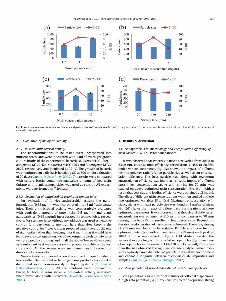
Fig. 1. Variation in nisin encapsulation efficiency and particle size with variation in (a) nisin to polymer ratio, (b) concentration of cross linker calcium chloride, (c) concentration ofnisin (d) stirring time.
M. Bernela et al. / LWT - Food Science and Technology 59 (2014) 1093e1099 1095
2.6. Evaluation of biological activity
2.6.1. In-vitro antibacterial activityThe nanoformulations to be tested were incorporated into
nutrient broth, and were inoculated with 1 ml of overnight grownculture broths of the experimental bacteria M. luteus MTCC 1809, P.aeruginosaMTCC 424, S. entericaMTCC 1253 and E. aerogenesMTCC2823, respectively and incubated at 37 �C. The growth of bacteriawas monitored on daily basis by taking OD at 600 nm for a durationof 20 days (Carson, Mee, & Riley, 2002). The results were comparedwith culture broths containing equivalent amount of free nisin.Culture with blank nanoparticles was used as control. All experi-ments were performed in Triplicate.
2.6.2. Evaluation of antimicrobial activity in tomato juiceFor evaluation of in vivo antimicrobial activity the nano-
formulation (0.68mg/ml) was incorporated into 15ml fresh tomatojuice. Their antimicrobial activity was comparatively evaluatedwith equivalent amount of pure nisin (0.5 mg/ml) and blanknanoparticles (0.68 mg/ml) incorporated in tomato juice, respec-tively. Pure tomato juice without any additive was used as negativecontrol. It is pertinent to mention here that after keeping thenegative control for 1 week, it was prepared again towards the endof six months rather than keeping it for 6 months, as it would haveled to severe contamination. For all experiments fresh tomato juicewas prepared by grinding, and in all the above Tween 40 was usedas a surfactant as it was necessary for proper solubility of the testsubstances. All the setups were maintained and observed forduration of six months.
Nisin activity is enhanced when it is applied in liquid media orfoods rather than in solid or heterogeneous products because it isdistributed more homogenously in liquid systems (Thomas &Delves-Broughton, 2005). All the solutions were prepared intween 40 because nisin shows antimicrobial activity in tomatowhen mixed along with surfactant (Hakovirta, Reunanen, & Saris,2006).
3. Results & discussion
3.1. Nanoparticle size, morphology and encapsulation efficiency ofnisin-loaded ALGeCSePF68 nanoparticles
It was observed that whereas, particle size varied from 208.2 to831.9 nm, encapsulation efficiency varied from 41.45% to 88.36%,under various treatments. Fig. 1(a) shows the impact of differentnisin to polymer ratio (v/v) on particle size as well as on encapsu-lation efficiency. The best particle size along with maximumencapsulation efficiency was found at 1:1 ratio. Impact of differentcross-linker concentrations along with stirring for 70 min, wasstudied on above optimized nisin concentration [Fig. 1(b)], with aresult that best size and loading efficiencywere obtained at 2mg/ml.The effect of different nisin concentrations was then studied at thesetwo optimized variables [Fig. 1(c)]. Maximum encapsulation effi-ciency along with least particle size was found at 1 mg/ml of nisin.Fig. 1(d) shows the impact of different stirring durations at theseoptimized parameters. It was observed that though a slightly lesserencapsulation was obtained at 210 min, in comparison to 70 minstirring time but 210 min resulted in least particle size. Beyond thisduration agglomeration of particles was observed. Therefore, stirringof 210 min was found to be suitable. Particle size curve for thisoptimised batch (i.e. with stirring time of 210 min) with peak at208.2 d nm is represented in Fig. 2. TEM studies revealed thespherical morphology of nisin-loaded nanoparticles (Fig. 3) and sizeof nanoparticles in the range of 130e178 nm. Expectedly this is lessthan the size observed through particle size analyzer, which mea-sures hydrodynamic diameter of particle in its native environmentand cannot distinguish between microparticulate impurities andsample (Kaur, Ahuja, Kumar, & Dilbaghi, 2012).
3.2. Zeta potential of nisin-loaded ALGeCSePF68 nanoparticles
Zeta potential is an indicator of stability of colloidal dispersions.A high zeta potential (>j30j mV) ensures electric repulsion strong

Fig. 2. Particle size of nisin loaded alginateechitosanepluronic F68 nanoparticles dispersed in distilled water as observed by particle size analyzer.
M. Bernela et al. / LWT - Food Science and Technology 59 (2014) 1093e10991096
enough to avoid aggregation of the particles (Labhatestwer, Song,&Levy, 1997). It also determines the in vivo interaction of nano-particles. With an increase in zeta potential, the repulsive in-teractions will be larger between the charged particles leading tothe formation of more stable suspension with a more uniform sizedistribution. Fig. 4 shows the zeta potential curve of nisin-loadednanoparticles with a zeta potential value of 36.4 mV. This valueof zeta potential indicates good stability of the nanoparticlepreparation.
3.3. FTIR analysis of nisin-loaded ALGeCSePF68 nanoparticles
Fig. 5 shows FTIR spectra of nisin, blank nanoparticles andnisin loaded nanoparticles. Spectra of nisin gives broad band at3288 cm�1 due to OH stretching of COOH group, peak at2960 cm�1 which can be attributed to CeH Stretching andpeak at 1232 cm�1 which can be ascribed to OeH group. Peak at1645 cm�1 attributed to amide group. Peak at 1527 cm�1 is dueto bending of primary amines. In spectra of nisin-loadednanoparticles, a peak at 3284 cm�1 is attributed to free OeHgroup of COOH, at 2935 cm�1 corresponds to the CeH stretchingand another at 1085 cm�1 corresponds to secondary hydroxylgroup. Spectral analysis indicated that the specific functionalgroups of polymeric material in the nanoparticles surface havealmost the same chemical characteristics as that of nisin. Thestudy suggests that chemical interactions between functionalgroup of nisin and polymer, that could have altered the chemicalstructure of nisin, did not occur. Earlier in another reportwhere alpha-1 antitrypsin was encapsulated in poly (D, L lactide-co glycolide) (PLGA) nanoparticles, no interaction was observed
Fig. 3. TEM image of nisin loaded alginateechitosanepluronic F68 nanoparticles at200 KV and �29,000 magnification.
in FTIR spectral results (Pirooznia, Hasannia, Lotfi, & Ghanei,2012).
3.4. In vitro release of nisin from nisin-loaded ALGeCSePF68nanoparticles
Release of nisin from loaded ALGeCSePF68 NPs occurred in asustained manner, and in the first few hours less than 40% ofnisin was released from the particles. It gradually increased up to60% in 24 h, and by the end of 240 h about 86% of nisin wasreleased from the nanoformulation. In contrast to this, more than80% of free nisin was released within 2 h from the dialysis baginto the saline solution. Earlier, Zohri et al. (2010) reportedrelease of nisin for 25 days, and that nisin release is pH depen-dent. In the present study also, nisin release was found todecrease with increase in pH, and data shown in Fig. 6 showsrelease of nisin at pH 7.4.
3.5. Biological activity
3.5.1. In-vitro antibacterial activityThe antimicrobial activity of free nisin and nisin-loaded
nanoparticles are shown in Fig. 7(aed). Free nisin displayedantimicrobial activity against (a) M. luteus MTCC 1809, (b)P. aeruginosa MTCC 424, (c) S. enterica MTCC 1253 and (d)E. aerogenes MTCC 2823 for up to 6th day and thereafter thebacterial growth commenced heavily, suggesting no significantantibacterial activity persisted beyond these time points. Incontrast, the inhibitory effect of nisin encapsulated inALGeCSePF68 nanoparticles against M. luteus MTCC 1809, P.aeruginosa MTCC 424 and S. enterica MTCC 1253 and E. aerogenesMTCC 2823 was prolonged for at least up to 20 days.
Blank nanoparticles (a negative control) showed reducedantimicrobial activity and nutrient medium (positive control) didnot show any antimicrobial activity. Earlier, Prombutara et al.(2012) had shown that the antibacterial activity of nisin-loadedSLNs against Listeria monocytogenes DMST 2871 and Lactoba-cillus plantarum TISTR 850 was evident for up to 20 and 15 days,respectively, compared to only one and three days, respectively,for free nisin. In milk and cheese samples also similar resultswere shown earlier (Zohri et al., 2010, 2013). Studies in thepresent work on new microbial strains and new substrate i.e.tomato juice, further expand the scope of biopreservatives infood.
In 2010, Santos, Ribeiro, Knirsch, Pessoa Jr., Penna demon-strated that Pluronic F68 improved the antimicrobial effective-ness of the ceftazidime drug, a third generation cephalosporinwith wide action spectrum, against Escherichia coli andP. aeruginosa microorganisms. In 2012, Santos, Jozala, Pessoa Jr.,

Fig. 4. Zeta potential curve of nisin loaded alginateechitosanepluronic F68 nanoparticles.
M. Bernela et al. / LWT - Food Science and Technology 59 (2014) 1093e1099 1097
and Seckler reported that the effectiveness improved to a MICof 12.5% in the presence of the Pluronic F68 costabilizer forPVP stabilized Ag NPs. In the present study Pluroniccopolymer was used to enhance stability as well as bactericidaleffect, therefore, the enhanced antimicrobial activitymay be attributed to a combination effect of chitosan andPluronic.
Fig. 5. FTIR spectra of nisin, blank alginateechitosanepluro
3.5.2. Biological activity in tomato juiceFig. 8 displays the in vivo evaluation of nisin loaded
ALGeCSePF68 NPs, blank nanoparticles and pure nisin in tomatojuice. Tomato juice without any additive i.e. control showed growthof microorganism in 2 days. In case of pure nisin, no growth wasseen till 5 months but seed colour turned black. For blank nano-particles, no visible indication of microorganism growth was
nic F68 nanoparticles and nisin loaded nanoparticles.

Fig. 6. Comparison of in vitro release profile of nisin and nisin loaded nanoparticles.
Fig. 7. In vitro antibacterial activity of nisin, blank nanoparticles, nisin loaded nanoparticles and control against (a)Micrococcus luteusMTCC 1809, (b) Pseudomonas aeruginosa MTCC424, (c) Salmonella enterica MTCC 1253 and (d) Enterobactor aerogenes MTCC 2823, over a period of 20 days.
M. Bernela et al. / LWT - Food Science and Technology 59 (2014) 1093e10991098
observed up to a period of two months whereas nisin loadednanoparticles exhibited bacteriostatic effect on tomato juice for 6months. The reason behind this may be attributed to the cumula-tive effect of nisin and polymers as well as sustained release of nisinfrom the nanoparticles into the juice. The results were consistentwith those previously reported by Zohri et al. (2013), that nisin-loaded chitosan/alginate nanoparticles show more antibacterialpotency than free nisinwithout any unwanted effects on the qualityof the original UF Feta cheese on which it was tested.
4. Conclusion
Nisin loaded ALGeCSePF68 nanoparticles were synthesized byionotropic pre-gelation method followed by polycationic cross-linking and were characterized using techniques such as PSA,TEM and FTIR. The results revealed that particle size and encap-sulation efficiency varied with change in nisin to polymer ratio,nisin concentration, cross linker concentration and stirring time.Electron microscopy studies showed that shape of the optimized

Fig. 8. Antimicrobial activity of pure nisin, blank nanoparticles, and nisin loadednanoparticles, against negative control in tomato juice over a period of 6 months(Excluding negative control).
M. Bernela et al. / LWT - Food Science and Technology 59 (2014) 1093e1099 1099
nanoparticles was spherical. The nanoparticulate formulationexhibited long lasting antimicrobial activity against microbesin vitro as well as in fresh tomato juice which can be attributed tothe synergistic effect of nisin and ALGeCSePF68 nanoparticlesalong with slow and sustained release of the loaded nisin.
Whereas, further in vivo studies shall determine the preciseconcentration of nisin in food products, nevertheless, the workpresents an excellent nanoparticulate system for delivery of nisinwith food permissible polymers in a manner such that the sub-stance delivered is protected against undesired interactions withthe food matrix material, and its efficacy is maintained over aprolonged period. The combination may be further investigated forin-vivo studies and may likely be included in future new foodpreservative systems. Moreover, the composite nanoparticles canbe used for delivery of other preservatives or antimicrobial agentsalso in a similar manner, in order to enhance the duration of theirefficacy.
Acknowledgements
The authors are thankful to Department of Science and Tech-nology (DST), New Delhi, India for providing kind support. Authorsare also grateful to AIIMS, New Delhi for analysis of sample by TEMand to Dr. Sandeep Kumar for FTIR analysis, and to Dr. Neeraj Dil-baghi, Chairperson, Department of Bio & Nanotechnology GuruJambheshwer University of Science & Technology, Hisar forproviding the necessary research facilities. Manju Bernela isthankful to DST IF120488 (2012) for INSPIRE Fellowship.
References
Carson, C. F., Mee, B. J., & Riley, T. V. (2002). Mechanism of action of Melaleucaalternifolia (tea tree) oil on Staphylococcus aureus determined by time-kill, lysis,leakage, and salt tolerance assays and electron microscopy. Antimicrobial AgentsChemotherapy, 48, 1914e1920.
Cha, D. S., Cooksey, K., Chinnan, M. S., & Park, H. J. (2003). Release of nisin fromvarious heat-pressed and cast films. LWT e Food Science and Technology, 36(2),209e213.
Chopra, M., Kaur, P., Bernela, M., & Thakur, R. (2012). Synthesis and optimization ofstreptomycin loaded chitosan-alginate nanoparticles. International Journal ofScientific & Technology Research, 1(10), 31e34.
Das, R. K., Kasoju, N., & Bora, U. (2010). Encapsulation of curcumin in alginate-chitosan-pluronic composite nanoparticles for delivery to cancer cells. Nano-medicine: Nanotechnology, Biology, and Medicine, 6, 153e160.
Delves-Broughton, J., & Gasson, M. J. (1994). Nisin. In V. M. Dillon, & R. G. Board(Eds.), Natural antimicrobial systems in food preservation (pp. 99e132). Wall-ingford, UK: CAB International.
Dilbaghi, N., Kaur, H., Ahuja, M., Arora, P., & Kumar, S. (2013). Synthesis and eval-uation of ciprofloxacin-loaded carboxymethyl tamarind kernel polysaccharidenanoparticles. Journal of Experimental Nanoscience, 1e11.
Dilbaghi, N., Kaur, H., Ahuja, M., & Kumar, S. (2013). Evaluation of tropicamide-loaded tamarind seed xyloglucan nanoaggregates for ophthalmic delivery.Carbohydrate Polymers, 94, 286e291.
Hakovirta, J., Reunanen, J., & Saris, P. E. J. (2006). Bioassay for nisin in milk processedcheese, salad dressings, canned tomatoes, and liquid egg products. Applied andEnvironmental Microbiology, 72, 1001e1005.
Iskakov, R. M., Kikuchi, A., & Okano, T. (2002). Time-programmed pulsatilerelease of dextran from calcium alginate gel beads coated with carboxy-n-propylacrylamide copolymers. Journal of Controlled Release, 80(1e3),57e68.
Jimenez-Escrig, A., & Sanchez-Muniz, F. J. (2000). Dietary fiber from edible sea-weeds: chemical structure, physicochemical properties, and effects on choles-terol metabolism. Nutrition Research, 20, 585e598.
Kaur, H., Ahuja, M., Kumar, S., & Dilbaghi, N. (2012). Carboxymethyl tamarind kernelpolysaccharide nanoparticles for ophthalmic drug delivery. International Journalof Biological Macromolecules, 50, 833e839.
Kaur, H., Yadav, S., Ahuja, M., & Dilbaghi, N. (2012). Synthesis, characterization andevaluation of thiolated tamarind seed polysaccharide as a mucoadhesivepolymer. Journal of Carbohydrate Polymers, 90, 1543e1549.
KFDA. (1995). Food additives code. Seoul: Korea Food and Drug Administration.Kong, M., Chen, X. G., Xing, K., & Park, H. J. (2010). Antimicrobial properties of
chitosan and mode of action: a state of the art review. International Journal ofFood Microbiology, 144, 51e63.
Labhatestwer, V., Song, C., & Levy, R. J. (1997). Nanoparticle drug delivery systems.Advanced Drug Delivery Reviews, 434(24), 63e85.
Meena, B., Aparna, V., & Shelef, L. A. (2004). Factors affecting the antilisterial effectsof nisin in milk. International Journal of Food Microbiology, 97, 215e219.
Moghimi, S. M., & Hunter, A. C. (2000). Poloxamers and poloxamines in nanoparticleengineering and experimental medicine. Trends in Biotechnology, 18(10),412e420.
Pirooznia, N., Hasannia, S., Lotfi, A. S., & Ghanei, M. (2012). Encapsulation of Alpha-1antitrypsin in PLGA nanoparticles: in vitro characterization as an effectiveaerosol formulation in pulmonary diseases. Journal of Nanobiotechnology,10(20), 1e15.
Pranoto, Y., Rakshit, S. K., & Salokhe, V. M. (2005). Enhancing antimicrobial activityof chitosan films by incorporating garlic oil, potassium sorbate and nisin. LWT e
Food Science and Technology, 38(8), 859e865.Prombutara, P., Kulwatthanasal, Y., Supaka, N., Sramala, I., & Chareonpornwattana, S.
(2012). Production of nisin-loaded solid lipid nanoparticles for sustained anti-microbial activity. Food Control, 24, 184e190.
Ren, D., Noda, H., Amano, H., Nishino, T., & Nishizana, K. (1994). Study on the hy-pertensive and antihyperlipidemic effect of marine algae. Fisheries Science, 60,83e88.
Rodeheaver, G. T., Kurtz, L., Kircher, B. J., & Edlich, R. F. (1980). Pluronic F-68: apromising new skin wound cleanser. Annals of Emergency Medicine, 9(11),572e576.
Rohani, S. M. R., Mehran, M., Tooraj, M., Seyyed, S. S., & Mansel, W. G. (2011). Theeffect of nisin and garlic (Allium sativum L.) essential oil separately and incombination on the growth of Listeria monocytogenes. LWT e Food Science andTechnology, 44(10), 2260e2265.
Samelis, J., Bedie, G. K., Sofos, J. N., Belk, K. E., Scanga, J. A., & Smith, G. C. (2005).Combinations of nisin with organic acids or salts to control Listeria mono-cytogenes on sliced pork bologna stored at 4 �C in vacuum packages. LWT e FoodScience and Technology, 38(1), 21e28.
Santander-Ortega, M. J., Jodar-Reyes, A. B., Csaba, N., Bastos-Gonzalez, D., & Ortega-Vinuesa, J. L. (2006). Colloidal stability of Pluronic F68-coated PLGA nano-particles: a variety of stabilisation mechanisms. Journal of Colloid and InterfaceScience, 302, 522e529.
Santos, C. A., Jozala, A. F., Pessoa, A., Jr., & Seckler, M. M. (2012). Antimicrobialeffectiveness of silver nanoparticles co-stabilized by the bioactive copolymerpluronic F68. Journal of Nanobiotechnology, 10(43).
Santos, C. A., Ribeiro, G. B., Knirsch, M. C., Pessoa, A., Jr., & Penna, T. C. V. (2010).Influence of pluronic F68 on ceftazidime biological activity in parenteral solu-tions. Journal of Pharmaceutical Sciences, 1, 1e6.
Thomas, L. V., & Delves-Broughton, J. (2005). Nisin. In P. M. Davidson, J. N. Sofos, &A. L. Branen (Eds.), Antimicrobials in food (3rd ed) (pp. 237e274). CRC Press.
Weiner, M. L. (1992). In C. J. Brine, P. A. Sandford, & J. P. Zikakis (Eds.), Advances inchitin and chitosan. London: Elsevier (663 pp.).
Xiao, D., Davidson, P. M., & Zhong, Q. (2011). Release and antilisterial properties ofnisin from zein capsules spray-dried at different temperatures. LWT e FoodScience and Technology, 44(10), 1977e1985.
Zohri, M., Alavidjeh, M. S., Mirdamadi, S. S., Behmadi, H., Nasr, S. M. H.,Gonbaki, S. E., et al. (2013). Nisin-loaded chitosan/alginate nanoparticles: ahopeful hybrid biopreservative. Journal of Food Safety, 33, 40e49.
Zohri, M., Shafiee Alavidjeh, M., Haririan, I., Shafiee Ardestani, M., SadatEbrahimi, S. E., Tarighati Sani, H., et al. (2010). A comparative study between theantibacterial effect of nisin and nisin-loaded chitosan/alginate nanoparticles onthe growth of staphylococcus aureus in raw and pasteurized milk samples.Probiotics Antimicrobial Proteins, 2, 258e266.


![[27] Ion channels in microbes](https://img.pdfslide.net/doc/110x75/636172af8838ef044c075453/27-ion-channels-in-microbes.jpg)


