Embed Size (px)
Citation preview
Full Paper
540
Synthesis, Degradability, and Drug ReleasingProperties of Methyl Esters of FungalPoly(b,L-malic acid)
Jose A. Portilla-Arias, Montserrat Garcıa-Alvarez,Antxon Martınez de Ilarduya, Eggerhard Holler, Juan A. Galbis,Sebastian Munoz-Guerra*
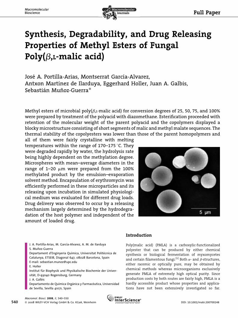
Methyl esters of microbial poly(b,L-malic acid) for conversion degrees of 25, 50, 75, and 100%were prepared by treatment of the polyacid with diazomethane. Esterification proceeded withretention of the molecular weight of the parent polyacid and the copolymers displayed ablockymicrostructure consisting of short segments ofmalic andmethylmalate sequences. Thethermal stability of the copolyesters was lower than those of the parent homopolymers andall of them were fairly crystalline with meltingtemperatures within the range of 170–175 8C. Theywere degraded rapidly by water, the hydrolysis ratebeing highly dependent on the methylation degree.Microspheres with mean–average diameters in therange of 1–20 mm were prepared from the 100%methylated product by the emulsion–evaporationsolvent method. Encapsulation of erythromycin wasefficiently performed in these microparticles and itsreleasing upon incubation in simulated physiologi-cal medium was evaluated for different drug loads.Drug delivery was observed to occur by a releasingmechanism largely determined by the hydrodegra-dation of the host polymer and independent of theamount of loaded drug.
J. A. Portilla-Arias, M. Garcıa-Alvarez, A. M. de IlarduyaS. Munoz-GuerraDepartament d’Enginyeria Quımica, Universitat Politecnica deCatalunya, ETSEIB, Diagonal 647, 08028 Barcelona, SpainE-mail: [email protected]. HollerInstitut fur Biophysik und Physikalische Biochemie der Univer-sitat, D-93040 Regensburg, GermanyJ. A. GalbisDepartamento de Quımica Organica y Farmaceutica, Universidadde Sevilla, Sevilla 41071, Spain
Macromol. Biosci. 2008, 8, 540–550
� 2008 WILEY-VCH Verlag GmbH & Co. KGaA, Weinheim
Introduction
Poly(malic acid) (PMLA) is a carboxylic-functionalized
polyester that can be produced by either chemical
synthesis or biological fermentation of myxomycetes
and certain filamentous fungi.[1] Both a- and b-structures,
either racemic or optically pure, may be obtained by
chemical methods whereas microorganisms exclusively
generate PMLA of extremely high optical purity. Since
production costs by both routes are fairly high, PMLA is a
hardly accessible product whose properties and applica-
tions have not been extensively investigated so far.
DOI: 10.1002/mabi.200700248
Synthesis, Degradability, and Drug Releasing Properties . . .
Nevertheless, PMLA displays exceptionally good features
for being used as a drug carrier. It is a perfectly biodegrad-
able and biocompatible polymer that is metabolized in
the mammalian tricarboxylic acid cycle.[2] Some PMLA
derivatives have been proposed for drug targeting[3]
as components of crosslinked prodrugs,[4] and also as
scaffolding for tissue regeneration.[5] Recently, a nano-
conjugate prototype of a drug delivery system based on
PMLA produced by Physarum polycephalum has been
reported for brain cancer chemotherapy.[6] PMLA is readily
water soluble and is hydrolyzed very fast in aqueous
environment.[7] Blocking of carboxylic side groups of
PMLA has been a strategy frequently used to modify its
properties and simultaneously to slow down its hydrolysis
under the assumption that the convenient biological
behavior of the parent polyacid is retained. A good amount
of work has been made on the benzyl[8] and methyl[9]
esters of chemically synthesized PMLA. Furthermore,
coupling of PMLA with alkyltrimethylammonium surfac-
tants bearing long alkyl chains has been shown to be a
reliable method to produce stoichiometric ionic complexes
with a biphasic amphiphilic nanostructure[10] able to lodge
hydrophobic drugs in the paraffinic subphase.[11]
So far, little has been reported on the biodegradability of
PMLA derivatives other than the chemically prepared
benzyl and hexyl esters. According to Holler[12] naturally
occurring PMLA can be degraded by the hydrolase of
P. polycephalum, an exohydrolytic enzyme that degrades
specifically L-units. The non-enzymatic hydrolysis of
synthetic PMLA was extensively studied by Braud and
Vert.[7] Regardless of chirality, degradation proceeds
rapidly according to an autocatalytic random scission of
the main chain ester bonds to yield malic acid as the final
degradation product. The hydrolytic degradation of PMLA
benzyl esters appeared much more complex. In contrast
to PMLA, the fully benzylated product is resistant to
hydrolysis whereas partially converted PMLA degrades
depending on the esterification degree because of the
faster degradation of acid rich segments. It has also been
shown that pellets made from these copolymers, as well as
large devices made of poly(lactic acid) degraded hetero-
geneously and much faster than corresponding films and
particles because of acidic autocatalysis and diffusion-
dependent phenomena.[13] Barbosa et al.[14] have recently
reported on the water degradation of nanoparticles made
of benzyl and hexyl esters of PMLA[15] and have observed
that hydrolysis is not greatly influenced by the esterifica-
tion degree. These authors found that nanospheres
degraded with a progressive decrease in the molecular
weight of the copolymers demonstrating that the degra-
dation proceeded by a random hydrolytic cleavage of the
main chain ester bond.
In this work we wish to report on the synthesis,
hydrolytic degradation, and drug delivering properties of
Macromol. Biosci. 2008, 8, 540–550
� 2008 WILEY-VCH Verlag GmbH & Co. KGaA, Weinheim
methyl esters of biosynthetic PMLA. Methylation with
diazomethane of fungal PMLA has proven to be an
effective method to produce poly(a-methyl b,L-malic acid)
(PMLA-Me) without perceivable breaking of the initial
polymer chain.[9b] The method is applied here to prepare a
set of partially methylated products with different con-
version degrees, which will be designated in this work as
coPMLA-(MexHy), where x and y refer to the percentages of
methyl malate and malic units contained in the copoly-
mer. The hydrolytic degradation of these products is
comparatively evaluated and the release of erythromycin
from microspheres made of PMLA-Me is quantitatively
examined. To the best of our knowledge it is the first time
that methyl esters of PMLA are investigated in this regard.
Experimental Part
Materials
Poly(b,L-malic acid) of microbial origin was used in this work. It
was obtained by cultivation of P. polycephalum and subsequent
purification as described in detail elsewhere.[12] The final polyacid
was NMR pure and had an Mw ¼34 300 Da and Mw=Mn ¼1.08, as
determined by GPC. Erythromycin, assay 95% (NT), was supplied
by Fluka and lipase from Candida cylindracea (EC 3.1.1.3, Type VII,
943 units �mg�1) was purchased from Sigma. Organic solvents
were of analytical grade and used without further purification.
Water used for buffer preparation was double distilled and
deionized in a ‘‘Milli-Q’’ system.
Esterification
A solution of diazomethane[9] in ether (0.25 M) was added in dif-
ferent ratios, according to the methylation degree to be attained,
to a solution of PMLA in dry acetone (500 mg, 4.3 mmol) and the
mixture was left under stirring at room temperature for 1 h. In all
cases, the reaction proceeded with precipitation of the partially
esterified PMLA. The reaction mixture was then evaporated under
vacuum and the residue was dissolved in a small amount of
N-methyl pyrrolidone (NMP) and added with cold diethyl ether,
the PMLA methyl esters being recovered by filtration as white
powders. coPMLA-(MexHy) with nominallymethylation degrees of
25, 50, and 75% in addition to fully methylated PMLA-Me were
prepared by this method.
Hydrolytic and Enzymatic Degradation
In the case of coPMLA-(Me25H75) and coPMLA-(Me50H50), the
degradation study was performed using a solution with a
concentration of 1 mg of copolymer in 1 mL of buffered saline
solutions of selected pHs (pH 4.0 citrate buffer, pH 11.0 Na2HPO4/
NaOH buffer, and pH 7.4 phosphate buffer). Degradation of the
nonwater-soluble coPMLA-(Me75H25) and PMLA-Me compounds
was performed on 5 mm diameter and 1 mm thick disks,
which were cut from polymer films prepared by casting from
www.mbs-journal.de 541
J. A. Portilla-Arias, M. Garcıa-Alvarez, A. M. de Ilarduya, E. Holler, J. A. Galbis, S. Munoz-Guerra
542
hexafluoroisopropanol solutions (10mg �mL�1). Disks were placed
into vials and coveredwith 5mL of buffered solutions. The sample
vials were sealed to avoid evaporation of the fluids and stored at
37 8C in a heat chamber. The enzymatic degradation was carried
out with lipase (0.1 mg �mL�1) in a phosphate buffered saline
solution (pH 7.4, 37 8C). At the end of the incubation period,
the disks were withdrawn, from the incubation medium,
washed with distilled water, dried, weighted, and analyzed by
gel permeation chromatography (GPC) by triplicate.
Preparation of Microspheres, Hydrodegradation,
and Erythromycin Release
Microsphereswere prepared by the emulsion–evaporation solvent
method. PMLA-Me was dissolved in chloroform (4 mg �mL�1), and
then erythromycin was dissolved in the polymer solution at
concentrations of 10, 20, and 30 wt.-% with respect to PMLA-Me.
The solution was added with different amounts of poly(vinyl
alcohol) (4, 6, 8, and 10%), which acts as emulsifier, and stirred
at different speeds (400, 800, 1 200, and 1 600 rpm) at room
temperature until the solvent evaporated completely. The
evolution of microspheres formation was followed by optical
microscopy. At the end of the process, the microspheres were
rinsed three times with distilled water and finally recovered by
freeze-drying.
The hydrolytic degradation of microspheres was performed
by placing 10 mg of the particles into vials and adding 5 mL of
buffered solutions at different pHs 4.0, 11.0, and 7.4. The sample
vials were sealed and stored at 37 8C in a heat chamber. The
temperature effect was evaluated at pH 7.4 in samples stored at
37, 60, and 80 8C. The enzymatic degradation was carried out in
a phosphate buffered saline solution (pH 7.4, 37 8C) with lipase
(0.1 mg �mL�1). At predetermined intervals, the microparticles
were withdrawn, washed with distilled water, filtered, and
analyzed by GPC. The analysis was carried out by triplicate.
The amount of active ingredient incorporated into the micro-
spheres was estimated by UV–Vis spectrophotometry. Randomly
selected loaded microspheres (10 mg) with drug loads of 10,
20, and 30 wt.-% were dissolved in CHCl3 and the erythromycin
present was determined using the appropriate blank and
calibration curve. The in vitro release test was carried out by
incubation of the 10 mg of loaded microspheres in 10 mL of the
phosphate saline buffer (pH 7.4 at 37 8C). Aliquots of 2 mL
were taken from the releasing media at scheduled times and the
removed volume being replaced by fresh medium every time.
The amount of delivered drug was estimated by spectrophoto-
metry and the cumulative drug concentration plotted against the
release time.
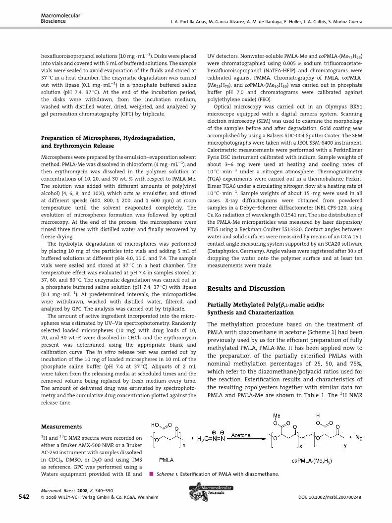
Scheme 1. Esterification of PMLA with diazomethane.
Measurements
1H and 13C NMR spectra were recorded on
either a Bruker AMX-500 NMR or a Bruker
AC-250 instrumentwith samples dissolved
in CDCl3, DMSO, or D2O and using TMS
as reference. GPC was performed using a
Waters equipment provided with IR and
Macromol. Biosci. 2008, 8, 540–550
� 2008 WILEY-VCH Verlag GmbH & Co. KGaA, Weinheim
UV detectors. Nonwater-soluble PMLA-Me and coPMLA-(Me75H25)
were chromatographied using 0.005 M sodium trifluoroacetate-
hexafluoroisopropanol (NaTFA-HFIP) and chromatograms were
calibrated against PMMA. Chromatography of PMLA, coPMLA-
(Me25H75), and coPMLA-(Me50H50) was carried out in phosphate
buffer pH 7.0 and chromatograms were calibrated against
poly(ethylene oxide) (PEO).
Optical microscopy was carried out in an Olympus BX51
microscope equipped with a digital camera system. Scanning
electron microscopy (SEM) was used to examine the morphology
of the samples before and after degradation. Gold coating was
accomplished by using a Balzers SDC-004 Sputter Coater. The SEM
microphotographs were taken with a JEOL SSM-6400 instrument.
Calorimetric measurements were performed with a PerkinElmer
Pyris DSC instrument calibrated with indium. Sample weights of
about 3–6 mg were used at heating and cooling rates of
10 8C �min�1 under a nitrogen atmosphere. Thermogravimetry
(TGA) experiments were carried out in a thermobalance Perkin-
Elmer TGA6 under a circulating nitrogen flow at a heating rate of
10 8C �min�1. Sample weights of about 15 �mg were used in all
cases. X-ray diffractograms were obtained from powdered
samples in a Debye–Scherrer diffractometer INEL CPS-120, using
Cu Ka radiation of wavelength 0.1541 nm. The size distribution of
the PMLA-Me microparticles was measured by laser dispersion/
PIDS using a Beckman Coulter LS13320. Contact angles between
water and solid surfaces were measured by means of an OCA 15þcontact angle measuring system supported by an SCA20 software
(Dataphysics, Germany). Angle values were registered after 30 s of
dropping the water onto the polymer surface and at least ten
measurements were made.
Results and Discussion
Partially Methylated Poly(b,L-malic acid)s:Synthesis and Characterization
The methylation procedure based on the treatment of
PMLA with diazomethane in acetone (Scheme 1) had been
previously used by us for the efficient preparation of fully
methylated PMLA, PMLA-Me. It has been applied now to
the preparation of the partially esterified PMLAs with
nominal methylation percentages of 25, 50, and 75%,
which refer to the diazomethane/polyacid ratios used for
the reaction. Esterification results and characteristics of
the resulting copolyesters together with similar data for
PMLA and PMLA-Me are shown in Table 1. The 1H NMR
DOI: 10.1002/mabi.200700248
Synthesis, Degradability, and Drug Releasing Properties . . .
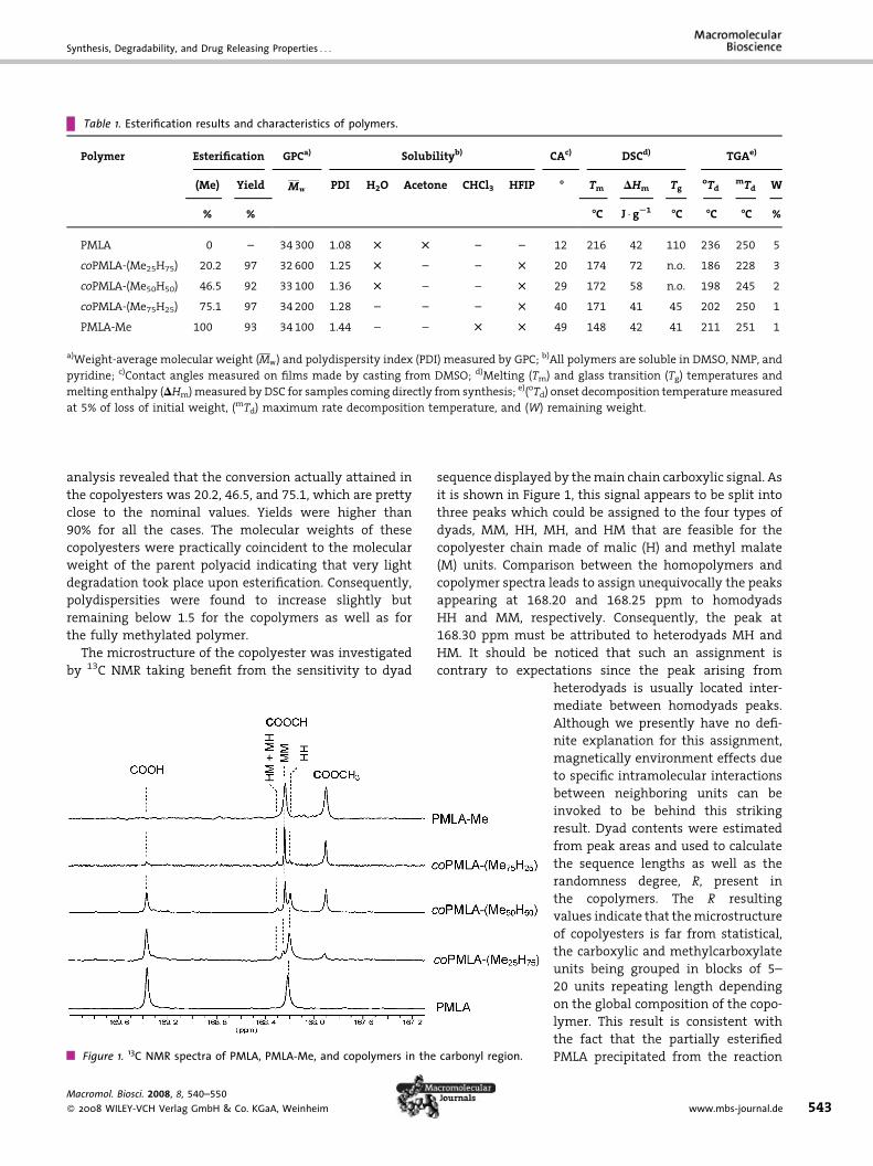
Table 1. Esterification results and characteristics of polymers.
Polymer Esterification GPCa) Solubilityb) CAc) DSCd) TGAe)
(Me) Yield Mw PDI H2O Acetone CHCl3 HFIP - Tm DHm TgoTd
mTd W
% % -C J � gS1 -C -C -C %
PMLA 0 – 34300 1.08 T T – – 12 216 42 110 236 250 5
coPMLA-(Me25H75) 20.2 97 32600 1.25 T – – T 20 174 72 n.o. 186 228 3
coPMLA-(Me50H50) 46.5 92 33100 1.36 T – – T 29 172 58 n.o. 198 245 2
coPMLA-(Me75H25) 75.1 97 34200 1.28 – – – T 40 171 41 45 202 250 1
PMLA-Me 100 93 34100 1.44 – – T T 49 148 42 41 211 251 1
a)Weight-average molecular weight (Mw) and polydispersity index (PDI) measured by GPC; b)All polymers are soluble in DMSO, NMP, and
pyridine; c)Contact angles measured on films made by casting from DMSO; d)Melting (Tm) and glass transition (Tg) temperatures and
melting enthalpy (DHm)measured byDSC for samples coming directly from synthesis; e)(oTd) onset decomposition temperaturemeasured
at 5% of loss of initial weight, (mTd) maximum rate decomposition temperature, and (W) remaining weight.
analysis revealed that the conversion actually attained in
the copolyesters was 20.2, 46.5, and 75.1, which are pretty
close to the nominal values. Yields were higher than
90% for all the cases. The molecular weights of these
copolyesters were practically coincident to the molecular
weight of the parent polyacid indicating that very light
degradation took place upon esterification. Consequently,
polydispersities were found to increase slightly but
remaining below 1.5 for the copolymers as well as for
the fully methylated polymer.
The microstructure of the copolyester was investigated
by 13C NMR taking benefit from the sensitivity to dyad
Figure 1. 13C NMR spectra of PMLA, PMLA-Me, and copolymers in the
Macromol. Biosci. 2008, 8, 540–550
� 2008 WILEY-VCH Verlag GmbH & Co. KGaA, Weinheim
sequence displayed by themain chain carboxylic signal. As
it is shown in Figure 1, this signal appears to be split into
three peaks which could be assigned to the four types of
dyads, MM, HH, MH, and HM that are feasible for the
copolyester chain made of malic (H) and methyl malate
(M) units. Comparison between the homopolymers and
copolymer spectra leads to assign unequivocally the peaks
appearing at 168.20 and 168.25 ppm to homodyads
HH and MM, respectively. Consequently, the peak at
168.30 ppm must be attributed to heterodyads MH and
HM. It should be noticed that such an assignment is
contrary to expectations since the peak arising from
carbonyl region.
heterodyads is usually located inter-
mediate between homodyads peaks.
Although we presently have no defi-
nite explanation for this assignment,
magnetically environment effects due
to specific intramolecular interactions
between neighboring units can be
invoked to be behind this striking
result. Dyad contents were estimated
from peak areas and used to calculate
the sequence lengths as well as the
randomness degree, R, present in
the copolymers. The R resulting
values indicate that themicrostructure
of copolyesters is far from statistical,
the carboxylic and methylcarboxylate
units being grouped in blocks of 5–
20 units repeating length depending
on the global composition of the copo-
lymer. This result is consistent with
the fact that the partially esterified
PMLA precipitated from the reaction
www.mbs-journal.de 543
J. A. Portilla-Arias, M. Garcıa-Alvarez, A. M. de Ilarduya, E. Holler, J. A. Galbis, S. Munoz-Guerra
Table 2. Composition, number–average sequence lengths, and randomness of methylated PMLA.
Polymer Composition
(mol-%)a)
Dyadsb) (mol-%) Number–average
sequence lengthb)
XM XH MM MHRHM HH nM nH Rb
PMLA 0 100 – – 100 – 274 0.0
coPMLA-(Me25H75) 20.2 79.8 18.6 13.0 68.4 3.9 11.5 0.3
coPMLA-(Me50H50) 46.5 53.5 41.2 7.6 51.2 11.8 14.5 0.2
coPMLA-(Me75H25) 75.1 24.9 74.0 9.0 17.0 17.4 4.8 0.3
PMLA-Me 100 0 100 – – 182 0.0
a)Determined from the signals arising from main chain methyne protons; b)Values estimated by deconvolution of main chain carbonyl
carbon peaks and calculations made according to ref.[16]
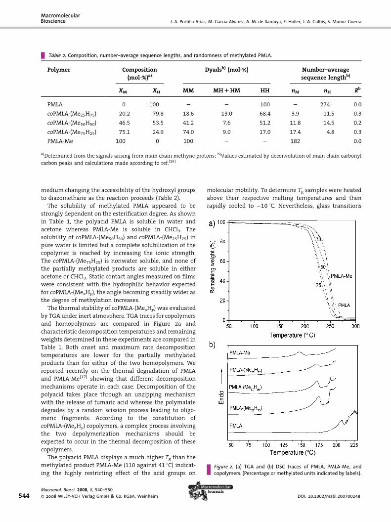
Figure 2. (a) TGA and (b) DSC traces of PMLA, PMLA-Me, andcopolymers. (Percentage or methylated units indicated by labels).
544
medium changing the accessibility of the hydroxyl groups
to diazomethane as the reaction proceeds (Table 2).
The solubility of methylated PMLA appeared to be
strongly dependent on the esterification degree. As shown
in Table 1, the polyacid PMLA is soluble in water and
acetone whereas PMLA-Me is soluble in CHCl3. The
solubility of coPMLA-(Me50H50) and coPMLA-(Me25H75) in
pure water is limited but a complete solubilization of the
copolymer is reached by increasing the ionic strength.
The coPMLA-(Me75H25) is nonwater soluble, and none of
the partially methylated products are soluble in either
acetone or CHCl3. Static contact angles measured on films
were consistent with the hydrophilic behavior expected
for coPMLA-(MexHy), the angle becoming steadily wider as
the degree of methylation increases.
The thermal stability of coPMLA-(MexHy) was evaluated
by TGA under inert atmosphere. TGA traces for copolymers
and homopolymers are compared in Figure 2a and
characteristic decomposition temperatures and remaining
weights determined in these experiments are compared in
Table 1. Both onset and maximum rate decomposition
temperatures are lower for the partially methylated
products than for either of the two homopolymers. We
reported recently on the thermal degradation of PMLA
and PMLA-Me[17] showing that different decomposition
mechanisms operate in each case. Decomposition of the
polyacid takes place through an unzipping mechanism
with the release of fumaric acid whereas the polymalate
degrades by a random scission process leading to oligo-
meric fragments. According to the constitution of
coPMLA-(MexHy) copolymers, a complex process involving
the two depolymerization mechanisms should be
expected to occur in the thermal decomposition of these
copolymers.
The polyacid PMLA displays a much higher Tg than the
methylated product PMLA-Me (110 against 41 8C) indicat-ing the highly restricting effect of the acid groups on
Macromol. Biosci. 2008, 8, 540–550
� 2008 WILEY-VCH Verlag GmbH & Co. KGaA, Weinheim
molecular mobility. To determine Tg samples were heated
above their respective melting temperatures and then
rapidly cooled to �10 8C. Nevertheless, glass transitions
DOI: 10.1002/mabi.200700248
Synthesis, Degradability, and Drug Releasing Properties . . .
were not clearly observable in copolymers coPMLA-
(MexHy) by DSC so that only the Tg of coPMLA-(Me75H25)
could be determinedwith acceptable confidence. The value
obtainedwas 45 8Cwhich is consistent with the increasing
trend that should be expected along the series for
decreasing contents in methylated units. PMLA has been
reported to show crystalline melting at 216 8C provided
that it is well dry[12b] but its crystal structure is not known.
On the other hand, PMLA-Me is a high crystalline polymer
able to crystallize from the melt displaying two crystal
modifications depending onmolecular weight and crystal-
lization conditions.[9b] The DSC traces of copolymers
coPMLA-(MexHy) are shown in Figure 2b revealing the
occurrence of melting in all of them at temperatures
around 170–175 8C with associated enthalpies of fairly
high values. Powder X-ray diffraction of these copolymers
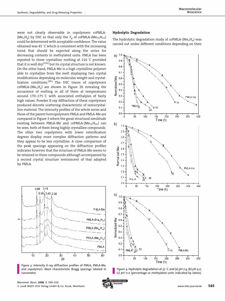
produced discrete scattering characteristic of semicrystal-
line material. The intensity profiles of the whole series and
those of the parent homopolymers PMLA and PMLA-Me are
compared in Figure 3 where the great structural similitude
existing between PMLA-Me and coPMLA-(Me75H25) can
be seen, both of them being highly crystalline compounds.
The other two copolymers with lower esterification
degrees display more complex diffraction patterns and
they appear to be less crystalline. A close comparison of
the peak spacings appearing on the diffraction profiles
indicates however that the structure of PMLA-Me seems to
be retained in these compounds although accompanied by
a second crystal structure reminiscent of that adopted
by PMLA.
Figure 3. Intensity X-ray diffraction profiles of PMLA, PMLA-Me,and copolymers. Most characteristic Bragg spacings labeled innanometer.
Macromol. Biosci. 2008, 8, 540–550
� 2008 WILEY-VCH Verlag GmbH & Co. KGaA, Weinheim
Hydrolytic Degradation
The hydrolytic degradation study of coPMLA-(MexHy) was
carried out under different conditions depending on their
Figure 4.Hydrolytic degradation at 37 8C and (a) pH 7.4, (b) pH 4.0,(c) pH 11.0 (percentage or methylated units indicated by labels).
www.mbs-journal.de 545
J. A. Portilla-Arias, M. Garcıa-Alvarez, A. M. de Ilarduya, E. Holler, J. A. Galbis, S. Munoz-Guerra
546
solubility in water. coPMLA-(Me25H75) and coPMLA-
(Me50H50) were dissolved in aqueous buffer and incubated
at 37 8C at pH 7.4 with or without added lipase. Conversely,
disks of the nonwater-soluble copolymer coPMLA-
(Me75H25) and the homopolymer PMLA-Me were incu-
bated at pH 7.4 and at 37 and 80 8C to evaluate the effect
of temperature. Additional incubations at pH 4.0 and
11.0 were carried out at 37 8C for all the polymers to
evaluate the influence of the medium pH. Results afforded
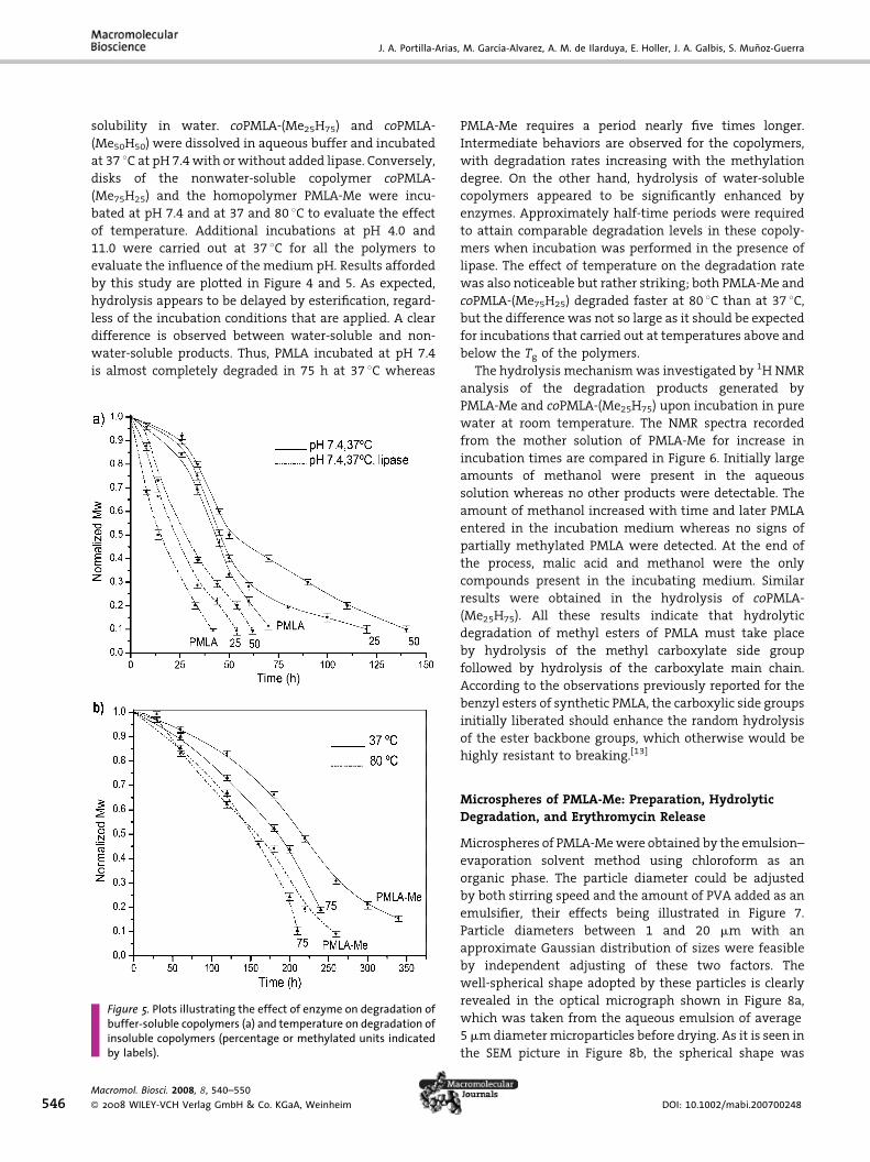
by this study are plotted in Figure 4 and 5. As expected,
hydrolysis appears to be delayed by esterification, regard-
less of the incubation conditions that are applied. A clear
difference is observed between water-soluble and non-
water-soluble products. Thus, PMLA incubated at pH 7.4
is almost completely degraded in 75 h at 37 8C whereas
Figure 5. Plots illustrating the effect of enzyme on degradation ofbuffer-soluble copolymers (a) and temperature on degradation ofinsoluble copolymers (percentage or methylated units indicatedby labels).
Macromol. Biosci. 2008, 8, 540–550
� 2008 WILEY-VCH Verlag GmbH & Co. KGaA, Weinheim
PMLA-Me requires a period nearly five times longer.
Intermediate behaviors are observed for the copolymers,
with degradation rates increasing with the methylation
degree. On the other hand, hydrolysis of water-soluble
copolymers appeared to be significantly enhanced by
enzymes. Approximately half-time periods were required
to attain comparable degradation levels in these copoly-
mers when incubation was performed in the presence of
lipase. The effect of temperature on the degradation rate
was also noticeable but rather striking; both PMLA-Me and
coPMLA-(Me75H25) degraded faster at 80 8C than at 37 8C,but the difference was not so large as it should be expected
for incubations that carried out at temperatures above and
below the Tg of the polymers.
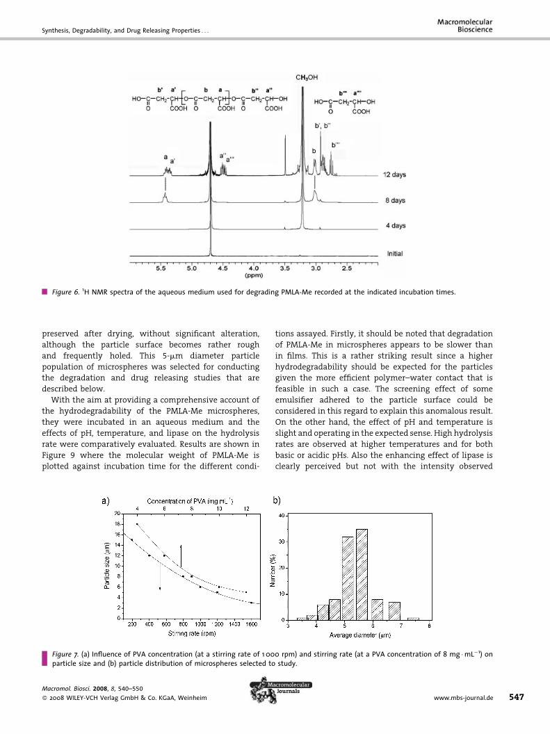
The hydrolysis mechanismwas investigated by 1H NMR
analysis of the degradation products generated by
PMLA-Me and coPMLA-(Me25H75) upon incubation in pure
water at room temperature. The NMR spectra recorded
from the mother solution of PMLA-Me for increase in
incubation times are compared in Figure 6. Initially large
amounts of methanol were present in the aqueous
solution whereas no other products were detectable. The
amount of methanol increased with time and later PMLA
entered in the incubation medium whereas no signs of
partially methylated PMLA were detected. At the end of
the process, malic acid and methanol were the only
compounds present in the incubating medium. Similar
results were obtained in the hydrolysis of coPMLA-
(Me25H75). All these results indicate that hydrolytic
degradation of methyl esters of PMLA must take place
by hydrolysis of the methyl carboxylate side group
followed by hydrolysis of the carboxylate main chain.
According to the observations previously reported for the
benzyl esters of synthetic PMLA, the carboxylic side groups
initially liberated should enhance the random hydrolysis
of the ester backbone groups, which otherwise would be
highly resistant to breaking.[13]
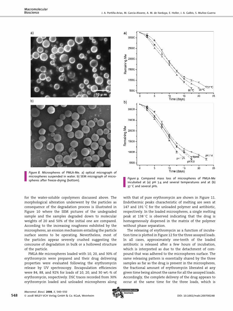
Microspheres of PMLA-Me: Preparation, HydrolyticDegradation, and Erythromycin Release
Microspheres of PMLA-Mewere obtained by the emulsion–
evaporation solvent method using chloroform as an
organic phase. The particle diameter could be adjusted
by both stirring speed and the amount of PVA added as an
emulsifier, their effects being illustrated in Figure 7.
Particle diameters between 1 and 20 mm with an
approximate Gaussian distribution of sizes were feasible
by independent adjusting of these two factors. The
well-spherical shape adopted by these particles is clearly
revealed in the optical micrograph shown in Figure 8a,
which was taken from the aqueous emulsion of average
5 mmdiametermicroparticles before drying. As it is seen in
the SEM picture in Figure 8b, the spherical shape was
DOI: 10.1002/mabi.200700248
Synthesis, Degradability, and Drug Releasing Properties . . .
Figure 6. 1H NMR spectra of the aqueous medium used for degrading PMLA-Me recorded at the indicated incubation times.
preserved after drying, without significant alteration,
although the particle surface becomes rather rough
and frequently holed. This 5-mm diameter particle
population of microspheres was selected for conducting
the degradation and drug releasing studies that are
described below.
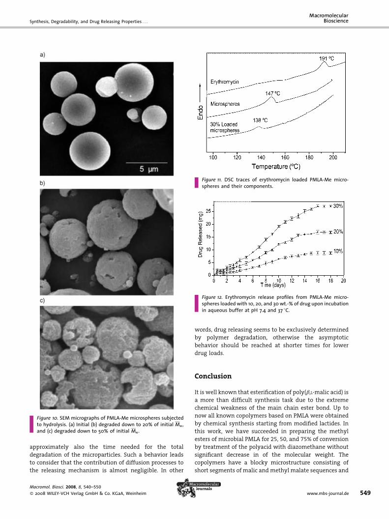
With the aim at providing a comprehensive account of
the hydrodegradability of the PMLA-Me microspheres,
they were incubated in an aqueous medium and the
effects of pH, temperature, and lipase on the hydrolysis
rate were comparatively evaluated. Results are shown in
Figure 9 where the molecular weight of PMLA-Me is
plotted against incubation time for the different condi-
Figure 7. (a) Influence of PVA concentration (at a stirring rate of 1 00particle size and (b) particle distribution of microspheres selected to
Macromol. Biosci. 2008, 8, 540–550
� 2008 WILEY-VCH Verlag GmbH & Co. KGaA, Weinheim
tions assayed. Firstly, it should be noted that degradation
of PMLA-Me in microspheres appears to be slower than
in films. This is a rather striking result since a higher
hydrodegradability should be expected for the particles
given the more efficient polymer–water contact that is
feasible in such a case. The screening effect of some
emulsifier adhered to the particle surface could be
considered in this regard to explain this anomalous result.
On the other hand, the effect of pH and temperature is
slight and operating in the expected sense. High hydrolysis
rates are observed at higher temperatures and for both
basic or acidic pHs. Also the enhancing effect of lipase is
clearly perceived but not with the intensity observed
0 rpm) and stirring rate (at a PVA concentration of 8 mg �mL�1) onstudy.
www.mbs-journal.de 547
J. A. Portilla-Arias, M. Garcıa-Alvarez, A. M. de Ilarduya, E. Holler, J. A. Galbis, S. Munoz-Guerra
Figure 8. Microspheres of PMLA-Me. a) optical micrograph ofmicrospheres suspended in water. b) SEM micrograph of micro-spheres after freeze-drying (bottom). Figure 9. Compared mass loss of microspheres of PMLA-Me
incubated at (a) pH 7.4 and several temperatures and at (b)37 8C and several pHs.
548
for the water-soluble copolymers discussed above. The
morphological alteration underwent by the particles as
consequence of the degradation process is illustrated in
Figure 10 where the SEM pictures of the undegraded
sample and the samples degraded down to molecular
weights of 20 and 50% of the initial one are compared.
According to the increasing roughness exhibited by the
microspheres, an erosionmechanism entailing the particle
surface seems to be operating. Nevertheless, most of
the particles appear severely crushed suggesting the
concourse of degradation in bulk or a hollowed structure
of the particle.
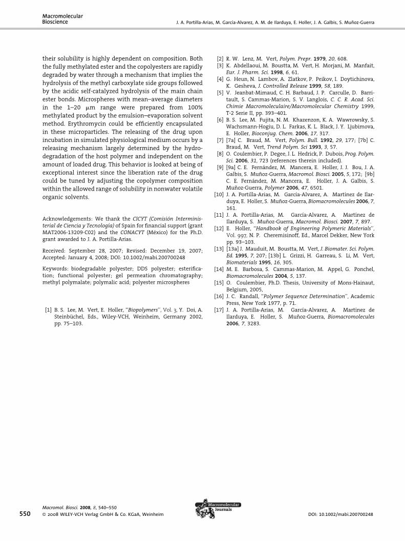
PMLA-Me microspheres loaded with 10, 20, and 30% of
erythromycin were prepared and their drug delivering
properties were evaluated following the erythromycin
release by UV spectroscopy. Encapsulation efficiencies
were 84, 88, and 92% for loads of 10, 20, and 30 wt.-% of
erythromycin, respectively. DSC traces recorded from 30%
erythromycin loaded and unloaded microspheres along
Macromol. Biosci. 2008, 8, 540–550
� 2008 WILEY-VCH Verlag GmbH & Co. KGaA, Weinheim
with that of pure erythromycin are shown in Figure 11.
Endothermic peaks characteristic of melting are seen at
147 and 191 8C for the unloaded polymer and antibiotic,
respectively. In the loaded microspheres, a single melting
peak at 138 8C is observed indicating that the drug is
homogeneously dispersed in the matrix of the polymer
without phase separation.
The releasing of erythromycin as a function of incuba-
tion time is plotted in Figure 12 for the three assayed loads.
In all cases, approximately one-tenth of the loaded
antibiotic is released after a few hours of incubation,
which is interpreted as due to the detachment of com-
pound that was adhered to the microspheres surface. The
same releasing pattern is essentially shared by the three
samples as far as the drug is present in the microspheres,
the fractional amount of erythromycin liberated at any
given time being almost the same for all the assayed loads.
Accordingly, the complete delivery of the drug appears to
occur at the same time for the three loads, which is
DOI: 10.1002/mabi.200700248
Synthesis, Degradability, and Drug Releasing Properties . . .
Figure 10. SEM micrographs of PMLA-Me microspheres subjectedto hydrolysis. (a) Initial (b) degraded down to 20% of initial Mw,and (c) degraded down to 50% of initial Mw.
Figure 11. DSC traces of erythromycin loaded PMLA-Me micro-spheres and their components.
Figure 12. Erythromycin release profiles from PMLA-Me micro-spheres loaded with 10, 20, and 30 wt.-% of drug upon incubationin aqueous buffer at pH 7.4 and 37 8C.
approximately also the time needed for the total
degradation of the microparticles. Such a behavior leads
to consider that the contribution of diffusion processes to
the releasing mechanism is almost negligible. In other
Macromol. Biosci. 2008, 8, 540–550
� 2008 WILEY-VCH Verlag GmbH & Co. KGaA, Weinheim
words, drug releasing seems to be exclusively determined
by polymer degradation, otherwise the asymptotic
behavior should be reached at shorter times for lower
drug loads.
Conclusion
It is well known that esterification of poly(b,L-malic acid) is
a more than difficult synthesis task due to the extreme
chemical weakness of the main chain ester bond. Up to
now all known copolymers based on PMLA were obtained
by chemical synthesis starting from modified lactides. In
this work, we have succeeded in preparing the methyl
esters of microbial PMLA for 25, 50, and 75% of conversion
by treatment of the polyacid with diazomethane without
significant decrease in of the molecular weight. The
copolymers have a blocky microstructure consisting of
short segments of malic andmethyl malate sequences and
www.mbs-journal.de 549
J. A. Portilla-Arias, M. Garcıa-Alvarez, A. M. de Ilarduya, E. Holler, J. A. Galbis, S. Munoz-Guerra
550
their solubility is highly dependent on composition. Both
the fully methylated ester and the copolyesters are rapidly
degraded by water through a mechanism that implies the
hydrolysis of the methyl carboxylate side groups followed
by the acidic self-catalyzed hydrolysis of the main chain
ester bonds. Microspheres with mean–average diameters
in the 1–20 mm range were prepared from 100%
methylated product by the emulsion–evaporation solvent
method. Erythromycin could be efficiently encapsulated
in these microparticles. The releasing of the drug upon
incubation in simulated physiological medium occurs by a
releasing mechanism largely determined by the hydro-
degradation of the host polymer and independent on the
amount of loaded drug. This behavior is looked at being of
exceptional interest since the liberation rate of the drug
could be tuned by adjusting the copolymer composition
within the allowed range of solubility in nonwater volatile
organic solvents.
Acknowledgements: We thank the CICYT (Comision Interminis-terial de Ciencia y Tecnologıa) of Spain for financial support (grantMAT2006-13209-C02) and the CONACYT (Mexico) for the Ph.D.grant awarded to J. A. Portilla-Arias.
Received: September 28, 2007; Revised: December 19, 2007;Accepted: January 4, 2008; DOI: 10.1002/mabi.200700248
Keywords: biodegradable polyester; DDS polyester; esterifica-tion; functional polyester; gel permeation chromatography;methyl polymalate; polymalic acid; polyester microspheres
[1] B. S. Lee, M. Vert, E. Holler, ‘‘Biopolymers’’, Vol. 3, Y. Doi, A.Steinbuchel, Eds., Wiley-VCH, Weinheim, Germany 2002,pp. 75–103.
Macromol. Biosci. 2008, 8, 540–550
� 2008 WILEY-VCH Verlag GmbH & Co. KGaA, Weinheim
[2] R. W. Lenz, M. Vert, Polym. Prepr. 1979, 20, 608.[3] K. Abdellaoui, M. Boustta, M. Vert, H. Morjani, M. Manfait,
Eur. J. Pharm. Sci. 1998, 6, 61.[4] G. Heun, N. Lambov, A. Zlatkov, P. Peikov, I. Doytichinova,
K. Gesheva, J. Controlled Release 1999, 58, 189.[5] V. Jeanbat-Mimaud, C. H. Barbaud, J. P. Carculle, D. Barri-
tault, S. Cammas-Marion, S. V. Langlois, C. C. R. Acad. Sci.Chimie Macromoleculaire/Macromolecular Chemistry 1999,T-2 Serie II, pp. 393–401.
[6] B. S. Lee, M. Fujita, N. M. Khazenzon, K. A. Wawrowsky, S.Wachsmann-Hogiu, D. L. Farkas, K. L. Black, J. Y. Ljubimova,E. Holler, Bioconjug. Chem. 2006, 17, 317.
[7] [7a] C. Braud, M. Vert, Polym. Bull. 1992, 29, 177; [7b] C.Braud, M. Vert, Trend Polym. Sci 1993, 3, 57.
[8] O. Coulembier, P. Degee, J. L. Hedrick, P. Dubois, Prog. Polym.Sci. 2006, 31, 723 (references therein included).
[9] [9a] C. E. Fernandez, M. Mancera, E. Holler, J. J. Bou, J. A.Galbis, S. Munoz-Guerra, Macromol. Biosci. 2005, 5, 172; [9b]C. E. Fernandez, M. Mancera, E. Holler, J. A. Galbis, S.Munoz-Guerra, Polymer 2006, 47, 6501.
[10] J. A. Portilla-Arias, M. Garcıa-Alvarez, A. Martınez de Ilar-duya, E. Holler, S. Munoz-Guerra, Biomacromolecules 2006, 7,161.
[11] J. A. Portilla-Arias, M. Garcıa-Alvarez, A. Martınez deIlarduya, S. Munoz-Guerra, Macromol. Biosci. 2007, 7, 897.
[12] E. Holler, ‘‘Handbook of Engineering Polymeric Materials’’,Vol. 997, N. P. Cheremisinoff, Ed., Marcel Dekker, New Yorkpp. 93–103.
[13] [13a] J. Mauduit, M. Boustta, M. Vert, J. Biomater. Sci. Polym.Ed. 1995, 7, 207; [13b] L. Grizzi, H. Garreau, S. Li, M. Vert,Biomaterials 1995, 16, 305.
[14] M. E. Barbosa, S. Cammas-Marion, M. Appel, G. Ponchel,Biomacromolecules 2004, 5, 137.
[15] O. Coulembier, Ph.D. Thesis, University of Mons-Hainaut,Belgium, 2005,
[16] J. C. Randall, ‘‘Polymer Sequence Determination’’, AcademicPress, New York 1977, p. 71.
[17] J. A. Portilla-Arias, M. Garcıa-Alvarez, A. Martınez deIlarduya, E. Holler, S. Munoz-Guerra, Biomacromolecules2006, 7, 3283.
DOI: 10.1002/mabi.200700248


