Embed Size (px)
Citation preview
UNIVERSIDADE FEDERAL DE SANTA MARIA CENTRO DE CIÊNCIAS DA SAÚDE
PROGRAMA DE PÓS-GRADUAÇÃO EM FARMACOLOGIA
Talita Rodrigues
EFEITO DA SILIBININA EM MODELO DE DISCINESIA OROFACIAL INDUZIDA PELO HALOPERIDOL EM CAMUNDONGOS
Santa Maria, RS 2019
Talita Rodrigues
EFEITO DA SILIBININA EM MODELO DE DISCINESIA OROFACIAL INDUZIDA
PELO HALOPERIDOL EMCAMUNDONGOS
Dissertação apresentada ao Curso de Pós-Graduação em Farmacologia, da Universidade Federal de Santa Maria (UFSM, RS), como requisito parcial para obtenção do título de Mestre em Farmacologia.
Orientadora: Prof.ª Dr.ª Roselei Fachinetto Co-orientador: Dr. Alcindo Busanello
Santa Maria, RS 2019
© 2019 Todos os direitos autorais reservados a Talita Rodrigues. A reprodução de partes ou do todo deste trabalho só poderá ser feita mediante a citação da fonte. E-mail: [email protected]
AGRADECIMENTOS
Para chegar até aqui, percorri um longo e nada tranquilo caminho, mas,
durante toda essa jornada tive a oportunidade de conhecer e conviver com pessoas
maravilhosas e empenhadas, eu gostaria de agradecer todos que estiveram
presentes nesse momento, tanto diretamente quanto indiretamente.
Primeiramente, eu gostaria de agradecer meus pais, Antônio L. C. Rodrigues
e Elza M. Rodrigues. Obrigada por sempre estarem ao meu lado, por todo incentivo
e apoio, principalmente nos momentos mais difíceis. Obrigada por me mostrarem
que sem esforço e dedicação não conquistamos nada. E por me ensinar a dar valor
às pequenas coisas da vida. Amo muito vocês!
À Deus, que sempre me guiou e me escutou nos momentos difíceis.
Obrigada à minha orientadora Roselei Fachinetto e meu co-orientador Alcindo
Busanello, pela oportunidade e confiança, que mesmo sem me conhecer,
acreditaram em meu potencial e me deram essa chance. Obrigada pela paciência e
ensinamentos.
Ao pessoal do Laboratório de Neurotoxicidade e Comportamento
(NEUROTOX), ou simplesmente Lab. 5209, Catiuscia, Bárbara, Getulio, Ana Paula,
Jeane, Janaína. Obrigada pelo acolhimento, paciência, disponibilidade, amizade, e
por compartilharem comigo momentos de alegrias e angústias, considero vocês
parte da minha família.
E por último, mas não menos importante, minhas amigas da graduação para a
vida, Gabriela, Flávia e Raísa, obrigada pelas palavras de apoio, por sempre me
escutarem e aconselharem. Irmãs de coração!

“A maior recompensa para o trabalho do homem não é o que ele ganha com isso, mas o que ele se torna com isso.”
(John Ruskin)
RESUMO
EFEITO DA SILIBININA EM MODELO DE DISCINESIA OROFACIAL INDUZIDA PELO HALOPERIDOL EM CAMUNDONGOS
AUTORA: Talita Rodrigues ORINTADORA: Roselei Fachinetto
CO-ORIENTADOR: Alcindo Busanello
A esquizofrenia é um distúrbio crônico e debilitante que afeta cerca de 1% da população. A utilização crônica de antipsicóticos, principalmente os antipsicóticos típicos, utilizados para o tratamento da esquizofrenia, causa, como efeito adverso, distúrbios motores debilitantes (discinesia). A discinesia tardia afeta de 20% a 40% dos pacientes, e é caracterizada por movimentos repetitivos e involuntários que envolvem principalmente a região oro-buco-facial. Não existe um tratamento eficaz para evitar e/ou tratar a discinesia tardia. Desta forma, é importante a busca por novos tratamentos e/ou adjuvantes terapêuticos que possam ser utilizados clinicamente. A Silibinina é o constituinte ativo majoritário da silimarina, que é um flavonóide isolado das sementes de Silybum marianum (L.) Gaerth, o qual possui ação antioxidante e potencial efeito neuroprotetor, inclusive em modelos animais de doenças motoras. O objetivo deste trabalho foi avaliar o efeito da silibinina em um modelo de discinesia orofacial induzida por haloperidol em camundongos. Dessa forma, camundongos machos foram tratados com veículo (NaCl 0,9%), haloperidol (1,25 mg / kg, i.p.), silibinina (20 mg / kg, i.p.) e haloperidol (1,25 mg / kg, i.p.) + silibinina (20 mg/ kg, i.p) intraperitonealmente durante 28 dias consecutivos. A quantificação comportamental (movimentos de mastigação vazios - MMVs, número de cruzamentos e levantamentos no campo aberto e tempo de imobilidade) foi realizada a cada 7 ou 14 dias durante o período experimental. Os parâmetros bioquímicos do estresse oxidativo foram avaliados em estruturas cerebrais (córtex, estriado e região contendo a substância negra), fígado e rim. O haloperidol causou discinesia orofacial aumentando a prevalência e a frequência de MMVs sem alterar os demais parâmetros comportamentais avaliados. Foram encontradas correlações negativas entre o número de cruzamentos ou levantamentos com MMVs e uma correlação positiva entre o tempo de imobilidade e os MMVs. A silibinina não evitou os efeitos do haloperidol nos parâmetros comportamentais. Além disso, nem o haloperidol nem a silibinina causaram alterações nos parâmetros de estresse oxidativo. Foi encontrada uma correlação positiva entre o número de MMVs e o teor de tiol não protéico no córtex de camundongos. Não foram encontrados resultados significativos na atividade da Na+/k+/ATPase nas diferentes estruturas cerebrais. Em conclusão, os dados deste estudo demonstram que, em camundongos, também é possível verificar o aumento na frequência de MMVs apenas em um percentual dos animais tratados mimetizando o que acontece em pacientes. Além disso, apesar de a silibinina não evitar os MMVs, o tratamento combinado com haloperidol não parece causar alterações em marcadores de estresse oxidativo nos animais. Palavras chave: Movimento de mascar no vazio. Campo aberto. Imobilidade. Estresse oxidativo.
ABSTRACT
EFFECT OF SILIBININ ON A MODEL OF OROFACIAL DYSKINESIA INDUCED BY HALOPERIDOL IN MICE
AUTHOR: Talita Rodrigues ADVISOR: Roselei Fachinetto
CO-ADVISOR: Alcindo Busanello
Schizophrenia is a chronic and debilitating disorder that affects about 1% of the population. Chronic use of antipsychotics, especially typical ones, used to treat schizophrenia, causes as adverse effect debilitating motor disorder (dyskinesia). Tardive dyskinesia affects 20% to 40% of patients, and is characterized by repetitive and involuntary movements involving mainly the oro-buco-facial region. There is no effective treatment either for avoiding or treating tardive dyskinesia. Therefore, it is important to search for new treatments and/or therapeutic adjuvants that can be clinically useful. Silibinin is the majoritary active constituent of silymarin, which is a flavonoid isolated from the seeds of Silybum marianum (L.) Gaerth, which has antioxidant action and potential neuroprotective effect, even in animal models of motor diseases. Thus, male mice were treated with vehicle (0,9% NaCl), haloperidol (1.25 mg / kg, i.p.), silibinin (20 mg / kg, i.p.) and haloperidol (1.25 mg / kg, i.p.) + silibinin (20 mg / kg, i.p.) intraperitoneally for 28 consecutive days. Behavioral quantification (vacuous chewing movements - VCMs, number of crossings and rearings in the open field and time of immobility) was performed every 7 or 14 days during the experimental period. The biochemical parameters of oxidative stress were evaluated in cerebral structures (cortex, striatum and region containing the substantia nigra), liver and kidney. Haloperidol caused orofacial dyskinesia by increasing the prevalence and frequency of VCMs without altering the other behavioral parameters evaluated. Negative correlations were found between the numbers of crossings or rearings with VCMs and a positive correlation between immobility time and VCMs. Silibinin did not avoid the effects of haloperidol on behavioral parameters. In addition, neither haloperidol nor silibinin caused changes in oxidative stress parameters. A positive correlation was found between the number of VCMs and the non-protein thiol content in the cortex of mice. No significant results were found in Na+/K+/ ATPase activity in the different brain structures. In conclusion, the data of this study demonstrates that in mice also it is possible to verify the increase in the frequency of VCMs only in a percentage of animals mimicking which occurs with the patients. Furthermore, although silibinin did not avoid the VCMs in mice its combined treatment with haloperidol seems not cause signals of oxidative stress markers in animals. Key words: Vacuous chewing movements. Open field. Immobility. Oxidative stress.
LISTA DE FIGURAS
APRESENTAÇÃO Figura 1 – Vias dopaminérgicas ................................................................................ 17
Figura 2 – Silimarina (Silybum marianum L. Gaernt.) ................................................ 25 Figura 3 – Diasteroisômeros da Silibinina ................................................................. 26 MANUSCRITO Figure 1. Effect of haloperidol (1.25 mg/kg) and silibinin (20 mg/kg) on VCM in
mice. The quantification of VCM was performed during 6 minutes on days 7 (a), 14 (b), 21 (c) and 28 (d). Data are expressed as means ± SEM. One-way ANOVA followed by Tukey's multiple comparisons test. *p<0.05, compared with the control group; (c) #p<0.05, compared with the haloperidol group -VCM. (d) #p<0.05, compared with the haloperidol+ silibinin group -VCM. (control, n=9; silibinin, n=9; haloperidol, n=9; haloperidol+silibinin, n=10) ............................................. 46
Figure 2. Effect of haloperidol (1.25 mg/kg) and silibinin (20 mg/kg) on open field test in mice. The quantification number of crossing was performed on days 14 (a) and 28 (b) and number of rearing was performed on days 14 (c) and 28 (d). Data are expressed as means ± SEM. One-way ANOVA followed by Tukey's multiple comparisons test.*p<0.05, compared with the control group (control, n=9; silibinin, n=9; haloperidol, n=9; haloperidol+silibinin, n=10) ......................................................................... 47
Figure 3. Effect of haloperidol (1.25 mg/kg) and silibinin (20 mg/kg) on open field test in mice. The quantification number of time of immobility on days 14 (a) and 28 (b) and number of rearing was performed on days 14 (c) and 28 (d). Data are expressed as means ± SEM. One-way ANOVA followed by Tukey's multiple comparisons test. @ p<0.05, compared with the silibinin group (control, n=9; silibinin, n=9; haloperidol, n=9; haloperidol+silibinin, n=10) ......................................................................... 48
Figure 4. Correlation between VCMs number with crossing (a), rearing (b) and time of immobility (c). Significant correlations (Pearson correlation – r) when p < 0.05 ............................................................................................. 49
Figure 5. Correlation between VCMs number with non-protein thiol in cortex. Significant correlations (Pearson correlation – r) when p < 0.05 ................ 50
LISTA DE TABELAS
MANUSCRITO Table 1. Effects of haloperidol and/or silibinin on total and non-protein thiol
groups and on Na+/K+-ATPase activity in brain structures. Data are expressed as means ± SEM ....................................................................... 51
Table 2. Effects of haloperidol and/or silibinin on total and non-protein thiol groups and on TBARS productionin liver and kidney. Data are expressed as means ± SEM ........................................................................................ 52
LISTA DE ABREVIATURAS E SIGLAS
AA Ácido ascórbico
ANVISA Agência Nacional de Vigilância Sanitária e Ambiental
CAT Catalase
DA Dopamina
DCFH-DA 2 ', 7'-dicloro fluoresceína diacetato
DO Discinesia orofacial
DT Discinesia tardia
D2 Receptores de dopamina do tipo D2
ERO Espécies reativas de oxigênio
ES Estresse oxidativo
FDA do inglês “Food and Drugs Administration”
GPx Glutationa peroxidase
GSH Glutationa reduzida
H2O2 Peróxido de hidrogênio
MAO Monoaminoxidase
MMVs Movimentos de mascar no vazio
MPP+ 1-metil-4- fenilpiridína
MPTP 1-Metil-4-fenil-1,2,3,6-tetra-hidropiridina
Na+K+-ATPase Sódio/ Potássio- ATPase
NO- Óxido nítrico
O2─ Íon superóxido
OH- Radical hidroxila
OMS Organização Mundial da Saúde
ONOO─ Peróxinitrito
PL Peroxidação lipídica
Redox Reação de redução e oxidação
SOD Superóxido dismutase
SNC Sistema nervoso central
TBARS Substâncias reativas ao ácido tiobarbitúrico
VMAT2 Transportador vesicular de monoaminas 2

SUMÁRIO
APRESENTAÇÃO .......................................................................................... 12
1 INTRODUÇÃO ................................................................................................ 13
2 REFERENCIAL TEÓRICO ............................................................................. 16 2.1 ESQUIZOFRENIA ........................................................................................... 16 2.1.1 Tratamento ..................................................................................................... 18
2.2 DISCINESIA TARDIA ...................................................................................... 20 2.3 DISCINESIA TARDIA E ESTRESSE OXIDATIVO .......................................... 21 2.4 SILIBININA ...................................................................................................... 24
3 OBJETIVOS .................................................................................................... 28
3.1 OBJETIVO GERAL ......................................................................................... 28 3.2 OBJETIVOS ESPECÍFICOS ........................................................................... 28
4 RESULTADOS ............................................................................................... 29
5 MANUSCRITO – EFFECTS OF SILIBININ ON HALOPERIDOL-INDUCED OROFACIAL DYSKINESIA IN MICE .............................................................. 30
6 CONCLUSÕES ESPECÍFICAS ...................................................................... 53
7 CONCLUSÃO GERAL .................................................................................... 54
REFERÊNCIAS .............................................................................................. 55
ANEXO A – CERTIFICADO DE APROVAÇÃO DE PROJETO EMITIDO PELA COMISSÃO DE ÉTICA NO USO DE ANIMAIS DA UNIVERSIDADE FEDERAL DE SANTA MARIA (CEUA/UFSM) ............................................... 67
ANEXO B – CARTA DE SUBMISSÃO DO MANUSCRITO ........................... 68
12
APRESENTAÇÃO
Esta dissertação está apresentada na forma de um manuscrito cientifico.
No item INTRODUÇÃO e REFERENCIAL TEÓRICO, está descrita uma
revisão sucinta sobre os temas abordados nesta dissertação.
Os resultados que fazem parte deste estudo estão apresentados sob a forma
de um manuscrito, o qual se encontra no item RESULTADOS. As seções Materiais e
Métodos, Resultados, Discussões dos Resultados e Referências Bibliográficas do
manuscrito encontram-se no próprio e representam a integra deste estudo.
O item CONCLUSÕES encontrado no final desta dissertação, apresenta
conclusão geral referente aos dados encontrados neste estudo e apresentados no
manuscrito.
O item REFERÊNCIAS BIBLIOGRÁFICAS referem-se somente às citações
que aparecem na INTRODUÇÃO e REFERENCIAL TEÓRICO desta dissertação.
13
1 INTRODUÇÃO
Distúrbios psicóticos são disfunções mentais, onde há um conjunto de
sintomas envolvidos, que alteram a percepção da realidade, afetando a capacidade
da pessoa realizar atividades corriqueiras e relacionar-se com outras pessoas
(STAHL, 2014). Dentre esses distúrbios psicóticos encontra-se a esquizofrenia
(STAHL, 2014). Segundo a Organização Mundial da Saúde (OMS), a esquizofrenia é
definida como um transtorno mental, incapacitante e grave (OMS, 2013), onde
ocorre distorção de pensamentos, diminuição da percepção da realidade e de
emoções assim como déficits cognitivos como perda de memória e isolamento social
(MCGORRY; MEI, 2018). A principal hipótese para a ocorrência destes sintomas é
de que eles decorrem de alterações nas funções das vias dopaminérgicas cerebrais,
os quais podem ser tratados com fármacos antipsicóticos típicos e atípicos (STAHL,
2014).
O haloperidol é o fármaco mais utilizado para o tratamento da esquizofrenia
(DOLD et al., 2015). Por ser um antipsicótico típico, seu mecanismo de ação
consiste no bloqueio dos receptores D2 de dopamina na via mesolímbica (STAHL,
2014). Dessa maneira, torna-se efetivo na amenização dos sintomas positivos
(alucinações, ilusões, delírios) da doença (MEYER, 2012). Com sua administração
prolongada, ocorrem alguns efeitos extrapiramidais (disfunções motoras), devido ao
bloqueio dos receptores D2 de dopamina na via nigroestriatal, região responsável
pelo controle motor (TURRONE et al., 2003) dentre eles a discinesia tardia (KANE;
SMITH, 1982). Dessa forma, a utilização do haloperidol para indução de sintomas
que mimetizam a discinesia tardia (DT) vem sendo amplamente utilizado na literatura
(ARAÚJO et al., 2017; DATTA et al., 2016; KRONBAUER et al., 2017; MACÊDO et
al., 2011).
A DT é uma doença iatrogênica caracterizada por movimentos anormais,
involuntários, repetitivos e indolores, envolvendo a região orofacial, músculos dos
lábios, pálpebras, língua, podendo afetar também o tronco e consequentemente os
membros superiores e inferiores (CASEY, 1985; MEYER, 2012; STAHL, 2014). A DT
aparece em torno de 20- 40% das pessoas que fazem uso crônico de antipsicóticos
típicos (ANDREASSEN; JØRGENSEN, 2000; BERGMAN et al., 2017).
Recentemente, dados da literatura demonstraram que, mesmo os antipsicóticos
atípicos causam DT, embora com uma prevalência menor do que os típicos
14
(CITROME, 2017; D'ABREU; AKBAR; FRIEDMAN, 2018). Muitas vezes esses
sintomas tornam-se irreversíveis mesmo com a suspensão do tratamento (KANE,
1995; STAHL, 2014).
Vários modelos animais vêm sendo utilizados no estudo da DT, dentre eles o
modelo de DT induzida por haloperidol (ARAÚJO et al., 2017; DHINGRA;
GOSWAMI; GAHALAIN, 2017; SONEGO et al., 2018). A DT induzida por haloperidol
tem sua patofisiologia relacionada à hipersensibilidade dopaminérgica, ocasionando
um aumento de receptores dopaminérgicos do tipo D2 e consequentemente, o
aparecimento dos efeitos adversos extrapiramidais (STAHL, 2014). Pelo fato de não
existir uma terapia efetiva para amenizar os sintomas da discinesia, acredita-se que
o desenvolvimento da DT é consequência do efeito neurotóxico mediado pelo
haloperidol, estando relacionada ao estresse oxidativo, devido aos radicais livres
gerados do metabolismo da dopamina e do aumento da transmissão glutamatérgica
por bloqueio dos receptores dopaminérgicos (BURGER et al., 2005; FACHINETTO
et al., 2005; PEROZA et al., 2013; SONEGO et al., 2018). Sendo assim, substâncias
com potencial antioxidante poderiam ser benéficas para atenuar os sintomas da
doença (LISTER et al., 2014; SAMAD; HALEEM, 2017).
Atualmente, os estudos estão voltando-se para a busca de compostos
naturais, que apresentem baixo potencial toxicológico, como a silimarina/silibinina
(SALLER; MEIER; BRIGNOLI, 2001), para o tratamento e/ou prevenção de
patologias do sistema nervoso central (SNC), (BUSANELLO et al., 2011; CRAGG;
NEWMAN, 2013; RECKZIEGEL et al., 2013; NADE; KAWALE; YADAV, 2010), uma
vez que, desde as civilizações mais antigas, busca-se na natureza a cura e
tratamento de doenças (ANDRADE et al., 2007). A literatura tem demonstrado que
diversos antioxidantes naturais podem minimizar os movimentos involuntários
induzidos em animais experimentais (BUSANELLO et al., 2012; PEROZA et al.,
2013; RECKZIEGEL et al., 2013).
Dessa forma, a silimarina, também conhecida como cardo-mariano ou cardo
de leite (de FREITAS, et al 2018;CHOI et al., 2012), é um flavonóide isolado das
sementes do fruto da planta Silybum marianum (L.) Gaerth, planta pertencente à
família Asteraceae (MORAZZONI; BOMBARDELLI, 1995; SVOBODOVÁ et al.,
2016), popularmente utilizada a mais de dois mil anos no tratamento de doenças
hepáticas de diferentes origens, principalmente em doenças relacionadas ao
consumo de álcool e distúrbios do trato gastrointestinais (KREN; WALTEROVA,
15
2005;RAINONE, 2005), devido ás suas propriedades antiinflamatórias, antioxidantes
e de regeneração tecidual (HADDADI et al., 2014; PÉREZ-H et al., 2014;
SRIVASTANA et al., 1994). A silimarina também possui propriedade neuroprotetoras
(FERNANDES et al., 2018; FREITAS et al., 2018; OLIVEIRA et al., 2015b), devido
sua capacidade de inibir o estresse oxidativo (BORAH, 2013; GALHARDI et al.,
2009), acreditando-se assim que essa propriedade é atribuída a seu principal
constituinte ativo, a silibinina (LEE et al., 2015; NENCINI; GIORGI; MICHELI, 2007),
uma vez que ela constitui cerca de 50% dos compostos polifenólicos que compõem
a silimarina (HACKETT et al., 2013). Dessa maneira, a silibinina poderia apresentar
resultados promissores na prevenção da discinesia orofacial (DO), uma vez que há
estudos constatando sua capacidade de reduzir o déficit motor e diminuir a perda da
dopamina nos neurônios dopaminérgicos (LEE et al., 2015).
Sendo assim, nesse estudo, investigamos o efeito da silibinina nos sintomas
da discinesia a fim de verificar se a silibinina poderia auxiliar no tratamento e/ou
prevenção das alterações motora induzidas por haloperidol em um modelo de
discinesia orofacial em camundongos.
16
2 REFERENCIAL TEÓRICO
2.1 ESQUIZOFRENIA
A esquizofrenia consiste em uma doença crônica e debilitante, que afeta 21
milhões de pessoas no mundo todo (OMS, 2018), cerca de 1% da população
mundial (MENDELSON; EATON, 2018). Essa patologia foi caracterizada no final do
século XIX por psiquiatras como Emil Kraepelin e Eugen Bleuler, criador do termo
“esquizofrenia” (MAATZ; HOFF, 2014; MAATZ; HOFF; ANGST, 2015). Do grego,
esquizofrenia significa: schizo= dividir e phrenia= mente, dividir a mente (MAATZ;
HOFF, 2014).
Os primeiros sintomas surgem na adolescência ou no início da fase adulta
(GANGULY; SOLIMAN; MOUSTAFA, 2018; MENDELSON; EATON, 2018), sendo
um distúrbio psiquiátrico de difícil diagnóstico, pois, seus estágios iniciais
assemelham-se com o transtorno bipolar e a depressão (KRYNICKI et al., 2018;
MENDELSON; EATON, 2018; SIRIS et al., 1988). Para o diagnóstico da
esquizofrenia, leva-se em consideração o histórico clínico e exames mentais do
paciente, com base nos critérios de Classificação Internacional de Doenças 10º
revisão (CID-10) e no Manual Estatístico e Diagnóstico de Transtornos Mentais 5ª
edição (DSM-V) (CORNETT et al., 2017; DOLD et al., 2015).
A esquizofrenia ainda não tem sua etiologia bem descrita (ATTADEMO;
BERNARDINI, 2017; BORÇOI et al., 2015), porém existem algumas hipóteses sobre
sua origem. Entre elas destacam-se poluição ambiental, hereditariedade, mutações
genéticas (polimorfismos), drogas de abuso (cocaína e anfetaminas), complicações
obstétricas (prematuridade, controle de peso, idade de mãe, hipóxia), falta de
vitamina D durante o período pré-natal, infecções (toxoplasmose, rubéola, influenza)
(ATTADEMO; BERNARDINI, 2017; BORÇOI et al., 2015; CANNON et al., 2002;
KAMEDA et al.,2013; KELLY et al., 2010; MCGRATH, 1999; OWEN; SAWA;
MORTENSEN, 2016; TORREY; YOLKEN, 1995).
Porém, a hipótese mais aceita e estudada para a sua etiologia é a hipótese
dopaminérgica (STEPNICKI; KONDEJ; KACZOR, 2018), a qual é a base para o
tratamento da esquizofrenia (LAU et al., 2013). Ela descreve o papel da dopamina
em diferentes regiões do cérebro (STEPNICKI; KONDEJ; KACZOR, 2018). Segundo
Carlsson e Lindqvist (1963), essa hipótese foi proposta pela primeira vez na década
17
de 1960 quando foi observado que a clorpromazina amenizava os sintomas positivos
da doença ao bloquear os receptores de dopamina. Combinado com a descoberta
de que a anfetamina produz psicose ao aumentar os níveis sinápticos de dopamina,
foi proposto que ocorre um aumento excessivo da neurotransmissão dopaminérgica,
podendo então, ser um dos motivos da esquizofrenia (STEPNICKI; KONDEJ;
KACZOR, 2018).
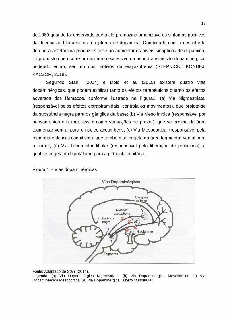
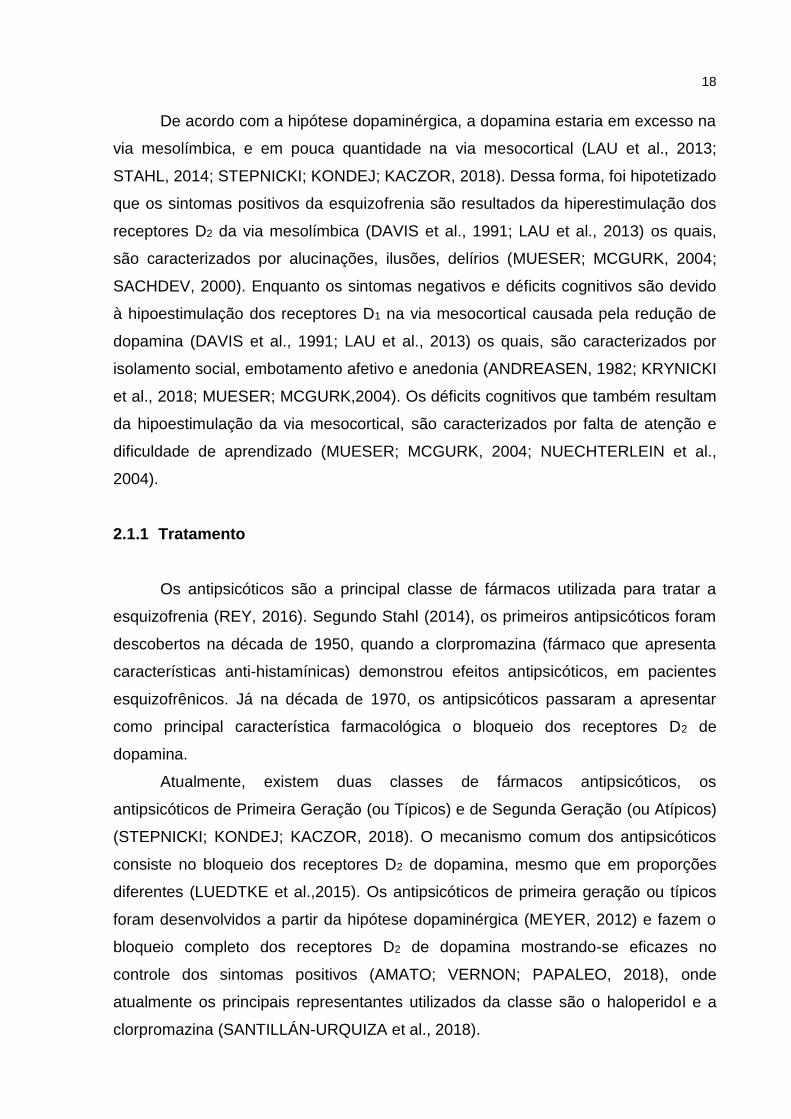
Segundo Stahl, (2014) e Dold et al. (2015) existem quatro vias
dopaminérgicas, que podem explicar tanto os efeitos terapêuticos quanto os efeitos
adversos dos fármacos, conforme ilustrado na Figura1, (a) Via Nigroestriatal
(responsável pelos efeitos extrapiramidais, controla os movimentos), que projeta-se
da substância negra para os gânglios da base; (b) Via Mesolímbica (responsável por
pensamentos e humor, assim como sensações de prazer), que se projeta da área
tegmentar ventral para o núcleo accumbens; (c) Via Mesocortical (responsável pela
memória e déficits cognitivos), que também se projeta da área tegmentar vental para
o cortex; (d) Via Tuberoinfundibular (responsável pela liberação de prolactina), a
qual se projeta do hipotálamo para a glândula pituitária.
Figura 1 – Vias dopaminérgicas
Fonte: Adaptado de Stahl (2014). Legenda: (a) Via Dopaminérgica Nigroestriatal (b) Via Dopaminérgica Mesolimbica (c) Via Dopaminergica Mesocortical (d) Via Dopaminérgica Tuberoinfundibular.
18
De acordo com a hipótese dopaminérgica, a dopamina estaria em excesso na
via mesolímbica, e em pouca quantidade na via mesocortical (LAU et al., 2013;
STAHL, 2014; STEPNICKI; KONDEJ; KACZOR, 2018). Dessa forma, foi hipotetizado
que os sintomas positivos da esquizofrenia são resultados da hiperestimulação dos
receptores D2 da via mesolímbica (DAVIS et al., 1991; LAU et al., 2013) os quais,
são caracterizados por alucinações, ilusões, delírios (MUESER; MCGURK, 2004;
SACHDEV, 2000). Enquanto os sintomas negativos e déficits cognitivos são devido
à hipoestimulação dos receptores D1 na via mesocortical causada pela redução de
dopamina (DAVIS et al., 1991; LAU et al., 2013) os quais, são caracterizados por
isolamento social, embotamento afetivo e anedonia (ANDREASEN, 1982; KRYNICKI
et al., 2018; MUESER; MCGURK,2004). Os déficits cognitivos que também resultam
da hipoestimulação da via mesocortical, são caracterizados por falta de atenção e
dificuldade de aprendizado (MUESER; MCGURK, 2004; NUECHTERLEIN et al.,
2004).
2.1.1 Tratamento
Os antipsicóticos são a principal classe de fármacos utilizada para tratar a
esquizofrenia (REY, 2016). Segundo Stahl (2014), os primeiros antipsicóticos foram
descobertos na década de 1950, quando a clorpromazina (fármaco que apresenta
características anti-histamínicas) demonstrou efeitos antipsicóticos, em pacientes
esquizofrênicos. Já na década de 1970, os antipsicóticos passaram a apresentar
como principal característica farmacológica o bloqueio dos receptores D2 de
dopamina.
Atualmente, existem duas classes de fármacos antipsicóticos, os
antipsicóticos de Primeira Geração (ou Típicos) e de Segunda Geração (ou Atípicos)
(STEPNICKI; KONDEJ; KACZOR, 2018). O mecanismo comum dos antipsicóticos
consiste no bloqueio dos receptores D2 de dopamina, mesmo que em proporções
diferentes (LUEDTKE et al.,2015). Os antipsicóticos de primeira geração ou típicos
foram desenvolvidos a partir da hipótese dopaminérgica (MEYER, 2012) e fazem o
bloqueio completo dos receptores D2 de dopamina mostrando-se eficazes no
controle dos sintomas positivos (AMATO; VERNON; PAPALEO, 2018), onde
atualmente os principais representantes utilizados da classe são o haloperidol e a
clorpromazina (SANTILLÁN-URQUIZA et al., 2018).
19
Os antipsicóticos de segunda geração ou atípicos, desenvolvidos na década
de 1990 (MILLER et al., 2007; ROSENHECK, 2007), antagonizam os receptores de
serotonina, enquanto bloqueiam os receptores de D2, menos potentemente do que
os antipsicóticos típicos, tornando-os eficazes tanto no controle dos sintomas
positivos como nos sintomas negativos da esquizofrenia (AMATO; VERNON;
PAPALEO, 2018), alguns dos representantes desta classe são clozapina,
olanzapina, ziprasidona, quetiapina, aripiprazol, risperidona, (STAHL, 2014).
Segundo a portaria SAS/MS nº 364, no Brasil, o tratamento recomendado, seria a
base dos seguintes fármacos: Risperidona, Quetiapina, Ziprasidona, Olanzapina,
Clozapina (antipsicótipos de segunda geração), Clorpromazina e Haloperidol
(antipsicóticos de primeira geração).
O Haloperidol por sua vez, é o fármaco antipsicótico mais utilizado para tratar
a esquizofrenia (DOLD et al., 2015; LOHSE; MÜLLER-OERLINGHAUSEN, 2009).
Por esse motivo, o haloperidol consta na lista dos medicamentos essenciais da OMS
(OMS, 2009), sendo que em 2008, foi prescrito para mais de 2.48 milhões de
pessoas (ALEXANDER et al., 2011). A primeira síntese do haloperidol ocorreu no
final da década de 1950, no laboratório do Dr. Paul Janssen, na Janssen
Pharmaceutica, na Bélgica (TYLER; ZALDIVAR-DIEZ; HAGGARTY, 2017).
Pertencente à primeira geração dos antipsicóticos, o haloperidol é quimicamente
classificado como uma butirofenona. Sua meia-vida é de 15 a 37 horas, enquanto
sua biodisponibilidade é de 60% a 70% (KUDO; ISHIZAKI, 1999).
Além de sua utilização como antipsicótico, o haloperidol, também pode ser
utilizado para o tratamento de náuseas, vômitos, enxaquecas, transtorno obsessivo-
compulsivo (TOC) e soluços (GAFFIGAN et al., 2015; MCDOUGLE et al., 1994;
MURRAY-BROWN; DORMAN, 2015). Além disso, também pode ser empregado em
associação com outros fármacos, ou em modo off- label, para tratar sintomas de
ansiedade, depressão, transtorno de déficit de atenção e hiperatividade (TDAH)
(ALEXANDER et al., 2011).
O haloperidol é altamente eficaz no controle dos sintomas positivos da
esquizofrenia, mas também é responsável por causar efeitos extrapiramidais
(transtorno de movimento), como o parkinsonismo, no tratamento agudo (CERETTA
et al., 2018; DOLD et al., 2015; MIKSYS et al., 2017; SCHAFFER et al., 2016) e a
discinesia tardia, com o tratamento crônico (FACHINETTO et al., 2007b;
FACHINETTO et al., 2005; LISTER et al., 2017; SOUZA et al., 2003).
20
2.2 DISCINESIA TARDIA
Segundo Meyer (2012), para um antipsicótico típico apresentar efeito
terapêutico, ele deve bloquear de 60%-75% dos receptores D2 de dopamina, mas,
bloqueando mais de 78% desses mesmos receptores, originam-se os efeitos
extrapiramidais. Enquanto isso, os antipsicóticos atípicos, bloqueiam em maior
porcentagem os receptores serotoninérgicos (75%-99%), ao invés dos receptores
dopaminérgicos, provocando assim menores chances de desenvolver os efeitos
extrapiramidais (TURRONE et al., 2003).
Já segundo Rey (2016), efeitos extrapiramidais, são transtornos de
movimento que ocorrem na via nigroestriatal, devido a um desequilíbrio entre os
receptores dopaminérgicos e os receptores colinérgicos, uma vez que os receptores
dopaminérgicos encontram-se bloqueados, gerando assim um excesso de atividade
colinérgica, e dessa forma, resultando nos efeitos extrapiramidais (distonias,
Parkinsonismo, acatisia e discinesia tardia) (CAROFF et al., 2011; TURRONE et al.,
2003).
A DT é uma doença iatrogênica e sua fisiopatologia ainda não é
completamente compreendida. Entretanto, a hipótese do mecanismo envolvido mais
aceito, é o da hipersensibilização e aumento do número dos receptores D2 de
dopamina principalmente na via nigrostriatal, na tentativa de superar o bloqueio dos
receptores D2 induzido por antipsicóticos no estriado por tempo prolongado (STAHL,
2014; TURRONE et al., 2003). A DT consiste em movimentos involuntários,
repetitivos e anormais principalmente da região orofacial, músculos dos lábios,
língua, pálpebras, que também pode afetar o tronco, ocasionando movimentos
rápidos dos membros inferiores e superiores (CASEY, 1985).
A DT aparece em torno de 20-40% (1 em cada 4) (SOLMI et al., 2018) das
pessoas que fazem uso crônico de antipsicóticos típicos (ANDREASSEN;
JØRGENSEN, 2000). Esses sintomas surgem meses ou anos após o início do
tratamento com o fármaco (REY, 2016), e muitas vezes tornam-se irreversíveis
mesmo com a suspensão do tratamento (KANE, 1995; REY, 2016), o que acaba
afetando a qualidade de vida das pessoas, uma vez que, pode dificultar a fala, a
respiração e a alimentação (CAROFF; CAMPBELL, 2016).
Com a utilização crônica dos antipsicóticos, principalmente os de primeira
geração ou típicos, como, haloperidol e flufenazina, é possível o estudo da
21
discinesia orofacial em roedores a qual apresenta similaridade com a DT
(ANDREASSEN; JØRGENSEN, 2000; BUSANELLO et al., 2017; BUSANELLO et
al., 2011; CERETTA et al., 2018; FACHINETTO et al., 2007b; PEROZA et al., 2013;
PEROZA et al., 2016; SCHAFFER et al., 2016).
Em roedores, antipsicóticos, como o haloperidol, causam a discinesia
orofacial, caracterizada por movimentos anormais, os chamados movimentos de
mascar no vazio, do inglês vacuous chewing movements (VCMs) (BUSANELLO et
al., 2017; CERETTA et al., 2018; FACHINETTO et al., 2007a; PEROZA et al., 2013;
RÖPKE et al., 2014), a qual é uma síndrome análoga a que ocorre em humanos
(ANDREASSEN; JØRGENSEN, 2000; RANA; CHAUDRY; BLANCHET, 2013).
Em 2017, a Food and Drugs Administration (FDA), órgão americano que
regula serviços de saúde nos Estados Unidos, aprovou a comercialização de dois
novos medicamentos no tratamento da DT, a Valbenazina e a Deutetrabenazina,
inibidores seletivos do transportador vesicular de monoaminas do tipo 2 (VMAT-2).
Estudos com estes fármacos demonstraram que eles possuem capacidade de
reduzir a DT, mas causam efeitos adversos como, problemas cardíacos e sonolência
(CITROME, 2017; CITROME, 2018; DAVIS et al., 2017; GRIGORIADIS et al., 2017).
No Brasil, não existem fármacos específicos aprovados pela Agência
Nacional de Vigilância Sanitária (ANVISA) para tratar a DT. Dessa forma, são
recomendadas outras estratégias farmacológicas na tentativa de redução desses
sintomas como, aumento da dose ou suspensão da utilização do fármaco, assim
como a utilização de agentes anticolinérgicos (biperideno) ou agentes Gabaérgicos
(baclofeno e benzodiazepínicos), podem amenizar esses sintomas (LIN; ONDO,
2018; MEYER, 2012; STAHL, 2014).
2.3 DISCINESIA TARDIA E ESTRESSE OXIDATIVO
Evidências demonstram que a utilização de antipsicóticos pode levar ao
estresse oxidativo (EO), devido à geração excessiva de espécies reativas de
oxigênio (ERO) e radicais livres (ABÍLIO et al., 2004; AKINTUNDE; ABUBAKAR,
2017; BUSANELLO et al., 2017; LISTER et al., 2014; PEROZA et al., 2013;
SCHAFFER et al., 2016). A concentração celular de pró-oxidantes e antioxidantes
deve estar em equilíbrio no organismo para que não haja o chamado estresse
oxidativo (KOHEN; NYSKA, 2002; PEROZA et al., 2013).
22
Dessa forma, um ambiente pró-oxidante, proporcionaria a presença de
radicais livres como, íons superóxido (O2─), óxido nítrico (NO-) e radical hidroxila
(OH-), além de outras espécies moleculares como peróxido de hidrogênio (H2O2) e
peroxinitrito (ONOO─) (WU; KOSTEN; ZHANG, 2013; BANSAL; SINGH, 2017),
produzindo assim, danos celulares favorecendo a peroxidação lipídica (PL),
oxidação de proteínas e, consequentemente, a morte celular neuronal. O tecido
cerebral é altamente suscetível ao estresse oxidativo, principalmente os neurônios
dopaminérgicos, devido ao seu alto consumo de oxigênio e seus mecanismos de
defesa antioxidante serem insuficientes (BANSAL; SINGH, 2017; FLOYD, 1999;
HADDADI et al., 2014), especialmente na região do estriado e substância negra, que
estão envolvidas na função motora (BRAVO; NASSIF, 2006; LISTER et al., 2014;
MARTELLI; NUNES, 2014).
A presença desses radicais livres pode estar envolvida no processo de
desenvolvimento da DT (SAMAD; HALEEM 2017; SOUNG et al., 2018; WU;
KOSTEN; ZHANG, 2013). Segundo Lister et al. (2014), devido à região estriatal ser
rica em dopamina, um antipsicótico ao bloquear os receptores de dopamina por
tempo prolongado, promoveria um aumento compensatório de síntese de dopamina.
Este excesso de dopamina, ao ser auto-oxidado, proporcionaria a formação de
ânions como, O2- e OH-, que interagiriam com o oxigênio e os lipídios presentes nas
células, ocasionando dano tecidual, morte celular (por necrose ou apoptose), e
assim deixando a pessoas mais suscetíveis a DT e consequentemente seus efeitos
extrapiramidais (BRAVO, NASSIF, 2006; FLATOW; BUCKLEY; MILLER, 2013;
MARTELLI; NUNES, 2014; MERRILL; LYON; MATIACO, 2013; ZHANG; YAO,
2013).
De particular importância, a silimarina apresenta características antioxidantes
devido à capacidade de prevenção da formação de radicais livres, através da
eliminação de ERO, inibição da PL e aumento dos níveis de glutationa reduzida
(GSH) (ABENAVOLI et al., 2018; NENCINI; GIORGI; MICHELI, 2007). Dados da
literatura demonstraram que a silimarina poderia ser útil para a prevenção e
tratamento de doenças neurodegenerativas associadas ao envelhecimento, assim
como melhorar as respostas fisiológicas a ERO nas células neurais (GALHARDI et
al., 2009). Assim, como em Nencini, Giorgi e Micheli, (2007), a silimarina apresentou
sua capacidade de prevenir a PL e restabelecer os níveis de GSH.
23
Estudos in vitro realizados por Oliveira et al. (2015b), sugerem que os efeitos
neuroprotetores da silimarina podem envolver efeitos modulatórios na atividade da
Monoaminoxidase (MAO) e sódio/potássio ATPase (Na+/K+-ATPase) (proteína
encontrada na membrana celular, sensível a ação redox), além de diminuir os níveis
de TBARS, evitar a perda de atividade da catalase (CAT) e dos tióis. Nos
experimentos in vitro realizados por Chtourou et al., (2011), foi constatado que a
silimarina melhorou os níveis de substâncias reativas ao ácido tiobarbitúrico
(TBARS), superóxido dismutase (SOD), CAT, glutationa peroxidase (GPx), e a
carbonilação de proteínas, além de atenuar os níveis de Na+/K+-ATPase em
comparação com as células expostas ao manganês. Já em Song et al., (2006), foi
observado os efeitos da silimarina na hepatotoxicidade causada por uso agudo do
etanol, através de análises de TBARS e GSH. A silimarina atenuou os níveis de
TBARS, e também atenuou a redução dos níveis de GSH hepático, demonstrando-
se assim um hepatoprotetor.
Lee et al. (2015), demonstrou que o pré tratamento com a silibinina em baixas
concentrações (1 e 10 mg/kg), não preveniu a perda de neurônios dopaminérgicos
em camundongos tratados com 1-metil-4-fenilpiridina (MPP), forma metabolizada do
1-Metil-4-fenil-1,2,3,6-tetra-hidropiridina (MPTP), em um teste realizado por 2 ', 7'-
diclorofluoresceína diacetato (DCFH-DA). Outro estudo com administração de MPP,
realizado por GEED et al., (2014), demonstrou que os níveis de TBARS e SOD
foram atenuados com o tratamento de silibinina (200 mg/kg).
Embora modelos experimentais demonstrem o efeito promissor da silimarina,
pouco se sabe sobre os mecanismos envolvidos nestes efeitos. Diversos estudos
têm demonstrado que o tratamento com substâncias antioxidantes pode atenuar os
resultados de EO, assim como um possível desenvolvimento de DT (BURGER et al.,
2005; BURGUER et al., 2003; COLPO et al., 2007; KRONBAUER et al., 2017;
KRONBAUER et al., 2015; THAAKUR; HIMABINDHU, 2009).
Além da silimarina/ silibinina, outros antioxidantes foram testados em modelo
redox, sendo que alguns foram capazes de atenuar, aumentar ou reduzir os níveis
dos marcadores de EO analisados, de forma parcial ou total como: Vitaminas do
complexo B (B1, B6 e B12), Bauhinia forficata, ácido lipóico, ácido alfa- lipóico,
ômega-3, L-teanina, hesperetina, óleo de farelo de arroz, Harpagophytum
procumbens, licopeno e canabidiol (ARAÚJO et al., 2016; DATTA et al., 2016;
DHINGRA; GOSWAMI; GAHALAIN, 2017; LISTER et al., 2017; MÂCEDO et al.,
24
2011; PEROZA et al., 2013; SCHAFFER et al., 2013; SCHAFFER et al., 2016;
SONEGO et al., 2018; SOUNG et al., 2018).
Por exemplo, em um estudo apresentado por Peroza et al., (2013), foi testada
a Bauhinia forficata (250 a 300mg/kg/ dia) em ratos tratados com haloperidol (38
mg/kg) durante 28 dias. A Bauhinia forficata além de prevenir parcialmente os
MMVs, impediu a PL, através de uma diminuição dos níveis de TBARS.
Assim como, Samad e Haleem, (2017), demonstraram que o óleo de farelo de
arroz (0,4 mL/kg, durante 15 dias) em ratos tratados com 0,2 mg/kg por dia de
haloperidol durante 5 semanas, foi capaz de reduzir os níveis de TBARS, aumentar
os níveis da SOD e de GPx, também houve um aumento da atividade da CAT.
Em contra partida, outros antioxidantes como, ácido gálico, Valeriana
officinalis, disseleneto de difenila, não foram capaz de alterar os parâmetros de EO
analisados (FACHINETTO et al., 2007a; FACHINETTO et al., 2007b; PEREIRA et
al., 2011; RECKZIEGEL et al., 2013). Fachinetto et al. (2007b), testou a Valeriana
officinalis (200-250 mg/ kg/ dia) em ratos tratados com haloperidol (38 mg/kg) por 12
semanas a cada 28 dias, não foi observada alterações dos níveis de oxidação da
DCFH-DA, TBARS, carbonilação de proteínas e atividade da SOD.
2.4 SILIBININA
Neste contexto, é de extrema importância o desenvolvimento de novas
estratégias terapêuticas para melhorar a qualidade de vida dos pacientes que
precisam utilizar antipsicóticos, a fim de evitar o desenvolvimento da DT. Assim, a
silimarina, conhecida no Brasil como cardo-mariano ou cardo de leite (CHOI et al.,
2012; MARRAZZO et al., 2011), conforme Figura 2, um flavonóide isolado pela
primeira vez em 1968 das sementes da planta Silybum marianum (L.) Gaertn,
pertencente à família Asteraceae, nativa do Mediterrâneo e naturalizada na Europa,
América do Sul, Austrália e regiões da Ásia (MORAZZONI; BOMBARDELLI, 1995;
NENCINI; GIORGI; MICHELI, 2007; WAGNER; HÖRHAMMER; MÜNSTER, 1968).
25
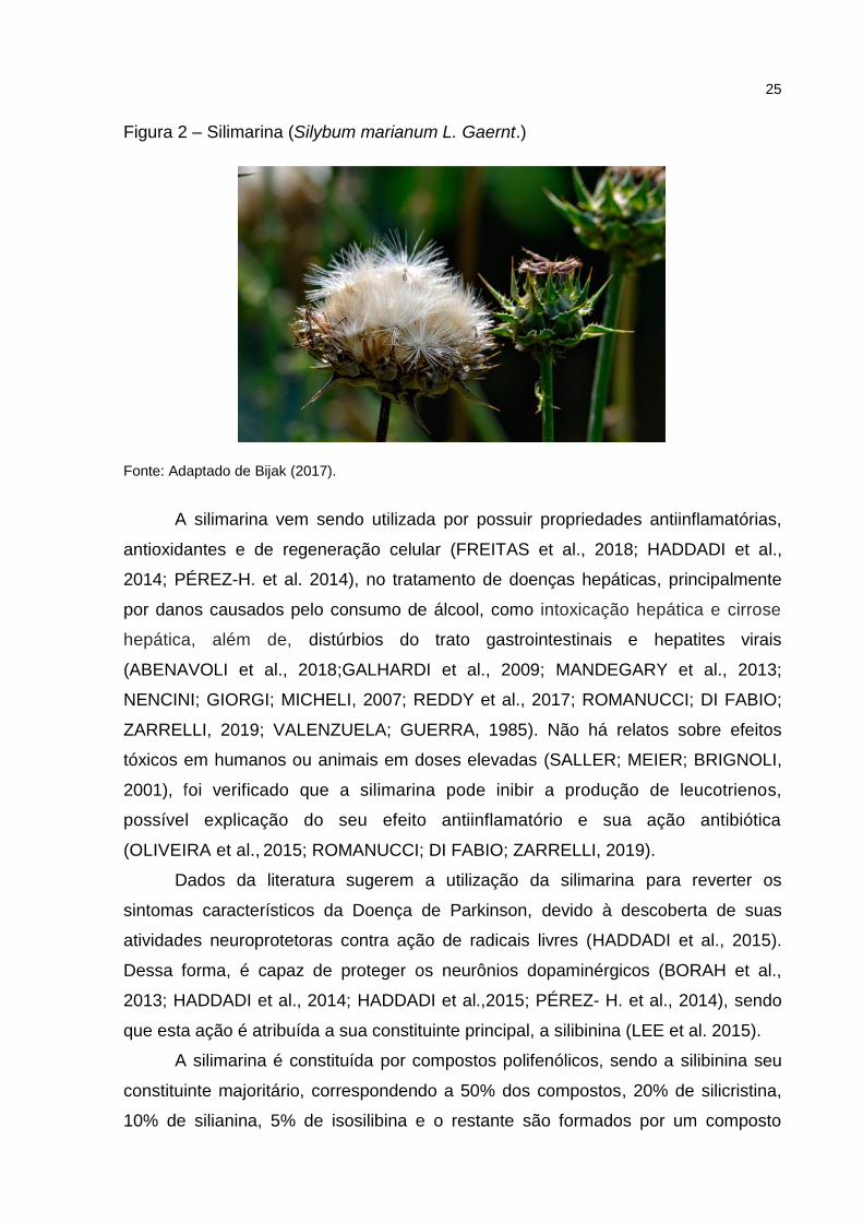
Figura 2 – Silimarina (Silybum marianum L. Gaernt.)
Fonte: Adaptado de Bijak (2017).
A silimarina vem sendo utilizada por possuir propriedades antiinflamatórias,
antioxidantes e de regeneração celular (FREITAS et al., 2018; HADDADI et al.,
2014; PÉREZ-H. et al. 2014), no tratamento de doenças hepáticas, principalmente
por danos causados pelo consumo de álcool, como intoxicação hepática e cirrose
hepática, além de, distúrbios do trato gastrointestinais e hepatites virais
(ABENAVOLI et al., 2018;GALHARDI et al., 2009; MANDEGARY et al., 2013;
NENCINI; GIORGI; MICHELI, 2007; REDDY et al., 2017; ROMANUCCI; DI FABIO;
ZARRELLI, 2019; VALENZUELA; GUERRA, 1985). Não há relatos sobre efeitos
tóxicos em humanos ou animais em doses elevadas (SALLER; MEIER; BRIGNOLI,
2001), foi verificado que a silimarina pode inibir a produção de leucotrienos,
possível explicação do seu efeito antiinflamatório e sua ação antibiótica
(OLIVEIRA et al., 2015; ROMANUCCI; DI FABIO; ZARRELLI, 2019).
Dados da literatura sugerem a utilização da silimarina para reverter os
sintomas característicos da Doença de Parkinson, devido à descoberta de suas
atividades neuroprotetoras contra ação de radicais livres (HADDADI et al., 2015).
Dessa forma, é capaz de proteger os neurônios dopaminérgicos (BORAH et al.,
2013; HADDADI et al., 2014; HADDADI et al.,2015; PÉREZ- H. et al., 2014), sendo
que esta ação é atribuída a sua constituinte principal, a silibinina (LEE et al. 2015).
A silimarina é constituída por compostos polifenólicos, sendo a silibinina seu
constituinte majoritário, correspondendo a 50% dos compostos, 20% de silicristina,
10% de silianina, 5% de isosilibina e o restante são formados por um composto
26
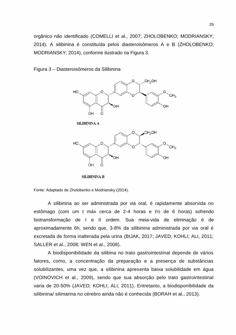
orgânico não identificado (COMELLI et al., 2007; ZHOLOBENKO; MODRIANSKY;
2014). A silibinina é constituída pelos diasteroisômeros A e B (ZHOLOBENKO;
MODRIANSKY; 2014), conforme ilustrado na Figura 3.
Figura 3 – Diasteroisômeros da Silibinina
Fonte: Adaptado de Zholobenko e Modriansky (2014).
A silibinina ao ser administrada por via oral, é rapidamente absorvida no
estômago (com um t máx cerca de 2-4 horas e t½ de 6 horas) sofrendo
biotransformação de I e II ordem. Sua meia-vida de eliminação é de
aproximadamente 6h, sendo que, 3-8% da silibinina administrada por via oral é
excretada de forma inalterada pela urina (BIJAK, 2017; JAVED; KOHLI; ALI, 2011;
SALLER et al., 2008; WEN et al., 2008).
A biodisponibilidade da silibina no trato gastrointestinal depende de vários
fatores, como, a concentração da preparação e a presença de substâncias
solubilizantes, uma vez que, a silibinina apresenta baixa solubilidade em água
(VOINOVICH et al., 2009), sendo que sua absorção pelo trato gastrointestinal
varia de 20-50% (JAVED; KOHLI; ALI, 2011). Entretanto, a biodisponibilidade da
silibinina/ silimarina no cérebro ainda não é conhecida (BORAH et al., 2013).
27
Em diferentes modelos experimentais, a silibinina vem apresentando efeito
neuroprotetor, como demonstrado por Geed et al. (2014), onde foram utilizados ratos
machos, os quais receberam uma única injeção de MPP + (modelo de indução do
parkinsonismo) e foram pré-tratados com silibinina (50, 100 e 200 mg/kg). A
silibinina, principalmente nas doses de 100 e 200 mg/kg, atenuou os déficits motores
causados pela injeção de MPP+ quando comparados com os grupos controle.
A administração de silibinina nas doses de 100 e 200 mg/kg por mais de 2
semanas preveniu a perda de memória induzida por estreptozotocina ou
lipopolissacarídeo (JOSHI et al., 2014; TOTA et al., 2011). Em um estudo realizado
por Wang et al., (2011), foi sugerido que o tratamento repetido com silibinina (50 mg/
kg) durante 6 semanas atenuou a senescência induzida pela D-galactose em
animais. Entretanto, ainda não existem evidências na literatura sobre o efeito da
silibinina em modelo experimental de indução de discinesia orofacial por haloperidol.
Embora modelos experimentais demonstrem o efeito promissor da silibinina,
pouco se sabe sobre os mecanismos envolvidos nestas respostas. Dessa forma,
nossa pesquisa visa estudar possíveis efeitos da silibinina, uma vez que, estudos de
substâncias naturais e desprovidos de efeitos tóxicos significativos devem ser
realizados em modelos de DT na tentativa de encontrar substâncias efetivas no
tratamento, mesmo que de forma adjuvante, desta alteração motora incapacitante.
28
3 OBJETIVOS
3.1 OBJETIVO GERAL
O objetivo deste estudo é investigar os efeitos da silibinina em um modelo de
discinesia orofacial induzido por haloperidol em camundongos.
3.2 OBJETIVOS ESPECÍFICOS
Em camundongos tratados com haloperidol e/ ou silibinina.
→ Avaliar o efeito da silibinina sobre os movimentos de mascar no vazio
induzidos por haloperidol;
→ Avaliar as possíveis alterações motoras através de avaliações
comportamentais (atividades locomotoras e exploratória, tempo de
imobilidade);
→ Investigar se as alterações motoras estão associadas a parâmetros de
estresse oxidativo (tiol total, tiol não- protéico e Na+K+-ATPase), no córtex,
estriado e na região contendo a substância negra;
→ Investigar possíveis alterações nos marcadores de estresse oxidativo (tiol
total, tiol não- protéico e TBARS) causados pela silibinina e haloperidol no
fígado e nos rins.
29
4 RESULTADOS
Os resultados que fazem parte desta dissertação estão apresentados sob a
forma de um manuscrito, o qual se encontra aqui organizado. Os itens Materiais e
Métodos, Resultados, Discussão dos Resultados e Referências Bibliográficas,
encontram-se no próprio manuscrito. O próprio manuscrito está disposto na forma
como está submetido para publicação na revista Pharmacology, Biochemistry and
Behavior.
30
5 MANUSCRITO – EFFECTS OF SILIBININ ON HALOPERIDOL-INDUCED
OROFACIAL DYSKINESIA IN MICE
EFFECTS OF SILIBININ ON HALOPERIDOL-INDUCED OROFACIAL
DYSKINESIA IN MICE
Talita Rodriguesa; Mariana Maikéli Dottoc; Jeane Binotto Reinheimerb; Catiuscia Molz de
Freitasb; Alcindo Busanelloa; Ana Paula Chiappinotto Cerettaa; Getulio Nicola Bressanb;
Bárbara Nunes Kruma; Roselei Fachinettoa, b, *
aPrograma de Pós-Graduação em Farmacologia, Universidade Federal de Santa Maria, RS,
Brazil.
bPrograma de Pós-Graduação em Ciências Biológicas: Bioquímica Toxicológica,
Universidade Federal de Santa Maria, RS, Brazil.
cCurso de Farmácia, Universidade Federal de Santa Maria, RS, Brazil.
Corresponding author:
Drª. Roselei Fachinetto
Centro de Ciências da Saúde
Departamento de Fisiologia e Farmacologia
97105-900, Santa Maria, RS, Brazil
Tel: x21-55-3220 8096
Fax: x21-55-3220 8241
e-mail:[email protected]
Acknowledgments: This study was financed in part by the Coordenação de Aperfeiçoamento
de Pessoal de Nível Superior - Brasil (CAPES) – Finance Code 001, CAPES/PROEX
(process number: 88882.182134/2018-01; support number: 0737/2018), CNPq (475210/2013-
1) and FAPERGS (PqG - 2080-2551/13-5-1). We also acknowledge fellowships from CNPq
(R.F.) and CAPES (T.R., J.B.R., C.M.F., A.B., A.P.C.C., G.N.B., B.N.K).
31
ABSTRACT
Haloperidol is an antipsychotic commonly used to treat schizophrenia, but its long-term use
causes involuntary motor alterations such as tardive dyskinesia in humans. There are few
alternatives to minimize the motor symptoms with low success. Therefore, we investigated
the effect of silibinin, the major component of silymarin, product isolated from seeds Silybum
marianum, which is suggested to reduce the motor alterations in Parkinson’s disease model,
in mice treated with haloperidol. Male mice were treated with vehicle (0.9% NaCl),
haloperidol (1.25 mg/kg, i.p.), silibinin (20 mg/kg, i.p.) and haloperidol (1.25 mg/kg) +
silibinin (20 mg/kg), intraperitoneally for 28 consecutive days. Behavioral quantifications
(vacuous chewing movements-VCMs, number of crossings and rearings in the open field and
time of immobility) were performed each 7 or 14 days during the experimental period. The
biochemical parameters of oxidative stress were evaluated in brain structures (cortex, striatum
and the region containing the substantia nigra), liver and kidney. Haloperidol increased the
prevalence and frequency of VCMs without alterating other behavioral parameters evaluated.
We found negative correlations between number of crossings or rearings with VCMs and a
positive correlation between the time of immobility and VCMs. Silibinin did not modify the
effects of haloperidol on behavioral parameters, and, neither haloperidol not silibinin
modulated oxidative stress parameters. A positive correlation was found between the number
of VCMs and the non-protein thiol content in cortex of mice. In conclusion, our data
demonstrates that in mice also it is possible to verify the increase in the frequency of VCMs
in a percentage of animals mimicking the clinic, since they are not all that develop the OD.
Furthermore, besides silibinin did not avoid the VCMs in mice its combined treatment with
haloperidol seems not cause signals of oxidative stress markers in animals.
Key-words: Vacuous chewing movements, open field, immobility, oxidative stress
32
INTRODUCTION
Schizophrenia is a psychiatric disorder that affects millions of people worldwide (Wu
et al., 2013). The haloperidol is a classical antipsychotic used for the treatment of psychiatric
disorders, such as schizophrenia (Seeman, 2010; Tarsy et al., 2011; Ibrahim and Tamminga,
2011). Its main pharmacological action involves the blockade of D2-type dopamine receptors
(Creese et al., 1976). However, chronic use of antipsychotics, as haloperidol, may lead to the
development of extrapyramidal effects, such as tardive dyskinesia (TD) in 20-40% of patients
increasing this prevalence with age (Andreassen and Jorgensen, 2000). TD is characterized by
repetitive involuntary movements mainly involving the orofacial region, and sometimes the
musculature of the trunk and upper limbs that appear during antipsychotic treatment or after
withdrawal of the antipsychotic (Andreassen and Jorgensen, 2000; Lister et al., 2014; Correll
and Schenk, 2008; Peluso et al., 2012; Tarsy et al., 2011; Woods et al., 2010; Kane, 1995).
In rodent models, occurs orofacial dyskinesia (OD) an analogous syndrome that occurs
in humans (Schaffer et al., 2016; Lister et al., 2014; Röpke et al., 2014; Turrone et al., 2003)
which is characterized by vacuous chewing movements (VCMs) (Lister et al., 2017; Turrone
et al., 2003). The main clinical problem involving TD is that it may become irreversible in
most cases (Casey, 1985; Jeste, 1979; Hashimoto et al., 1998) and forms of effective
treatments, are still unknown (Andreassen et al., 2003; Lohr et al., 2003), due to the lack of
clarity of its pathophysiological mechanism.
Natural products with antioxidant and other pharmacological properties have been
investigated to prevent motor alterations. Silymarin is isolated from the seeds of the Silybum
marianum, and silibinin, a flavonoid, is the major component of silymarin whose
pharmacological potential has been attributed to it (Morazzoni and Bombadelli, 1995).
Sylimarin presented anti-inflammatory, antioxidant and neuroprotective properties (de Freitas
et al., 2018; de Oliveira et al., 2015b; Haddadi et al., 2014; Pérez- H et al., 2014; Nencini et
al., 2007), and it is popularly used in the treatment of liver diseases (Mandegary et al., 2013;
Nencini et al., 2007; Reddy et al., 2017), since there are no reports of toxic effects in humans
or animals at high doses (Saller et al., 2001). Studies have shown that silibinin reduces motor
deficit and decreases dopamine loss in dopaminergic neurons (Lee et al., 2015). However,
there are no data showing the effect of silibinin in motor alterations caused by antipsychotics.
In this study we investigated the effect of silibinin in an OD model induced by haloperidol in
mice, in a tentative to find an effective treatment, even if adjuvant, for this debilitating motor
disease.
33
MATERIALS AND METHODS
Animals
Forty male Albino Swiss mice (2 months old, weighing 20-35 g) were obtained from the
central laboratory of experimentation of the Federal University of Santa Maria. Animals were
acclimatized during at least 7 days and maintained during all the experimental period in the
experimental room of the Department of Physiology and Pharmacology. The room has the
controlled temperature (22 ± 3°C) and light / dark cycle of 12 hours (beginning the clear cycle
at 7:00 am) and the animals were housed in polycarbonate cages (5 animals per cage), with
free access to water and food. All experiments were performed in accordance with the
guidelines of the National Council of Control of Animal Experimentation (CONCEA). This
protocol was approved by the Committee for Ethics in Animal Use (CEUA) of the Federal
University of Santa Maria under the number 139/2014. Every effort was made to reduce the
number of animals used and minimize your suffering.
Drugs
Silibinin was obtained from Sigma-Aldrich (São Paulo, Brazil) and administered at a dose of
20 mg/kg (Marrazzo et al., 2011). Haloperidol (Teuto, Brazil) was obtained in commercial
pharmacy and administered at the dose of 1.25 mg/kg (Éthier et al., 2004). All drug solutions
were prepared in physiological saline (0.9% NaCl, vehicle) before administration and were
administered intraperitoneally (i.p.) with a constant volume of 5 mL/kg body weight each, for
28 days.
Experimental design and treatment
Initially, the mice were randomly divided into four experimental groups with 10 animals each:
control (which receive two i.p. administrations of 0.9% NaCl, n=9), silibinin (0.9% NaCl and
silibinin 20 mg/kg), haloperidol (0.9% NaCl and haloperidol 1.25 mg/kg) and haloperidol +
silibinin (silibinin 20 mg/kg and haloperidol 1.25 mg/kg). During the experimental period one
animal from control and one animal from haloperidol group died. Then these groups had 9
animals at the final of the experiment. Animals received the treatment intraperitoneally (i.p.)
during 28 days (Ceretta et al., 2018). To evaluate the effects of haloperidol and or silibinin,
34
the VCMs and immobility were quantified on days 7, 14, 21 and 28 while open field test was
performed on days 14 and 28 of the experimental period. Then, the animals were anesthetized
with ketamine:xylazine (100:10 mg/kg, i.p.), and euthanized by cervical dislocation. The
brain was removed and the cortex, striatum and the region containing the substantia nigra
were immediately separated, or well in the liver and kidney. All tissues were stored at −80 °C
until the biochemical analysis.
Behavioral testing
One hour before the behavioral tests, the animals were taken to the behavioral assessment
room for adaptation. The tests were performed between 7 and 11 a.m. Observers were blinded
with regard to the treatment conditions. All behaviors were quantified at the moment that the
animal stay into de apparatus and also filmed.
Quantification of vacuous chewing movements (VCM)
VCMs were quantified on days 7, 14, 21 and 28 of experimental design. Mice were placed
individually in a glass box (20x20x19 cm), containing a mirror positioned just below the floor
of the box. The number of VCM was measured continuously for 6 minutes, after a period of 6
minutes of adaptation (Busanello et al., 2011; de Freitas et al., 2016; Ceretta et al., 2018).
VCM were defined as single mouth openings in the vertical plane not directed towards
physical object and they were not counted if they occurred during the grooming period. As the
animals were evaluated during four section behavior, it was possible to separate the animals
that developed VCMs after haloperidol treatment and the animals that did not developed an
increase of VCM intensity. Clinically, only a percentage of patients develop tardive
dyskinesia after antipsychotic treatment (Andreassen and Jorgensen, 2000) and in rats also it
occurs when treated with antipsychotics to develop OD (Kane and Smith, 1982; Shirakawa
and Tamminga, 1994). In our study, only the animals presenting more than 8 VCMs during 6
min in at least two behavioral evaluations were considered +VCMs. This value was choosed
because control animals did not present this value in two consecutive analyses. In our
experiment, one animal of silibinin group showed +VCM. Thus, as it is impossible to perform
statistical analyses with a group containing one animal, it was removed from the graphs and
the group of silibinin appears with 9 animals.
35
Quantification of immobility time
During the VCM session, the animals were filmed for 6 minutes for later quantification of the
total time of immobility (absence of movement).
Open field test
The spontaneous locomotor and exploratory activities were evaluated on days 14 and 28 of
experimental protocol before VCM session. Mice were placed individually in the center of an
open field arena (44×44×44 cm), divided into 9 equal squares. The number of lines crossed,
and number of rearing were measured during 5 minutes without habituation (Broadhurst,
1960; Busanello et al., 2011).
Tissue preparation and assays
Samples were centrifuged at 500 x g at 4 °C for 10 min. The supernatant was used to
determine protein concentration using the Lowry method (Lowry et al., 1951) and
biochemical assays:
Total thiol and non-protein thiol groups
The levels of total and non-protein thiol were quantified by the methodology described by
Ellman (1959). To total thiol an aliquot of the supernatant was incubated with 0.5 M 5, 5'-
dithiobis-(2-nitrobenzoic acid) (DTNB). To non-protein thiol, the supernatants of the cortex,
striatum, region containing the substantia nigra, liver and kidney were mixed with 10%
trichloroacetic acid (TCA) at a proportion of 1:1, centrifuged at 500 x g for 10 min. Then, one
aliquot of supernatant was incubated with 0.5 M DTNB. The chromogen formed was
measured spectrophotometrically at 412 nm. Glutathione (GSH) was used as standard. Results
were expressed as nmol total thiol/mg protein or nmol non-protein thiol/mg protein.
Na+/K+-ATPase activity
Na+/K+ ATPase activity was quantified according to colorimetric methodology by Fiske and
Subarrow (1925). Samples of cortex, striatum and region containing the substantia nigra were
incubated with or without ouabain (250 μM) and pre-incubated 10 min at 37ºC. After, the 3
mM of ATP was added to the reaction and incubated for 30 min at 37 °C. Reading was
36
carried out at 405 nm and phosphate was used as standard. Result is expressed as nmol Pi/mg
protein.
Measurement of Thiobarbituric Acid Reactive Substances (TBARS)
Aliquots of liver and kidney supernatants were used to determine the TBARS production as
described by Ohkawa et al. (1979). Malondialdehyde (MDA was used as standard). Reading
was carried out at 532 nm and results were expressed in nmol MDA/mg protein.
Statistical analysis
Data are expressed as mean ± standard error of the mean (SEM). Statistical analysis of data
was carried out by one-way analysis of variance (ANOVA) followed by post hoc Tukey’s
multiple comparisons test when appropriated. Statistical significance was assumed at p <
0.05. F values are presented in the text only if p value associated with it was p<0.05.
Pearson’s correlation test was applied to check for possible significant correlations of the
data.
RESULTS
Effect of haloperidol and/or silibinin on VCMs
On days 7 [F (5, 31) = 17.37, p<0.05, fig. 1a] and 14 [F (5, 31) = 7.48, p<0.05, fig. 1b], the
treatment with haloperidol alone or haloperidol and silibinin significantly increased VCM on
mice when compared to the control group. On day 21, the increase was caused only by the
treatment with control [F (5, 31) = 3.95, p<0.05, fig. 1c] while on day 28, the increase
occurred only at the co-treated group [F (5, 31) = 4.87, p<0.05, fig. 1d]. Treatment with
haloperidol induced a VCM prevalence of 55% compared to its vehicle (Chi-square=6.92;
p<0.05), with 5 out of 9 animals actually having VCMs. In fact, the co-treatment of
haloperidol with silibinin developed VCMs in 30% of the mice. The treatment with silibinin
did reduce neither the prevalence nor the intensity of VCMs in those mice that developed
VCMs.
Effect of haloperidol and/or silibinin on immobility
There was no significant difference in the immobility (Fig 2a and 2b) on days 7 (Fig 3a), 14
(Fig 3b), 21 (Fig 3c) and 28(Fig 3d) compared with control group. However, a positive
37
correlation was detected between VCMs with the time of immobility on day 28 [r = 0.6038,
p< 0.0001] (Fig. 4c).
Effect of haloperidol and/or silibinin on open field test
There was no significant difference in the number of crossings (Fig 2a and 2b) on days 14 and
28. With regard to the number of rearings, only the group treated with haloperidol –VCM
presented an increase on day 28 [F (5, 31) = 3.03, p<0.05, Fig 2d] without any effect on day
14 (Fig 2c evaluated on the open field test in any of the evaluated days, 14 and 28. However,
a negative correlation was detected between VCMs with the number of crossings [r = -0.5474,
p = 0.0005] (Fig. 4a) and VCMs and the number of rearings [r = -0.3834, p = 0.0192] (Fig.
4b).
Effect of haloperidol and/or silibinin on total and non-protein thiol groups and Na+/K+-
ATPase in brain structures
There were no significant differences among the groups on total and non-protein thiol groups
and Na+/K+-ATPase in brain structures of mice treated with haloperidol and/or silibinin
(Table 1). However, there was a positive correlation between the number of VCMs and the
non-protein thiol groups in the cortex of mice [r = 0.4353, p = 0.008] (Fig. 5).
Effect of haloperidol and/or silibinin on total and non-protein thiol groups and TBARS
production in liver and kidney
There were no significant differences among the groups on total and non-protein thiol groups
and TBARS production in liver and kidney of mice treated with haloperidol and/or silibinin
(Table 1).
DISCUSSION
The present study investigated if silibinin could avoid the development of OD induced
by haloperidol in mice. Haloperidol increased the prevalence and frequency of VCMs without
alterations in other behavioral parameters evaluated. The animals were divided into high and
low VCMs, because, as in the clinic, not all of them develop dyskinesia. Negative correlations
38
between number of crossings or rearings with VCMs and a positive correlation between the
time of immobility and VCMs were found. Silibinin did not modify the effects of haloperidol
on behavior. Furthermore, neither haloperidol nor silibinin caused alterations in oxidative
stress parameters. A positive correlation was found between the number of VCMs and the
non-protein thiol content in cortex of mice.
Antipsychotics have been used for a numerous of pathological conditions like autism,
bipolarity and is the main treatment to psychosis like schizophrenia (Canitano and Scandurra,
2011; Pickar et al., 2008; Seeman, 2010; Tarsy et al., 2011; Ibrahim and Tamminga, 2011).
However, the long-term use of antipsychotics is associated with the development of TD which
can be irreversible after its appearance (Andreassen and Jorgensen, 2000). Although of a
numerous of studies searching for substances either natural or synthetics, little success has
been obtained to avoid the appearance of motor symptoms in patients (Busanello et al., 2017;
Ceretta et al., 2018; Dhingra et al., 2018; Kronbauer et al., 2017). Other problem is that the
pathophysiology of TD remains unclear, which contributes to difficulties in to establish a
pharmacological target to finding a substance to treating or preventing TD.
Studies demonstrate that silymarin can ameliorate motor alterations in different
models in rodents (Haddadi et al., 2014; Borah et al., 2013; Farnebo et al., 1971; Scatton et
al., 1970; De Freitas et al., 2018) and has promissory benefits in humans (Lee et al., 2015)
without presenting toxicity in the used doses (Saller et al., 2001). Furthermore, De Oliveira et
al. (2015b) demonstrated silymarin inhibits the activity of MAO and has antioxidant potential
in vitro. The beneficial effects of silymarin are mainly attributed to the presence of silibinin
(Morazzoni and Bombadelli, 1995). In this study, it was investigated if silibinin, the major
component of silymarin, could avoid the OD induced by haloperidol in mice.
We observed that silibinin was not effective in reducing the intensity of VCMs in mice
under treatment with haloperidol, however it showed a reduction in the prevalence of VCMs
compared with the group treated with haloperidol. However, this effect did not reach
significant. Other studies from our group also demonstrated that some substances can reduce
the intensity of VCMs (Peroza et al., 2013; Röpke et al., 2014; Schaffer et al., 2016;
Busanello et al., 2017; Ceretta et al., 2018) while other reduced the prevalence of VCMs
(Fachinetto et al., 2007a; Busanello et al., 2012) and in some studies any effect was obtained
(Fachinetto et al., 2007b; Reis et al., 2013).
Data demonstrate the decrease of locomotion after administration of haloperidol
(Sonego et al., 2018; Peroza et al., 2013; Fachinetto et al., 2007b; Busanello et al., 2012)
which was not observed by other authors (Sonego et al., 2018). In the present study the
39
decrease in the locomotor activity was not observed although it was found a negative
correlation between VCM intensity with number of crossings and rearings suggesting those
animals presenting high intensity of VCMs showed a reduction in locomotion. As the animals
with high intensity of VCMs received haloperidol we can indirectly suggest that there was a
reduction in locomotor activity. Other data which reinforce this suggestion was the positive
correlation between the VCMs with the time of immobility since the animals with high VCMs
presented a higher time without movements. The time of immobility is considered a
stereotyped behavior, as are VCMs, which are increased by haloperidol (Wallnau et al., 1978;
Sanberg et al., 1980; Thakur et al., 2015).
As the possible interaction between haloperidol and silibinin is unknown, it was
investigated its effects on total thiol, non-protein thiol groups and Na+/K+-ATPase in brain
structures as well as total thiol, non-protein thiol groups and TBARS in liver and kidney of
mice treated. No significant effect in the tested markers suggesting the concomitant use of
acute haloperidol and silibinin did not presentsigns of oxidative stress. In chronic studies,
antipsychotics presented oxidative stress markers to liver and kidney (Dalla Corte et al., 2008;
Corte et al., 2009; Akintude and Abubakar, 2017). By the other hand, silymarin is clinically
used for offering protection against hepatotoxins (Mandegary et al., 2013; Nencini et al.,
2007; Reddy et al., 2017) and these effects are also attributed to silibinin.
In conclusion, our data demonstrates that in mice also it is possible to verify the
increase in the frequency of VCMs only in a percentage of animals mimicking which occurs
in the clinic. Furthermore, besides silibinin did not avoid the VCMs in mice its combined
treatment with haloperidol seems not cause signals of oxidative stress markers in animals.
40
References
Andreassen, O.A., JØrgensen, H.A. (2000). Neurotoxicity associated with neuroleptic-
induced oral dyskinesias in rats: Implications for tardive dyskinesia?. Prog. Neurobiol. 61 (5),
525–541.
Akintunde, J.K., Abubakar, O.K. (2017). Novel therapeutic approaches of natural oil from
black seeds and its underlying mechanisms against kidney dysfunctions in haloperidol
induced male rats. Drug Metab Pers Ther. 32 (2), 97-107.
Borah, A., Paul, R., Choudhury, S., Choudhury, A. Bhuyan, B., Das Talukdar, A., Dutta, C.
M., Mohanakumar, K. P. (2013).Neuroprotective Potential of Silymarin against CNS
Disorders: Insight into the Pathways and Molecular Mechanisms of Action.CNS Neurosci.
Ther. 19 (11), 847–853
Broadhurst, P.L. (1960). Experiments in psychogenetics. In: Eysenk HJ, editor. Experiments
inpersonality. London: Routledge & Kegan Paul, pp. 76.
Busanello, A., Barbosa, N.B., Peroza, L.R., Farias, L.E., Burger, M.E., Barreto,
K.P.,Fachinetto, R. (2011). Resveratrol protects against a model of vacuous
chewingmovements induced by reserpine in mice. Behav. Pharmacol. 22 (1), 71–75.
Busanello, A., Peroza, L.R., Wagner, C., Sudati, J.H., Pereira, R.P., Prestes, A. S., Rocha,
J.B.T.,Fachinetto, R., Barbosa, N. B. V. (2012).Resveratrol reduces vacuous chewing
movements induced by acute treatment with Fluphenazine. Pharmacol. Biochem. Behav. 101
(2), 307-310.
Busanello, A., Leal, C. Q., Peroza, L. R., Röpke, J., de Moraes, E.R.,de Freitas, C. M.,
Libardoni, M., Barbosa, N. B. V., Fachinetto, R. (2017).Resveratrol Protects Against Vacuous
Chewing Movements Induced by Chronic Treatment with Fluphenazine. Neurochem. Res. 42
(11), 3022- 3040.
Canitano, R., Scandurra, V. (2011). Psychopharmacology in autism: an update. Prog
Neuropsychopharmacol Biol Psychiatry. 35(1), 18-28.
Casey, D.E.(1985). Tardive dyskinesia: reversible and irreversible. Psychopharmacology
(Berl). v. 2, p. 88–97.
Ceretta, A. P. C., de Freitas, C. M., Schaffer, L. F., Reinheimer, J. B., Dotto, M.M., Reis, E.
M., Scussel, R.,Machado-de-Ávila, R. A., Fachinetto, R. (2018).Gabapentin reduces
haloperidol-induced vacuous chewing movements in mice. Pharmacol Biochem Behav.166,
21-26.
Correll, C.U., Schenk, E.M.(2008). Tardive dyskinesia and new antipsychotics. Curr. Opin.
Psychiatry 21, 151–156.
Corte, C.L., Fachinetto, R., Puntel, R., Wagner, C., Nogueira, C.W., Soares, F.A., Rocha, J.B.
(2009). Chronic treatment with fluphenazine alters parameters of oxidative stress in liver and
kidney of rats.Basic ClinPharmacolToxicol. 105(1), 51-57.
41
Creese, I., Burt, D.R., Snyder, S.H. (1976). Dopamine receptor binding predicts clinical
andpharmacological potencies of antischizophrenic drugs. Science. 192(4238), :481-483.
Dalla Corte, C.L., Fachinetto, R., Colle, D., Pereira, R.P., Avila, D.S., Villarinho, J.G.,
Wagner,C., Pereira, M.E., Nogueira, C.W., Soares, F.A., Rocha, J.B(2008). Potentially
adverse interactions between haloperidol and valerian.Food Chem Toxicol.46(7), 2369-2375.
de Freitas, C.M., Busanello, A., Schaffer, L.F., Peroza, L.R., Krum, B.N., Leal, C.Q.,
Ceretta,A.P., da Rocha, J.B., Fachinetto, R. ( 2016). Behavioral and neurochemical effects
induced by reserpine in mice.Psychopharmacology (Berl).233 (3), 457–467.
de Freitas, C.M., Krum, B. N., Ceretta, A.P.C., Schaffer, L. F., Reis, E.M.,Schwerz, J.P.,
Barbosa, C.P., Soares, F. A. A., Fachinetto, R. (2018). Silymarin recovers 6-
hydroxydopamine-induced motor deficits in mice. Food Chem Toxicol.118, 549-556.
de Oliveira, D. R., Schaffer, L. F., Busanello, A., Barbosa, C. P., Peroza, L.R., de Freitas, C.
M., Krum, B. N., Bressan, G. N., Boligon, A. A., Athayde, M. L., de Menezes, I.R,.A.,
Fachinetto, R. (2015b). Silymarin has antioxidant potential and changes the activity
ofNa+/K+-ATPase and monoamine oxidase in vitro. Ind. Crops Prod.70, 347–355.
Dhingra, D., Goswami, S., Gahalain, N. (2017).Protective effect of hesperetin against
haloperidol induced orofacial dyskinesia and catalepsy in rats. NutrNeurosci.21 (9), 667-675.
Ellman, G.L. (1959). Tissue sulphydryl groups. Archives of Biochemistry and Biophysics. 82
(1),70-71.
Éthier, I., Kagechika, H., Shudo, K., Rouillard, C., Lévesque, D. (2004). Docosahexaenoic
acid reduces haloperidol-induced dyskinesias in mice: involvement of Nur77 and retinoid
receptors.Biol Psychiatry. 56(7), 522-526.
Fachinetto, R., Villarinho, J.G., Wagner, C., Pereira, R. P., Ávila, D. S., Burger, M. E.,
Calixto, J.B., Rocha.,J. B.T., Ferreira, J.(b). (2007). Valeriana officinalis does not alter
theorofacial dyskinesia induced by haloperidol in rats: role of dopamine transporter. Prog.
Neuro-Psychopharmacology Biol. Psychiatry.31 (7), 1478–1486.
Fachinetto, R.,. Villarinho, J. G., Wagner, C., Pereira, R.P., Puntel, R.L., Paixão, M.W.,
Braga, A.L., Calixto, J.B., Rocha, J.B.T., Ferreira, J. (2007a). Diphenyl diselenide decreases
the prevalence of vacuous chewing movements induced by fluphenazine in rats.
Psychopharmacology, 194 (3), 423-432.
Farnebo, L.O., Fuxe,K., Goldstein,M., Hamberger,B., Ungerstedt,U. (1971). Dopamine and
noradrenaline releasing action of amantadine in the central and peripheral nervous system: a
possible mode of action in Parkinson's disease. Eur J. Pharmacol.16 (1), 27-38.
Fiske, C.H., Subbarow, Y. (1925). The colorimetric determination of phosphorus. J. of Biol.
Chem. 66, 375–400.
Haddadi, R., Nayebi, A. M., Farajniya, S., Brooshghalan, S. E., Sharifi, H. (2014). Silymarin
improved 6-OHDA-induced motor impairment in hemi parkisonian rats: behavioral and
molecular study. DARU, J. Pharm. Sci. 22 (1), 22-38.
42
Hashimoto, T., Ross, D.E., Gao, X.M., Medoff, D.R., Tamminga, C.A. (1998). Mixture in
thedistribution of haloperidol-induced oral dyskinesias in the rat supports an animal
model of tardive dyskinesia. Psychopharmacology (Berl).137 (2), 107–112.
Ibrahim, H.M., Tamminga, C.A. (2011). Schizophrenia: treatment targets beyond monoamine
systems. Annu Rev PharmacolToxicol. 51, 189-209.
Jeste, D.V., Potkin, S.G., Sinha, S., Feder, S., Wyatt, R.J. (1979). Tardive dyskinesia-
reversible and persistent. Arch Gen Psychiatry. 36, 585–590.
Kane, J.M. (1995). Tardive dyskinesia: epidemiology and clinical presentation. In: Bloom,
F.E., Kupfer, D.J. (Eds.), Psychopharmacology: The Fourth Generation of Progress, vol. 39.
Raven Press, Ney York, pp. 1485-1495.
Kane, J.M., Smith, J.M.(1982). Tardive dyskinesia: prevalence and risk factors, 1959 to1979.
Arch Gen Psychiatry . 39, 473–481.
Kronbauer, M., Metz, V.G., Roversi, K., Dias, V.T., de David, C.T.A., da Silva, R.C. B.,
Burger, M.E. (2017). Influence of magnesium supplementation on movement side effects
related to typical antipsychotic treatment in rats.Behav Brain Res. 320, 400-411.
Lee, Y., Park, H.R., Chun, H. J., Lee, J. (2015).Silibinin Prevents Dopaminergic Neuronal
Loss in a Mouse Model of Parkinson’s Disease Via Mitochondrial Stabilization. J. Neurosci.
Res. 93 (5), 755–765.44
Lister, J., Nobrega, J. N., Fletcher, P. J., Remington, G. (2014). Oxidative stress and the
antipsychotic-induced vacuous chewingmovement model of tardive dyskinesia: evidence for
antioxidant-based prevention strategies. Psychopharmacology (Berl).231(11), 2237-2249.
Lister, J., Andreazza, A.C., Navaid, B., Wilson, V.S., Teo, C., Nesarajah, Y., Wilson,
A.A.,Nobrega, J.N., Fletcher, P.J., Remington, G.(2017). Lipoic acid and haloperidol-
inducedvacuous chewing movements: implications for prophylactic antioxidant use intardive
dyskinesia. Prog. Neuro-Psychopharmacol. Biol. Psychiatry 72, 23–29.
Lohr, J.B., Kuczenski, R., Niculescu, A.B. (2003). Oxidative mechanisms and tardive
dyskinesia. CNS Drugs. 17, 47–62.
Lowry, O.H., Rosebrough, N.J., Farr, A.L., Randall, R.J. (1951) . Protein measurement with
the Folin phenol reagent. J. Biol. Chem. 193 (1), 265–275.
Mandegary, A., Saeedi, A., Eftekhari, A., Montazeri, V., Sharif, E. (2013). Hepatoprotective
effect of silyamarin in individuals chronically exposed to hydrogen sulfide; modulating
influence of TNF-α cytokine genetic polymorphism. DARU, J. Pharm. Sci. 21 (1), 21-28.
Marrazzo, G., Bosco, P., Delia, F.L., Scapagnini, G., Giacomo, C.D., Malaguarnera,M.,
Galvano,F., Nicolosia, A., Volti, G.L. (2011).Neuroprotective effect of silibinin in diabetic
mice. Neurosci Lett.504, 252– 256.
Morazzoni, P., Bombardelli, E. (1995). Silybum marianum (Carduus marianus), Fitoterapia.
66 (1), 3–42.
43
Nencini, C., Giorgi, G., Micheli, L. (2007). Protective effect of silymarin on oxidative stress
in rat brain. Phytomedicine. 14, 129–135.
Ohkawa, H., Ohishi, N., Yagi, K. (1979).Assay for lipid peroxidation in animal tissues by
thiobarbituric acid reaction.Anal Biochem. 95 (2), 351- 358.
Peluso, M.J., Lewis, S.W., Barnes, T.R., Jones, P.B. (2012).Extrapyramidal motor side-effects
of first- and second-generation antipsychotic drugs. Br. J. Psychiatry. 200, 387–392.
Pérez-H, J., Carrillo-S, C., García, E., Ruiz-Mar, G., Pérez-Tamayo, R.,Chavarría, A. (2014).
Neuroprotective effect of silymarin in a MPTP mouse model of Parkinson’s disease.
Toxicology. 319, 38–43.
Peroza, L.R., Busanello, A., Leal, C. Q., Röpke, J., Boligon, A. A., Meinerz, D., Libardoni,
M., Athayde, M. L., Fachinetto, R. (2013).Bauhinia Forficata prevents vacuous chewing
movements induced by haloperidol in rats and has antioxidant potential in vitro. Neurochem.
Res. 38 (4), 789–796.
Pickar, D., Vinik, J., Bartko, J.J. (2008). Pharmacotherapy of schizophrenic patients:
preponderance of off-label drug use. PLoS One. 3 (9), e3150.
Reddy, M. K., Reddy, A. G., Kumar, B. K., Madhuri, D., Boobalan, G., Reddy, M. A.
(2017). Protective effect of rutin in comparison to silymarin against induced hepatotoxicity in
rats. Vet. World. 10 (1),74-80.
Reis, E.M., Röpke, J., Busanello, A., Reckziegel, P., Leal, C.Q., Wagner, C., Boligon, A.A.,
Athayde, M.L., Fachinetto, R. (20130. Effect of Hypericum perforatum on different models of
movement disorders in rats.BehavPharmacol. 24 (7), 623-627.
Röpke, J., Busanello, A., Leal, C.Q., de Moraes Reis, E., de Freitas, C.M., Villarinho, J.G.,
Figueira, F.H., Mello, C.F., Ferreira, J., Fachinetto, R. (2014). Anandamide attenuates
haloperidol-induced vacuous chewing movements in rats. Prog. Neuro- Psychopharmacol.
Biol. Psychiatry 54, 195–199.
Saller, R., Meier, R., Brignoli, R. (2001). The use of Silymarin in the treatment of liver
diseases. Drugs. 61, 2035-2063.
Sanberg,P.R., Pisa, M., Faulks, I.J., Fibiger, H.C. (1980). Experimental influences on
catalepsy. Psychopharmacology (Berl). 69 (2), 225-226.
Seeman, P.(2010). Dopamine D2 receptors as treatment targets in schizophrenia. Clin.
Schizophr. Relat. Psychoses. 4, 56–73.
Sonego, A. B., Prado, D. S., Vale, G. T., Sepulveda-Diaz, J.E., Cunha, T. M., Tirapelli, C.R.,
Del Bel, E. A., Raisman-Vozari, R., Guimarães, F. S. (2018).Cannabidiol prevents
haloperidol-induced vacuos chewing movements and inflammatory changes in mice via
PPARγ receptors. Brain Behav Immun.. 74, 241-251.
Scatton B, Cheramy A, Besson MJ, Glowinski J. (1970). Increased synthesis and release of
dopamine in the striatum of the rat after amantadine treatment. Eur J Pharmacol. 13 (1), 131-
133.
44
Schaffer, L. F., de Freitas, C. M., Ceretta, A. P.C., Peroza, L. R.,Reis, E. M., Krum, B. N.,
Busanello, A., Boligon, A. A., Sudati, J. H., Fachinetto, R., Wagner, C. (2016).
Harpagophytum Procumbens Ethyl Acetate Fraction Reduces Fluphenazine-Induced Vacuous
Chewing Movements and Oxidative Stress in Rat Brain.Neurochem. Res. 41 (5), 1170-1184.
Shirakawa, O., Tamminga, C.A.(1994). Basal ganglia GABAA anddopamine D1 binding site
correlates of haloperidol-induced oral dyskinesias in rat. ExpNeurol. 127, 62–69.
Tarsy, D., Lungu, C., Baldessarini, R.J. (2011). Epidemiology of tardive dyskinesia before
and during the era of modern antipsychotic drugs. Handb. Clin. Neurol. 100, 601–616.
Thakur, K.S., Prakash, A.,Bisht, R., Bansal, P. K. (2015). Beneficial effect of candesartan and
lisinopril against haloperidol-induced tardive dyskinesia in rat. J Renin Angiotensin
Aldosterone Syst. 16 (4), 917–929.
Turrone, P., Remington, G., Kapur, S., Nobrega, J. N. (2003). The relationship between
dopamine D2 receptor occupancy and the vacuous chewing movement syndrome in rats.
Psychopharmacology (Berl). 165 (2), 166- 171.
Wallnau, L.B., Carnrike,C.L. Jr.,Dewey,G.I. (1978). Tonic immobility: effects of dopamine
receptor blockade and stimulation. Pharmacol. Biochem. Behav.10 (2), 177-181.
Woods, S.W., Morgenstern, H., Saksa, J.R., Walsh, B.C., Sullivan, M.C., Money,
R., Hawkins, K.A., Gueorguieva, R.V., Glazer ,W.M. (2010). Incidence of tardive dyskinesia
with atypical versus conventional antipsychotic medications: a prospective cohort study. J.
Clin. Psychiatry. 71, 463–474.
Wu, J.Q., Kosten, T.R.,Zhang, X. Y.(2013). Free radicals, antioxidant defense systems, and
schizophrenia.Prog. Neuro-Psychopharmacology Biol. Psychiatry. 46, 200-206.
45
LEGENDS FOR FIGURES AND TABLES
Figure 1. Effect of haloperidol (1.25 mg/kg) and silibinin (20 mg/kg) on VCM in mice. The
quantification of VCM was performed during 6 minutes on days 7 (a), 14 (b), 21 (c) and 28
(d). Data are expressed as means ± SEM. One-way ANOVA followed by Tukey's multiple
comparisons test. *p<0.05, compared with the control group; (c) #p<0.05, compared with the
haloperidol group -VCM. (d) #p<0.05, compared with the haloperidol+ silibinin group -VCM.
(control, n=9; silibinin, n=9; haloperidol, n=9; haloperidol+silibinin, n=10).
Figure 2. Effect of haloperidol (1.25 mg/kg) and silibinin (20 mg/kg) on open field test in
mice. The quantification number of crossing was performed on days 14 (a) and 28 (b) and
number of rearing was performed on days 14 (c) and 28 (d). Data are expressed as means ±
SEM. One-way ANOVA followed by Tukey's multiple comparisons test.*p<0.05, compared
with the control group (control, n=9; silibinin, n=9; haloperidol, n=9; haloperidol+silibinin,
n=10).
Figure 3. Effect of haloperidol (1.25 mg/kg) and silibinin (20 mg/kg) on open field test in
mice. The quantification number of time of immobility on days 14 (a) and 28 (b) and number
of rearing was performed on days 14 (c) and 28 (d). Data are expressed as means ± SEM.
One-way ANOVA followed by Tukey's multiple comparisons test. @p<0.05, compared with
the silibinin group (control, n=9; silibinin, n=9; haloperidol, n=9; haloperidol+silibinin,
n=10).
Figure 4: Correlation between VCMs number with crossing (a), rearing (b) and time of
immobility (c). Significant correlations (Pearson correlation – r) when p < 0.05.
Figure 5: Correlation between VCMs number with non-protein thiol in cortex. Significant
correlations (Pearson correlation – r) when p < 0.05.
Table 1: Effects of haloperidol and/or silibinin on total and non-protein thiol groups and on
Na+/K+-ATPase activity in brain structures. Data are expressed as means ± SEM
Table 2: Effects of haloperidol and/or silibinin on total and non-protein thiol groups and on
TBARS productionin liver and kidney. Data are expressed as means ± SEM.
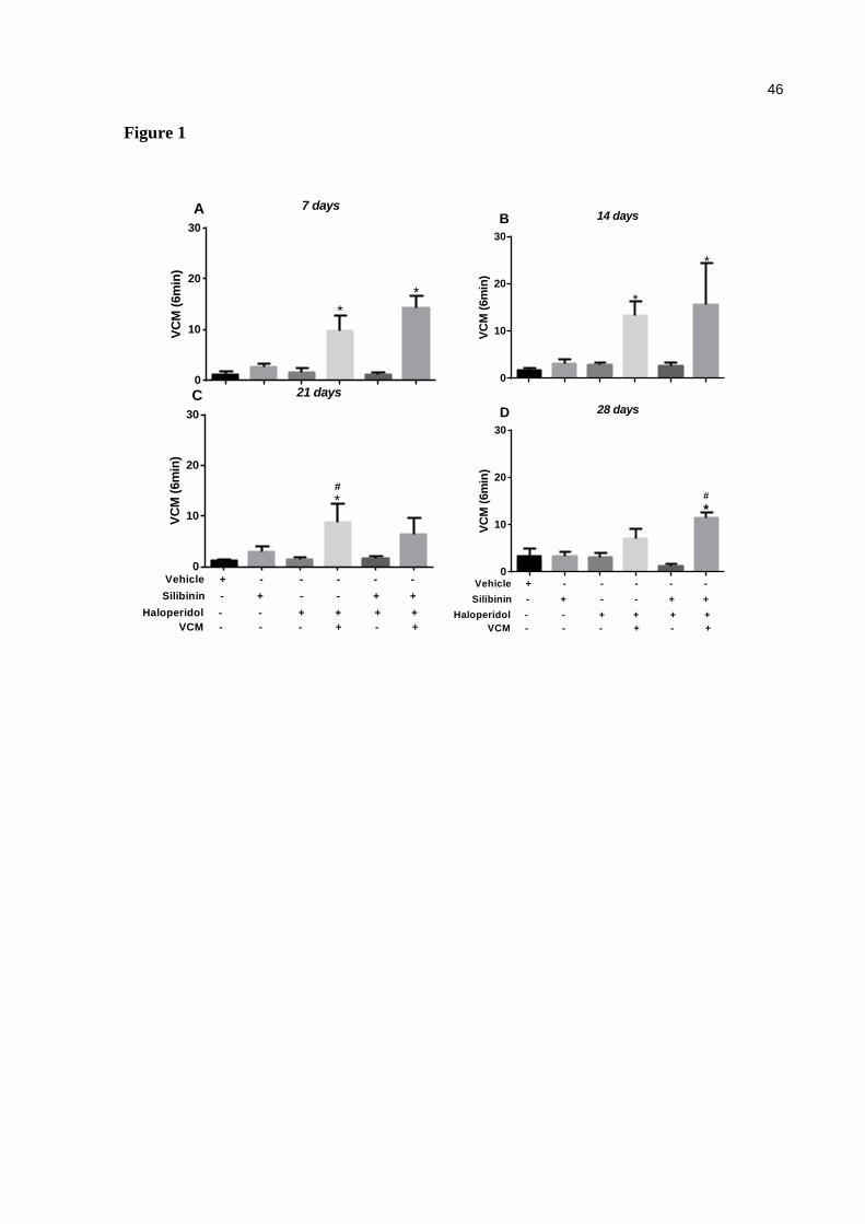
46
Figure 1
7 days
VC
M (
6m
in)
0
10
20
30
*
*
A14 days
VC
M (
6m
in)
0
10
20
30
*
*
B
21 days
VC
M (
6m
in)
0
10
20
30
*#
Vehicle + - - - - -
Silibinin - + - - + +
Haloperidol - - + + + +
VCM - - - + - +
C28 days
VC
M (
6m
in)
0
10
20
30
*#
Vehicle + - - - - -
Silibinin - + - - + +
Haloperidol - - + + + +
VCM - - - + - +
D
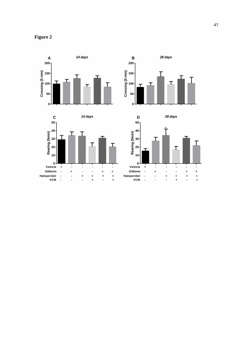
47
Figure 2
14 days
Cro
ssin
g (
5 m
in)
0
50
100
150
200
A 28 days
Cro
ssin
g (
5 m
in)
0
50
100
150
200
B
14 days
Reari
ng
(5m
in)
0
10
20
30
40
50
Vehicle + - - - - -
Silibinin - + - - + +
Haloperidol - - + + + +
VCM - - - + - +
C 28 days
Reari
ng
(5m
in)
0
10
20
30
40
50
*
Vehicle + - - - - -
Silibinin - + - - + +
Haloperidol - - + + + +
VCM - - - + - +
D
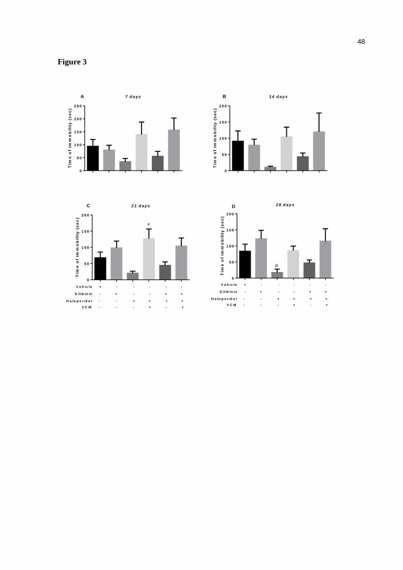
48
Figure 3
7 d a y s
Tim
e o
f im
mo
bil
ity
(s
ec
)
0
5 0
1 0 0
1 5 0
2 0 0
2 5 0
A 1 4 d a y s
Tim
e o
f im
mo
bil
ity
(s
ec
)
0
5 0
1 0 0
1 5 0
2 0 0
B
2 1 d a y s
Tim
e o
f im
mo
bil
ity
(s
ec
)
0
5 0
1 0 0
1 5 0
2 0 0
#
V e h ic le + - - - - -
S i l ib in in - + - - + +
H a lo p e r id o l - - + + + +
V C M - - - + - +
C 2 8 d a y s
Tim
e o
f im
mo
bil
ity
(s
ec
)
0
5 0
1 0 0
1 5 0
2 0 0
@
V e h ic le + - - - - -
S i l ib in in - + - - + +
H a lo p e r id o l - - + + + +
V C M - - - + - +
D
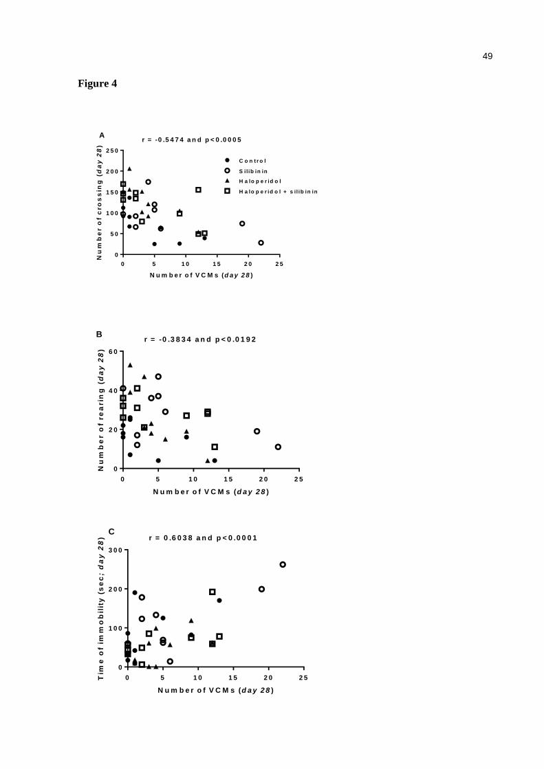
49
Figure 4
N u m b e r o f V C M s (d a y 2 8 )
Nu
mb
er o
f c
ro
ss
ing
(d
ay
28
)
0 5 1 0 1 5 2 0 2 5
0
5 0
1 0 0
1 5 0
2 0 0
2 5 0
r = -0 .5 4 7 4 a n d p < 0 .0 0 0 5A
S ilib in in
H a lo p e r id o l
H a lo p e r id o l + s il ib in in
C o n t r o l
N u m b e r o f V C M s (d a y 2 8 )
Nu
mb
er o
f re
arin
g (
da
y 2
8)
0 5 1 0 1 5 2 0 2 5
0
2 0
4 0
6 0
r = -0 .3 8 3 4 a n d p < 0 .0 1 9 2B
N u m b e r o f V C M s (d a y 2 8 )
Tim
e o
f im
mo
bil
ity
(s
ec
; d
ay
28
)
0 5 1 0 1 5 2 0 2 5
0
1 0 0
2 0 0
3 0 0
r = 0 .6 0 3 8 a n d p < 0 .0 0 0 1C
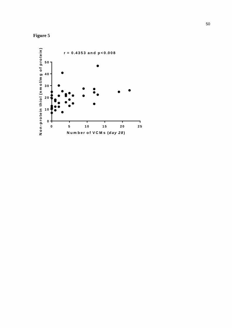
50
Figure 5
N u m b e r o f V C M s (d a y 2 8 )No
n-p
ro
tein
th
iol
(nm
ol/
mg
of
pro
tein
)
0 5 1 0 1 5 2 0 2 5
0
1 0
2 0
3 0
4 0
5 0
r = 0 .4 3 5 3 a n d p < 0 .0 0 8
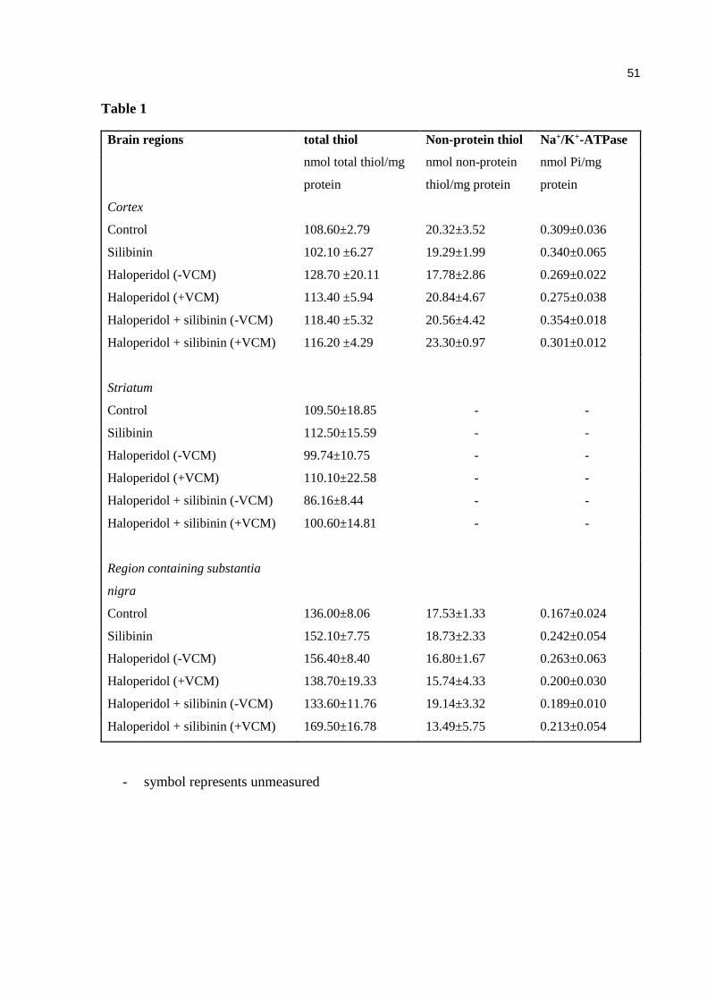
51
Table 1
Brain regions total thiol Non-protein thiol Na+/K+-ATPase
nmol total thiol/mg
protein
nmol non-protein
thiol/mg protein
nmol Pi/mg
protein
Cortex
Control 108.60±2.79 20.32±3.52 0.309±0.036
Silibinin 102.10 ±6.27 19.29±1.99 0.340±0.065
Haloperidol (-VCM) 128.70 ±20.11 17.78±2.86 0.269±0.022
Haloperidol (+VCM) 113.40 ±5.94 20.84±4.67 0.275±0.038
Haloperidol + silibinin (-VCM) 118.40 ±5.32 20.56±4.42 0.354±0.018
Haloperidol + silibinin (+VCM) 116.20 ±4.29 23.30±0.97 0.301±0.012
Striatum
Control 109.50±18.85 - -
Silibinin 112.50±15.59 - -
Haloperidol (-VCM) 99.74±10.75 - -
Haloperidol (+VCM) 110.10±22.58 - -
Haloperidol + silibinin (-VCM) 86.16±8.44 - -
Haloperidol + silibinin (+VCM) 100.60±14.81 - -
Region containing substantia
nigra
Control 136.00±8.06 17.53±1.33 0.167±0.024
Silibinin 152.10±7.75 18.73±2.33 0.242±0.054
Haloperidol (-VCM) 156.40±8.40 16.80±1.67 0.263±0.063
Haloperidol (+VCM) 138.70±19.33 15.74±4.33 0.200±0.030
Haloperidol + silibinin (-VCM) 133.60±11.76 19.14±3.32 0.189±0.010
Haloperidol + silibinin (+VCM) 169.50±16.78 13.49±5.75 0.213±0.054
- symbol represents unmeasured
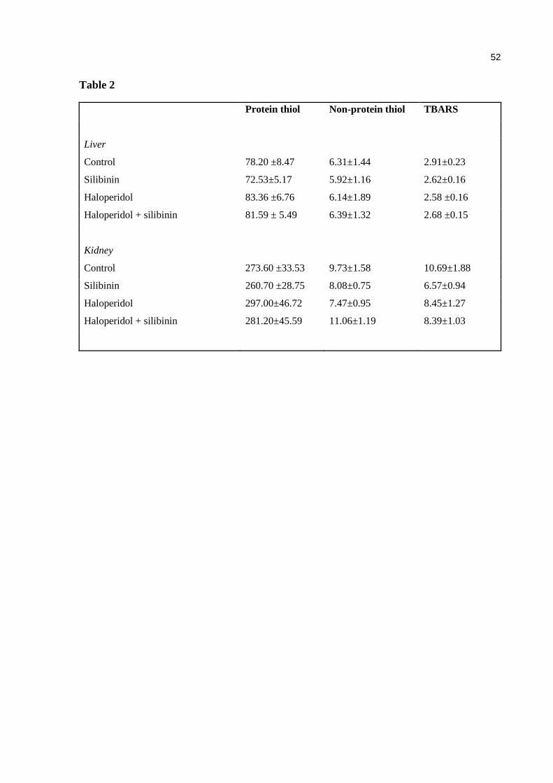
52
Table 2
Protein thiol Non-protein thiol TBARS
Liver
Control 78.20 ±8.47 6.31±1.44 2.91±0.23
Silibinin 72.53±5.17 5.92±1.16 2.62±0.16
Haloperidol 83.36 ±6.76 6.14±1.89 2.58 ±0.16
Haloperidol + silibinin 81.59 ± 5.49 6.39±1.32 2.68 ±0.15
Kidney
Control 273.60 ±33.53 9.73±1.58 10.69±1.88
Silibinin 260.70 ±28.75 8.08±0.75 6.57±0.94
Haloperidol 297.00±46.72 7.47±0.95 8.45±1.27
Haloperidol + silibinin 281.20±45.59 11.06±1.19 8.39±1.03
53
6 CONCLUSÕES ESPECÍFICAS
De acordo com os resultados apresentados neste trabalho de mestrado pode-
se concluir que:
Haloperidol
O haloperidol demonstrou que causa DO nos camundongos, provocando um
aumento na prevalência e na frequência de MMVs;
Entretanto, não se mostrou capaz de alterar os demais parâmetros
comportamentais avaliados (cruzamentos e levantadas no campo aberto, e o tempo
de imobilidade).
Embora tenha sido encontrada uma correlação negativa entre a intensidade
da MMVs com o número de cruzamentos e levantamentos sugerindo que os animais
com alta intensidade de MMVs apresentaram uma redução na locomoção. Uma vez
que, esses animais receberam haloperidol.
Silibinina
A silibinina, por sua vez, não demonstrou efetividade sobre os efeitos do
haloperidol nos parâmetros comportamentais. A pesar de não ter sido capaz de
reduzir a intensidade dos MMVs, reduziu a prevalência de MMVs em comparação ao
grupo tratado com haloperidol.
Tanto o haloperidol como a silibinina não apresentaram sinais de marcadores
de estresse oxidativo nas estruturas cerebrais, fígado e rins, sobre o tiol total, tiol
não-protéico, Na+ / K+-ATPase e TBARS.

54
7 CONCLUSÃO GERAL
Este estudo demonstrou que, a silibinina não foi capaz de evitar os MMVs em
camundongos, apesar da redução de sua prevalência. Assim como, não se mostrou
promissora com os demais parâmetros motores, mas, seu tratamento combinado
com haloperidol não parece causar sinais de marcadores de estresse oxidativo nos
animais.
São necessários mais estudos para elucidar por quais mecanismos de ação a
silibinina atua, assim como, são necessários mais estudos voltados à busca de
tratamentos eficazes para esta doença motora incapacitante.
55
REFERÊNCIAS
ABENAVOLI, L. et al. Milk thistle (Silybum marianum): A concise overview on its chemistry, pharmacological, and nutraceutical uses in liver diseases. Phytotherapy Research, v. 32, n.11, p. 2202-2213, 2018. DOI: 10.1002/ptr.6171 ABÍLIO, V.C. et al. Important role of striatal catalase in aging- and reserpine-induced oral dyskinesia. Neuropharmacology, v. 47, n. 2, p. 263-272, 2004. DOI: 10.1016/j.neuropharm.2004.04.003 AKINTUNDE, J. K.; ABUBAKAR, O. K. Novel therapeutic approaches of natural oil from black seeds and its underlying mechanisms against kidney dysfunctions in haloperidol induced male rats. Drug Metabolism and Personalized Therapy, v. 32, n. 2, p. 97-107, 2017. DOI: 10.1515/dmpt-2016-0044 ALEXANDER, G. C. et al. Increasing off-label use of antipsychotic medications in the United States, 1995-2008. Pharmacoepidemiology Drug Safety, v. 20, n. 2, p. 177-184, 2011. DOI: 10.1002/pds.2082 AMATO, D.; VERNON, A. C.; PAPALEO, F. Dopamine, the antipsychotic molecule: A perspective on mechanisms underlying antipsychotic response variability. Neuroscience and Biobehavioral Reviews, v. 85, p. 146-159, 2018. DOI: 10.1016/j.neubiorev.2017.09.027 ANDREASEN, N. C. Negative symptoms in schizophrenia. Definition and reliability. Archives Of General Psychiatry, v. 39, n. 7, p. 784-788, 1982. ANDREASSEN, O. A.; JØRGENSEN, H. A. Neurotoxicity associated with neuroleptic-induced oral dyskinesias in rats: Implications for tardive dyskinesia?. Progress in Neurobiology, n.5, v.61, p. 525-541, 2000. DOI: 10.1016/S0301-0082(99)00064-7 ANDRADE, S. F. et al. Anti-inflammatory and antinociceptive activities of extract, fractions and populnoic acid from bark Wood of Austroplenckia populnea. Journal of Ethnopharmacology, v. 109, n. 3, p. 464-471, 2007. DOI: 10.1016/j.jep.2006.08.023 ARAÚJO, D. P. de et al. Behavioral and neurochemical effects of alpha lipoic acid associated with omega-3 in tardive dyskinesia induced by chronic haloperidol in rats.Canadian Journal of Physiology and Pharmacology, v. 95, n. 7, p. 837-843, 2017. DOI: 10.1139/cjpp-2016-0307 ATTADEMO, L.; BERNARDINI, F. Air pollution and urbanicity: common risk factors for dementia and schizophrenia? The Lancet Planetary Health, v. 1, n. 3, p. 90-91, 2017. DOI: 10.1016/S2542-5196(17)30042-6 BANSAL, R.; SINGH, R. Exploring the potential of natural and synthetic neuroprotective steroids against neurodegenerative disorders: A literature review. Medicinal Research Reviews, v. 38, n. 4, p. 1126-1158, 2017. DOI: 10.1002/med.21458

56
BARÇOI, A. R. et al. Effects of prenatal immune activation on amphetamine-induced addictive behaviors: Contributions from animal models. Progress in Neuro-Psychopharmacology & Biological Psychiatry, v. 63, p. 63-69, 2015. DOI: 10.1016/j.pnpbp.2015.05.015 BERGMAN, H. et al. Systematic review of interventions for treating or preventing antipsychotic-induced tardive dyskinesia. Health Tecnology Assessment, v. 21, n. 43, p. 1-218, 2017. DOI: 10.3310/hta21430. BIJAK, M. Silybin, a Major Bioactive Component of Milk Thistle (Silybum marianum L. Gaernt.)—Chemistry, Bioavailability, and Metabolism. Molecules, v. 22, n. 11, p. 1-11, 2017. DOI: 10.3390/molecules22111942 BORAH, A. et al. Neuroprotective Potential of Silymarin against CNS Disorders: Insight into the Pathways and Molecular Mechanisms of Action. CNS Neuroscience & Therapeutics, v.19, n. 11, p. 847-853, 2013. DOI: 10.1111/cns.12175 BURGER, M. E. et al. Ebselen attenuates reserpine-induced orofacial dyskinesia and oxidative stress in rat striatum. Progress in Neuro-Psychopharmacology & Biological Psychiatry, v. 27, n.1, p. 135-140, 2003. BURGER, M. E. et al. Ebselen attenuates haloperidol-induced orofacial dyskinesia and oxidative stress in rat brain. Pharmacology, Biochemistry and Behavior, v. 81, n. 3, p. 608-615, 2005. DOI: 10.1016/j.pbb.2005.05.002 BUSANELLO, A. et al. Resveratrol protects against a model of vacuoua chewing movements induced by reserpine in mice. Behavioural Pharmacology, n. 1, p. 71-75, 2011. DOI: 10.1097/FBP.0b013e328341e9b4 BUSANELLO, A. et al. Resveratrol reduces vacuous chewing movements induced by acute treatment with Fluphenazine. Pharmacology Biochemistry Behavioural, v. 101, n. 2, p. 307-310, 2012. DOI:10.1016/j.pbb.2012.01.007 BUSANELLO, A. et al. Resveratrol Protects Against Vacuous Chewing Movements Induced by Chronic Treatment with Fluphenazine. Neurochemical Research, v. 42, p. 3022-3040, 2017. DOI 10.1007/s11064-017-2335-4 BRAVO, P. A. F.; NASSIF, M. C. Doença de Parkinson: Terapêutica atual e avançada. Infarma. Conselho Federal de Farmácia, v. 18, n. 9/10, 2006. CANNON, M.; JONES, P. B.; MURRAY, R. M. Obstetric complications and schizophrenia: historical and meta-analytic review. The American Journal Psychiatry, v. 159, n. 7, p.1080-1092, 2002. DOI: 10.1176/appi.ajp.159.7.1080 CARLSSON, A.; LINDQVIST, M. Effect of chlorpromazine or haloperidol on formation of 3- Methoxyramine and normetanephrine in mouse brain. Acta Pharmacologica et Toxicologica, v. 20, p. 140-144, 1963.
57
CAROFF, S. N. et al. Movement disorders induced by antipsychotic drugs: implications of the CATIE schizophrenia trial. Neurologic Clinics, v. 29, n.1, p. 127-148, 2011. DOI: 10.1016/j.ncl.2010.10.002 CAROFF, S. N.; CAMPBELL, C. Drug-Induced Extrapyramidal Syndromes: Implications for Contemporary Practice. Psychiatric Clinics of North America, v. 39, n. 3, p. 391-411, 2016. DOI: 10.1016/j.psc.2016.04.003 CASEY, D. E. Tardive dyskinesia: reversible and irreversible. Psychopharmacology, v. 2, p. 88-97, 1985. CERETTA, A. P. et al. Gabapentin reduces haloperidol-induced vacuous chewing movements in mice. Pharmacology, Biochemistry and Behavior, v. 166, p. 21-26, 2018. DOI: 10.1016/j.pbb.2018.01.003 CHOI, Y. H. et al. Silibinin attenuates allergic airway inflammation in mice. Biochemical and Biophysical Research Communications, v. 427, n. 3, p. 450-455, 2012. DOI: 10.1016/j.bbrc.2012.07.112 CHTOUROU, Y. et al. Manganese Induces Oxidative Stress, Redox State Unbalance and Disrupts Membrane Bound ATPases on Murine Neuroblastoma Cells In Vitro: Protective Role of Silymarin. Neurochemical Research, v. 36, n. 8, p. 1546-1557, 2011. DOI: 10.1007/s11064-011-0483-5 CITROME, L. Reprint of: Clinical management of tardive dyskinesia: Five steps to success. Journal of the Neurological Sciences, v.389, p. 61-66, 2017. DOI: 10.1016/j.jns.2018.02.037 CITROME, L. Tardive dyskinesia: placing vesicular monoamine transporter Type 2 (VMAT2) Inhibitors into Clinical Perspective. Expert Review of Neurotherapeutics, v. 18, n. 4, p. 323-332, 2018. DOI: 10.1080/14737175.2018.1455504 COLPO, G. et al. Ilex paraguariensis has antioxidant potential and attenuates haloperidol-induced orofacial dyskinesia and memory dysfunction in rats. Neurotoxicity Research, v. 12, n.3, p. 171-180, 2007. COMELLI, M. C. et al. Toward the definition of the mechanism of action of silymarin: Activies related to cellular protection from toxic damage induced by chemotherapy. Integrative Cancer Therapies, v. 6, p. 120-129, 2007. CORNETT, E. M. et al. Medication-Induced Tardive Dyskinesia: A Review and Update. Ochsner Journal, v. 17, n. 2, p. 162-174, 2017. CRAGG, G. M.; NEWMAN, D. J. Natural products: a continuing source of novel drug leads. Biochimica et Biophysica Acta, v. 1830, n. 6, p. 3670-3695, 2013. DOI: 10.1016/j.bbagen.2013.02.008 D'ABREU, A.; AKBAR, U.; FRIEDMAN,J. H. Tardive dyskinesia: Epidemiology. Journal of the Neurological Sciences, v. 389, p. 17-20, 2018. DOI:10.1016/j.jns.2018.02.007
58
DATTA, S. et al. Beneficial effects of lycopene against haloperidol induced orofacial dyskinesia in rats: Possible neurotransmitters and neuroinflammation modulation. European Journal of Pharmacology, v. 771, p. 229-235, 2016. DOI: 10.1016/j.ejphar.2015.12.032 DAVIS, K.L. et al. Dopamine in schizophrenia: A review and reconceptualization. The American Journal of Psychiatry, v. 148, n. 11, p. 1474-1486, 1991.DOI: 10.1176/ajp.148.11.1474 DAVIS, M. C. et al. Efficient trial design-FDA approval of Valbenazine for Tardive Dyskinesia. The New England Journal of Medicine, v. 376, n. 26, p. 2503-2506, 2017. DOI:10.1056/NEJMp1704898 DHINGRA, D.; GOSWAMI, S.; GAHALAIN, N. Protective effect of hesperetin against haloperidol induced orofacial dyskinesia and catalepsy in rats. Nutritional Neuroscience, v. 21, n.9, p.667-675, 2017. DOI: 10.1080/1028415X.2017.1338549 DOLD, M. et al. Haloperidol versus first-generation antipsychotics for the treatment of schizophrenia and other psychotic disorders. Cochrane Database of Systematic Reviews, v.1, n.9831. p. 1-200, 2015. DOI:10.1002/14651858.CD009831.pub2. FACHINETTO, R. et al. High fat diet increases the incidence of orofacial dyskinesia and oxidative stress in specific brain regions of rats. Pharmacology Biochemistry and Behavior, v. 81, n. 3, p. 585-592, 2005. DOI: 10.1016/j.pbb.2005.05.001 FACHINETTO, R. et al. Diphenyl diselenide decreases the prevalence of vacuous chewing movements induced by fluphenazine in rats. Psychopharmacology, v.194, n. 3, p. 423-432, 2007a. DOI: 10.1007/s00213-007-0831-y FACHINETTO, R. et al. Valeriana officinalis does not alter the orofacial dyskinesia induced by haloperidol in rats: role of dopamine transporter. Progress in Neuro-Psychopharmacology & Biological Psychiatry, v. 31, n. 7, p. 1478-1486, 2007b. DOI: 10.1016/j.pnpbp.2007.06.028 FERNANDES, V. et al. Neuroprotective effects of silibinin: an in silico and in vitro study. International Journal of Neuroscience, v. 128, p. 935-945, 2018. FLATOW, J.; BUCKLEY, P.; MILLER, B.J. Meta-analysis of oxidative stress in Schizophrenia. Biological Psychiatry, v. 74, n. 6, p. 400-409, 2013. DOI: 10.1016/j.biopsych.2013.03.018 FLOYD, R. A. Antioxidant oxidative stress, and degenerative neurological disorders. Proceedings of the Society for Experimental Biology and Medicine, v. 222, n.3, p. 236-245, 1999. FREITAS, C. M. de et al. Silymarin recovers 6-hydroxydopamine-induced motor deficits in mice. Food and Chemical Toxicology, v. 118, p. 549-556. 2018. DOI: 10.1016/j.fct.2018.05.062
59
GAFFIGAN, M. E. et al. A Randomized Controlled Trial of Intravenous Haloperidol vs. Intravenous Metoclopramide for Acute Migraine Therapy in the Emergency Department. The Journal of Emergency Medicine, v. 49, n. 3, p. 326-334, 2015. DOI: 10.1016/j.jemermed.2015.03.023 GALHARDI, F. Effect of Silymarin on biochemical parameters of oxidative stress in aged and young rat brain. Food and Chemical Toxicology, v. 47, n. 10, p. 2655-2660, 2009. DOI: 10.1016/j.fct.2009.07.030 GANGULY, P.; SOLIMAN, A.; MOUSTAFA, A.A. Holistic Management of Schizophrenia Symptoms Using Pharmacological and Non-pharmacological Treatment. Frontiers in Public Health, v. 6, n. 166, p. 1-9, 2018. DOI: 10.3389/fpubh.2018.00166 GEED, M. et al. Silibinin pretreatment attenuates biochemical and behavioral changes induced by intrastriatal MPP+ injection in rats. Pharmacology, Biochemistry and Behavior, n. 117, p. 92-103, 2014. DOI: 10.1016/j.pbb.2013.12.008 GRIGORIADIS, D.E. et al. Pharmacologic Characterization of Valbenazine (NBI-98854) and Its Metabolites. The Journal of Pharmacology and Experimental Therapeutics, v. 361, n. 3, p. 454-461, 2017.DOI:10.1124/jpet.116.239160 HACKETT, E.S. et al. Milk thistle and its derivative compounds: A review of opportunities for treatment of liver disease. Journal of Veterinary Internal Medicine, v. 27, p. 10-16, 2013. HADDADI, R. et al. Silymarin improved 6-OHDA-induced motor impairment in hemi parkisonian rats: behavioral and molecular study. DARU Journal of Pharmaceutical Sciences, v. 22, p. 38, 2014. DOI: 10.1186/2008-2231-22-38 HADDADI, R. et al. Short-Term Treatment with Silymarin Improved 6-OHDA-Induced Catalepsy and Motor Imbalance in Hemi-Parkisonian Rats. Advanced Pharmaceutical Bulletin, v. 5, n.4, p. 463-469, 2015.DOI: 10.15171/apb.2015.063 JAVED, S.; KOHLI, K.; ALI, M. Reassessing bioavailability of silymarin. Alternative Medicine Review, v. 16, n. 3, p. 239-249, 2011. JOSHI, R. et al. Silibinin ameliorates LPS-induced memory deficits in experimental animals. Neurobiology of Learning and Memory, v. 116, p.117-131, 2014. DOI: 10.1016/j.nlm.2014.09.006 KAMEDA, S. R. et al. Opposite effects of neonatal hypoxia on acute amphetamine-induced hyperlocomotion in adult and adolescent mice. Psychiatry Research, v. 208, n. 1, p. 74-77, 2013. DOI: 10.1016/j.psychres.2013.03.021 KANE, J. M. Tardive dyskinesia: epidemiology and clinical presentation. In: BLOOM, F. E.; KUPFER, D. J. (Ed.). Psychopharmacology: the fourth generation of progress, v. 39. New York: Raven Press, p. 1485-95, 1995.
60
KANE, J. M; SMITH, J. M. Tardive dyskinesia: prevalence and risk factors, 1959 to 1979. Archives of General Psychiatry, v. 39, n. 4, p. 473-481, 1982. KELLY, B. D. et al. Schizophrenia and the city: A review of literature and prospective study of psychosis and urban city in Ireland. Schizophrenia Research, v. 116, n. 1, p. 75-89, 2010. DOI: 10.1016/j.schres.2009.10.015 KOHEN, R.; NYSKA, A. Oxidation of Biological Systems: Oxidative Stress Phenomena, Antioxidants, Redox Reactions, and Methods for Their Quantication. Toxicologic Pathology, v. 30, n. 6, p. 620-650, 2002. DOI: 10.1080/0192623029016672 4 KREN, V.; WALTEROVA, D. Silybin and silymarin - new effects and applications. Biomedical Papers, v. 149, n. 1, p. 29-41, 2005. KRYNICKI, C. R. et al. The relationship between negative symptoms and depression in schizophrenia: a systematic review. Acta Psychiatrica Scandinavica, v. 137, n. 5, p. 380-390, 2018. DOI: 10.1111/acps.12873 KRONBAUER, M. et al. Magnesium Supplementation Prevents and Reverses Experimentally Induced Movement Disturbances in Rats: Biochemical and Behavioral Parameters. Biological Trace Element Research, v. 166, n. 2, p. 163-172, 2015. DOI: 10.1007/s12011-015-0268-9 KRONBAUER, M. et al. Influence of magnesium supplementation on movement side effects related to typical antipsychotic treatment in rats. Behavioural Brain Research, v. 320, p. 400-411, 2017. DOI: 10.1016/j.bbr.2016.10.049 KUDO, S.; ISHIZAKI, T. Pharmacokinetics of haloperidol: an update. Clinical Pharmacokinetics, v. 37, n. 6. p. 435-456, 1999. DOI: 10.2165/00003088-199937060-00001 LAU, C. I. et al. Does the dopamine hypothesis explain schizophrenia? Reviews in the Neurosciences, v. 24, n. 4, p. 389-400, 2013. DOI: 10.1515/revneuro-2013-0011 LEE, Y.et al. Silibinin Prevents Dopaminergic Neuronal Loss in a Mouse Model of Parkinson’s Disease Via Mitochondrial Stabilization. Journal of Neuroscience Research, v. 93, p. 755-765, 2015. DOI: 10.1002/jnr.23544 LIN, C. C.; ONDO, W. G. Non-VMAT2 inhibitor treatments for the treatment of tardive dyskinesia. Journal of the Neurological Sciences, v. 389, p. 48-54, 2018. DOI: 10.1016/j.jns.2018.02.014 LISTER, J. et al. Oxidative stress and the antipsychotic-induced vacuous chewing movement model of tardive dyskinesia: evidence for antioxidant-based prevention strategies. Psychopharmacology, v. 23, n. 11, p. 2237-2249, 2014. DOI: 10.1007/s00213-014-3582-6
61
LISTER, J. et al. Lipoic acid and haloperidol-induced vacuous chewing movements: Implications for prophylactic antioxidant use in tardive dyskinesia. Progress in Neuro-Psychopharmacology & Biological Psychiatry, v. 72, p. 23-29, 2017. DOI: 10.1016/j.pnpbp.2016.08.010 LOHSE, M.J.; MÜLLER-OERLINGHAUSEN, B. Psychopharmaka. In: SCHWABE, U.; PFAFFRATH, D. (Ed.). Arzneimittelverordnungsreport. Heidelberg: Springer, 2009. p. 767-810. LUEDTKE, R.R. et al. Bitropic D3 Dopamine Receptor Selective Compounds as Potential Antipsychotics. Current Pharmaceutical Design, v. 21, n. 26, p. 3700-3724, 2015. MAATZ, A.; HOFF, P. The birth of schizophrenia or a very modern Bleuler: a close reading of Eugen Bleuler’s ‘Die Prognose der Dementia praecox’ and a re-consideration of his contribution to psychiatry. History of Psychiatry, v. 25, n. 4, p. 431-440, 2014. DOI: 10.1177/0957154X14546606 MAATZ, A.; HOFF, P.; ANGST, J. Eugen Bleuler’s schizophrenia—a modern Perspective. Dialogues in Clinical Neuroscience, v. 17, n.1, p. 1-7, 2015. MACÊDO, D. S. et al. B vitamins attenuate haloperidol-induced orofacial dyskinesia in rats: possible involvement of antioxidant mechanisms. Behavioural Pharmacology, v. 22, n. 7, p. 674-680, 2011. DOI: 10.1097/FBP.0b013e32834aff6d MANDEGARY, A. et al. Hepatoprotective effect of silyamarin in individuals chronically exposed to hydrogen sulfide; modulating influence of TNF-α cytokine genetic polymorphism. DARU Journal of Pharmaceutical Sciences, v. 21, p. 28, 2013. DOI: 10.1186/2008-2231-21-28 MARRAZZO, G. et al. Neuroprotective effect of silibinin in diabetic mice. Neuroscience Letters, v. 504, p. 252-256, 2011. DOI: 10.1016/j.neulet.2011.09.041 MARTELLI, F.; NUNES, F. M. F. Radicais livres: em busca do equilíbrio. Ciência e Cultura, Universidade Estadual de Campinas, São Paulo, v. 66, n. 3, 2014. ISSN 2317-6660
MENDELSON, T.; EATON, W. W. Recent advances in the prevention of mental disorders. Social Psychiatry and Psychiatric Epidemiology, v. 53, n. 4, p. 325-339, 2018. DOI: 10.1007/s00127-018-1501-6 MERRILL, R. M.; LYON, J. L.; MATIACO, P. M. Tardive and spontaneous dyskinesia in the general population. BMC Psychiatry, v. 13, p. 152, 2013. DOI: 10.1186/1471-244X-13-152 MEYER, J. M. Farmacoterapia da psicose e da mania. In: BRUNTON, L. L.; CHABNER, B. A.; KNOLLMANN, B. C. (Ed.). As bases farmacológicas da terapêutica de Goodman & Gilman. 12. ed. Porto Alegre: AMGH, 2012.cap.22, p. 609-628.
62
MCDOUGLE, C. J. et al. Haloperidol addition in fluvoxamine- refractory obsessive- compulsive disorder. A double-blind, placebo-controlled study in patients with and without tics. Archives of General Psychiatry, v. 51, n. 4, p. 302-308,1994. MCGORRY, P. D.; MEI, C. Early intervention in youth mental health: progress and future directions. Evidence-Based Mental Health, v. 21, n. 4, p. 182-184, 2018. DOI: 10.1136/ebmental-2018-300060 MCGRATH, J. Hypothesis: is low prenatal vitamin D a risk-modifying factor for schizophrenia? Schizophrenia Research, v. 40, n. 3, p.173-177, 1999. MILLER, D. D. et al. Comparative assessment of the incidence and severity of tardive dyskinesia in patients receiving aripiprazole or haloperidol for the treatment of schizophrenia: a post hoc analysis. The Journal of Clinical Psychiatry, v. 68, n. 12, p. 1901-1906, 2007. DOI: 10.4088/JCP.v68n1210 MIKSYS, S. et al. Rat brain CYP2D enzymatic metabolism alters acute and chronic haloperidol side-effects by different mechanisms. Progress in Neuro Psychopharmacology and Biological Psychiatry, v. 78, p. 140-148, 2017. DOI:10.1016/j.pnpbp.2017.04.030 MORAZZONI, P. BOMBARDELLI, E. Silybum marianum (Carduus marianus), Fitoterapia, v. 66, n. 1, p. 3-42, 1995. MUESER, K. T.; MCGURK, S. R. Schizophrenia. The Lancet, v. 363, n. 9426, p. 2063- 2072, 2004. MURRAY-BROWN, F.; DORMAN, S. Haloperidol for the treatment of nausea and vomiting in palliative care patients. Cochrane Library, v. 11, n. 3, 2015.DOI:10.1002/14651858.CD006271.pub3 NADE, V. S.; KAWALE, L. A.; YADAV, A. V. Protective effect of Morus alba leaves on haloperidol-induced orofacial dyskinesia and oxidative stress. Pharmaceutical Biology, v. 48, n. 1, p. 17-22, 2010. DOI: 10.3109/13880200903029357 NENCINI, C.; GIORGI, G.; MICHELI, L. Protective effect of silymarin on oxidative stress in rat brain. Phytomedicine, v. 14, p. 129-135, 2007. DOI:10.1016/j.phymed. 2006.02.005 NUECHTERLEIN, K. H. et al. Identification of separable cognitive factors in schizophrenia. Schizophrenia Research, v. 72, n. 1, p. 29-39, 2004. DOI: 10.1016/j.schres.2004.09.007 OLIVEIRA, D. R. de et al. Silymarin has antioxidant potential and changes the activity of Na+/K+ -ATPase and monoamine oxidase in vitro. Industrial Crops and Products, v. 70, p. 347-355, 2015b. DOI: 10.1016/j.indcrop.2015.03.060 OLIVEIRA, D. R. de. In Vitro Antimicrobial and Modulatory Activity of the Natural Products Silymarin and Silibinin. BioMed Research International, v. 2015, 2015a. DOI: 10.1155/2015/292797
63
ORGANIZAÇÃO MUNDIAL DA SAÚDE - OMS. Lista de Medicamentos Essenciais, 16. ed. 2009. Disponível em: <https://www.who.int/medicines/publications/ essentialmedicines/en/>. Acesso em: 29 nov. 2018. ORGANIZAÇÃO MUNDIAL DA SAÚDE - OMS. Esquizofrenia. 2013. Disponível em: <http://www.who.int/es/news-room/fact-sheets/detail/schizophrenia>. Acesso em: 29 nov. 2018. ORGANIZAÇÃO MUNDIAL DA SAÚDE - OMS. Esquizofrenia. 2018. Disponível em: <http://www.who.int/topics/schizophrenia/es/>. Acesso em: 12 jan. 2018.
OWEN, M. J.; SAWA, A.; MORTENSEN, P. B. Schizophrenia. The Lancet, v. 388, n. 10039, p. 86-97, 2016. DOI: 10.1016/S0140-6736(15)01121-6 PÉREZ-H, J. et al. Neuroprotective effect of silymarin in a MPTP mouse model of Parkinson’s disease. Toxicology, v. 319, p. 38-43, 2014. DOI: 10.1016/j.tox. 2014.02.009 PEREIRA, R. P. et al. Valeriana officinalis ameliorates vacuous chewing movements induced by reserpine in rats. Journal of Neural Transmission, v. 118, n. 11, p. 1547-1557, 2011. DOI: 10.1007/s00702-011-0640-7 PEROZA, L. R. et al. Bauhinia Forficata prevents vacuous chewing movements induced by haloperidol in rats and has antioxidant potential in vitro. Neurochemical. Research, v. 38, n. 4, p. 789-796, 2013. DOI: 10.1007/s11064-013-0981-8 PEROZA, L. R. et al. Alteration of Cytokines Levels in the Striatum of Rats: Possible Participation in Vacuous Chewing Movements Induced by Antipsycotics. Neurochemical Research, v. 41, p. 2481-2489, 2016. DOI 10.1007/s11064-016-1961-6 RAINONE, F. Milk thistle. American Family Physician, v. 72, n. 7, p. 1285-1288, 2005. RANA, A. Q.; CHAUDRY, Z. M.; BLANCHET, P. J. New and emerging treatments for symptomatic tardive dyskinesia. Drug Design, Development and Therapy, v. 7, p. 1329-1340, 2013. DOI:10.2147/DDDT.S32328 RECKZIEGEL, P. et al. Gallic acid decreases vacuous chewing movements induced by reserpine in rats. Pharmacology, Biochemistry and Behavior, v. 104, p. 132-137, 2013. DOI: 10.1016/j.pbb.2013.01.001. REDDY, M. K. et al. Protective effect of rutin in comparison to silymarin against induced hepatotoxicity in rats. Veterinary World, v. 10, p. 74-80, 2017. EISSN: 2231-0916. DOI: 10.14202/vetworld.2017.74-80 ROSENHECK, R. A. Evaluating the cost-effectiveness of reduced tardive dyskinesia with second-generation antipsychotics. British Journal of Psychiatry, v. 191, p. 238-245, 2007. DOI: 10.1192/bjp.bp.106.035063
64
REY, J. A. Antipsicóticos. In: WHALEN, K.; FINKEL, R.; PANAVELI, T. A. (Ed). Farmacologia Ilustrada. 6. ed. Porto Alegre: ARTMED, 2016. cap. 11, p. 147-155. ROMANUCCI, V.; DI FABIO, G.; ZARRELLI, A. A New Class of Synthetic Flavonolignan-Like Dimers: Still Few Molecules, but with Attractive Properties. Molecules, v. 24, n. 1, p. 108-119, 2019. DOI: 10.3390/molecules24010108 RÖPKE, J. et al. Anandamide attenuates haloperidol-induced vacuous chewing movements in rats. Progressin Neuro-Psychopharmacology & Biological Psychiatry, v. 54, p. 195-199, 2014. DOI: 10.1016/j.pnpbp.2014.04.006 SACHDEV, P. S. The current status of tardive dyskinesia. Australian and New Zealand Journal of Psychiatry, v. 34, n. 3, p. 355-369, 2000. SALLER, R.; MEIER, R.; BRIGNOLI, R. The use of Silymarin in the treatment of liver diseases. Drugs, v. 61, p. 2035-2063, 2001. SALLER, R. et al. An updated systematic review with meta-analysis for the clinical evidence of silymarin. Forschende Komplementarmedizim (2006), v. 15, n. 1, p. 9-20, 2008.DOI: 10.1159/000113648 SAMAD, N.; HALEEM, D. J. Antioxidant effects of rice bran oil mitigate repeated haloperidol-induced tardive dyskinesia in male rats. Metabolic Brain Disease, v. 32, n. 4, p. 1099-1107, 2017. DOI: 10.1007 / s11011-017-0002-8 SANTILLÁN-URQUIZA, M. A. et al. Pharmacological interaction of Galphimia glauca extract and natural galphimines with Ketamine and Haloperidol on different behavioral tests. Biomedicine & Pharmacotherapy, v. 103, p. 879-888, 2018. DOI: 10.1016/j.biopha.2018 SCHAFFER, L. F. et al. Harpagophytum procumbens Prevents Oxidative Stress and Loss of Cell Viability In Vitro. Neurochemical Research, v. 38, n. 11, p. 2256-2267, 2013. DOI: 10.1007/s11064-013-1133-x SCHAFFER, L. F.et al. Harpagophytum Procumbens Ethyl Acetate Fraction Reduces Fluphenazine-Induced Vacuous Chewing Movements and Oxidative Stress in Rat Brain. Neurochemical Research, v. 41, p. 1170-1184, 2016. DOI 10.1007/s11064-015-1811-y SIRIS, S. G. et al. Postpsychotic depression and negative symptoms: an investigation of syndromal overlap. American Journal of Psychiatry, v. 145, n. 12, p. 1532-1537, 1988. DOI: 10.1176/ajp.145.12.1532 SOLMI, M. et al. Clinical risk factors for the development of tardive dyskinesia. Journal of the Neurological Sciences, v. 389, p. 21-27, 2018. DOI: 10.1016/j.jns.2018.02.012 SONG, Z. et al. Silymarin Protects Against Acute Ethanol-Induced Hepatotoxicity in Mice. Alcoholism: Clinical and Experimental Research, v. 30, n. 3, p. 407-413, 2006. DOI: 10.1111/j.1530-0277.2006.00063.x
65
SONEGO, A. B. et al. Cannabidiol prevents haloperidol-induced vacuos chewing movements and inflammatory changes in mice via PPARγ receptors. Brain, Behavior, and Immunity, 2018.DOI: 10.1016/j.bbi.2018.09.014 SOUNG, H. S. et al. L-Theanine Decreases Orofacial Dyskinesia Induced by Reserpine in Rats. Neurotoxicity Research, v. 34, n. 3, p. 375-387, 2018. DOI: 10.1007/s12640-018-9897-z SOUZA, I. E. J. de et al. Relationship of orofacial movements to behavioural repertoire as assessed topographically over the course of 6-month haloperidol treatment followed by 4-month withdrawal. Psychopharmacology, v. 1, n. 169, p. 28-34, 2003. DOI: 10.1007/s00213-003-1466-2 SRIVASTAVA, S. Effect of picroliv and Silymarin on liver regeneration in rats. Indian Journal of Pharmacology, v. 26, p. 19-22, 1994. STAHL, S. M. Psicose e Esquizofrenia. In: STAHL, S. M. (Ed.). Psicofarmacologia bases neurocientíficas e aplicações prática. 4. ed. Rio de Janeiro: Guanabara, 2014. cap. 4. STEPNICKI, P.; KONDEL, M.; KACZOR, A. A. Current Concepts and Treatments of Schizophrenia. Molecules, v. 23, n. 8, p. 1-29, 2018. DOI:10.3390/molecules 23082087 SVOBODOVÁ, A. R. et al. Phototoxic potential of silymarin and its bioactive components. Journal of Photochemistry and Photobiology B: Biology, v. 156, p. 61-68, 2016. DOI: 10.1016/j.jphotobiol.2016.01.011 THAAKUR, S.; HIMABINDHU, G. Effect of alphalipoic acid on the tardive dyskinesia and oxidative stress induced by haloperidol in rats. Journal of Neural Transmission (VIENNA), v. 116, n. 7, p. 807-814, 2009. DOI: 10.1007/s00702-009-0232-y TOTA, S. et al. Improvement of brain energy metabolism and cholinergic functions contributes to the beneficial effects of silibinin against streptozotocin induced memory impairment. Behavioural Brain Research, v. 221, n. 1, p. 207-215, 2011. DOI: 10.1016/j.bbr.2011.02.041 TORREY, E. F.; YOLKEN, R. H. Could schizophrenia be a viral zoonosis transmitted from house cats? Schizophrenia Bulletin, v. 21, n. 2, p. 167-171, 1995. TURRONE, P. et al. Differential Effects of Within-Day Continuous Vs Transient Dopamine D2 Receptor Occupancy in the Development of Vacuous Chewing Movements (VCMs) in Rats. Neuropsychopharmacology, v. 28, n.8, p. 1433-1439, 2003. DOI: 10.1038/sj.npp.1300233 TURRONE, P. et al. The relationship between dopamine D2 receptor occupancy and the vacuous chewing movement syndrome in rats. Psychopharmacology, v. 165, p. 166-171, 2003. DOI: 10.1007/s00213-002-1259-z
66
TYLER, M. W.; ZALDIVAR-DIEZ, J.; HAGGARTY, S. J. Classics in Chemical Neuroscience: Haloperidol. ACS Chemical Neuroscience, v. 8, n. 4, p. 444-453, 2017. DOI: 10.1021/acschemneuro.7b00018 VALENZUELA, A.; GUERRA, R. Protective effect of the flavonoid silybin dihemisuccinate on the toxicity of phenylhydrazine on rat liver. FEBS Letters, v. 181, n. 2, p. 291-294, 1985. VOINOVICH, D. et al. Solid state mechanochemical activation of Silybum marianum dry extract with betacyclodextrins: Characterization and bioavailability of the coground systems. Journal of Pharmaceutical Sciences, v. 99, n. 11, p. 4119-4129, 2009. DOI: 10.1002/jps.21704 WAGNER, H.; HÖRHAMMER, L.; MÜNSTER, R. On the chemistry of Silymarin (silybin) the active principle of the fruits from Silybum marianum (L.) Gaertn. (Carduus marianus L.). Arzneimittelforschung, v. 18, n. 6, p. 688-696, 1968. WANG, Q. et al. Inhibiting NF-κB activation and ROS production are involved in the mechanism of silibinin's protection against D-galactose-induced senescence. Pharmacology Biochemistry and Behavior, v. 98, n. 1, p. 140-149, 2011. DOI: 10.1016/j.pbb.2010.12.006 WEN, Z. et al. Pharmacokinetics and metabolic profile of free, conjugated, and total silymarin flavonolignans in human plasma after oral administration of milk thistle extract. Drug Metabolism and Disposition, v. 36, n. 1, p. 65-72, 2008. DOI: 10.1124/dmd.107.017566 WU, J. Q.; KOSTEN, T. R.; ZHANG, X. Y. Free radicals, antioxidant defense systems, and schizophrenia. Progress in Neuro-Psychopharmacology & Biological Psychiatry, v. 46, p. 200-206, 2013. DOI: 10.1016/j.pnpbp.2013.02.015 ZHANG, X. Y.; YAO, J. K. Oxidative stress and therapeutic implications in psychiatric disorders. Progress in Neuro-Psychopharmacology & Biological Psychiatry, v. 46, p. 197-199, 2013. DOI: 10.1016/j.pnpbp.2013.03.003 ZHOLOBENKO, A.; MODRIANSKY, M. Silymarin and its constituents in cardiac preconditioning. Fitoterapia, v. 97, p. 122-132, 2014. DOI: 10.1016/j.fitote. 2014.05.016
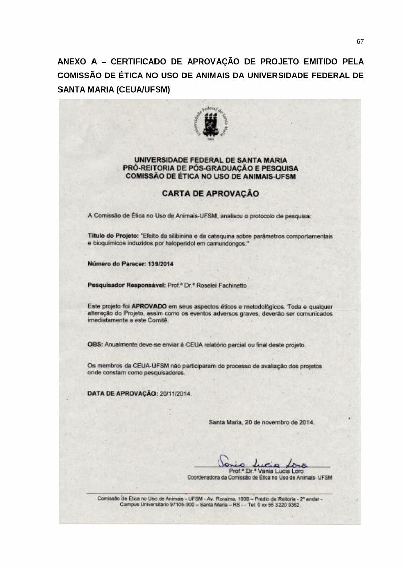
67
ANEXO A – CERTIFICADO DE APROVAÇÃO DE PROJETO EMITIDO PELA
COMISSÃO DE ÉTICA NO USO DE ANIMAIS DA UNIVERSIDADE FEDERAL DE
SANTA MARIA (CEUA/UFSM)
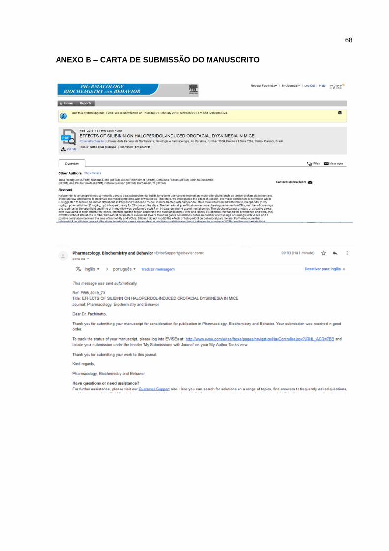
68
ANEXO B – CARTA DE SUBMISSÃO DO MANUSCRITO


![Correspondência entre José Maria Álvarez Blázquez e Rodrigues Lapa [2008]](https://img.pdfslide.net/doc/110x75/6322a198078ed8e56c0a7fb0/correspondencia-entre-jose-maria-alvarez-blazquez-e-rodrigues-lapa-2008.jpg)


