Embed Size (px)
Citation preview

Bioorganic & Medicinal Chemistry 14 (2006) 169–180
Targeting integrins: Insights into structure and activity of cyclicRGD pentapeptide mimics containing azabicycloalkane amino acids
Laura Belvisi,a Anna Bernardi,a Matteo Colombo,a,� Leonardo Manzoni,b
Donatella Potenza,a Carlo Scolastico,a,* Giuseppe Giannini,c Marcella Marcellini,c
Teresa Riccioni,c Massimo Castorina,c Pietro LoGiudicec and Claudio Pisanoc,*
aDipartimento di Chimica Organica e Industriale and Centro Interdisciplinare Studi bio-molecolari e applicazioni Industriali, (CISI),
Universita degli Studi di Milano, via G. Venezian 21, I-20133 Milan, ItalybCNR-Istituto di Scienze e Tecnologie Molecolari, via C. Golgi 19, I-20133 Milan, ItalycResearch and Development, Sigma-Tau, via Pontina Km 30,400, I-00040 Pomezia, Italy
Received 15 February 2005; accepted 2 August 2005
Available online 7 October 2005
Abstract—A small library of cyclic RGD pentapeptide mimics incorporating stereoisomeric 5,6- and 5,7-fused bicyclic lactams wassynthesized. This library was found to contain high-affinity ligands for the avb3 integrin. The aim of this study was to investigate activ-ity, selectivity, and structure of these ligands in order to identify new specific av-integrin antagonists that could be evaluated as tumorangiogenesis inhibitors. In vitro screening, including receptor-binding assays to purified avb3, avb5, and a5b1 integrins, and plateletaggregation assay, revealed ST1646 as a potent, highly selective avb3/avb5 integrin antagonist. Structure determination of the cyclicRGD pentapeptide mimics performed by a combination of NMR spectroscopy, and molecular mechanics and dynamics calculationsshowed a strong dependence of the RGD cyclopeptide conformation on lactam ring size and stereochemistry. ST1646 revealed thehighest ability within the library to adopt the proper RGD orientation required for binding to the avb3 integrin, as deduced from therecently solved crystal structure of the extracellular segment of integrin avb3 in complex with a cyclic pentapeptide ligand.� 2005 Elsevier Ltd. All rights reserved.
1. Introduction
Cell adhesion is essential for the proper functionality ofmany physiological processes such as embryogenesis,cell differentiation, hemostasis, wound healing, and im-mune response, but also for pathophysiological eventssuch as tumor cell invasion, formation of metastases,and tumor-induced angiogenesis.1–4 These cell adhesionprocesses are mediated by cell-surface receptors, amongwhich integrins represent the most diverse and promi-nent class.5–8
Integrins are a family of membrane-spanning adhesionreceptors composed of noncovalently linked a and b sub-
0968-0896/$ - see front matter � 2005 Elsevier Ltd. All rights reserved.
doi:10.1016/j.bmc.2005.08.048
Keywords: Integrin receptor antagonists; Peptidomimetics; Conforma-
tional analysis; Angiogenesis inhibitors.* Corresponding authors. Tel.: +39 02 50314090; fax: +39 02 50314072
(C.S.); tel.: +39 06 91393760; fax: +39 06 91393988 (C.P.); e-mail
addresses: [email protected]; [email protected]� Present address: Nikem Research, via Zambeletti 25, I-20021,
Baranzate di Bollate (MI), Italy.
units which combine to give a wide amount of heterodi-mers. Besides cell adhesion to extracellular matrix,integrins also mediate intracellular events that controlcell shape, migration, proliferation, and survival.5,8
Many integrins recognize polypeptide domains contain-ing the Arg-Gly-Asp (RGD) aminoacid sequence presentin several matrix associated adhesive glycoproteins.9,10
Specificity and efficacy of these molecular recognitionprocesses are determined by the context of the RGD se-quence, including flanking residues, conformational pre-sentation of the triad, and individual features of theintegrin binding pockets.11,12
Among such RGD-dependent integrins, the vitronectinreceptors aVb3 and aVb5 have recently received increas-ing attention as interesting therapeutic targets, becauseof their critical role in tumor-induced angiogenesis andmetastasis formation.13
avb3 is not generally expressed on epithelial cells andnormal endothelial cells (EC), but it is significantlyupregulated on activated EC and in metastatic tumor

NCOOH
ONH2
N
OHN
Arg
O
AspGly
n=1,2
123
III
( )n( )n
Figure 1. Azabicycloalkane scaffolds of type I and general formula II
of the library cyclo(Arg-Gly-Asp-Lactam).
N
OHN
Arg
O
AspGly
X1
X2
H
H
N
OHN
Arg
O
AspGly
X1
X2
H
N
OHN
Arg
O
AspGly
X1
X2
H
N
O
HN
Arg
O
AspGly
X1
X2
H
1a (5,6 cis S)1b (5,6 cis S)
a : X1= Pmc; X2 = tBub : X1 = X2 = H
2a (5,6 trans R)2b (5,6 trans R)
3a (5,6 trans S)3b (5,6 trans S)
H
H
4a (5,7 trans S)4b (5,7 trans S)
5a (5,7 trans R)5b (5,7 trans R) ST1646
6a (5,7 cis S)6b (5,7 cis S)
7a (5,7 cis R)7b (5,7 cis R)
N
O
HN
Arg
O
AspGly
X1
X2
H
H
N
O
HN
Arg
O
AspGly
X1
X2
H
N
O
HN
Arg
O
AspGly
X1
X2
H
H
Figure 2. Cyclic RGD pentapeptide mimics cyclo(Arg-Gly-Asp-Lac-
tam) 1–7. The ring size and the stereochemistry at the bridgehead (cis
or trans) and at the C3 (S or R) carbon of the bicyclic lactam are
reported in parentheses.
170 L. Belvisi et al. / Bioorg. Med. Chem. 14 (2006) 169–180
cells. Growth factors such as fibroblast growth factor-2(FGF2) and tumor necrosis factor-a (TNFa) stimulateavb3 expression in the developing chick chorioallantoicmembrane (CAM) and in the angiogenesis model ofthe rabbit cornea.14 The importance of avb3 in tumorangiogenesis is also demonstrated by the fact that avb3antagonists, including cyclic RGD peptides and mono-clonal antibodies, were successfully used to inhibit bloodvessel development and tumor growth in different mod-els.15,16 It is noteworthy that avb3 antagonists have verylittle effect on pre-existing blood vessels, indicating theusefulness of targeting this receptor for therapeutic ben-efit without adverse side effects.
Recent studies have implicated a related integrin, aVb5,in angiogenesis, possibly via a signaling pathway distinctfrom that of avb3 and activated by a different growthfactor.17 The existence of distinct angiogenic pathwayscan be explained with the prevalence of specific growthfactors and/or cell-adhesive proteins in differentconditions.
Furthermore, many steps that characterize the acquisi-tion of invasive and metastatic potential by tumor cells,such as intravasation, adhesion to the vessel wall,extravasation, infiltration, and proliferation into targettissue, involve integrins. This implies that migratory tu-mor cells express the integrin receptors and the enzymat-ic machinery for recognition, adhesion, matrixdegradation, and penetration involved in cell–cell andcell–ECM interactions. For example, integrin avb3expression is upregulated on certain invasive tumorsincluding metastatic melanoma18 and late stage glioblas-toma.19 Thus, inhibition of avb3 receptor, in addition toblocking tumor-induced angiogenesis, has impact on anumber of mechanisms involved in tumor progression.
Taken together, these results suggest that selective avb3and/or avb5 antagonists may represent a promising ap-proach for the inhibition of tumor angiogenesis and tu-mor growth. Various RGD-containing cyclic peptideshave been developed by different groups as active andselective integrin antagonists that compete with matrixmolecules for specific integrin receptors.20–22
An efficient procedure of spatial screening, performed byKessler et al. and based on the synthesis of stereoisomer-ic cyclic peptide libraries, led to the highly active aVb3-se-lective first-generation cyclic pentapeptide cyclo(Arg-Gly-Asp-D-Phe-Val).23–25 Extensive modifications ofthis lead structure with different peptidomimetics andcarbohydrate scaffolds have been performed, and newpotent antagonists have been identified.26–28 The system-atic derivatization of the lead peptide resulted in the N-alkylated cyclopeptide cyclo(Arg-Gly-Asp-D-Phe-[NMe]Val),29 which has entered clinical phase II studiesas anticancer drug (cilengitide, EMD121974).
Recently, our group has reported a library of cyclicRGD pentapeptide mimics, based on azabicycloalkaneamino acid scaffolds of type I (Fig. 1).30 Stereoisomeric5,6- and 5,7-fused bicyclic lactams showing different re-verse-turn mimetic properties31–33 were exploited as
dipeptide analogs for the synthesis of a library of generalformula cyclo(Arg-Gly-Asp-Lactam) II (Fig. 1). This li-brary was found to contain specific high-affinity ligands

L. Belvisi et al. / Bioorg. Med. Chem. 14 (2006) 169–180 171
of aVb3 integrin, that are presently being evaluated asvery promising antiangiogenic drugs. Among the pep-tides tested, compound ST1646 (Fig. 2 and Table 1,compound 5b) showed the highest affinity to aVb3,inhibiting echistatin binding to aVb3 with an IC50 of3.8 ± 0.9 nM.
Herein, we report the in vitro screening of activity andselectivity, and the structure determination by spectro-scopic and computational means of the small libraryof cyclic RGD pentapeptide mimics cyclo(Arg-Gly-Asp-Lactam). Effects of the structural constraint intro-duced by the bicyclic template on the conformation ofthe RGD sequence have been especially investigatedby examining the dependence of the cyclopeptide con-formation on lactam ring size and stereochemistry.Moreover, efforts to evaluate the ability of the librarymembers to adopt the proper RGD orientation requiredfor binding to the avb3 integrin have been carried out,using the recently solved crystal structure of the cyclicpentapeptide ligand EMD121974 in complex with theextracellular segment of avb3 integrin.34
2. Results
2.1. Screening in the solid-phase receptor-binding assay
The small library of seven cyclic RGD pentapeptidemimics incorporating stereoisomeric 5,6- and 5,7-fusedbicyclic lactams was screened in vitro for activity andselectivity. The replacement of the DD-Phe-Val or the DD-Phe-[NMe]Val dipeptide present in the lead structures
Table 1. In vitro binding of RGD peptides to purified integrin
receptorsa
Compound IC50 ± SD (nM)
[125I]Echistatin
binding to avb3
[125I]Echistatin
binding to avb5
[125I]Fibrinogen
binding to a5b1
Echistatin 0.29 ± 0.08 0.29 ± 0.02
c(RGDfV) 196 ± 17 0.11 ± 0.03
EMD121974 18.9 ± 3.1 0.13 ± 0.01 >10,000
Vitronectin 44 ± 17 11.8 ± 2.7
Fibronectin 835 ± 287 162 ± 45
Fibrinogen 17408 ± 2966
1b 154 ± 21 >1000
2b 14.3 ± 4.7 9.02 ± 1.9
3b 202 ± 23 >1000
4b 461 ± 122 1.3 ± 0.4
5b (ST1646) 3.8 ± 0.9 1.39 ± 0.2 >10,000
6b 491 ± 131 >1000
7b 3343 ± 230 >1000
a The compounds were tested in a solid-phase receptor assay for their
ability to compete for the binding of [125I]echistatin to either purified
avb3 or avb5 and for the binding of [125I]fibrinogen to purified a5b1.Integrin-coated 96-well plates were incubated with radiolabeled
ligand in the presence of serially diluted competing compounds. After
incubation, plates were washed and radioactivity was measured with
a c-counter. The IC50 was calculated as the concentration of com-
pound required for 50% inhibition of ligand binding as estimated by
the Allfit program. Values are means ± standard deviation of the
determinations from three or four independent experiments.
c(RGDfV)23–25 or EMD121974,29 respectively, with aza-bicycloalkane scaffolds showing different reverse-turnmimetic properties could constrain the RGD sequenceinto different conformations and possibly provide the re-quired activity and selectivity for integrin antagonism.To test this hypothesis the compounds were screenedin a solid-phase assay for their ability to compete withradiolabeled echistatin for the binding to purified hu-man avb3 and avb5 and to compete with radiolabeledfibrinogen for the binding to a5b1 integrin. The well-characterized integrin antagonists cyclic pentapeptidesc(RGDfV) and EMD121974, as well as the natural inte-grin ligands vitronectin, fibronectin, and fibrinogen,were used as positive controls (Table 1).
Among the seven peptides tested, compounds 2b and 5bshowed the highest affinities to aVb3, and inhibited radi-olabeled echistatin binding to aVb3 with an IC50 of14.3 ± 4.7 and 3.8 ± 0.9 nM, respectively. Interestingly,the affinity of 5b for the aVb3 integrin in this kind of as-say was almost 50 and 5 times higher than the affinity ofthe lead structures c(RGDfV) and EMD121974, used asreference compounds.
The most active compound 5b (ST1646) inhibited radio-labeled echistatin binding to avb5 with IC50 equal to1.4 ± 0.2 nM. Like echistatin, compounds 2b and 5bhave similar potency against aVb3 and aVb5 whereasc(RGDfV) and EMD121974 are, respectively, over1000- and 100-fold more selective for aVb5 in this kindof assay (Table 1).35
Selectivity of compound 5b was demonstrated by itsinability to compete with radiolabeled fibrinogen fora5b1 interaction (IC50 > 10�5 M). Similar to c(RGDfV),ST1646 was 500 times less active than echistatin in pre-venting in vitro platelet aggregation in response tothrombin receptor-activating peptide (Table 2). Thus,the inability of ST1646 to inhibit aIIbb3-mediated plate-let aggregation confirmed the selectivity of thiscompound.
Table 2. Effect of RGD peptides on TRAP-induced guinea pig platelet
aggregationa
Compound IC50 ± SD (lM)
Echistatin 0.018 ± 0.0001
c(RGDfV) 10.5 ± 1.7
EMD121974 >10
2b >10
5b (ST1646) 12 ± 2.2
a Peptide-mediated inhibition of platelet aggregation was determined
by incubating platelet-rich plasma (PRP) with different concentra-
tions of the peptides or with vehicle and quantifying the extent of
platelet aggregation by a turbidimetric method within 4 min after the
addition of the agonist 11-mer thrombin receptor activating peptide
(TRAP). The results are plotted (Allfit program) and expressed as the
antagonist concentration that inhibited 50% of platelet aggregation.
Values are means ± standard deviation of the determinations from
three or four independent experiments. As shown, ST1646 was 500-
fold less active than echistatin in preventing in vitro platelet aggre-
gation, demonstrating a low affinity for the aIIbb3 receptor.

Table 3. Effect of RGD peptides on BMEC adhesion to vitronectin
and fibronectina
Compound IC50 ± SD (lM)
EC adhesion to
vitronectin
EC adhesion to
fibronectin
c(RGDfV) 10.3 ± 2.6 >100
EMD121974 2.7 ± 0.3 66.8 ± 14
1b >100 >100
2b 53.1 ± 6.1 53.9 ± 13.9
3b >100 >100
4b 43.2 ± 5.6 —
5b (ST1646) 0.9 ± 0.2 37.5 ± 2.6
6b >100 >100
7b >100 >100
a Bovine microvascular endothelial cell (BMEC) adhesion was per-
formed allowing the cells to adhere to 96-well plates coated with
either fibronectin or vitronectin for 1–3 h at 37 �C, in the presence or
absence of various concentrations of RGD peptides. Toluidine-
stained adherent cells were quantified on a microtiter plate reader at
600 nm. Results, expressed as mean compound concentration ± SD
that inhibited 50% of cell adhesion (from three independent experi-
ments), show that ST1646 was the most active among the compounds
tested.
172 L. Belvisi et al. / Bioorg. Med. Chem. 14 (2006) 169–180
2.2. Inhibition of cellular adhesion to vitronectin andfibronectin
To assess the ability of the cyclic RGD pentapeptidemimics to behave as integrin antagonists for ECs,BMECs were allowed to adhere to immobilized vitro-nectin or fibronectin in the presence of increasing con-centrations of the synthetic pseudopeptides (rangingbetween 0.1 and 100 lM). As shown in Table 3, com-pound 5b (ST1646) significantly inhibited cell adhesion
Table 4. Characteristics of low-energy conformers (MC/EM, AMBER*,
cyclo(Ala-Gly-Ala-Lactam)a
Compound (no. of conf. <3 kcal/mol) DE (kcal/mol) C
1c cyclo(Ala-Gly-Ala-5,6 cis S) (6 conformers) 0.0 Ty
2.4 Ty
2.9 Ty
2c cyclo(Ala-Gly-Ala-5,6 trans R) (3 conformers) 0.0 Ty
0.8 Ty
3c cyclo(Ala-Gly-Ala-5,6 trans S) (4 conformers) 0.0 Ty
0.5 Ty
0.6 Ty
0.9 Ty
4c cyclo(Ala-Gly-Ala-5,7 trans S) (10 conformers) 0.0 Ty
0.4 Ty
0.9 Ty
1.4 Ty
5c cyclo(Ala-Gly-Ala-5,7 trans R) (5 conformers) 0.0 Ty
0.2 Ty
1.0 Ty
6c cyclo(Ala-Gly-Ala-5,7 cis S) (3 conformers) 0.0 Ty
0.1 Ty
7c cyclo(Ala-Gly-Ala-5,7 cis R) (5 conformers) 0.0 Ty
0.7 Ty
a The lowest energy conformer of each conformational family within 3 kcal/b Distorted type II0 b-turn.c Distorted type I b-turn.
to either vitronectin or fibronectin in a dose-dependentmanner with IC50 values equal to 0.9 and 37.5 lM,respectively. For both cell-adhesion proteins, the activi-ty of this compound was higher, on a molar basis, thanthose of c(RGDfV) and of EMD121974. In contrast, allthe other RGD-containing cyclic pseudopeptides didnot exert a significant antagonist activity. Similar resultswere obtained when the compounds were tested on hu-man microvascular endothelial cells (data not shown).
Taken together, the in vitro screening results indicatethat 5b (ST1646) represents a potent and selectiveavb3/avb5 integrin antagonist to be tested for anti-angio-genic activity in vivo. To this aim, a multigram scalesynthesis of 5b was developed by optimizing both thebicyclic lactam preparation and the peptide synthesis.36
2.3. Computational studies
Monte Carlo/energy minimization (MC/EM) conforma-tional searches designed to investigate the effects of thebicyclic lactam on the cyclopeptide conformation wereperformed on the cyclo(Ala-Gly-Ala-Lactam) pentapep-tide analogs 1c–7c (Table 4). The principal types ofbackbone geometries calculated for these compoundsand their relative stabilities are given in Table 4. Theminimum energy conformations of these cyclic penta-peptide mimics are characterized by the formation ofpeptide secondary structures, in particular c- and b-turns, that may be stabilized by intramolecular hydro-gen bonds. In c-turns, the C@O of the first residue (i)may be hydrogen bonded to the NH of the third residue(i + 2), giving rise to a seven-membered ring. In b-turns,the C@O of the first residue (i) may be hydrogen bonded
and H2O GB/SA) calculated for cyclic pentapeptide mimics 1c–7c
onformation CbArg–CbAsp distance (A)
pe SII—c(Gly)/bII 0(Gly-Asp)b 8.1
pe SIII—Inv.c(Asp)/bII0(Gly-Asp)b 8.6
pe SI—bII0(lactam)/c(Gly) 7.4
pe SIV—bI(Pro-Arg)/Inv.c(Asp) 9.4
pe SIII—Inv.c(Asp)/bII0(Gly-Asp)b 8.6
pe SI—bII0(lactam)/c(Gly) 7.7
pe SIV—bI(Pro-Arg)c/Inv.c(Asp) 9.4
pe SII—c(Gly)/bII 0(Gly-Asp)b 8.2
pe SIII—Inv.c(Asp)/bII0(Gly-Asp)b 8.8
pe SII—c(Gly)/bII 0(Gly-Asp)b 8.3
pe SIII—Inv.c(Asp)/bII0(Gly-Asp)b 8.7
pe SIV—bI(Pro-Arg)c/Inv.c(Asp) 9.4
pe SI—bII0(lactam)/c(Gly) 7.7
pe SIII—Inv.c(Asp)/bII0(Gly-Asp)b 8.5
pe SII—c(Gly)/bII 0(Gly-Asp)b 8.2
pe SIV—bI(Pro-Arg)/Inv.c(Asp) 9.3
pe SIV—bI(Pro-Arg)/Inv.c(Asp) 9.4
pe SIII—Inv.c(Asp)/bII0(Gly-Asp)b 8.7
pe SIII—Inv.c(Asp)/bII0(Gly-Asp)b 8.6
pe SIV—bI(Pro-Arg)/Inv.c(Asp) 9.3
mol of the global minimum is described.

L. Belvisi et al. / Bioorg. Med. Chem. 14 (2006) 169–180 173
to the NH of the fourth residue (i + 3), forming a ten-membered ring. The classification into specific b-turnor c-turn structural types was based on the geometryof the peptide backbone, as described by the / and wtorsion angles in residues i + 1 and i + 2 (b-turn) or inresidue i + 1 (c-turn). Accordingly, assignment of alow-energy conformation to a particular turn type wasmade, where possible, on the basis of the ideal / andw torsion angles (±30�) reported by Rose et al.37
The structural constraints introduced by the cyclic penta-peptide system and the azabicycloalkane amino acid forcethe compounds to assume only few conformations. Asshown in Table 4, four significant geometries (denotedas SI, SII, SIII, and SIV) can be detected among the con-formers within 3 kcal/mol of the global minimum calcu-lated for compounds 1c–7c in water, as implicitlyrepresented by theGB/SA solvation model.47 Each cyclo-peptide geometry shows a specific b/c-turn arrangementthat induces a differently kinked conformation of theAGA sequence with a characteristic Cb(Ala)–Cb(Ala)distance value. The strongest kink and the shortest Cb–Cb distances can be observed in SI structural type, featur-ing a bII 0/c-turn arrangementwith the bicyclic template inthe i + 1 and i + 2 positions of the bII 0-turn and the Glyresidue in the i + 1 position of the c-turn. On the opposite
A Type SI B
C Type SIII D
Figure 3. Minimum energy conformations of compound 3c cyclo(Ala-Gly-A
distorted bII 0(Gly-Asp). (C) Type SIII, inverse c(Asp)/distorted bII 0(Gly-
hydrogen bonds in which HO distance < 2.5 A, N–HO bond angle > 120�, a
site, the most extended conformation of the AGA se-quence and the highest Cb(Ala)–Cb(Ala) distance valuescan be observed in SIV geometry, featuring a bI-turn/in-verse c-turn arrangement with the Pro residue of the lac-tam in the i + 1 position of the bI-turn and the Alareplacing the Asp residue in the i + 1 position of the in-verse c-turn. Intermediate situations in terms of kinkand distance are introduced by the SII and SIII structuraltypes, featuring a c-turn at Gly or an inverse c-turn at Ala(Asp), respectively, and a common distorted bII 0-turnwith Gly at the i + 1 position. As an example, the con-formers calculated for compound 3c are reported in Fig-ure 3 to show the four cyclopeptide conformations.Moreover, a strong dependence of the preferred cyclopep-tide conformations on lactam ring size and stereochemis-try can be observed in Table 4. For instance, the SIstructural type can be detected only among the conform-ers of cyclopentapeptides incorporating b-turn inducerbicyclic lactams, like 1c, 3c, and 4c, with a relative stabilitythat increases as the turn mimetic properties of the scaf-fold improve.38
MC/SD simulations of the cyclic RGD pentapeptidemimics 1b–7b were then performed at 300 K, startingfrom the cyclopeptide backbone geometries located bythe previous MC/EM step. For each compound, conver-
Type SII
Type SIV
la-5,6 trans S). (A) Type SI, bII 0(lactam)/c(Gly). (B) Type SII, c(Gly)/
Asp). (D) Type SIV, bI(Pro-Arg)/inverse c(Asp). The formation of
nd HO@C angle > 90� is indicated by a violet line.
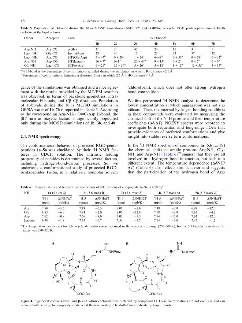
Table 5. Population of H-bonds during the 10 ns MC/SD simulations (AMBER*, H2O GB/SA) of cyclic RGD pentapeptide mimics 1b–7b
cyclo(Arg-Gly-Asp-Lactam)
Donor Acceptor Turn % H-bonda
1b 2b 3b 4b 5b 6b 7b
Asp NH Arg CO c(Gly) 31 3 45 24 11 3 5
Lact. NH Gly CO Inv. c(Asp) 23 40 16 23 33 57 32
Lact. NH Arg CO bII0(Gly-Asp) 9 + 47b 0 + 20b 1 + 14b 6+48b 0 + 50b 0 + 28b 0 + 42b
Arg NH Asp CO bII0(lactam) 16 + 7b 0+1b 20 + 44b 9 + 15b 0 + 2b 0 + 2b 0 + 0b
Gly NH Lact. CO bI(Pro-Arg) 6 + 21b 26 + 34b 5 + 26b 5 + 18b 1 + 12b 13 + 55b 4 + 15b
a % H-bond is the percentage of conformations sampled during the simulation in which HO distance <2.5 Ab Percentage of conformations featuring a distorted b-turn in which 2.5 A < HO distance < 4 A.
174 L. Belvisi et al. / Bioorg. Med. Chem. 14 (2006) 169–180
gence of the simulations was obtained and a nice agree-ment with the results provided by the MC/EM searcheswas observed, in terms of backbone geometries, intra-molecular H-bonds, and Cb–Cb distances. Populationof H-bonds during the 10 ns MC/SD simulations inGB/SA water of 1b–7b is reported in Table 5. Accordingto the corresponding Arg-NH� � �O@C-Asp H-bond, thebII 0-turn at bicyclic lactam is significantly populatedonly during the MC/SD simulations of 1b, 3b, and 4b.
2.4. NMR spectroscopy
The conformational behavior of protected RGD-penta-peptides 1a–5a was elucidated by their 1H NMR fea-tures in CDCl3 solution. The intrinsic foldingpropensity of peptides is determined by several factors,including hydrogen-bond-driven processes. So, weundertook a conformational study of protected RGD-pentapeptides 1a–5a, in a relatively nonpolar solvent
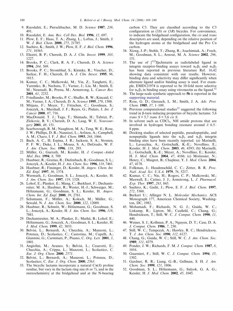
Table 6. Chemical shifts and temperature coefficients of NH protons of com
NH 1a (5,6 cis S) 2a (5,6 trans R) 3a (5
1H d(ppm)
DdNH/DT(ppb/K)
1H d(ppm)
DdNH/DT(ppb/K)
1H d(ppm)
Arg 7.80 �5.6 7.18 �0.5 7.06
Gly 8.83 �6.5 7.93 �2.0 8.06
Asp 7.62 �0.8 7.56 �8.0 7.02
Lactam 6.79 +1.8 7.35 �0.7 7.59
a The temperature coefficients for 5,6 bicyclic derivatives were obtained in t
range was 290–320 K.
NN
N
OH5
N
O
H
O
HH7
O NH
HHN
O
COOtBu
H
H
HH
NH
NHPmc
A B
Figure 4. Significant contacts NOE and b- and c-turn conformations prefer
occur simultaneously; for simplicity we depicted them separately. The dotted
(chloroform), which does not offer strong hydrogenbond competition.
We first performed 1H NMR analysis to determine thelowest concentration at which aggregation was not sig-nificant. Then, the internal hydrogen-bonding equilibriain these compounds were evaluated by measuring thechemical shift of the N–H protons and their temperaturecoefficients (Dd/DT). NOESY spectra were recorded toinvestigate both sequential and long-range nOe�s thatprovide evidences of preferred conformations and giveinsight into stable reverse turn conformations.
In the 1H NMR spectrum of compound 1a (5,6 cis 3S)the chemical shifts of amide protons Arg-NH, Gly-NH, and Asp-NH (Table 6)39 suggest that they are allinvolved in a hydrogen bond interaction, but each to adifferent extent. The temperature dependence (DdNH/DT) (Table 6) also reflects this behavior and suggeststhat the participation of the hydrogen bond of Arg-
pounds 1a–5a in CDCl3a
,6 trans S) 4a (5,7 trans S) 5a (5,7 trans R)
DdNH/DT(ppb/K)
1H d(ppm)
DdNH/DT(ppb/K)
1H d(ppm)
DdNH/DT(ppb/K)
�1.6 7.19 �3.0 6.99 �12.0
�12.8 7.78 �8.0 7.61 �4.2
�9.5 7.94 �12.0 7.62 �12.0
�5.7 7.64 �6.0 7.49 �1.2
he temperature range (250–300 K); for the 5,7 bicyclic derivatives the
NN
N
OH5
HN
O
HO
HH7O
NH
H
HN
OCOOtBu
H
H
H
NH
NHPmc
red by compound 1a These conformations are not exclusive and can
lines indicate hydrogen bonds.

L. Belvisi et al. / Bioorg. Med. Chem. 14 (2006) 169–180 175
NH and Gly-NH in 1a becomes much more significantin the low temperature region. At the same time, the po-sitive value of DdNH/DT (+1.8 ppb/K, 6.79 ppm) forLactam-NH indicates that at low temperature, also thesmall amount of hydrogen bond experienced by Lac-tam-NH was broken.
The NOE patterns and the spin–spin coupling 3JHNHa
expected for a b-turn conformation are observed incompound 1a. In fact, the long range NOEs of Gly-NH with Arg-NH and H7 and the extreme value of 3J(ArgNH-H8 = 9 Hz) are indicative of a b-turn confor-mation (Fig. 4B). This b-turn was stabilized by a hydro-gen bond between Gly-NH and C@O of the bicycliclactam. Moreover, Arg-NH forms an additional hydro-gen bond with C@O of Asp, stabilizing a b-turn with theproline residue at the i + 2 position (Fig. 4A). The longrange NOE between Arg-NH and Asp-Ha confirms thisconformation.
At the same time, the presence of a c-turn (Fig. 4A), ob-served by modeling, was confirmed by the temperaturedependence and the value of chemical shift of the amideproton Asp-NH. The contact NOE between Lactam-NH and H5 suggests a conformation where Lactam-NH is not involved in an internal hydrogen bond. Anindication of the rigidity of this part of the cycle comesfrom the observation that the side-chain protons of theAsp residue have restricted rotation: the chemical shiftsfor the two b protons are very different (0.5 ppm). InFigures 4A and B, the significant contacts NOE, andb- and c-turn conformations preferred by compound1a are reported. These conformations are not exclusiveand can occur simultaneously; for simplicity, we depict-ed them separately. Thus, in CDCl3 solution, thepseudopeptide 1a assumes a very compact folding stabi-lized by three hydrogen bonds.
The peptidomimetic 2a is more prone to aggregation sothe spectra were performed below 2 mM CDCl3 solu-tion. In this compound, the configuration of the scaffold(5,6 trans R) arranges the peptidic cycle so that the Argresidue is found above the plane of the bicyclic lactam,
N H7
H N
N
O
OH6
H
N
H
H
H
HO
O
COOtBu
H
NH
N
H
H
O
NH
NHPmc
Figure 5. Significant contacts NOE and conformation preferred by
compound 2a. Pseudopeptide 2a can fold into a b-hairpin-likeconformation where the b-turn is stabilized by a second hydrogen
bond to form (10 + 14) hydrogen bonded rings.
while the Lactam-NH bond is oriented below the plane.Hence, the Arg-NH cannot form a 10-membered ringhydrogen bond (b-turn) with C@O Asp (below themedium plane) but, if any, a c-turn with lactamicC@O. In fact, Arg-NH exhibits a medium NOE cross-peak with H6 (Fig. 5). The chemical shifts and the tem-perature dependence of amide protons Gly-NH andLactam-NH (respectively, 7.93 ppm and �2 ppb/K forGly-NH; 7.35 ppm and �0.7 ppb/K for Lactam-NH)(Table 6) are typical of protons locked in a hydrogenbond. NOESY spectra of compound 2a show NOEs be-tween Gly-NH and Arg-NH, and between Gly-NH andArg-Ha (Fig. 5). These are indicative of a b-turn confor-mation stabilized by a hydrogen bond between Gly-NHand lactamic C@O. Moreover, Gly-NH shows a NOEcross-peak with Lactam-NH as well as the amidic pro-ton Lactam-NH that is inside to the cyclic pentapeptideand can experience a hydrogen bond with C@O of Gly.These data indicate that 2a can fold into a b-hairpin-likeconformation where the b-turn is stabilized by a secondhydrogen bond (Fig. 5).
The 1H NMR spectrum of compound 3a (5,6 trans S)shows broad signals, also in the CHa region, indicativeof a slow equilibrium between more equivalent confor-mations. In fact, the spectra of the same compoundrecorded at different temperature (CDCl3, 330 K) or inDMSO solution show sharp signals. The 5,6 trans 3S-fused bicyclic lactam used as external constraints forthe RGD tripeptide in the mimic 3a is a very goodinductor of b-turn.32 Indeed, the chemical shift of ami-dic Arg-NH of compound 3a was at 7.06 ppm (DdNH/DT = �1.6 ppb/K) and it represents the behavior of aamide proton in the hydrogen-bonded state. But, thetemperature dependence (DdNH/DT) and the chemicalshift value (Table 6) of amide proton Lactam-NH hinttoward involvement in a hydrogen bond. Most likely,Lactam-NH is inside to the cyclopeptide and, in agree-ment with modeling, experiences a seven-memberedhydrogen-bonded ring with C@O of Gly. The formationof this c-turn is exclusive with the presence of b-turnwhere Arg-NH experiences a hydrogen bond. Compila-tion of NOE data (Fig. 6) and NH chemical shifttemperature dependence for protons Gly-NH and Asp-NH (respectively, �12.8 and �9.5 ppb/K) in compound3a provide further evidence for multiple folded confor-mations in equilibrium. So, the NMR data obtainedfor 3a do not give evidence of a unique solutionconformation.
N N
NH
H6
H
O
O
H
H NH
ON
HO
H
NO
H
HH
H
COOtBu
NH
NHPmc
Figure 6. Key NOE connectivities found for mimetic 3a.

NN
N
H
HO
O
H
H
N
O
H
O N
COOtBu
O
H
HH
H
H
H7
HN
NH
NHPmc
Figure 7. Key NOE connectivities found for mimetic 4a.
NN
N
H6
HO
O
H
H
N
O
H
O N
COOR'
O
H
HH
H
H
NH
H7
NH
NHR
5a R=Pmc, R'=tBu5b R=H, R'=H
Figure 8. Key NOE connectivities found for mimetics 5a and 5b.
Table 7. Chemical shifts (ppm) of NH protons of compounds 5a in
CDCl3 and 5b in H2O/D2O (9:1) solution
NH 5a 5b Down-field shift (ppm)1H (CDCl3)
1H (H2O)
Arg 6.99 8.22 1.23
Gly 7.61 7.65 0.04
Asp 7.62 8.92 1.3
Lactam 7.49 7.95 0.54
176 L. Belvisi et al. / Bioorg. Med. Chem. 14 (2006) 169–180
In previous studies,32 we observed that the (5,7 trans 3S)lactam included in the pentapeptide 4a is considered bymodeling a very good b-turn inducer.
The 1H NMR spectrum of 4a in 3 mM CDCl3 solutionsexhibited amide protons in the range 7.19–7.94 ppm.These chemical shifts were characteristic of peptidebackbone NHs strongly involved in hydrogen bonding.Since these hydrogen bonds cannot occur simultaneous-ly, we propose that 4a equilibrates between differentfolded conformations, similar to compound 3a.
The middle- and long-range NOE cross-peaks observedin NOESY spectrum are summarized in Figure 7. Asingle conformation cannot account for all of theselong-range NOEs; therefore, the NOESY data providefurther evidence of multiple folded conformations. TheNOE between Arg-NH and Gly-NH could arise fromconformation SIV (Fig. 3). The NOE between Asp-NH and Lactam-NH is consistent with the c-turn-likefolding pattern SII (Fig. 3).
Compound 5a, like compound 2a, is more prone toaggregation so the spectra are recorded in 2 mM CDCl3solution. In this compound, the Arg residue is above theplane of the bicyclic lactam, while the R configuration ofthe stereocenter in position 3 orients the aspartic residuebelow the plane. So, Arg-NH cannot form a 10-mem-bered ring hydrogen bonding with C@O(Asp). Thechemical shift value of the amide protons Gly-NH(7.61 ppm) and Lactam-NH (7.49 ppm), and the tem-perature coefficients (DdNH/DT = �4.2 and �1.2 ppb/K, respectively) indicate that these protons are lockedin an intramolecularly H-bonded state. Lactam-NH,inside to the cyclic pentapeptide, can form a c-turn cen-tered on aspartic residue. Protons Arg-NH and Asp-NH(dNH = 6.99 and 7.62 ppm, respectively; DdNH/DT = �12.0 ppb/K) are in equilibrium between anonH-bonded and a H-bonded state. The NOESY spec-trum shows the following cross-peaks: Asp-NH/Lactam-NH, Gly-NH/Arg-NH, Arg-NH/H6 (weak), Arg-NH/H7 and Arg-NH/Arg-Ha strong (Fig. 8). These dataprovide further evidence for multiple folded conforma-tions in equilibrium (type SII, SIII, and SIV geometries).
Amide proton–deuterium exchange rates provide infor-mation regarding the possible participation of an amideproton in a stable intramolecular H-bond within a sec-
ondary or tertiary structural element (a-helix, b-sheet,and reverse turns). So, we analyzed the deprotectedcyclopentapeptide mimics 1b–5b (Fig. 2) in D2O solu-tion. In this solvent, the amide protons Arg-NH andGly-NH of mimic 1b exchange very slowly and thisbehavior suggests their involvement in strong intramo-lecular hydrogen bonds. The exchange is slow also forLactam-NH of 2b and 5b, and for Arg-NH of com-pound 3b.
After these results, we decided to study the conforma-tional features of the most active compound 5b inH2O/D2O (9:1) solution in order to observe the behaviorof amide protons (Table 7). It is remarkable that theamide proton Gly-NH (7.65 ppm) is locked in an intra-molecularly H-bonded state also in a competitive sol-vent like H2O. The NOESY spectrum shows NOEsbetween Gly-NH and Arg-NH, and between Gly-NHand Arg-Ha. These are indicative of a b-turn conforma-tion stabilized by a hydrogen bond between Gly-NHand lactamic C@O. Moreover, the chemical shift andthe slow exchange rate of Lactam-NH are indicative ofa hydrogen-bonded proton that can form a c-turn cen-tered on aspartic residue. Protons Arg-NH and Asp-NH are solvent exposed; other significant NOE contactsare between Asp-NH/Lactam-NH, Asp-NH/Gly-Ha1,and Asp-NH/Gly-Ha2 (Fig. 8). It is interesting to ob-serve that the NOE contacts and the conformationalpreferences of compound 5b are similar to those of ana-log pseudopeptide 5a.
3. Discussion and conclusions
The crystal structure of the extracellular segment of inte-grin aVb3 in complex with the cyclic pentapeptide ligandEMD121974 c(Arg-Gly-Asp-D-Phe-[NMe]Val) hasbeen reported.34 This structure provides the exact con-

L. Belvisi et al. / Bioorg. Med. Chem. 14 (2006) 169–180 177
formation of EMD121974 bound to aVb3 integrin andcan serve as a basis for understanding the general modeof interaction of integrins with other RGD-containingligands.
Examination of the three-dimensional structure of thecyclic pentapeptide antagonist EMD121974 bound tothe integrin aVb3 (Protein Data Bank, entry 1L5G) re-veals a conformation characterized by an inverse c-turnwith Asp at position (i + 1) and by a distorted bII 0-turnwith Gly and Asp at the (i + 1) and (i + 2) positions(Fig. 9). A distance between the Cb atoms of Asp andArg of 8.9 A is observed in this pentapeptide bound con-formation. Contrary to what had been assumed previ-ously,23 the inhibition of aVb3 integrin does notrequire a strong kink of the RGD sequence. The typeSIII geometry obtained by computational methods forthe cyclopentapeptide mimics (see above, Fig. 3 and Ta-ble 4) is very similar to the X-ray binding conformationof EMD121974, in terms of backbone arrangement,intramolecular H-bonds, and Cb–Cb distance.
With the aim of identifying highly selective integrinantagonists, we synthesized and tested a small libraryof cyclic pseudopeptides in which the triad arginine, gly-cine, and aspartic acid (RGD) was attached to (5,6)- or(5,7)-fused bicyclic lactams with different configurationat two stereocenters. The use of rigid peptidomimeticscaffolds and conformationally constrained cyclic pep-tides that match biologically active conformation mightenhance ligand binding for entropic reasons.
Type SIIIA B
Figure 10. Conformations of 5b sampled during the 10 ns MC/SD simulatio
bII 0(Gly-Asp). (B) Type SIV, distorted bI(Pro-Arg)/inverse c(Asp).
Figure 9. X-ray, aVb3-bound conformation of EMD121974 from
1L5G.34
According to the results provided by spectroscopic andcomputational studies, a strong dependence of the cyclo-peptide conformations on lactam ring size and stereo-chemistry has been observed. The effects of thestructural constraint introduced by the bicyclic templateon the conformation of the RGD sequence are mainlydictated by the turn mimetic properties of the scaffold.32
Compounds 2b and 5b, containing the poor b-turninducers bicyclic lactams 5,6 trans 3R and 5,7 trans3R, respectively, show preferred cyclopeptide geometriesfeaturing a lightly kinked or an almost extended confor-mation of the RGD sequence. Only type SIII (inverse c-turn at Asp and distorted bII 0-turn at Gly-Asp) and typeSIV (bI-turn at Pro-Arg and inverse c-turn at Asp)geometries can be detected among the conformers with-in 3 kcal/mol of the global minimum calculated for thesimplified AGA cyclopeptide 2c (Table 4). Accordingto the H-bond analysis, these turns are populated by40% (inverse c-turn at Asp), 20% (distorted bII 0-turnat Gly-Asp), and 26% (bI-turn at Pro-Arg) during the10 ns MC/SD simulation of 2b (Table 5). The averageof the Cb(Arg)–Cb(Asp) distance is 8.8 A over the sametrajectory. The chemical shifts and the temperaturedependence of amide protons Gly-NH and Lactam-NH of the protected cyclopeptide 2a (Table 6), as wellas NOE data (Fig. 5) and the slow exchange rate of Lac-tam-NH of 2b in D2O, provide evidence for the impor-tance of the b-hairpin-like conformation of type SIV.
The lowest energy conformer of the simplified AGAcyclopeptide 5c is characterized by an inverse c-turn atAsp and by a distorted bII 0-turn at Gly-Asp (type SIIIgeometry). Type SII and SIV geometries can be alsodetected among the conformers within 3 kcal/mol ofthe global minimum (Table 4). NMR data of compound5a provide evidence for different folded conformationsin equilibrium, showing, in particular, the involvementof Lactam-NH in strong intramolecular hydrogen bonds(type SIII and SIV geometries). Compound 5b in H2Osolution keeps the conformational preferences of theprotected analog 5a. Again Gly-NH and Lactam-NHare inner to the pentapeptide ring and one of thepreferred conformations is stabilized by a b-turn andan inverse c-turn (type SIV geometry). According to
Type SIV
n after energy minimization. (A) Type SIII, inverse c (Asp)/distorted

178 L. Belvisi et al. / Bioorg. Med. Chem. 14 (2006) 169–180
the H-bond analysis, the inverse c-turn at Asp (33%)and the distorted bII 0-turn at Gly-Asp (50%) and bI-turn at Pro-Arg (13%) are mainly populated duringthe 10 ns MC/SD simulation of 5b (Table 5). The aver-age of the Cb(Arg)–Cb(Asp) distance is 8.5 A over thesame trajectory. Remarkably, the 5,7 trans 3R bicyclicscaffold of compound 5b appears to force the cyclopep-tide to assume preferred conformations very similar tothe X-ray aVb3-bound conformation of EMD121974.Energy-minimized conformations of 5b obtained fromframes featuring the binding requirements (type SIIIgeometry) or the SIV geometry and corresponding tothe most populated geometries of the 10 ns MC/SD tra-jectory are shown in Figure 10. The root-mean-square(RMS) deviation in the rigid superimposition betweenthe type SIII conformation of 5b (Fig. 10A) and theX-ray structure of bound EMD121974 is 0.16 A forthe backbone atoms of the RGD sequence. Binding of5b might actually be enhanced by the high structuralpreorganization.
Compounds 2b and 5b showed the highest affinities toaVb3 and inhibited radiolabeled echistatin binding toaVb3 with an IC50 of 14.3 ± 4.7 and 3.8 ± 0.9 nM,respectively. The conformational studies describedabove suggested that the type SIII and SIV geometriesare the conformations mainly contributing to the con-formational equilibria of cyclopeptides 2b and 5b. Boththese geometries feature a minimal kink of the RGDsequence and an orientation of the Asp NH groupsuitable to maintain the same electrostatic and hydrogenbond ligand–receptor interactions observed in thecrystalline complex of EMD121974 with aVb3.
Conformational studies of compounds 1b, 3b, and 4b, orrelated derivatives, containing efficient reverse-turn andb-turn inducer bicyclic lactams,38 provide evidence formultiple folded conformations in equilibrium. In partic-ular, the contribution of the SI structural type to theseconformational equilibria is worth noting. This geome-try features a strong kink of the RGD motif as a conse-quence of the bII 0/c-turn arrangement with the bicyclictemplate in the i + 1 and i + 2 positions of the bII 0-turnand the Gly residue in the i + 1 position of the c-turn atthe opposite side. According to the correspondingArg-NH� � �O@C-Asp H-bond, the bII 0-turn at bicycliclactam is significantly populated during the MC/SDsimulations of 1b, 3b, and 4b (Table 5). The presencein conformational equilibria of geometries featuringkinked backbone conformations of the RGD sequence,short Cb(Arg)–Cb(Asp) distances, and inside orienta-tion of the amide proton Asp-NH (c-turn at Gly) mightbe responsible for reduced structural preorganizationfor binding and therefore for lower affinities to aVb3(Table 1).
Computational studies of 6b–7b and 6c–7c derivativessuggest that both the poor turn inducer bicylic scaffolds5,7 cis force the cyclopeptide to assume mainly the typeSIII and SIV geometries. In spite of the proper orienta-tion thus achieved by the pharmacophoric groups re-quired for binding to the aVb3 integrin, compounds 6band 7b showed low affinities toward this receptor (Table
1). Docking studies40 are currently in progress to gaindeeper insights into ligand–receptor interactions of cyc-lic RGD pentapeptide mimics containing azabicycloal-kane amino acids.
4. Materials and methods
4.1. Cell cultures
Primary cultures of bovine microvascular endothelialcells (BMECs) were obtained from bovine adrenalglands as described by Folkman.41 BMECs were main-tained in DMEM supplemented with 20% fetal calf ser-um (FCS), 50 U/ml heparin (Sigma, St. Louis, MO),50 lg/ml bovine brain extract, and 100 U/ml gentamicin.Human umbilical vein endothelial cells (HUVECs)and human microvascular dermal endothelial cells(HMECs) were obtained from BioWhittaker (Walkers-ville, MD) and grown in EGM-2 (BioWhittaker).
4.2. Solid-phase receptor-binding assay
The receptor-binding assays were performed as de-scribed previously.42,43 Purified receptors avb3 andavb5 (Chemicon International Inc., Temecula, CA) werediluted, respectively, to 500 ng/ml and 1000 ng/well incoating buffer [20 mM Tris–HCl (pH 7.4), 150 mMNaCl, 2 mM CaCl2, 1 mM MgCl2, and 1 mM MnCl2],whereas a5b1 was diluted to 1000 ng/ml in 20 mM Tris–HCl (pH 7.4), 150 mM NaCl, and 1 mM MnCl2. An ali-quot of the diluted receptors (100 ll/well) was added to96-well microtiter plates and incubated overnight at4 �C. The coating solution was removed by aspiration,and 200 ll of blocking solution (coating buffer contain-ing 1% bovine serum albumin (BSA)) was added to thewells, which were incubated for an additional 2 h atroom temperature. After incubation, the plates wererinsed with 200 ll of blocking solution (3·) and incubat-ed with appropriate radiolabeled ligands for 3 h at roomtemperature. 0.05 and 0.1 nM [125I]echistatin (Amer-sham Pharmacia) were used, respectively, for avb3 andavb5, whereas 20 nM [125I]fibrinogen (Amersham Phar-macia) was used for a5b1 receptor-binding assay. Afterincubation, the plates were sealed and counted in thec-counter (Packard). Each data point is the result ofthe average of triplicate well, and was analyzed by non-linear regression analysis with the Allfit program.
4.3. Platelet aggregation assay
Blood samples were collected in guinea pigs via cardiacpuncture in 3.8% sodium citrate at a final dilution of 1–10. Platelet-rich plasma (PRP) was prepared by centrifu-gation at 120g for 10 min at room temperature. Plateletaggregation was assayed following the addition ofTRAP 42-55 (Sigma), 25–100 lM, to PRP at 37 �C bylight transmission (4 channel aggregometer, PACKS-4,Helena Laboratories, Beaumont, Texas).
Vehicle or peptide solutions at different concentrationswere added to aliquots of the same PRP, two minutesprior to TRAP addition. The extent of platelet aggrega-

L. Belvisi et al. / Bioorg. Med. Chem. 14 (2006) 169–180 179
tion was quantified as the maximum change in lighttransmission within 4 min after the addiction of agonist.
The results were plotted (Allfit program) and expressedas the antagonist concentration that inhibited 50% ofplatelet aggregation.
4.4. Cell-adhesion assay
Ninety-six-well plates were coated with either fibronec-tin (Sigma, St. Louis, MO) or vitronectin (Calbiochem,San Diego, CA) (both at 5 lg/ml in phosphate-bufferedsaline) overnight at 4 �C. Approximately 4–5 · 104 cells/100 ll were seeded in each well and allowed to adherefor 1–3 h at 37 �C in the presence of various concentra-tions of RGD peptides. Nonadherent cells were re-moved with PBS and the remaining cells were fixedwith 4% paraformaldehyde for 10 min. Adherent cellswere stained with 1% toluidine blue for 10 min andrinsed with water. Stained cells were solubilized with1% SDS and quantified on a microtiter plate reader at600 nm (Victor2, Wallac). Experiments were performedin quadruplicate and repeated at least three times. Re-sults are expressed as mean compound concentra-tion ± SEM that inhibited 50% of cell adhesion.
4.5. Computational studies
Conformational preferences of the RGD cyclopeptideshave been investigated by molecular mechanics44 calcu-lations within the framework of MacroModel45 version5.5, using the MacroModel implementation of the Am-ber all-atom force field46 (denoted AMBER*) and theimplicit water GB/SA solvation model of Still et al.47
A two-step protocol was used.
Monte Carlo/energy minimization (MC/EM) conforma-tional searches48 of the AGA (Ala-Gly-Ala) cyclopep-tide analogs containing methyl groups instead of theArg and Asp side chains were performed as the first step.The torsional space of each AGA cyclopeptide wasrandomly varied with the usage-directed Monte Carloconformational search of Chang, Guida, and Still.48
Ring-closure bonds were defined in the six- and seven-membered rings of the 5,6- and 5,7-fused bicyclic lac-tams, respectively, and in the cyclopeptide ring. Amidebonds were included among the rotatable bonds. Foreach search, at least 1000 starting structures for eachvariable torsion angle were generated and minimized un-til the gradient was less than 0.05 kJ/Amol using thetruncated Newton–Raphson method49 implemented inMacroModel. Duplicate conformations and those withan energy greater than 6 kcal/mol above the global min-imum were discarded. The nature of the stationarypoints individuated was tested by computing the eigen-values of the second-derivative matrix.
Free simulations of the complete RGD cyclic peptides(Asp and Arg side chains were considered ionized) werethen performed at 300 K using the metropolis MonteCarlo/stochastic dynamics (MC/SD) hybrid simulationalgorithm,50 starting from the cyclopeptide backbonegeometries located by the previous MC/EM step.
RGD side-chain dihedral angles were defined as internalcoordinate degrees of freedom in the Monte Carlo partof the algorithm. A time step of 1 fs was used for the sto-chastic dynamics (SD) part of the algorithm. At leasttwo 10 ns simulations were run for each RGD cyclopep-tide starting from different conformations to test theconvergence. Samples were taken at 2 ps intervals dur-ing each simulation, yielding 5000 conformations foranalysis.
4.6. NMR spectroscopy
All the spectra were acquired at a 400 MHz Bruker spec-trometer equipped with pulsed field gradients. The fol-lowing experiments were carried out (at 300 K):TOCSY, NOESY (mixing time 200, 400, and 600 ms),and HSQC.
The protected RGD-pentapeptides 1a–5a (Fig. 2) wereanalyzed by NMR spectroscopy in CDCl3 solution.We first performed 1H NMR analysis to determine thelowest concentration at which intermolecular hydrogenbonding occurs.51 The data reported in this paper wereobtained from samples at 300 K and 1–5 mM concentra-tions, at which aggregation was not significant. Tracesof acid were removed from CDCl3 by passing it throughan alumina column. Amide hydrogen temperature coef-ficients were measured from 1D experiments carried outin the range 240–320 K. The cyclopentapeptide mimics1b–5b (Fig. 2) were analyzed by NMR spectroscopy inD2O solution; compound 5b was also studied in H2O/D2O (9:1) solution.
Acknowledgments
The authors thank CNR and MIUR (COFIN andFIRB research programs) for financial support andCILEA for computing facilities.
Supplementary data
Description of the synthesis and experimental data forthe preparation of compound 5b. Tables S1–S6 listingthe 1H and 13C NMR chemical shifts d (ppm) of pseudo-pentapeptides 1a–7a and 1b–7b. Supplementary dataassociated with this article can be found in the onlineversion at doi:10.1016/j.bmc.2005.08.048.
References and notes
1. Gumbiner, B. M. Cell 1996, 84, 345.2. Aplin, A. E.; Howe, A.; Alahari, S. K.; Juliano, R. L.
Pharmacol. Rev. 1998, 50, 197.3. Eliceiri, B. P.; Cheresh, D. A. Curr. Opin. Cell Biol. 2001,
13, 563.4. Hood, J. D.; Cheresh, D. A. Nat. Rev. Cancer 2002, 2, 91.5. Howe, A.; Aplin, A. E.; Alahari, S. K.; Juliano, R. L.
Curr. Opin. Cell Biol. 1998, 10, 220.6. Hynes, R. O. Cell 2002, 110, 673.7. Hynes, R. O. Cell 1987, 48, 549.8. Giancotti, F. G.; Ruoslahti, E. Science 1999, 285, 1028.
180 L. Belvisi et al. / Bioorg. Med. Chem. 14 (2006) 169–180
9. Ruoslahti, E.; Pierschbacher, M. D. Science 1987, 238,491.
10. Ruoslahti, E. Ann. Rev. Cell Dev. Biol. 1996, 12, 697.11. Plow, E. F.; Haas, T. A.; Zhang, L.; Loftus, J.; Smith, J.
W. J. Biol. Chem. 2000, 275, 21785.12. Suehiro, K.; Smith, J. W.; Plow, E. F. J. Biol. Chem. 1996,
271, 10365.13. Eliceiri, B. P.; Cheresh, D. A. J. Clin. Invest. 1999, 103,
1227.14. Brooks, P. C.; Clark, R. A. F.; Cheresh, D. A. Science
1994, 264, 569.15. Brooks, P. C.; Stromblad, S.; Klemke, R.; Visscher, D.;
Sarkar, F. H.; Cheresh, D. A. J. Clin. Invest. 1995, 96,1815.
16. Kumar, C. C.; Malkowski, M.; Yin, Z.; Tanghetti, E.;Yaremko, B.; Nechuta, T.; Varner, J.; Liu, M.; Smith, E.M.; Neustadt, B.; Presta, M.; Armstrong, L. Cancer Res.2001, 61, 2232.
17. Friedlander, M.; Brooks, P. C.; Shaffer, R. W.; Kincaid, C.M.; Varner, J. A.; Cheresh, D. A. Science 1995, 270, 1500.
18. Mitjans, F.; Meyer, T.; Fittschen, C.; Goodman, S.;Jonczyk, A.; Marshall, J. F.; Reyes, G.; Piulats, J. Int. J.Cancer 2000, 87, 716.
19. MacDonald, T. J.; Taga, T.; Shimada, H.; Tabrizi, P.;Zlokovic, B. V.; Cheresh, D. A.; Laug, W. E. Neurosur-gery 2001, 48, 151.
20. Scarborough, R. M.; Naughton, M. A.; Teng, W. E.; Rose,J. W.; Phillips, D. R.; Nannizzi, L.; Arfsten, A.; Campbell,A. M.; Charo, I. F. J. Biol. Chem. 1993, 268, 1066.
21. Bach, A. C., II; Espina, J. R.; Jackson, S. A.; Stouten,P. F. W.; Duke, J. L.; Mousa, S. A.; DeGrado, W. F.J. Am. Chem. Soc. 1996, 118, 293.
22. Muller, G.; Gurrath, M.; Kessler, H. J. Comput.-AidedMol. Des. 1994, 8, 709.
23. Haubner, R.; Gratias, R.; Diefenbach, B.; Goodman, S. L.;Jonczyk, A.; Kessler, H. J. Am. Chem. Soc. 1996, 118, 7461.
24. Haubner, R.; Finsinger, D.; Kessler, H. Angew. Chem. Int.Ed. Engl. 1997, 36, 1374.
25. Wermuth, J.; Goodman, S. L.; Jonczyk, A.; Kessler, H.J. Am. Chem. Soc. 1997, 119, 1328.
26. Lohof, E.; Planker, E.; Mang, C.; Burkhart, F.; Dechants-reiter, M. A.; Haubner, R.; Wester, H.-J.; Schwaiger, M.;Holzemann, G.; Goodman, S. L.; Kessler, H. Angew.Chem. Int. Ed. Engl. 2000, 39, 2761.
27. Schumann, F.; Muller, A.; Koksch, M.; Muller, G.;Sewald, N. J. Am. Chem. Soc. 2000, 122, 12009.
28. Haubner, R.; Schmitt, W.; Holzemann, G.; Goodman, S.L.; Jonczyk, A.; Kessler, H. J. Am. Chem. Soc. 1996, 118,7881.
29. Dechantsreiter, M. A.; Planker, E.; Matha, B.; Lohof, E.;Holzemann, G.; Jonczyk, A.; Goodman, S. L.; Kessler, H.J. Med. Chem. 1999, 42, 3033.
30. Belvisi, L.; Bernardi, A.; Checchia, A.; Manzoni, L.;Potenza, D.; Scolastico, C.; Castorina, M.; Cupelli, A.;Giannini, G.; Carminati, P.; Pisano, C. Org. Lett. 2001, 3,1001.
31. Angiolini, M.; Araneo, S.; Belvisi, L.; Cesarotti, E.;Checchia, A.; Crippa, L.; Manzoni, L.; Scolastico, C.Eur. J. Org. Chem. 2000, 2571.
32. Belvisi, L.; Bernardi, A.; Manzoni, L.; Potenza, D.;Scolastico, C. Eur. J. Org. Chem. 2000, 2563.
33. The bicyclic lactams incorporate a natural Ca(S) prolineresidue, but vary in the lactam ring size (6 or 7), and in thestereochemistry at the bridgehead and at the N-bearing
carbon C3. They are classified according to the C3configuration as (3S) or (3R) bicycles. For convenience,to indicate the bridgehead configuration, the cis and transdescriptors are used, depending on the relative position ofthe hydrogen atoms at the bridgehead and the Pro Cacarbon.
34. Xiong, J.-P.; Stehle, T.; Zhang, R.; Joachimiak, A.; Frech,M.; Goodman, S. L.; Arnout, M. A. Science 2002, 296,151.
35. The use of [125I]echistatin as radiolabeled ligand inintegrin receptor-binding assays toward aVb3 and aVb5has been reported in previous literature Ref. 16,42showing data consistent with our results. However,binding data and selectivity may differ significantly whenalternate ligand and/or binding assay is used. For exam-ple, EMD121974 is reported to be 10-fold more selectivefor aVb3 in binding assay using vitronectin as the ligand.52
36. The large-scale synthetic approach to 5b is reported in thesupporting material.
37. Rose, G. D.; Gierasch, L. M.; Smith, J. A. Adv. Prot.Chem. 1985, 37, 1–109.
38. Previous computational studies32 suggested the followingtrend in b-turn inducing properties of bicyclic lactams: 5,6trans S > 5,7 trans S > 5,6 cis S.
39. In solvent such as CDCl3, NH amide protons that areinvolved in hydrogen bonding resonate around d 6.5–8 ppm.
40. Docking studies of selected peptidic, pseudopeptidic, andnonpeptidic ligands into the avb3 and avb5 integrinbinding sites have been recently reported. (a) Marinelli,L.; Lavecchia, A.; Gottschalk, K.-E.; Novellino, E.;Kessler, H. J. Med. Chem. 2003, 46, 4393; (b) Marinelli,L.; Gottschalk, K.-E.; Meyer, A.; Novellino, E.; Kessler,H. J. Med. Chem. 2004, 47, 4166; (c) Moitessier, N.;Henry, C.; Maigret, B.; Chapleur, Y. J. Med. Chem. 2004,47, 4178.
41. Folkman, J.; Haudenschild, C. C.; Zetter, B. R. Proc.Natl. Acad. Sci. U.S.A. 1979, 76, 5217.
42. Kumar, C. C.; Nie, H.; Rogers, C. P.; Malkowski, M.;Maxwell, E.; Catino, J. J.; Armstrong, L. J. Pharmacol.Exp. Ther. 1997, 283, 843.
43. Suehiro, K.; Gailit, J.; Plow, E. F. J. Biol. Chem. 1997,272, 5360.
44. Burkert U.; Allinger N. L. Molecular Mechanics. ACSMonograph 177, American Chemical Society, Washing-ton, DC, 1982.
45. Mohamadi, F.; Richards, N. G. J.; Guida, W. C.;Liskamp, R.; Lipton, M.; Caufield, C.; Chang, G.;Hendrickson, T.; Still, W. C. J. Comput. Chem. 1990, 11,440.
46. Weiner, S. J.; Kollman, P. A.; Nguyen, D. T.; Case, D. A.J. Comput. Chem. 1986, 7, 230.
47. Still, W. C.; Tempczyk, A.; Hawley, R. C.; Hendrickson,T. J. Am. Chem. Soc. 1990, 112, 6127.
48. Chang, G.; Guida, W. C.; Still, W. C. J. Am. Chem. Soc.1989, 111, 4379.
49. Ponder, J. W.; Richards, F. M. J. Comput. Chem. 1987, 8,1016.
50. Guarnieri, F.; Still, W. C. J. Comput. Chem. 1994, 15,1302.
51. Gardner, R. R.; Liang, G.-B.; Gellman, S. H. J. Am.Chem. Soc. 1999, 121, 1806.
52. Goodman, S. L.; Holzemann, G.; Sulyok, G. A. G.;Kessler, H. J. Med. Chem. 2002, 45, 1045.


