Embed Size (px)
Citation preview
TAT hitchhiker selection expanded to folding helpers,multimeric interactions and combinations with proteinfragment complementation
Janina Speck1,2, Christina Rauber1,2,3,Tim Kukenshoner1,2, Christoph Niemoller2, KatelynJ.Mueller2,4, Paula Schleberger2,Padmarupa Dondapati2,5, Jochen Hecky2,6, KatjaM.Arndt1,2,3,5 and Kristian M.Muller1,2,3,7
1Institute for Biochemistry and Biology, University of Potsdam, Germany,2Department of Biology, Albert-Ludwigs-University, Freiburg, Germany,3Centre for Biological Signalling Studies (BIOSS), Freiburg, Germany,4Simon Fraser University, Vancouver, Canada, 5Freiburg Institute forAdvanced Studies (FRIAS), Germany and 6Present address: CellGenixTechnologie Transfer GmbH, Freiburg, Germany
7To whom correspondence should be addressed.E-mail: [email protected]
Received January 3, 2012; revised October 15, 2012;accepted October 30, 2012
Edited by Dek Woolfson
The twin-arginine translocation (TAT) pathway of the bac-terial cytoplasmic membrane mediates translocation only ofproteins that accomplished a native-like conformation. Wedeploy this feature in modular selection systems for directedevolution, in which folding helpers as well as dimeric oroligomeric protein–protein interactions enable TAT-dependent translocation of the resistance marker TEMb-lactamase (bL). Specifically, we demonstrate and analyzeselection of (i) enhancers for folding by direct TAT trans-location selection of a target protein interposed between theTorA signal sequence and bL, (ii) dimeric or oligomericprotein–protein interactions by hitchhiker translocation(HiT) selection of proteins fused to the TorA signal se-quence and to the bL, respectively and (iii) heterotrimericprotein–protein interactions by combining HiT withprotein fragment complementation selection of proteinsfused to two split bL fragments and TorA, respectively.The lactamase fragments were additionally engineered forimproved activity and stability. Applicability was bench-marked with interaction partners of known affinity andmultimerization whereby cellular fitness correlated wellwith biophysical protein properties. Ultimately, the HiT se-lection was employed to identify peptides, which specificallybind to leukemia- and melanoma-relevant target proteins(MITF and ETO) by coiled-coil or tetra-helix-bundleformation with high affinity. The various versions ofTAT selection led to inhibiting peptides (iPEPs) of disease-promoting interactions and enabled so far difficult toachieve selections.Keywords: HiT selection/NHR2/TAT selection/three hybrid/two hybrid
Introduction
Protein–protein interactions play fundamental roles in virtu-ally all biological processes, making protein engineering animportant area of research. To date, the most successfulapproaches to engineering proteins have been based on direc-ted evolution (Stemmer, 1994a; Arnold, 1998). Mimickingnatural evolutionary processes, this strategy relies on theselection of proteins from a (partially) randomized library ofup to 1014 different molecules, possibly combined with invitro DNA recombination (DNA shuffling) (Stemmer,1994a,b; Muller et al., 2005). The quality of the library aswell as the suitability of the selection system account for thesuccess of the outcome. Therefore, the development ofrobust yet adjustable selection systems, which allow theapplication of a well-defined and focused selection pressureon a specific trait of a protein, is of great interest for theengineering of proteins for medical or industrial use.
Several techniques have emerged to identify and character-ize protein–protein interactions, including standard biochem-
ical methods, the yeast two-hybrid system (Y2H; Fields andSong, 1989) and protein display strategies, such as phage
display (Smith, 1985; Speck et al., 2011), ribosome display
(Hanes and Pluckthun, 1997), mRNA display (Roberts andSzostak, 1997), cell surface display (Daugherty et al., 1999;
Wittrup, 2001; Wernerus and Stahl, 2004) or the SOS- or
Ras-recruitment systems, which allow the investigation ofprotein–protein interactions at the cytoplasmic site of the
cell membrane (Aronheim et al., 1997; Broder et al., 1998;
Kohler and Muller, 2003). For monitoring dynamic intermo-lecular interactions at a subcellular level of spatial resolution,
Forster resonance energy transfer (Adams et al., 1991) is
probably the most accurate method currently available.
However, the stringent steric requirements for the energytransfer limit the applicability of this technique. An alterna-
tive method now referred to as protein fragment complemen-
tation assay (PCA) was described in 1994 using splitubiquitin (Johnsson and Varshavsky, 1994). This type of
assay relies on the reconstitution of a whole protein from in-
active protein fragments, which could be driven, forexample, by the interaction of complementary domains fused
to these fragments. The full scope of the PCA has been rea-
lized by deploying a variety of other enzymatic or fluorescent
reporter proteins, including murine dihydrofolate reductase(mDHFR; Pelletier et al., 1998, 1999), green fluorescent
protein (GFP; Ghosh et al., 2000), luciferase (Luker et al.,
2004; Paulmurugan et al., 2004; Remy and Michnick, 2006)and b-lactamase (bL) (Garlaneau et al., 2002; Wehrman
et al., 2002).
# The Author 2012. Published by Oxford University Press. All rights reserved.
For Permissions, please e-mail: [email protected]
225
Protein Engineering, Design & Selection vol. 26 no. 3 pp. 225–242, 2013Published online December 6, 2012 doi:10.1093/protein/gzs098
at University of Potsdam
, University L
ibrary on March 19, 2013
http://peds.oxfordjournals.org/D
ownloaded from

In comparison with methods routinely used for proteinlibrary selections, such as protein display technologies, PCAbased on split bL or split mDHFR offer attractive advantages.These include powerful selection-by-survival principles and norequirement for purifying the target protein, because it isco-expressed with the library. This also holds true for anothergroup of selection strategies, which employ the twin-argininetranslocation (TAT) pathway of Escherichia coli and use eithermaltose-binding protein (MBP) or bL as the reporter for trans-location of the protein of interest (Strauch and Georgiou, 2007;Waraho and DeLisa, 2009). It has been shown that the TATpathway not only promotes translocation of fully folded poly-peptides, but excludes proteins from translocation as long asthey have not accomplished a native-like conformation (Berkset al., 2000; Bruser et al., 2003; DeLisa et al., 2003). This in-trinsic folding quality control mechanism, which is most likelybased on sensing the surface hydrophobicity of not correctlyfolded polypeptides (Berks et al., 2000), can be used as a selec-tion tool for proper folding and solubility (Fisher et al., 2006;Ribnicky et al., 2007). Physiological substrates of the TATpathway are mainly co-factor-containing proteins, certain mem-brane proteins and proteins showing premature folding (Weineret al., 1998; Lee et al., 2006). Furthermore, co-translocation ofinteracting proteins, with only one protein containing aTAT-directing signal peptide, has been described (Rodrigueet al., 1999). This so-called ‘hitchhiker export’ can be exploitedas a dimeric protein–protein interaction assay by fusing one ofthe putative interaction partners to the signal peptide and theother one to a reporter protein for periplasmic localization, e.g.MBP, DsbA (Strauch and Georgiou, 2007) or bL (Waraho andDeLisa, 2009).
In the present study, we established new protein–proteininteraction assays that rely on the intrinsic folding control of
the TAT pathway and its ability to co-translocate proteins.These systems were tested using a number of peptides (sum-marized in Table I) with known interaction partners as di-merization or oligomerization domains. A more detaileddescription of these peptides as well as of the coiled-coilmotif is given in Supplementary data.
We continued to study the use of the hitchhiker transloca-tion (HiT) as a selection system for homo- and heterodimericprotein–protein interactions and extended the system. First,we proved that the HiT selection system can also beemployed for higher homo-oligomers. Second, we demon-strated that the system can be adjusted to select for proteinsthat support folding, solubility and/or stability of a targetprotein interposed between TAT-directing signal peptide andbL. And third, we combined the principles of HiT and PCAusing optimized split bL fragments to establish a system forthe identification and characterization of hetero-trimericprotein–protein interactions. To our knowledge, the latter isthe first direct trimeric protein interaction assay in bacteriathat can be used for screening and selection.
Materials and methods
ReagentsAll chemicals were of pro-analysis quality and purchasedfrom Sigma-Aldrich, Merck Darmstadt, VWR or Carl Rothunless stated otherwise. Restriction enzymes and bufferswere from New England Biolabs, and PCR extender systemfrom 5 Prime. Oligonucleotides were synthesized byMicrosynth, Eurofins MWG Operon or Sigma-Aldrich, andsynthetic peptides were purchased from Peptide ProteinResearch. Helper phages VCS M13 and E.coli strain
Table I. Overview of peptides used in the present work
Used acronym Original or alternativeacronyms
Origin Main interactionpartner(s)a,b
Homomericinteraction at 378Ca
AF10cc Leucine zipper domain of AF10 that interacts with hDot1L bycoiled-coil formation
hDcc3reg2 (2) Yes (2)
hDcc3R2 Domain of hDot1L that interacts with AF10 by coiled-coil formation,part of hDcc3FL
AF10cc (2) No (2)
hDcc3FL hDcc3R2 domain of hDot1L that interacts with AF10 by coiled-coilformation plus 21 additional residues
AF10cc (2)a No (2)a
iM6 MITFcc-derived variant selected by HiT MITFcc (2) Yes (2)MITFcc Dimerization domain of MITF (microphthalmia-associated transcription
factor)MITFcc (2) Yes (2)
NHR2 Nervy homology region2, oligomerization domain of ETO (eighttwenty-one)
NHR2 (4/8)c Yes (4/8)c
NHR2_HiT1 to 5 NHR2-derived variants selected by HiT NHR2 (4/8)c Not determinedNHR2_HiT6, 7 NHR2-derived variants selected by phage display plus HiT selection NHR2 (4/8)c Not determinedNHR2_PhD1 NHR2-derived variant selected by phage display NHR2 (4/8)c Not determinedTriA TriB TriC A
BC
Designed peptides which form a heterotrimeric coiled coil together interact witheach other (3)
Yes (3)
winzipA2 WinZip-A2 Artificial leucine zipper derived from split mDHFR-based library vs.library selection
winzipB1 (2) Yes (2)
winzipB1 Winzip-B1 Artificial leucine zipper derived from split mDHFR-based library vs.library selection
winzipA2 (2) No (weak) (2)
aNumber of peptides involved in protein interaction are given in parentheses; references: AF10cc, hDcc3R2, hDcc3FL (Rauber C. et al. in preparation); iM6(present work and Kukenshoner T. et al. in preparation); MITFcc (Levy et al., 2006; Vachtenheim and Borovansky, 2010); NHR2, (Liu et al., 2006);NHR2_HiT1–7 and NHR2_PhD (present work); TriA-C (Nautiyal et al., 1995); winzip peptides (Arndt et al., 2002).bGiven are only interaction partners relevant for the present work.cNHR2 has been described to form tetramers (Liu et al., 2006), in our hands, size-exclusion chromatography at 208C revealed formation of octamers (notshown), which might derive from the interaction of two tetramers.
J.Speck et al.
226
at University of Potsdam
, University L
ibrary on March 19, 2013
http://peds.oxfordjournals.org/D
ownloaded from

XL1-Blue were from Stratagene and E.coli RV308 from theGerman Resource Centre for Biological Material (DSMZ).
CloningAll vectors used in the present work were based on deriva-tives of the pAK400 vector (Krebber et al., 1997) besides thevariations described here. These vectors contained in thefollowing order the lacI repressor, a terminator, the CAPbinding site, the lac promotor / operator region, theShine-Dalgarno sequence of lacZ, a short sequence coding foran lacZ peptide, the Shine-Dalgarno sequence of the bacterio-phage T7 gene10 and the gene of interest, the lpp terminator,the f1 origin, the antibiotic resistance gene and the pUCorigin. To generate vectors with different resistances, thechloramphenicol resistance marker was exchanged with akanamycin or tetracycline resistance. For this, the kanamycinresistance cassette of the pREP4 vector (Qiagen) was ampli-fied by polymerase chain reaction (PCR) with the primer pairpr_fwd_KANres (AAATAATTAG AATGCTCCAT GGTTTATGGA CAGCAAGCGA ACC) and pr_rev_KANres (TTAATAAATT AAGCATTCCA TGGTTCGAACCCCAGAGTCC CGC) and was inserted into the chloramphenicol cassettevia the BsmI restriction sites. The kanamycin resistance wasthen replaced with the tetracycline resistance cassette fromvector pAK600 (Krebber et al., 1997) via NcoI restrictionsites.
For the nucleotide sequences of the expression cassettessee Supplementary data. All peptide-encoding sequenceswere obtained by fill-in reactions of two overlapping synthe-sized oligonucleotides encoding the full-peptide sequence(Supplementary Table SII) flanked by NheI and AscI restric-tion sites for cloning, which resulted in an N-cap by alanineand serine and a C-terminal tag of glycine, alanine andproline. Sequences encoding the TEM-1 wild-type split bLfragments were obtained by PCR of pBR322 and sequencesfor the optimized split bL fragments were synthesized byGeneart. The sequence of TEM-116 bL was amplified byPCR with pUC19. Serine-glycine linkers (of 11 or 15 aminoacids, see Supplementary data) were introduced between allfusions of a peptide and full-length bL or split bL fragments.No linkers were introduced after signal peptides.
The phagemid vectors were based on pAK100 (Krebberet al., 1997) and contained a weaker Shine-Dalgarno se-quence (SD2) upstream of the PelB signal sequence, a shortflag tag (Knappik and Pluckthun, 1994), the NHR2 sequenceand the truncated geneIII. In contrast to pAK100, no amberstop codon was introduced between the NHR2 encoding se-quence and the truncated geneIII.
Vectors for the expression of glutathione S-transferase(GST)- and GFP-fusion constructs were derived from vectorspAR200-GST-cFos and pAR200-helix-GFP described byHagemann et al. (2008) by inserting the respective peptideencoding sequences via NheI/AscI restriction sites.
Evaluation of ampicillin resistanceOvernight cultures with Luria–Bertani (LB) media contain-ing 1% glucose and marker antibiotics (25 mg/ml chloram-phenicol (Cm) and/or 50 mg/ml kanamycin (Kan)) wereinoculated from glycerol stocks of transformed E.coliXL1-Blue and incubated at 378C with orbital shaking. Onthe next day 15 ml 2 � YT supplemented with the respectiveantibiotics were inoculated with overnight cultures to an
OD600 of 0.1 and then grown at 378C or—for the trimericinteraction assay—308C. As soon as OD600 of 0.5 wasreached, cells were diluted and plated on agar plates contain-ing 1 mM isopropyl b-D-thiogalactopyranoside (IPTG) anddifferent ampicillin concentrations or 1 mM IPTG and oneof the marker antibiotics. After incubation for 16–24 h (thesame time was used for all plates of one assay) at 378C(288C for the trimeric interaction assay), the colonies werecounted and percentages of surviving clones were calculatedin relation to the number of colonies on ampicillin freeplates, which was set to 100%.
For comparison of ampicillin resistances in liquid culture,cells were treated analogously, but instead of plating, cellswere diluted to a start OD600 of 0.03 in ampicillin-containingor ampicillin-free 2 � YT. Cell growth was evaluated byrepeated OD600 measurements.
HiT selectionFor the test selection, single clones were grown as describedfor the evaluation of ampicillin resistance. On reaching anOD600 of 0.4–0.6, the pre-cultures were diluted to an OD600
of 0.0002 for the controls and an OD600 of 0.00001 for theclone harboring the vectors encoding TorA-MITFcc andMITFcc-bL. Equal volumes of each dilution were combined,and 250 ml of this mixture (¼test library) was plated onampicillin-containing agar plates (145 mm diameter) and25 ml per control plate (94 mm diameter). The plates wereincubated at 28, 32.5 or 378C until the colonies reached onaverage a diameter of �1 mm. After each selection round,single clones were tested for the presence of the plasmidcombination encoding TorA-MITFcc and MITFcc-bL by testdigest. All clones of one test library (MixA or MixB, seeTable II) that were selected at the same temperature andampicillin concentration were pooled, diluted to OD600 of0.0002 and reselected under the same conditions as before.
A similar procedure was followed for the real selection ofMITFcc- and NHR2-binding peptides. In these cases, the15 ml pre-culture was started with an OD600 of 0.1 by directinoculation from a glycerol stock containing the respectivelibrary of co-transformed E.coli XL1-Blue cells. For theMITFcc targeting, one single selection was performed platingthe cells on various ampicillin concentrations in parallel. Incontrast, for the NHR2-targeting library, three successive se-lection rounds with increasing ampicillin concentrations wereperformed (50 mg/ml in the first, 100 mg/ml in the second and200 mg/ml in the third selection round, each with 1 mMIPTG). After the second and third selection round, theremaining library was re-cloned into the original vector topurge any mutations that may have spontaneously occurred inthe vector backbone or host genome.
Phage productionPhage particles were produced as described (Speck et al.,2011). In brief: 60 ml cultures were grown at 378C to OD600
of 0.3, infected with helper phage VCS M13, induced with0.8 mM IPTG and then further incubated at 288C for 5–6 h.Cells were removed by two consecutive centrifugation steps(5000 � g, 15 min, 48C) and phage particles were recoveredby two successive precipitation steps with PEG/NaCl (20%polyethylenglycol 6000, 2.5 M NaCl) and resuspension inTBS (25 mM Tris/HCl, 150 mM NaCl, pH 7.5). Phage con-centrations were determined by absorption measurement
TAT hitchhiker selection technologies
227
at University of Potsdam
, University L
ibrary on March 19, 2013
http://peds.oxfordjournals.org/D
ownloaded from

according to Wiseman et al. (1976): phage concentration(phages/ml) ¼ ((A269 – A320) 6 � 1016)/(phage genome innt) � dilution factor.
Phage enzyme-linked immunosorbent assayMicrotiter plates (96 wells, Maxisorb surface, Nunc) werecoated overnight at 48C with 100 ml per well of 0.3 mg/mlNHR2-GST fusion protein in 0.1 M sodium bicarbonatebuffer, pH 9.0 or M1 anti-flag antibody (Sigma-Aldrich)diluted 1 : 2500 in TBS. Wells coated with wild-type GST orwithout coated protein served to assess unspecific interactionof phage particles or phage-displayed peptides with the platesurface, GST or bovine serum albumin (BSA). After removalof the protein solution, plates were blocked with 1% BSA/TBS (300 ml/well) for 1–2 h at 208C and washed five timeswith TBS containing 0.1% Tween 20 (TBST). One hundredmicroliters of 0.5% BSA/TBST containing 1012 phage parti-cles was added to each well and incubated for 1–2 h at208C. Unbound phage particles were removed by fivewashing steps with TBST. An anti-M13 antibody (horserad-ish peroxidase conjugate, 100 ml/well, GE Healthcare) 1 :1000 diluted in 0.5% BSA/TBST was added and incubatedfor 1 h at 208C. Wells were washed five times with TBST,and 100 ml/well of a substrate solution containing ABTS(2,20-azino-bis(3-ethylbenzthiazoline-6-sulphonic acid)) wereadded. The change in absorbance at 405 nm was measuredwith a microplate absorbance reader (Sunrise, Tecan). All in-cubation steps were performed with mild agitation. For thedetection of flag-tagged fusion proteins, all solutions weresupplemented with 1 mM CaCl2.
Protein purificationProteins were produced in E.coli strain RV308 at 268C (bLand split bL fragments) or 308C (GST and GST fusion pro-teins). Expression cultures (up to 9 � 600 ml 2�YT contain-ing antibiotics) were inoculated from overnight cultures to anOD600 of 0.1 and incubated on an orbital shaker. For GST orGST fusion constructs, expression was induced with 1 mMIPTG at an OD600 of 0.5. In the case of bL andco-expression of split bL, protein production was inducedwith 0.5 mM IPTG, and 100 mg/ml Amp was added 45 minafter induction. Cells were harvested by centrifugation(4000 � g, 10 min, 48C) 4–6 h after induction. Cell pellets
of GST or GST fusion constructs producing cells were storedat 2808C until further processing. For bL and split bL frag-ments, cell pellets were directly resuspended in TES buffer(100 mM Tris, 1 mM EDTA, 500 mM sucrose, pH 8.0) andincubated on ice for 90 min with occasional agitation. Thecell suspension was centrifuged (43000 � g, 45 min, 48C)and the supernatant containing the periplasmic protein frac-tion was dialysed thrice against 20 mM sodium phosphate,500 mM NaCl, pH 7.0. After filtration (0.45 mm polyvinyli-dene difluoride (PVDF) syringe filter, Carl Roth), affinitypurification of bL and co-expressed split bL fragments wasperformed using phenylboronate-superose affinity matrix(MoBiTec) and Ni-NTA-superflow columns (Qiagen).
For protein purification of GST and GST fusions, frozencell pellets were thawed and resuspended in ice-coldphosphate-buffered saline supplemented with 1 mM phenyl-methylsulfonyl fluoride (PMSF), sonicated for 5 min on ice(Branson sonifier S250, 50% duty cycle, output 5–6) andcentrifuged (43000 � g, 45 min, 48C). The supernatant wasagain sonicated for 1 min, filtered through a 0.45-mm PVDFsyringe filter (Carl Roth) and subjected to affinity purificationusing a 1 ml-GSTrap cartridge (GE Healthcare) followed bysize exclusion chromatography on a Superdex200 column(10/300 GL, GE Healthcare). All proteins used for bindinganalysis were over 95% pure as judged by Coomassie-stained12.5% polyacrylamide gels.
Analysis by circular dichroismCircular dichroism (CD) spectra were recorded with a spec-tropolarimeter (Jasco J-810) in the range of 190–300 nm.High-voltage signal amplification was required for the0.5-cm cuvettes at wavelengths below 200 nm, and thusthese values are less reliable. Raw data were converted tomean residue ellipticity (MRE) using QMRE ¼ Q (c � d �nr)21 with c being the molar concentration, d the opticalpath length and nr the number of residues of the respectivepeptide. For historic reasons, MRE is given in deg � cm2 �dmol21.The values of the negative ellipticity ‘peaks’ at 208and 222 nm in CD spectra of peptides served to assess theira-helical content as well as their ability to form coiled-coilstructures. According to Chen et al. (Chen et al., 1974) thehelical content was calculated from the observed meanresidue molar ellipticity as percentage of the predicted molar
Table II. Outcome of MITF test selection
Temperature (8C) Amp-concentration (mg/ml) Number of MITF-containing clones/tested clonesa
S0b S1b S2b S3b
Mix Ac 28.0 15 0/24 (�1%)d 3/14 (21%) 7/15 (47%) 8/15 (53%)28.0 30 0/24 (�1%) 2/15 (13%) 7/15 (47%) 11/15 (73%)32.5 15 0/24 (�1%) 8/15 (53%) 7/15 (47%) 9/15 (60%)32.5 30 0/24 (�1%) 8/10 (80%) 14/15 (93%) 13/15 (87%)37.0 15 0/24 (�1%) 6/10 (60%) 13/15 (87%) 14/15 (93%)
Mix Bc 28.0 15 0/24 (�1%)d 1/15 (7%) 0/15 (0%) 2/15 (13%)28.0 30 0/24 (�1%) 3/15 (20%) 9/14 (64%) 8/15 (53%)32.5 15 0/24 (�1%) 1/15 (7%) 7/15 (47%) 9/15 (60%)32.5 30 0/24 (�1%) 11/11 (100%) 15/15 (100%) 15/15 (100%)37.0 15 0/24 (�1%) 15/15 (100%) 15/15 (100%) 15/15 (100%)
aCalculated percentage is given in parentheses.bS0: before selection; S1–S3: after 1–3 selection rounds, respectively.cMixA: TorA-MITFcc, library-bL; MixB: TorA-library, MITFcc-bL.dCalculated from the optical densities at 600 nm of the cultures used for mixing the test library (see Materials and methods).
J.Speck et al.
228
at University of Potsdam
, University L
ibrary on March 19, 2013
http://peds.oxfordjournals.org/D
ownloaded from

ellipticity for an a-helix of n residues length: [Q]222 ¼39500 deg cm2 dmol21 � (1–2.57/n). In addition, greaternegative ellipticity values at 222 nm than at 208 nm wereconsidered as typical for a-helixes in a coiled-coil conform-ation (Cooper and Woody, 1990). For thermal melts, the tem-perature was ramped with 0.58C/min and ellipticity wasrecorded at 222 nm. In the case of bL and split bL, 20 mMprotein in 50 mM phosphate buffer, pH 7.0 were heated in a0.1-cm quartz cuvette from 8 to 968C. In the case of the syn-thetic MITF-related peptides, 60 mM peptide, and in the caseof other peptides (hDcc3R2 and AF10cc), 20 mM peptidewas heated in 100 mM KF, 20 mM potassium phosphate, pH7.0 in a 0.5-cm cuvette from 28 to 928C. KF was used toenable comparison with previous measurements. KF may in-fluence the peptide behavior; however, only minor differ-ences were detected when comparing melts in KF versusNaCl using a test peptide (helixLL, data not shown).Although thermal unfolding transitions were only partiallyreversible in most cases, apparent Tm values were determinedaccording to Pace (1986) assuming a two-state transition.Fitting of the experimental data was performed usingSigmaPlot version 11.0 (Systat Software).
GdmCl-induced chemical denaturationChemically induced unfolding of bL and split bL was inves-tigated by fluorescence measurements of protein samplescontaining increasing GdmCl concentrations. One-millilitersamples (0.2 mM bL, 0–6 M GdmCl, 50 mM sodium phos-phate, pH 7.0) were equilibrated overnight at room tempera-ture and changes in tertiary structure were studied byintrinsic tryptophan fluorescence measurements using a spec-trofluorimeter (Jasco FP-6500). Samples were excited at280 nm and the decrease of fluorescence intensity as a func-tion of increasing GdmCl concentrations was monitored at340 nm. Since an intermediate state was barely visible forthe full-length enzyme and indiscernible for the split bL, wefitted the experimental data according to Pace (1986) assum-ing a two-state transition. Curve-fitting was performed usingSigmaPlot version 11.0 (Systat Software).
Enzyme kineticsCatalytic constants (kcat values) were determined at 258Cusing the chromogenic cephalosporin-derivative nitrocefin(Becton Dickinson) (0.2 mM nitrocefin, 50 mM potassiumphostphate, 0.5% dimethyl sulfoxide, pH 7.0). Twenty micro-liters of a freshly prepared 50 nM enzyme solution weremixed with 980 ml nitrocefin solution and the change in ab-sorbance at 486 nm was recorded for 60 s. The maximum re-action rate served to calculate apparent kcat values with thechange in the extinction coefficient from non-hydrolyzed tohydrolyzed nitrocefin at 486 nm D1486 ¼ 16 000 M21 cm21.
Results
Correlation of cellular fitness with interaction in a dimericbL hitchhiker systemWe semi-quantitatively tested the utility of the HiT mechan-ism for the selection of dimeric protein–protein interactionsby fusing one of the proteins to the TAT-directing signalpeptide TorA and putative interaction partners to the enzymeb-lactamase (bL) lacking its natural signal sequence
(Fig. 1A and B). In this configuration, bL should only trans-locate to the periplasm if the two proteins interact with eachother. And since bL mediates antibiotic resistance only uponreaching the periplasm, this system provides a very conveni-ent way to screen or select for protein interactions bygrowing bacteria in the presence of b-lactam antibiotics (e.g.ampicillin).
At first, we aimed to validate the suitability of bL as re-porter for TAT-mediated translocation and the correlation ofthe in vivo performance of E.coli cells (i.e. ampicillin resist-ance) with the interaction strength of the two coiled-coildomains fused to TorA and bL. Therefore, we performed aproof-of-concept experiment, in which coiled-coil-forminghelices of known affinities (winzipA2 and winzipB1; Arndtet al., 2002) served as dimerization domains (for a descrip-tion of the coiled-coil motif and details on all peptides men-tioned in this manuscript see Supplementary data; a shortoverview is given in Table I). Using CD, the midpoint ofthermal transition (Tm) of the winzipA2-winzipB1 heterodi-mer has been determined to be 63.28C, while the homodi-mers melt at 54.28C (winzipA2) and 27.68C (winzipB1),respectively (Arndt et al., 2002). In perfect agreement withthe described in vitro stability of the helix pairs, the heterodi-mer conferred resistance to higher ampicillin concentrationsthan the winzipA2 homodimer and cells co-expressing theTorA-winzipB1 and winzipB1-bL fusion constructs did noteven survive the lowest ampicillin concentration, if incubatedat 378C (Fig. 1C). As controls, we also tested eight otherpeptide combinations by replacing either winzipA2 orwinzipB1 with one of two ‘control peptides’ (NHR2 orAF10) that were expected to interact with winzipA2 andwinzipB1 only very weakly or not at all. Only for one of theeight controls (combination of AF10cc fused to TorA andwinzipA2 fused to bL), just a few cells (,20%) survived thelowest ampicillin concentration. All other controls werenegative (Fig. 1C).
Application of the dimeric bL hitchhiker system for MITFcoiled-coil-interacting peptidesThe excellent signal-to-noise ratio and the good correlationof the ampicillin resistance in vivo with the stability of thecorresponding peptide interaction in vitro encouraged us toapply the HiT system as a selection system and interactionreporter for a peptide library targeting the coiled-coil regionof the human transcription factor MITF (MITFcc), a proteininvolved in melanocyte proliferation and differentiation andin the development of melanomas (Levy et al., 2006).
MITFcc is known to homodimerize under physiologicalconditions. Therefore, we first tested the homomeric inter-action of the wild-type MITFcc fused to both the TorAsignal peptide and bL, and observed a very low ampicillinresistance at 378C (Fig. 2A), indicating a relative weak inter-action of the two helices in the MITFcc homodimer.
Second, we established the selection settings with twominimal ‘library’ compositions. In this test setting homodi-meric MITFcc interaction should promote survival and thusenrichment in the clone pool, whereas combinations ofMITFcc with non-interacting control peptides should bedepleted. These control peptides were: winzipA2, winzipB1,NHR2, AF10cc and hDcc3R2. For the first test library, namedMixA, we mixed clones co-expressing TorA-MITFcc andMITFcc-bL with clones co-expressing TorA-MITFcc and a
TAT hitchhiker selection technologies
229
at University of Potsdam
, University L
ibrary on March 19, 2013
http://peds.oxfordjournals.org/D
ownloaded from

control peptide fused to bL. In a second mixture, namedMixB, the position of MITFcc and the peptide test-librarywas switched. The cell mixtures were grown under six condi-tions: at 28, 32.5 or 378C in the presence of either 15 or30 mg/ml ampicillin, and plasmid content of individual cloneswas analyzed after each round of plating (Table II). Underconditions with the highest selection pressure (30 mg/ml
ampicillin, 378C) no colonies grew. All other conditions ledto the expected enrichment of clones co-expressingTorA-MITFcc and MITFcc-bL, out-performing all otherclones. Importantly, the speed of enrichment correlated notonly with the ampicillin concentration but also with tempera-ture, demonstrating that temperature as well as antibiotic con-centration can be adjusted to fine-tune the selection pressure.
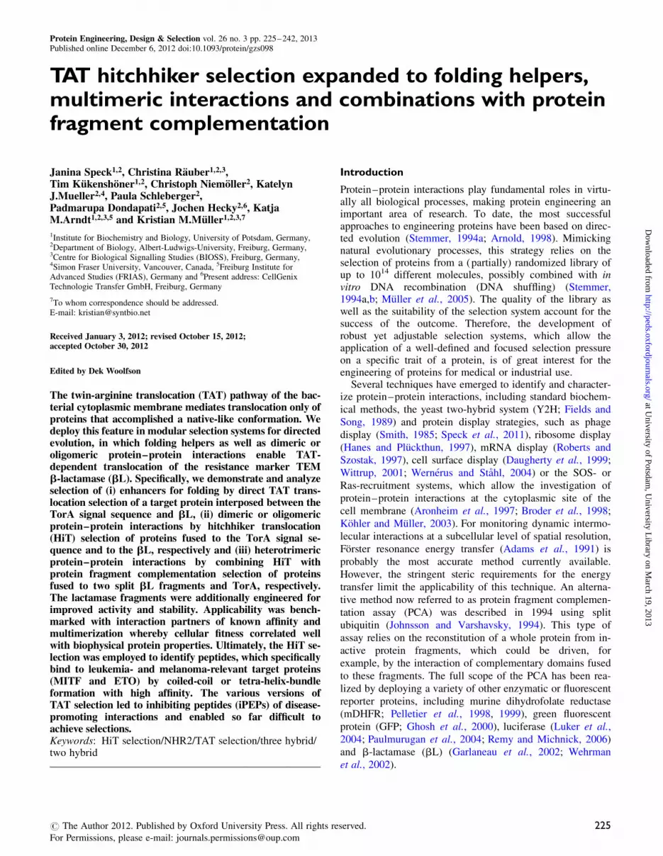
Fig. 2. Characterization of iM6, an MITFcc variant selected by HiT. (A) Ampicillin resistance test of single clones expressing different plasmid combinations.The first of the two acronyms below the x-axis gives the peptide fused to TorA and the second acronym refers to the peptide fused to bL. Error bars indicatestandard deviation of colony numbers of three plates each. (B) Thermal unfolding transitions of chemically synthesized peptides (labels shown in inset, datafrom CD spectroscopy, 60 mM peptide, 0.1 cm cuvette). The solid lines represent fits of experimental data assuming a two-state transition. The dotted line isthe mathematical average of the fitted homodimeric peptide measurement and approximates a curve, which would be seen if peptides are mixed but would notinteract. The mixture of the selected variant iM6 with MITFcc (grey diamond) shows an increased apparent Tm value for the interaction, which is shifted by13.48C compared to MITFcc homodimer. Both tests demonstrate that the interaction of iM6 with MITFcc is significantly stronger than that of the MITFcchomodimer.
Fig. 1. The HiT selection system. (A) Genetic organization of the two plasmids with the expression cassettes downstream of the lacI gene, a terminator (t) andthe lac promotor/operator region (lacp/o). (B) Schematic representation of the principle underlying the HiT selection system, illustrating that the translocation ofbL to the periplasm depends on the interaction of the peptides fused to TorA and bL. Since bL only confers resistance to lactam antibiotics if located in theperiplasm, cell survival correlates with the interaction of the peptides. (C) Ampicillin resistance test of single clones expressing different plasmidcombinations. The first of the two acronyms below the x-axis gives the peptide fused to TorA and the second acronym refers to the peptide fused to bL (wz:winzip). Only four of eight negative controls are shown; the respective negative controls with winzipB1 were all negative. The number of colonies relative tothe control plate is given on the y-axis; error bars indicate standard deviation of colony numbers of 3–4 plates each.
J.Speck et al.
230
at University of Potsdam
, University L
ibrary on March 19, 2013
http://peds.oxfordjournals.org/D
ownloaded from

Due to the good selection performance achieved with30 mg/ml ampicillin at 32.58C, these conditions were subse-quently chosen for the selection of MITFcc-binding peptidesfrom a ‘real’ library.
Our peptide library was created by partially randomizingseven amino acids of the wild-type MITFcc peptide. Six of
the seven positions are located in the hydrophobic interface
of the homodimeric MITFcc coiled-coil (positions a and d
in Table III; Supplementary Fig. SIII). This library was fused
genetically to bL and E.coli cells co-transformed with the
library vectors and the vector encoding for TorA-MITFcc
were selected once at 32.58C on plates containing either 30,
50, 80, 160 or 240 mg/ml ampicillin. After overnight incuba-
tion, several colonies formed on all plates, and all clones
grown at the same ampicillin concentration were pooled. The
sequencing chromatograms of all plasmid mixtures showed
overlaying signals at the randomized positions (not shown).
For the plasmid pool of clones grown at 240 mg/ml ampicil-
lin, all randomized positions showed a clear preferences
for one amino acid. This consensus sequence is given in
Table III. Without performing additional selection rounds we
analyzed single clones picked from the 240 mg/ml ampicillin
plate. As an example, Fig. 2A shows an ampicillin resistance
test of one of the selected clones (named iM6) in comparison
with the wild-type MITFcc homodimer. For this test, the
encoding sequence of the selected peptide was re-cloned in
both the TorA and the bL-containing vectors.This semi-quantitative assay showed that the heterodimer
of the selected peptide (iM6) and MITFcc is much more
stable than the MITFcc homodimer. Furthermore, a compari-
son with Fig. 1C indicated that the strength of the
iM6:MITFcc interaction ranges between the strength of the
winzipA2-winzipB1 heterodimer (Tm ¼ 63.28C) and that of
the winzipA2 homodimer (Tm ¼ 54.28C) (Arndt et al.,
2002). In accordance with this observation, CD measure-
ments monitoring the structural loss caused by thermal de-
naturation of the respective synthetic peptides revealed a Tm
of 58.58C for an equimolar mixture of the selected peptide
with MITFcc, significantly surpassing the Tm of 45.28C for
the MITFcc homodimer (Fig. 2B). The iM6 homodimer had
a Tm of 57.48C. The details of the library design, course of
further selections and more detailed biophysical characteriza-
tion of the best selected MITFcc-binding peptides will be
described elsewhere (Kukenshoner T. et al., in preparation).
In summary, the described experiments demonstrated thatbL is indeed a reliable and convenient reporter for periplas-mic localization and thus for HiT, and that the achievedampicillin resistance levels (hence, the in vivo performance)correlated very well with the Tm values of the correspondingpeptide interactions in vitro. Furthermore, the example ofiM6 showed that the HiT system can easily be used to iden-tify peptides or proteins from a library of closely relatedpolypeptides that bind to a specific target protein withimproved affinity.
Hitchhiker export of bL as selective tool for oligomericprotein–protein interactionsThe established HiT selection system was further used toinvestigate interactions of a homo-oligomeric helix bundle,which has been described to be formed by four NHR2domains of the ETO protein (Liu et al., 2006). In our hands,when performing size-exclusion chormatography of fusionswith a monomeric variant of GFP even higher oligomers (i.e.octamers) were detected (not shown).
Similar to the experiments with the MITF homodimer, theNHR2 wild-type peptide was fused genetically to both theTorA signal peptide and bL. Co-transformation of the twovectors rendered E.coli cells resistant to low ampicillin con-centrations (Fig. 3A). This indicated that folded structures(presumably tetramers or higher oligomers) containing atleast one TorA-NHR2 and one NHR2-bL fusion wereformed and translocated successfully to the periplasm.Consequently, we tested whether the HiT selection can iden-tify peptides that bind to NHR2 with improved affinity,replacing at least one strand of wild-type NHR2 in the helixbundle. To this end, a randomized NHR2 library (describedin Supplementary data) was cloned in fusion to the bL geneand co-transformed into E.coli with the vector encoding forTorA-wild-type NHR2. The same library was additionallycloned into a phagemid vector and selected forNHR2-binding peptides by phage panning (details of thephage panning procedure are given in Supplementary data).Although several attempts have been made, the success ofthe phage selection was hampered by genetic instability (ac-cumulation of non-sense mutations and frame shifts) and atendency to enrich peptides with low binding specificity(data not shown). Hence, the phage display pre-selectedlibrary was also cloned into the vector for HiT selection.
Table III. Amino acid sequences of wild-type and selected MITFcc variants
1 2 3Heptad position g a b c d e f g a b c d e f g a b c d e f gMITFcc A S V D Y I R K L Q R E Q Q R A K E L E N Rpool A I V D Y I R K I Q R E Q Q R V K E L E N RiM6 A I V D Y I R K I Q R E E Q R V K E L E N R
4 5 6 7Heptad position a b c d e f g a b c d e f g a b c d e f g a b c dMITFcc Q K K L E H A N R H L L L R I Q E L E M Q A R A Hpool L K K L E H A N R H L L Q R A Q E L E Q Q L R A HiM6 L K K L E H A N R H L L Q R A Q E L E Q Q I R A H
In the wild-type MITFcc, the underlined positions correspond to the partially randomized residues of the library; in the iM6 sequence, the highlightedpositions mark the two residues where iM6 differs from the consensus sequence of the pool.
TAT hitchhiker selection technologies
231
at University of Potsdam
, University L
ibrary on March 19, 2013
http://peds.oxfordjournals.org/D
ownloaded from

The native library, as well as the phage display pre-selected library, was subjected to three successive rounds ofHiT selection with increasing ampicillin concentrations (50,100 and 200 mg/ml). After the third round, single cloneswere sequenced (Table IV). The clones from the HiT selec-tion of the native library showed five different sequences,four of them were similar to each other and the fifth se-quence was found six times. In the case of the pre-selectedlibrary, seven of eight clones were sequence identical andthe remaining clone showed the same sequence plus two add-itional mutations. As indicated in Table IV, three clones(named NHR2_HiT2, -5 and -6) were chosen for further in-vestigation: the two clones which were found several timesand one ‘representative’ of the four clones with similarsequences.
To rule out that the increased ampicillin resistance wascaused by spontaneous mutations in the vector backboneor host genome, the peptide-encoding sequences ofNHR2_HiT2, -5 and -6 were re-cloned into the originalvector. The improved ampicillin resistance of co-transformedbacteria was confirmed in all cases (Fig. 3A). In addition, theampicillin resistance of the HiT-selected NHR2 mutants wascompared with that of wild-type NHR2 and the best cloneidentified by phage display (named NHR2_PhD1). All threeHiT-selected clones showed significantly increased resistanceto ampicillin in comparison with the wild-type NHR2 inter-action. In contrast, bacteria co-expressing TorA-NHR2 andNHR2_PhD1-bL did not grow on 25 mg/ml ampicillin,which was the lowest concentration tested. To verify thatthe translocation of bL depended on interaction with theTorA-NHR2 construct and was mediated by the TATpathway, co-transformants with a construct that encoded fora mutated TorA peptide (twin arginines replaced by lysins)served as negative controls. These clones did not grow in thepresence of ampicillin (not shown).
As we wondered how HiT-selected peptides would func-tion in other selection systems, we confirmed NHR2-specific
binding of NHR2_HiT2, -5 and -6 using phage display. Theencoding genes were cloned into the phagemid vector andbinding to a GST fusion of the wild-type NHR2 helix wasinvestigated by phage enzyme-linked immunosorbent assay(ELISA; Fig. 3B). All selected clones, includingNHR2_PhD1, showed increased binding to NHR2 in com-parison with the wild-type interaction. Remarkably,NHR2_PhD1 showed a relatively high unspecific interactionwith GST and BSA, while NHR2_HiT2, -5 and -6 boundvery specifically to the NHR2-GST fusion. Since weemployed a phagemid system for binding analysis, variationsin phage display levels can influence the ELISA signals andneeded to be taken into account. Therefore, we assessed therelative display levels of the flag-tagged peptides with ananti-flag antibody (Fig. 3B) and used the flag-tag detectionsignal as reference for normalization of ELISA data(Fig. 3C). These data indicated superior binding of the cloneNHR2_HiT6, which was derived from HiT selection of thephage display pre-selected library, over the two clones onlyselected by HiT, and, importantly, also over NHR2_PhD1.The latter clone succeeded in the phage display selectionprobably by increased display levels rather than improvedbinding. Overall, the identification of three peptides bindingmore firmly to NHR2 than the wild-type counterpart con-firmed that the HiT system is not only applicable for dimericinteractions but also for the targeting of oligomeric interac-tions. As a side note, an NHR2_HiT5 fusion with GFP wasfound to form the same oligomeric state (i.e. octameric) asNHR2 wild type in size exclusion chromatography.
Selection of folding helper molecules employing the TATfolding quality controlThe intrinsic folding quality control of the TAT pathwayhinders translocation of poorly folded polypeptides. Hence,this control mechanism should be applicable as a selectiontool to improve a polypeptide’s folding efficiency by directedevolution as well as a screen or selection tool for conditions
Fig. 3. Characterization of NHR2-binding peptides selected by HiT and/or phage display. Note that the NHR2 helices form a tetramer in the crystal structure(Liu et al., 2006). (A) Ampicillin resistance test of single clones expressing different plasmid combinations encoding for wild-type NHR2 fused to TorA anddifferent NHR2 variants fused to bL as indicated below the x-axis. Error bars indicate standard deviation of colony numbers of 3–4 plates each. (B) Analysisof affinity and specificity of phage displayed peptides to NHR2 by phage ELISA. Wells were coated with NHR2-GST fusion protein, anti-flag antibody orwild-type GST and blocked with BSA or contained only BSA. Helper phages without display served as negative control (neg ctrl). Error bars indicate standarddeviations of triplicate measurements. To take the differences in display level into account, the NHR2 binding signal was divided by the anti-flag signal, whichis proportional to the peptide display level. The result of this normalization is given in (C).
J.Speck et al.
232
at University of Potsdam
, University L
ibrary on March 19, 2013
http://peds.oxfordjournals.org/D
ownloaded from

under which the folding is improved without modifying theprotein itself. Besides variations of culture conditions,folding improvement without protein modification is bestachieved by co-expressing molecules that support the foldingprocess.
To ascertain that the folding quality control of the TATpathway can be employed to screen or select for ‘foldinghelper molecules’, we conducted the following experiments.Initially, we investigated the ability of the TAT machinery todiscriminate between polypeptides by their differences infolding, stability and/or solubility. To this end, vectors en-coding for fusion proteins of TorA signal peptide, a polypep-tide and bL (TorA-peptide-bL) were constructed (Fig. 4C)and the resulting TAT-translocation efficiency was investi-gated by monitoring the growth of transformed E.coli cellsin the presence of ampicillin. As shown in Fig. 4A, the con-structs containing peptide hDcc3R2 or hDcc3FL (which con-sists of hDcc3R2 and 21 additional residues) did not conferresistance to ampicillin under the tested conditions, whileclones expressing the AF10cc, winzipA2 or winzipB1 con-taining constructs survived. This correlated very well withour observation that the hDcc3R2 peptide is poorly solublein aqueous solution (100 mM KF, 10 or 20 mM potassiumphosphate, pH 7.0) and that its CD spectrum shows relativelya low a-helical structure content even at 288C and no ten-dency to form a coiled-coil structure (Fig. 4B and Table V).In contrast, AF10cc is much more soluble in aqueous solu-tion and yields typical a-helical CD spectra at 58C or even208C. For comparison, the previously selected peptideswinzipA2 and winzipB1 yield at 150 mM and 58C a helicalcontent of 100 or 85%, respectively (Arndt et al., 2002)
To further illuminate the properties of the selectionsystem, we tested if the substitution of four solvent-exposedhydrophobic residues (2 � leucine, isoleucine and valine) byalanine in the hDcc3R2 peptide enhances the translocation
efficiency of the TorA-peptide-bL fusion protein and therebyimproves ampicillin resistance of the cell. Indeed, cellsexpressing this modified construct (hDcc3R2-bfmut inFig. 4A) showed even higher ampicillin resistance thanclones expressing the AF10cc, winzipA2 or winzipB1constructs.
The hDcc3R2 peptide forms a coiled coil with AF10cc(Rauber C. et al. in preparation). Thus, the presence ofAF10cc is expected to improve folding and solubility ofhDcc3R2 as well as the overall structure of aTorA-hDcc3R2-bL fusion protein and its resistance againstdegradation (see scheme in Fig. 4D). Consequently, AF10ccco-expression was expected to improve translocation efficacyof the TorA-hDcc3R2-bL construct and—possibly to aminor extent—of the longer TorA-hDcc3FL-bL construct.Interestingly, a mixture of hDcc3R2 and AF10cc peptideswas nicely soluble in aqueous solution—while hDcc3R2alone was not—and the CD spectrum of the peptide mixtureshowed a high content of a-helical structure and coiled-coilformation (Fig. 4B and Table V). By testing the ampicillinresistance of clones expressing the TorA-hDcc3R2-bL orTorA-hDcc3FL-bL fusion constructs either alone or in com-bination with AF10cc, we additionally detected that the pres-ence of AF10cc significantly increased the translocation byTAT and thus the resistance to ampicillin (Fig. 4E). This ob-servation confirmed our assumption that the folding controlmechanism of the TAT pathway can also be exploited toscreen for ‘folding helper molecules’ that support the foldingprocess of a target protein without modifying the protein’samino acid sequence.
Design of a trimeric protein–protein interaction assaycomprising optimization of split bL fragmentsFurther, we intended to develop a trimeric protein–proteininteraction assay exploiting HiT selection in combination
Table IV. Amino acid sequences of wild-type NHR2a and selected NHR2 variants
NHR2a G T R Q E E M I D H R L T D R E W A E E W K H L D H L L N SNHR2_PhD 1 D G V PNHR2_HiT 1 VNHR2_HiT 2 QNHR2_HiT 3 Q SNHR2_HiT 4 QNHR2_HiT 5 (6�)NHR2_HiT 6 (7�) DNHR2_HiT 7 P D
NHR2a I M D M V E K T R R S L T V L R R S Q E A D R E E L N Y WNHR2_PhD 1 L V A TNHR2_HiT 1 I V I V T LNHR2_HiT 2 L A T LNHR2_HiT 3 L I TNHR2_HiT 4 LNHR2_HiT 5 (6�) V I P TNHR2_HiT 6 (7�) V P I L RNHR2_HiT 7 L V P I P R
aThe first 59 of 67 residues are given. No mutations were found in the remaining eight amino acids (IRRYSDAE). The library used for the selection procedurewas based on the mixture of an error-prone library and a designed library as described in Supplementary data. The positions that were partially randomized inthe designed library are underlined. Sequences were grouped according to their origin: NHR2_PhD1 was derived from phage display selection, NHR2_HiT1–5were gained by HiT selection and NHR2_HiT6 and -7 were found in clones derived from HiT selection of the phage display pre-selected library. NHR2_PhD1and NHR2_HiT2, -5 and -6 (written in italics) were chosen for further characterization (Fig. 3).
TAT hitchhiker selection technologies
233
at University of Potsdam
, University L
ibrary on March 19, 2013
http://peds.oxfordjournals.org/D
ownloaded from

with protein fragment complementation. For this purpose, wefused one peptide each of a heterotrimeric coiled-coil motif(TriA, TriB and TriC; Nautiyal et al., 1995) to the TorAsignal peptide, the aminoterminal (a-) fragment and the car-boxyterminal (v-) fragment of TEM-1 bL (residues 26–199
and 200–290, respectively; numbering according to Ambleret al., 1991; Fig. 5A). Trimerization of the peptides TriA,TriB and TriC to a stable helix bundle was expected todrive protein fragment complementation and subsequentTAT-dependent translocation of the properly folded and
Fig. 4. The folding control mechanism of the TAT pathway as selective tool for folding helper molecules. (A) Influence of peptides (names shown in theinset) on the growth of transformed E.coli expressing the respective TorA-peptide-bL fusion constructs by monitoring the optical density at 600 nm (OD600) inthe presence of ampicillin in liquid culture. (B) Characterization of the helicity of chemically synthesized peptides by CD spectroscopy wavelength scans with20 mM peptide at 28, 5 and 208C. The calculated a-helical contents as well as the ratios of ellipticity at 222 and 208 nm are given in Table V. (C) Geneticorganization of the two co-expressed plasmids (see legend of Fig. 1) for the ‘folding helper’ assay. (D) Schematic representation of the principle underlyingthe ‘folding helper’ effect illustrating that the translocation of bL, and thus the resistance of the host cell toward b-lactam antibiotics, depends on the foldingof the peptide fused between TorA and bL and can be improved by co-expressing a ‘folding helper’ (E) Ampicillin resistance test of single clones expressingthe fusion construct TorA-hDcc3R2-bL or TorA-hDcc3FL-bL in the absence or presence of AF10cc, which forms a coiled coil with hDcc3R2 (Rauber C.et al. in preparation) and thereby improves its folding into a-helical structure. Error bars indicate standard deviation of colony numbers of 3–4 plates each.
Table V. CD spectroscopy of synthetic peptidesa
MRE � 1023; a-helical content (%)b at [Q]222/[Q]208c at
208C 58C 288C 208C 58C 288C
hDcc3R2 212.0; 30 216.3; 41 220.5; 51 0.81 0.92 1.00AF10cc 228.1; 75 233.9; 91 235.9; 96 1.02 1.09 1.12hDcc3R2:AF10cc 229.5; 79 231.1; 83 232.2; 86 1.05 1.08 1.10
aTotal peptide concentration was 20 mM.bThe helical content was calculated from the observed mean residue molar ellipticity (MRE) as percentage of the predicted molar ellipticity for an a-helix of nresidues length according to: [Q]222 ¼ 39 500 deg cm2 dmol21� (1–2.57/n) (Chen et al., 1974).cAccording to Cooper and Woody (1990) a [Q]222/[Q]208 ratio � 1 indicates coiled-coil formation.
J.Speck et al.
234
at University of Potsdam
, University L
ibrary on March 19, 2013
http://peds.oxfordjournals.org/D
ownloaded from

catalytically active bL into the periplasm (Fig. 5B).Nonetheless, co-expressing the three respective plasmids inE.coli did not confer resistance to ampicillin (not shown).
Assuming that trimerization and stability of the helixbundle should meet the requirements for TAT-dependenttranslocation, we focused on optimizing stability and activityof the split bL fragments to improve the interaction assay.Therefore, we slightly shifted the split position and intro-duced nine presumably stabilizing mutations into the two bLfragments (Fig. 6A and B): V31A, R120G, H153R, M182T,L201P, I208M, E212K, A224V and R275L. The applicabilityof the re-designed split bL fragments was first tested in adimeric PCA. In this PCA, the two enzyme fragments (eachfused to either winzipA2 or winzipB1) were independentlytranslocated into the periplasm by the general secretorypathway (SEC). After translocation, reconstitution of thewhole enzyme was driven by the interaction of the winzippeptides. Again, ampicillin resistance (Fig. 6C and D) corre-lated with published in vitro affinities (Arndt et al., 2002) for
the tested peptide combinations. Cells expressing only onebL fragment, both fragments but one without a peptide (notshown) or one of the two fragments fused to AF10cc insteadof a winzip peptide, served as negative controls and did noteven grow on the lowest ampicillin concentration tested. Tofurther evaluate the effect of our split bL re-design, we puri-fied the bL fragments fused to winzipA2 and winzipB1 andinvestigated activity and stability of the engineered enzymefragments by determining the catalytic activity (kcat) andthe midpoints of transition for thermal (Tm) as well asguanidine-induced (D1
2) unfolding (Fig. 6E and F). From the
latter experiment we derived a DGH2O value. As summarizedin Table VI, the activity, as well as chemical and thermal sta-bility of the reassembled enzyme, was at or above the levelof full-length wild-type bL, and most remarkably, the activ-ity was far superior to earlier published values for other splitbL variants (kcat of 0.1–20 s21; de las Heras et al., 2008).
Finally, the improved split bL fragments were successfullyutilized in the trimeric protein–protein interaction assay
Fig. 5. The trimeric HiT selection system. (A) Genetic organization of the three components (see also legend of Fig. 1) required for the trimeric protein–protein interaction assay. (B) Schematic representation of the principle underlying the trimeric selection system combining HiT and PCA. The schemeillustrates that protein fragment complementation and subsequent translocation of functional bL into the periplasm depend on the interaction of all threeinteraction domains (e.g. helical peptides). For clarity, the split bL fragments are shown in a native-like conformation although their structure beforereassembly is unknown. (C) Ampicillin resistance test of single clones expressing different plasmid combinations. The first of the three letters below the x-axisgives the peptide fused to TorA, the second letter refers to the peptide fused to the a-fragment of split bL, and the third letter refers to the peptide fused to thev-fragment of split bL (A: TriA, B: TriB, C: TriC, N: NHR2). Combinations including an NHR2 peptide served as negative controls.
TAT hitchhiker selection technologies
235
at University of Potsdam
, University L
ibrary on March 19, 2013
http://peds.oxfordjournals.org/D
ownloaded from

described above (Fig. 5A and B). As shown in Fig. 5C, cellsexpressing the peptides TriA, TriB, TriC fused to TorA, a-and v- bL fragments, respectively, were resistant to
ampicillin, regardless of orientation. Replacing one of thepeptides with the peptide NHR2 served as negative control,since it was not expected that this peptide would firmly
Fig. 6. Optimized split bL fragments. (A and B) Ribbon structure of full-length bL with the parts corresponding to the a- and v-fragments of split bL shownin marine and light blue, respectively (image generated from pdb 1BTL (Jelsch et al., 1993) with PyMol). (A) Illustration of the shifted split position. ResiduesL199 and L198, which are now part of the a-fragment and mainly contact residues in the N-terminal part of the full-length enzyme are highlighted as pinkspheres. (B) Distribution of all mutations introduced to stabilize the split bL fragments. Mutated residues are shown as spheres and colored according to atomtypes, with gray, blue, red and yellow for carbon, nitrogen, oxygen and sulfur atoms, respectively. The last residue of the a-fragment is highlighted in pink. (C)The functionality of the newly designed split bL fragments was tested in a dimeric PCA as shown schematically. Both fragments were translocatedindependently to the periplasm by the SEC pathway. Interaction of the fused peptides (shown in orange and yellow) was expected to drive reconstitution offunctional bL and thus to result in resistance to ampicillin. (D) Ampicillin resistance test of clones co-expressing the split bL fragments fused to differentpeptide combinations as indicated below the x-axis. The first acronym refers to the peptide fused to the a-fragment and the second acronym refers to thepeptide fused to the v-fragment (wz: winzip). (E and F) Comparison of the stability of full-length wild-type bL and optimized reassembled split bL bythermal unfolding measuring ellipticity at 222 nm (E) as well as chemically induced unfolding by monitoring the intrinsic tryptophan fluorescence (F). Linesin panel E and F represent fits of experimental data according to Pace (1986) assuming a two-state transition.
J.Speck et al.
236
at University of Potsdam
, University L
ibrary on March 19, 2013
http://peds.oxfordjournals.org/D
ownloaded from

interact with any of the TriA-C peptides. To our knowledge,this is the first published trimeric interaction assay in whichall three parts are absolutely required to give a positivesignal.
Discussion
The HiT as selective toolWe have engineered several screening and selection systemsthat build upon the TAT pathway and its intrinsic foldingquality control mechanism. We chose the TorA signal se-quence to target the protein (or protein complex) of interestto the translocon, and bL as reporter for the periplasmic lo-calization. TorA was chosen since this leader peptide hasbeen shown to be highly specific for the TAT pathway(DeLisa et al., 2003; Tullman-Ercek et al., 2007). The use ofbL as a reporter protein that confers a phenotype (ampicillinresistance) only after export to the periplasmic space was inour opinion much more attractive than employing GFP, MBPor DsbA as described in other publications (DeLisa et al.,2002; Strauch and Georgiou, 2007). Most importantly, theuse of bL provides a very economic and straight forwardselection-by-survival principle ideal for high-throughputapplications without the need for specific instrumentationsuch as a cell sorter or fluorescent microscope. Only MBPallows for similar selection by survival when using anMBP-deficient E.coli strain and minimal medium withmaltose as the sole carbon source. The growth on minimalmedium, however, is very slow compared with LB or 2 �YT media. In addition, the possibility to fuse proteins toMBP without impairing its physiological function seemsto be more limited than for bL (Fisher et al., 2008).Furthermore, due to its frequent use as a model enzyme forprotein engineering, TEM bL is a very well-known protein,with a multitude of available information on its function,structure and stability as well as on stabilizing mutations(Osuna et al., 2002; Hecky and Muller, 2005; Kather et al.,2008). It might be interesting to test stabilized and fastfolding bL variants to further improve the HiT selectionsystem by enhancing the focus of the selection pressure onthe protein fused to bL. In addition, bL can also be split intotwo fragments that can reassemble to a functional enzyme(Garlaneau et al., 2002; Wehrman et al., 2002), which pro-vided the possibility to combine the principles of HiT andPCA.
Yet, there are also some minor drawbacks when using bLas reporter protein. Many b-lactam antibiotics including
ampicillin are relatively unstable and heat sensitive.Consequently, not only the release of enzymes from deadcells but also prolonged incubation times result in degradationof ampicillin and thus a decrease of selection pressure overtime, which can be problematic when working with liquidcultures. Therefore, we used liquid cultures only for referenceexperiments, while library selections were always performedon freshly prepared agar plates. It should also be mentionedthat more stable analogs (e.g. carbenicillin) exist but are rela-tively expensive. Although the usefulness of bL has beenquestioned by others (Strauch and Georgiou, 2007), in ourhands, bL is a very effective and convenient indicator forTAT-mediated translocation and gave good signal-to-noiseratios in all described selection systems.
In the first step, we established a dimeric HiT selectionsystem comparable with the one described by Waraho andDeLisa (2009). As demonstrated with the winzipA2 andwinzipB1 peptides, this system can be used to investigateprotein–protein interactions semi-quantitatively. The systemwas further tested exploring the homodimeric interaction ofthe MITF coiled-coil domain. The relative low resistanceto ampicillin, compared with clones bearing the winzipconstructs, is mainly explained by the lower Tm of theMITFcc homodimer and by competition of all three possibledimerizations (TorA-MITFcc:MITFcc-bL; TorA-MITFcc:TorA-MITFcc and MITFcc-bL:MITFcc-bL), of which onlythe combination TorA-MITFcc:MITFcc-bL leads to trans-location of bL. As we have shown, reducing incubation tem-perature is an effective alternative to reducing antibioticconcentration to recover survival of E.coli cells co-expressingthe MITFcc constructs. We further tested the HiT system intest selections, varying the temperature and the ampicillinconcentration. With all tested settings, cells co-expressing therespective MITFcc constructs outperformed all controls andthe speed of MITFcc-homodimer enrichment correlated withthe selection pressure. Besides the parameters temperatureand ampicillin concentration, which we analyzed, many otherparameters could be varied to adjust the required selectionpressure. First of all, the amount of inducer (e.g. IPTG) canbe modified. The translocation capacity and efficiency can beimproved by overexpressing the genes tatABC (Barrett et al.,2003) encoding for the translocon components or the gene en-coding the chaperone TorD, which specifically enhances thetranslocation of proteins with a TorA leader peptide (Li et al.,2006).
Furthermore, we designed an MITFcc-derived peptidelibrary by varying residues mainly in the core of the MITFcchomodimer, and we successfully identified peptides thatinteract more strongly with the wild-type peptide after apply-ing just a single round of HiT selection at 32.58C. The dom-inant pool sequence of the clones selected on 240 mg/mlampicillin differed from the wild-type MITFcc in six of theseven partly randomized positions, and the iM6 variant in allseven positions. Interestingly, sequencing the library poolrevealed a clear signal at one of the randomized positions(a6), which resulted in an exchange of isoleucine by alanine.All other a positions were occupied by isoleucine, leucineand valine. Residues at the a and d positions are not only im-portant for the formation and stability of the coiled-coil corebut have also a major impact on the oligomerization states ofcoiled coils (Harbury et al., 1993; Zhu et al., 1993). Thepresence of isoleucine at a positions and leucine at d
Table VI. Activity and stability of reassembled split bL and full-length bL
TEM-116
kcat
(s21)DGH2O
(kJ mol21)aD1
2
(M)aTm
(8C)
Full-length bLTEM-116
628 13.6 0.92 50.8
Split Bla 665 44.1 1.34 56.9
aThermodynamic parameters refer to GdmCl-induced unfolding transitionsand were determined using the fluorescence intensity data shown in Fig. 6F.Data were analyzed according to the linear extrapolation method (Pace,1986) assuming a monophasic unfolding transition.
TAT hitchhiker selection technologies
237
at University of Potsdam
, University L
ibrary on March 19, 2013
http://peds.oxfordjournals.org/D
ownloaded from

positions is known to favor the formation of dimeric coiledcoils. However, a more detailed characterization and com-parison of additional MITFcc-derived variants are needed toevaluate the importance of the three isoleucine at the a1, a2,and a7 positions in iM6 for the interaction with wild-typeMITFcc.
Applicability of the HiT selection system for homo-oligomersWe also tested the applicability of the HiT selection systemfor higher-order homo-oligomers. As an example, we aimedto identify peptides that can replace at least one of thea-helices in a homo-oligomeric NHR2 helix bundle and thatbind to NHR2 with higher affinity than the wild-type coun-terpart. In the described setting of HiT selection, a dimericinteraction of one peptide each fused to TorA and to bLwould theoretically have been sufficient to give a positivereadout. However, in the HiT selection procedure, the puta-tive NHR2-interaction partners of the library fused to bL hadto compete with homo-oligomerization of the TorA-NHR2fusion. Since NHR2 has been described to form very stablehomo-tetramers with a Tm of 858C (Wichmann et al., 2010),it was rather unlikely to identify NHR2-derived peptides thatinteract with wild-type NHR2 in a dimeric way firmlyenough to compete with tetramers of TorA-NHR2. To testthe oligomerization state of NHR2 and its selected derivativeNHR2_HiT6, we purified GFP fusion constructs of thesepeptides and analyzed the homomeric complexes bysize-exclusion chromatography at �208C (data not shown).Surprisingly, both, NHR2-GFP and NHR2_HiT6-GFP, werefound to form octameric complexes, making it even moreunlikely that TorA-NHR2 and NHR2_HiT6-bL interacted invivo in a dimeric way. The discrepancy of our size exclusiondata and the results from NHR2 crystallization as well asultracentrifugation experiments (Liu et al., 2006) might indi-cate that the octamers found in SEC resulted from metastableassembly of two tetra-helix bundles. Nevertheless, an in vivoshift to dimeric interactions would only be expected, if thehigher oligomers exceed the size limit for TAT-mediatedtranslocation, which has been estimated to �160–180 kDa(Strauch and Georgiou, 2007). The calculated molecularweights of the TorA signal peptide, NHR2 and bL are �4.2,8.7 and 28.9 kDa, respectively. Thus, it is even possible thattranslocation competent octamers formed in vivo (e.g. acomplex of six TorA-NHR2 plus two NHR2_HiT6-bL has acalculated molecular weight of �153 kDa). Since the HiTselection with the NHR2 library was successful and selectedNHR2 library clones maintained the oligomeric state, we canconclude that multimerization and the potentially exposedhydrophobic surface of this case did not pose a problem tothe system and that for the analyzed cases selection did notwork against multimerization.
To identify NHR2-derivatives that bind with higher affin-ity to NHR2 than NHR2 itself, the selection pressure wasprogressively increased with ongoing selection rounds. Evenat an ampicillin concentration that completely prevented thegrowth of clones expressing the NHR2 wild-type constructs,several colonies formed. This indicated that HiT selectionwas indeed able to identify peptides that interact morestrongly with NHR2 than the wild-type peptide. Sequenceanalysis of single clones showed a significant accumulationof only a few sequences that were similar to one another. Itwas therefore rather unlikely that further selection without
additional sequence diversification (e.g. random mutagenesisor DNA shuffling), would lead to further improved variants.
In a parallel experiment, the same library was also sub-jected to phagemid display with very limited success. Thismight partially be explained by the oligomeric character ofthe homomeric NHR2-interaction and the low (typicallymonovalent) display levels achieved in a phagemid system.Although the average binding of the phage libraries to NHR2peptide was somewhat increased with on-going panningrounds (data not shown), the library remained highlydiverse—as revealed by sequencing—and additionally suf-fered from genetic instability. Furthermore, the specificity ofinteraction diminished continually, indicating that ‘sticky’peptides with the tendency to interact non-specifically withother polypeptides were enriched. This was further corrobo-rated when transferring the best clone identified by phagedisplay (NHR2_PhD1) into the HiT system. As would beexpected for any ‘sticky’ peptide this clone did not conferampicillin resistance, probably because it interacts not onlywith the target protein but also with several other cytosolicproteins, impeding TAT-mediated translocation. In ouropinion, the intrinsic selection against hydrophobic, unstruc-tured or sticky proteins is one of the most valuable featuresof the HiT selection system.
We also combined the two selection systems by subjectingthe phagemid-preselected library to additional selection byHiT. In the ampicillin resistance test the predominant clonefound after three HiT selection rounds (NHR2_HiT6) per-formed equally well as one of the clones selected only byHiT, but outperformed all identified clones in phage ELISA,in particular when taking the display level into account.Since we worked with a phagemid system, the number of dis-played proteins per phage particle could theoretically varybetween zero and all gene3 proteins (approximately five perphage particle) substituted by the peptide-gene3 fusionprotein. Even though the flag tag (Knappik and Pluckthun,1994) allowed for the evaluation of the relative displaylevels, the conducted normalization (division of signal byrelative display level) of the NHR2-specific interaction stillunderestimated the impact of the display level since the oli-gomeric nature of this interaction results in a disproportionaterelation between display level and signal intensity. However,a comparison of the ELISA signals clearly showed that thephage particles displaying HiT-selected peptides (especiallyNHR2_HiT6) bind stronger to wild-type NHR2 than NHR2itself despite the relatively low display levels.
Interestingly, independent of the selection system, cloneswith incorporated prolines were identified. This was surpris-ing since this amino acid results in a local disruption ofa-helical structure and is not expected to improve coiled-coilstructures. However, the accumulation of proline residues intwo different selection systems indicates that the resultingbend in the polypeptide chain might be advantageous for theformation of higher-order helix bundles. It is also possiblethat a local structural disintegration provided beneficialflexibility.
Although we investigated NHR2-binding only by ampicil-lin resistance test and phage ELISA, we were able to showthat the HiT system with only two components(TorA-‘polypeptide 1’ and ‘polypeptide 2’-bL) can also beused to investigate and target homomeric interactions ofhigher order. Furthermore, based on the described
J.Speck et al.
238
at University of Potsdam
, University L
ibrary on March 19, 2013
http://peds.oxfordjournals.org/D
ownloaded from

observations, we hypothesize that the combination of selec-tion systems has important advantages over increasing thenumber of selection rounds without changing the selectionsystem. Any selection system has its own bias, for example,the accumulation of ‘sticky’ clones may occur when usingphage display. However, clones arising from unwanted fea-tures of one selection system typically do not have a similaradvantage in another selection system, and thus can bepurged by shifting to or alternating with another selectionstrategy. Advantageous effects arising from the combinationof an in vitro selection method (e.g. phage or ribosomedisplay) with an in vivo selection step (e.g. PCA or Y2H)have also been observed by others (Visintin et al., 2002;Amstutz et al., 2006; Secco et al., 2009).
The folding control mechanism as a selective toolThe first efforts to employ the TAT-pathway for directed evo-lution focused mainly on improving folding and solubility ofthe target protein by selecting variants from a genetically di-versified library (DeLisa et al., 2002; Fisher et al., 2006;Ribnicky et al., 2007). We raised the question whether thesame selection mechanism would also be applicable to selectfor specific conditions under which folding, solubility and/orstability of a protein are improved without modifying theprotein’s amino acid sequence.
At first, we confirmed that translocation efficiency ofTorA-peptide-bL fusion constructs correlated with the pep-tides’ solubility and helical structure, which was determinedby CD spectroscopy of chemically synthesized peptides. Asexpected, the very poor solubility and low helical content ofthe hDcc3R2 (and hDcc3FL) peptide resulted in low trans-location efficiency, while the AF10cc-, winzipA2- andwinzipB1-containing constructs showed a more efficientTAT-mediated translocation according to the better solubilityand folding of these peptides. Additionally, winzipA2 formsstable homodimers, which probably not only increases itssolubility and content of a-helical structure but also the sta-bility against proteolytic degradation. However, one shouldbe aware that the readout of in vivo selection systems alwaysresults from the sum of several different factors, includingfor example expression levels and toxicity.
As an example, we demonstrated that the efficiency ofTAT-mediated translocation of TorA-hDcc3R2-bL andTorA-hDcc3FL-bL can be improved by co-expression of aninteracting peptide (i.e. AF10cc), which acts as a ‘foldinghelper’, instead of modifying the hDcc3R2 (or hDcc3FL)peptide itself.
We conclude that the selection or screening for efficientTAT-mediated translocation of a poorly soluble or slowlyfolding protein should be an optimal platform for the investiga-tion or selection of function and relative activity of(e. g. genetically modified) molecular chaperones. Furthermore,the efficiency of TAT-mediated translocation may also be a valu-able readout for evaluating and improving culture conditions forrecombinant expression whenever polypeptide yields gainedwith standard procedures are insufficient, for example due tolow solubility, toxicity, fast degradation and/or inclusion bodyformation. A similar strategy has been reported by Lee et al.(2009), who applied the selectivity of the TAT-pathway forscreening a chemical compound library for molecules that alle-viate the aggregation tendency of Ab42 amyloid.
In rare cases of homology between sequences precedingthe lactamase gene and genomic sequences coding for signalpeptides targeting the SEC pathway, recombination eventsbetween the plasmid and the genome can generate false-positive clones (see Supplementary data).
Optimization of split bL fragments and trimericprotein–protein interaction assayWe re-engineered split fragments of TEM bL for better sta-bility and activity. In 2002, the design of split bL for the in-vestigation of protein–protein interactions was independentlydescribed by Garlaneau et al. (2002) as well as Wehrmanet al. (2002). Both publications describe a favorable effect ofutilizing fragments with improved stability, which has beenachieved by either introducing the global suppressor mutationM182T or by the addition of a selected trimeric peptide atthe C-terminus of the a-fragment. The resulting fragmentswere successfully employed to investigate protein–proteininteractions in vivo or in cell lysates. However, de las Heraset al. (2008) described difficulties regarding activity andsignal-to-noise ratio using similar fragments in vitro. The en-zymatic activity of the reassembled enzyme was significantlybelow that of the wild-type full-length enzyme, even if con-taining the M182T mutation. To reduce spontaneous re-assembly and thus improve signal-to-noise ratios, they testedthe introduction of mutations in the fragments’ interface,which indeed improved the signal-to-noise ratio but furtherdiminished the enzymatic activity.
Based on structural modeling using X-ray structures offull-length bL, we decided to shift the described split pos-ition so that the amino acids L198 and L199 now made partof the a-fragment. Besides an H-bond between the backboneoxygen atom of L199 to the side chain of R204, these resi-dues contact many residues that are located moreN-terminally in the enzyme and thus make part of thea-fragment. Therefore, we speculate that the presence ofthese two hydrophobic residues at the N-terminus of thev-fragment might at least partly account for the shortcom-ings of the previously described fragments. However, usingfragments derived from the wild-type TEM-1 bL with a splitbetween position 199 and 200, we did not get positive read-outs in our newly designed trimeric protein–protein inter-action assay. Although other features of this assay might alsobe improvable, we focused on further optimizing the enzymefragments. To this end, we chose to design new fragmentsbased on the enzyme TEM-116 (which differs from TEM-1by two substitutions: V84I and A184V) and introduced ninemutations, which have been shown to stabilize full-lengthbL. It should be mentioned that much, if not most, ofthe published work on TEM-1 was actually based on theenzyme variant TEM-116, including other split bL studies(Garlaneau et al., 2002; Wehrman et al., 2002; de las Heraset al., 2008) as well as studies on full-length and truncatedbL (Hecky and Muller, 2005; Bershtein et al., 2008; Katheret al., 2008). Since most of the data relevant for our split bLdesign was based on TEM-116, and since the substitutionA184V has been reported to increase the Tm value of TEM-1bL by 1.48C (Brown et al., 2010), we chose to maintain I84and V184 (as in TEM-116) even though the catalytic effi-ciency of TEM-1 for aminopenicillins is about 2-fold higherthan that of TEM-116 (Chaibi et al., 1997). Further, weintroduced the substitutions V31A, R120G, H153R and
TAT hitchhiker selection technologies
239
at University of Potsdam
, University L
ibrary on March 19, 2013
http://peds.oxfordjournals.org/D
ownloaded from

M182T in the a-fragment as well as L201P, I208M, E212K,A224V and R275L in the v-fragment to stabilize both frag-ments, as will be discussed below.
To characterize the new fragments in more detail, we firstinvestigated the fragments’ functional reassembly in adimeric protein–protein interaction assay in vivo and found avery good signal-to-noise ratio. Furthermore, we purified theproteins and demonstrated that the activity and stability ofthe reassembled enzyme was significantly improved com-pared with published data on previously described bL frag-ments (de las Heras et al., 2008) and even slightly superiorto wild-type full-length bL. Finally, the improved enzymefragments allowed for successful engineering of a trimericprotein–protein interaction assay based on a combination ofHiT selection with PCA.
As mentioned before, there is still room to improve thedescribed trimer assay. Especially for the work with libraries,the design of a single vector encoding for all three compo-nents (with or without polycistronic organization) would beadvantageous, due to much better transformation efficiencies.Additionally, the use of only one marker antibiotic to avoidplasmid loss would alleviate the stress imposed on the cells,resulting in accelerated cell growth and reduced incubationtimes. For some applications it might also be beneficial tointroduce different promoters, so that the expression levels ofthe different components could be varied independently.
Nevertheless, we want to emphasize that our trimericscreening and selection system is engineered so that only theinteraction of all three parts can result in positive readouts.To our knowledge, this is in sharp contrast to other describedtrimeric interaction assays (SenGupta et al., 1996; Pelletieret al., 1998), in which a direct interaction of ‘protein 1’ and‘protein 3’ without the bridging ‘component 2’ may result infalse-positive signals.
Assumed contributions of individual mutations in the splitbL contextThe V31A substitution was present in functional variants ofTEM-116 bL either truncated N-terminally by five residues(ND5; Hecky and Muller, 2005) or circular permutated withnew termini in the second cross-over loop (Osuna et al.,2002). V31 is located in the N-terminal helix H1 (numberingof secondary structure elements according to Jelsch et al.(1993) with its hydrophobic side chain largely exposed to thesolvent. Substitution by alanine presumably reduces thehydrophobic surface area and improves helix propensity,which likely improved folding of helix H1 and native statestability of the whole protein. We also found the substitutionsR120G, H153R and M182T in the ND5 study (Hecky andMuller, 2005) as well as in two follow-up studies, whichimproved TEM-116 stability and activity by selection formutations compensating for more severe structural perturba-tions (Speck et al. 2012 and manuscript in preparation). Thesame mutations were also described to compensate for thedetrimental effects of high mutational load (Bershtein et al.,2008) and overall, M182T is probably the most frequentglobal suppressor mutation identified in in vitro evolutionstudies as well as in clinically isolated extened spectrumbeta-lactamases (ESBL) (see www.lahey.org/Studies; Huangand Palzkill, 1997). The positive effect of substituting R120,which is located at the N-terminal part of helix H4, is mostlyattributable to the removal of the bulky arginine side chain,
which unfavorably interacts with the macrodipole of helixH4 and additionally packs against the side chain of L91, pre-venting optimal solvation of the guanidino group. Accordingto Bershtein et al. (2008), the substitution H153R, whichconverts residue 153 to consensus (Ambler et al., 1991), hasan even stronger stabilizing effect than R120G, although thesubstitution of H153 results in removal of p-stacking withH158. According to the structure of a TEM-116 7-foldmutant that we have recently solved (manuscript in prepar-ation), this destabilizing effect is overcompensated by exten-sive contacts of R153 with residues 155–160. Furthermore,this as well as several structures of TEM bL variants(Minasov et al., 2002; Kather et al., 2008) show that theM182T substitution stabilizes helix H8 as an N-cap residueby forming a hydrogen bond to the amide nitrogen of A185and additionally recruits water molecules that hydrogen bondwith the side chain of E63 (Wang et al., 2002).
Four (L201P, I208M, A224V and R275L) of the five pre-sumably beneficial mutations that we introduced into thev-fragment of split bL were—besides other publications—identified in a directed evolution study based on Proside(protein stability increased by directed evolution) and weredemonstrated to increase the Tm value of wild-type TEM-116by 1.4, 1.0, 3.1 and 5.08C, respectively (Kather et al., 2008).The fifth mutation, E212K, was identified in the first selectionround for mutations compensating for detrimental effectsinduced by circular permutation of TEM-116 bL (Osunaet al., 2002). In the wild-type protein, L201 is located at thefirst position of a-helix H9 with its aliphatic side chain over60% solvent exposed. The first position of a-helices are espe-cially suited for proline residues (Richardson and Richardson,1988) and crystal structures of an L201P containing bLmutants show a well-accommodated side chain of P201(Kather et al., 2008; Marciano et al., 2008). Furthermore, theintroduction of proline residues in polypeptides decreases theentropy difference of folded and unfolded states. The substitu-tions I208M and A224V increase overall stability of full-length bL by improving local packing densities between helixH9 and the b-sheets S3-S5 and between helix H10 and helixH11 as well as the underlying b-sheet, respectively (Katheret al., 2008). In contrast to the other mutations, the E212Ksubstitution has rarely been found and its effect on stabilityand/or enzymatic activity has not been investigated yet.However, we introduced the mutation in our split bL becausethe structure of wild-type TEM-116 (Jelsch et al., 1993) sug-gests that this substitution improves electrostatic interactionswith D209. According to the comparison of the wild-typestructure (Jelsch et al., 1993) and the structure of a TEM bLvariant containing the substitution R275L (Kather et al.,2008), the stabilizing effect of this substitution is mainly at-tributable to the removal of the charged guanidino group ofR275, which—in the wild-type protein—is partially buriedand additionally unfavorably interacts with the macrodipoleof helix H11.
Conclusion
In the present work we demonstrated that the TAT pathwaycan be exploited in multiple ways to create selection systemsfor protein–protein interactions as well as improved proteinfolding and solubility. In comparison with other selectionsystems, the use of bL as reporter protein provides a very
J.Speck et al.
240
at University of Potsdam
, University L
ibrary on March 19, 2013
http://peds.oxfordjournals.org/D
ownloaded from

convenient selection procedure, with the potential to performone selection round each day and to automate the procedure.
Using the HiT selection system, we identified peptides,which bind with higher affinity to their target proteinsMITFcc and NHR2 than their wild-type counterparts.Further investigations will help to clarify the principlesunderlying these improved coiled-coil and oligomeric helix-bundle formations. In addition, the identified peptides mightalso prove valuable for the investigation of the wild-typeinteractions in more physiological settings.
Furthermore, we designed the first trimeric interactionassay that involved new split bL fragments with significantlyimproved activity and stability and does not permit falsepositives by dimeric interactions. These optimized split bLfragments are also very interesting for other PCA-basedapplications independent of HiT selection or the TATpathway.
Supplementary data
Supplementary data are available at PEDS online.
Acknowledgements
We thank Prof. Dr. Andreas Pluckthun for providing the pAK400 plasmid,Dr. Ingrid Haas for helpful discussions and critically reading the manuscriptand Christoph Deeg for technical assistance.
Funding
This study was supported by the Excellence Initiative of theGerman Federal and State Governments (EXC 294, BIOSS),the DFG (Priority Program SPP1170; Collaborative ResearchCentre (SFB) 850), and the Deutsche Jose CarrerasLeukamie-Stiftung (DJCLS R 07/10v; DJCLS R 06/12).K.J.M. was supported by the DAAD RISE Program and anundergraduate scholarship.
ReferencesAdams,S.R., Harootunian,A.T., Buechler,Y.J., Taylor,S.S. and Tsien,R.Y.
(1991) Nature, 349, 694–697.Ambler,R.P., Coulson,A.F., Frere,J.M., et al. (1991) Bioch. J., 276,
269–270.Amstutz,P., Koch,H., Binz,H.K., Deuber,S.A. and Pluckthun,A. (2006)
Protein Eng. Des. Select., 19, 219–229.Arndt,K.M., Pelletier,J.N., Muller,K.M., Pluckthun,A. and Alber,T. (2002)
Structure, 10, 1235–1248.Arnold,F.H. (1998) Acc. Chem. Res., 31, 125–131.Aronheim,A., Zandi,E., Hennemann,H., Elledge,S.J. and Karin,M. (1997)
Mol. Cell. Biol., 17, 3094–3102.Barrett,C.M.L., Ray,N., Thomas,J.D., Robinson,C. and Bolhuis,A. (2003)
Biochem. Biophys. Res. Commun., 304, 279–284.Berks,B.C., Sargent,F. and Palmer,T. (2000) Mol. Microbiol., 35, 260–274.Bershtein,S., Goldin,K. and Tawfik,D.S. (2008) J. Mol. Biol., 379,
1029–1044.Broder,Y.C., Katz,S. and Aronheim,A. (1998) Curr. Biol., 8, 1121–1124.Brown,N.G., Pennington,J.M., Huang,W., Ayvaz,T. and Palzkill,T. (2010)
J. Mol. Biol., 404, 832–846.Bruser,T., Yano,T., Brune,D.C. and Daldal,F. (2003) Eur. J. Biochem. FEBS,
270, 1211–1221.Chaibi,E.B., Peduzzi,J., Barthelemy,M. and Labia,R. (1997) J. Antimicrob.
Chemother., 39, 668–669.Chen,Y.H., Yang,J.T. and Chau,K.H. (1974) Biochemistry, 13, 3350–3359.Cooper,T.M. and Woody,R.W. (1990) Biopolymers, 30, 657–676.Daugherty,P.S., Olsen,M.J., Iverson,B.L. and Georgiou,G. (1999) Protein
Eng., 12, 613–621.
de las Heras,R., Fry,S.R., Li,J., Arel,E., Kachab,E.H., Hazell,S.L. andHuang,C.-Y. (2008) Biochem. Biophys. Res. Commun., 370, 164–168.
DeLisa,M.P., Samuelson,P., Palmer,T. and Georgiou,G. (2002) J. Biol.Chem., 277, 29825–29831.
DeLisa,M.P., Tullman,D. and Georgiou,G. (2003) Proc. Natl Acad. Sci.USA, 100, 6115–6120.
Fields,S. and Song,O. (1989) Nature, 340, 245–246.Fisher,A.C., Kim,W. and DeLisa,M.P. (2006) Protein Sci., 15, 449–458.Fisher,A.C., Kim,J.-Y., Perez-Rodriguez,R., Tullman-Ercek,D., Fish,W.R.,
Henderson,L.A. and DeLisa,M.P. (2008) Microb. Biotechnol., 1,403–415.
Garlaneau,A., Primeau,M., Trudeau,L.-E. and Michnick,S.W. (2002) Nat.Biotechnol., 20, 619–622.
Ghosh,I., Hamilton,A.D. and Regan,L. (2000) J. Am. Chem. Soc., 122,5658–5659.
Hagemann,U.B., Mason,J.M., Muller,K.M. and Arndt,K.M. (2008) J. Mol.Biol., 381, 73–88.
Hanes,J. and Pluckthun,A. (1997) Proc. Natl Acad. Sci. USA, 94,4937–4942.
Harbury,P.B., Zhang,T., Kim,P.S. and Alber,T. (1993) Science, 262,1401–1407.
Hecky,J. and Muller,K.M. (2005) Biochemistry, 44, 12640–12654.Huang,W. and Palzkill,T. (1997) Proc. Natl Acad. Sci. USA, 94, 8801–8806.Jelsch,C., Mourey,L., Masson,J.M. and Samama,J.P. (1993) Proteins, 16,
364–383.Johnsson,N. and Varshavsky,A. (1994) Proc. Natl Acad. Sci. USA, 91,
10340–10344.Kather,I., Jakob,R.P., Dobbek,H. and Schmid,F.X. (2008) J. Mol. Biol., 383,
238–251.Knappik,A. and Pluckthun,A. (1994) BioTechniques, 17, 754–761.Kohler,F. and Muller,K.M. (2003) Nucleic Acids Res., 31, e28.Krebber,A., Bornhauser,S., Burmester,J., Honegger,A., Willuda,J.,
Bosshard,H.R. and Pluckthun,A. (1997) J. Immunol. Methods, 201,35–55.
Lee,P., Tullman-Ercek,D. and Georgiou,G. (2006) Annu. Rev. Microbiol.,60, 373–395.
Lee,L.L., Ha,H., Chang,Y.-T. and DeLisa,M.P. (2009) Protein Sci., 18,277–286.
Levy,C., Khaled,M. and Fisher,D.E. (2006) Trends Mol. Med., 12, 406–414.Li,S.-Y., Chang,B.-Y. and Lin,S.-C. (2006) J Biotechnol., 122, 412–421.Liu,Y., Cheney,M.D., Gaudet,J.J., et al. (2006) Cancer Cell, 9, 249–260.Luker,K.E., Smith,M.C.P., Luker,G.D., Gammon,S.T., Piwnica-Worms,H.
and Piwnica-Worms,D. (2004) Proc. Natl Acad. Sci. USA, 101,12288–12293.
Marciano,D.C., Pennington,J.M., Wang,X., et al. (2008) J. Mol. Biol., 384,151–164.
Minasov,G., Wang,X. and Shoichet,B.K. (2002) J. Am. Chem. Soc., 124,5333–5340.
Muller,K.M., Stebel,S.C., Knall,S., Zipf,G., Bernauer,H.S. and Arndt,K.M.(2005) Nucleic Acids Res., 33, e117.
Nautiyal,S., Woolfson,D.N., King,D.S. and Alber,T. (1995) Biochemistry,34, 11645–11651.
Osuna,J., Perez-Blancas,A. and Soberon,X. (2002) Protein Eng. Des. Sel.,15, 463–470.
Pace,C.N. (1986) Methods Enzymol., 131, 266–280.Paulmurugan,R., Massoud,T.F., Huang,J. and Gambhir,S.S. (2004) Cancer
Res., 64, 2113–2119.Pelletier,J.N., Campbell-Valois,F.X. and Michnick,S.W. (1998) Proc. Natl
Acad. Sci. USA, 95, 12141–12146.Pelletier,J.N., Arndt,K.M., Pluckthun,A. and Michnick,S.W. (1999) Nat.
Biotechnol., 17, 683–690.Remy,I. and Michnick,S.W. (2006) Nat. Methods, 3, 977–979.Ribnicky,B., Van Blarcom,T. and Georgiou,G. (2007) J. Mol. Biol., 369,
631–639.Richardson,J.S. and Richardson,D.C. (1988) Science, 240, 1648–1652.Roberts,R.W. and Szostak,J.W. (1997) Proc. Natl Acad. Sci. USA, 94,
12297–12302.Rodrigue,A., Chanal,A., Beck,K., Muller,M. and Wu,L.F. (1999) J. Biol.
Chem., 274, 13223–13228.Secco,P., D’Agostini,E., Marzari,R., et al. (2009) Protein Eng. Des. Sel., 22,
149–158.SenGupta,D.J., Zhang,B., Kraemer,B., Pochart,P., Fields,S. and Wickens,M.
(1996) Proc. Natl Acad. Sci. USA, 93, 8496–8501.Smith,G.P. (1985) Science, 228, 1315–1317.Speck,J., Arndt,K.M. and Muller,K.M. (2011) Protein Eng. Des. Sel., 24,
473–484.
TAT hitchhiker selection technologies
241
at University of Potsdam
, University L
ibrary on March 19, 2013
http://peds.oxfordjournals.org/D
ownloaded from

Speck,J., Hecky,J., Tam,H.-K., Arndt,K.M., Einsle,O. and Muller,K.M.(2012) Biochemistry, 51, 4850–4867.
Stemmer,W.P.C. (1994a) Proc. Natl Acad. Sci. USA, 91, 10747–10751.Stemmer,W.P.C. (1994b) Nature, 370, 389–391.Strauch,E.-M. and Georgiou,G. (2007) Protein Sci., 16, 1001–1008.Tullman-Ercek,D., DeLisa,M.P., Kawarasaki,Y., Iranpour,P., Ribnicky,B.,
Palmer,T. and Georgiou,G. (2007) J. Biol. Chem., 282, 8309–8316.Vachtenheim,J. and Borovansky,J. (2010) Exp. Dermatol., 19, 617–627.Visintin,M., Settanni,G., Maritan,A., Graziosi,S., Marks,J.D. and
Cattaneo,A. (2002) J. Mol. Biol., 317, 73–83.Wang,X., Minasov,G. and Shoichet,B.K. (2002) J. Mol. Biol., 320, 85–95.Waraho,D. and DeLisa,M.P. (2009) Proc. Natl Acad. Sci. USA, 106,
3692–3697.Wehrman,T., Kleaveland,B., Her,J.-H., Balint,R.F. and Blau,H.M. (2002)
Proc. Natl Acad. Sci. USA, 99, 3469–3474.Weiner,J.H., Bilous,P.T., Shaw,G.M., et al. (1998) Cell., 93, 93–101.Wernerus,H. and Stahl,S. (2004) Biotechnol. Appl. Biochem., 40, 209–228.Wichmann,C., Becker,Y., Chen-Wichmann,L., et al. (2010) Blood, 116,
603–613.Wiseman,R.L., Berkowitz,S.A. and Day,L.A. (1976) J. Mol. Biol., 102,
549–561.Wittrup,K.D. (2001) Curr. Opin. Biotechnol., 12, 395–399.Zhu,B.Y., Zhou,N.E., Kay,C.M. and Hodges,R.S. (1993) Protein Sci., 2,
383–394.
J.Speck et al.
242
at University of Potsdam
, University L
ibrary on March 19, 2013
http://peds.oxfordjournals.org/D
ownloaded from