Embed Size (px)
Citation preview
of October 30, 2013.This information is current as
of Human Dendritic Langerhans Cells1 Prevents the Noncognate MaturationβTGF-
Amigorena, Olivier Hermine and Anne DurandyLepelletier, Michel Dy, Nicole Brousse, Sebastian Frederic Geissmann, Patrick Revy, Armelle Regnault, Yves
http://www.jimmunol.org/content/162/8/45671999; 162:4567-4575; ;J Immunol
Subscriptionshttp://jimmunol.org/subscriptions
is online at: The Journal of ImmunologyInformation about subscribing to
Permissionshttp://www.aai.org/ji/copyright.htmlSubmit copyright permission requests at:
Email Alertshttp://jimmunol.org/cgi/alerts/etocReceive free email-alerts when new articles cite this article. Sign up at:
Print ISSN: 0022-1767 Online ISSN: 1550-6606. Immunologists All rights reserved.Copyright © 1999 by The American Association of9650 Rockville Pike, Bethesda, MD 20814-3994.The American Association of Immunologists, Inc.,
is published twice each month byThe Journal of Immunology
by guest on October 30, 2013
http://ww
w.jim
munol.org/
Dow
nloaded from
by guest on October 30, 2013
http://ww
w.jim
munol.org/
Dow
nloaded from
by guest on October 30, 2013
http://ww
w.jim
munol.org/
Dow
nloaded from
by guest on October 30, 2013
http://ww
w.jim
munol.org/
Dow
nloaded from
by guest on October 30, 2013
http://ww
w.jim
munol.org/
Dow
nloaded from
by guest on October 30, 2013
http://ww
w.jim
munol.org/
Dow
nloaded from
by guest on October 30, 2013
http://ww
w.jim
munol.org/
Dow
nloaded from
by guest on October 30, 2013
http://ww
w.jim
munol.org/
Dow
nloaded from
by guest on October 30, 2013
http://ww
w.jim
munol.org/
Dow
nloaded from
by guest on October 30, 2013
http://ww
w.jim
munol.org/
Dow
nloaded from
TGF-b1 Prevents the Noncognate Maturation of HumanDendritic Langerhans Cells1
Frederic Geissmann,2*† Patrick Revy,‡ Armelle Regnault,§ Yves Lepelletier,* Michel Dy,*Nicole Brousse,† Sebastian Amigorena,§ Olivier Hermine,* and Anne Durandy ‡
TGF-b1 is critical for differentiation of epithelial-associated dendritic Langerhans cells (LC). In accordance with the character-istics of in vivo LC, we show that LC obtained from human monocytes in vitro in the presence of TGF-b1 1) express almostexclusively intracellular class II Ags, low CD80, and no CD83 and CD86 Ags and 2) down-regulate TNF-RI (p55) and do notproduce IL-10 after stimulation, in contrast to dermal dendritic cells and monocyte-derived dendritic cells. Surprisingly, while LCexhibit E-cadherin down-regulation upon exposure to TNF-a and IL-1, TGF- b1 prevents the final LC maturation in response toTNF-a, IL-1, and LPS with respect to Class II CD80, CD86, and CD83 Ag expression, loss of FITC-dextran uptake, productionof IL-12, and Ag presentation. In sharp contrast, CD40 ligand cognate signal induces full maturation of LC and is not inhibitedby TGF-b1. The presence of emigrated immature LCs in human reactive skin-draining lymph nodes provides in vivo evidence thatLC migration and final maturation may be differentially regulated.
Therefore, due to the effects of TGF-b1, inflammatory stimuli may not be sufficient to induce full maturation of LC, thusavoiding potentially harmful immune responses. We conclude that TGF-b1 appears to be responsible for both the acquisition ofLC phenotype, cytokine production pattern, and prevention of noncognate maturation. The Journal of Immunology,1999, 162:4567–4575.
D endritic cells (DC)3 are the most potent APC for initi-ating primary and secondary immune responses (1, 2).They differentiate from their precursors into so-called
“immature” DCs, which are present in most tissues, in a sentinelposition (1). The best characterized immature DC is the Langer-hans cell (LC), located above the basal layer of epithelial cells inthe skin, oral, nasal, esophagal, pulmonary, vaginal, and rectal mu-cosae. Immature DCs are efficient in Ag uptake but need to matureand migrate into lymphoid organs before acquiring the capacity toprime T cells efficiently (1). Migration and maturation of DCs aftercapture of Ags are thus key events in the induction of immunity.Upon Ag exposure, DCs travel to the lymphoid tissues where theymay complete their maturation (1). Mature DC express high levelsof class I and II Ags, CD80, CD86, and CD83, produce IL-12, and
can prime naive CD4-helper and CD8-cytotoxic T cells (1–3). Thematuration process of DCs in the human was best studied onmonocyte-derived DC in the presence of GM-CSF and IL-4 (4–8).In this model, the maturation process requires activation of theimmature DC by various stimuli, including bacterial components(e.g., LPS), inflammatory cytokines (e.g., TNF-a and IL-1), andcognate CD41 T cell help, mediated by CD40L (5, 7, 8). Recentstudies showed that stimulation of CD81 T cells by DCs wasachieved after a two step process: first, DCs were induced to ma-ture by Th cells via CD40/CD40L interaction or by viral infection,and second, this so-called “licensed” DC may directly stimulatecytotoxic T cells (3, 9, 10).
At this time it is difficult, however, to reconcile the ability ofDCs to mature in response to both the T cell-dependent signal(CD40L) and nonspecific stimuli with the specificity of the cog-nate immune response, which requires CD41 T cell help for theresponse to most Ags. If inflammatory stimuli had the same effectas CD40L, LPS alone should be able to license DCs to activatekiller T cells in vivo, bypassing the need for CD41 help. Also, LCfrom mucosal barriers, which are frequently challenged with LPSand inflammatory signals, should be continuously activated andshould stimulate CD4 helper as well as CD8 killer T cells in theabsence of actual danger (11).
However, monocyte-derived DCs do not behave like LC. In-deed, while TNF-a-induced maturation in GM-CSF1 IL-4 mono-cyte-derived DC is mediated exclusively via TNFRIp55 (6), onlyTNFRIIp75 mediates effects of TNF-a in human LC (12). Thecytokine TGF-b1, which is present in the mucosal barriers, is re-quired for differentiation of epithelial-associated dendritic LC fromtheir precursors, including the monocyte (13–16). We have previ-ously shown that while GM-CSF- and IL-4-treated monocytes giverise to non-Langerhans DCs, the addition of TGF-b1 allows mono-cytes to differentiate toward dendritic LC (15).
We show here that TGF-b1, indeed, drastically changes the re-quirements for DC maturation in this model. First, cells grown in
*Unite de Recherche Associe´e 1461, Centre National de la Recherche Scientifique,†Pathology Department, and‡Institut National de la Sante´ et de la Recherche Me´di-cale, Unite429, Hopital Necker-Enfants Malades, Faculte´ Necker, Universite´ Paris-VRene-Descartes, Paris, France; and§Institut National de la Sante´ et de la RechercheMedicale, Contrat Jeune Formation 95–01, Institut Curie, Section Recherche, Paris,France
Received for publication October 23, 1998. Accepted for publication January20, 1999.
The costs of publication of this article were defrayed in part by the payment of pagecharges. This article must therefore be hereby markedadvertisementin accordancewith 18 U.S.C. Section 1734 solely to indicate this fact.1 This work was supported by the French Histiocytosis Study Group and by grantsfrom the Association pour la Recherche sur le Cancer No. 4023 and the Comite´ deParis de la Ligue Nationale contre le Cancer No. 97/RS-RC/52. F.G. was supportedby a fellowship from Institut National de la Sante´ et de la Recherche Me´dicale andP.R. by a fellowship from the French Ministry of Research and Technology. A.R. isfunded by the Ligue Nationale contre le Cancer and Y.L. by the Ligue du Cher contrele Cancer and the French Histiocytosis Study Group.2 Address correspondence and reprint requests to Dr. F. Geissmann, URA CNRS1461, Hopital Necker-Enfants Malades, 161 rue de Sevres, 75743 Paris Cedex 15,France. E-mail address: [email protected] Abbreviations used in this paper: DC, dendritic cell; LC, Langerhans cell; CD40L,ligand for CD40; TRITC, tetraethylrhodamine isothiocyanate; TT, tetanus toxin;CLA, cutaneous lymphocyte-associated Ag; LAMP, lysosome-associated membraneprotein.
Copyright © 1999 by The American Association of Immunologists 0022-1767/99/$02.00
the presence of TGF-b1 (LC) exhibit a more immature phenotype,lose TNFRIp55 expression, and do not produce IL-10 after stim-ulation. Second, and more important, TGF-b1 inhibits LC matu-ration in response to nonspecific signals such as LPS, TNF-a, andIL-1, but not to the cognate signal CD40L, while it does not inhibitdown-regulation of E-cadherin expression upon exposure toTNF-a and IL-1. Furthermore, we have observed immature LC inhuman reactive skin-draining lymph nodes; this may reflect in vivothe relevance of our findings.
These results are consistent with the crucial role of CD40-me-diated activation for the final maturation and licensing of DCs (3).By differentially regulating the differentiation, maturation, andfunctions of LC (and possibly of other DC subsets) in response tocognate T-dependent and nonspecific inflammatory signals,TGF-b1 appears to be physiologically involved in the fine tuningof the immune response by DCs.
Materials and MethodsMedia, reagents, Abs, and cell lines
The medium used was RPMI 1640 supplemented with 2 mML-glutamine,100 U/ml penicillin, 100 mg/ml streptomycin, and 10% heat-inactivatedFCS Myoclone (all from Life Technologies, Gaithersburg, MD), referred tobelow as complete medium. Recombinant human GM-CSF was providedby Sandoz (Bale, Switzerland), recombinant human IL-4 was purchasedfrom Genzyme (Cambridge, MA), and recombinant human TGF-b1,TNF-a, and IL-1b were all purchased from R&D Systems (Minneapolis,MN). LPS from Escherichia coli0127-B8 and 026-B6 were purchasedfrom Sigma Immunochemicals (St. Louis, MO). Lysine-fixable FITC-dex-tran (Mr 5 40,000) was purchased from Molecular Probes (Eugene, OR).Murine fibroblast cell lines transfected with human CD40L (LcCD40L) orCD32 (LcCD32) were kindly provided by Dr. J. Banchereau and Dr. F.Briere (Schering-Plough, Dardilly, France) (17). Tetanus toxoid was a kindgift of Dr. F. Le Deist (Laboratoire d’Immunologie Clinique, Necker, Paris,France). Anti-CD40 BB20-activating Ab (IgG1) was obtained from Dia-clone (Besanc¸on, France). FITC-conjugated CD1a (clone BL1, IgG1),MHC-I (MHC ABC, IgG2a), MHC-II (IgG2), CD83 (IgG2b), and uncou-pled CD80 (IgG1) and CD40 (IgG1) were obtained from Immunotech(Marseille, France). Phycoerythrin (PE)-conjugated CD14 (Leu-M3,IgG2b) and CD86 (IgG2b) were obtained respectively from Becton Dick-inson (Le Pont de Claix, France) and PharMingen (San Diego, CA). Un-coupled anti-E-cadherin (HECD-1, mouse IgG1) was obtained from R&DSystems. Lag Ab (mouse IgG1) was a kind gift of Dr F. Furukawa (Hama-mastu University, Hamdacho, Japan). CD120a (anti-TNFRIp55, cloneMR1–2) was obtained from Genzyme. Anti-DR (L-243, mouse IgG2a) wasobtained from the American Type Culture Collection (Manassas, VA). Anti-lysosome-associated membrane protein-1 (LAMP-1) rabbit antiserum waskindly provided by Dr. S. Carlsson, Umeå University, Umeå, Sweden (18).
Culture of peripheral blood monocytes
DC and LC were prepared as previously described (15). Fresh CD141
monocytes were isolated from PBMC of healthy volunteers obtained bythe standard Ficoll-Paque method and immediately separated by nega-tive magnetic depletion using hapten-conjugated CD3, CD7, CD19,CD45RA, CD56, and anti-IgE Abs (MACS; Miltenyi Biotec, BergischGladbach, Germany) (19) and a magnetic cell separator (MACS) ac-cording to the manufacturer’s instructions, routinely resulting in.95%purity of CD141 cells. Cells were cultured in flasks, or in 6- or 24-welltissue culture plates (Costar, Cambridge, MA) for 5 to 7 days in com-plete medium supplemented with 250 ng/ml GM-CSF and 100 ng/mlIL-4, resulting in their differentiation into CD1a1 DCs, or 250 ng/mlGM-CSF, 100 ng/ml IL-4, and 10 ng/ml TGF-b1, resulting in theirdifferentiation into CD1a1, E-cadherin1, cutaneous lymphocyte-asso-ciated Ag (CLA)1, Lag, and Birbeck1 dendritic LC. At days 2 and 4,fresh medium, supplemented with the above-mentioned cytokines, wasadded. FCS was absolutely required to obtain reproducibly homoge-neous populations of CD1a1CD142CD832CD862 DC (but not LC) inthe presence of GM-CSF and IL-4. However, we have determined, in aprevious report (15), that concentration of TGF-b1 found in FCS-sup-plemented medium was below 0.1 ng/ml, which is 100-fold less thanrequired for acquisition of the LC phenotype by monocytes. The presentstudy was done with a single FCS batch, but several differents batcheswere tested with the same efficiency to generate DC and LC.
Stimulation of DC and LC
DC and LC were collected at days 5–7 of culture, washed three times incomplete medium at 37°, and resuspended in 24-well tissue culture platesat a concentration of 53 105 cell/ml in complete medium supplementedwith 250 ng/ml GM-CSF and 100 ng/ml IL-4 with or without TGF-b1(10 ng/ml) for stimulation. TNF-a, IL-1b, LPS, or medium alone wasadded at various doses for 40 h for stimulation with inflammatory cyto-kines and LPS. For study of CD40L-mediated activation, fibroblastic Lcells transfected with either CD40L, or CD32 as control, were irradiated at80 Gy and added to the culture wells in a proportion of 1/10. Alternatively,DC and LC were cocultured with fibroblastic L cells transfected with CD32(FcgRII) together with increasing concentrations of activating anti-CD40Abs. In all conditions, cells and supernatants were collected after 40 h ofactivation. LC grown in the presence of TGF-b1 were either stimulated inthe absence of TGF-b1 (and referred to as T2), or stimulated in the pres-ence of TGF-b1 (and referred to as T1).
Flow cytometry analysis of PBMCs and PBMC-derived cells
For single- and two-color flow cytometry, 33 105 cells were incubated in96-well plates (Becton Dickinson) for 15 min at 4°C in PBS, 2% humanAB serum, and 0.01 M NaN3, mAbs at the appropriate concentration, orwith control isotype-matched irrelevant mAbs at the same concentration.After washing, cells were incubated when appropriate with F(ab9)2 goatanti-mouse (GAM)-FITC (Immunotech) for 15 min at 4°C in the samebuffer, washed again, and then 104 events were analyzed with a FACS-calibur (Becton Dickinson) using CellQuest software (Becton Dickinson).
Quantitation of endocytosis in single cell by FACS analysis
FITC-dextran uptake of DCs was assessed as previously described (6).Cells were resuspended in complete medium and incubated at 37°C with5% CO2. FITC-dextran was added at a final concentration of 1 mg/ml. Thecells were washed four times with cold PBS, 2% human AB serum, and0.01 M NaN3 and were analyzed with a FACScalibur (Becton Dickinson)using CellQuest software (Becton Dickinson).
Confocal microscopy
Cells were adhered to glass slides coated with 50mg/ml polyL-lysine (Sig-ma), fixed in 4% paraformaldehyde in Ca21/Mg21-free PBS, and quenchedwith 0.1 M glycine. Cells were permeabilized in PBS/saponin (0.01%)/gelatin (0.25%)/Nonidet P-40 (0.1%) and sequentially incubated with mAbL243 and anti-mouse FITC-conjugated secondary Abs, anti-LAMP-1 rab-bit serum and TRITC-conjugated secondary Ab, or appropriate controls.Mounted slides were analyzed with a confocal laser microscope systemattached to a microscope.
Quantitation of cytokine production by ELISA
Supernatants were stored at270°C until cytokine measurements. Produc-tion of IL-10 and bioactive IL-12 p70 were measured in duplicate usingELISA Quantikine Kits (R&D Systems) according to the manufacturer’sinstructions. Sensitivity of IL-10 and IL-12 detection was, respectively, 1.5pg/ml and 0.5 pg/ml.
Autologous response to TT
DCs were collected, washed three times, pulsed for 48 h with TT or me-dium alone, with or without LPS (10 ng/ml) or LcCD40L. Cells were thenwashed two times in PBS, and half of the cells were fixed with 0.001%glutaraldehyde for 20 min on ice. Cells were washed again two times inPBS, resuspended in RPMI with 10% human AB serum, and added intriplicate at various concentrations to 105 autologous T cells/well in 96-well tissue cultue plates (Falcon, Oxnard, CA). T cells were isolated by thestandard Ficoll-Paque method followed by magnetic depletion of non-Tcells (MACS; Miltenyi Biotec). [3H]Thymidine (Amersham Life Science,Buckinghamshire, U.K.) incorporation was measured in newly synthesizedDNA over 18 h, using pulses initiated at days 4 or 5 of the culture with 1mCi/well of [3H]thymidine. Cells were then harvested with a 96-well Har-vester (Pharmacia, St. Quentin, France) and collected on glass-fiber filters(Pharmacia); the incorporation of thymidine was measured with ab-platemicroscintillation counter (LKB, Pharmacia).
Immunohistochemistry
Serial cryostat sections of skin-draining reactive lymph node biopsies fromthree patients with dermatopathic lymphadenopathy were stained withCD1a, CD80, CD83, or CD86 mouse primary Abs and then labeled with agoat anti-mouse alkaline phosphatase-conjugated Ab. Double-stainings
4568 CONTROL OF LC MATURATION BY TGF-b1
were performed using peroxidase and alkaline phosphatase-antialkalinephosphatase (APAAP) protocols according to published procedures (20–22). Fast Blue (Sigma) and 3 amino-9 ethylcarbazole (Sigma) were used assubstrates for alkaline phosphatase and peroxidase, respectively.
ResultsMonocyte-derived LC behave as fully immature LC
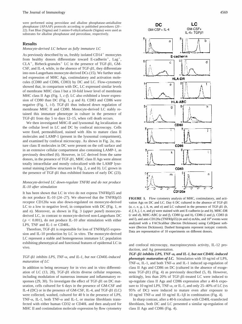
As previously described by us, freshly isolated CD141 monocytesfrom healthy donors differentiate toward E-cadherin1, Lag1,CLA1, Birbeck-granules1 LC in the presence of TGF-b1, GM-CSF, and IL-4, while, in the absence of TGF-b1, they differentiateinto non-Langerhans monocyte-derived DCs (15). We further stud-ied expression of MHC Ags, costimulatory and activation mole-cules (CD80 and CD86, CD83) by DC and LC. Flow-cytometryshowed that, in comparison with DC, LC expressed similar levelsof membrane MHC class I but a 10-fold lower level of membraneMHC class II Ags (Fig. 1,c-f). LC also exhibited a lower expres-sion of CD80 than DC (Fig. 1,g and h). CD83 and CD86 werenegative (Fig. 1,i-l). TGF-b1 thus induced down regulation ofmembrane MHC II and CD80. Monocyte-derived LC stably re-tained this immature phenotype in culture in the presence ofTGF-b1 from day 5 to days 12–15, when cell death occurs.
We then investigated MHC-II and lysosomal Ag localization atthe cellular level in LC and DC by confocal microscopy. Cellswere fixed, permeabilized, stained with Abs to mature class IImolecules and LAMP-1 (present in the lysosomal compartment),and examined by confocal microscopy. As shown in Fig. 2a, ma-ture class II molecules in DC were present on the cell surface andin an extensive cellular compartment also containing LAMP-1, aspreviously described (6). However, in LC derived from the samedonors, in the presence of TGF-b1, MHC class II Ags were almosttotally intracellular and mostly colocalized with the LAMP lyso-somal staining (yellow structures in Fig. 2,a andb). LC grown inthe presence of TGF-b1 thus exhibited features of early DC (23).
Monocyte-derived LC down-regulate TNFRI and do not produceIL-10 after stimulation
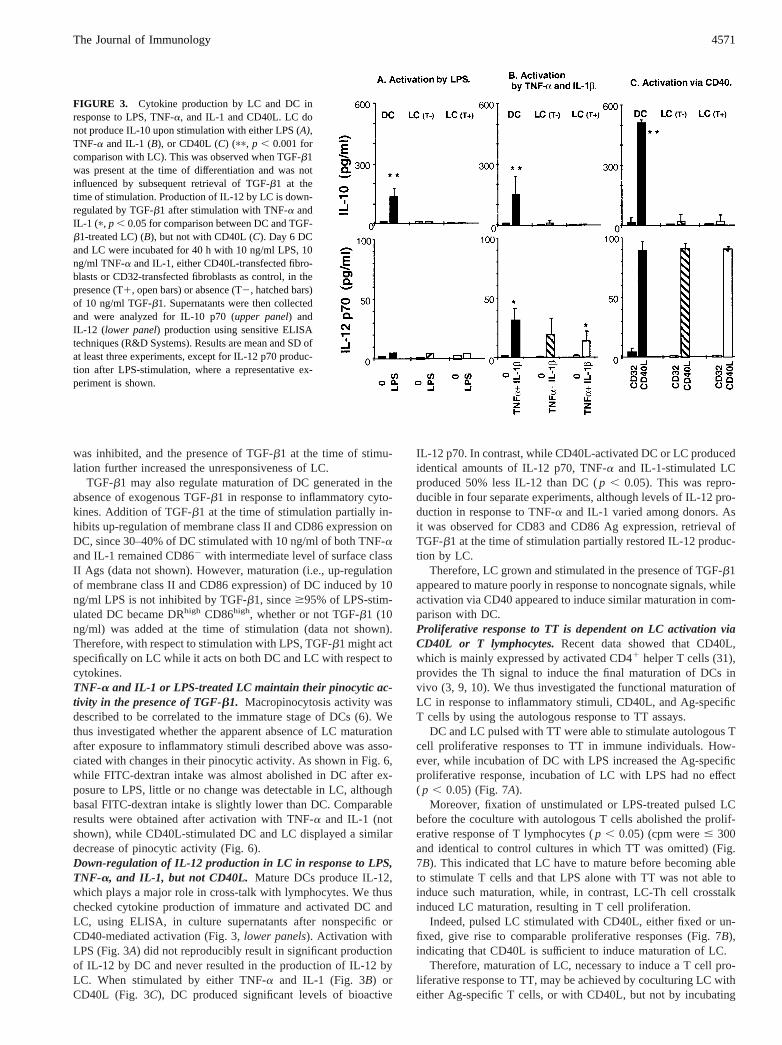
It has been shown that LC in vivo do not express TNFRIp55 anddo not produce IL-10 (24–27). We observed that the TNFRIp55receptor CD120a was also down-regulated on monocyte-derivedLC to a low to negative level, in comparison with DC (Fig. 1,mand n). Moreover, as shown in Fig. 3 (upper panel) monocyte-derived LC, in contrast to monocyte-derived non-Langerhans DC( p , 0.001), do not produce IL-10 after stimulation with eitherLPS, TNF and IL-1 or CD40L.
Therefore, TGF-b1 is responsible for loss of TNFRIp55 expres-sion and IL-10 production by LC in vitro. The monocyte-derivedLC represent a stable and homogeneous immature LC populationexhibiting phenotypical and functional features of epidermal LC invivo.
TGF-b1 inhibits LPS, TNF-a, and IL-1, but not CD40L-inducedmaturation of LC
In addition to being necessary for in vivo and in vitro differenti-ation of LC (13, 28), TGF-b1 elicits diverse cellular responses,including modulation of numerous immune and inflammatory re-sponses (29, 30). To investigate the effect of TGF-b1 on DC mat-uration, cells cultured for 6 days in the presence of GM-CSF andIL-4 (DC) or in the presence of GM-CSF, IL-4, and TGF-b1 (LC)were collected, washed, cultured for 40 h in the presence of LPS,TNF-a, IL-1, both TNF-a and IL-1, or murine fibroblasts trans-fected with either human CD32 or CD40L and then analyzed forMHC II and costimulation molecule expression by flow cytometry
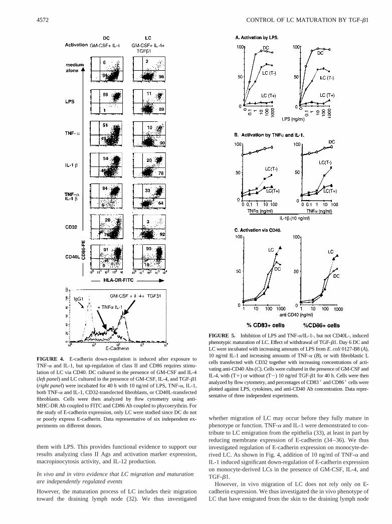
and confocal microscopy, macropinocytosis activity, IL-12 pro-duction, and Ag presentation.TGF-b1 inhibits LPS, TNF-a, and IL-1, but not CD40L-inducedphenotypic maturation of LC.Stimulation with 10 ng/ml of LPS,TNF-a, IL-1, and both TNF-a and IL-1 induced up-regulation ofclass II Ags and CD86 on DC (cultured in the absence of exoge-nous TGF-b1) (Fig. 4) as previously described (5, 8). However,strikingly, less than 20% of TGF-b1-treated LC were induced toup-regulate class II Ags and CD86 expression after a 40-h expo-sure to 10 ng/ml LPS, TNF-a, or IL-1, and only 25–40% of LC (vs95% of DC) were induced to mature even after exposure to10 ng/ml TNF-a and 10 ng/ml IL-1b in combination (Fig. 4).
In sharp contrast, after a 40-h coculture with CD40L-transfectedfibroblasts, both DC and LC presented a similar up-regulation ofclass II Ags and CD86 (Fig. 4).
FIGURE 1. Flow cytometry analysis of MHC, costimulatory, and acti-vation Ags on DC and LC. Day 6 DC cultured in the absence of TGF-b1(a, c, e, g, i, k, m, ando) and LC cultured in the presence of TGF-b1 (b,d, f, h, j, l, n, andp) were stained with anti E-cadherin (a andb), MHC-DR(c andd), MHC-ABC (e andf), CD80 (g andh), CD86 (i and j), CD83 (kandl), and anti-CD120a (TNFRIp55) (mandn) mAbs, and 104 events wereanalyzed with a FACScalibur (Becton Dickinson) using CellQuest soft-ware (Becton Dickinson). Dashed histograms represent isotypic controls.Data are representative of 10 experiments on different donors.
4569The Journal of Immunology
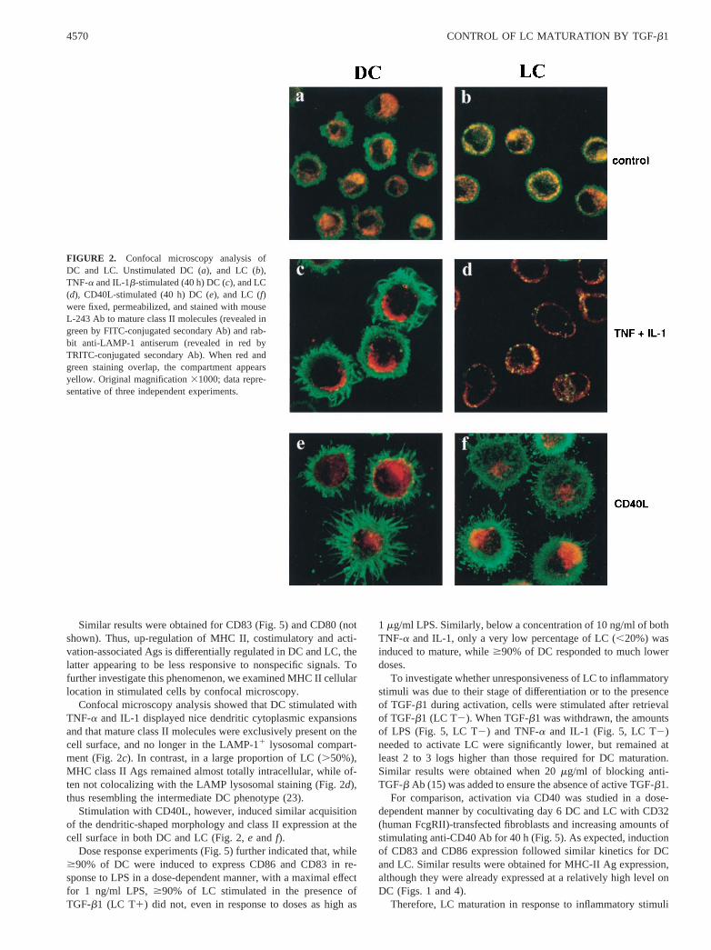
Similar results were obtained for CD83 (Fig. 5) and CD80 (notshown). Thus, up-regulation of MHC II, costimulatory and acti-vation-associated Ags is differentially regulated in DC and LC, thelatter appearing to be less responsive to nonspecific signals. Tofurther investigate this phenomenon, we examined MHC II cellularlocation in stimulated cells by confocal microscopy.
Confocal microscopy analysis showed that DC stimulated withTNF-a and IL-1 displayed nice dendritic cytoplasmic expansionsand that mature class II molecules were exclusively present on thecell surface, and no longer in the LAMP-11 lysosomal compart-ment (Fig. 2c). In contrast, in a large proportion of LC (.50%),MHC class II Ags remained almost totally intracellular, while of-ten not colocalizing with the LAMP lysosomal staining (Fig. 2d),thus resembling the intermediate DC phenotype (23).
Stimulation with CD40L, however, induced similar acquisitionof the dendritic-shaped morphology and class II expression at thecell surface in both DC and LC (Fig. 2,e and f).
Dose response experiments (Fig. 5) further indicated that, while$90% of DC were induced to express CD86 and CD83 in re-sponse to LPS in a dose-dependent manner, with a maximal effectfor 1 ng/ml LPS,$90% of LC stimulated in the presence ofTGF-b1 (LC T1) did not, even in response to doses as high as
1 mg/ml LPS. Similarly, below a concentration of 10 ng/ml of bothTNF-a and IL-1, only a very low percentage of LC (,20%) wasinduced to mature, while$90% of DC responded to much lowerdoses.
To investigate whether unresponsiveness of LC to inflammatorystimuli was due to their stage of differentiation or to the presenceof TGF-b1 during activation, cells were stimulated after retrievalof TGF-b1 (LC T2). When TGF-b1 was withdrawn, the amountsof LPS (Fig. 5, LC T2) and TNF-a and IL-1 (Fig. 5, LC T2)needed to activate LC were significantly lower, but remained atleast 2 to 3 logs higher than those required for DC maturation.Similar results were obtained when 20mg/ml of blocking anti-TGF-b Ab (15) was added to ensure the absence of active TGF-b1.
For comparison, activation via CD40 was studied in a dose-dependent manner by cocultivating day 6 DC and LC with CD32(human FcgRII)-transfected fibroblasts and increasing amounts ofstimulating anti-CD40 Ab for 40 h (Fig. 5). As expected, inductionof CD83 and CD86 expression followed similar kinetics for DCand LC. Similar results were obtained for MHC-II Ag expression,although they were already expressed at a relatively high level onDC (Figs. 1 and 4).
Therefore, LC maturation in response to inflammatory stimuli
FIGURE 2. Confocal microscopy analysis ofDC and LC. Unstimulated DC (a), and LC (b),TNF-a and IL-1b-stimulated (40 h) DC (c), and LC(d), CD40L-stimulated (40 h) DC (e), and LC (f)were fixed, permeabilized, and stained with mouseL-243 Ab to mature class II molecules (revealed ingreen by FITC-conjugated secondary Ab) and rab-bit anti-LAMP-1 antiserum (revealed in red byTRITC-conjugated secondary Ab). When red andgreen staining overlap, the compartment appearsyellow. Original magnification31000; data repre-sentative of three independent experiments.
4570 CONTROL OF LC MATURATION BY TGF-b1
was inhibited, and the presence of TGF-b1 at the time of stimu-lation further increased the unresponsiveness of LC.
TGF-b1 may also regulate maturation of DC generated in theabsence of exogenous TGF-b1 in response to inflammatory cyto-kines. Addition of TGF-b1 at the time of stimulation partially in-hibits up-regulation of membrane class II and CD86 expression onDC, since 30–40% of DC stimulated with 10 ng/ml of both TNF-aand IL-1 remained CD862 with intermediate level of surface classII Ags (data not shown). However, maturation (i.e., up-regulationof membrane class II and CD86 expression) of DC induced by 10ng/ml LPS is not inhibited by TGF-b1, since$95% of LPS-stim-ulated DC became DRhigh CD86high, whether or not TGF-b1 (10ng/ml) was added at the time of stimulation (data not shown).Therefore, with respect to stimulation with LPS, TGF-b1 might actspecifically on LC while it acts on both DC and LC with respect tocytokines.TNF-a and IL-1 or LPS-treated LC maintain their pinocytic ac-tivity in the presence of TGF-b1. Macropinocytosis activity wasdescribed to be correlated to the immature stage of DCs (6). Wethus investigated whether the apparent absence of LC maturationafter exposure to inflammatory stimuli described above was asso-ciated with changes in their pinocytic activity. As shown in Fig. 6,while FITC-dextran intake was almost abolished in DC after ex-posure to LPS, little or no change was detectable in LC, althoughbasal FITC-dextran intake is slightly lower than DC. Comparableresults were obtained after activation with TNF-a and IL-1 (notshown), while CD40L-stimulated DC and LC displayed a similardecrease of pinocytic activity (Fig. 6).Down-regulation of IL-12 production in LC in response to LPS,TNF-a, and IL-1, but not CD40L. Mature DCs produce IL-12,which plays a major role in cross-talk with lymphocytes. We thuschecked cytokine production of immature and activated DC andLC, using ELISA, in culture supernatants after nonspecific orCD40-mediated activation (Fig. 3,lower panels). Activation withLPS (Fig. 3A) did not reproducibly result in significant productionof IL-12 by DC and never resulted in the production of IL-12 byLC. When stimulated by either TNF-a and IL-1 (Fig. 3B) orCD40L (Fig. 3C), DC produced significant levels of bioactive
IL-12 p70. In contrast, while CD40L-activated DC or LC producedidentical amounts of IL-12 p70, TNF-a and IL-1-stimulated LCproduced 50% less IL-12 than DC (p , 0.05). This was repro-ducible in four separate experiments, although levels of IL-12 pro-duction in response to TNF-a and IL-1 varied among donors. Asit was observed for CD83 and CD86 Ag expression, retrieval ofTGF-b1 at the time of stimulation partially restored IL-12 produc-tion by LC.
Therefore, LC grown and stimulated in the presence of TGF-b1appeared to mature poorly in response to noncognate signals, whileactivation via CD40 appeared to induce similar maturation in com-parison with DC.Proliferative response to TT is dependent on LC activation viaCD40L or T lymphocytes.Recent data showed that CD40L,which is mainly expressed by activated CD41 helper T cells (31),provides the Th signal to induce the final maturation of DCs invivo (3, 9, 10). We thus investigated the functional maturation ofLC in response to inflammatory stimuli, CD40L, and Ag-specificT cells by using the autologous response to TT assays.
DC and LC pulsed with TT were able to stimulate autologous Tcell proliferative responses to TT in immune individuals. How-ever, while incubation of DC with LPS increased the Ag-specificproliferative response, incubation of LC with LPS had no effect( p , 0.05) (Fig. 7A).
Moreover, fixation of unstimulated or LPS-treated pulsed LCbefore the coculture with autologous T cells abolished the prolif-erative response of T lymphocytes (p , 0.05) (cpm were# 300and identical to control cultures in which TT was omitted) (Fig.7B). This indicated that LC have to mature before becoming ableto stimulate T cells and that LPS alone with TT was not able toinduce such maturation, while, in contrast, LC-Th cell crosstalkinduced LC maturation, resulting in T cell proliferation.
Indeed, pulsed LC stimulated with CD40L, either fixed or un-fixed, give rise to comparable proliferative responses (Fig. 7B),indicating that CD40L is sufficient to induce maturation of LC.
Therefore, maturation of LC, necessary to induce a T cell pro-liferative response to TT, may be achieved by coculturing LC witheither Ag-specific T cells, or with CD40L, but not by incubating
FIGURE 3. Cytokine production by LC and DC inresponse to LPS, TNF-a, and IL-1 and CD40L. LC donot produce IL-10 upon stimulation with either LPS (A),TNF-a and IL-1 (B), or CD40L (C) (pp, p , 0.001 forcomparison with LC). This was observed when TGF-b1was present at the time of differentiation and was notinfluenced by subsequent retrieval of TGF-b1 at thetime of stimulation. Production of IL-12 by LC is down-regulated by TGF-b1 after stimulation with TNF-a andIL-1 (p, p , 0.05 for comparison between DC and TGF-b1-treated LC) (B), but not with CD40L (C). Day 6 DCand LC were incubated for 40 h with 10 ng/ml LPS, 10ng/ml TNF-a and IL-1, either CD40L-transfected fibro-blasts or CD32-transfected fibroblasts as control, in thepresence (T1, open bars) or absence (T2, hatched bars)of 10 ng/ml TGF-b1. Supernatants were then collectedand were analyzed for IL-10 p70 (upper panel) andIL-12 (lower panel) production using sensitive ELISAtechniques (R&D Systems). Results are mean and SD ofat least three experiments, except for IL-12 p70 produc-tion after LPS-stimulation, where a representative ex-periment is shown.
4571The Journal of Immunology
them with LPS. This provides functional evidence to support ourresults analyzing class II Ags and activation marker expression,macropinocytosis activity, and IL-12 production.
In vivo and in vitro evidence that LC migration and maturationare independently regulated events
However, the maturation process of LC includes their migrationtoward the draining lymph node (32). We thus investigated
whether migration of LC may occur before they fully mature inphenotype or function. TNF-a and IL-1 were demonstrated to con-tribute to LC emigration from the epithelia (33), at least in part byreducing membrane expression of E-cadherin (34–36). We thusinvestigated regulation of E-cadherin expression on monocyte-de-rived LC. As shown in Fig. 4, addition of 10 ng/ml of TNF-a andIL-1 induced significant down-regulation of E-cadherin expressionon monocyte-derived LCs in the presence of GM-CSF, IL-4, andTGF-b1.
However, in vivo migration of LC does not rely only on E-cadherin expression. We thus investigated the in vivo phenotype ofLC that have emigrated from the skin to the draining lymph node
FIGURE 4. E-cadherin down-regulation is induced after exposure toTNF-a and IL-1, but up-regulation of class II and CD86 requires stimu-lation of LC via CD40. DC cultured in the presence of GM-CSF and IL-4(left panel) and LC cultured in the presence of GM-CSF, IL-4, and TGF-b1(right panel) were incubated for 40 h with 10 ng/ml of LPS, TNF-a, IL-1,both TNF-a and IL-1, CD32-transfected fibroblasts, or CD40L-transfectedfibroblasts. Cells were then analyzed by flow cytometry using anti-MHC-DR Ab coupled to FITC and CD86 Ab coupled to phycoerythrin. Forthe study of E-cadherin expression, only LC were studied since DC do notor poorly express E-cadherin. Data representative of six independent ex-periments on different donors.
FIGURE 5. Inhibition of LPS and TNF-a/IL-1-, but not CD40L-, inducedphenotypic maturation of LC. Effect of withdrawal of TGF-b1. Day 6 DC andLC were incubated with increasing amounts of LPS fromE. coli 0127-B8 (A),10 ng/ml IL-1 and increasing amounts of TNF-a (B), or with fibroblastic Lcells transfected with CD32 together with increasing concentrations of acti-vating anti-CD40 Abs (C). Cells were cultured in the presence of GM-CSF andIL-4, with (T1) or without (T2) 10 ng/ml TGF-b1 for 40 h. Cells were thenanalyzed by flow cytometry, and percentages of CD831 and CD861 cells wereplotted against LPS, cytokines, and anti-CD40 Ab concentration. Data repre-sentative of three independent experiments.
4572 CONTROL OF LC MATURATION BY TGF-b1
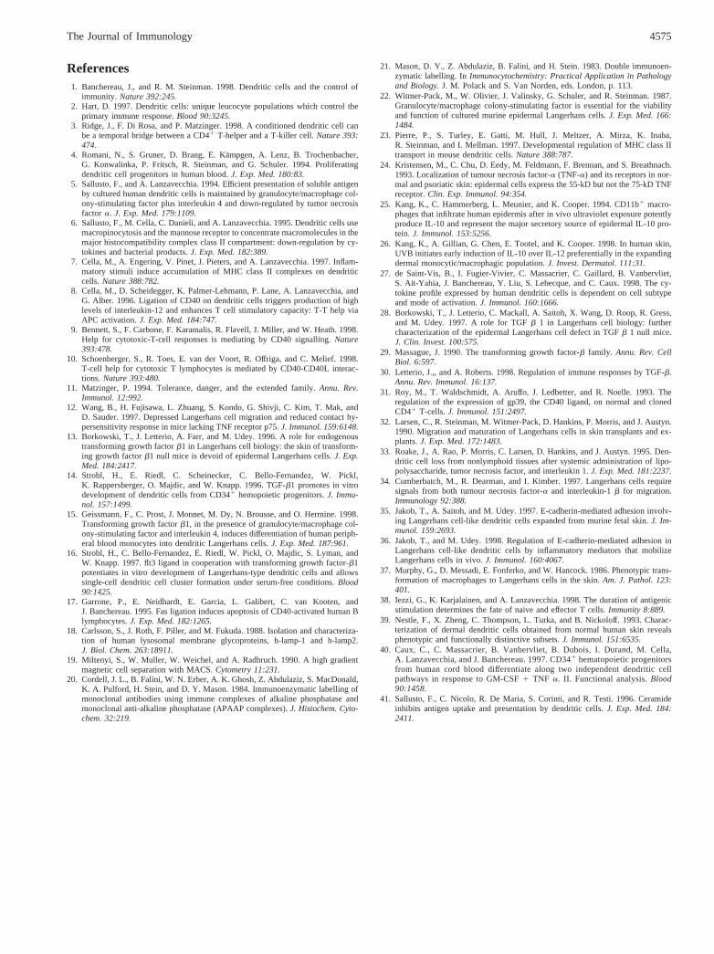
on three patients with dermatopathic lymphadenopathy, a benignreactive condition in which skin-draining lymph nodes are en-larged, due to the accumulation of LC in T cell zones. In three ofthree patients, immunohistochemical staining on serial sectionsshowed that a large number of cells in the T cell zones expressedhigh levels of CD1a and are thus poorly mature LC; most of thesecells did not express detectable levels of CD83, CD86, and CD80.A double immunostaining using CD1a and CD83 Abs is shown inFig. 8; CD1a1CD83low/2 are numerous in the T cell zone (closedarrowhead), while some CD1a1CD831 cells are also present (ar-row). CD1a2CD831 (open arrowhead) cells are mainly found inthe B cell follicle (F). This result suggests that phenotypically im-mature LCs can migrate into the draining lymph node in responseto an inflammatory signal.
Therefore, TGF-b1, which allows differentiation of immatureLCs from monocytes, may not inhibit migration of LC to the para-cortical area of draining lymph nodes in response to inflammatorystimuli such as TNF-a and IL-1. Rather, our results suggest thatTGF-b1 prevents the functional maturation of these cells unless acognate signal such as CD40L is provided.
DiscussionWe have previously shown that TGF-b1, in the presence of GM-CSF and IL-4, induces differentiation of human peripheral bloodmonocytes into CD1a1, E-cadherin1, CLA1, and Lag1 dendriticLC expressing Birbeck granules (15). In the present study, wefurther extend this finding and show that monocyte-derived LCsrepresent a stable population of immature LCs, with intracellularclass II Ags, which do not produce IL-10 after stimulation and donot express the TNFRIp55.
These results further indicate that monocyte-derived LC behaveas do in vivo LC in many aspects. Indeed, it has been shown thatin vivo LC only express TNFRIIp75 (24) and respond to TNF-avia TNFRIIp75 and not TNFRIp55 (12) while, in contrast, mono-cyte-derived DC respond to TNF-a by TNFRIp55 and not TN-FRIIp75 (6). Also, in vivo LC and in vitro CD341-derived LC donot produce IL-10 (25–27), while dermal CD11b1 macrophages/DCs in vivo (25, 26), in vitro CD34-derived (27), and monocyte-derived non-Langerhans DC produce IL-10 upon stimulation.These findings distinguish LC from dermal DC, which resemble
more closely the monocyte-derived DCs generated in the presenceof GM-CSF and IL-4 without exogenous TGF-b1, while the twoDC subsets (dermal DC and LC) may be closely related (37).
More important, the present study also shows that maturation ofLC is differentially regulated by TGF-b1: 1) TGF-b1 allowsdown-regulation of E-cadherin expression by the whole LC pop-ulation in response to TNF-a and IL-1 but inhibits LC maturationafter exposure to LPS, TNF, and IL-1, with respect to Class II,CD80, CD86, and CD83 Ag expression, loss of FITC-dextran up-take, production of IL-12, and Ag presentation; 2) in contrast, cog-nate T cell-dependent stimuli (i.e., CD40-mediated) induce fullmaturation of LC and are not influenced by TGF-b1; 3) in vivomaturation (i.e., induction of CD80, CD86, and CD83 Ag expres-sion) does not necessarily precede migration, and, in accordance,it has been shown that LC do not need to be fully mature in phe-notype or function before they leave the skin (32).
These results are consistent with the crucial role of CD40-me-diated activation for the final maturation and “licensing” of DCs(3, 9, 10). LC are the DCs of the epithelial barriers, including theskin and oral, nasal, esophagal, pulmonary, vaginal, and rectal mu-cosae. These cells are thus challenged frequently with numerouspathogens and traumatic events and reside in epithelia whereTNF-a and IL-1 are produced at relatively high levels (28). LCrepresent the main population of APC in the mucosal barriers, andthe main source of IL-12 after stimulation, but do not produceIL-10 and drive Th1 responses such as delayed-contact hypersen-sitivity. It would be dangerous for the host if inflammatory stimuliwere able to “license” LC in a way that enable them to stimulatean effector response. Our results indicate that TGF-b1 may beresponsible for the cognate T cell dependence of LC maturation.By dampering the effect of inflammatory cytokines and LPS on thefunctional maturation of LC, TGF-b1 may prevent the noncognatematuration of LC via bystander inflammatory cytokines present inepithelia, thereby avoiding potentially harmful immune responses.
After Ag exposure, TNF-a and IL-1 contribute to LC emigrationfrom the epithelia (33, 34), at least in part by reducing membraneexpression of E-cadherin (35, 36). Our results are in accordancewith these data; however, we propose that these stimuli are notsufficient to induce final maturation of DC, due to the presence of
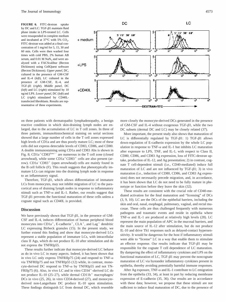
FIGURE 6. FITC-dextran uptakeby DC and LC: TGF-b1 maintain fluidphase intake in LPS-treated LC. Cellswere resuspended in complete mediumand incubated at 37°C with 5% CO2.FITC-dextran was added at a final con-centration of 1 mg/ml for 5, 15, 30 and60 min. Cells were then washed fourtimes with cold PBS, 2% human ABserum, and 0.01 M NaN3 and were an-alyzed with a FACScalibur (BectonDickinson) using CellQuest software(Becton Dickinson).Upper panel, DC,cultured in the presence of GM-CSFand IL-4 (left), LC cultured in thepresence of GM-CSF, IL-4, andTGF-b1 (right). Middle panel, DC(left) and LC (right) stimulated by 10ng/ml LPS.Lower panel, DC (left) andLC (right) stimulated by CD40L-transfected fibroblasts. Results are rep-resentative of three experiments.
4573The Journal of Immunology
TGF-b1. Once in lymphoid organs, LC interact with naive Ag-specific helper T cells. This contact, which may be facilitated byattracting chemokines, can be sustained for 20 h in certain circum-stances (38). If cognate recognition occurs, this crosstalk may leadto activation of both the helper T cell and the LC, thus initiating ahelper CD41 immune response and “licensing” mature LC (so-called interdigitating DCs) to stimulate cytotoxic T cells (3). Itcould be tempting to further speculate that this crosstalk in the Tcell area of lymph nodes may also result in tolerance.
Uncontrolled maturation of LC by nonspecific bystander signalsand consecutive inappropriate T cell activation might break toler-ance. However, DCs grown in the absence of TGF-b1, which maycorrespond to dermal (39) and some nonmucosal DCs, becomemature in response to cytokines or LPS and produce IL-10. Thesecells have been proposed to mediate humoral rather than cellularimmune responses (40). In addition, LPS or TNF-a, away from theperiphery, e.g. in the spleen, represent “danger” (e.g., septicemia).Conversely, production of TGF-b1 by tumoral cells may locallyhamper inflammatory-induced maturation of DC and reduce anti-tumoral immune responses.
We have shown here that several DC activation pathways may beinhibited by TGF-b1, while another (i.e., CD40L) remains unaffected.Although a common ceramide-mediated signaling pathway was de-scribed for CD40L, TNF-a, and IL-1 in DC (41), it is clearly notaffected in TGF-b1-treated LC, since CD40L-induced activation andAg presentation are not inhibited. CD14, the main known LPS recep-tor, was barely detectable in both DC and LC (15). We have shownin this study that TGF-b1 down-regulates membrane TNFRIp55 onLC vs DC. However, stimulation via CD40 induces IL-12 productionin both DC and LC at similar levels, but LC do not produce IL-10,while DC produce large amounts of this cytokine. Therefore, differ-ential regulation of receptor expression is not the only mechanisminvolved in the alternative responses to external stimuli by DC andLC. CD40 may engage different downstream signaling molecules inthe two cell types.
In conclusion, TGF-b1 appears to be a major cytokine in DCbiology, responsible for the acquisition of the LC phenotype andthe prevention of noncognate maturation.
AcknowledgmentsWe thank Drs. O. Lantz and M. Throsby for helpful discussions and criticalreading of the manuscript and Drs. B. Varet, C. Bonnerot, B. Pron, andJ. L. Gaillard for helpful discussions. Murine fibroblast cell lines trans-fected with human CD40L (LcCD40L) or CD32 (LcCD32) were kindlyprovided by Dr. F. Brie`re. We also thank M. Leborgne for excellent tech-nical assistance.
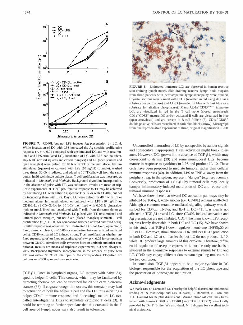
FIGURE 7. CD40L but not LPS induces Ag presentation by LC.A,While incubation of DC with LPS increased the Ag-specific proliferativeresponse (p, p , 0.01 compared with unstimulated DC and with unstimu-lated and LPS-stimulated LC), incubation of LC with LPS had no effect.Day 6 DC (closed squares and closed triangles) and LC (open squares andopen triangles) were pulsed for 48 h with TT or medium alone, left un-stimulated (squares) or cultured with LPS (10 ng/ml) (triangle), washedthree times, 30-Gy-irradiated, and added to 105 T cells/well from the samedonor, in 96-well tissue culture plates. T cell proliferation was measured asindicated inMaterials and Methods. Background thymidine incorporation,in the absence of pulse with TT, was subtracted; results are mean of trip-licate experiments.B, T cell proliferative response to TT may be achievedby coculturing LC with either Ag-specific T cells, or with CD40L, but notby incubating them with LPS. Day 6 LC were pulsed for 48 h with TT ormedium alone, left unstimulated or cultured with LPS (10 ng/ml) orCD40L-Lc (1 CD40L-Lc for 10 LC), then fixed with 0.001% glutaralde-hyde or mock fixed and cocultured with T cells from the same donor asindicated inMaterials and Methods. LC pulsed with TT, unstimulated andunfixed (open triangles) but not fixed (closed triangles) stimulate T cellproliferation (p, p , 0.05 for comparison between unfixed and fixed cells).Similar response was obtained for LPS-treated LC (not fixed, open circle;fixed, closed circle) (p, p , 0.05 for comparison between unfixed and fixedcells). CD40-activated LC induced strong T cell proliferation whether un-fixed (open squares) or fixed (closed squares) (pp, p , 0.05 for comparisonbetween CD40L-stimulated cells (whether fixed or unfixed) and other con-ditions). Results are means of triplicate experiments; SD was always#
20%. Background thymidine incorporation, in the absence of pulse withTT, was either#10% of total cpm of the corresponding TT-pulsed LCcultures or,300 cpm and was subtracted.
FIGURE 8. Emigrated immature LCs are observed in human reactiveskin-draining lymph nodes. Skin-draining reactive lymph node biopsiesfrom three patients with dermatopathic lymphadenopathy were studied.Cryostat sections were stained with CD1a (revealed in red using AEC as asubstrate for peroxidase) and CD83 (revealed in blue with fast blue as asubstrate for alkaline phosphatase). Many CD1a1CD83low/2 immatureLCs are visualized in red in the T cell zone (closed arrowhead).CD1a2CD831 mature DC and/or activated B cells are visualized in blue(open arrowhead) and are present in B cell follicle (F). CD1a1CD831
double positive cells are visualized in dark blue-black (arrow). Micrographfrom one representative experiment of three, original magnification3200.
4574 CONTROL OF LC MATURATION BY TGF-b1
References1. Banchereau, J., and R. M. Steinman. 1998. Dendritic cells and the control of
immunity. Nature 392:245.2. Hart, D. 1997. Dendritic cells: unique leucocyte populations which control the
primary immune response.Blood 90:3245.3. Ridge, J., F. Di Rosa, and P. Matzinger. 1998. A conditioned dendritic cell can
be a temporal bridge between a CD41 T-helper and a T-killer cell.Nature 393:474.
4. Romani, N., S. Gruner, D. Brang, E. Ka¨mpgen, A. Lenz, B. Trochenbacher,G. Konwalinka, P. Fritsch, R. Steinman, and G. Schuler. 1994. Proliferatingdendritic cell progenitors in human blood.J. Exp. Med. 180:83.
5. Sallusto, F., and A. Lanzavecchia. 1994. Efficient presentation of soluble antigenby cultured human dendritic cells is maintained by granulocyte/macrophage col-ony-stimulating factor plus interleukin 4 and down-regulated by tumor necrosisfactor a. J. Exp. Med. 179:1109.
6. Sallusto, F., M. Cella, C. Danieli, and A. Lanzavecchia. 1995. Dendritic cells usemacropinocytosis and the mannose receptor to concentrate macromolecules in themajor histocompatibility complex class II compartment: down-regulation by cy-tokines and bacterial products.J. Exp. Med. 182:389.
7. Cella, M., A. Engering, V. Pinet, J. Pieters, and A. Lanzavecchia. 1997. Inflam-matory stimuli induce accumulation of MHC class II complexes on dendriticcells.Nature 388:782.
8. Cella, M., D. Scheidegger, K. Palmer-Lehmann, P. Lane, A. Lanzavecchia, andG. Alber. 1996. Ligation of CD40 on dendritic cells triggers production of highlevels of interleukin-12 and enhances T cell stimulatory capacity: T-T help viaAPC activation.J. Exp. Med. 184:747.
9. Bennett, S., F. Carbone, F. Karamalis, R. Flavell, J. Miller, and W. Heath. 1998.Help for cytotoxic-T-cell responses is mediating by CD40 signalling.Nature393:478.
10. Schoenberger, S., R. Toes, E. van der Voort, R. Offriga, and C. Melief. 1998.T-cell help for cytotoxic T lymphocytes is mediated by CD40-CD40L interac-tions.Nature 393:480.
11. Matzinger, P. 1994. Tolerance, danger, and the extended family.Annu. Rev.Immunol. 12:992.
12. Wang, B., H. Fujisawa, L. Zhuang, S. Kondo, G. Shivji, C. Kim, T. Mak, andD. Sauder. 1997. Depressed Langerhans cell migration and reduced contact hy-persensitivity response in mice lacking TNF receptor p75.J. Immunol. 159:6148.
13. Borkowski, T., J. Letterio, A. Farr, and M. Udey. 1996. A role for endogenoustransforming growth factorb1 in Langerhans cell biology: the skin of transform-ing growth factorb1 null mice is devoid of epidermal Langerhans cells.J. Exp.Med. 184:2417.
14. Strobl, H., E. Riedl, C. Scheinecker, C. Bello-Fernandez, W. Pickl,K. Rappersberger, O. Majdic, and W. Knapp. 1996. TGF-b1 promotes in vitrodevelopment of dendritic cells from CD341 hemopoietic progenitors.J. Immu-nol. 157:1499.
15. Geissmann, F., C. Prost, J. Monnet, M. Dy, N. Brousse, and O. Hermine. 1998.Transforming growth factorb1, in the presence of granulocyte/macrophage col-ony-stimulating factor and interleukin 4, induces differentiation of human periph-eral blood monocytes into dendritic Langerhans cells.J. Exp. Med. 187:961.
16. Strobl, H., C. Bello-Fernandez, E. Riedl, W. Pickl, O. Majdic, S. Lyman, andW. Knapp. 1997. flt3 ligand in cooperation with transforming growth factor-b1potentiates in vitro development of Langerhans-type dendritic cells and allowssingle-cell dendritic cell cluster formation under serum-free conditions.Blood90:1425.
17. Garrone, P., E. Neidhardt, E. Garcia, L. Galibert, C. van Kooten, andJ. Banchereau. 1995. Fas ligation induces apoptosis of CD40-activated human Blymphocytes.J. Exp. Med. 182:1265.
18. Carlsson, S., J. Roth, F. Piller, and M. Fukuda. 1988. Isolation and characteriza-tion of human lysosomal membrane glycoproteins, h-lamp-1 and h-lamp2.J. Biol. Chem. 263:18911.
19. Miltenyi, S., W. Muller, W. Weichel, and A. Radbruch. 1990. A high gradientmagnetic cell separation with MACS.Cytometry 11:231.
20. Cordell, J. L., B. Falini, W. N. Erber, A. K. Ghosh, Z. Abdulaziz, S. MacDonald,K. A. Pulford, H. Stein, and D. Y. Mason. 1984. Immunoenzymatic labelling ofmonoclonal antibodies using immune complexes of alkaline phosphatase andmonoclonal anti-alkaline phosphatase (APAAP complexes).J. Histochem. Cyto-chem. 32:219.
21. Mason, D. Y., Z. Abdulaziz, B. Falini, and H. Stein. 1983. Double immunoen-zymatic labelling. InImmunocytochemistry: Practical Application in Pathologyand Biology. J. M. Polack and S. Van Norden, eds. London, p. 113.
22. Witmer-Pack, M., W. Olivier, J. Valinsky, G. Schuler, and R. Steinman. 1987.Granulocyte/macrophage colony-stimulating factor is essential for the viabilityand function of cultured murine epidermal Langerhans cells.J. Exp. Med. 166:1484.
23. Pierre, P., S. Turley, E. Gatti, M. Hull, J. Meltzer, A. Mirza, K. Inaba,R. Steinman, and I. Mellman. 1997. Developmental regulation of MHC class IItransport in mouse dendritic cells.Nature 388:787.
24. Kristensen, M., C. Chu, D. Eedy, M. Feldmann, F. Brennan, and S. Breathnach.1993. Localization of tumour necrosis factor-a (TNF-a) and its receptors in nor-mal and psoriatic skin: epidermal cells express the 55-kD but not the 75-kD TNFreceptor.Clin. Exp. Immunol. 94:354.
25. Kang, K., C. Hammerberg, L. Meunier, and K. Cooper. 1994. CD11b1 macro-phages that infiltrate human epidermis after in vivo ultraviolet exposure potentlyproduce IL-10 and represent the major secretory source of epidermal IL-10 pro-tein. J. Immunol. 153:5256.
26. Kang, K., A. Gillian, G. Chen, E. Tootel, and K. Cooper. 1998. In human skin,UVB initiates early induction of IL-10 over IL-12 preferentially in the expandingdermal monocytic/macrophagic population.J. Invest. Dermatol. 111:31.
27. de Saint-Vis, B., I. Fugier-Vivier, C. Massacrier, C. Gaillard, B. Vanbervliet,S. Ait-Yahia, J. Banchereau, Y. Liu, S. Lebecque, and C. Caux. 1998. The cy-tokine profile expressed by human dendritic cells is dependent on cell subtypeand mode of activation.J. Immunol. 160:1666.
28. Borkowski, T., J. Letterio, C. Mackall, A. Saitoh, X. Wang, D. Roop, R. Gress,and M. Udey. 1997. A role for TGFb 1 in Langerhans cell biology: furthercharacterization of the epidermal Langerhans cell defect in TGFb 1 null mice.J. Clin. Invest. 100:575.
29. Massague, J. 1990. The transforming growth factor-b family. Annu. Rev. CellBiol. 6:597.
30. Letterio, J.,, and A. Roberts. 1998. Regulation of immune responses by TGF-b.Annu. Rev. Immunol. 16:137.
31. Roy, M., T. Waldschmidt, A. Aruffo, J. Ledbetter, and R. Noelle. 1993. Theregulation of the expression of gp39, the CD40 ligand, on normal and clonedCD41 T-cells.J. Immunol. 151:2497.
32. Larsen, C., R. Steinman, M. Witmer-Pack, D. Hankins, P. Morris, and J. Austyn.1990. Migration and maturation of Langerhans cells in skin transplants and ex-plants.J. Exp. Med. 172:1483.
33. Roake, J., A. Rao, P. Morris, C. Larsen, D. Hankins, and J. Austyn. 1995. Den-dritic cell loss from nonlymphoid tissues after systemic administration of lipo-polysaccharide, tumor necrosis factor, and interleukin 1.J. Exp. Med. 181:2237.
34. Cumberbatch, M., R. Dearman, and I. Kimber. 1997. Langerhans cells requiresignals from both tumour necrosis factor-a and interleukin-1b for migration.Immunology 92:388.
35. Jakob, T., A. Saitoh, and M. Udey. 1997. E-cadherin-mediated adhesion involv-ing Langerhans cell-like dendritic cells expanded from murine fetal skin.J. Im-munol. 159:2693.
36. Jakob, T., and M. Udey. 1998. Regulation of E-cadherin-mediated adhesion inLangerhans cell-like dendritic cells by inflammatory mediators that mobilizeLangerhans cells in vivo.J. Immunol. 160:4067.
37. Murphy, G., D. Messadi, E. Fonferko, and W. Hancock. 1986. Phenotypic trans-formation of macrophages to Langerhans cells in the skin.Am. J. Pathol. 123:401.
38. Iezzi, G., K. Karjalainen, and A. Lanzavecchia. 1998. The duration of antigenicstimulation determines the fate of naive and effector T cells.Immunity 8:889.
39. Nestle, F., X. Zheng, C. Thompson, L. Turka, and B. Nickoloff. 1993. Charac-terization of dermal dendritic cells obtained from normal human skin revealsphenotypic and functionally distinctive subsets.J. Immunol. 151:6535.
40. Caux, C., C. Massacrier, B. Vanbervliet, B. Dubois, I. Durand, M. Cella,A. Lanzavecchia, and J. Banchereau. 1997. CD341 hematopoietic progenitorsfrom human cord blood differentiate along two independent dendritic cellpathways in response to GM-CSF1 TNF a. II. Functional analysis.Blood90:1458.
41. Sallusto, F., C. Nicolo, R. De Maria, S. Corinti, and R. Testi. 1996. Ceramideinhibits antigen uptake and presentation by dendritic cells.J. Exp. Med. 184:2411.
4575The Journal of Immunology


