Embed Size (px)
Citation preview
Diabetologia (1995) 38:509-517 Diabetologia �9 Springer-Verlag 1995
b
The ACE-inhibitor captopril improves myocardial perfusion in spontaneously diabetic (BB) rats R. Riisen 1, A . E E . Rump 1, P. RiJsen 2
1 Institute of Pharmacology University of Cologne, Cologne, Germany 2 Diabetes Research Institute at the Heinrich-Heine-University, D%seldorf, Germany
Summary The aim of this study was to examine the influence of inhibition on angiotensin converting en- zyme (ACE) of myocardial function and perfusion of the rat impaired by diabetes. Spontaneously dia- betic rats were treated with the ACE-inhibitor capto- pril for 4 months. Cardiac performance was analysed in the isolated heart perfused at constant volume. Epicardial perfusion was determined by measuring changes in epicardial fluorescence after injection of a bolus of fluoresceinisothiocyanate-dextrane (3 kDa) as described previously. As compared to un- treated diabetic controls, captopril prevented the in- crease of end diastolic pressure, coronary perfusion pressure and vascular resistance. The intravascular volume was enlarged and the epicardial perfusion rate increased in hearts of diabetic rats treated with captopril as compared to diabetic controls. Treat-
ment of diabetic rats with the ACE-inhibitor capto- pril (1) increases the number of perfused capillaries, and (2) can partly prevent the development of cardi- ac dysfunction in diabetes. Together with morpholo- gical data demonstrating an inhibition of interstitial and perivascular fibrosis in hearts of diabetic rats treated with captopril, our data suggest that ACE-in- hibition is cardioprotective in diabetes. These obser- vations are also compatible with the assumption that an accelerated generation of angiotensin II may be involved in the pathophysiological chain of events leading to diabetic cardiopathy. [Diabetologia (1995) 38: 509-517]
Key words Diabetes mellitus, cardiopathy, ACE-inhi- bition, captopril, BB rats, regional perfusion, myocar- dial function.
The incidence of cardiovascular complications is sev- erely increased in diabetes mellitus by the develop- ment of macrovascular complications. However, cor- onary heart disease can only partly explain the in- creased rate of cardiovascular complications in dia- betes, since cardiac risk remains elevated even in the absence of atherosclerosis [1-3]. In patients, impair- ment of diastolic relaxation and signs of cardiac neu-
Received: 19 April 1994 and in revised form: 20 October 1994
Corresponding author: Professor Dr. E R6sen, Diabetes-For- schungsinstitut, Auf'm Hennekamp 65, D-40225 Dt~sseldorf, Germany Abbreviations: ACE, Angiotensin converting enzyme; CVR, coronary vascular resistance; EDR end diastolic pressure; LVR left ventricular pressure; CPE coronary peffusion pres- sure.
ropathy have been consistently reported [3, 4]. Addi- tionally, cardiac performance is further impaired in diabetic patients by development of small vessel dis- ease [5, 6]. In animal models using chemically-in- duced forms of insulin deficiency or spontaneously diabetic rats and mice, the metabolic disturbances following insulin deficiency cause impairment of dia- stolic and systolic heart performance, which can be totally prevented by treatment of the animals with in- sulin [7-11]. The various forms of cardiac dysfunction in diabetes which develop independently from coro- nary heart disease, are often called "diabetic cardio- myopathy" [12-14].
The mechanisms underlying these complex distur- bances of heart function in diabetes are not fully un- derstood and several hypotheses have been suggest- ed to link the metabolic disturbances observed in the diabetic heart with impairment of heart function [4,

510 R. R6sen et al.: Influence of captopril on heart function in diabetic rats
7, 10]. Microangiopathic changes [14-16], abnormali- ties in vascular reactivity [15, 17], a l tered au tonomic funct ion [9, 18], increased stiffness of the ventr icular wall and perivascular thickening, interstit ial accumu- lat ion of connect ive tissue and matr ix proteins [4, 9, 18, 19], and defects in the regulat ion of calcium homeostasis [20] have been claimed to contr ibute as aetiological factors to disturbances of hear t funct ion in diabetes.
We suggested recent ly [4] that the renin-angioten- sin-system is act ivated in diabetes with increasing work load because of the l imited capacity of the dia- betic hear t to supply oxygen and to genera te energy in sufficient amounts. A n enhanced genera t ion of an- giotensin II could explain some of the changes in car- diac per formance observed in diabetes: act ivation of the sympathet ic nervous system [21, 22], p romot ion of cardiac and vascular fibrosis result ing in increased myocardia l stiffness and impaired myocardia l con- tractility, and disturbances in the regulat ion of coro- nary flow [23-25]. Moreover , coronary vasoconstric- t ion and myocardia l necrosis have been repor ted to be caused by an enhanced genera t ion of angiotensin II in o ther forms of hear t fai lure [23, 24], which can contr ibute to a fur ther restriction and impai rment of cardiac function. In various forms of hear t failure the use of angiotensin convert ing enzyme (ACE)- in- hibitors is now an established therapy [23]. In clinical settings, ACE- inh ib i t ion has been shown to improve survival and clinical symptoms [23, 26]. In exper imen- tal studies evidence has been presented that ACE- in- hibi t ion can inhibit the deve lopment of hyper t rophy and fibrosis as well as improve endothel ia l funct ion [27]. However , influence of ACE-inhibi tors on the funct ion of the diabetic hear t has not yet been stu- died, a l though inhibi t ion of A C E m a y enlarge the availability of bradykin in in diabetic hearts which has been shown to improve the insulin sensitivity and the conversion of glucose in hearts of diabetic rats and in skeletal muscle of pat ients [28-30].
To test the hypotheses that an enhanced genera- t ion of angiotensin II contr ibutes largely to cardiac dysfunct ion as observed in diabetes, we t rea ted dia- betic rats with captopril as an A C E inhibi tor in con- centrat ions which do no t interfere only with the pe- r ipheral angiotensin II format ion, but p resumably also with the local myocardia l genera t ion of angio- tensin II. As a mode l of insul in-dependent diabetes melli tus ( I D D M ) we used the diabetic BB (Bio- breeding) rat which is a close counterpar t to h u m a n I D D M [4, 101.
Materials and methods
Animals. Male BB rats were delivered by M0Uegaard, Ejby, Denmark, at the age of 60 days. The animals were kept in the animal laboratory of the Diabetes-Forschungsinstitut under
standard housing conditions. The animals had free access to water and received a standard diet (Sniff, Soest, Germany). At the age of 80-100 days, 70 % of tile rats became spontane- ously diabetic and had to be treated with insulin (Insulin super lente; Novo, Bagsvaerd., Denmark, 3-4 IU/day) according to the metabolic demand to prevent ketoacidosis and excessive hyperglycaemia [9]. The animals were controlled daily for glu- cosuria (Gluketur sticks; Boehringer Mannheim, Mannheim, Germany), weekly for weight and every 4 weeks for blood glu- cose by the hexokinase method using a glucose analyser (Beckman, Munich, Germany). As controls, diabetes-resistant rats were used, which develop diabetes at an incidence of less than 1%. Just after killing, blood was taken for the determina- tion of plasma insulin using a commercial RIA-kit (Pharmacia, Freiburg, Germany) with rat insulin (Novo) as standard and glycated haemoglobins by affinity chromatography (Isolab, Munich, Germany).
Sodium, potassium and haematocrit were determined by standard laboratory techniques, triglycerides by the glycerol- oxidase method according to the instructions of the manufac- turer (Boehringer Mannheim) and ACE-activity according to Cushman and Cheung [31].
After verification of the diabetic state, one group of ani- mals was treated with captopril for 4 months. Captopril was supplied in the drinking water. Since the amount of drinking water was different (18 ml/day in controls and 112 ml/day for diabetic rats), the amount of drinking water was regularly con- trolled and the concentration of captopril adjusted to ensure an uptake of 30 mg/kg body weight and day. The plasma con- centration of sodium, potassium and the haematocrit were rou- tinely checked every month. Each group of rats consisted of at least five to eight animals.
Heart function in vivo. Heart function was determined in an- aesthetised rats (Nembutal, 30 mg/kg body weight) after in- serting a Millar tip catheter into the carotid artery as de- scribed previously [8].
Isolated perfused heart. Additionally, hearts were isolated and perfused according to Langendorff with Krebs-Henseleit-bi- carbonate buffer (Na + 143, K + 5.9, Ca 2+ 2.5, Mg 2+ 1.18, C1- 127.7, HCO 3- 25, HzPO 4- 1.18, sulfate 1.18 and glucose 10.1 (mmol/1), pH 7.4) saturated with 5 % CO2-95 % 02 as previous- ly published [17].
There was no ACE-inhibitor applied during heart perfu- sion. Global coronary flow was kept constant at 5.0, 10.0, and 13.0 ml/min with a roller pump. Hearts were allowed to beat spontaneously. End systolic and end diastolic leff ventricular pressure were measured via an intraventricular catheter. Re- gional perfusion parameters on the left ventricular epicar- dium were analysed from indicator elution kinetics registered by surface fluorescence after bolus injection of fluorescein- isothiocyanate-dextrane (MG 3 kDa, 100 ~1) as described pre- viously [17, 32]. The elution profiles could be attributed to two components of regional perfusion: a fast, vascular and a slow, extravascular component with half-lives in the range of seconds and minutes, respectively. It has been shown previous- ly that the slow component was absent when fluorescein- isothiocyanate-dextrane of high molecular weight (70 kDa) was used [17]. The maximal signal height after bolus injection was proportional to the intravascular volume, whereas the ex- travascular indicator concentration could be extrapolated from half-log plots of the slow component and corresponded to the transcapillary permeability. The half-lives of the elution kinetics corresponded to the intraluminal flow rate (vascular perfusion rate, T/2 in seconds) and to the interstitial-extravas- cular exchange rate (extravascular clearance, t/2 in minutes).
R. R6sen et al.: Influence of captopril on heart function in diabetic rats 511
Table 1. Influence of ACE-inhibition by captopril on the bio- chemical properties of diabetic BB rats
Parameter Controls Diabetes Diabetics treated with captopril
Body weight (g) 462 +_ 15 426 • 19 422 _+ 7 a
Blood glucose (retool/l) 7.3 + 1.5 15.6 +_ 1.5 a 16.5 _+ 1.3 a
HbA~ (%) 5.7 + 0.5 10.7 + 1.4 a 8.5 + 0.9" ACE (U/l) 60.3 + 8.2 98.9 +__ 8.5 a 11. d . b
Sodium (meq/1) 142 _+ 2 139 + 2 140 + 3
Potassium (meq/1) 6.6 +- 0.4 7.5 + 0.5 7.2 + 0.4
Haematocrit (%) 42.2 +_ 2.3 41.5 + 1.3 42.0 + 1.8
After manifestation of diabetes, the diabetic BB rats were treated with insulin. One group of rats additionally received captopril (30 mg. kg b. w. -1 �9 day-l). After a diabetes duration of 4 months the given parameters were determined as describ- ed in Methods. The data are given as mean + SEM, n = 5-8 animals per group, n. d. not detectable. ~p < 0.05 statistically significant difference between controls and diabetic rats; b p < 0.05 statistically significant difference between untreated and treated diabetic rats
Statistical analysis
Data are given as mean + SEM. In any experimental group five to eight animals were studied. Differences between groups were analysed by analysis of variance (ANOVA), differences between means by the Scheff6 test, assuming a statistically sig- nificant difference at p < 0.05 and a normal distribution of data.
topri l had no significant inf luence on the body weight, the b lood glucose, and p lasma insulin, bu t re- duced the level of glycated haemoglobin . The con- cent ra t ions of sodium and potass ium in p lasma were no t significantly a f fec ted by the diabet ic s tate or by t r e a tm en t with captopr i l (Table 1). The haema toc r i t was not significantly d i f ferent b e tw een the experi- menta l groups (42 + 2, 41 _+ 3 and 39.5 + 2.5 % in con- trol, diabet ic and cap topr i l - t rea ted diabet ic rats, re- spectively).
The activity of angiotensin conver t ing en zyme was e levated in the serum of diabet ic rats as c o m p a r e d to controls (60.3 + 8.2 vs 98.9 + 8.5 U/l, p < 0.05), bu t not be de tec tab le in se rum of cap topr i l - t rea ted dia- bet ic or cont ro l rats.
Heart function in vivo. As a l ready publ ished [9], car- diac p e r f o r m a n c e was r ed u ced in the diabet ic BB rat: hear t rate , the maximal systolic pressure in the left ventricle, and the diastolic re laxa t ion veloci ty were reduced , whereas the veloci ty of con t rac t ion was un- changed by the d e v e l o p m e n t of the diabet ic state.
T rea tmen t of the animals with captopr i l had no ef- fect on hear ts of control , hea l thy rats. In diabet ic ani- mals, the left vent r icular pressure, and the systolic aor t ic pressure were slightly reduced , the end diasto- lic pressure significantly r educed in the captopr i l group as c o m p a r e d to u n t r ea t ed diabet ic BB rats (Table 2). The m a x i m u m of con t rac t ion and relaxa- t ion were no t inf luenced by the t r e a tm en t with capto- pril. The end diastolic pressure was significantly re- duced in the captopr i l group as c o m p a r e d to unt rea t - ed diabet ic BB rats (Table 2).
Results
Despi te daily adminis t ra t ion of insulin, the diabet ic BB rats were hyperg lycaemic as ascer ta ined f ro m the e leva ted pos tprandia l b lood glucose levels and the increased amoun t s of g tycated haemoglobin . Tri- glycerides ranged b e t w e e n 3.5 and 8.5 mmol/1. Glu- cosuria, ke tonur ia , and polydipsia were addi t ional ly typical symptoms found in the diabet ic BB rats. Cap-
Performance o f the isolated perfused heart. Trea tmen t with captopri l had no significant inf luence on perfor- m an ce of the isolated hear t of control , hea l thy rats pe r fused at constant vo lume (data not shown). H e a r t ra te was no t d i f ferent b e tw een controls and diabet ic rats and was not inf luenced by the co rona ry flow or t r e a tm en t with captopril . With increasing co rona ry f low left vent r icu lar pressure (LVP) rose sharply in the hear ts of controls, but only slightly in those of un-
Table 2. Influence of ACE-inhibition by captopril on heart performance of diabetic BB rats in vivo
Parameter
Heart rate (beats/min) Left ventricular systolic pressure (mm Hg) End diastolic pressure (mm Hg) Maximal rate of contraction (mm Hg/s) Maximal rate of relaxation (mm Hg/s) Systolic aortic pressure
Controls Diabetes Diabetics treated with captopril
386 _+ 14 348 + 12 a 361 + 22 133 _+ 13 115 + 3" 101 + 8 a 0.5 • 0.2 2.3 _+ 0.4 a i.i _+ 0 . 2 b
6740 _+ 791 6965 + 386 6767 + 344 6943 +_ 673 5961 + 357 a 5367 + 819 ~
104 ! 3.5 105 +_ 4.9 98 + 7.3
Cardiac performance was determined in diabetic BB-rats, one group of which was additionally treated with captopril (30 mg. kg b.w. -1. day-~). After a duration of diabetes for 4 months a Milar tip catheter was inserted into the carotid ar- tery and heart performance determined as described. Data are given as mean + SEM, n = 5-8 rats per group.
a p < 0.05 statistically significant difference between controls and diabetic rats; Up < 0.05 statistically significant difference between untreated and treated diabetic rats

512 R. ROsen et al.: Influence of captopril on heart function in diabetic rats
A
,I- E g n > - - I
80"
60
40"
20
Left ventricular pressure (BB)
I
*
-7
10 13
Coronary flow (ml /min)
"B
E g a. a. o
Coronary perfusion pressure (BB)
5 10 13
Coronary flow (ml /min)
E g a.
ku
End diastolic pressure (BB) 8] ~ T T
6
5 10 13
Coronary flow (ml /min)
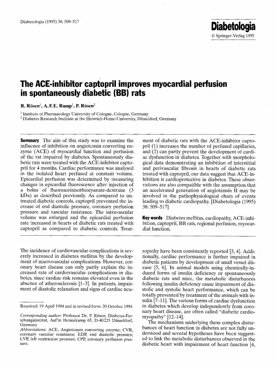
Fig. 1A, B. Influence of diabetes and captopril on (A) left ven- tricular (LVP) and (B) and end diastolic pressure (EDP) of Langendorff-perfused rat hearts at different flow rates. Hearts of captopriI-treated and untreated diabetic BB and control rats were isolated and perfused at constant volume. The duration of diabetes and treatment with captopril was 4 months. Capto- pril did not influence the LVP and EDP in hearts of control rats. Data are given as mean + SEM, n = 5-8 animals per group. *p < 0.05 statistically significant difference between controls and untreated diabetic rats; **p < 0.05 statistically sig- nificant difference between untreated and treated diabetic rats. �9 non-diabetic controls; [ ] diabetics; [ ] diabetics treat- ed with captopril
.~=
-r- E g
> 0
6"
2 �84
Vascular resistance (BB)
5 10 13
Coronary flow (ml /min)
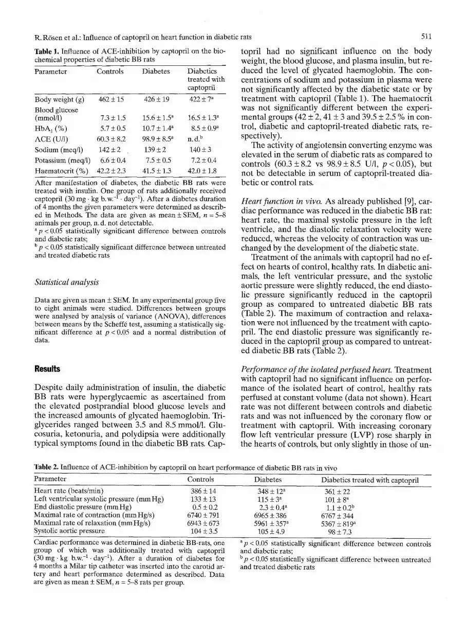
Fig. 2A, B. Influence of diabetes and captopril on (A) the cor- onary perfusion pressure (CPP) and (B) coronary vascular re- sistance (CVR) of Langendorff-perfused rat hearts at differ- ent flow rates. Hearts of captopril-treated and untreated dia- betic BB and control rats were isolated and perfused at con- stant volume. The duration of diabetes and treatment with cap- topril was 4 months. Captopril did not influence the EDP and CPP in hearts of control rats. Data are given as mean + SEM, n = 5-8 animals per group. *p < 0.05 statistically significant difference between controls and untreated diabetic rats; **p < 0.05 statistically significant difference between untreat- ed and treated diabetic rats. �9 Non-diabetic controls; [ ] dia- betics; [ ] diabetics treated with captopril
t r ea t ed and cap topr i l - t rea ted diabet ic BB rats. Inde- pend e n t l y f rom c o rona ry f low L V P was r edu ced in d iabetes as c o m p a r e d to controls and no t significant- ly in f luenced by captopr i l (Fig. I A ) . Thus, pres- sure x hea r t ra te index was r educed in d iabetes as c o m p a r e d to non-d iabe t ic controls, and not changed by the t r e a t m e n t with the ACE- inh ib i to r .
The end diastolic pressure ( E D P ) was e leva ted in hear ts of d iabet ic rats as c o m p a r e d to controls inde- pend e n t l y f r om the co rona ry flow. T rea tmen t with captopr i l did no t inf luence E D P in controls, bu t re- duced the e leva ted E D P significantly in diabetes. A
normal i sa t ion of this p a r a m e t e r was, however , not r e ach ed by the captopr i l t r e a tm en t (Fig. 1 B).
A t low flow, co rona ry perfus ion pressure (CPP) was d i f ferent in hear ts of cont ro l and diabet ic BB rats. In diabetes, CPP increased, however , m o r e s teeply with increasing co rona ry f low than in con- trols, but r e ach ed the same level at the highest f low rates ( i3 ml/min). In the hear ts of cap topr i l - t rea ted diabet ic rats CPP was significantly r ed u ced and in- c reased strictly depending on co rona ry f low (Fig. 2 A).
C o r o n a r y vascular res is tance (CV R) was near ly i n d e p e n d e n t f r o m co ro n a ry f low in cont ro l hearts. In

R. R6sen et al.: Influence of captopril on heart function in diabetic rats
Vascular perfusion volume (BB) A A
~" 2000
E _= o
_~ 1000
>
5 10 13
Coronary flow (ml/min)
80'
~" 60 �84
ca o = 40 o m
~ 20
Transcapillary permeability (BB)
32
I0
Coronary. flow (ml/min)
13
513
B
Vascular perfusi0n rate (BB) ~ t *
�86186
Fig. 3 A, B.
5 10 13
Coronary flow (ml/min)
Influence of diabetes and captopril on vascular perfusion parameters of the left epicardium in Langendorff- perfused rat hearts at different flow rates. Hearts of captopril- treated and untreated diabetic BB rats and controls were iso- lated and perfused at constant volume. The duration of dia- betes and of treatment with captopril was 4 months. Vascular perfusion volume (A) and intravascular elution half-life (B) of fluorescence indicator wash-out was analysed at different coronary flow rates after bolus injection of fluorescein- isothiocyanate-dextrane (3 kDa) by determination of the time course of epicardial fluorescence as described previously [17]. Data are given as mean + SEM, n = 5-8 animals per group. *p < 0.05 statistically significant difference between controls and untreated diabetic rats; **p < 0.05 statistically significant difference between untreated and treated diabetic rats. �9 Non-diabetic controls; ~] diabetics; [ ] diabetics treated with captopril
I,~ I 0,8
0,6"
0,4 "
Interstitial vascular exchange rate
(BB)
0 , 2 "
0 , 0 "
5 10 13
Coronary flow (ml/min)
Fig. 4A, B. Influence of diabetes and captopril on extravascu- lar perfusion parameters of Langendorff-perfused rat hearts at different coronary flow rates. Hearts of captopril-treated and untreated diabetic BB rats and controls were isolated and perfused at constant volume. The duration of diabetes and of treatment with captopril was 4months. Permeability of fluoresceinisothiocyanate-dextrane (A) was determined by the amount of indicator in the interstitium after bolus transit. (B) Half-life of interstitial-vascular indicator exchange was measured by analysing the slow component of the time course of epicardial fluorescence. Further methodological details are described in [17]. Data are given as mean _+ SEM, n = 5-8 ani- mals per group. *p < 0.05 statistically significant difference be- tween controls and untreated diabetic rats; **p < 0.05 statisti- cally significant difference between untreated and treated dia- betic rats. �9 Non-diabetic controls; [ ] diabetics; [~ diabetics treated with captopril
d iabe tes C V R was significantly increased at f low ra tes of 5 and 10 ml /min as c o m p a r e d to controls. T r e a t m e n t of d iabet ic rats wi th cap topr i l r educed C V R to abou t 2 m m Hg/ml . In cont ras t to contro ls and d iabet ic hearts , a g radua l increase of C V R was o b s e r v e d in hear t s of d iabet ic BB rats t r e a t e d with cap topr i l d e p e n d e n t l y f r o m c o r o n a r y flow. A t the h ighes t c o r o n a r y f low s tudied C V R was not any long- er d i f fe ren t in the th ree e x p e r i m e n t a l g roups (F ig .2B) .
In controls, the local vascu la r pe r fus ion ra te was acce le ra t ed in para l le l to the global co rona ry flow. The in t ravascu la r vo lume , on the o the r hand, in- c reased only if the c o r o n a r y f low was inc reased f r o m 5 to 10 ml /min and dec l ined at ve ry high f low rates. Thus, the increase in global c o r o n a r y f low caused a reg iona l ly acce le ra t ed f low ra te and the co ron a ry ves- sels we re only d i la ted at the low f low rates, bu t not at h igh f low ra tes (overper fus ion) . As c o m p a r e d to con- trols the ra te of local vascu la r pe r fus ion was initially

514 R. R6sen et al,: Influence of captopril on heart function in diabetic rats
Table 3. Influence of ACE-inhibition by captopril on perfor- mance of the isolated heart perfused at constant flow of 10 ml/min
Parameter Controls Diabetics Treated diabetics
Heart rate (beats/rain) 204 • 21 190 + 16 193 + 7 Left ventricular pressure (mmHg) 62 • 4 41 + 4 ~ 44 + 4 End diastolic pressure (mmHg) 3.6 _+ 0.5 6.3 _+ 0.9 a 5.1 • 0 . 6 b
Coronary perfusion pressure (mmHg) 42.0 • 2.0 52.6 • 3.8 ~ 31.0 _+ 2.2 b Coronary vascular resistance (mmHg/ml) 4.1 • 0.2 5.2 • 0.4 ~ 2.9 • 0.2 b T/2 (s) 1.47 • 0.11 1.34 • 0.13 1.17 • 0.03 b t/2 (min) 0.49 • 0.03 0.52 • 0.06 0.43 • 0.03 u eA (A.U.) 724 • 564 1888 • 503 a 963 • 214 b ea (A.U.) 38.0 s 3.4 24.9 • 3.7 ~ 27.6 • 2.6
After a diabetes duration of 4 months, hearts of control and diabetic BB-rats were isolated and perfused at constant perfu- sion volume. Cardiac performance was determined as describ- ed in Methods. Groups of control and diabetic rats were addi- tionally treated with captopril (30 mg. kg b . w . -1 �9 day-Z). Cap- topril had no significant influence on cardiac performance of control rats. Data are given as mean + SEM, n = 5-8 rats per group. T/2, Half time of the fast elution kinetics = vascular perfusion rate; t/2, half time of the slow elution kinetics = interstitial-extravas- cular exchange rate; e A, intravascular volume; e a, transcapillary permeability; AU, arbitrary units; ap < 0.05 statistically significant difference between controls and untreated diabetic rats; b p < 0.05 statistically significant difference between untreated and treated diabetic rats
higher in diabetic hearts (5 ml/min), but not different at higher coronary flow rates (10 and 13 ml/min). The intravascular volume was the same in both kinds of hearts (Fig. 3). Treatment of diabetic BB rats with captopril normalised the initially regional elevated perfusion rate (5 ml/min). Increasing the coronary flow, however, caused an acceleration of the local vas- cular perfusion rate. In contrast to hearts of controls and untreated diabetic rats, the intravascular volume was not influenced in hearts of captopriI-treated dia- betic hearts by changes in the global coronary flow (Table 3, Fig. 4).
The smaller the regional vascular perfusion vol- ume, the smaller is the luminal surface for the ex- change of the indicator across the vessel wall. The more accelerated the vascular transit (perfusion rate), the shorter is the exposition time of the vascu- lar surface to the indicator. Both factors caused a sig- nificant diminution of permeability in the hearts of the diabetic BB rats (Fig. 4 A), similarly as previous-
ly reported for streptozotocin diabetic rats [17], al- though the driving force of the exchange (CVR) was enhanced. Treatment with captopril increased the transcapillary permeability at low flow rates.
Discussion
Previously we and others have shown that the diabet- ic BB rat is a reliable model for IDDM and that a dis- tinct form of cardiopathy rapidly develops in these hyperglycaemic and insulin-treated rats affecting the vasculature, the cardiac nervous systems and the muscle cells [4, 9, 10]. We hypothesised that the re- nin-angiotensin system becomes activated in dia- betes and that an enhanced formation or action of an- giotensin II significantly contributes to the develop- ment of diabetic cardiopathy. The results of this study at least partly confirm the hypothesis that the renin-angiotensin system is activated as follows from the increased plasma ACE-activity in BB rats. A sim- ilar increase has been observed by others in experi- mental studies using the streptozotocin diabetic rats [33, 34]. An augmented plasma ACE-activity has also been determined in IDDM patients with micro- angiopathy [35]. We also show that treatment of dia- betic rats with the ACE-inhibitor captopril exerts dis- tinct, protective effects on myocardial perfusion of the diabetic heart and can partly prevent the develop- ment of vascular and nervous cardiac dysfunction in diabetes.
The EDP was clearly reduced by treatment of dia- betic rats with captopril in both experimental models. This diminution of EDP can be taken as an indicator of an improved relaxation of the diabetic heart. Since the velocity of relaxation delayed in diabetes was not influenced by captopril and since the dia- betes-induced increase in EDP largely reflects the in- creased stiffness of ventricles in diabetes by accumu- lation of collagen and other matrix proteins [4, 9, 19, 25], we assume that the diminution of the end diasto- lic pressure reflects changes in the texture of cardiac vasculature and wall, but not an effect of the ACE in- hibitor on the electric properties of the heart. In line with this assumption it has been shown that ACE in- hibitors inhibit the deposition of collagen and other matrix proteins and thus can prevent the develop- ment of interstitial fibrosis [25]. In diabetic hearts, we showed by morphological techniques that the de- velopment of interstitial and perivascular fibrosis is significantly inhibited in diabetic BB rats by ACE in- hibition [36]. That captopril can directly alter the dia- stolic-pressure relationship has also been demon- strated previously by Raya et a1.[37]. In agreement with these data, a close relationship between the in- terstitial fibrosis and the activity of the renin-angio- tensin system has been observed in humans. Treat- ment with A C E inhibitors did not only result in a re-

R. R6sen et al.: Influence of captopril on heart function in diabetic rats
duction of blood pressure, but also normalised im- paired diastolic filling in the left ventricle [26]. On the other hand, it has to be considered that the re- duced left ventricular stiffness might also be associat- ed with a reduced end diastolic volume, which could contribute to the beneficial effect described.
Heart rate, left ventricular pressure, and contrac- tion and relaxation velocities were not significantly altered by treatment of control and diabetic rats with captopril. It has been consistently reported that the heart rate is reduced in the perfused heart of dia- betic rats [8, 10, 38]. Since we did not observe a simi- lar alteration in vivo, the reduced heart rate ob- served in an isolated heart preparation indicates a specific defect in the sympathetic cardiac nerves [9], which is compensated in vivo by central nervous mechanisms. In humans and rats a neuropathy of the heart develops in diabetes, but it seems that the cardi- ac sympathetic nerves are affected more severely and earlier in the rat as compared to humans and other species [9, 39, 40]. In healthy control rats we did not observe any effect of the used dosage of captopril on heart performance at all, presumably because the ac- tivity of the ACE inhibitor is balanced by counterreg- ulatory mechanisms to maintain blood pressure and LVR In diabetes, the systolic aortic pressure was only slightly reduced by the treatment with capto- pril. Our data suggest furthermore that angiotensin independent mechanisms seem to be relevant for changes in the velocities of contraction and relaxa- tion observed in diabetic rat hearts [7, 8, 10, 38, 41].
In contrast to humans [42] the dosage of 30 mg- kg -1- day -1 is well-tolerated in rats in agreement with observations of others [33] and does not lead to disturbances in the regulation of systemic blood pres- sure and myocardial mechanics. For extrapolation of the observations reported for humans, it has to be taken into account that the rat is obviously less sensi- tive to ACE inhibition than humans, since all effects of captopril were achieved by drug concentrations which are very high for humans, but did not signifi- cantly reduce the blood pressure in rats. Further- more, in rats the conversion of angiotensin I to II can be totally inhibited by captopril [43], whereas in human myocardium angiotensin has been suggested to be mainly metabolised by a chymase activity which is insensitive to captopril [43].
A closer analysis of the effect of captopril on cardi- ac performance was performed using the isolated heart perfused at constant volume to enable the mea- surement of the time courses of epicardial fluores- cence changes after injection of a bolus of fluores- ceinisothiocyanate-dextrane. By this technique, the local perfusion rate and the local perfusion volume at the epicardium as well as the transcapillary ex- change rates can be determined as discussed previ- ously [17]. Several lines of evidence suggest that the autoregulation of coronary flow is disturbed in dia-
515
betes and can be significantly improved by treatment of diabetic rats with captopril. Firstly, coronary perfu- sion pressure and coronary vascular resistance elevat- ed in diabetes are significantly reduced by treatment with captopril. Secondly, the intravascular volume which remained nearly constant at increasing global coronary flow in hearts of captopril-treated diabetic BB rats, as well as the alterations in the vascular per- fusion rate suggest that treatment with captopril al- ters the kinetics of the indicator elution. We inter- pret these observations as an indication of an in- creased number of perfused capillaries. This interpre- tation of our results is in line with morphological data showing that the mean capillary diameter is enlarged by about 25 % in the diabetic heart at spontaneous coronary flow (8 to 9 ml/min) as compared with con- trols, but that the density of capillaries in the left ven- tricle of diabetic BB rats is reduced [9]. Treatment with captopril causes a reduction in diameter of myo- cardial capillaries without changing the spontaneous coronary flow, but increases the density of myocar- dial capillaries [36]. Such an angiogenic action of cap- topril and other ACE-inhibitors has been demon- strated by others [27]. Thirdly, captopril enhances the permeability and partly normalises the disturbed permeability properties of epicardial vessels. The in- dicator permeation and the transcapillary exchange rates were reduced in hearts of diabetic BB rats indi- cating an increased tightness of coronary epicardial vessels in diabetes. These findings are in good agree- ment with earlier observations of hearts of streptozo- tocin diabetic rats [17]. It has to be mentioned, how- ever, that the used indicator dilution technique repre- sents a dynamic approach and that the local perme- ability of epicardial coronary vessels is determined by this method. With our technique we observe a lo- cal event at the epicardial surface during passage of the indicator bolus through the vessel lumen and do not measure the global exchange of indicator across the total myocardial vessel surfaces. These results cannot be extrapolated to the subepicardial vessels and the whole myocardial wall. Therefore our results are not comparable with data obtained for the whole heart by the indicator dilution technique [44], by which the distribution of the indicator is measured after complete equilibration and the exchange of in- dicator mostly occurs in the postcapillary venules of the heart. In any case, our findings indicate that de- spite the absence of apparent morphological changes in endothelium [9, 38] the function of myocardial en- dothelium is altered in diabetes and that at least the changes in permeability and tightness as measured by this technique can be prevented by ACE inhibi- tion. It remains to be proved whether other early en- dothelial dysfunctions in diabetes such as the dis- turbed synthesis and release of the endothelial medi- ators prostacyclin and nitric oxide [45] can also be prevented by ACE inhibition.

516
Together with the morphological data recently published [36] our observations clearly suggest that inhibition of A C E is cardioprotective in diabetes with respect to the myocardial texture and coronary perfusion system. Both effects are important in im- proving myocardial relaxation and perfusion of the heart in diabetes.
From these studies we can conclude that activa- tion of A C E may play an important role in the de- velopment of cardiac dysfunction in diabetes. This conclusion is in line with other studies demonstrat- ing cardioprotective effects of A C E inhibition in other experimental and clinical settings [22, 25, 27, 46]. Two different mechanisms can contribute to the observed cardioprotection: inhibition of the genera- tion of angiotensin II or the delayed degradation of bradykinin. Angiotensin II has been shown to exert vasoconstriction, to stimulate growth of fibroblasts and deposition of collagen and other matrix pro- teins, to induce growth of myocardium [25, 46] and has numerous further actions on the central and per- ipheral nervous tissue [47, 21]. It is therefore obvious that an accelerated generation of angiotensin II greatly influences the function and structure of myo- cardium and that inhibition of an enhanced forma- tion of angiotensin can explain many of the ob- served alterations in function and structure of the heart caused by the diabetic state. Additionally or al- ternatively, our results could be explained by a de- layed degradation of bradykinin. Bradykinin has been shown to increase the nutritional flow and the permeabili ty of coronary vasculature leading to an improved local myocardial perfusion [17, 19, 30]. Furthermore, it has been shown that A C E inhibi- tion and delayed degradation of bradykinin can pre- vent endothelial dysfunction induced by a variety of pathophysiological states [27, 48]. Thus, there is some probability that cardioprotection by A C E inhi- bition is indirectly exerted by improvement of myo- cardial perfusion and prevention of hypoxic or isch- aemic episodes. Additionally some evidence has ac- cumulated that A C E inhibition and bradykinin ac- celerate the uptake and conversion of glucose in ske- letal muscle and heart [28, 29]. A better provision of the heart with glucose and oxygen might help econo- mise myocardial metabolism and to prevent an ener- gy deficit because of the inability of the diabetic heart to adapt the glucose metabolism to the meta- bolic need [8, 28]. Since there is no evidence in the rat of a specific myocardial A C E activity such as the chymase in human heart [43], we cannot distin- guish be tween the two mechanisms and decide to which degree both mechanisms are involved in the cardioprotective effect of A C E inhibition by capto- pril.
In summary, our data confirm the initial hypothe- sis that the myocardial A C E activity may play an im- portant pathophysiological role in the development
R. R6sen et al.: Influence of captopril on heart function in diabetic rats
of myocardial disturbances in diabetes either by an enhanced formation of angiotensin II or an accelerat- ed degradation of bradykinin.
Acknowledgements. This work was supported by the Ministe- rium ffir Frauen, Familie und Gesundheit der Bundesrepublik Deutschland and the Wissenschaftsministerium des Landes NRW, the Deutsche Forschungsgemeinschaft (SFB 351), Bonn, and the "Klinische Zellbiologie und Biophysik" e.V., D~isseldorf, Germany.
References
1. Kannel WB, McGee FL (1979) Diabetes and cardiovas- cular disease: the Framingham study. JAMA 241: 2035- 2038
2. Palumbo PJ, Elveback CR, Conolly DC (1981) Coronary artery disease and congestive heart failure in the diabetic: epidemiological aspects: The Rochester Project. In: Scott RC (ed) Clinical cardiology and diabetes, pp 13-34
3. Uusitupa MIJ, Mustonen JN, Airaksinen KE (1990) Dia- betic heart muscle disease. Ann Med 22:377-386
4. R6sen R Pogatsa G, Tsch6pe D, Addicks K, Reinauer H (1992) Diabetische Kardiopathie: Pathophysiologische Konzepte und therapeutische Ans~tze. Klin Wochenschr. 69:3-15
5. Zoneraich S (1988) Small vessel disease, coronary vasodila- tion reserve, and diabetic microangiopathy. Chest 94:5-7
6. Cannon RO, Cunnion RE, Parillo JE et al. (1987) Dynamic limitation of coronary vasodilator reserve in patients with dilated cardio-myopathy and chest pain. J Am Coll Cardi- ol 10:1190-1200
7. Fein FS, Sonnenblick EH (1985) Diabetic cardiomyopathy. Prog Cardiovasc Dis 27:255-270
8. R6sen R Windeck R Zimmer HG, Frenzel H, Burrig KF, Reinauer H (1986) Myocardial performance and metabo- lism in non-ketotic, diabetic rat hearts: myocardial func- tion and metabolism in vivo and in the isolated perfused heart under the influence of insulin and octanoate. Basic Res Cardio181:620-635
9. R6sen R Kiesel U, Reinauer H, Boy C, Addicks K (1991) Cardiopathy in the spontaneously diabetic (BB)rat: evi- dence for microangiopathy and autonomic neuropathy in the diabetic heart.In: Nagano M, Dhalla NS (eds) The dia- betic heart. Kluver Press, New York, pp 145-157
10. Rodrigues B, McNeill JH (1992) The diabetic heart: meta- bolic causes for the development of a cardiomyopathy. Cardiovasc Res 26:913-922
11. Bhimji S, Godin DV, McNeill JH (1986) Insulin reversal of biochemical changes in hearts from diabetic rats. Am J Physiol 251:H670-H675
12. Regan TJ, Lyons MM, Ahmed SS (1977) Evidence for car- diomyopathy in familial diabetes mellitus. J Clin Invest 60: 885-889
13. Hamby RI, Zoneraich S, Shermann L (1974) Diabetic car- diomyopathy. JAMA 229:1749-1754
14. Shapiro LM (1985) Diabetes-induced heart muscle disease and left ventricular dysfunction. Practical Cardio111: 79-91
15. Koltai MZ, R6sen R Hadhazy P, Ballagi-Pordany G, Koszeghy A, Pogatsa G (1988) Effects of hypoxia and adre- nergic stimulation induced alterations in PGI2 synthesis by diabetic coronary arteries. J Diabetic Complications 2: 5-7
16. Tahiliani AG, McNeill JH (1986) Diabetes-induced abnor- malities in the myocardium. Life Sci 38:959-974

R. R6sen et al.: Influence of captopril on heart function in diabetic rats
17. R6sen R R6sen R, Hohl C, Reinauer H, Klaus W (1984) 34. Reduced transcoronary exchange and prostaglandin syn- thesis in diabetes rat heart. Am J Physio1247:H563-H569
18. Addicks K, Boy C, R6sen P (1993) Sympathetic autonomic 35. neuropathy in the heart of the spontaneous diabetic BB rat. Annals of Anatomy - Anatomischer Anzeiger 175: 253- 257
19. Regan TJ, Ettinger PO, Khan MI et al. (1974) Altered myo- 36. cardial function and metabolism in chronic diabetes melli- tus without ischemia in dogs. Circ Res 35:222-237
20. Pierce GN, Dhalla NS (1985) Mechanisms of the defect in cardiac myofibrillar function during diabetes. Am J Phy- sio1248:E170-E175 37.
21. Dominiak P (1993) Modulation of sympathetic control by ACE inhibitors. Eur Heart J 14:169-172
22. Meisel AS, Phillips C, Michel MC, Ziegler MG, Carter SM (1989) Regulation of cardiac [3-adrenergic receptors by captopril. Circ 80:669-675 38.
23. Lindpaitner K, Ganten D (1991) The cardiac renin-angio- tensin system. An appraisal of present experimental and clinical evidence. Circ Res 68:905-921
24. Dostal DE, Baker KM (1993) Evidence for a role of an 39. intracardic renin-angiotensin system in normal and failing heart. TCM 3:67-74
25. Weber K, Brilla CG (1991) Pathological hypertrophy and cardiac interstitium. Fibrosis and renin-angiotensin-aldos- 40. terone system. Circ 83:1849-1865
26. Dietz R, Haberbosch W, Osterziel KJ, F6rderer T, Busch C (1992) Kardioreparative Effekte der ACE-Hemmer. Klin Wochenschr 69 [Suppl] (XXIX): 16--24
27. Clozel M, Kuhn H, Hefti F, Baumgartner HR (1991). En- dothelial dysfunction and sub-endothelial monocyte macrophages in hypertension. Effect of angiotensin en- zyme inhibition. Hypertension 18:132-141
28. R6sen R Eckel J, Reinauer H (1983) Influence of bradyki- nin on glucose uptake and metabolism studied in isolated cardiac myocytes and isolated perfused rat hearts. J Biol Chem 364:1431-1438
29. Rett K, Jauch KW, Wicklmayr M, Dietze G, Fink E, Mehnert H (1986) Angiotensin converting enzyme inhibi- tors in diabetes: experimental and human experience. Post- grad Med J 62:59-64
30. Jauch KW, Hartl W, Guenther B, Rett K, Dietze G (1987) Captopril enhances insulin responsiveness of forearm mus- cle tissue in non-insulin dependent diabetes mellitus. Eur J Clin Invest 17:448-457
31. Cushman DW, Cheung HS (1971) Spectropbotometric as- 46. say and properties of the angiotensin converting enzyme of rabbit lung. Biochem Pharmaco120:1637-1648
32. R6sen R, Beck E, R6sen P (1988) Early vascular altera- tions in the diabetic heart. Acta Physiol Huu'g 72:3-11 47.
33. Hartmann JF, Szemplinski M, Hayes NS, Keegan ME, Sla- ter EE (1988) Effects of the angiotensin enzyme inhibitor, 48. lisinopril, on normal and diabetic rats. J Hypertension 6: 677-683
517
Valentovic M, Elliott C (1986) Angiotensin converting en- zyme activity as function of the duration of the streptozoto- cin (STZ)-induced diabetes. Fed Proc 45:463 Toop M, Dallinger K, Jennings P, Barnett A (1986) Angio- tensin converting enzyme (ACE): relationship to insulin- dependent diabetes and microangiopathy. Diabet Med 3: 455-457 Boy C, R6sen R Bloch W, Hess A, Happich D, Addicks K (1993) Diabetic cardiopathy in the spontaneous diabetic BB rat is diminished by long term application of the angio- tensin converting enzyme inhibitor captopril. Proc 3rd Int Syrup on ACE (in press) Raya TE, Gay RG, Aguirre M, Goldman S (1989) Impor- tance of venodilation in the prevention of left ventricular dilatation after chronic large myocardial infarction in rats: a comparison of captopril and hydralazine. Circ Res 64: 330-337 Jackson CV, McGrath GM, Tahiliani AG, Vadlamudi RV, McNeill JH (1985) A functional and ultrastructural analy- sis of experimental diabetic rat myocardium. Manifesta- tion of a cardiomyopathy. Diabetes 34:876-883 McDowell TS, Chapleau MW, Hajduzcok G, Abboud FM (1994). Baroreflex dysfunction in diabetes mellitus. I. Se- lective impairment of parasympathetic control of heart rate. Am J Physio1266:H235-H243 McDowell TS, Chapleau MW, Hajduzcok G, Abboud PM (1994). Baroreflex dysfunction in diabetes mellitus. II. Site of baroreflex impairment in diabetic rabbits. Am J Physiol 266:H244-H249
41. Fein FS, Miller-Green B, Sonnenblick EH (1985) Altered myocardial mechanics in diabetic rabbits. Am J Physiol 248:H729-H736
42. Ward WF, Molteni A, Tsao Ch, Hinz JM (1990) Captopril reduces collagen and mast cell accumulation in irradiated rat lung. Int J Radiation Oncol 19:1405-1409
43. Lindpaitner K, Jim MW, Niedermaier N, Wilhelm MJ, Ganten D (1990) Cardiac angiotensinogen and its local ac- tivation in the isolated perfused beating heart. Circ Res 67:564-573
44. Yamaji T, Fakuhara T, Kinoshita M (1993) Increased capil- lary permeability to albumin in diabetic rat my0cardium. Circ Res 72:947-957
45. Wiemer G, Sch61kens BA, Becker RHA, Busse R (1991) Ramiprilat enhances endothelial autocoid formation by in- hibiting breakdown of endothelium-derived bradykinin. Hypertension 18:558-563 Raya TE, Lee RW, Westhoff T, Goldman S (1989) Capto- pril restores hemodynamic responsiveness to atrial na- triuretic peptide in rats with heart failure. Circ 80: 1886- 1892 Baker KM (1991) Cardiac actions of angiotensin. J Vasc Med Biol 3:30-37 Peach MJ (1977) Renin-angiotensin systems: biochemistry and mechanisms of action. Physiol Rev 57:317-370


