Embed Size (px)
Citation preview

REVIEW Open Access
The cell cycle checkpoint inhibitors in thetreatment of leukemiasA. Ghelli Luserna di Rora’1*, I. Iacobucci1,2 and G. Martinelli1
Abstract
The inhibition of the DNA damage response (DDR) pathway in the treatment of cancers has recently reached anexciting stage with several cell cycle checkpoint inhibitors that are now being tested in several clinical trials incancer patients. Although the great amount of pre-clinical and clinical data are from the solid tumor experience,only few studies have been done on leukemias using specific cell cycle checkpoint inhibitors. This review aims tosummarize the most recent data found on the biological mechanisms of the response to DNA damages highlightingthe role of the different elements of the DDR pathway in normal and cancer cells and focusing on the main geneticalteration or aberrant gene expression that has been found on acute and chronic leukemias. This review, for the firsttime, outlines the most important pre-clinical and clinical data available on the efficacy of cell cycle checkpointinhibitors in single agent and in combination with different agents normally used for the treatment of acute andchronic leukemias.
Keywords: DNA damage response, Checkpoint kinase inhibitor, Acute lymphoblastic leukemia, Acute myeloidleukemia, Chronic myeloid leukemia, Chronic lymphocytic leukemia
BackgroundThe DNA damage response (DDR) pathwayIn the eukaryotic cells, the mechanism of response toDNA damages is generally termed DNA damage re-sponse (DDR) pathway (Fig. 1). The crucial functionof the DDR pathway is to maintain genomic stabilityand to prevent tumor transformation. This pathwayincludes different regulators involved in the recogni-tion of DNA damage (DNA damage sensors), in therecruitment of proteins on the site of DNA damages(DNA damage mediators) and in the response toDNA damages (DNA damage effectors) [1]. Three arethe most important consequences of the DDR activa-tion: (i) the regulation of the cell cycle, throughoutthe activation of different cell cycle checkpoints, (ii)the activation of the mechanisms of DNA repair and(iii) the induction of the apoptosis when the errorsare too extended to be fixed.
DNA damage sensors and mediatorsSeveral deleterious attacks from extrinsic agents, such asionizing radiations or genotoxic agents, as well as intrin-sic sources, such as reactive oxygen species (ROS), canmine the DNA stability. In eukaryotic cells, appropriateintracellular levels of ROS play a crucial role in regulat-ing several biologic processes. On the other hand, exces-sive production of ROS (due to primary oxidativemetabolism in the mitochondria, metabolic processes,and inflammation) or inadequacy in a normal cell’s anti-oxidant defense system can cause oxidative stress and,eventually, DNA damages (ROS directly damages onDNA structure or base modification). Independentlyfrom the intrinsic or extrinsic sources, different types ofDNA damage can be recognized, like adduction, strandtorsions, or single breaks; however, the most deleteriousare the double-strand breaks (DSBs) that arise whenboth the DNA strands are lesioned. Although DSBs arephysiologically generated (i.e., immunoglobulin re-arrangement or as a consequence of controlled oxidativemetabolism), uncontrolled DSB generation is associatedwith genetic instability [2, 3]. Due to their high cytotox-icity, the generation of DSBs is the basis for conventionalchemotherapy currently used in the treatment of
* Correspondence: [email protected] of Hematology and Medical Sciences “L. and A. Seràgnoli”,Bologna University, Bologna, ItalyFull list of author information is available at the end of the article
© The Author(s). 2017 Open Access This article is distributed under the terms of the Creative Commons Attribution 4.0International License (http://creativecommons.org/licenses/by/4.0/), which permits unrestricted use, distribution, andreproduction in any medium, provided you give appropriate credit to the original author(s) and the source, provide a link tothe Creative Commons license, and indicate if changes were made. The Creative Commons Public Domain Dedication waiver(http://creativecommons.org/publicdomain/zero/1.0/) applies to the data made available in this article, unless otherwise stated.
Ghelli Luserna di Rora’ et al. Journal of Hematology & Oncology (2017) 10:77 DOI 10.1186/s13045-017-0443-x

different kinds of cancer [4]. Three kinases, members ofthe phosphoinositide three-kinase-related kinase (PIKK)family, the DNA-dependent protein kinase (DNA-PK),the ataxia-telangiectasia mutated (ATM), and the ATMand Rad3 related (ATR) have a relevant biological role inthe initial phase of the DDR. In particular, both DNA-PK and ATM are involved in the response to DSBs whileATR is mostly involved in the response to DNA replica-tion stress and in particular in the resolution of damagesthat involve only one strand of the DNA structure(single-strand breaks, SSBs) [5, 6]. In eukaryotic cells,the Mre11-Rad50-Nbs1 (MRN) complex is fundamentalfor the response to DSBs, for the localization of thesites of damages and for the activation of ATM itself.Indeed, it has been demonstrated that mutations,down-expression, degradation, or mislocalization ofMRN components deeply affects ATM functionality[7–9]. MRE11 is a protein structurally composed by aMn2+/Mg2+-dependent phosphoesterase domain andtwo DNA-binding domains [10]. The main function ofMRE11 is to bind the DNA and, thanks to an exo-and endonuclease activity, to synapse the DNA ends[11]. RAD50 is a protein structurally homolog to agroup of proteins involved in “the maintenance of thehigher order structure of chromatin” called, SMC fam-ily proteins. The function of RAD50 is to maintain theDNA ends in close proximity thanks to an ATPase ac-tivity [12]. The third member of the MRN complex,NBS1, recruits different DNA repair and cell cyclecheckpoint proteins (ATM itself ) in the site of DNA
damages [13]. In general, due to their central role inthe early phase of the DSBs response, the members ofthe MRN complex are always present during the dif-ferent phases of the cell cycle and are localized in anuclear compartment known as promyelocytic leukemia(PML) bodies [14]. In presence of DSBs, MRN membersrapidly, within seconds, delocalize from the PML bodiesto the site of damages. Also, ATM is constitutivelypresent in the nucleus of eukaryotic cells as an inactivedimer. In presence of DNA damages, ATM dissociates inactive monomers and rapidly auto-phosphorylates mul-tiple serine residues (Ser367, Ser1893, Ser1981, andSer2996) to avoid the reconstitution of the inactive dimer[15, 16]. Further, post-transcriptional modifications likethe acetylation of a lysine residue (Lys3016) and thephosphorylation of an additional threonine residue(Thr1885) complete the stabilization and the activationprocess [16]. ATM recruitment has been shown to re-quire its binding to the C-terminus of NBS1, which isfundamental also for the kinase activity of ATM. WhenATM is associated to the sites of damage, it rapidly phos-phorylates the histone variant H2AX (ser139). This is akey event of both ATM and ATR transduction pathwaysand is necessary to amplify the signal of DNA damagesand to facilitate the recruitment of other mediators of theDDR. Following the detection of a damage, ATM plays acentral role in the activation of the G1/S cell cycle check-point, which prevents cells with damaged DNA fromstarting the S phase. This mechanism that will be betterexplained in the further section is primarily mediated
Fig. 1 Schematic representation of the DNA damage response (DDR) pathway. DNA damages are sensed and repaired in multi-protein complexes.Signaling caused by this damage results in the activation of different mediators of the damage response and then results in cell cycle arrest and achoice between repair or progression to apoptosis
Ghelli Luserna di Rora’ et al. Journal of Hematology & Oncology (2017) 10:77 Page 2 of 14

through activation of the tumor suppressor protein p53and of the checkpoint kinase 2 (CHK2). Single-strandDNA (ssDNA) is physiologically generated during DNAreplication in all proliferating cells. Indeed, during the Sphase, replication blocks are generated to allow the DNApolymerase to duplicate the two strands of DNA. Thefirst event for the generation of the replication blocks isthe activation of the replicative elicase; MCM (mini-chromosome maintenance) that ahead of the polymeraseunwinds the double chain of DNA, generating ssDNA.Single strands are extremely fragile. Different insults canmine the stability of the replication forks, like the expos-ure to UV ray, causing the break of one strand and, con-sequently, generating SSBs. During DNA replication, thereplicative blocks are keeping opened thanks to the activ-ity of proteins termed replication protein A (RPA). Theseproteins bind the ssDNA and prevent the reconstitutionof double helix. The first step of the response to SSB isthe activation of ATR-interacting protein (ATRIP), theregulatory partner of ATR, directly binds RPA, therebyallowing the ATR–ATRIP complex to recognize theRPA-ssDNA at DNA damage sites or stressed replicationforks [17]. The activation of ATR is strictly associatedwith the constitution of the ssDNA-RPA complex [18].Then, the complex ATR-ATRIP-ssDNA-RPA stimulates
the binding to the damage sites of second critical groupof proteins, the RAD17/RFC2-5 clamp-loader complex.Consequently, the site of damage recruited the RAD9/HUS1/RAD1 (9–1–1) heterotrimer that in turn recruitstopoisomerase II binding protein 1 (TopBP1) which acti-vates ATR [19]. Recent studies have clarified on howTopBP1 engages and stimulates the ATR-ATRIP complexon RPA-ssDNA. Two independent studies showed thatATR is phosphorylated on a threonine residue (Thr1989)in the FAT domain after DNA damage, and this event isdependent to ATIP, RPA, and ATR itself activity [20, 21].The interactions between TopBP1 and the ATR-ATRIPcomplex are believed to lead to conformational changesof the kinase that increases the activity of its kinase do-main and/or its binding to substrates [22]. Once acti-vated, ATM and ATR delay the cell cycle progressionallowing the cells to resolve DNA damages before con-tinuing the cell replication (Fig. 2).
DNA damage effectorsDifferent effectors are substrates of ATM and ATR ki-nases, and most of them are involved in cell cycle regu-lation (cell cycle checkpoint kinases) and in themechanisms of DNA repair. Here, we will focus on thecell cycle regulation ATM/ATR-mediated. The most
Fig. 2 DNA damages sensor and mediators in the response to DSBs and SSBs. DNA damages trigger the recruitment of specific damage sensorprotein complexes. On one hand, the MRN (MRE11–RAD50–NBS1) complex is required for the activation of ataxia-telangiectasia mutated (ATM) inresponse to double-strand breaks (DSBs). On the other hand, the ATM- and Rad3-related (ATR)-interacting protein (ATRIP) complex, formed byATR-ATRIP-9-1-1 complex, is recruited to sites of single-strand breaks and activates ATR. The activation of ATM and ATR promotes respectively theactivation of two different effectors, CHK2 and CHK1. Although currently, the activator of WEE1 is unknown, it is believed that CHK1 promotesWEE1 activation
Ghelli Luserna di Rora’ et al. Journal of Hematology & Oncology (2017) 10:77 Page 3 of 14

important substrates of ATR and ATM are the check-point kinase 1 (CHK1) and 2 (CHK2), respectively.CHK1 kinase is activated by ATR through the phosphor-ylation on serine 317 (ser317) and on serine 345(ser345). Rapidly, CHK1 auto-phosphorylates on serine296 stabilizing its structure and creating a binding sitefor the interaction with its direct substrates, the phos-phatases CDC25 (CDC25A/B/C). The activation ofCHK2 is enhanced by ATM through the phosphoryl-ation on threonine 68 (thr68) and followed by severalauto-phosphorylation events. CHK2 shares the substratehomology with CHK1 and inhibits CDC25A/B/C phos-phatases in a similar way [23]. In eukaryotic cells, thecell cycle is finely regulated by the oscillation in the ac-tivity of different cyclin-dependent kinases, CDKs, whichare positively regulated by proteins, called cyclins, andnegatively regulated by CDK inhibitors (CDKI) and by amechanisms of inhibitory phosphorylation [24, 25]. Thetransition from a phase of the cell cycle to another isregulated by different cell cycle checkpoints and in par-ticular by the G1/S (transition through the G1 phase tothe S phase), the intra-S and the G2/M checkpoints(transition to the G2 phase and entry in the mitosis).The activation of the G1/S checkpoint is mainly regu-lated through the activity of the tumor suppressor p53which has been showed to be one of the direct substrateof ATM/ATR activation via the phosphorylation onserine 15 (ser15). Different sequential phosphorylationscontribute to p53 stabilization and prevent the ubiquiti-nation and consequently degradation enhanced by thenegative regulator of p53, MDM2 [26]. The regulation ofp53 in the G1/S checkpoint is also related to the activationof two direct substrates of both ATM and ATR, respect-ively, the checkpoint kinase 2 (CHK2) and the checkpointkinase 1 (CHK1), that promote the activatory phosphoryl-ation of p53 on serine 20 (ser20) [27, 28]. Once fully acti-vated, p53 promotes the transcription of different genesinvolved in cell cycle regulation, like CDKN1A (cyclin-dependent kinase inhibitor 1A (P21, Cip1)), and inductionof apoptosis, like BAX/PUMA/NOXA proteins [29]. Thetransition through the S phase is mainly regulated by aspecific phosphatase, CDC25A [30]. This protein is neces-sary to remove the inhibitory phosphorylation on tyrosine15 (tyr15) and threonine 14 (thr14) on cyclin-dependentkinase 2 (CDK2, CDC1). During normal replication,CDC25A activates CDK2 promoting the formation of thecomplex CDK2-cyclin E/cyclin A necessary for the entryinto the S phase and for the DNA synthesis. In the pres-ence of DNA damages, both CHK1 and CHK2 phosphor-ylate CDC25A on serine 136 (ser136) promoting itsubiquitination by SCF/TrCP ubiquitin ligase complex andfollowing proteasomal degradation. The inhibition ofCDC25A causes an S phase delay. Similar to the regula-tion of the S phase, also, the transition from the G2 to the
M phase is strictly dependent on the activation of specificphosphates and in particular on the activation of bothCDC25B and CDC25C. During the checkpoint activation,CDC25B is phosphorylated at serine 323 (ser323), byCHK1 and bound by 14-3-3 that blocks its catalytic activ-ity [31, 32]. Both CHK1 and CHK2 negatively regulateCDC25C via phosphorylation of a serine residue (ser216);this event creates a site for the binding to 14-3-3 proteinresulting in its cytoplasmic sequestration and G2/Mcheckpoint activation. The events that follow CDC25C se-questration are similar to those follow CDC25A degrad-ation. The segregation of this phosphatase in thecytoplasm prevents its accumulation into the nucleus andconsequently the inactivation of a protein complex crucialfor the transition through the G2/M phase, the CDK1(CDC2)-cyclin B complex. This complex is finely regu-lated not only by CHK1 or CHK2 but also by two proteinsof the WEE1 family, WEE1, and MYT1. While both ki-nases can inhibit CDK1 through the phosphorylation ontyrosine 15 (Tyr15), MYT1 can also phosphorylate onthreonine 14 (thr14), which has been shown to negativelyregulate CDK1 as well. Thus, after the activation of theG2/M checkpoint, CHK1, CHK2, and WEE1 cooperate tonegatively regulate CDK1 to prevent the formation of thecomplex with the cyclin B [33]. Although the regulation ofWEE1 during normal cell cycle has been established [34],the mechanisms by which WEE1/MYT1 are activated inresponse to DNA damage in human is still not fullyunderstood [35]. During normal cell division polo kinase 1(PLK1) phosphorylates WEE1 promoting its degradationand, consequently, the beginning of mitosis. After DDRactivation, both ATM and ATR promote the inhibitoryphosphorylation of PLK1, leading to the nuclear accumu-lation of WEE1 [36].
Cell cycle checkpoint-related proteins alteration in acuteand chronic leukemiasAlthough in normal cells the cell cycle checkpoint ki-nases as well as other elements of the DDR pathway actas tumor suppressor and are crucial for the maintenanceof genetic stability, in cancer, they have been found toprotect tumor cells from different stresses and, conse-quently, to promote tumor progression [37]. Indeed, innormal cells, DNA errors are fixed by the repair mecha-nisms and if not, cell proliferation is arrested and celldeath often ensues. The following section summarizesthe main genetic alterations (mutations, copy number al-teration, and gene expression alterations) that, althoughrare, have been reported in leukemias.
Mutations and copy number alterations in key cell cyclecheckpoint genes in leukemiasThe loss of function of ATM leads to the genetic disorderataxia-telangiectasia (A-T), characterized by cerebellar
Ghelli Luserna di Rora’ et al. Journal of Hematology & Oncology (2017) 10:77 Page 4 of 14

degeneration, immunodeficiency, radiation sensitivity,chromosomal instability, and cancer pre-disposition[38, 39]. Mutations in ATM pre-dispose A-T patients tothe development of lymphoid neoplasms, with a risk forleukemia approximately 70 times higher than the normalpopulation [40]. Inactivating mutations and copy numberalterations have been reported in both acute and chronicleukemia subtypes. In acute leukemia, Haidar and col-leagues reported a high frequency of ATM deletions (10out of 36; 28%), including 7 (19.4%) cases with loss of het-erozygosity (LOH) and 3 (8.4%) cases with homozygousdeletions in adult acute lymphoblastic leukemia (ALL) pa-tients. Interestingly, in the ALL subgroup, the ATM pro-tein deficiency (due to LOH or homozygous deletions)correlates with a favorable prognosis [41]. Copy numbergains of ATM in 3 out of 191 (1.6%) adult patients with denovo acute myeloid leukemia (AML) have been reportedby the Cancer Genome Atlas Research Network [42]. Inchronic myeloid leukemia (CML), ATM was investigatedas a potential candidate gene for the increased genetic in-stability following the evolution from chronic phase toblasts crisis (BC). Initial mutational analysis of 57 CMLcases in BC highlighted no deleterious nucleotide changesin ATM and lack of correlation with BC progression [43].However, the correlation between the loss of ATM and theacceleration of BC has been recently reported in CMLmouse models [44]. LOH events involving the ATM locusand ATM protein deficiency occur in 14% and 34%, re-spectively, of patients with chronic lymphocytic leukemia(CLL) and have been found to correlate with aggressivedisease and worse outcome [45]. Recent studies in largecohorts of CLL primary samples revealed a high frequencyof missense/truncating mutation of ATM and deletion ofATM (associated with 11q22.3-23.2 deletion) [46–48]. ATRmutations, as well as copy number alterations, are rare intumor cells due to the fundamental biological role of thiskinase. Currently, no mutations affecting ATR have beenannotated in acute and chronic leukemia patients, and onlyone case of single-nucleotide variant (SNV) out of 50 sam-ples has been described in AML patients [49]. The down-stream target of ATM, CHK2, has been found insteadmutated in low rate in several kinds of cancer and in par-ticular in hereditary cancers (CHK2 1100delC protein-truncating mutation confers a twofold increased risk ofbreast cancer) [50, 51]. In both acute (AML) and chronic(CLL) leukemias, only few studies reported mutations orcopy number alterations of CHK2 and with a very low per-centage [52–54]. Similarly to ATR, no mutations have beenreported in CHK1 in acute and chronic leukemias.
Gene expression alteration of key cell cycle checkpointgenes in leukemiaIn highly proliferating tumor cells, the activation of dif-ferent oncogenes causes the so called replicative stress
and, consequently, the activation of different elements ofthe DDR [55, 56]. This phenomenon has been thoughtto participate in the early phases of tumor progressionand, at least in solid tumors, with the development ofpre-neoplastic lesions. In particular, the dysregulation ofDDR-related genes together with the activation of spe-cific oncogenes is responsible for the high genetic in-stability that characterizes acute leukemia. Differentgroups have reported that the activation of oncogenes,like MYC, BCR-ABL1, and FLT3/ITD, alters the expres-sion of different genes involved in the response to DNAdamages. Today is generally believed that MYC-drivencells in order to sustain the high proliferative state in-duced by MYC itself need to up-regulate the expressionof genes involved in both ATR/CHK1 and ATM/CHK2pathway. In particular, in MYC-driven B cell lymphomas,the hyper-activation of the ATR/CHK1 pathway isthought to be fundamental to protect the replicativeforks from collapse [57, 58]. MYC has been found over-expressed not only in lymphoma cells but also in chronicmyeloid leukemia (CML) patients [59], in ALL patientsharboring the translocations t(8;14), t(8;22), and t(2;8)[60] and in AML [61]. In a recent study, Muvarak andcolleagues showed that in BCR-ABL1 and FLT3/ITD-positive leukemia cells, the constitutive activation ofthese kinases, via the overexpression of MYC, triggersintracellular pathways that increase genomic instabilitythrough generation of ROS, DSBs, and error-prone re-pair [62]. A study from Cavelier C. and colleaguesshowed that in primary AML samples with complexkaryotype, the level of DNA damage detected byphospho-H2AX as well as the level of activated CHK1 ishigher than in AML samples with normal karyotype andin normal hematopoietic precursors [63]. In ALL, differ-ent studies have confirmed the overexpression of thekinase CHK1 in leukemic blasts in comparison with itsexpression in normal lymphoid precursors [64, 65].Moreover the ATR/CHK1 pathway has been found toprotect BCR-ABL1-positive leukemic cells from thecytotoxicity of conventional therapies, slowing the cellcycle progression and allowing the leukemic cells to re-pair the DNA damages induced by the therapeutic treat-ment [66] (Fig. 3).
Cell cycle checkpoint kinase inhibitors against leukemiasDue to the central role in the DNA damage response,different cell cycle checkpoint inhibitors (ATM/ATR/CHK1/CHK2/WEE1 inhibitors) have been developed tospecifically inhibit the mechanisms by which tumor cellsrespond to DNA damaging agents. Initially, this class ofcompounds has been developed for the treatment of p53mutated tumors because of their impaired G1/S check-point, and then, their applicability has been extendedalso to p53 wild-type tumors [42, 43]. These compounds
Ghelli Luserna di Rora’ et al. Journal of Hematology & Oncology (2017) 10:77 Page 5 of 14

have been developed to potentiate the efficacy of differ-ent chemotherapeutic compounds especially for thetreatment of solid tumors [44]. The following sectionsummarizes the main studies that have been performedto explore the efficacy of different cell cycle checkpointinhibitors in acute (ALL and AML) and chronic (CLLand CML) leukemias.
ATM/ATR inhibitors against leukemiasATM inhibitors KU-55933 was the first developedpotent ATM inhibitor (KuDOS Pharmaceuticals,AstraZeneca). Hickinson and colleagues showed thatKU-55933 confers marked sensitization to ionizing ra-diation and DNA DSB-inducing chemotherapeutics,such as the topoisomerase II inhibitors (etoposide anddoxorubicin), in cancer cells [67]. The efficacy of KU-55933 against leukemic cells was evaluated in Jurkatcells with or without etoposide. The combination betweenthe DSBs inducer and the ATM inhibitor deeply affectedthe cell viability of the leukemic cells [68]. Although KU-55933 showed strong efficacy in vitro, its high lipophilicitylimited the use in in vivo studies.KU-59403 is a novel ATM inhibitor with improved po-
tency, solubility, and bioavailability over the KU-55933.Batey and colleagues demonstrated a good tissue distri-bution and a good efficacy in mice [69]. In acuteleukemia, Grosjean-Raillard and colleagues demon-strated that treatment with KU-59403 represses theantiapoptotic transcription factor nuclear factor-κB
(NF-κB) pathway, which has been found to be consti-tutively activated in CD34+ myeloblasts of high-riskmyelodysplastic syndrome (MDS) and AML patientsand consequently, it induces cell death via apoptosis[70]. Despite none clinical trials have been yet per-formed using ATM inhibitors, the results of several invitro studies carried out that the pharmacologicalinhibition of this protein has great potential as acancer therapy in combination with radiotherapy orcertain chemotherapeutic drugs (like topoisomeraseinhibitors).
ATR inhibitors Schisandrin B was the first ATR-selective small molecule inhibitor that has been evalu-ated in vitro. Nishida and colleagues reported that schi-sandrin B was able to abrogate UV-induced intra-Sphase and G2/M cell cycle checkpoints and increase thecytotoxicity of UV radiation in human lung cancer cells[71]. Then, Vertex Pharmaceuticals using a large high-throughput screening led to the discovery of the firstseries of both potent and selective ATR kinase inhibitors[72]. The first selective ATR inhibitor, VE-821, had>100-fold selectivity for ATR versus ATM, PI3K, DNA-PK, and mTOR and sensitized leukemic cell lines toradiotherapy [72, 73].VE-822 (VX-970), a further analogs of VE-821, has
been improved with increased solubility, potency, select-ivity, and pharmacodynamic properties [74]. Several pre-clinical studies have shown that VX-970 robustly
Fig. 3 The DDR pathway in cancer cells. The high proliferation rate induced by different oncogenes (MYC or BCR-ABL1) can led to the so calledreplicative stress which is a negative signal for proliferation. In order to sustain the replicative stress and continue to proliferate, leukemic cellsneed to up-regulate different key elements of the DDR pathway, like CHK1. The two most important consequences of DDR elements up-regulation are(1) genetic instability due to the increment of tolerable level of DNA damages and (2) resistance to DNA damaging agents, such as chemotherapies,due to the up-regulation of the mechanisms involved in the DNA repair
Ghelli Luserna di Rora’ et al. Journal of Hematology & Oncology (2017) 10:77 Page 6 of 14

sensitizes multiple tumor cell lines to cisplatin, ionizingradiation, gemcitabine, PARP inhibitors, topoisomerase Iinhibitors, etoposide, and oxaliplatin in vitro [75–80]. Invivo studies using both VE-821 and VX-970 showed ro-bust results. Indeed, these two ATR inhibitors synergizedwith radiotherapy and gemcitabine in pancreatic cancerxenograft models [76, 77] and with irinotecan in a colo-rectal cancer model [79]. Nowadays, different clinical tri-als are ongoing against solid tumors to assess the safety,tolerability, and pharmacokinetics of VX-970 in combin-ation with cytotoxic chemotherapy (NCT02157792,NCT02595931, NCT02567422, and NCT02595892).AZD6738 is the second ATR inhibitor currently in
clinical development that possesses significantly im-proved solubility bioavailability and pharmacokineticproperties compared to other ATR inhibitors and is suit-able for oral dosing [81]. Treatments with AZD6738 in-hibit the phosphorylation of CHK1 while increasingphosphorylation of γH2AX in vitro. In in vivo models,combinatorial studies with carboplatin or ionizing radi-ation (IR) demonstrated significantly reduction of tumorprogression in comparison with the effects of the single
treatments [81, 82]. In hematological malignances,AZD6738 showed activity as monotherapy in mantle celllymphoma xenograft mouse models with ATM and p53deficiencies [83] and in primary CLL patient-derived xe-nografts with 11q deletion (ATM deficient) and 17p de-letion (p53 deficient) [84]. Finally, preliminary datahighlighted that AZD6738 synergizes with carboplatin,bendamustine, and cyclophosphamide in an ATM-deficient diffuse large B cell lymphoma model. Currently,no data have been published using ATR inhibitors inacute leukemia.
CHK1/CHK2 inhibitors against leukemiasIn the last decade, the number of publications evaluatingthe pre-clinical and clinical efficacy of small molecule in-hibitors of CHK1 has constantly grown [85] as well asthe number of molecules against this kinase [86, 87](Fig. 4). The first inhibitor of CHK1 was the UCN-01(known as 7-hydroxystaurosporine). This moleculeshowed to inhibit not only CHK1 but also other differentkinases (CHK2, CDK1, CDK2, PKC 7, and MK2) and topromote the G2/M checkpoint override upon treatment
Fig. 4 Schematic representation of the mechanism of action of CHK1/CHK2 inhibitor. In both normal and tumor cells, the recognition ofdamages on DNA by the DDR-sensors activates different cell cycle checkpoints. The central event of checkpoint activation is the inhibition of thephosphatases CDC25s which is necessary for the activation of the complexes CDK-cyclins. Both ATR/CHK1 and ATM/CHK2 pathways promoteCDC25s inhibition (ubiquitin-dependent degradation) and, consequently, they arrest cell cycle in response to DNA damages. Tumor cells canactivate these pathways in response to DNA damaging agents and survive. The treatment with a CHK1/CHK2 inhibitor avoids the degradation ofthe phosphatase CDC25s, inducing cell cycle progression even in the presence of DNA damages. For this reason, different CHK1/CHK2 inhibitorshave been developed to enhance the DNA damaging from chemotherapeutic drugs by inhibiting the cell cycle checkpoint negative signals
Ghelli Luserna di Rora’ et al. Journal of Hematology & Oncology (2017) 10:77 Page 7 of 14

with DNA damaging agents such as cisplatin or topo-isomerase inhibitor. UCN-01 was tested in several clin-ical trials; however, the low specificity of the compoundcaused many harmful side effects and this avoided itsprogression beyond phase II clinical trials [88, 89].MK-8776 (SCH900776) is a potent and selective
CHK1 inhibitor in clinical development. It rapidly, lessthan 2 h, induced γH2AX accumulation and suppressedCHK1 functionality (shown by the reduction of theauto-phosphorylation site of serine 296). The efficacy ofthis inhibitor was assessed not only in single agent butalso in combination with different genotoxic compoundsshowing chemotherapy sensitization by increasing thelevel of DSBs. Many other studies confirmed the greatefficacy of this compound for the treatment of differ-ent kinds of tumor, and today, MK-8776 entered inphase II clinical trials in combination with chemother-apy [86, 90–92]. The efficacy of the compound wasalso evaluated in hematological malignances. Day andcolleagues demonstrated that MK-8776 synergisticallypotentiated the histone deacetylase (HDAC) inhibitor(HDACI) vorinostat in both AML cell lines and primarycells [92]. Moreover, they showed that efficacy of the com-bination was independent on the mutational status of p53and that the synergistic interactions were associated withinhibition of CHK1 activity, interference with the intra-Sphase checkpoint, disruption of DNA replication, anddown-regulation of proteins involved in DNA replicationand repair [92]. Zemanova J. and colleagues reported thatSCH900776 enhanced the cytotoxicity of different nucleo-side analogs (fludarabine, cytarabine, and gemcitabine) onthe p53-deficient CLL cell line MEC1 and primary cellsisolated from CLL patients [93].AZD7762 is an ATP competitive CHK1/CHK2 inhibi-
tor. This compound was evaluated in different trials as achemo-sensitizer agent for conventional chemotherapy.It has been described that lung cancer cells expressinghigh levels of CHK1 were hyper-sensitive to AZD7762.This suggests a correlation between CHK1 inhibitor-mediated sensitivity and elevated amounts of CHK1. Dif-ferent further studies were performed to investigate theefficacy of ASD7762 in combination with different com-pounds. Indeed, it has been reported that combinationof AZD7762 with gemcitabine and ionizing radiationdeeply sensitized pancreatic cells to radiation [94]. Theefficacy of the compound was evaluated also inhematologic malignances, e.g., in different myelomamultiple (MM) cell lines. The combination of AZD7762with alkylating agents (melphalan) promoted apoptosisand mitotic catastrophe of p53-mutated MM cells [95].Moreover, Didier et al. showed that AZD7762 enhancesgenotoxic treatment efficacy in immature KG1 AML cellline and in AML primary leukemic cells [96]. In thisstudy, they also found a correlation between the
sensitivity to the checkpoint kinase inhibitors and acomplex karyotype, usually a poor prognostic marker toconventional chemotherapy. Thus, the basal level ofDNA damage (γH2AX, CHK1, and phosphorylatedATM/ATR substrates) could be a useful marker to selectAML patients susceptible to receive this type of combin-ation therapy [96].PF-0477736 is a selective and competitive inhibitor for
the CHK1 ATP site. Its specificity is 100 times strongerfor CHK1 than that for CHK2. The efficacy of this com-pound has been well established against different kindsof tumor. In ovarian cancer, it has been shown thattumor cells strongly respond to treatment with PF-0477736 but they generate metastasis and chemo-resistant clones [97]. The efficacy of PF-0477736 hasbeen evaluated also in leukemia. Sarmento et al. [65]demonstrated that the T-ALL primary samples expresshigher level of CHK1 kinase in comparison to normalthymocytes. The treatment with PF-0477736 promotedapoptotic cell death and CHK1 inhibition and conse-quently impaired replication and abrogation of G2/Mcheckpoint in T-ALL cells. Interestingly, in vitro treat-ment did not significantly affect the viability of normalthymocyte cells [65]. Similar results have been shown byour group. The inhibition of CHK1/CHK2 by PF-0477736 as single agent deeply reduced the cell viabilityof ALL primary cells and leukemia cell lines. The resultsfrom the in vitro/ex vivo studies were further confirmedusing an in vivo model [64]. Recently, Nguyen T. andcolleagues reported the in vitro/in vivo synergic efficacyof PF-00477736 in combination with the Src/ABL inhibi-tor bosutinib (SKI-606) in BCR-ABL1-positive CML orALL cells, focusing on highly imatinib-resistant modelswith ABL kinase mutations. The authors speculated thatthe combination acts through a BCR-ABL1-independentprocess that may involve multiple mechanisms, includ-ing inactivation of ERK1/2 and Src, up-regulation ofBIM, down-regulation of MCL-1 (BCL-2-like protein),activation of CDK1, and induction of DNA damage [98].LY2603618, a potent and selective inhibitor of CHK1,
is the first second-generation checkpoint kinase inhibitorthat has been evaluated in a clinical trial [99]. King andcolleagues [100] reported that the treatment withLY2603618 produced a cellular phenotype similar to thatreported for depletion of CHK1 by RNA interference(RNAi). Moreover, they reported that the inhibition ofCHK1 caused impaired DNA synthesis, elevated H2AXphosphorylation, and pre-mature entry into mitosis. Fi-nally, they showed that LY2603618 was able to overridethe G2/M checkpoint activated after the exposure todoxorubicin, resulting in cells entering into metaphasewith poorly condensed chromosomes [100]. In severalstudies, LY2603618 potentiated the effect of DNA dam-age compounds like pemetrexed and cisplatin in vitro.
Ghelli Luserna di Rora’ et al. Journal of Hematology & Oncology (2017) 10:77 Page 8 of 14
This result was confirmed in vivo using a tumor xeno-graft model and placed the bases for a phase I clinicaltrial evaluating the effectiveness of LY2603618 in com-bination with pemetrexed and cisplatin in patients withadvanced cancer [99]. Zhao J. and colleagues have re-cently reported the efficacy of LY2603618 in combin-ation with the BCL-2 inhibitor ABT-199 in AML celllines and primary cells (n = 26). The authors demon-strated that the treatment with LY2606368 reducedthe total amount of MCL-1 and, consequently, en-hanced the efficacy of ABT-199 in terms of inductionof apoptosis [101].LY2606368 (prexasertib) is a novel CHK1/CHK2 in-
hibitor which has been reported to cause as a singleagent DBSs while simultaneously removing the protec-tion of the DNA damage checkpoints. King and col-leagues reported that LY2606368 increases extensiveDNA damage in the cell population in S phase highlight-ing the possible mechanism of death through replicationcatastrophe [102]. In a recent study from our group inacute lymphoblastic leukemia, the efficacy of LY2606368was evaluated both as single agent and in combinationwith different compounds currently used in clinicalpractice. This study showed that LY2606368 deeply
sensitized both primary and leukemic cells to the anti-metabolite, clofarabine, and to tyrosine kinase inhibitors(imatinib and dasatinib) [103].
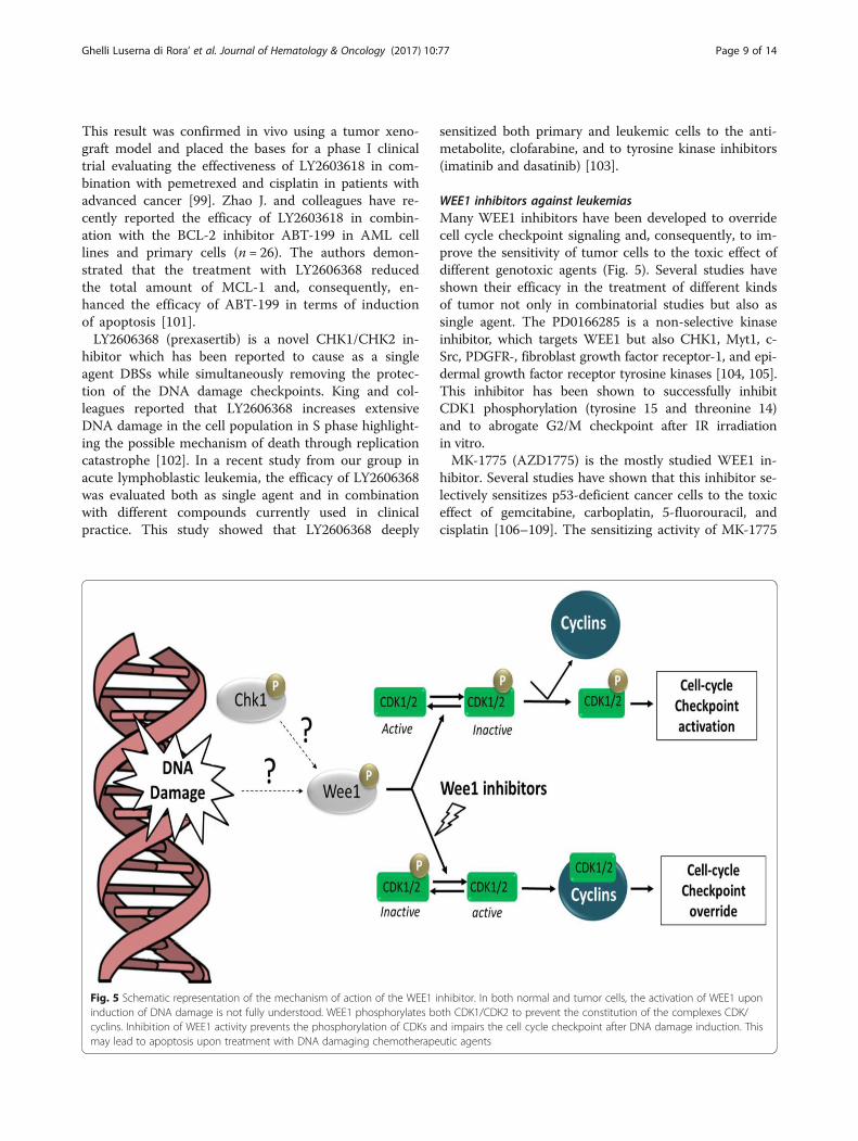
WEE1 inhibitors against leukemiasMany WEE1 inhibitors have been developed to overridecell cycle checkpoint signaling and, consequently, to im-prove the sensitivity of tumor cells to the toxic effect ofdifferent genotoxic agents (Fig. 5). Several studies haveshown their efficacy in the treatment of different kindsof tumor not only in combinatorial studies but also assingle agent. The PD0166285 is a non-selective kinaseinhibitor, which targets WEE1 but also CHK1, Myt1, c-Src, PDGFR-, fibroblast growth factor receptor-1, and epi-dermal growth factor receptor tyrosine kinases [104, 105].This inhibitor has been shown to successfully inhibitCDK1 phosphorylation (tyrosine 15 and threonine 14)and to abrogate G2/M checkpoint after IR irradiationin vitro.MK-1775 (AZD1775) is the mostly studied WEE1 in-
hibitor. Several studies have shown that this inhibitor se-lectively sensitizes p53-deficient cancer cells to the toxiceffect of gemcitabine, carboplatin, 5-fluorouracil, andcisplatin [106–109]. The sensitizing activity of MK-1775
Fig. 5 Schematic representation of the mechanism of action of the WEE1 inhibitor. In both normal and tumor cells, the activation of WEE1 uponinduction of DNA damage is not fully understood. WEE1 phosphorylates both CDK1/CDK2 to prevent the constitution of the complexes CDK/cyclins. Inhibition of WEE1 activity prevents the phosphorylation of CDKs and impairs the cell cycle checkpoint after DNA damage induction. Thismay lead to apoptosis upon treatment with DNA damaging chemotherapeutic agents
Ghelli Luserna di Rora’ et al. Journal of Hematology & Oncology (2017) 10:77 Page 9 of 14

selectively on p53-deficient cells has been shown alsoafter gamma ray irradiation. Although all the abovementioned studies, recent findings highlighted that theeffectiveness of this compound in different types oftumor is independent on the functional status of p53[110]. In hematological malignancies, recent studiesmainly on acute myeloid leukemia have shown the effi-cacy of this compound, not only as single agent [111]but also in combination with different compounds likeHDAC (vorinostat), CDK inhibitor (roscovitine),LY2603618, or cytarabine [111–116]. The combinationof cytarabine and MK-1775 enhanced chemotherapycytotoxicity by abrogating the mechanisms of DNA re-pair and by the inhibition of the S phase arrest inducedby cytarabine [110, 117]. Similar results were recentlyfound in T-ALL cell lines and in in vivo models [118].Tibes and colleagues showed the efficacy of MK-1775 inboth CML primary cell and cell lines as single agent andin combination with cytarabine. In this study, theyshowed that the inhibition of WEE1 significantly sensi-tizes leukemic cells to cytarabine in terms of reductionof cell viability and induction of apoptosis [117]. Thesedata support the development of clinical trials includingAZD1775 in combination with conventional chemother-apeutic compounds for leukemias.
Data available from clinical trials in solid tumorsA phase I dose-escalation study aiming to examine thesafety and tolerability of LY2603618 in combination withpemetrexed 500 mg/m2 every 21 days in patients withcancer defined a maximum tolerated dose (MTD) of150 mg/m2. A following phase I study using LY2603618in combination with gemcitabine in patients with solidtumors showed that among the 50 patients enrolled, fre-quent adverse events, possibly related to study drugtreatment, included fatigue (44%), decreased platelets(42%), decreased neutrophils (32%), nausea (26%), anddecreased hemoglobin (20%). Systemic exposure ofLY2603618 increased dose dependently, while clearancewas relatively dose independent. The mean LY2603618half-life varied. However, the durations were still suitablefor maintaining human exposures while minimizing ac-cumulation. LY2603618 pharmacokinetic (PK) was notaltered by gemcitabine administration. Plasma exposuresthat correlate with the maximal pharmacodynamic effectin non-clinical models were achieved for all doses. Onepatient with non-small cell lung cancer carcinomaachieved a partial response; 22 patients had stable dis-ease. They conclude that the MTD of LY2603618 com-bined with gemcitabine was 200 mg/m2, but a fixedLY2603618 dose of 230 mg combined with gemcitabinewas selected as the recommended phase II dose [99]. Aconsequent phase II study evaluating the effect ofLY2603618 in combination with pemetrexed in patients
with advanced or metastatic non-small cell lung cancerhighlighted no significant improvement of pemetrexedefficacy as single agent in non-small cell lung cancer[119]. Until now, only a phase I study has been doneusing LY2606368 as single agent in advanced solid tu-mors. Forty-five patients were treated with two differentdose-escalation schedule: from 10 to 50 mg/m2 onschedule 1 (days 1 to 3 every 14 days) or from 40 to130 mg/m2 on schedule 2 (day 1 every 14 days); sevenexperienced dose-limiting toxicities (all hematologic).The MTDs were 40 mg/m2 (schedule 1) and 105 mg/m2
(schedule 2). The most common related grade 3 or 4treatment-emergent adverse events were neutropenia,leukopenia, anemia, thrombocytopenia, and fatigue.Grade 4 neutropenia occurred in 73.3% of patients andit was transient (typically <5 days). Febrile neutropeniaincidence was low (7%). The LY2606368 exposure overthe first 72 h (area under the curve from 0 to 72 h) atthe MTD for each schedule coincided with the exposurein mouse xenografts that resulted in maximal tumor re-sponses. Minor intra- and intercycle accumulation ofLY2606368 was observed at the MTDs for both sched-ules. Two patients (4.4%) had a partial response. Fifteenpatients (33.3%) had a best overall response of stable dis-ease (range, 1.2 to 6.7 months), six of whom had squa-mous cell carcinoma. An LY2606368 dose of 105 mg/m2
once every 14 days is being evaluated as the recom-mended phase II dose in dose-expansion cohorts for pa-tients with squamous cell carcinoma. A phase I study ofsingle-agent AZD-1775 involving 25 patients with refrac-tory solid tumors showed that the MTD was establishedas 225 mg twice per day orally over 2.5 days per weekfor 2 weeks per 21-day cycle. Confirmed partial re-sponses were observed in two patients carrying BRCAmutations: one with head and neck cancer and one withovarian cancer. Common toxicities were myelosup-pression and diarrhea. The on-target efficacy of thecompound was assessed looking at the levels of phos-phorylated Tyr15-Cdk (pY15-Cdk) and γH2AX inpaired tumor biopsies obtained at the MTD [120]. A sec-ond phase I study demonstrated target inhibition (Tyr15-Cdk) at MTD in combination with carboplatin adultpatients with advanced solid tumors (NCT00648648). Pa-tients with p53 mutated ovarian cancer refractory or re-sistant (<3 months) to standard first line therapy(carboplatin plus paclitaxel) were re-exposed to carbopla-tin (AUC 5), plus five bi-daily doses of 225 mg AZD-1775 in a 21-day cycle (MTD). Bone marrow toxicity,fatigue, diarrhea, nausea, and vomiting were the mostcommon adverse events. Out of 24 patients enrolled, 22patients were evaluable for study endpoints. As best re-sponse (RECIST 1.0), six patients (27%) showed con-firmed partial response (PR) with a median progression-free survival (PFS) of 10.9 months. Nine patients (41%)
Ghelli Luserna di Rora’ et al. Journal of Hematology & Oncology (2017) 10:77 Page 10 of 14

had stable disease and seven patients (32%) had progres-sive disease as best response, with a median PFS of 5.3and 1.3 months, respectively (NCT01164995).
ConclusionsNowadays, the amount of pre-clinical data has con-firmed the efficacy of different cell cycle checkpoint in-hibitors against different kinds of hematologic as well assolid tumors, as single agent, or in combination with awide number of drugs. The efficacy as well as the safetyof different combinations is now being established alsoin several phase I/II clinical trials. Most of the studieswere based on the use of cell cycle checkpoint inhibitorsin combination with standard chemotherapy in order toenhance its effectiveness. Although the good successesthat have been achieved have many questions needed tobe answered regarding the safety and the effectiveness ofthis class of compounds. Some acute leukemia subtypesare characterized by high genetic instability that shouldmake this kind of tumor very sensitive to cell cyclecheckpoint inhibitors. However, few clones can take ad-vantage from the inhibition of DNA repair, acquire novelinvasive features, and start to proliferate. Long-periodsafety of cell cycle checkpoint inhibitors should be ad-dressed also in normal tissues in order to exclude tumortransformation of healthy cells. A second crucial ques-tion that should be answered, at least in hematologicalmalignances, is the ability of cell cycle checkpoint inhibi-tors to eradicate leukemic stem cells in the contest ofbone marrow niche. Indeed, until today, very few studieshave addressed, for example, the efficacy of the check-point inhibitors under hypoxic condition or more gen-eral in experimental settings that mime the niche micro-environment. Finally, prognostic markers should be eval-uated to stratify patients that could be more sensitive tocheckpoint kinase inhibitors. One predictive markercould be the evaluation of basal expression of elementsinvolved in the DDR and the level of genetic instability(γH2AX expression). In our opinion, based on the re-sults from the clinical trials, a last important questionshould be answered: can we substitute DNA damagingagents (chemotherapy) with DDR inhibitors in standardtherapeutic regimens in which a specific inhibitor, forexample, BCR-ABL1 inhibitors, is associated with con-ventional chemotherapy? Several studies have been doneto evaluate the chemotherapy-induced genetic instabilityin various types of cancers [121–123]. It is generallybelieved that DNA damaging compounds can posi-tively select tumor cells that harbor particular muta-tions or can increase genetic instability leading to thegeneration of novel clones with more aggressive phe-notypes. These two scenarios are the biological expla-nations for the failure of standard chemotherapy andfor tumor relapses. For the abovementioned reasons,
we speculate that a winning strategy to avoid relapsemay be to substitute chemotherapy with cell cyclecheckpoint inhibitors in the treatment of hematologicalmalignances that can be treated with specific targetedinhibitors.
AbbreviationsALL: Acute lymphoblastic leukemia; AML: Acute myeloid leukemia;ATM: Ataxia-telangiectasia mutated; ATR: ATM and Rad3 related;ATRIP: ATR-interacting protein; CDC25: Cell division cycle 25; CDK: Cyclin-dependent kinases; CDKI: CDK inhibitors; CDKN1A: Cyclin-dependentkinase inhibitor 1A; CHK1: Checkpoint kinase 1; CHK2: Checkpoint kinase2; CLL: Chronic lymphocytic leukemia; CML: Chronic myeloid leukemia;DDR: DNA damage response; DNA-PK: DNA-dependent protein kinase;MCM: Mini-chromosome maintenance; MRN: Mre11-Rad50-Nbs1;MYT1: Membrane-associated tyrosine/threonine 1; ROS: Reactive oxygenspecies; RPA: Replication protein A; SSB: Single-strand break;ssDNA: Single-strand DNA; TopBP1: Topoisomerase II binding protein 1
AcknowledgementsNot applicable
FundingThe study was funded by the University of Bologna and by the ItalianAssociation for Cancer Research (AIRC).
Availability of data and materialsData sharing is not applicable to this article as no datasets were generatedor analyzed during the current study.
Authors’ contributionsAGLDR drafted the first version of the manuscript and created the figures. IIand GM equally contributed to the final version of the manuscript and gavethe final approval. All authors read and approved the final manuscript.
Competing interestsGM has competing interests with Novartis, BMS, Roche, Pfizer, ARIAD, MSD.
Consent for publicationNot applicable
Ethics approval and consent to participateNot applicable
Publisher’s NoteSpringer Nature remains neutral with regard to jurisdictional claims inpublished maps and institutional affiliations.
Author details1Department of Hematology and Medical Sciences “L. and A. Seràgnoli”,Bologna University, Bologna, Italy. 2Present: Department of Pathology, St.Jude Children’s Research Hospital, Memphis, TN, USA.
Received: 19 January 2017 Accepted: 15 March 2017
References1. Jackson SP, Bartek J. The DNA-damage response in human biology and
disease. Nature. 2009;461:1071–8.2. Karanjawala ZE, Murphy N, Hinton DR, Hsieh CL, Lieber MR. Oxygen
metabolism causes chromosome breaks and is associated with the neuronalapoptosis observed in DNA double-strand break repair mutants. Curr Biol.2002;12:397–402.
3. Sallmyr A, Fan J, Rassool FV. Genomic instability in myeloid malignancies:increased reactive oxygen species (ROS), DNA double strand breaks (DSBs)and error-prone repair. Cancer Lett. 2008;1–9.
4. Velic D, Couturier A, Ferreira M, Rodrigue A, Poirier G, Fleury F, Masson J-Y.DNA damage signalling and repair inhibitors: the long-sought-after Achilles’heel of cancer. Biomolecules. 2015;5:3204–59.
Ghelli Luserna di Rora’ et al. Journal of Hematology & Oncology (2017) 10:77 Page 11 of 14

5. Shiloh Y, Ziv Y. The ATM protein kinase: regulating the cellular response togenotoxic stress, and more. Nat Rev Mol Cell Biol. 2013;14:197–210.
6. Falck J, Coates J, Jackson SP. Conserved modes of recruitment of ATM, ATRand DNA-PKcs to sites of DNA damage. Nature. 2005;434:605–11.
7. Uziel T, Lerenthal Y, Moyal L, Andegeko Y, Mittelman L, Shiloh Y.Requirement of the MRN complex for ATM activation by DNA damage.EMBO J. 2003;22:5612–21.
8. Lamarche BJ, Orazio NI, Weitzman MD. The MRN complex in double-strandbreak repair and telomere maintenance. FEBS Lett. 2010;3682–3695.
9. Girard P-M, Riballo E, Begg AC, Waugh A, Jeggo PA. Nbs1 promotes ATMdependent phosphorylation events including those required for G1/S arrest.Oncogene. 2002;4191–9.
10. Williams RS, Williams JS, Tainer JA, Williams RS, Williams JS, Tainer JA.Mre11–Rad50–Nbs1 is a keystone complex connecting DNA repairmachinery, double-strand break signaling, and the chromatin template.Biochem Cell Biol. 2007;85:509–20.
11. Williams RS, Moncalian G, Williams JS, Yamada Y, Limbo O, Shin DS,Groocock LM, Cahill D, Hitomi C, Guenther G, Moiani D, Carney JP, Russell P,Tainer JA. Mre11 dimers coordinate DNA end bridging and nucleaseprocessing in double-strand-break repair. Cell. 2008;135:97–109.
12. De Jager M, Van Noort J, Van Gent DC, Dekker C, Kanaar R, Wyman C.Human Rad50/Mre11 is a flexible complex that can tether DNA ends. MolCell. 2001;8:1129–35.
13. Lee J-H, Paull TT. ATM activation by DNA double-strand breaks through theMre11-Rad50-Nbs1 complex. Sci (New York, NY). 2005;308:551–4.
14. Mirzoeva OK, Petrini JH. DNA damage-dependent nuclear dynamics of theMre11 complex. Mol Cell Biol. 2001;21:281–8.
15. Bakkenist CJ, Kastan MB. DNA damage activates ATM through intermolecularautophosphorylation and dimer dissociation. Nature. 2003;421:499–506.
16. Kozlov SV, Graham ME, Jakob B, Tobias F, Kijas AW, Tanuji M, Chen P,Robinson PJ, Taucher-Scholz G, Suzuki K, So S, Chen D, Lavin MF.Autophosphorylation and ATM activation: additional sites add to thecomplexity. J Biol Chem. 2011;286:9107–19.
17. Zou L, Elledge SJ. Sensing DNA damage through ATRIP recognition of RPA-ssDNA complexes. Science. 2003;300(June):1542–8.
18. Byun TS, Pacek M, Yee MC, Walter JC, Cimprich KA. Functional uncouplingof MCM helicase and DNA polymerase activities activates the ATR-dependent checkpoint. Genes Dev. 2005;19:1040–52.
19. Weber AM, Ryan AJ. ATM and ATR as therapeutic targets in cancer.Pharmacol Ther. 2015;124–138.
20. Liu S, Shiotani B, Lahiri M, Maréchal A, Tse A, Leung CCY, Glover JNM,Yang XH, Zou L. ATR autophosphorylation as a molecular switch forcheckpoint activation. Mol Cell. 2011;43:192–202.
21. Nam EA, Zhao R, Glick GG, Bansbach CE, Friedman DB, Cortez D. Thr-1989phosphorylation is a marker of active ataxia telangiectasia-mutated andRad3-related (ATR) kinase. J Biol Chem. 2011;286:28707–14.
22. Mordes DA, Cortez D. Activation of ATR and related PIKKs. Cell Cycle.2008;2809–2812.
23. Cuadrado M, Martinez-Pastor B, Murga M, Toledo LI, Gutierrez-Martinez P,Lopez E, Fernandez-Capetillo O. ATM regulates ATR chromatin loading inresponse to DNA double-strand breaks. J Exp Med. 2006;203:297–303.
24. Satyanarayana A, Kaldis P. Mammalian cell-cycle regulation: several Cdks,numerous cyclins and diverse compensatory mechanisms. Oncogene.2009;28:2925–39.
25. Lim S, Kaldis P. Cdks, cyclins and CKIs: roles beyond cell cycle regulation.Development. 2013;140:3079–93.
26. Khosravi R, Maya R, Gottlieb T, Oren M, Shiloh Y, Shkedy D. Rapid ATM-dependent phosphorylation of MDM2 precedes p53 accumulation inresponse to DNA damage. Proc Natl Acad Sci. 1999;96:14973–7.
27. Origanti S, Cai S, Munir AZ, White LS, Piwnica-Worms H. Synthetic lethalityof Chk1 inhibition combined with p53 and/or p21 loss during a DNAdamage response in normal and tumor cells. Oncogene. 2013;32(5):577–88.
28. Bartek J, Lukas J. Chk1 and Chk2 kinases in checkpoint control and cancer.Cancer Cell. 2003;421–429.
29. Chipuk JE, Green DR. Dissecting p53-dependent apoptosis. Cell Death Differ.2006;13:994–1002.
30. Donzelli M, Draetta GF. Regulating mammalian checkpoints through Cdc25inactivation. EMBO Rep. 2003;4:671–7.
31. Schmitt E, Boutros R, Froment C, Monsarrat B, Ducommun B, Dozier C. CHK1phosphorylates CDC25B during the cell cycle in the absence of DNAdamage. J Cell Sci. 2006;119(Pt 20):4269–75.
32. Forrest A, Gabrielli B. Cdc25B activity is regulated by 14-3-3. Oncogene.2001;20:4393–401.
33. Do K, Doroshow JH, Kummar S. Wee1 kinase as a target for cancer therapy.Cell Cycle. 2013;12(19):3159–64.
34. Watanabe N, Arai H, Iwasaki J-I, Shiina M, Ogata K, Hunter T, Osada H.Cyclin-dependent kinase (CDK) phosphorylation destabilizes somatic Wee1via multiple pathways. Proc Natl Acad Sci U S A. 2005;102:11663–8.
35. Perry JA, Kornbluth S. Cdc25 and Wee1: analogous opposites? Cell Div.2007;2:12.
36. Guardavaccaro D, Pagano M. Stabilizers and destabilizers controlling cellcycle oscillators. Mol Cell. 2006;1–4.
37. Malumbres M, Barbacid M. Cell cycle, CDKs and cancer: a changingparadigm. Nat Rev Cancer. 2009;9:153–66.
38. Lavin MF. Ataxia-telangiectasia: from a rare disorder to a paradigm for cellsignalling and cancer. Nat Rev Mol Cell Biol. 2008;9:759–69.
39. Teive HA, Moro A, Moscovich M, Arruda WO, Munhoz RP, Raskin S, Ashizawa T.Ataxia-telangiectasia—a historical review and a proposal for a newdesignation: ATM syndrome. J Neurol Sci. 2015;355:3–6.
40. Negrini S, Gorgoulis VG, Halazonetis TD. Genomic instability—an evolvinghallmark of cancer. Nat Rev Mol Cell Biol. 2010;11:220–8.
41. Haidar MA, Kantarjian H, Manshouri T, Chang CY, O’Brien S, Freireich E,Keating M, Albitar M. ATM gene deletion in patients with adult acutelymphoblastic leukemia. Cancer. 2000;88:1057–62.
42. Network TCGAR. Genomic and epigenomic landscapes of adult de novoacute myeloid leukemia. N Engl J Med. 2013;368:2059–74.
43. Melo JV, Kumberova A, van Dijk AG, Goldman JM, Yuille MR. Investigationon the role of the ATM gene in chronic myeloid leukaemia. Leukemia.2001;15:1448–50.
44. Takagi M, Sato M, Piao J, Miyamoto S, Isoda T, Kitagawa M, Honda H,Mizutani S. ATM-dependent DNA damage-response pathway as adeterminant in chronic myelogenous leukemia. DNA Repair (Amst).2013;12:500–7.
45. Starostik P, Manshouri T, Brien SO, Leukemia BCL, Lerner S, Keating M.Deficiency of the ATM protein expression defines an aggressive subgroupof B-cell chronic lymphocytic leukemia. Cancer Res. 1998;4552–4557.
46. Puente XS, Beà S, Valdés-Mas R, Villamor N, Gutiérrez-Abril J, Martín-Subero JI,Munar M, Rubio-Pérez C, Jares P, Aymerich M, Baumann T, Beekman R,Belver L, Carrio A, Castellano G, Clot G, Colado E, Colomer D, Costa D,Delgado J, Enjuanes A, Estivill X, Ferrando AA, Gelpí JL, González B,González S, González M, Gut M, Hernández-Rivas JM, López-Guerra M, etal. Non-coding recurrent mutations in chronic lymphocytic leukaemia.Nature. 2015;526:519–24.
47. Jiang Y, Chen H-C, Su X, Thompson P, Liu X, Do K-A, Wierda W, Keating M,Plunkett W. ATM function and its relationship with ATM gene mutations inchronic lymphocytic leukemia with the recurrent deletion (11q22.3-23.2).Blood Cancer J. 2016;6:e465.
48. Rossi D, Gaidano G. ATM and chronic lymphocytic leukemia: mutations, andnot only deletions, matter. Haematologica. 2012;97:5–8.
49. Dolnik A, Engelmann JC, Scharfenberger-schmeer M, Mauch J,Haldemann B, Fries T, Krönke J, Kühn MWM, Kayser S, Wolf S, Gaidzik VI,Schlenk RF, Rücker FG, Döhner H, Lottaz C, Döhner K, Bullinger L, Kelkenberg-schade S, Kro J, Ku MWM. Commonly altered genomic regions in acutemyeloid leukemia are enriched for somatic mutations involved in chromatinremodeling and splicing commonly altered genomic regions in acute myeloidleukemia are enriched for somatic mutations involved in chromatin. Blood.2013;120:83–93.
50. Offit K, Pierce H, Kirchhoff T, Kolachana P, Rapaport B, Gregersen P,Johnson S, Yossepowitch O, Huang H, Satagopan J, Robson M, Scheuer L,Nafa K, Ellis N. Frequency of CHEK2*1100delC in New York breast cancercases and controls. BMC Med Genet. 2003;4:1.
51. Thompson D, Seal S, Schutte M, McGuffog L, Barfoot R, Renwick A, Eeles R,Sodha N, Houlston R, Shanley S, Klijn J, Wasielewski M, Chang-Claude J,Futreal PA, Weber BL, Nathanson KL, Stratton M, Meijers-Heijboer H,Rahman N, Easton DF. A multicenter study of cancer incidence inCHEK2 1100delC mutation carriers. Cancer Epidemiol Biomarkers Prev.2006;15:2542–5.
52. Hangaishi A, Ogawa S, Qiao Y, Wang L, Hosoya N, Yuji K, Imai Y, Takeuchi K,Miyawaki S, Hirai H. Neoplasms, mutations of Chk2 in primary hematopoietic.Blood. 2002;99:3075–8.
53. Hofmann WK, Miller CW, Tsukasaki K, Tavor S, Ikezoe T, Hoelzer D,Takeuchi S, Koeffler HP. Mutation analysis of the DNA-damage
Ghelli Luserna di Rora’ et al. Journal of Hematology & Oncology (2017) 10:77 Page 12 of 14

checkpoint gene CHK2 in myelodysplastic syndromes and acutemyeloid leukemias. Leuk Res. 2001;25:333–8.
54. Rudd MF, Sellick GS, Webb EL, Catovsky D, Houlston RS. Variants in theATM-BRCA2-CHEK2 axis predispose to chronic lymphocytic leukemia. Blood.2006;108:638–44.
55. Bartkova J, Hamerlik P, Stockhausen M-T, Ehrmann J, Hlobilkova A,Laursen H, Kalita O, Kolar Z, Poulsen HS, Broholm H, Lukas J, Bartek J.Replication stress and oxidative damage contribute to aberrantconstitutive activation of DNA damage signalling in human gliomas.Oncogene. 2010;29:5095–102.
56. Halazonetis TD, Gorgoulis VG, Bartek J. An oncogene-induced DNA damagemodel for cancer development. Science. 2008;319:1352–5.
57. Brown EJ, Baltimore D. Essential and dispensable roles of ATR in cell cyclearrest and genome maintenance. Genes Dev. 2003;17:615–28.
58. De Klein A, Muijtjens M, Van Os R, Verhoeven Y, Smit B, Carr AM, Lehmann AR,Hoeijmakers JHJ. Targeted disruption of the cell-cycle checkpoint gene ATRleads to early embryonic lethality in mice. Curr Biol. 2000;10:479–82.
59. Sawyers CL. Molecular consequences of the BCR-ABL translocation inchronic myelogenous leukemia. Leuk Lymphoma. 1993;11 Suppl 2:101–3.
60. Kim KT, Baird K, Davis S, Piloto O, Levis M, Li L, Chen P, Meltzer P, Small D.Constitutive Fms-like tyrosine kinase 3 activation results in specificchanges in gene expression in myeloid leukaemic cells. Br J Haematol.2007;138:603–15.
61. Faderl S, O’Brien S, Pui C-H, Stock W, Wetzler M, Hoelzer D, Kantarjian HM.Adult acute lymphoblastic leukemia: concepts and strategies. Cancer.2010;116:1165–76.
62. Muvarak N, Kelley S, Robert C, Baer MR, Perrotti D, Gambacorti-Passerini C,Civin C, Scheibner K, Rassool F. c-MYC generates repair errors via increasedtranscription of alternative-NHEJ factors, LIG3 and PARP1, in tyrosine kinase-activated leukemias. Mol Cancer Res. 2015;13:699–712.
63. Cavelier C, Didier C, Prade N, Mansat-De Mas V, Manenti S, Recher C, Demur C,Ducommun B. Constitutive activation of the DNA damage signaling pathwayin acute myeloid leukemia with complex karyotype: potential importance forcheckpoint targeting therapy. Cancer Res. 2009;69:8652–61.
64. Iacobucci I, Di Rorà AGL, Falzacappa MVV, Agostinelli C, Derenzini E,Ferrari A, Papayannidis C, Lonetti A, Righi S, Imbrogno E, Pomella S,Venturi C, Guadagnuolo V, Cattina F, Ottaviani E, Abbenante MC, Vitale A,Elia L, Russo D, Zinzani PL, Pileri S, Pelicci PG, Martinelli G. In vitro and invivo single-agent efficacy of checkpoint kinase inhibition in acutelymphoblastic leukemia. J Hematol Oncol. 2015;8:125.
65. Sarmento LM, Póvoa V, Nascimento R, Real G, Antunes I, Martins LR,Moita C, Alves PM, Abecasis M, Moita LF, Parkhouse RME, Meijerink JPP,Barata JT. CHK1 overexpression in T-cell acute lymphoblastic leukemia isessential for proliferation and survival by preventing excessivereplication stress. Oncogene. 2015;34(23):2978–90.
66. Nieborowska-Skorska M, Stoklosa T, Datta M, Czechowska A, Rink L,Slupianek A, Koptyra M, Seferynska I, Krszyna K, Blasiak J, Skorski T. ATR-Chk1axis protects BCR/ABL leukemia cells from the lethal effect of DNA double-strand breaks. Cell Cycle. 2006;5:994–1000.
67. Hickson I, Zhao Y, Richardson CJ, Green SJ, Martin NMB, Orr AI, Reaper PM,Jackson SP, Curtin NJ, Smith GCM. Identification and characterization of anovel and specific inhibitor of the ataxia-telangiectasia mutated kinase ATM.Cancer Res. 2004;64:9152–9.
68. Korwek Z, Sewastianik T, Bielak-Zmijewska A, Mosieniak G, Alster O,Moreno-Villaneuva M, Burkle A, Sikora E. Inhibition of ATM blocks theetoposide-induced DNA damage response and apoptosis of restinghuman T cells. DNA Repair (Amst). 2012;11:864–73.
69. Batey MA, Zhao Y, Kyle S, Richardson C, Slade A, Martin NMB, Lau A,Newell DR, Curtin NJ. Preclinical evaluation of a novel ATM inhibitor,KU59403, in vitro and in vivo in p53 functional and dysfunctionalmodels of human cancer. Mol Cancer Ther. 2013;12:959–67.
70. Grosjean-Raillard J, Tailler M, Adès L, Perfettini J-L, Fabre C, Braun T,De Botton S, Fenaux P, Kroemer G. ATM mediates constitutive NF-kappaB activation in high-risk myelodysplastic syndrome and acutemyeloid leukemia. Oncogene. 2009;28:1099–109.
71. Nishida H, Tatewaki N, Nakajima Y, Magara T, Ko KM, Hamamori Y, Konishi T.Inhibition of ATR protein kinase activity by schisandrin B in DNA damageresponse. Nucleic Acids Res. 2009;37:5678–89.
72. Charrier JD, Durrant SJ, Golec JMC, Kay DP, Knegtel RMA, MacCormick S,Mortimore M, O’Donnell ME, Pinder JL, Reaper PM, Rutherford AP, Wang PSH,Young SC, Pollard JR. Discovery of potent and selective inhibitors of ataxia
telangiectasia mutated and Rad3 related (ATR) protein kinase as potentialanticancer agents. J Med Chem. 2011;54:2320–30.
73. Šalovská B, Fabrik I, Ďurišová K, Link M, Vávrová J, Řezáčová M, Tichý A.Radiosensitization of human leukemic HL-60 cells by ATR kinase inhibitor(VE-821): phosphoproteomic analysis. Int J Mol Sci. 2014;15:12007–26.
74. Fokas E, Prevo R, Hammond EM, Brunner TB, McKenna WG, Muschel RJ.Targeting ATR in DNA damage response and cancer therapeutics. CancerTreat Rev. 2014;109–117.
75. Pires IM, Olcina MM, Anbalagan S, Pollard JR, Reaper PM, Charlton PA,McKenna WG, Hammond EM. Targeting radiation-resistant hypoxic tumourcells through ATR inhibition. Br J Cancer. 2012;107:291–9.
76. Prevo R, Fokas E, Reaper PM, Charlton PA, Pollard JR, McKenna WG,Muschel RJ, Brunner TB. The novel ATR inhibitor VE-821 increasessensitivity of pancreatic cancer cells to radiation and chemotherapy.Cancer Biol Ther. 2012;13:1072–81.
77. Fokas E, Prevo R, Pollard JR, Reaper PM, Charlton PA, Cornelissen B, Vallis KA,Hammond EM, Olcina MM, Gillies McKenna W, Muschel RJ, Brunner TB.Targeting ATR in vivo using the novel inhibitor VE-822 results in selectivesensitization of pancreatic tumors to radiation. Cell Death Dis. 2012;3, e441.
78. Vávrová J, Zárybnická L, Lukášová E, Řezáčová M, Novotná E, Sinkorová Z,Tichý A, Pejchal J, Durišová K. Inhibition of ATR kinase with the selectiveinhibitor VE-821 results in radiosensitization of cells of promyelocyticleukaemia (HL-60). Radiat Environ Biophys. 2013;52:471–9.
79. Jossé R, Martin SE, Guha R, Ormanoglu P, Pfister TD, Reaper PM, Barnes CS,Jones J, Charlton P, Pollard JR, Morris J, Doroshow JH, Pommier Y. ATRinhibitors VE-821 and VX-970 sensitize cancer cells to topoisomerase Iinhibitors by disabling DNA replication initiation and fork elongationresponses. Cancer Res. 2014;74:6968–78.
80. Hall AB, Newsome D, Wang Y, Boucher DM, Eustace B, Gu Y, Hare B,Johnson MA, Milton S, Murphy CE, Takemoto D, Tolman C, Wood M,Charlton P, Charrier J-D, Furey B, Golec J, Reaper PM, Pollard JR. Potentiationof tumor responses to DNA damaging therapy by the selective ATRinhibitor VX-970. Oncotarget. 2014;5:5674–85.
81. Jones CD, Blades K, Foote KM, Guichard SM, Jewsbury, Philip J. McGuire T,Nissink JW, Odedra R, Tam K, Thommes P, Turner P, Wilkinson G, Wood C,Yates JW. Discovery of AZD6738, a potent and selective inhibitor with thepotential to test the clinical efficacy of ATR kinase inhibition in cancerpatients. [abstract]. In: Proceedings of the 104th Annual Meeting of theAmerican Association for Cancer Research; 2013 Apr 6-10; 2013.
82. Vendetti FP, Lau A, Schamus S, Conrads TP, O’Connor MJ, Bakkenist CJ. Theorally active and bioavailable ATR kinase inhibitor AZD6738 potentiates theanti-tumor effects of cisplatin to resolve ATM-deficient non-small cell lungcancer in vivo. Oncotarget. 2015;6:44289–305.
83. Menezes DL, Holt J, Tang Y, Feng J, Barsanti P, Pan Y, Ghoddusi M, Zhang W,Thomas G, Holash J, Lees E, Taricani L. A synthetic lethal screen revealsenhanced sensitivity to ATR inhibitor treatment in mantle cell lymphoma withATM loss-of-function. Mol Cancer Res. 2014;13:0240.2014.
84. Kwok M, Davies N, Agathanggelou A, Smith E, Petermann E, Yates E, Brown J,Lau A, Stankovic T. Synthetic lethality in chronic lymphocytic leukaemia withDNA damage response defects by targeting the ATR pathway. Lancet. 2015;385 Suppl 1:S58.
85. Curtin NJ. DNA repair dysregulation from cancer driver to therapeutictarget. Nat Rev Cancer. 2012;12:801–17.
86. Daud AI, Ashworth MT, Strosberg J, Goldman JW, Mendelson D, Springett G,Venook AP, Loechner S, Rosen LS, Shanahan F, Parry D, Shumway S,Grabowsky JA, Freshwater T, Sorge C, Kang SP, Isaacs R, Munster PN. Phase Idose-escalation trial of checkpoint kinase 1 inhibitor MK-8776 asmonotherapy and in combination with gemcitabine in patients withadvanced solid tumors. J Clin Oncol. 2015;33:1060–6.
87. Prudhomme M. Novel checkpoint 1 inhibitors. Recent Pat Anticancer DrugDiscov. 2006;1:55–68.
88. Welch S, Hirte HW, Carey MS, Hotte SJ, Tsao MS, Brown S, Pond GR, Dancey JE,Oza AM. UCN-01 in combination with topotecan in patients with advancedrecurrent ovarian cancer: a study of the Princess Margaret Hospital Phase IIconsortium. Gynecol Oncol. 2007;106:305–10.
89. Ma CX, Ellis MJC, Petroni GR, Guo Z, Cai SR, Ryan CE, Craig Lockhart A,Naughton MJ, Pluard TJ, Brenin CM, Picus J, Creekmore AN, Mwandoro T,Yarde ER, Reed J, Ebbert M, Bernard PS, Watson M, Doyle LA, Dancey J,Piwnica-Worms H, Fracasso PM. A phase II study of UCN-01 in combinationwith irinotecan in patients with metastatic triple negative breast cancer.Breast Cancer Res Treat. 2013;137:483–92.
Ghelli Luserna di Rora’ et al. Journal of Hematology & Oncology (2017) 10:77 Page 13 of 14

90. Engelke CG, Parsels LA, Qian Y, Zhang Q, Karnak D, Robertson JR, Tanska DM,Wei D, Davis MA, Parsels JD, Zhao L, Greenson JK, Lawrence TS, Maybaum J,Morgan MA. Sensitization of pancreatic cancer to chemoradiation by the Chk1inhibitor MK8776. Clin Cancer Res. 2013;19:4412–21.
91. Grabauskiene S, Bergeron EJ, Chen G, Chang AC, Lin J, Thomas DG,Giordano TJ, Beer DG, Morgan MA, Reddy RM. CHK1 levels correlate withsensitization to pemetrexed by CHK1 inhibitors in non-small cell lungcancer cells. Lung Cancer. 2013;82:477–84.
92. Dai Y, Chen S, Kmieciak M, Zhou L, Lin H, Pei X-Y, Grant S. The novel Chk1inhibitor MK-8776 sensitizes human leukemia cells to HDAC inhibitors bytargeting the intra-S checkpoint and DNA replication and repair. Mol CancerTher. 2013;12:878–89.
93. Zemanova J, Hylse O, Collakova J, Vesely P, Oltova A, Borsky M, Zaprazna K,Kasparkova M, Janovska P, Verner J, Kohoutek J, Dzimkova M, Bryja V,Jaskova Z, Brychtova Y, Paruch K, Trbusek M. Chk1 inhibition significantlypotentiates activity of nucleoside analogs in TP53-mutated B-lymphoid cells.Oncotarget. 2016;7.
94. Morgan MA, Parsels LA, Zhao L, Parsels JD, Davis MA, Hassan MC,Arumugarajah S, Hylander-Gans L, Morosini D, Simeone DM, Canman CE,Normolle DP, Zabludoff SD, Maybaum J, Lawrence TS. Mechanism ofradiosensitization by the Chk1/2 inhibitor AZD7762 involves abrogation ofthe G2 checkpoint and inhibition of homologous recombinational DNArepair. Cancer Res. 2010;70:4972–81.
95. Landau HJ, McNeely SC, Nair JS, Comenzo RL, Asai T, Friedman H, Jhanwar SC,Nimer SD, Schwartz GK. The checkpoint kinase inhibitor AZD7762 potentiateschemotherapy-induced apoptosis of p53-mutated multiple myeloma cells. MolCancer Ther. 2012;1781–1788.
96. Didier C, Demur C, Grimal F, Jullien D, Manenti S, Ducommun B. Evaluationof checkpoint kinase targeting therapy in acute myeloid leukemia withcomplex karyotype. Cancer Biol Ther. 2012;13:307–13.
97. Kim MK, James J, Annunziata CM. Topotecan synergizes with CHEK1(CHK1) inhibitor to induce apoptosis in ovarian cancer cells. BMCCancer. 2015;15:1–10.
98. Nguyen T, Hawkins E, Kolluri A, Kmieciak M, Park H, Lin H, Grant S. Synergismbetween bosutinib (SKI-606) and the Chk1 inhibitor (PF-00477736) in highlyimatinib-resistant BCR/ABL+ leukemia cells. Leuk Res. 2015;39:65–71.
99. Calvo E, Chen VJ, Marshall M, Ohnmacht U, Hynes SM, Kumm E, Diaz HB,Barnard D, Merzoug FF, Huber L, Kays L, Iversen P, Calles A, Voss B, Lin AB,Dickgreber N, Wehler T, Sebastian M. Preclinical analyses and phase Ievaluation of LY2603618 administered in combination with pemetrexedand cisplatin in patients with advanced cancer. Investig New Drugs.2014;955–968.
100. King C, Diaz H, Barnard D, Barda D, Clawson D, Blosser W, Cox K, Guo S,Marshall M. Characterization and preclinical development of LY2603618: aselective and potent Chk1 inhibitor. Invest New Drugs. 2014;32:213–26.
101. Zhao J, Niu X, Li X, Edwards H, Wang G, Wang Y, Taub JW, Lin H, Ge Y.Inhibition of CHK1 enhances cell death induced by the Bcl-2-selectiveinhibitor ABT-199 in acute myeloid leukemia cells. Oncotarget. 2016;7:16–8.
102. King C, Diaz HB, McNeely S, Barnard D, Dempsey J, Blosser W, Beckmann R,Barda D, Marshall MS. LY2606368 causes replication catastrophe and anti-tumor effects through CHK1-dependent mechanisms. Mol Cancer Ther. 2015.
103. Di Rorà AGL, Iacobucci I, Imbrogno E, Papayannidis C, Derenzini E, Ferrari A,Guadagnuolo V, Robustelli V, Parisi S, Sartor C, Abbenante MC, Paolini S,Martinelli G. Prexasertib, a Chk1/Chk2 inhibitor, increases the effectivenessof conventional therapy in B-/T-cell progenitor acute lymphoblasticleukemia. Oncotarget. 2016;7:53377–91.
104. Panek RL, Lu GH, Klutchko SR, Batley BL, Dahring TK, Hamby JM, Hallak H,Doherty AM, Keiser JA. In vitro pharmacological characterization of PD166285, a new nanomolar potent and broadly active protein tyrosine kinaseinhibitor. J Pharmacol Exp Ther. 1997;283:1433–44.
105. Wang Y, Li J, Booher RN, Kraker A, Lawrence T, Leopold WR, Sun Y.Radiosensitization of p53 mutant cells by PD0166285, a novel G2checkpoint abrogator. Cancer Res. 2001;61:8211–7.
106. Hirai H, Iwasawa Y, Okada M, Arai T, Nishibata T, Kobayashi M, Kimura T,Kaneko N, Ohtani J, Yamanaka K, Itadani H, Takahashi-Suzuki I, Fukasawa K,Oki H, Nambu T, Jiang J, Sakai T, Arakawa H, Sakamoto T, Sagara T,Yoshizumi T, Mizuarai S, Kotani H. Small-molecule inhibition of Wee1 kinaseby MK-1775 selectively sensitizes p53-deficient tumor cells to DNA-damaging agents. Mol Cancer Ther. 2009;8:2992–3000.
107. Hirai H, Arai T, Okada M, Nishibata T, Kobayashi M, Sakai N, Imagaki K,Ohtani J, Sakai T, Yoshizumi T, Mizuarai S, Iwasawa Y, Kotani H. MK-1775, a
small molecule Wee1 inhibitor, enhances antitumor efficacy of various DNA-damaging agents, including 5-fluorouracil. Cancer Biol Ther. 2010;9:514–22.
108. Bridges KA, Hirai H, Buser CA, Brooks C, Liu H, Buchholz TA, Molkentine JM,Mason KA, Meyn RE. MK-1775, a novel wee1 kinase inhibitor, radiosensitizesp53-defective human tumor cells. Clin Cancer Res. 2011;17:5638–48.
109. Kreahling JM, Gemmer JY, Reed D, Letson D, Bui M, Altiok S. MK1775, aselective Wee1 inhibitor, shows single-agent antitumor activity againstsarcoma cells. Mol Cancer Ther. 2012;11:174–82.
110. Van Linden AA, Baturin D, Ford JB, Fosmire SP, Gardner L, Korch C, Reigan P,Porter CC. Inhibition of Wee1 sensitizes cancer cells to antimetabolitechemotherapeutics in vitro and in vivo, independent of p53 functionality.Mol Cancer Ther. 2013;12:2675–84.
111. Qi W, Xie C, Li C, Caldwell J, Edwards H, Taub JW, Wang Y, Lin H, Ge Y.CHK1 plays a critical role in the anti-leukemic activity of the wee1 inhibitorMK-1775 in acute myeloid leukemia cells. J Hematol Oncol. 2014;7:53.
112. Zhou L, Zhang Y, Chen S, Kmieciak M, Leng Y, Lin H, Rizzo KA, Dumur CI,Ferreira-Gonzalez A, Dai Y, Grant S. A regimen combining the Wee1inhibitor AZD1775 with HDAC inhibitors targets human acute myeloidleukemia cells harboring various genetic mutations. Leukemia. 2015;29:807–18.
113. Qi W, Zhang W, Edwards H, Chu R, Madlambayan GJ, Taub JW, Wang Z,Wang Y, Li C, Lin H, Ge Y. Synergistic anti-leukemic interactions betweenpanobinostat and MK-1775 in acute myeloid leukemia ex vivo. Cancer BiolTher. 2015.
114. Chaudhuri L, Vincelette ND, Koh BD, Naylor RM, Flatten KS, Peterson KL,McNally A, Gojo I, Karp JE, Mesa RA, Sproat LO, Bogenberger JM,Kaufmann SH, Tibes R. CHK1 and WEE1 inhibition combinesynergistically to enhance therapeutic efficacy in acute myeloidleukemia ex vivo. Haematologica. 2014;99:688–96.
115. Guertin AD, Martin MM, Roberts B, Hurd M, Qu X, Miselis NR, Liu Y, Li J,Feldman I, Benita Y, Bloecher A, Toniatti C, Shumway SD. Uniquefunctions of CHK1 and WEE1 underlie synergistic anti-tumor activityupon pharmacologic inhibition. Cancer Cell Int. 2012;12:45.
116. Russell MR, Levin K, Rader J, Belcastro L, Li Y, Martinez D, Pawel B,Shumway SD, Maris JM, Cole KA. Combination therapy targeting theChk1 and Wee1 kinases shows therapeutic efficacy in neuroblastoma.Cancer Res. 2013;73:776–84.
117. Tibes R, Bogenberger JM, Chaudhuri L, Hagelstrom RT, Chow D, Buechel ME,Gonzales IM, Demuth T, Slack J, Mesa RA, Braggio E, Yin HH, Arora S,Azorsa DO. RNAi screening of the kinome with cytarabine in leukemias.Blood. 2012;119:2863–72.
118. Ford JB, Baturin D, Burleson TM, Van Linden AA, Kim Y, Porter CC. AZD1775sensitizes T cell acute lymphoblastic leukemia cells to cytarabine bypromoting apoptosis over DNA repair. Oncotarget. 2015;6:28001–10.
119. Scagliotti G, Kang JH, Smith D, Rosenberg R, Park K, Kim SW, Su WC,Boyd TE, Richards DA, Novello S, Hynes SM, Myrand SP, Lin J, Smyth EN,Wijayawardana S, Lin AB, Pinder-Schenck M. Phase II evaluation of LY2603618,a first-generation CHK1 inhibitor, in combination with pemetrexed in patientswith advanced or metastatic non-small cell lung cancer. Invest New Drugs.2016;34:625–35.
120. Do K, Wilsker D, Ji J, Zlott J, Freshwater T, Kinders RJ, Collins J, Chen AP,Doroshow JH, Kummar S. Phase I study of single-agent AZD1775 (MK-1775),a Wee1 kinase inhibitor, in patients with refractory solid tumors. J ClinOncol. 2015;33:3409–15.
121. Lopez de Mesa R, Lopez de Cerain Salsamendi A, Ariznabarreta LS,Calasanz Abinzano MJ, Patino-Garcia A. Measurement and analysis ofthe chemotherapy-induced genetic instability in pediatric cancerpatients. Mutagenesis. 2002;17:171–5.
122. Kamat N, Khidhir MA, Hussain S, Alashari MM, Rannug U. Chemotherapyinduced microsatellite instability and loss of heterozygosity in chromosomes 2,5, 10, and 17 in solid tumor patients. Cancer Cell Int. 2014;1–9.
123. Finette BA, Homans AC, Albertini RJ. Emergence of genetic instability inchildren treated for leukemia. Science. 2000;288:514–7.
Ghelli Luserna di Rora’ et al. Journal of Hematology & Oncology (2017) 10:77 Page 14 of 14


