Embed Size (px)
Citation preview

Published Ahead of Print 9 December 2013. 2014, 34(4):673. DOI: 10.1128/MCB.01359-13. Mol. Cell. Biol.
O'Meara, Aaron P. Mitchell and J. Andrew AlspaughTeresa R. O'Meara, Wenjie Xu, Kyla M. Selvig, Matthew J. Genes Required for Adaptation to the HostTranscription Factor Directly Regulates The Cryptococcus neoformans Rim101
http://mcb.asm.org/content/34/4/673Updated information and services can be found at:
These include:
SUPPLEMENTAL MATERIAL Supplemental material
REFERENCEShttp://mcb.asm.org/content/34/4/673#ref-list-1at:
This article cites 68 articles, 44 of which can be accessed free
CONTENT ALERTS more»articles cite this article),
Receive: RSS Feeds, eTOCs, free email alerts (when new
http://journals.asm.org/site/misc/reprints.xhtmlInformation about commercial reprint orders: http://journals.asm.org/site/subscriptions/To subscribe to to another ASM Journal go to:
on August 12, 2014 by U
NIV
OF
TO
RO
NT
Ohttp://m
cb.asm.org/
Dow
nloaded from
on August 12, 2014 by U
NIV
OF
TO
RO
NT
Ohttp://m
cb.asm.org/
Dow
nloaded from

The Cryptococcus neoformans Rim101 Transcription Factor DirectlyRegulates Genes Required for Adaptation to the Host
Teresa R. O’Meara,a Wenjie Xu,b Kyla M. Selvig,a Matthew J. O’Meara,c Aaron P. Mitchell,b J. Andrew Alspaugha
Departments of Medicine and Molecular Genetics and Microbiology, Duke University School of Medicine, Durham, North Carolina, USAa; Department of BiologicalSciences, Carnegie Mellon University, Pittsburgh, Pennsylvania, USAb; Department of Computer Science, University of North Carolina—Chapel Hill, Chapel Hill, NorthCarolina, USAc
The Rim101 protein is a conserved pH-responsive transcription factor that mediates important interactions between several fun-gal pathogens and the infected host. In the human fungal pathogen Cryptococcus neoformans, the Rim101 protein retains con-served functions to allow the microorganism to respond to changes in pH and other host stresses. This coordinated cellular re-sponse enables this fungus to effectively evade the host immune response. Preliminary studies suggest that this conservedtranscription factor is uniquely regulated in C. neoformans both by the canonical pH-sensing pathway and by the cyclic AMP(cAMP)/protein kinase A (PKA) pathway. Here we present comparative transcriptional data that demonstrate a strong concor-dance between the downstream effectors of PKA and Rim101. To define Rim101-dependent gene expression during a murinelung infection, we used nanoString profiling of lung tissue infected with a wild-type or rim101� mutant strain. In this setting, wedemonstrated that Rim101 controls the expression of multiple cell wall-biosynthetic genes, likely explaining the enhanced im-munogenicity of the rim101� mutant. Despite its divergent upstream regulation, the C. neoformans Rim101 protein recognizesa conserved DNA binding motif. Using these data, we identified direct targets of this transcription factor, including genes in-volved in cell wall regulation. Therefore, the Rim101 protein directly controls cell wall changes required for the adaptation of C.neoformans to its host environment. Moreover, we propose that integration of the cAMP/PKA and pH-sensing pathways allowsC. neoformans to respond to a broad range of host-specific signals.
Microorganisms adapt to the stressful conditions of the in-fected host by translating stimuli from multiple signal trans-
duction cascades into a coordinated cellular response. These sig-nal transduction cascades can be activated by a number of hostenvironmental stimuli, including high temperature, host physio-logical pH, and nutrient limitation (1). In Cryptococcus neofor-mans, these stimuli induce the expression of several virulence-associated phenotypes that allow this pathogenic fungus to survivein the host and cause disease. One important virulence factor is thepolysaccharide capsule, which is the primary method of avoidingthe host immune response and allowing effective survival withinthe host (2–6). Encapsulation requires increased polysaccharidebiosynthesis, secretion of this polysaccharide across the cell wall,and remodeling of the cell wall to allow capsule attachment andmaintenance around the cell (2, 7). The Rim101 transcriptionfactor appears to regulate encapsulation primarily through medi-ating cell wall changes that render the fungal cell competent tomaintain the polysaccharide capsule at its surface (8). Withoutthese adaptive cell wall changes, the rim101� mutant is unable tomaintain the antigen masking provided by the capsule, thus re-sulting in an excessive inflammatory response by the host (9). Ourfocus here is to determine the mechanisms of Rim101 activationand effector function that specifically allow C. neoformans to sur-vive in the host.
The Rim101/PacC zinc finger transcription factor was firstidentified in Saccharomyces cerevisiae and Aspergillus nidulans as amajor effector of alkaline pH responses (10, 11). Extracellular pHsignals are recognized at the plasma membrane and are trans-ferred to the endosomal membranes through arrestin-like signal-ing and the ESCRT (endosomal sorting complex required fortransport) machinery (12–20). At the endosomal membranes,Rim101 is cleaved at the C terminus and is then localized to the
nucleus (21, 22). The members of this pH response pathway arehighly conserved and have been identified in most fungal species.The C. neoformans genome also contains many elements of thisconserved pathway, but there are no obvious homologues of themembrane-associated pH sensors. We propose that, instead, C.neoformans Rim101 has additional or alternative activating signalsthrough the cyclic AMP (cAMP)/protein kinase A (PKA) path-way (8).
In C. neoformans, Rim101 is important in the regulation of anumber of responses to host stresses. In addition to exhibitingaltered cell wall composition and defects in the capsule, therim101� mutant is sensitive to low iron levels, high salt concen-trations, and alkaline pHs (8, 23, 24). Many of these mutant phe-notypes are conserved among rim101� mutants in other fungi,including the pathogens Candida albicans, Aspergillus fumigatus,and Ustilago maydis (25–31). Extensive work with these fungi hasalso revealed a conserved 5=-GCCAAG-3= binding motif for theRim101 transcription factor, allowing the prediction of direct tar-gets that may influence adaptation to external pH signals (32–35).
Previously identified direct targets of Rim101 in C. albicansinclude the cell wall glycosidase genes PHR1 and PHR2 and the
Received 10 October 2013 Returned for modification 5 November 2013Accepted 1 December 2013
Published ahead of print 9 December 2013
Address correspondence to J. Andrew Alspaugh, [email protected].
Supplemental material for this article may be found at http://dx.doi.org/10.1128/MCB.01359-13.
Copyright © 2014, American Society for Microbiology. All Rights Reserved.
doi:10.1128/MCB.01359-13
February 2014 Volume 34 Number 4 Molecular and Cellular Biology p. 673– 684 mcb.asm.org 673
on August 12, 2014 by U
NIV
OF
TO
RO
NT
Ohttp://m
cb.asm.org/
Dow
nloaded from
ferric reductase genes FRE1 and FRP1 (26, 28, 32, 35). Rim101activates the expression of both ferric reductases but represses theexpression of PHR2 (34, 35). In S. cerevisiae, however, Rim101 actsprimarily as a negative regulator of gene expression, and majorRim101 targets include the Nrg1 and Smp1 transcription factors(33). Moreover, Rim101 may bind to promoters in an additivemanner; the A. nidulans ipnA gene requires PacC/Rim101 bindingat all three sites in the promoter for full activation of expression(36). Therefore, although the Rim101 motif is conserved in diver-gent fungal species, the specific effector genes and the mode ofRim101 regulation appear to differ.
To date, the role of C. neoformans Rim101 in transcriptionaladaptations to the host has only been inferred from comparativegene expression data. In this work, we demonstrate that the C.neoformans Rim101 protein interacts directly with genes that arenecessary for adaptation to the host, especially for processes in-volved in cell wall remodeling and capsule attachment. We hy-pothesize that the connections between Rim101 and Pka1 in C.neoformans allow a wider range of activating signals for theRim101 transcription factor than in other fungi. Using chroma-tin immunoprecipitation, we demonstrate that C. neoformansRim101 can act both as an activator and as a repressor of transcrip-tion, depending on the target gene. Moreover, we use in vivo RNAprofiling to examine the transcriptional response of this pathogento the host. This analysis revealed the limitations of extrapolatingresults from simple in vitro growth conditions and emphasized theimportance of examining a pathogen in the context of infection todetermine the precise regulatory pathways controlling biologi-cally relevant phenotypes. We propose a model in which C. neo-formans uses both the cAMP/PKA and pH-sensing pathways toactivate Rim101 in response to host signals. Rim101 activation isrequired for direct regulation of multiple cell wall components. Inturn, active cell wall remodeling creates a surface that minimizesthe exposure of immunogenic epitopes, favoring fungal cell sur-vival within the host by facilitating immune evasion.
MATERIALS AND METHODSStrains and media. The Cryptococcus neoformans strains used in this studywere H99 and the rim101�, pka1�, rim20�, rim101� pHis-GFP-RIM101,
and rim101� pRim101-GFP-RIM101 strains. Host conditions were ap-proximated in vitro using two types of tissue culture medium, CO2-inde-pendent medium and Dulbecco’s modified Eagle’s medium (DMEM),buffered to pH 7.4. These two media result in similar levels of Rim101 andcapsule activation. CO2-independent tissue culture medium was obtainedfrom Gibco (Invitrogen). An alkaline-pH medium was created by buffer-ing yeast extract-peptone-dextrose (YPD) with 25 mM HEPES and ad-justing the medium to the target pH with NaOH. A salt medium wasprepared by adding 1.5 M NaCl to YPD.
RNA sequencing and transcript analysis. To determine the transcrip-tion changes dependent on the pka1� mutation, the pka1� mutant wasincubated under conditions identical to those for strains used in priortranscriptome sequencing (RNA-Seq) experiments. Briefly, the cells wereincubated in YPD medium to mid-log phase, washed twice, and thenincubated for 3 h at 37°C either in DMEM with 5% CO2 or in YPD. Cellswere then washed twice, frozen on dry ice, and lyophilized for 3 h. TotalRNA was extracted using the Qiagen RNeasy plant minikit (Qiagen, Va-lencia, CA), as described previously (8, 9).
All library preparation and RNA sequencing were performed by theDuke Sequencing Core Facility, as described previously (9). The pka1�mutant was sequenced with 36-bp single-end reads. All reads weremapped to the C. neoformans reference genome provided by the BroadInstitute by using TopHat, version 1.3.0, as described previously (9, 37).All data were uploaded to the NCBI GEO database. Genes were consideredsignificantly differentially expressed if P values were greater than the falsediscovery rate after Benjamini-Hochberg corrections for multiple testingand if the fold change was greater than 2.0.
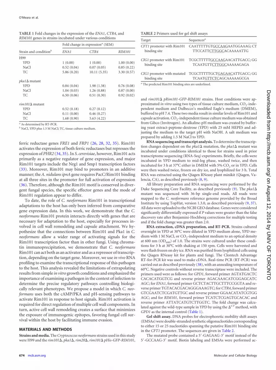
RNA extraction, cDNA preparation, and RT-PCR. Strains culturedovernight in YPD at 30°C were diluted in YPD medium alone, YPD me-dium with 1 M NaCl, or CO2-independent medium to an optical densityat 600 nm (OD600) of 1.0. The strains were cultured under these condi-tions for 3 h at 30°C with shaking at 150 rpm. Cells were harvested andwere flash frozen on dry ice. RNA was purified from lyophilized cells usingthe Qiagen RNeasy kit for plants and fungi. The Clontech AdvantageRT-for-PCR kit was used to make cDNA. Real-time PCR (RT-PCR) wascarried out as described previously (38), with an annealing temperature of60°C. Negative controls without reverse transcriptase were included. Theprimers used were as follows: for GPD1, forward primer AGTATGACTCCACACATGGTCG and reverse primer AGACAAACATCGGAGCATCAGC; for ENA1, forward primer GCTCTACTTGCTTTCCGGTA and re-verse primer TGTACACGACAGGGAAAGTC; for CTR4, forward primerGTCGAATCTCGATGTTGC and reverse primer GGAACATATCGTGGAGC; and for RIM101, forward primer TCATCTCGAGTGCACAC andreverse primer ATTATCATGTCTTGGTC. The fold change was calcu-lated against the wild-type sample in YPD by using the �CT method, withGPD1 as the internal control (Table 1).
Gel shift assay. DNA probes for electrophoretic mobility shift assays(EMSAs) were double-stranded synthetic oligonucleotides correspondingto either 15 or 25 nucleotides spanning the putative Rim101 binding sitein the CFT1 promoter. The sequences are given in Table 2.
The mutated probe contained a 5=-GAGAAG-3= motif instead of the5=-GCCAAG-3= motif. Biotin labeling and EMSAs were performed ac-
TABLE 1 Fold changes in the expression of the ENA1, CTR4, andRIM101 genes in strains incubated under various conditions
Strain and conditionb
Fold change in expressiona (SEM)
ENA1 CTR4 RIM101
H99YPD 1 (0.00) 1 (0.00) 1.00 (0.00)NaCl 0.52 (0.04) 0.07 (0.05) 0.85 (0.22)TC 5.86 (0.20) 10.11 (5.35) 3.30 (0.57)
pka1� mutantYPD 0.84 (0.04) 1.98 (1.38) 0.76 (0.08)NaCl 1.04 (0.03) 1.26 (0.88) 0.87 (0.00)TC 6.50 (0.06) 0.51 (0.30) 0.92 (0.02)
rim101� mutantYPD 0.52 (0.18) 0.27 (0.12)NaCl 0.11 (0.00) 0.46 (0.27)TC 1.68 (0.90) 5.63 (4.22)
a As determined by RT-PCR.b NaCl, YPD plus 1.5 M NaCl; TC, tissue culture medium.
TABLE 2 Primers used for gel shift assays
Target Sequencesa
CFT1 promoter with Rim101binding site
CAATTTTTGTGCCAAGAATGGAAAG; CTTTCCATTCTTGGCACAAAAATTG
CIG1 promoter with Rim101binding site
TCGCTTTTTGCCAAGAACATTGACC; GGTCAATGTTCTTGGCAAAAAGAGA
CIG1 promoter with mutatedbinding site
TCGCTTTTTGCTGAGAACATTGACC; GGTCAATGTTCTCAGCAAAAAGCGA
a The predicted Rim101 binding sites are underlined.
O’Meara et al.
674 mcb.asm.org Molecular and Cellular Biology
on August 12, 2014 by U
NIV
OF
TO
RO
NT
Ohttp://m
cb.asm.org/
Dow
nloaded from

cording to the manufacturer’s instructions (Pierce LightShift chemilumi-nescent EMSA kit; Thermo Scientific, Rockford, IL, USA). For each lane,6 �l of protein extract was used. Protein extracts were obtained as de-scribed previously (8).
Chromatin immunoprecipitation. Strains were incubated in CO2-independent tissue culture medium for 3 h and were then cross-linked for2 h in 1% formaldehyde before quenching with glycine. Chromatin im-munoprecipitation was performed as described previously (39), with mi-nor modifications. An antibody against green fluorescent protein (GFP)(Roche) was used to immunoprecipitate the Gfp-Rim101 fusion proteinand the associated DNA. A mock-antibody-treated strain and the wild-type strain (without GFP) were used as controls. Enrichment was deter-mined by real-time PCR of candidate promoters, by comparing the im-munoprecipitated sample to the mock-antibody control by use of thecomparative threshold cycle (CT) method (40).
Microscopy. Fluorescent images were captured using a DeltaVisionElite deconvolution microscope equipped with a CoolSnap HQ2 high-resolution charge-coupled device (CCD) camera. Cells were cultured ei-ther in CO2-independent medium (pH 7.4) (Gibco) or in YPD (pH 6.0)plus 150 mM HEPES for 4 to 5 h. To visualize nuclei, the Hoechst 33342nucleic acid stain (Invitrogen) was added to 10 �g/ml and was incubatedfor 15 min. To minimize background fluorescence, cells were washed andwere resuspended in synthetic complete medium (pH 6.0) containingMcIlvaine’s buffer or in CO2-independent medium. At an acidic pH, theHoechst 33342 nuclear stain shows membrane staining (41).
Cell wall analysis. For cell wall staining, strains were first washed andthen stained with wheat germ agglutinin (WGA) conjugated to AlexaFluor 488 (chitin) (Molecular Probes, Eugene, OR) (42, 43). After stain-ing, cells were washed twice before observation by fluorescence micros-copy. WGA was observed using a 488-nm wavelength for fluorescence.Images were captured with a Zeiss Axio Imager.A1 fluorescence micro-scope equipped with an AxioCam MRm digital camera.
In vivo RNA profiling. Female A/J mice were infected with 5 � 105
cells by using the inhalational model of cryptococcal infection, as de-scribed previously (9). The mice were sacrificed 4 days postinfection, andthe lungs were harvested. Immediately after harvesting, infected lungswere cut into the separate lobes and were flash frozen in dry ice. The frozenlungs were then homogenized by three rounds of 30-s beating on a mini-bead beater using 0.2-�m acid-washed glass beads. RNA was extractedfrom the tissue homogenate by using the Qiagen RNeasy Plant minikitaccording to the manufacturer’s instructions (Qiagen, Valencia, CA). Allanimal experiments were performed in accordance with Duke Universityinstitutional guidelines for the ethical care of experimental animals.
Fungal gene expression in vivo was analyzed by nanoString profiling asdescribed previously (44). Briefly, 10 �g of C. neoformans-infected mousetissue RNA was mixed with a custom-designed probe set and was pro-cessed according to the manufacturer’s instructions. Six hundred fieldsper sample were scanned on the nanoString digital analyzer. The rawcounts were adjusted for technical variability by using irrelevant RNAsequences included in the code set. The adjusted counts were then nor-malized to the expression of five housekeeping genes (expressing aldosereductase [CNAG_02722], cofilin [CNAG_02991], microtubule bindingprotein [CNAG_00816], mitochondrial protein [CNAG_00279], andphosphoglycerate kinase [CNAG_03358]).
RESULTSPka1 and Rim101 share downstream targets. In C. neoformans,there is evidence that the PKA signaling pathway helps to controlRim101 localization and function. Therefore, we hypothesizedthat examining the transcriptional profiles of the rim101� andpka1� mutants in comparison to that of the wild type would revealthe extent of PKA1 regulation of Rim101 activity (8). To define theoverlapping and unique sets of genes with Pka1- and Rim101-dependent transcription, we compared the global transcriptional
profiles of the pka1� and rim101� mutant strains to that of thewild type (45, 46).
Previously, we performed deep mRNA sequencing of wild-type and rim101� cells after incubation in tissue culture mediumfor 3 h (9). This condition results in heat stress, slight pH stress(since C. neoformans has a preference for acidic pHs), and alterednutrient availability. We noted that 1,257 genes have significantlydifferent expression in the rim101� mutant strain and the wildtype. We subsequently performed transcriptional analysis of thepka1� mutant strain incubated under identical conditions. Thiscomparative transcriptional profiling revealed 1,476 genes with atleast 2-fold differences in expression between the pka1� mutantand the wild type (see Table S1 in the supplemental material). Ofthese, 1,077 genes were common to both sets; these genes demon-strated transcriptional dependence on both Pka1 and Rim101.
To examine the network structure, we performed pairwise cor-relation analysis of the entire transcriptomes of the pka1� andrim101� mutant strains (46). This analysis revealed a strong cor-relation between Pka1- and Rim101-dependent genes (adjustedr2, 0.515; P, �0.001). This correlation increased dramaticallywhen we examined only those genes that were at least 2-fold dif-ferentially expressed in the wild-type and mutant strains (adjustedr2, 0.923; P, �0.001). Moreover, the majority of genes withRim101-dependent expression demonstrated a similar directionand magnitude of transcriptional control by Pka1 (Fig. 1A). Thisstrong correlation between the rim101� and pka1� mutant tran-scriptomes supports our hypothesis that C. neoformans Pka1 andRim101 are in the same intracellular signaling pathway. It alsosuggests a model in which Pka1 is required for the majority of theRim101 transcriptional regulation activity (Fig. 1B).
Among the coregulated biological processes, we observed sim-ilar differences in the expression of multiple proteins involved incell wall biosynthesis and remodeling (especially in processes re-lated to �-1,3 glucan, �-1,3 glucan, and �-1,6 glucan synthesis),consistent with previously documented changes in rim101� cellwalls (9). Additionally, 5 of the 7 genes involved in chitin andchitosan biosynthesis (47–50) were differentially expressed in thetwo mutant strains compared with the wild type.
To further explore the altered cell wall phenotype, we exam-ined chitin localization in the wild-type strain and the pka1�,rim101�, and rim20� mutant strains by using a fluorescently con-jugated lectin (fluorescein isothiocyanate [FITC]-conjugatedwheat germ agglutinin [WGA]). Rim20 is part of the scaffold thatis required for Rim101 cleavage as part of the canonical pH-sens-ing pathway (51). Each strain was incubated in tissue culture me-dium for 24 h prior to WGA addition. We observed similar pat-terns of staining in the rim101�, rim20�, and pka1� mutantstrains (Fig. 2A). Unlike the wild-type strain, which shows stain-ing only at the bud necks, the rim101�, rim20�, and pka1� strainsshow a more diffuse pattern of WGA-associated fluorescencearound the entire cell, consistent with similar alterations in cellwall chitin content. These results suggest that cell wall remodelingvia Rim101 is dependent on both the pH pathway and the cAMPpathway.
Despite the striking similarities between Pka1- and Rim101-dependent gene expression, several genes were transcriptionallyregulated by only one of these proteins. This suggests both Pka1-dependent and Pka1-independent regulation of Rim101. This ob-servation may explain some of the phenotypic differences betweenthe pka1� and rim101� mutant strains.
Analysis of Rim101 Targets in C. neoformans
February 2014 Volume 34 Number 4 mcb.asm.org 675
on August 12, 2014 by U
NIV
OF
TO
RO
NT
Ohttp://m
cb.asm.org/
Dow
nloaded from

For example, the ENA1 gene, encoding a sodium transporter,was regulated only by the Rim101 pathway; rim101� mutantstrains failed to support the level of ENA1 expression that wasobserved in the wild type and the pka1� mutant. The functionalconsequence of this Rim101-Ena1 association is suggested by theobservation that both ena1� and rim101� mutants fail to grow onmedia with high concentrations of salt or with a high pH. Addi-tionally, the divergent dependence of Ena1 on Rim101 and Pka1may explain the phenotypic differences between the sensitivitiesof the rim101� and pka1� mutants to high pHs and high saltconcentrations (Fig. 2B) (52). Importantly, the rim20� mutantshares the salt and pH sensitivities of the rim101� strain, suggest-ing that tolerance to alkaline pHs and high salt concentrations isregulated primarily by the Rim/pH-sensing pathway.
To support these findings, we used targeted RT-PCR analysisof ENA1 expression in YPD, tissue culture medium, and a high-salt medium (Fig. 2C). We observed that ENA1 expression is
highly induced under these conditions in the wild-type and pka1�mutant strains but not in the rim101� mutant. This also suggeststhat Rim101 is able to induce ENA1 expression in the absence ofPka1 phosphorylation, presumably through activation by distinctcomponents of the Rim/pH-sensing pathway. Together, thesedata support an emerging model in which the C. neoformans PKAand Rim pathways control overlapping targets regulating cell walldynamics, as well as distinct targets involved in other cell pro-cesses, such as tolerance to different salt concentrations (Fig. 1B).
Rim101 binds a conserved motif. The set of Rim101-depen-dent genes that we identified through comparative transcriptionalprofiling is composed of both direct and indirect targets of theRim101 transcription factor. To determine experimentally the di-rect targets of Rim101 action, we first investigated whether a pre-viously established Rim101-binding motif from other fungi isconserved in C. neoformans. Rim101 homologues in other speciesbind to the 5=-GCCAAG-3=motif in the promoters of direct target
Differential expression: pka1 vs. rim101
pka1 ln(FoldChange)
rim10
1 ln
(Fol
dCha
nge)
4
2
0
2
4
6
4 2 0 2 4
Rim20 Pka1
Rim101
Cell wallMetal homeostasis
Capsule biosynthesisMelanin
pH responsesSalt tolerance
Tissue culture conditionsAlkaline pHHigh salt
A
B
Rim13
FIG 1 (A) Pairwise correlation analysis of the entire transcriptomes of the rim101� and pka1� mutant strains. Gene expression levels for the two mutant strainswere determined in comparison with those for the wild-type strain. The transcriptomes of the pka1�, rim101�, and wild-type strains were defined using RNA-Seqanalysis after incubation in tissue culture medium (DMEM) for 3 h. Shaded circles represent genes that were �2-fold differentially expressed in the wild-type andmutant strains. The adjusted r2 for the entire transcriptomes was 0.515 (P, �0.001). The adjusted r2 for the comparison of the significantly differentially expressedgenes only was 0.923 (P, �0.001). (B) The concordance between Rim101- and Pka1-dependent gene expression supports a model in which the Rim101transcription factor is controlled by the classical Rim/pH-responsive pathway (including Rim20 and Rim13) as well as by the cAMP/Pka1 pathway to regulateseveral cellular processes.
O’Meara et al.
676 mcb.asm.org Molecular and Cellular Biology
on August 12, 2014 by U
NIV
OF
TO
RO
NT
Ohttp://m
cb.asm.org/
Dow
nloaded from

genes (30, 32). As reported previously, this putative target se-quence is present in the promoters of many genes with Rim101-dependent expression (8). We used electrophoretic mobility shiftassays to determine whether C. neoformans Rim101 (CnRim101)actually binds this motif. For these experiments, we chose 25-mersspanning the 5=-GCCAAG-3= motif from the promoter of the C.neoformans iron transporter gene CFT1, which is highly differen-tially expressed in the rim101� and wild-type strains. We observeddistinct mobility shifts of the labeled oligomer when it was incu-bated with protein extracts from the wild-type strain but not withextracts from the rim101� mutant (Fig. 3A, lane 1 versus lane 2).To determine the specificity of protein binding, we added excessunlabeled 15-mer oligomers containing this motif, and we ob-served a competitive reversal of the electrophoretic shift (Fig. 3A,lanes 3 and 4). Incubation of the labeled oligomer with proteinextracts from the rim101� Gfp-Rim101 strain resulted in a super-shift in electrophoretic mobility (Fig. 3B). Together, these datastrongly suggest that Rim101 binds directly to the 5=-GCCAAG-3=motif in this target gene promoter. Additionally, when we incubatedprotein extracts with a mutated oligomer (5=-GCCAAG-3=¡ 5=-GAGAAG-3=), we did not observe a shift in mobility, indicating the spec-ificity of interactions of Rim101 with its DNA binding site (Fig. 3C).
Then we analyzed the promoters of the Rim101-regulated
genes for the presence of this motif. Of the 1,257 genes differen-tially regulated by Rim101, 564 had this conserved motif in the1,000 bp upstream of their ATG start sites. In other fungal species,there is evidence that Rim101 can bind a divergent sequence;for example, C. albicans Rim101 binds the 5=-CCAAGAA-3=motif(35). Indeed, the CHI22 endochitinase, which demonstratesRim101-dependent expression, has this divergent motif in the pu-tative promoter region. However, for the most stringent initialanalysis, we chose to limit our studies to genes containing themost-conserved Rim101 binding motif. We hypothesized thatgenes that are direct targets of Rim101 would maintain this spe-cific, functional promoter sequence across the Cryptococcus genus,including the sister species Cryptococcus gattii. Therefore, we ex-amined the promoter regions of orthologs of the Rim101-regu-lated genes in C. gattii (strain R265). Of the 564 Rim101-regulatedgenes with promoters containing Rim101 binding sites in Crypto-coccus neoformans var. grubii, 310 maintained this promoter se-quence in their C. gattii orthologs (Fig. 3D). The majority of genes(76.7%) with Rim101 sites in both C. neoformans var. grubii and C.gattii displayed higher expression in the rim101� mutant than inthe wild type, suggesting that Rim101 may act primarily as a neg-ative regulator of gene expression for its direct targets.
We then mapped the position of each potential binding site in
Wild type pka1rim101
A
B pH 6 YPD pH 8 YPD
rim20
1.5 M NaCl YPDWild type
pka1
rim101
rim20
Fold
cha
nge
Wild type rim101 pka1
ENA1 expression
Tissue Culture
YPD
High salt
C
1
2
3
4
5
6
7
FIG 2 The rim101� and pka1� strains have both overlapping and divergent phenotypes. (A) The rim101� and pka1� mutants demonstrate similar alterationsin WGA binding patterns. Cells were incubated for 24 h in tissue culture medium before staining with FITC-conjugated WGA. All micrographs were taken at thesame exposure in order to distinguish the fluorescence levels of the different strains. (B) The rim101� and rim20� mutants have defects at an alkaline pH andunder high-salt conditions. A total of 1 � 106 cells were 10-fold serially diluted onto plates under the conditions indicated and were incubated at 30°C. (C) ENA1shows Rim101- but not Pka1-dependent changes in expression. RNA was extracted from wild-type, rim101�, and pka1� strains after incubation for 3 h in a richmedium (YPD), under tissue culture conditions, or with high salt concentrations. ENA1 transcript levels were determined by quantitative reverse transcriptasePCR. The fold change was calculated relative to wild-type levels in YPD and was normalized to the expression of the internal control, GPD1.
Analysis of Rim101 Targets in C. neoformans
February 2014 Volume 34 Number 4 mcb.asm.org 677
on August 12, 2014 by U
NIV
OF
TO
RO
NT
Ohttp://m
cb.asm.org/
Dow
nloaded from

these promoters relative to the ATG site in order to examinewhether certain positions were correlated with positive or nega-tive gene regulation (Fig. 3E). This analysis suggested that Rim101sites that occurred less than 100 bp from the ATG position wereassociated mainly with Rim101-activated genes. In contrast,Rim101 sites that were approximately 150, 500, or 900 bp up-stream of the ATG position were associated mainly with Rim101-repressed genes. The divergence in binding motif position sug-gests the possibility of interactions between Rim101 and othertranscriptional regulators in these promoters, thus allowing a sin-gle protein to act as both an activator and a repressor of geneexpression.
Rim101 binds a conserved motif in vivo. To verify thatRim101 actually binds this conserved motif in the cell, we per-
formed chromatin immunoprecipitation after incubating cellsunder Rim101-activating conditions for 3 h (DMEM tissue cul-ture medium at 37°C). After formaldehyde cross-linking andDNA sonication, we used an anti-GFP antibody to enrich for se-quences that were bound to the GFP-Rim101 protein and exam-ined candidate promoters from the subset of Rim101-regulatedgenes that had Rim101 sites in all species examined. As a control,we examined cells for enrichment at the actin promoter, whichdoes not contain a Rim101 binding site and is not transcription-ally regulated by Rim101.
After incubation under tissue culture conditions for 3 h, wedemonstrated enrichment for Rim101 binding at the promoters ofthree candidate cell wall genes that are among the predicted set ofdirect targets of Rim101. For CDA1 and KRE61, enrichment forRim101 binding correlated with an increase in gene expression inthe wild-type strain. In contrast, the AGS1 promoter, which con-tains a Rim101 binding site 944 bp away from the ATG position,demonstrated 2.8-fold enrichment for Rim101 binding (Fig. 4), aswell as higher expression in the rim101� mutant strain (9). Theseresults suggest that C. neoformans Rim101 directly regulates theexpression of cell wall genes by binding to their promoters but thatthe consequences of binding may be modulated by other factors,such as interactions with other proteins and the promoter posi-tion of Rim101 binding.
Rim101 has also been implicated in the regulation of iron andcopper homeostasis both in C. neoformans and in other fungalspecies (24, 34, 35, 53). Therefore, we examined the Rim101 bind-ing of the promoters of the HAPX, CTR4, and CFT1 genes (Fig. 4).Although there was enrichment at all three promoters, HAPXdemonstrated increased expression in the rim101� strain, whichwas consistent with a Rim101 binding motif 519 bp upstream ofthe ATG site and subsequent Rim101 repression of gene expres-sion. CTR4 and CFT1, with binding sites 279 and 253 bp away,respectively, showed increased expression in the wild-type strain,consistent with activation by Rim101 (Fig. 4). Finally, we exam-ined the binding of Rim101 at the ENA1 promoter, which also hasa conserved Rim101 binding motif. In agreement with the previ-ously discussed phenotypic and gene expression results, we ob-
A B C
1 321 2 31 2 3 4
H99 747 GGTTTTACCGCCAAGGATGCACATAACCTAAA 779 R265 752 GAGTCTAACGCCAAGAACGCATATAACCTAAC 784 cons 1050 * * ** ******* * *** ********* 1083
D
E
Distance from ATG (bp)
coun
t
0
10
20
30
40
50
1000 800 600 400 200 0
ATG-1000 bp
FIG 3 (A to C) Rim101 binds a conserved motif, as determined by EMSAs. (A)A biotin-labeled 25-mer containing the 5=-GCCAAG-3= motif was incubatedwith protein extracts from wild-type (lanes 1 and 4) or rim101� (lanes 2 and 3)cells. The mixtures were assessed by alterations in electrophoretic mobility byPAGE and immunoblotting using streptavidin detection. Excess unlabeled15-mers were added to lanes 3 and 4. (B and C) A 25-mer containing the5=-GCCAAG-3=motif (B) or a mutated Rim101 binding motif (5=-GAGAAG-3=) (C) was incubated with protein extracts from the wild-type (lanes 1),rim101� (lanes 2), or rim101�/GFP-RIM101 (lanes 3) strain prior to electro-phoresis. (D) The promoter of the ENA1 gene from C. neoformans var. grubiiand its homologous sequence in C. gattii were aligned. The conserved Rim101binding motif is highlighted. Identical nucleotides in a consensus (cons) se-quence of this region are indicated by asterisks. (E) Genes with Rim101 bind-ing sites in both C. neoformans and C. gattii were examined for Rim101-de-pendent transcription and were separated on the basis of induction (green) orrepression (red). The positions of the binding sites were determined, and thenumber of genes was plotted along a representation of the distance of theRim101 binding site from the ATG translation start site. All sequences andRim101 site positions were obtained from FungiDB (www.fungidb.org).
CDA1 AGS1 CFT1KRE61 CTR4
Fold
enr
ichm
ent o
ver m
ock
ENA1HAPXACT1
2
4
6
8
Cell wall Metal homeostasis
FIG 4 Chromatin immunoprecipitation to detect Gfp-Rim101 DNA binding.Cells were incubated in tissue culture medium for 3 h before fixation andchromatin immunoprecipitation. PCRs were performed using primers thatflanked a presumed Rim101 binding site in target gene promoters. The foldchange was determined by determining enrichment in the immunoprecipi-tated sample relative to the no-antibody control. Actin (ACT1), which doesnot contain a Rim101 binding site, was used as a control.
O’Meara et al.
678 mcb.asm.org Molecular and Cellular Biology
on August 12, 2014 by U
NIV
OF
TO
RO
NT
Ohttp://m
cb.asm.org/
Dow
nloaded from

served striking enrichment for Rim101 binding at the ENA1 pro-moter.
nanoString profiling of virulence gene expression in vitro.Recently, nanoString RNA profiling has been used to accuratelyquantify RNA levels in many organisms, including pathogenicfungi such as C. albicans (44). This technique avoids the biasesintroduced during the creation of cDNA libraries for sequencing,and it can be used to compare expression directly across multiplemutant strains and conditions. It has been successfully applied toassess the relative transcript levels of target genes in biologicalsamples. Therefore, we used this methodology to complement ourRNA sequencing results and to allow us to examine gene expres-sion under multiple growth conditions.
Taking a targeted approach, we surveyed the expression of 26genes that were likely to be involved in infection-related processes,such as capsule or melanin induction, metal acquisition, osmoticstress resistance, cell wall remodeling, and pH responses (2). As acontrol for RNA extraction in each sample, we examined the ex-pression of 5 genes that did not demonstrate variable expression inmultiple previous microarray, RNA-Seq, and RT-PCR analyses (8,24, 54–56). These control genes were chosen to represent a rangeof absolute expression levels, allowing for the normalization forboth weakly and highly expressed genes.
To ensure that the nanoString data would correlate with othermethods of transcriptional measurement, we first compared thenanoString data to the RNA-Seq data under in vitro growth con-ditions for the wild-type and rim101� mutant strains after 3 h ofincubation in tissue culture medium. For most genes, we observeda strong concordance with the changes in gene expression that weobserved by RNA-Seq and by RT-PCR. However, the RNA-Seqresults tended to suggest larger differences in expression (Table 3).
Using nanoString profiling, we were able to distinguish differ-ences in gene expression based both on Rim101 activity and on theincubation condition (Table 4). For example, ENA1 is stronglyinduced in the wild-type strain by incubation in CO2-indepen-dent medium. However, the absolute expression levels of this geneare also strongly Rim101 dependent, with 6.7- and 5.9-fold differ-ences in expression between the wild-type and rim101� mutantstrains in a rich medium or CO2-independent medium, respec-tively. The expression of SKN1, a �-glucan synthase gene, andCDA1, a chitin deacetylase, also follows this pattern. In contrast,KRE6 expression was repressed in a CO2-independent medium inthe wild-type background and was induced in the rim101� mu-tant background. Figure 5A displays the normalized RNA countsin the wild-type and rim101� mutant strains under the twogrowth conditions for genes that were at least 2-fold differentiallyexpressed in the two strains.
Interestingly, we documented 3.4-fold induction of RIM101gene expression in the wild-type strain under tissue culture con-ditions. The expression of the RIM101 and pacC genes in otherfungal species is highly regulated by growth conditions, contrib-uting to the levels of nucleus-localized protein (57). In C. neofor-mans, the RIM101 gene contains six Rim101 binding motifs in thepromoter, suggesting that its expression could be highly autoregu-lated. The C. albicans RIM101 promoter contains only twoRim101 binding sites (32). Therefore, we created a Gfp-Rim101fusion protein expressed under the control of the endogenouspromoter (pRIM101-Gfp-Rim101) in order to examine the sub-cellular pattern of protein localization that occurs during growth
under host-mimicking conditions with physiological levels ofprotein expression.
In contrast to the constitutively nuclear signal of the histone-driven GFP-Rim101 fusion protein, we observed a limitedamount of fluorescent signal in cells expressing the pRIM101-Gfp-RIM101 allele when incubated in rich medium, consistent with thelow levels of expression observed by using nanoString profiling.However, when these cells were shifted to a tissue culture mediumat 37°C, we observed a clear accumulation of fluorescence in thenucleus by 3 h (Fig. 5B). This observation is consistent withRim101 transcriptional induction and protein activation underthese physiologically relevant growth conditions.
Rim101 regulates expression changes in animal models ofinfection. A major advantage of nanoString profiling is the abilityto examine the low levels of fungus-specific RNA present in thecontext of a host sample. Therefore, in addition to studying therole of Rim101 in regulating gene expression in vitro, we also ex-plored the Rim101-dependent expression of a set of physiologi-cally relevant genes in the setting of a murine cryptococcal lunginfection. We used nanoString profiling to examine the changes inexpression of a set of C. neoformans genes in a murine inhalationalmodel of C. neoformans infection. This fungus typically initiatesinfection in the lung, making this a highly relevant model of hu-man infection. Also, we demonstrated recently that infection withthe rim101� mutant causes a dramatically greater lung inflamma-tory response than infection with the wild type (9).
TABLE 3 Comparative transcriptional profiling results for selected C.neoformans genes in the rim101 mutant versus the wild type usingnanoString and RNA-Seq techniques
Gene product Gene ID
Fold change in expression (wildtype/rim101 mutant)
By nanoStringprofiling By RNA-Seq
Cfo1 CNAG_06241 1.02 Below thresholdAgs1 CNAG_03120 0.83 0.49Cck1 CNAG_00556 0.84 0.39Cda1 CNAG_05799 2.80 4.78Cda3 CNAG_01239 0.89 Below thresholdCdc24 CNAG_04243 0.70 Below thresholdCft1 CNAG_06242 0.77 2.25Chs4 CNAG_05581 0.77 0.37Chs4 CNAG_00546 0.88 0.35Chs5 CNAG_05818 0.73 1.26Chs8 CNAG_07499 0.74 0.19Cir1 CNAG_04864 1.07 0.24Ctr4 CNAG_00979 1.43 43.16Ena1 CNAG_00531 5.88 4.67Fks1 CNAG_06508 0.83 0.15HapX CNAG_01242 0.59 0.18Kre6 CNAG_00914 0.30 0.37Lrg1 CNAG_05703 0.83 0.28Ova1 CNAG_02008 0.85 13.36Pbs2 CNAG_00769 0.81 0.29pH response
regulator Rim9CNAG_05654 0.57 0.36
Pka1 CNAG_00396 1.03 0.84Rim101 CNAG_05431Skn1 CNAG_00897 2.67 10.08Sp1 CNAG_00156 0.63 0.24Ssk2 CNAG_05063 0.76 0.30
Analysis of Rim101 Targets in C. neoformans
February 2014 Volume 34 Number 4 mcb.asm.org 679
on August 12, 2014 by U
NIV
OF
TO
RO
NT
Ohttp://m
cb.asm.org/
Dow
nloaded from

To examine infection-specific gene expression changes, we ex-tracted total RNA from the lungs of female A/J mice infected with5 � 105 cells at 4 days postinfection (n � 5) (Table 4). We deter-mined the contribution of the Rim101 transcription factor to theinfection process by examining RNA levels of the rim101� mutantin the lung in identical murine infections.
Overall, we found that most of the genes induced by in vitroincubation in tissue culture medium were also induced duringinfection of a mouse lung. However, a number of genes showedaltered expression only in the lung, suggesting the limitations of invitro culture conditions in mimicking the host. Some of theseinfection-specific genes include the chitin synthase-encodinggenes CHS4 and CHS5, as well as the �-1,3 glucan synthase geneFKS1, all of which are involved in cell wall biosynthesis. Addition-ally, we observed that the expression of the RIM101 gene increasedin the wild-type strain during infection and was approximately9.4-fold induced in the lung by day 4 relative to expression in richmedium, and an additional 2.75-fold increased relative to expres-sion under tissue culture conditions.
When we compared the gene expression profiles of wild-typeand rim101� mutant strains in the lungs (n � 5), 13 of the 26genes that we examined were significantly differentially expressedin the two strains (P, �0.05) (Fig. 6). These included several chitinbiosynthesis genes and the �-glucan synthase gene AGS1. How-ever, only four genes (CDA1, CTR4, ENA1, and KRE6) showed at
least a 2-fold difference in expression levels between the strains(Fig. 6). Interestingly, only CTR4 expression and KRE6 expressionwere regulated by both Rim101 and Pka1 in our in vitro RNA-Seqanalysis. Together, these results are consistent with a model inwhich the Rim101 transcription factor participates directly in cellwall remodeling within the infected host in response to activationfrom multiple signaling cascades. These cell wall changes are likelydue to direct binding of the Rim101 transcription factor to thepromoters of target genes controlling the expression of multiplecell wall components. Intact Rim101 signaling in vivo results incell wall changes that promote capsular polysaccharide bindingand antigen masking, allowing more-efficient survival within thehost.
DISCUSSION
We hypothesized that in C. neoformans, the Rim101 transcriptionfactor responds to signals from both the pH-responsive Rim signaltransduction cascade and the cAMP/PKA pathway (8). So far, thisconnection has been proposed only for Cryptococcus; in other fun-gal species, Rim101 activation has not been demonstrated to re-quire phosphorylation by PKA (13, 21, 58). To confirm the con-nections between the cAMP/PKA pathway and the Rim101transcription factor, we performed deep RNA sequencing of therim101� and pka1� mutants in comparison to the wild-typestrain after incubation under tissue culture conditions. Compar-
TABLE 4 nanoString quantification of transcript abundancea
Genename Putative function Gene ID
Relative transcript abundance in the following strain under thefollowing condition:
WT inYPD
WT inTCb
rim101mutantin YPD
rim101mutantin TC
WT inlung(n � 5)
rim101mutant inlung (n � 5)
AGS1 Alpha-glucan synthase CNAG_03120 1,321 1,960 1,400 2,356 1,040 1,418CCK1 Casein kinase protein CNAG_00556 589 748 582 894 547 728CDA1 Chitin deacetylase CNAG_05799 1,893 3,464 671 1,239 8,110 3,046CDA3 Chitin deacetylase CNAG_01239 433 1,619 376 1,830 675 509CDC24 Rho-family GTPase CNAG_04243 235 199 264 284 146 196CFO1 Ferric oxidase CNAG_06241 373 911 96 889 5,176 5,083CFT1 High-affinity iron permease CNAG_06242 1,203 2,295 251 2,963 5,985 5,281CHS4 Chitin synthase CNAG_05581 556 672 561 878 1,117 1,457CHS4 Chitin synthase CNAG_00546 211 378 221 431 1,310 1,863CHS5 Chitin synthase CNAG_05818 254 404 239 555 713 1,189CHS8 Chitin synthase CNAG_07499 241 268 229 364 261 425CIR1 Iron regulator CNAG_04864 343 364 286 340 799 684CTR4 Copper transporter CNAG_00979 2,443 2,025 214 1,418 3,366 8,524ENA1 Sodium transporter CNAG_00531 660 4,946 99 841 5,282 2,702FKS1 Beta-glucan synthase CNAG_06508 107 153 119 183 623 946HAPX Iron transcription factor CNAG_01242 96 217 76 368 720 1,065KRE6 Beta-glucan synthase CNAG_00914 812 348 791 1,147 700 2,291LRG1 GTPase-activating protein for the PKC pathway CNAG_05703 104 102 102 122 144 165OVA1 Putative phosphatidylethanolamine-binding protein CNAG_02008 890 1,191 702 1,406 3,348 1,927PBS2 Member of the HOG signaling cascade CNAG_00769 339 348 311 431 392 544RIM9 Conserved pH-responsive protein CNAG_05654 496 376 462 658 2,543 3,712PKA1 Protein kinase CNAG_00396 271 323 227 312 235 260RIM101 Transcription factor CNAG_05431 36 122 0 1 336 16SKN1 Beta-glucan synthase CNAG_00897 1,304 3,980 379 1,490 3,405 3,183SP1 CRZ1 transcription factor homologue CNAG_00156 235 225 194 358 649 773SSK2 MAPKKKc CNAG_05063 192 150 193 197 88 108a Relative transcript abundance for selected C. neoformans genes was quantified using nanoString profiling after incubation under various in vitro and in vivo conditions.b TC, tissue culture medium.c MAPKKK, mitogen-activated protein kinase kinase kinase.
O’Meara et al.
680 mcb.asm.org Molecular and Cellular Biology
on August 12, 2014 by U
NIV
OF
TO
RO
NT
Ohttp://m
cb.asm.org/
Dow
nloaded from

ison of the downstream transcriptional responses to the rim101�and pka1� mutations revealed a striking degree of coordinatedgene regulation, providing further evidence for a functional rela-tionship between these genes. Integration of these two pathwaysappears to give Rim101 flexibility in the upstream signals thatallow for activation. Instead of acting primarily as a pH responsefactor, C. neoformans Rim101 can respond to multiple host stim-uli via the cAMP/PKA pathway, including such diverse signals aslow iron levels or tissue culture medium (59).
This analysis also revealed some targets that demonstrated di-vergent regulation in the two mutant strains, including genes re-lated to salt and pH sensitivity (52, 60–62). Although it is clear thatthe Pka1 kinase has multiple downstream targets, the identifica-tion of apparently Pka1 independent and Rim101 specific targetsraises intriguing questions about alternative mechanisms for theactivation of the Rim101 transcription factor.
The overlapping but partially divergent phenotypes of therim101� and pka1� mutants contrast with the absolutely identicalphenotypes of the rim101� mutant and a strain with a mutation inthe RIM20 gene. Rim20 is a member of the conserved pH-sensingpathway, and it acts as a scaffold for Rim101 cleavage and activa-tion (51). This suggests that C. neoformans Rim101 activationmaintains dependence on the conserved, upstream, pH-respon-sive signaling elements, even though the expected pH-sensingmembrane receptors are not clearly present in the C. neoformansgenome. In contrast to other fungi for which the pH response hasbeen studied, C. neoformans has a strong preference for acidic pHsand a defect in growth above pH 7.4, potentially due to alterationsin the upstream members of the pH response pathway.
Despite the divergence in Rim101 activation in C. neoformans,there is significant conservation in the downstream targets ofRim101. One of the major cellular processes regulated by Rim101is the remodeling of the cell wall in response to host signals. Wedemonstrated previously the importance of this remodeling in thewild-type strain as a mechanism for evading the host immune
1000
2000
3000
4000
KRE6CFO1 CTR4 CFT1 CDA1 SKN1 ENA1
Nor
mal
ized
RN
A co
unts
WT YPD
WT TC
rim101 YPD
rim101 TC
A
B
Tissue Culture Media pH 7.4
YPD pH 6.0
GFP-Rim101Hoechst 33342nuclear stain
Metal homeostasis Cell wall
FIG 5 Differences in gene expression and localization due to induction intissue culture medium. (A) Normalized RNA levels of genes with differentialexpression in the wild-type and rim101� mutant strains. Strains were grown inYPD or under tissue culture (TC) conditions for 3 h before RNA extraction.RNA levels were determined by nanoString profiling. RNA levels were normal-ized to those of control genes as described in Materials and Methods. (B)Rim101 shows increased nuclear localization in tissue culture medium. Cellscontaining the Gfp-Rim101 fusion protein expressed under the control of theendogenous promoter were incubated in YPD medium (pH 6.0) or in tissueculture medium. Nuclei were stained using the Hoechst 33342 nucleic acidstain.
Nor
mal
ized
RN
A co
unts
CH
S8
RIM
101
PB
S2
FKS
1
KR
E6
CH
S5
HA
PX
AG
S1
CH
S4
CH
S6
OVA
1
CTR
4
EN
A1
CD
A1
2000
4000
6000
8000
000
FIG 6 In vivo profiling of gene expression. RNA was harvested from mouse lungs (from 5 mice for each strain) infected with either the wild-type (open bars) orthe rim101� mutant (shaded bars) strain. Expression levels were determined by nanoString profiling of candidate genes. The graph includes all genes withsignificantly different expression in the wild-type and rim101� strains (P, �0.05).
Analysis of Rim101 Targets in C. neoformans
February 2014 Volume 34 Number 4 mcb.asm.org 681
on August 12, 2014 by U
NIV
OF
TO
RO
NT
Ohttp://m
cb.asm.org/
Dow
nloaded from

responses (9). In many other fungi, Rim101 also regulates cell wallprocesses. For example, C. albicans uses Rim101 to allow for theyeast-hypha transition in response to neutral/alkaline pHs, whichis a vital step for tissue invasion and virulence (63, 64). Addition-ally, the C. albicans Rim101 pathway regulates the levels of chitinin the cell (21). The S. cerevisiae Rim101 transcription factor is alsorequired for cell wall assembly, and the U. maydis Rim101 tran-scription factor modulates cell wall sensitivity to lytic enzymes(25, 65). Using transcriptional profiling and cell wall staining, wedemonstrated previously that C. neoformans Rim101 also regu-lates these genes (9). Here we show that the cell wall of the pka1�mutant has features similar to those of the rim101� mutant, andmany cell wall genes are also differentially regulated in the wild-type and pka1� mutant strains.
A potential mechanism for this conservation in Rim101 func-tions is the conservation of the binding motif across these fungalspecies. Using EMSAs and chromatin immunoprecipitation, wedemonstrated that the C. neoformans Rim101 protein binds the5=-GCCAAG-3=motif, which had been documented previously inAspergillus, Candida, and Saccharomyces species (32, 33, 36). Pre-sumably, the upstream rewiring of the Rim101 activating signalallows C. neoformans to induce the expression of a conserved suiteof Rim101 targets, such as those involved in cell wall modification,in response to a wider range of activating signals.
In this work, we also observed that Rim101 binding was notcompletely associated with either induction or repression of thetarget genes, suggesting that there is interplay between Rim101and other transcription factors in these promoter regions. In A.nidulans, the gabA promoter contains overlapping Rim101 andIntA binding sites, resulting in competition for binding and tran-scriptional regulation (66). Both Rim101 and a CBF binding fac-tor control the expression of the C. albicans FRP1 ferric reductase(34). A C. neoformans ferric reductase transmembrane compo-nent (CNAG_06821) promoter also contains both a Rim101binding site and a CCAAT motif, suggesting that this gene couldbe regulated both by Rim101 and by a CBF protein.
Finally, we were able to use nanoString profiling to examine thetranscriptional responses to the host. This technology allows fordirect quantification of fungal RNA levels within the context of theinfected tissue—in our case, the mouse lung. We first confirmedthat nanoString profiling recapitulates the in vitro transcriptionalprofiling of cells incubated either in rich medium or under tissueculture conditions.
We were then able to use nanoString profiling to examine fun-gal gene expression in the mouse lung. This in vivo analysis re-vealed the inadequacies of extrapolating results from in vitrogrowth conditions, since many genes showed a much higher de-gree of induction in the mouse lung than in tissue culture me-dium. This is likely due to the large number of stresses in the hostthat are not completely recapitulated under in vitro culture con-ditions, including interactions with the host immune system ordeprivation of particular nutrients. For example, HAPX, a geneencoding an important regulator of iron homeostasis, was 2.2-fold differentially regulated in YPD and tissue culture medium but7.5-fold differentially regulated in YPD and the mouse lung. Fur-thermore, only 7 of the 26 candidate genes were differentially reg-ulated in YPD and tissue culture medium in the wild-type strain.When YPD expression was compared to in vivo expression, 17 ofthe 26 genes were differentially expressed. The genes with infec-tion-specific induction or repression included the cell wall biosyn-
thesis genes CDA1, CHS4, CHS6, and FKS1 and the iron genesCIR1 and CFT1.
We were also able to examine the transcriptional profile of therim101� mutant and the wild-type strain under host conditions.The major differences in expression between the strains were inthe CDA1, KRE6, CTR4, and ENA1 genes. The CDA1 gene regu-lates chitosan levels in the cell, and KRE6 is involved in �-glucansynthesis, consistent with the role of Rim101 in the regulation ofcell wall remodeling. ENA1 is also required for full proliferationwithin the cerebrospinal fluid (CSF), although we have not ob-served a difference in neurotropism or proliferation in the CSFbetween the rim101� mutant and the wild type (67). Interestingly,the wild-type strain expressed more CTR4 transcripts than therim101� strain under both in vitro growth conditions but 2.5-foldfewer in the context of the host lung. This suggests that Rim101 isrequired for CTR4 induction during infection but is dispensableduring in vitro growth. These results emphasize the importance ofusing animal models to accurately assess disease processes, since invitro assays may not be able to replicate the host conditions fully.Induction of CTR4 transcripts during lung infections was recentlydemonstrated by luciferase assays, suggesting that this may be animportant factor during infection (68).
In conclusion, we propose that C. neoformans has integratedcAMP signaling in the activation of a conserved cell wall-regulat-ing transcription factor to allow for active remodeling of the host-pathogen interface during infection. Since C. neoformans is aneffective intracellular pathogen, this may reflect the need to re-spond to host conditions in addition to extracellular, alkaline pHsignals. By assaying gene expression during infection, we couldobserve the importance of actively regulating cell surface changesto allow for colonization and pathogenesis.
In C. neoformans, induction of the polysaccharide capsule is amajor component of virulence. In these studies, we demonstratethat other cell surface components, including chitin and chitosan,�-glucan, and �-glucan, are also highly regulated during infec-tion. Our profiling data support previous characterizations of hostresponses in illustrating a role for Rim101-responsive gene ex-pression during lung infection and in identifying cell wall genesamong the in vivo Rim101 targets. Moreover, the strong inductionof Rim101 transcripts in the host lung suggests that Rim101 is amajor regulator of these phenotypes.
ACKNOWLEDGMENTS
We thank the Duke Sequencing Core Facility and the Duke Light Micros-copy Core Facility for assistance.
These studies were supported by NIH grants AI050128 and AI074677(to J.A.A.), an American Heart Association Pre-doctoral Fellowship (toT.R.O.), Department of Energy grant DE023311 (to A.P.M.), and supportfrom the Richard King Mellon Foundation (to A.P.M.).
REFERENCES1. Alspaugh J, Perfect J, Heitman J. 1998. Signal transduction pathways
regulating differentiation and pathogenicity of Cryptococcus neoformans.Fungal Genet. Biol. 25:1–14. http://dx.doi.org/10.1006/fgbi.1998.1079.
2. O’Meara TR, Alspaugh JA. 2012. The Cryptococcus neoformans capsule: asword and a shield. Clin. Microbiol. Rev. 25:387– 408. http://dx.doi.org/10.1128/CMR.00001-12.
3. Dykstra MA, Friedman L, Murphy JW. 1977. Capsule size of Cryptococ-cus neoformans: control and relationship to virulence. Infect. Immun. 16:129 –135.
4. Kozel T, Gotschlich E. 1982. The capsule of Cryptococcus neoformanspassively inhibits phagocytosis of the yeast by macrophages. J. Immunol.129:1675–1680.
O’Meara et al.
682 mcb.asm.org Molecular and Cellular Biology
on August 12, 2014 by U
NIV
OF
TO
RO
NT
Ohttp://m
cb.asm.org/
Dow
nloaded from

5. Chang YC, Kwon-Chung KJ. 1994. Complementation of a capsule-deficient mutation of Cryptococcus neoformans restores its virulence. Mol.Cell. Biol. 14:4912– 4919.
6. Chang YC, Penoyer LA, Kwon-Chung KJ. 1996. The second capsule geneof Cryptococcus neoformans, CAP64, is essential for virulence. Infect. Im-mun. 64:1977–1983.
7. Doering TL. 2009. How sweet it is! Cell wall biogenesis and polysaccharidecapsule formation in Cryptococcus neoformans. Annu. Rev. Microbiol. 63:223–247. http://dx.doi.org/10.1146/annurev.micro.62.081307.162753.
8. O’Meara TR, Norton D, Price MS, Hay C, Clements MF, Nichols CB,Alspaugh JA. 2010. Interaction of Cryptococcus neoformans Rim101 andprotein kinase A regulates capsule. PLoS Pathog. 6:e1000776. http://dx.doi.org/10.1371/journal.ppat.1000776.
9. O’Meara TR, Holmer SM, Selvig K, Dietrich F, Alspaugh JA. 2013.Cryptococcus neoformans Rim101 is associated with cell wall remodelingand evasion of the host immune responses. mBio 4(1):e00522–12. http://dx.doi.org/10.1128/mBio.00522-12.
10. Caddick MX, Brownlee AG, Arst HN. 1986. Regulation of gene expres-sion by pH of the growth medium in Aspergillus nidulans. Mol. Gen.Genet. 203:346 –353. http://dx.doi.org/10.1007/BF00333978.
11. Su SSY, Mitchell AP. 1993. Molecular characterization of the yeast mei-otic regulatory gene RIM1. Nucleic Acids Res. 21:3789 –3797. http://dx.doi.org/10.1093/nar/21.16.3789.
12. Li W, Mitchell AP. 1997. Proteolytic activation of Rim1p, a positiveregulator of yeast sporulation and invasive growth. Genetics 145:63–73.
13. Lamb T, Xu W, Diamond A, Mitchell AP. 2001. Alkaline response genes ofSaccharomyces cerevisiae and their relationship to the RIM101 pathway. J.Biol. Chem. 276:1850 –1856. http://dx.doi.org/10.1074/jbc.M008381200.
14. Herranz S, Rodriguez JM, Bussink H-J, Sanchez-Ferrero JC, Arst HN,Jr, Penalva MA, Vincent O. 2005. Arrestin-related proteins mediate pHsignaling in fungi. Proc. Natl. Acad. Sci. U. S. A. 102:12141–12146. http://dx.doi.org/10.1073/pnas.0504776102.
15. Calcagno-Pizarelli AM, Negrete-Urtasun S, Denison SH, Rudnicka JD,Bussink H-J, Munera-Huertas T, Stanton L, Hervás-Aguilar A, EspesoEA, Tilburn J, Arst HN, Jr, Peñalva MA. 2007. Establishment of theambient pH signaling complex in Aspergillus nidulans: PalI assists plasmamembrane localization of PalH. Eukaryot. Cell 6:2365–2375. http://dx.doi.org/10.1128/EC.00275-07.
16. Galindo A, Hervás-Aguilar A, Rodríguez-Galán O, Vincent O, Arst HN,Jr, Tilburn J, Peñalva MA. 2007. PalC, one of two Bro1 domain proteinsin the fungal pH signalling pathway, localizes to cortical structures andbinds Vps32. Traffic 8:1346 –1364. http://dx.doi.org/10.1111/j.1600-0854.2007.00620.x.
17. Cornet M, Bidard F, Schwarz P, Da Costa G, Blanchin-Roland S,Dromer F, Gaillardin C. 2005. Deletions of endocytic components VPS28and VPS32 affect growth at alkaline pH and virulence through bothRIM101-dependent and RIM101-independent pathways in Candida albi-cans. Infect. Immun. 73:7977–7987. http://dx.doi.org/10.1128/IAI.73.12.7977-7987.2005.
18. Hayashi M, Fukuzawa T, Sorimachi H, Maeda T. 2005. Constitutiveactivation of the pH-responsive Rim101 pathway in yeast mutants defec-tive in late steps of the MVB/ESCRT pathway. Mol. Cell. Biol. 25:9478 –9490. http://dx.doi.org/10.1128/MCB.25.21.9478-9490.2005.
19. Rothfels K, Tanny JC, Molnar E, Friesen H, Commisso C, Segall J. 2005.Components of the ESCRT pathway, DFG16, and YGR122w are requiredfor Rim101 to act as a corepressor with Nrg1 at the negative regulatoryelement of the DIT1 gene of Saccharomyces cerevisiae. Mol. Cell. Biol.25:6772– 6788. http://dx.doi.org/10.1128/MCB.25.15.6772-6788.2005.
20. Herrador A, Herranz S, Lara D, Vincent O. 2010. Recruitment of theESCRT machinery to a putative seven-transmembrane-domain receptoris mediated by an arrestin-related protein. Mol. Cell. Biol. 30:897–907.http://dx.doi.org/10.1128/MCB.00132-09.
21. Li M, Martin SJ, Bruno VM, Mitchell AP, Davis DA. 2004. Candidaalbicans Rim13p, a protease required for Rim101p processing at acidic andalkaline pHs. Eukaryot. Cell 3:741–751. http://dx.doi.org/10.1128/EC.3.3.741-751.2004.
22. Mingot JM, Espeso EA, Diez E, Penalva MA. 2001. Ambient pH signal-ing regulates nuclear localization of the Aspergillus nidulans PacC tran-scription factor. Mol. Cell. Biol. 21:1688 –1699. http://dx.doi.org/10.1128/MCB.21.5.1688-1699.2001.
23. Cadieux B, Lian T, Hu G, Wang J, Biondo C, Teti G, Liu V, MurphyMEP, Creagh AL, Kronstad JW. 2013. The mannoprotein Cig1 supportsiron acquisition from heme and virulence in the pathogenic fungus Cryp-
tococcus neoformans. J. Infect. Dis. 207:1339 –1347. http://dx.doi.org/10.1093/infdis/jit029.
24. Jung WH, Saikia S, Hu G, Wang J, Fung CK-Y, D’Souza C, White R,Kronstad JW. 2010. HapX positively and negatively regulates the tran-scriptional response to iron deprivation in Cryptococcus neoformans. PLoSPathog. 6:e1001209. http://dx.doi.org/10.1371/journal.ppat.1001209.
25. Aréchiga-Carvajal E, Ruiz-Herrera J. 2005. The RIM101/pacC homo-logue from the basidiomycete Ustilago maydis is functional in multiplepH-sensitive phenomena. Eukaryot. Cell 4:999 –1008. http://dx.doi.org/10.1128/EC.4.6.999-1008.2005.
26. Davis D, Wilson RB, Mitchell AP. 2000. RIM101-dependent and -inde-pendent pathways govern pH responses in Candida albicans. Mol. Cell.Biol. 20:971–978. http://dx.doi.org/10.1128/MCB.20.3.971-978.2000.
27. Davis D. 2003. Adaptation to environmental pH in Candida albicans andits relation to pathogenesis. Curr. Genet. 44:1–7. http://dx.doi.org/10.1007/s00294-003-0415-2.
28. Bensen E, Martin SJ, Li M, Berman J, Davis DA. 2004. Transcriptionalprofiling in Candida albicans reveals new adaptive responses to extracel-lular pH and functions for Rim101p. Mol. Microbiol. 54:1335–1351. http://dx.doi.org/10.1111/j.1365-2958.2004.04350.x.
29. Espeso EA, Tilburn J, Arst HN, Jr, Penalva MA. 1993. pH regulation isa major determinant in expression of a fungal penicillin biosynthetic gene.EMBO J. 12:3947–3956.
30. Tilburn J, Sarkar S, Widdick DA, Espeso EA, Orejas M, Mungroo J,Penalva MA, Arst HN, Jr. 1995. The Aspergillus PacC zinc finger tran-scription factor mediates regulation of both acid- and alkaline-expressedgenes by ambient pH. EMBO J. 14:779 –790.
31. Peñalva MA, Tilburn J, Bignell E, Arst HN, Jr. 2008. Ambient pH generegulation in fungi: making connections. Trends Microbiol. 16:291–300.http://dx.doi.org/10.1016/j.tim.2008.03.006.
32. Ramón AM, Fonzi WA. 2003. Diverged binding specificity of Rim101p,the Candida albicans ortholog of PacC. Eukaryot. Cell 2:718 –728. http://dx.doi.org/10.1128/EC.2.4.718-728.2003.
33. Lamb TM, Mitchell AP. 2003. The transcription factor Rim101p governsion tolerance and cell differentiation by direct repression of the regulatorygenes NRG1 and SMP1 in Saccharomyces cerevisiae. Mol. Cell. Biol. 23:677– 686. http://dx.doi.org/10.1128/MCB.23.2.677-686.2003.
34. Baek Y, Li M, Davis D. 2008. Candida albicans ferric reductases aredifferentially regulated in response to distinct forms of iron limitation bythe Rim101 and CBF transcription factors. Eukaryot. Cell 7:1168 –1179.http://dx.doi.org/10.1128/EC.00108-08.
35. Baek Y-U, Martin SJ, Davis DA. 2006. Evidence for novel pH-dependentregulation of Candida albicans Rim101, a direct transcriptional repressorof the cell wall beta-glycosidase Phr2. Eukaryot. Cell 5:1550 –1559. http://dx.doi.org/10.1128/EC.00088-06.
36. Espeso EA, Penalva MA. 1996. Three binding sites for the Aspergillusnidulans PacC zinc-finger transcription factor are necessary and sufficientfor regulation by ambient pH of the isopenicillin N synthase gene pro-moter. J. Biol. Chem. 271:28825–28830. http://dx.doi.org/10.1074/jbc.271.46.28825.
37. Trapnell C, Pachter L, Salzberg SL. 2009. TopHat: discovering splicejunctions with RNA-Seq. Bioinformatics 25:1105–1111. http://dx.doi.org/10.1093/bioinformatics/btp120.
38. Cramer KL, Gerrald QD, Nichols CB, Price MS, Alspaugh JA. 2006.Transcription factor Nrg1 mediates capsule formation, stress response,and pathogenesis in Cryptococcus neoformans. Eukaryot. Cell 5:1147–1156. http://dx.doi.org/10.1128/EC.00145-06.
39. Chun CD, Brown JCS, Madhani HD. 2011. A major role for capsule-independent phagocytosis-inhibitory mechanisms in mammalian infec-tion by Cryptococcus neoformans. Cell Host Microbe 9:243–251. http://dx.doi.org/10.1016/j.chom.2011.02.003.
40. Schmittgen TD, Livak KJ. 2008. Analyzing real-time PCR data by thecomparative CT method. Nat. Protoc. 3:1101–1108. http://dx.doi.org/10.1038/nprot.2008.73.
41. Chang YC, Lamichhane AK, Kwon-Chung KJ. 2012. Role of actin-bundling protein Sac6 in growth of Cryptococcus neoformans at low oxygenconcentration. Eukaryot. Cell 11:943–951. http://dx.doi.org/10.1128/EC.00120-12.
42. Shedletzky E, Unger C, Delmer DP. 1997. A microtiter-based fluores-cence assay for (1,3)-�-glucan synthases. Anal. Biochem. 249:88 –93. http://dx.doi.org/10.1006/abio.1997.2162.
43. Fox DS, Cox GM, Heitman J. 2003. Phospholipid-binding protein Cts1controls septation and functions coordinately with calcineurin in Crypto-
Analysis of Rim101 Targets in C. neoformans
February 2014 Volume 34 Number 4 mcb.asm.org 683
on August 12, 2014 by U
NIV
OF
TO
RO
NT
Ohttp://m
cb.asm.org/
Dow
nloaded from
coccus neoformans. Eukaryot. Cell 2:1025–1035. http://dx.doi.org/10.1128/EC.2.5.1025-1035.2003.
44. Fanning S, Xu W, Solis N, Woolford CA, Filler SG, Mitchell AP. 2012.Divergent targets of Candida albicans biofilm regulator Bcr1 in vitro and invivo. Eukaryot. Cell 11:896 –904. http://dx.doi.org/10.1128/EC.00103-12.
45. Boj SF, Petrov D, Ferrer J. 2010. Epistasis of transcriptomes revealssynergism between transcriptional activators Hnf1a and Hnf4a. PLoSGenet. 6:e1000970. http://dx.doi.org/10.1371/journal.pgen.1000970.
46. Fanning S, Xu W, Beaurepaire C, Suhan JP, Nantel A, Mitchell AP.2012. Functional control of the Candida albicans cell wall by catalyticprotein kinase A subunit Tpk1. Mol. Microbiol. 86:284 –302. http://dx.doi.org/10.1111/j.1365-2958.2012.08193.x.
47. Baker LG, Specht CA, Donlin MJ, Lodge JK. 2007. Chitosan, the deacety-lated form of chitin, is necessary for cell wall integrity in Cryptococcusneoformans. Eukaryot. Cell 6:855– 867. http://dx.doi.org/10.1128/EC.00399-06.
48. Baker LG, Specht CA, Lodge JK. 2009. Chitinases are essential for sexualdevelopment but not vegetative growth in Cryptococcus neoformans. Eu-karyot. Cell 8:1692–1705. http://dx.doi.org/10.1128/EC.00227-09.
49. Baker LG, Specht CA, Lodge JK. 2011. Cell wall chitosan is necessary forvirulence in the opportunistic pathogen Cryptococcus neoformans. Eu-karyot. Cell 10:1264 –1268. http://dx.doi.org/10.1128/EC.05138-11.
50. Banks IR, Specht CA, Donlin MJ, Gerik KJ, Levitz SM, Lodge JK. 2005. Achitin synthase and its regulator protein are critical for chitosan productionand growth of the fungal pathogen Cryptococcus neoformans. Eukaryot. Cell4:1902–1912. http://dx.doi.org/10.1128/EC.4.11.1902-1912.2005.
51. Xu W, Mitchell AP. 2001. Yeast PalA/AIP1/Alix homolog Rim20p associateswith a PEST-like region and is required for its proteolytic cleavage. J. Bacteriol.183:6917–6923. http://dx.doi.org/10.1128/JB.183.23.6917-6923.2001.
52. Idnurm A, Walton FJ, Floyd A, Reedy JL, Heitman J. 2009. Identifica-tion of ENA1 as a virulence gene of the human pathogenic fungus Cryp-tococcus neoformans through signature-tagged insertional mutagenesis.Eukaryot. Cell 8:315–326. http://dx.doi.org/10.1128/EC.00375-08.
53. Jung WH, Sham A, Lian T, Singh A, Kosman DJ, Kronstad JW. 2008.Iron source preference and regulation of iron uptake in Cryptococcus neo-formans. PLoS Pathog. 4:e45. http://dx.doi.org/10.1371/journal.ppat.0040045.
54. O’Meara TR, Hay C, Price MS, Giles S, Alspaugh JA. 2010. Cryptococcusneoformans histone acetyltransferase Gcn5 regulates fungal adaptation tothe host. Eukaryot. Cell 9:1193–1202. http://dx.doi.org/10.1128/EC.00098-10.
55. Ko Y-J, Yu YM, Kim G-B, Lee G-W, Maeng PJ, Kim S, Floyd A,Heitman J, Bahn Y-S. 2009. Remodeling of global transcription patternsof Cryptococcus neoformans genes mediated by the stress-activated HOGsignaling pathways. Eukaryot. Cell 8:1197–1217. http://dx.doi.org/10.1128/EC.00120-09.
56. Haynes BC, Skowyra ML, Spencer SJ, Gish SR, Williams M, Held EP,Brent MR, Doering TL. 2011. Toward an integrated model of capsuleregulation in Cryptococcus neoformans. PLoS Pathog. 7:e1002411. http://dx.doi.org/10.1371/journal.ppat.1002411.
57. Caracuel Z, Roncero MIG, Espeso EA, González-Verdejo CI, García-
Maceira FI, Di Pietro A. 2003. The pH signalling transcription factor PacCcontrols virulence in the plant pathogen Fusarium oxysporum. Mol. Micro-biol. 48:765–779. http://dx.doi.org/10.1046/j.1365-2958.2003.03465.x.
58. Peñas MM, Hervas-Aguilar A, Munera-Huertas T, Reoyo E, Penalva MA,Arst HN, Jr, Tilburn J. 2007. Further characterization of the signaling pro-teolysis step in the Aspergillus nidulans pH signal transduction pathway.Eukaryot. Cell 6:960 –970. http://dx.doi.org/10.1128/EC.00047-07.
59. Pukkila-Worley R, Gerrald QD, Kraus PR, Boily M-J, Davis MJ, GilesSS, Cox GM, Heitman J, Alspaugh JA. 2005. Transcriptional network ofmultiple capsule and melanin genes governed by the Cryptococcus neofor-mans cyclic AMP cascade. Eukaryot. Cell 4:190 –201. http://dx.doi.org/10.1128/EC.4.1.190-201.2005.
60. Jung K-W, Strain AK, Nielsen K, Jung K-H, Bahn Y-S. 2012. Two cationtransporters Ena1 and Nha1 cooperatively modulate ion homeostasis, an-tifungal drug resistance, and virulence of Cryptococcus neoformans via theHOG pathway. Fungal Genet. Biol. 49:332–345. http://dx.doi.org/10.1016/j.fgb.2012.02.001.
61. D’Souza CA, Alspaugh JA, Yue C, Harashima T, Cox GM, Perfect JR,Heitman J. 2001. Cyclic AMP-dependent protein kinase controls viru-lence of the fungal pathogen Cryptococcus neoformans. Mol. Cell. Biol.21:3179 –3191. http://dx.doi.org/10.1128/MCB.21.9.3179-3191.2001.
62. Hu G, Steen BR, Lian T, Sham AP, Tam N, Tangen KL, Kronstad JW.2007. Transcriptional regulation by protein kinase A in Cryptococcus neo-formans. PLoS Pathog. 3:e42. http://dx.doi.org/10.1371/journal.ppat.0030042.
63. Nobile CJ, Solis N, Myers CL, Fay AJ, Deneault J-S, Nantel A, MitchellAP, Filler SG. 2008. Candida albicans transcription factor Rim101 medi-ates pathogenic interactions through cell wall functions. Cell. Microbiol.10:2180 –2196. http://dx.doi.org/10.1111/j.1462-5822.2008.01198.x.
64. Villar CC, Kashleva H, Nobile CJ, Mitchell AP, Dongari-Bagtzoglou A.2007. Mucosal tissue invasion by Candida albicans is associated with E-cadherin degradation, mediated by transcription factor Rim101p and pro-tease Sap5p. Infect. Immun. 75:2126 –2135. http://dx.doi.org/10.1128/IAI.00054-07.
65. Castrejon F, Gomez A, Sanz M, Duran A, Roncero C. 2006. The RIM101pathway contributes to yeast cell wall assembly and its function becomesessential in the absence of mitogen-activated protein kinase Slt2p. Eu-karyot. Cell 5:507–517. http://dx.doi.org/10.1128/EC.5.3.507-517.2006.
66. Espeso EA, Arst HN. 2000. On the mechanism by which alkaline pHprevents expression of an acid-expressed gene. Mol. Cell. Biol. 20:3355–3363. http://dx.doi.org/10.1128/MCB.20.10.3355-3363.2000.
67. Lee A, Toffaletti DL, Tenor J, Soderblom EJ, Thompson JW, MoseleyMA, Price M, Perfect JR. 2010. Survival defects of Cryptococcus neofor-mans mutants exposed to human cerebrospinal fluid result in attenuatedvirulence in an experimental model of meningitis. Infect. Immun. 78:4213– 4225. http://dx.doi.org/10.1128/IAI.00551-10.
68. Ding C, Festa RA, Chen Y-L, Espart A, Palacios O, Espin J, CapdevilaM, Atrian S, Heitman J, Thiele DJ. 2013. Cryptococcus neoformans copperdetoxification machinery is critical for fungal virulence. Cell Host Mi-crobe 13:265–276. http://dx.doi.org/10.1016/j.chom.2013.02.002.
O’Meara et al.
684 mcb.asm.org Molecular and Cellular Biology
on August 12, 2014 by U
NIV
OF
TO
RO
NT
Ohttp://m
cb.asm.org/
Dow
nloaded from


