Embed Size (px)
Citation preview

REVIEW ARTICLE
The diversity and commonalities of gastroenteropancreaticneuroendocrine tumors
Simon Schimmack & Bernhard Svejda &
Benjamin Lawrence & Mark Kidd & Irvin M. Modlin
Received: 3 January 2011 /Accepted: 7 January 2011 /Published online: 28 January 2011# Springer-Verlag 2011
AbstractBackground Recent data demonstrate that the incidence ofgastroenteropancreatic neuroendocrine tumors (GEP-NETs)has increased exponentially (overall ~500%) over the lastthree decades, thus refuting the erroneous concept of rarity.GEP-NETs comprise 2% of all malignancies and in termsof prevalence, are the second commonest gastrointestinalmalignancy after colorectal cancer. Diagnosis is usually latesince there is no biochemical screening test and symptomsare protean and overlooked. As a consequence, 60–80%exhibit metastases with a consequent suboptimal outcome.Discussion The gastrointestinal tract and pancreas exhibit~17 different neuroendocrine cell types, but neither the cell oforigin nor the biological basis of GEP-NETs is understood.This review examines GEP-NETs from the cellular andmolecular perspective and addresses the distinct patterns offunctional tumor biology pertinent to clinicians. Althoughgrouped as a neoplastic entity (NETs), each lesion is derivedfrom distinct cell precursors, produces specific bioactiveproducts, exhibits distinct chromosomal abnormalities andsomatic mutation events and has uniquely dissimilar clinicalpresentations. GEP-NETs demonstrate very different survivalrates reflecting the intrinsic differences in malignant potentialand variations in proliferative regulation. Apart from the
identification of the inhibitory role of the somatostatinreceptors, there is limited biological knowledge of the keyregulators of proliferation and hence a paucity of successfultargeted therapeutic agents. IGF-I, TGFβ and a variety oftyrosine kinases have been postulated as key regulatoryelements; rigorous data is still required to define predictablyeffective and rational therapeutic strategy in an individualtumor. A critical issue in the clinical management of GEP-NETs is the need to appreciate both the neuroendocrinecommonalities of the disease as well as the unique character-istics of each tumor. The further acquisition of a detailedbiological and molecular appreciation of GEP-NETs is vital tothe development of effective management strategy.
Keywords Chromogranin . Enterochromaffin .
GEP-NET. Neuroendocrine . Serotonin . Somatostatin
Introduction
Although initially considered rare tumors, recent data indicatethat the incidence of gastroenteropancreatic neuroendocrinetumors (GEP-NETs) (Fig. 1) has increased exponentially overthe last three decades, and they are as common as myeloma,testicular cancer, and Hodgkin's lymphoma [1]. As such,they comprise 2% of all malignancies and in terms ofprevalence, GEP-NETs represent the second commonestgastrointestinal malignancy after colorectal cancer [1]. Theincrease in incidence and prevalence most likely reflectsimprovement in disease awareness and diagnostic techniques[1]. GEP-NETs present a considerable diagnostic andtherapeutic challenge since their clinical presentation isnonspecific. Diagnosis is usually therefore late in the naturalhistory of the disease with metastases evident at presentationin 60–80% [2]. Most GEP-NETs are sporadic lesions,
Supported by NIH: DK080871
S. Schimmack :B. Svejda :B. Lawrence :M. Kidd :I. M. Modlin (*)Gastrointestinal Pathobiology Research Group, Department ofGastroenterological Surgery, Yale University School of Medicine,PO Box 208602, New Haven, CT, USAe-mail: [email protected]
S. SchimmackVisceral- and Transplantation-Surgery of Heidelberg,University Hospital of General-,Heidelberg, Germany
Langenbecks Arch Surg (2011) 396:273–298DOI 10.1007/s00423-011-0739-1

although some, especially pancreatic neuroendocrine tumors(pNETs), may occur as part of familial tumor syndromessuch as multiple endocrine neoplasia type 1 (MEN1syndrome), von Hippel-Lindau disease (VHL), neurofibro-matosis type 1 (NF-1), and tuberous sclerosis (TSC) [3].
The origin of the cells from which GEP-NETs arise is notwell understood. Overall, the gastrointestinal tract includingthe pancreas has at least 17 different neuroendocrine celltypes (Fig. 2). The term “neuroendocrine” is a compositedescription of a cell type that exhibits mixed morphologicaland physiological attributes of both the neural and endocrineregulatory systems. The bicameral cell embraces the pheno-typic relationship of tumor precursor cells to neural cells inthe expression of certain proteins, such as synaptophysin,neuron-specific enolase, and chromogranin A (CgA) and thephysiological secretory/regulatory role classically ascribed toendocrine cells [4]. Individual GEP-NETs usually originatefrom a neuroendocrine cell that is specific to a particular partof the gut or pancreas. In some circumstances, neuroendo-crine cells may be part of complex lesions that haveadenocarcinomatous elements and the precise lineage ofsuch cells is unclear. This review addresses differences anddistinct patterns of each GEP-NET, and focuses on the celland organ of origin as well as the functionality of the tumor.
Neuroendocrine cell phenotypes: developmentand embryology
Broadly speaking, the neuroendocrine cell system can bedivided into two systems, namely aggregations of cells thatconstitute glands (the pituitary, the parathyroids, the para-
ganglia, and the adrenal medulla) and diffusely distributeddispersed cells that constitute a disseminated system (diffuseneuroendocrine system (DNES)) and comprise at least 17different cells. These cells, either individually or in aggrega-tions, populate the skin, thyroid, lung, thymus, pancreas orgastrointestinal tract (Table 1), biliary tract, and urogenitaltract; and are the largest group of hormone-producing cells inthe body. In some organs (e.g., stomach, pancreas) thepresence of a distinct neuroendocrine system provides afunctional duality whereby an endocrine and an exocrinefunction are biologically assimilated. Thus, the acid pepsindigestive function of the oxyntic part of the stomach isintrinsically dependent upon the endocrine role of thegastrin-secreting antrum. Similarly, the digestive role of thepancreas is enmeshed with the glucose homeostatic role ofthe pancreatic islets embedded within the exocrine paren-chyma of the pancreas. Presumably, such specializedregulatory cell collections represent biology in the processof transformation much as diffuse collections of sympatheticneurons evolved into para-renal endocrine organs (adrenals).
There has been considerable and prolonged controversyand debate in respect of the developmental origin of gutneuroendocrine cells [5–7]. In the past, it was consideredthat the neuroendocrine cell system was based uponmigration from the primitive neural crest to specificanatomical sites [8]. Currently the most accepted proposalis the “Unitarian Theory” of intestinal cytogenesis, whichopines that gastrointestinal cell lineages are derived from acommon stem-cell precursor, located in the base of intestinalcrypts or in the neck region of gastric glands (Fig. 3) [9].
Recent studies of gastric and intestinal epithelia haveidentified that neuroendocrine cells are among the progeny of
Fig. 1 Neuroendocrine cell types and tumors. Individual neuroendo-crine cells (circle) and their associated tumors (rectangles) representthe most commonly clinically encountered gastroenteropancreaticneuroendocrine tumors (GEP-NETs). Chromogranin A (CgA) positivestaining (central immunohistochemical image) in a NET (red = Cy-5
labeled CgA, blue = DAPI (nuclear stain)) is the common denomi-nator histopathology biomarker for NETs. The majority (>90%) oftumors express CgA but poorly differentiated lesions (NEC) may losetheir neuroendocrine phenotype and be CgA-negative
274 Langenbecks Arch Surg (2011) 396:273–298

suchmultipotential stem cell [10–13]. It therefore seems likelythat gastrointestinal neuroendocrine cells are derived fromlocal tissue-specific stem cells, probably through a committedprecursor cell. In the pancreas, it is thought that the commonprecursor cell resides within the ductal epithelium and thatthis structure provides the basis for the genesis of thepancreatic islets [14]. A second hypothesis suggests that isletneogenesis or development of an islet-precursor cell mayoccur from an already differentiated pancreatic cell (i.e.,transdifferentiation of an acinar cell) [15].
Within the gastrointestinal tract and pancreas, at least 17individual neuroendocrine cell types have been identified asderived from local multipotent gastrointestinal stem cells. Theprecise mechanism of differentiation of cells of the DNES isstill poorly understood although individual transcription factorsincluding protein atonal homolog 1 (PATCH1), neurogenin-3(NGN3) and neuroD have been identified as regulatorycomponents responsible for lineage transformation. An exam-ple of this cell specification role is the effect of loss-of-functionmutations in NGN3 evident in individuals with congenitalmalabsorptive diarrhea. This is associated with failure topromote neuroD transcription, resulting in a specific loss ofintestinal enteroendocrine cell populations [16]. In general,neuroendocrine cells are terminally differentiated and consid-ered non-proliferating as demonstrated by the absence of
proliferation makers for example Ki67 in CgA-expressingcells [17]. An alternative proliferative mechanism, however,probably exists since neuroendocrine cells are able to adapt topathological and physiological stimuli within their environ-ment [18]. Evidence for this phenomenon is provided in theinstance of gastric enterochromaffin-like (ECL) cells [19]. TheECL cells, located in the oxyntic mucosa, interact with antralG-cells, which secrete gastrin and activate ECL-cell histamineproduction which, in turn, drives acid secretion from parietalcells. Loss of parietal cells (e.g., in atrophic gastritis) or acidsuppression results in increased gastric pH, increased gastrinsecretion, and culminates in increased ECL-cell hyperplasiaand even neoplasia. If ECL cells are terminally differentiated,this suggests that the mechanism of proliferation likely resideswithin the ECL-cell stem cell or progenitor. Since a similarphenomenon occurs as a component of hypergastrinemia-associated MEN1 syndrome, it is plausible that an intrinsicgastrin-activated genetic event may also be implicated [20].
Neuroendocrine cell phenotypes: secretory function
A characteristic of neuroendocrine cells is the production ofa variety of bioactive peptides and amines (Table 1).Secretory products are stored in large dense-core vesicles
Fig. 2 Gut neuroendocrine cellmorphology. Electron micro-graph of an isolated EC celldemonstrates the typical admix-ture of electron dense andelectron-luscent granules (inset)that characterize neuroendocrinesecretory cells (top left). Neuro-endocrine cells are largely dis-tributed at the base of the gland(bottom left—yellow arrows:immunofluorescent CgA stain(FITC green), nuclei are blue(DAPI)). A higher magnificationof the mucosa (demonstrates ECcells predominantly located atthe gland periphery (brownDAB staining of CgA; glandcross section—top right). DABstaining of isolated, fixed ECcells exhibits long, axonal-likestructures (bottom right) that“synapse” with either adjacentmucosal cells or neurons withinthe gut mucosa providing theneural phenotype component
Langenbecks Arch Surg (2011) 396:273–298 275

(LDCV) and in small synaptic-like vesicles (SSV), andsome proteins associated with these vesicles (e.g., CgA orsynaptophysin) have been utilized as specific markers ofNECs [21]. Peptide hormones for regulated secretion arepackaged into secretory granules (LDCV) that bud from thetrans-Golgi network where prohormones and proneuropep-tides are stored and processed (Fig. 4). The size, shape, andelectron density of the secretory granules have been usedwith a varying degree of efficacy to characterize individualNEC types. Different granules store individual peptidehormones; however, in some neuroendocrine cells, severaldifferent peptides or amines may co-localize in the samegranule [22]. A key protein in the genesis of vesicles isCgA which, among other functions, regulates the biogen-esis of dense-core secretory granules.
Other granins (e.g., chromogranin B (CgB)) adjust proteo-lytic processing of peptide precursors and promoteaggregation-mediated sorting into mature secretory granules,enabling granules to mature into regulatable exocytoticcarriers. In certain neuroendocrine cell types, for exampleECL cell—histamine, or enterochromomaffin (EC) cell—serotonin (5-HT), amines are co-packaged with chromogra-nins in secretory vesicles. This process is energy dependent,
driven by proton gradients and involves vesicular monoaminetransporters (VMAT) [23]; ECL cells are identified byVMAT2 and EC cells by VMAT1 [24, 25].
Neuroendocrine cell secretion is regulated by a complexvariety of G-protein-coupled receptors, ion-gatedreceptors, and receptors with tyrosine-kinase activity [1].Secretagogue-evoked stimulation (via cAMP/PKA signal-ing, through MAPK or via ion channel-mediated depolar-ization [26]) induces actin re-organization throughsequential ordering of carrier proteins at the interfacebetween granules and the plasma membrane. Thiscalcium-dependent step is a prerequisite for regulatedexocytosis, and it allows granule membrane traffickingand release of neuroendocrine contents. Regulators includeneural, for example α- or β-adrenergic, muscarinic (bothstimulatory and inhibitory), and VPAC/PAC1 receptors;hormonal, for example gastrin/CCK2, histamine H1–4, or 5-HT1–7 receptors and somatostatin (usually types 2 and 5)which are invariably inhibitory (Fig. 5) [26].
Exocytosis, the mechanistic process by which bioactiveproducts are delivered from the cell into the adjacentmilieu, comprises a series of sequential intracellular events.In general the exocytotic process involves three steps: (1)
Table 1 Gastroenteropancreatic neuroendocrine (GEP-NET) cell types: distribution and bioactive products
Cell type STOM Oxy STOM Ant DUOD PANC JEJ ILEUM APPX COLON RECTUM Bioactive product
A n +++ Glugagon
B +++ Insulin
D +++ +++ +++ +++ +++ + + + + Somatostatin
EC +++ +++ +++ + +++ +++ +++ +++ ++ 5-HT
ECL +++ Histamine
G +++ +++ n Gastrin
Gr + + + + + Ghrelin
GIP +++ +++ + + GIP/Xenin
I +++ +++ + Cholecystokinin
L + + + +++ GLI/PYY
M +++ +++ + Motilin
N + +++ +++ Neurotensin
P/D1 +++ + +++ +, n +++ +++
PP n +++ Pancreatic Polypeptide
S +++ +++ Secretin/5-HT
VIP + + + ++ + + + + + VIP
X + + Amylin
Although neuroendocrine cells are distributed throughout the gut, there is evidence of some spatial restriction, for example ECL cells—oxynticgastric mucosa, G cells to the antrum, and duodenum. Neuroendocrine cells of the pancreas are, however, tightly spatially aggregated into isletarchitecture. The majority of gut neuroendocrine cells are scattered throughout the gastrointestinal tract, for example somatostatin (D) andserotonin (EC) cells. This suggests a more ubiquitous role for the products of these cells. Despite a similarity in distribution, EC cell-derivedtumors are ~30× more common than somatostatinomas (yellow rows)
STOM = stomach, APPX = appendix, DUOD = duodenum, JEJ = jejunum, PANC = pancreas,
Ant = antrum, Oxy = oxyntic mucosa,
n = neonatal and fetal period, + = few cells, ++ = cells present, +++, major site
276 Langenbecks Arch Surg (2011) 396:273–298

the transport of dense core vesicles to the plasma membrane(recruitment step), (2) their initial interaction with theplasma membrane (docking step), and (3) their subsequentfusion with the plasma membrane (fusion step). Molecularmechanisms leading to regulated exocytosis have been
summarized in the SNARE (synaptosomal-associated pro-tein receptor) hypothesis [27]. These include the following.
1. Docking of the vesicle to the plasma membrane.SNAREs are present on both the vesicle membrane
Fig. 4 Calcium-dependent exocytosis. CgA is a key protein in thegenesis of vesicles and regulates the biogenesis of dense-coresecretory granules. Secretory products are stored in large dense-corevesicles (LDCV) and in small synaptic-like vesicles (SSV). Proteinsassociated with these vesicles (e.g., CgA or synaptophysin) have been
utilized as biomarkers of neuroendocrine cells [21]. Prohormones andproneuropeptides are stored and processed in the trans-Golgi networkprior to packaging into secretory granules (LDCV) as bioactivepeptides for regulated secretion (from [2], with permission)
Fig. 3 Neuroendocrine cell dif-ferentiation in the gastrointesti-nal tract. Basal crypt stem(totipotential and pluripotential)cells give rise to a variety ofmucosal cell types: Math1expression directs cells to thesecretory lineage and NGN3 tothe neuroendocrine lineage.Specific hormone transcriptionis regulated by several tran-scription factors such as Pax4,Pax6, and BETA2 (from [9],with permission)
Langenbecks Arch Surg (2011) 396:273–298 277

(v-SNAREs) and on target membranes (t-SNAREs). Thedocking site is formed by the tight binding of one v-SNARE, synaptobrevin or vesicle-associated membraneprotein (VAMP), with two t-SNAREs, syntaxin, andsynaptosome-associated protein (SNAP-25), resulting ina stable trimeric core complex (Fig. 6).
2. Priming of the exocytotic machinery. After docking,vesicles are not immediately competent to fuse withtheir target membrane. The trimeric core complex
serves as a binding site for N-ethylmaleimide-sensitivefusion protein (NSF) and thereafter NSF crosslinksmultiple core complexes leading to hemifusion ofvesicle and target membrane.
3. Triggering of exocytosis by calcium. Synaptotagmins arev-SNAREs that comprise a component of a clampingapparatus that prevents spontaneous fusion of vesicleswith their target membrane. Synaptotagmins most likelyfunction as calcium sensors and after a sub-plasmalemmal
Fig. 5 Regulation of serotonin release from enterochromomaffin (EC)cells. Tryptophan is transported into the cell from the apical luminalcompartment and converted to serotonin (5-HT) by tryptophanhydroxylase. Serotonin accumulates in secretory vesicles whichundergo exocytosis in response to cell activation. Positive regulators(green) include noradrenaline, dopamine, serotonin itself and pituitaryadenylate cyclase-activating peptide (PACAP), which excite secretion
through receptor activation and either cAMP or calcium ([Ca2+])signaling pathways. Inhibitors (red) of secretion include serotonin,dopamine, acetylcholine, glutamic acid, and somatostatin. Therapeuticactivation of somatostatin receptors by somatostatin analogs haveproved effective in the inhibition of excess serotonin secretion in“carcinoid” syndrome
Fig. 6 Mechanism of vesicledocking and exocytosis at theplasma membrane. SNAREshave been identified both on thevesicle membrane (v-SNAREs)and on target membranes(t-SNAREs). The docking site isformed by the tight binding ofone v-SNARE, synaptobrevin orvesicle-associated membraneprotein (VAMP), with twot-SNAREs, syntaxin andsynaptosome-associated protein(SNAP-25), resulting in a stabletrimeric core complex. At thecompletion of docking, vesiclecontent is released into theparacellular space
278 Langenbecks Arch Surg (2011) 396:273–298

rise in intracellular calcium, interact with syntaxins(t-SNAREs) as well as membrane phospholipids andother yet-unidentified target proteins to allow.
4. Fusion of the vesicular membrane with the plasmalemma.This final step results in exocytosis [28].
Neuroendocrine tumors: etiology and pathogenesis
NETs are generally thought to represent malignant trans-formations of either terminally differentiated neuroendo-crine cells or a precursor/stem cell. The mechanism of theseevents is largely unknown. It is postulated that damage toearly, neuroendocrine precursor cell types leads to thedevelopment of high grade or poorly differentiated neuro-endocrine carcinomas (NECs). G1-NETs (previously calledNETs) and G2-NETs (previously called WDNEC (welldifferentiated neuroendocrine carcinoma)) develop fromlater stage or partially differentiated cells (Fig. 7).
The mechanisms for “damage” are not known but thesequelae are largely considered to be either epigeneticmodifications, for example differences in histone acetyla-tion or chromosomal methylation, or spontaneous muta-tions in critical genes, for example MEN1 [29]. Althoughan attractive pathological concept, there is little evidence tosupport the proposal of progression from a GEP-NET-G1 toNET-G2 and finally to a high-grade NEC (the so-called“NET–NEC sequence”) [30].
While the majority (>95%) of GEP-NETs are sporadic[1], a small percentage are either familial or associated withfive independent autosomal dominant inherited syndromes.Evidence for the familial basis of gastrointestinal NETs hasbeen assessed in large cancer databases, for exampleSwedish Family Cancer database [31] which indicated thatthe risk of developing NETs was significantly higheramong individuals with a parental history of NETs (relativerisk (RR): 4.33) and in individuals with a sibling history ofNETs (RR 2.88). Parental NETs were strongly associatedwith the development of small intestinal (RR 11.80) andcolon NETs (RR 2.78) in the offspring. Although this typeof analysis cannot identify candidate genes, it indicates thatthere exists an individual predisposition to GEP-NETdevelopment. A separate defined group of NET geneticdisorders includes MEN types 1 and 2, which are the mostcommon forms, VHL disease, von Recklinghausen diseaseor neurofibromatosis (NF1), tuberous sclerosis (TSC), andCarney complex (CNC).
MEN1 is an inherited disease classically constituted byparathyroid hyperplasia/adenoma, pancreatic endocrinetumors, and pituitary tumors. Variations of MENI includein addition adrenocortical secreting or nonfunctional
tumors, thymic NETs, and bronchial NETs [3, 32]. Thediversity of MEN1-related lesions and the divergentembryonic origins of affected tissues implicate the MEN1gene as exhibiting a critical role in early embryogenesis.The MEN1 gene is located on the long arm of chromosome11, band q13 [33], and comparative genomic analysis oftumoral and constitutional genotypes has identified evi-dence of somatic loss of heterozygosity (LOH). This isconsistent with the likelihood that development of MEN1-associated tumors is a two-step process, a germlinemutation affecting the first MEN1 allele, and a secondsomatic inactivation of the unaffected allele (LOH).Tumorigenesis in MEN1 likely involves loss of functionof the growth-suppressor gene MEN1 [34]. Menin is a 610-amino acid nuclear protein encoded by the MEN1 gene andinteracts with Jun D and the AP1 transcription factors tomodify growth-regulatory signaling. It also interacts with aputative tumor metastasis suppressor nm23H1/nucleoside
Fig. 7 Transformative events (putative) in the development of GEP-NETs. NETs develop in inherited/familial tumors of the stomach(gastric type II) and pancreas (pNETs) as a consequence of either asecond hit or LOH. Somatic mutations, the most common event,perhaps due to environmental damage at a committed neuroendocrineprecursor stage, lead to well-differentiated NETs (NET-G1). If damageoccurs early in stem cell progress (e.g., stem cell 1), poorlydifferentiated neuroendocrine carcinomas develop. If damage occursat a later stage, for example to a pluripotent cell (stem cell 2), then awell-differentiated NEC (G2-NET) is the consequence. There is littleevidence for evolution from a NET to a NEC in this schema [29]
Langenbecks Arch Surg (2011) 396:273–298 279

diphosphate kinase (nm23), and exerts GTPase activity[35]. Truncation or instability of MEN1 gene products hasbeen proposed to culminate in loss of transcriptionalregulation and/or GTP hydrolysis, thereby providing apossible mechanism of MEN1-driven tumor formation [34].
Germline mutations of the RET proto-oncogene encodinga transmembrane tyrosine-kinase-receptor, confer predispo-sition to clinical variants of MEN2, which can be subdividedinto 2A (Sipple’s syndrome), 2B (Gorlin’s syndrome), andFamilial Medullary Thyroid Cancer [36, 37]. In MEN2A,medullary thyroid carcinoma is associated with pheochro-mocytoma (30–50%) and primary hyperparathyroidism (10–20%). In MEN2B, the major clinical features are medullarythyroid carcinoma, pheochromocytoma, mucosal neuromas,and cranio-skeletal abnormalities sometimes associated witha marfanoid habitus. Additionally, angioneuromatosis of thegastrointestinal tract may also occur [38]. It is noteworthythat the majority of lesions associated with the MEN2/RETabnormality are NETs outside of the gastrointestinal system.
VHL disease is an autosomal dominant syndrome whosecardinal features include a predisposition to renal cancers,retinal and/or cerebellar hemangioblastoma, pheochromo-cytoma, and cystic and/or pancreatic endocrine tumors [39].Ten percent to fifteen per cent of patients with VHLdevelop pancreatic islet or ductal endocrine cell tumors [40]and more than 50% exhibit multiple tumors. The VHL geneis located on chromosome 3p35-26 [41], and its productinteracts with the elongin family of proteins to regulatetranscriptional elongation [42]. Other functions involvingthe VHL protein are hypoxia-induced cell regulation andextracellular matrix fibronectin expression and localization[43].
NF1- and TSC -related GEP-NETs include multiple tumorsin the pancreas and/or duodenum with psammomatousglandular histological features and immunohistochemicalexpression of somatostatin and/or insulin [3]. The NF gene,located on chromosome 17q11.2, acts as a tumor suppressor.Mutated (non-functional) neurofibromin, the NF-1 geneproduct, results in a loss of normal function, downregulationof the P21ras signaling pathway; this loss leads to aconstitutively activated GTP which results in abnormal cellproliferation [44].
TSC-determining loci have been mapped to chromo-somes 9q34 (TSC1) and 16p13 (TSC2). The proteinproducts of the tuberous sclerosis complex genes, hamartin(TSC1), and tuberin (TSC2), have important cellularregulatory functions. These include a role in cell signalingin growth and translation regulation via the PI3K/AKTpathway, in cell adhesion via the glycogen synthase kinase3 pathway, and in proliferation via the mitogen-activatedprotein kinase (MAPK) pathway [45].
The CNC, described in 1985 [46], is an autosomaldominant disease comprising skin pigmentation, myxomas,
melanotic Schwannomas and endocrine tumors of theadrenal glands, Sertoli cells, somatotrophs, thyroid, andovary [47]. The CNC gene, located on chromosome 17q22-q23, encodes PRKARIA, the protein kinase A (PKA)regulatory subunit 1α (R1α), and is a tumor suppressorgene. The role of this gene in GEP-NETs is unclear.
Neuroendocrine tumors: “functional” versus“non-functional”
Some NETs are associated with specific symptomatologyconsequent upon the release of bioactive peptides andamines, for example insulin and 5-HT into the systemiccirculation. Such tumors have in the past been designated as“functioning” tumors and recognized as the cause of avariety of syndromes, for example hypoglycemia related toinsulinoma (insulin), peptic ulceration and gastrinoma(gastrin), and the diarrhea, abdominal pain, sweating,flushing, bronchospasm, tachycardia, and fibrotic heartdisease of "carcinoid syndrome" (5-HT). Other functioningtumor syndromes reflect pancreatic primary lesions associ-ated with either excessive secretion of glucagon (glucago-nomas) or vasoactive intestinal polypeptide (VIPomas). Incontrast, many NETs (~50%) are not associated with aclinically defined “hypersecretory” symptom complex(syndrome) and were previously termed “non-functioning”.The wide variation previously reported (~10–85%) reflectsthe limitations in current reporting systems and thedifference between older series and more recent ones wheremore sophisticated biological tools are available. Thisclinical distinction has artificially led to the conclusion thatthere are two separate types of NETs. In general, however,NETs are indistinguishable at pathological, immunohisto-chemical, and transcriptomic level, while therapeuticallythey appear to respond in an almost indistinguishablefashion. There currently appears to be little scientificinformation to support the concept that functional NETs arein any biological fashion different to non-functional NETs.
Gastric nets—predominantly ECL-cell tumors
The stomach contains at least five types of endocrine cellswhich collectively comprise ~2% of the cells in the gastricmucosa [48]. Each endocrine cell secretes a “dominant”chemical messenger: ECL cells secrete histamine, G cellssecrete gastrin, while EC, D and P cells secrete 5-HT,somatostatin, and ghrelin, respectively [49]. The histamine-secreting ECL cells are the most common gastric neuroen-docrine cell type, constitute up to 80% of oxyntic mucosalneuroendocrine cells, and constitute the predominantneuroendocrine tumor type in the stomach. Gastric (ECL
280 Langenbecks Arch Surg (2011) 396:273–298
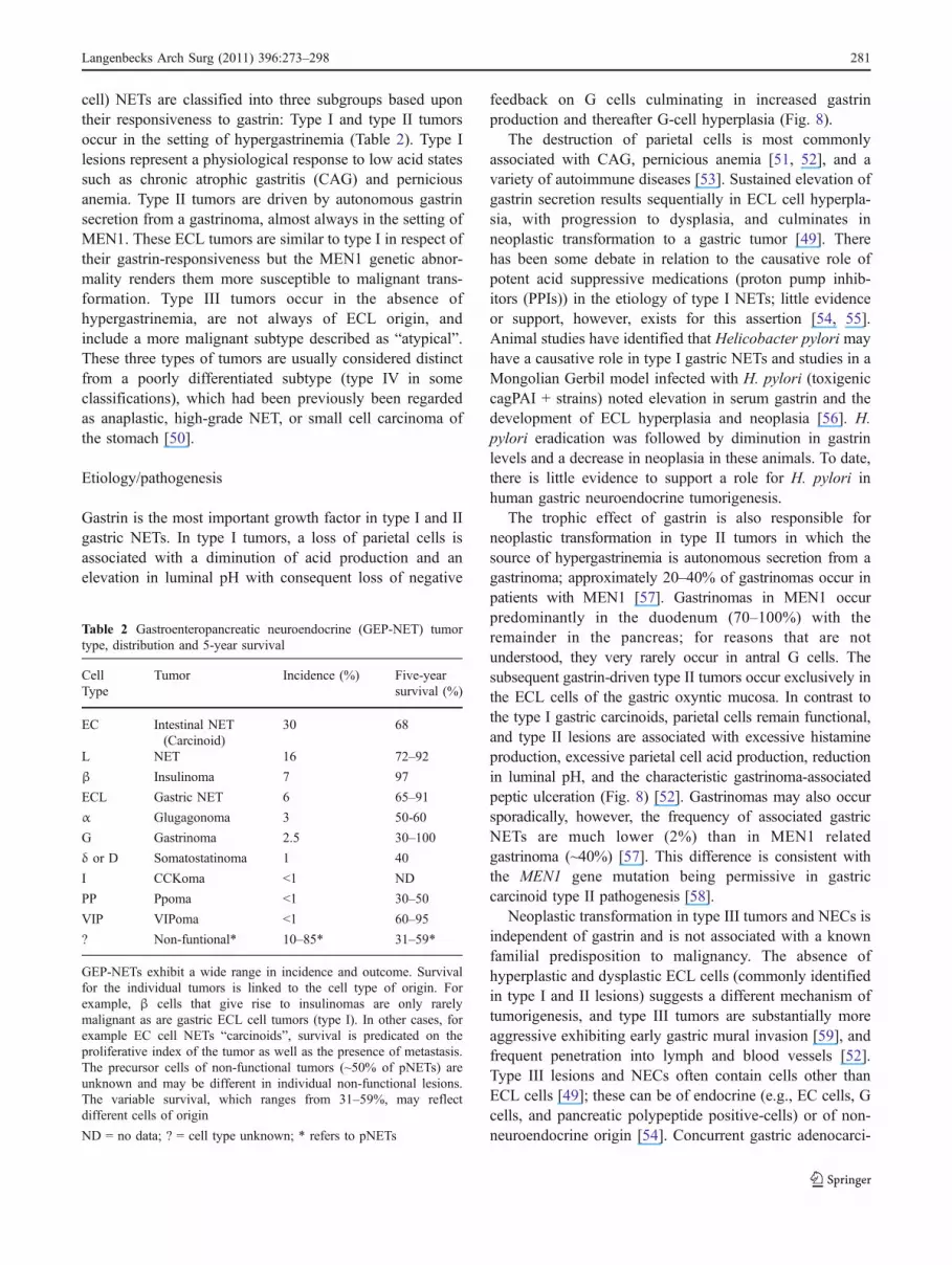
cell) NETs are classified into three subgroups based upontheir responsiveness to gastrin: Type I and type II tumorsoccur in the setting of hypergastrinemia (Table 2). Type Ilesions represent a physiological response to low acid statessuch as chronic atrophic gastritis (CAG) and perniciousanemia. Type II tumors are driven by autonomous gastrinsecretion from a gastrinoma, almost always in the setting ofMEN1. These ECL tumors are similar to type I in respect oftheir gastrin-responsiveness but the MEN1 genetic abnor-mality renders them more susceptible to malignant trans-formation. Type III tumors occur in the absence ofhypergastrinemia, are not always of ECL origin, andinclude a more malignant subtype described as “atypical”.These three types of tumors are usually considered distinctfrom a poorly differentiated subtype (type IV in someclassifications), which had been previously been regardedas anaplastic, high-grade NET, or small cell carcinoma ofthe stomach [50].
Etiology/pathogenesis
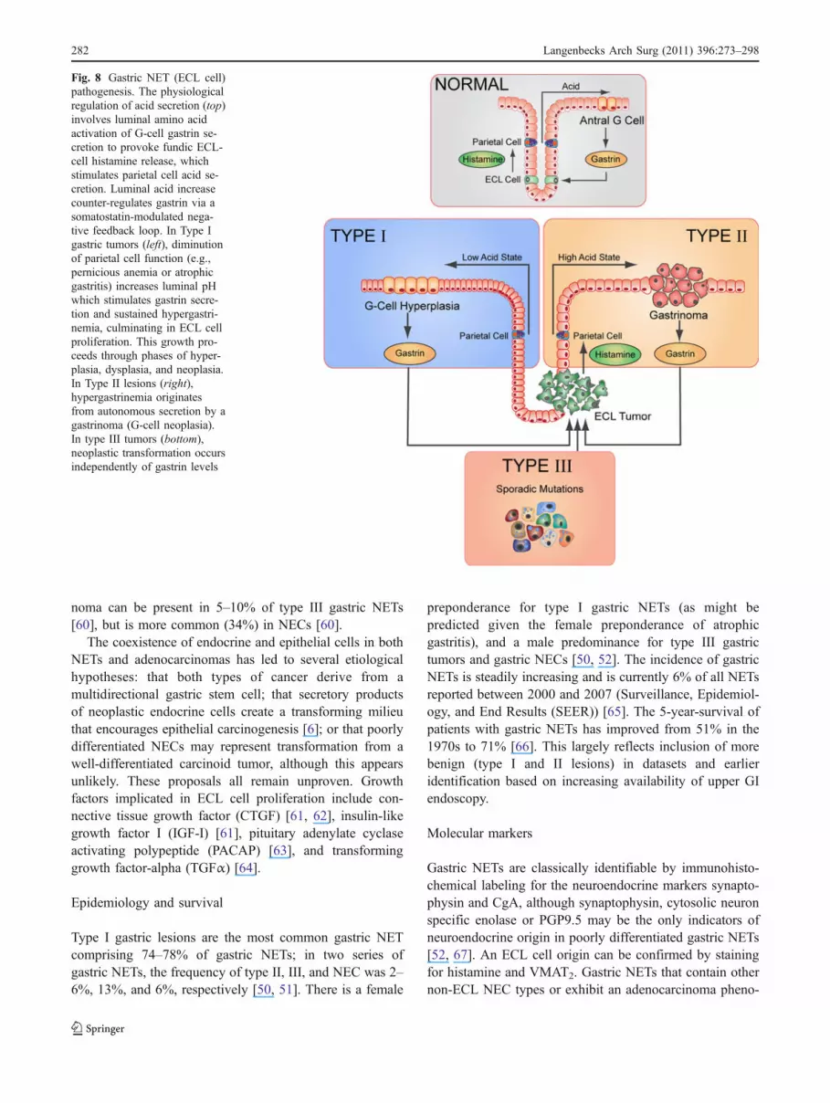
Gastrin is the most important growth factor in type I and IIgastric NETs. In type I tumors, a loss of parietal cells isassociated with a diminution of acid production and anelevation in luminal pH with consequent loss of negative
feedback on G cells culminating in increased gastrinproduction and thereafter G-cell hyperplasia (Fig. 8).
The destruction of parietal cells is most commonlyassociated with CAG, pernicious anemia [51, 52], and avariety of autoimmune diseases [53]. Sustained elevation ofgastrin secretion results sequentially in ECL cell hyperpla-sia, with progression to dysplasia, and culminates inneoplastic transformation to a gastric tumor [49]. Therehas been some debate in relation to the causative role ofpotent acid suppressive medications (proton pump inhib-itors (PPIs)) in the etiology of type I NETs; little evidenceor support, however, exists for this assertion [54, 55].Animal studies have identified that Helicobacter pylori mayhave a causative role in type I gastric NETs and studies in aMongolian Gerbil model infected with H. pylori (toxigeniccagPAI + strains) noted elevation in serum gastrin and thedevelopment of ECL hyperplasia and neoplasia [56]. H.pylori eradication was followed by diminution in gastrinlevels and a decrease in neoplasia in these animals. To date,there is little evidence to support a role for H. pylori inhuman gastric neuroendocrine tumorigenesis.
The trophic effect of gastrin is also responsible forneoplastic transformation in type II tumors in which thesource of hypergastrinemia is autonomous secretion from agastrinoma; approximately 20–40% of gastrinomas occur inpatients with MEN1 [57]. Gastrinomas in MEN1 occurpredominantly in the duodenum (70–100%) with theremainder in the pancreas; for reasons that are notunderstood, they very rarely occur in antral G cells. Thesubsequent gastrin-driven type II tumors occur exclusively inthe ECL cells of the gastric oxyntic mucosa. In contrast tothe type I gastric carcinoids, parietal cells remain functional,and type II lesions are associated with excessive histamineproduction, excessive parietal cell acid production, reductionin luminal pH, and the characteristic gastrinoma-associatedpeptic ulceration (Fig. 8) [52]. Gastrinomas may also occursporadically, however, the frequency of associated gastricNETs are much lower (2%) than in MEN1 relatedgastrinoma (~40%) [57]. This difference is consistent withthe MEN1 gene mutation being permissive in gastriccarcinoid type II pathogenesis [58].
Neoplastic transformation in type III tumors and NECs isindependent of gastrin and is not associated with a knownfamilial predisposition to malignancy. The absence ofhyperplastic and dysplastic ECL cells (commonly identifiedin type I and II lesions) suggests a different mechanism oftumorigenesis, and type III tumors are substantially moreaggressive exhibiting early gastric mural invasion [59], andfrequent penetration into lymph and blood vessels [52].Type III lesions and NECs often contain cells other thanECL cells [49]; these can be of endocrine (e.g., EC cells, Gcells, and pancreatic polypeptide positive-cells) or of non-neuroendocrine origin [54]. Concurrent gastric adenocarci-
Table 2 Gastroenteropancreatic neuroendocrine (GEP-NET) tumortype, distribution and 5-year survival
CellType
Tumor Incidence (%) Five-yearsurvival (%)
EC Intestinal NET(Carcinoid)
30 68
L NET 16 72–92
β Insulinoma 7 97
ECL Gastric NET 6 65–91
α Glugagonoma 3 50-60
G Gastrinoma 2.5 30–100
δ or D Somatostatinoma 1 40
I CCKoma <1 ND
PP Ppoma <1 30–50
VIP VIPoma <1 60–95
? Non-funtional* 10–85* 31–59*
GEP-NETs exhibit a wide range in incidence and outcome. Survivalfor the individual tumors is linked to the cell type of origin. Forexample, β cells that give rise to insulinomas are only rarelymalignant as are gastric ECL cell tumors (type I). In other cases, forexample EC cell NETs “carcinoids”, survival is predicated on theproliferative index of the tumor as well as the presence of metastasis.The precursor cells of non-functional tumors (~50% of pNETs) areunknown and may be different in individual non-functional lesions.The variable survival, which ranges from 31–59%, may reflectdifferent cells of origin
ND = no data; ? = cell type unknown; * refers to pNETs
Langenbecks Arch Surg (2011) 396:273–298 281
noma can be present in 5–10% of type III gastric NETs[60], but is more common (34%) in NECs [60].
The coexistence of endocrine and epithelial cells in bothNETs and adenocarcinomas has led to several etiologicalhypotheses: that both types of cancer derive from amultidirectional gastric stem cell; that secretory productsof neoplastic endocrine cells create a transforming milieuthat encourages epithelial carcinogenesis [6]; or that poorlydifferentiated NECs may represent transformation from awell-differentiated carcinoid tumor, although this appearsunlikely. These proposals all remain unproven. Growthfactors implicated in ECL cell proliferation include con-nective tissue growth factor (CTGF) [61, 62], insulin-likegrowth factor I (IGF-I) [61], pituitary adenylate cyclaseactivating polypeptide (PACAP) [63], and transforminggrowth factor-alpha (TGFα) [64].
Epidemiology and survival
Type I gastric lesions are the most common gastric NETcomprising 74–78% of gastric NETs; in two series ofgastric NETs, the frequency of type II, III, and NEC was 2–6%, 13%, and 6%, respectively [50, 51]. There is a female
preponderance for type I gastric NETs (as might bepredicted given the female preponderance of atrophicgastritis), and a male predominance for type III gastrictumors and gastric NECs [50, 52]. The incidence of gastricNETs is steadily increasing and is currently 6% of all NETsreported between 2000 and 2007 (Surveillance, Epidemiol-ogy, and End Results (SEER)) [65]. The 5-year-survival ofpatients with gastric NETs has improved from 51% in the1970s to 71% [66]. This largely reflects inclusion of morebenign (type I and II lesions) in datasets and earlieridentification based on increasing availability of upper GIendoscopy.
Molecular markers
Gastric NETs are classically identifiable by immunohisto-chemical labeling for the neuroendocrine markers synapto-physin and CgA, although synaptophysin, cytosolic neuronspecific enolase or PGP9.5 may be the only indicators ofneuroendocrine origin in poorly differentiated gastric NETs[52, 67]. An ECL cell origin can be confirmed by stainingfor histamine and VMAT2. Gastric NETs that contain othernon-ECL NEC types or exhibit an adenocarcinoma pheno-
Fig. 8 Gastric NET (ECL cell)pathogenesis. The physiologicalregulation of acid secretion (top)involves luminal amino acidactivation of G-cell gastrin se-cretion to provoke fundic ECL-cell histamine release, whichstimulates parietal cell acid se-cretion. Luminal acid increasecounter-regulates gastrin via asomatostatin-modulated nega-tive feedback loop. In Type Igastric tumors (left), diminutionof parietal cell function (e.g.,pernicious anemia or atrophicgastritis) increases luminal pHwhich stimulates gastrin secre-tion and sustained hypergastri-nemia, culminating in ECL cellproliferation. This growth pro-ceeds through phases of hyper-plasia, dysplasia, and neoplasia.In Type II lesions (right),hypergastrinemia originatesfrom autonomous secretion by agastrinoma (G-cell neoplasia).In type III tumors (bottom),neoplastic transformation occursindependently of gastrin levels
282 Langenbecks Arch Surg (2011) 396:273–298

type can be identified based on properties of the other celltypes (e.g., immunostaining of a specific peptide). At atranscript level, CgA discriminates gastric NETs from othergastric neoplasms (including GISTs) [68]. Over expressionof MAGE-D2 and MTA1 differentiate type III/IV from typeI/II GCs and MTA1 appears to be a marker of tumorinvasion [68].
Duodenal GEP-NETs—G- and D-cell tumors
These are usually small, non-functioning tumors, which donot cause any symptoms, and are often diagnosed by agastroduodenoscopy performed with a different indication.Most duodenal NETs are discovered at an early treatablestage (tumor diameter ≤10 mm) [69, 70]. In the instanceswhen tumors are associated with a syndrome, for exampleZES, lesions are often metastatic [71].
G cell—gastrinomas
Gastrinomas arise in the duodenum and in the pancreas. Up to70% occur in the duodenum, arise directly from G cells, andtend to be small, multiple, and exhibit a less malignant clinicalcourse than those arising in the pancreas [72]. Since G cellsare not identifiable in the adult pancreas it is assumed thatpancreatic gastrinomas arise from a different neuroendocrinecell or precursor. The more aggressive nature of mostpancreatic gastrinomas would support the concept of a differentmodel of tumorigenesis compared to duodenal gastrinomas[73]. Clinically, gastrin secreting tumors, irrespective of theirorigin, are associated with ZES. This syndrome comprisesgastric acid hypersecretion, recurrent, multiple, and intractablepeptic ulceration (duodenum and proximal jejunum) and isoften accompanied by a secretory diarrhea.
Etiology/pathogenesis
Sporadic duodenal gastrinomas constitute 80% of all gastri-nomas, and their etiology and pathogenesis is unknown. Theresidual 20% of duodenal gastrinomas are part of the MEN1syndrome. Duodenal gastrinomas are located in the first andsecond part of the duodenum (90%) [69] and are limited tothe submucosa in ~50% of patients [69]. The anatomical areacomprising the head of the pancreas, the superior, anddescending portion of the duodenum, and the relevant lymphnodes have been entitled the “gastrinoma triangle”, since itharbors the vast majority of these tumors [74]. In someinstances, gastrinomas in peri-pancreatic and peri-duodenallymph-nodes have been considered to represent a primarytumor rather than metastases from an occult lesion in theduodenum, and reports exist of occasional individuals havingbeen cured after resection of the lymph nodes [75, 76].
Duodenal tumors comprise 50–88% of gastrinomas insporadic ZES patients and 70–100% of gastrinomas inMEN1/ZES patients. In rare cases, non-pancreatico-duodenal gastrinomas have been described in the stomach,liver, bile duct, ovary (5–15%), and extra-abdominal (heart,lung) locations [69, 76]. Overall, more than 50% of duodenalgastrinomas have liver metastases at the time of diagnosis.
Epidemiology and survival
The reported incidence of gastrinomas is between 0.5 and 4per million of the population per year [69]. They are the mostcommon functional duodenal tumor. Approximately 0.1% ofpatients with duodenal ulcers have evidence of ZES. Ingeneral, the progression of gastrinomas is relatively slowwith a 5-year survival rate of 65% and 10-year survival rateof 51% [72]. The most significant predictor of survival is thepresence and extent of liver metastases at diagnosis [76, 77].The 10-year survival of patients with local tumors is 96%and only 30% in those with metastatic tumors [77].
Molecular markers
There is no established marker to predict the biologicalbehavior of a gastrinoma, although LOH at 11q13 occurs in44–48% of sporadic duodenal gastrinomas [78, 79]. Hyper-methylation (with subsequent gene silencing) of the promoterregion of p16INK4A occurs in ~50% of gastrinomas [80]; therelevance of this is unknown. Some investigators havesuggested that HER-2/neu amplification, over expression ofepidermal growth factor (EGF), and hepatocyte growth factor(HGF) may be associated with an aggressive growthphenotype [76, 81, 82].
D cell—somatostatinomas
Somatostatinomas secrete the bioactive product, somatostatin.This tetradecapeptide is a ubiquitous neurotransmitter,paracrine agent that in general exerts a widespread inhibitoryeffect on exocrine and endocrine secretion and bowel motility.These tumors usually produce local symptoms secondary tolocal mass effects, for example bile duct obstruction, jaundice,abdominal pain, and gastrointestinal bleeding [83, 84]. Thesomatostatinoma syndrome (steatorrhea, cholelithiasis,diabetes mellitus-like symptoms) is very rare, and a smallproportion are associated with the MEN1 or NF1 disorders.The majority of somatostatinomas are solitary, sporadic, andmalignant.
Etiology/pathogenesis
Approximately 14–43% of somatostatinomas develop in NF1patients. The lesions occur predominantly in the duodenum, in
Langenbecks Arch Surg (2011) 396:273–298 283

the vicinity of the ampulla of Vater but rarely in the pancreas[85, 86]. Tumors are often small, well differentiated, and lowgrade. Nevertheless, lymph node and liver metastases occurin ~35%, and the risk of metastases significantly increaseswhen tumors are >20 mm [87]. Tumors that occur in thesetting of NF1 are usually identified earlier due to increasedsurveillance [84].
Epidemiology and survival
To date, more than 100 cases of duodenal somatostatinomashave been reported [88], and the relative frequency ofduodenal primaries is ~6 times higher than pancreaticprimaries [85]. There is no statistically significant differ-ence in the rate of metastases and malignancy betweenpancreatic and extra pancreatic tumors [89]. The overall 5-year survival rate is 75% when the tumor is localized butdecreases to 60% when metastases are present [90].
Molecular markers
Somatostatinomas exhibit positivity for endocrine markers,especially synaptophysin and CgA, and most cells expresssomatostatin receptors [85]. Expression of the latterindicates tumors may be amenable to somatostatin receptortargeting.
Pancreatic endocrine tumors
Pancreatic neuroendocrine tumors (pNETs) represent 1–2%of all pancreatic neoplasia [91].The majority of pNETs areG1 or G2 neuroendocrine tumors in the WHO classificationof 2010, and approximately half secrete measurable levelsof site-specific bioactive peptides (insulin, gastrin, VIP,glucagon), or other non-pancreatic hormones, for exampleadrenocorticotropic hormone (ACTH) or growth hormone(GH). These peptides are associated with characteristicsyndromes (ZES, Verner-Morrison syndrome, glucagonomasyndrome, Cushing’s syndrome, and acromegaly) and less-specific symptoms (hypoglycemia, hyperglycemia) [4].Depending on the predominant bioactive agent secreted,individual tumors are identified as insulinomas, gastrino-mas, VIPomas, glucagonomas, etc. (Table 2). However,approximately half of pNETs (~10–85%) are not associatedwith symptoms from a clinically defined “hypersecretory”syndrome and are termed “non-functioning”. Irrespective ofthe secretory status, pNETs are usually well demarcated,often solitary, ovoid tumors that can occur in all parts of thepancreas. Although pNETs are histologically “well differ-entiated”, they are frequently malignant, with the exceptionof insulinomas. Tumor characteristics associated with poorprognosis include metastases to regional lymph nodes and
the liver, invasion of adjacent organs, tumor size more than2 cm, angioinvasion, and elevated proliferative activity(more than 2% of cells positive for Ki67) [92]. Recentreports suggest that angioinvasion may represent a morecritical role than previously assumed [93]. It is probablethat a tumor be considered malignant if angioinvasion isevident even if no other criteria of malignancy aredemonstrable.
Embryology, development, and phenotype
Human islets consist of approximately 3,000 cells produc-ing insulin (β cells, 54%), glucagon (α cells, 34%),somatostatin (δ cells, 10%), VIP (δ 2 cells), pancreaticpolypeptide (PP) (PP cells), and substance P/5-HT (ECcells). Gastrin-producing G cells are present in fetal but notnormal adult pancreatic islets [94, 95]. The quotient of allpancreatic endocrine cells is 1–2% of the entire pancreaticcell mass [96]. Both, alpha and beta cells appear to arisefrom the same precursor cell, namely the ductal epithelialcell. By the fifth month of fetal life, islet cells produceinsulin and glucagon [96, 97]. Cytokeratin 20, a marker foradult rat pancreatic ductal cells [98, 99] which is not found innormal adult islet cells, is co-expressed with insulin orglucagon during islet cell neogenesis in the fetal [14] andneonatal [99] pancreas.
Pathogenesis and pathology
The pathogenesis of pNETs is not clear. However, sinceductal cells may be a precursor cell type, at least for normalpancreatic endocrine cells, these are also consideredprecursors for pNETs themselves. The mechanism ofmalignant transformation is unknown but for non-hereditable tumors, is considered similar to other GEP-NETs, that is, reflects an environmental damage-drivensomatic mutation event. The etiology of inherited tumors,for example MEN1, has been discussed but usually is basedon the general schema of an overactive/unregulated growthphenomenon.
Histological examination is not able to define whether alesion is functional or identify the type of hormoneproduction. There are two exceptions to this rule: amyloiddeposits are indicative of insulinomas, and glandularstructures containing psammoma bodies are commonlyobserved in somatostatin-producing tumors [100–102].Poorly differentiated endocrine carcinomas can be misdiag-nosed as pancreatic adenocarcinoma unless appropriateimmunohistochemical is undertaken to define their neuro-endocrine phenotype. Such tumors exhibit pleomorphiccells with high mitotic index (≥10/10 HPF) and oftenangioinvasion. PNETs can be identified using antibodies tomarkers common to all or most NECs: that is, CgA,
284 Langenbecks Arch Surg (2011) 396:273–298

synaptophysin, NSE, and protein gene product 9.5 (PGP9.5) but can also express cytokeratin 8, 18, and 19 [103]and sometimes either VMAT1 or VMAT2.
Molecular markers
Under some conditions, the molecular basis of familial pNETshas been identified and comprises inherited mutations and asecond-hit somatic mutation of MEN1 and VHL genes.However, in contrast to other tumors, for example pancreaticadenocarcinoma, activation of classical oncogene-mediatedpathways does not seem a common event in pNETs. Little isknown in respect of pancreatic neuroendocrine oncogenesisand the molecular basis of the progression of sporadic NETs[103]. Thus, mutations in k-ras, P53, myc, fos, jun, src, andthe Rb gene have not been specifically implicated [104,105]. In contrast, copy number alterations, for example inproteins that regulate some of these pathways, MDM2 andP53, have been noted [106].
Transcription factors (TFs) regulate organogenesis andPAX8 (paired box gene 8) is a part of a group of TFs thatare cell-lineage specific in multiple organ systems [107]. TFanalysis has identified that PAX8 is expressed in normaladult pancreatic islet cells, and is also expressed in asignificant proportion of primary and metastatic well-differentiated PETs [108]. It has been proposed that lossof PAX8 may have prognostic significance since PAX8-negative tumors are significantly larger, associated withmalignant behavior, and are associated with an increasedincidence of liver metastases [108].
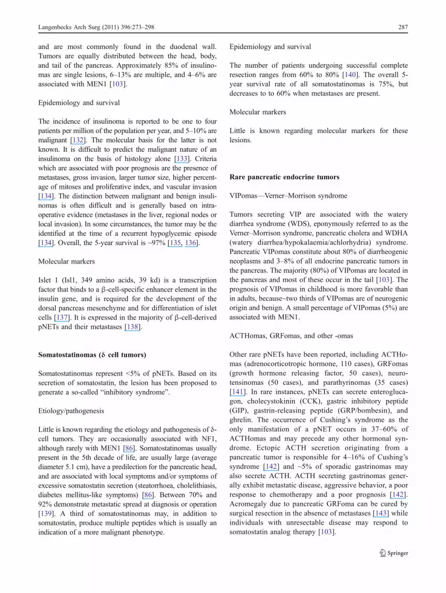
At a chromosomal level, molecular and cytogeneticanalyses have identified a number of chromosomal alter-ations in pNETs. Comparative genomic hybridizationstudies indicate that chromosomal losses have occurredslightly more frequently than gains, while amplificationsare uncommon (Fig. 9 top) [109, 110].
Furthermore, the total number of genomic changes pertumor appears to be associated with both the tumor volume(size) and disease stage, indicating that genetic alterationsaccumulate during the natural history of the lesion. Thus,large tumors with increased malignant potential—andespecially metastases—tend to harbor more genetic alter-ations than small and clinically benign neoplasms. Thissuggests the loss of tumor suppressor pathway(s) andgenomic instability as important mechanisms associatedwith progression but not initiation of a pNET. However,losses of chromosomes 1 and 11q as wells as gains on 9qappear to be early events in the development of pNETs,since they may already be present in small tumors.Prevalent chromosomal alterations common in metastasesinclude gains of both chromosome 4 and 7 and losses of21q, implying that these chromosome imbalances maycontribute to tumor metastasis (Fig. 9 top) [111, 112].
Deletions of 9p which occur in ~30% of pNETs, contain thelocation of the p16INK4A and p14ARF genes, both of whichencode tumor suppressors; loss of this gene locus may leadto tumorigenesis due to deregulation of the p53 and CyclinD1/Rb pathways. Alterations in the cyclin D1 pathway inpNETs indicate over expression of this proto-oncogene in43% of tumors [113]. Chromosome 16p, which containsTSC2 (a tumor suppressor of the AKT/mTOR pathwaywith GTPase activating function), is lost in ~40% of PETs[111, 114], while PTEN a second tumor suppressor at thislocus, is lost in 10–29% of lesions [111, 114, 115]. Lowexpression of either TSC2 or PTEN correlates with pNETaggressiveness, a “non-functional” status, proliferationindex, presence of liver metastasis at diagnosis or follow-up, and with time to progression [116]. This suggests theinvolvement of the AKT/mTOR pathway in pNET tumor-igenesis and progression. PNETs also over-express MDM2,MDM4, and WIP1, all of which may attenuate the functionof p53. Since p53 is critical in maintaining genomicstability, alterations in regulators of p53 are thereforeconsidered potentially permissive for pNET pathogenesis[106]. Fibroblast growth factor 13 (FGF13) is upregulatedin metastatic compared to non-metastatic pNETs [116], andis an independent predictor for shorter progression freesurvival [116]. Little, however, is known about themechanisms by which FGF13 regulates pNET proliferationand metastasis. Deletions on the X-chromosome wereassociated in one study with 100% of pNETs [117]. Twostudies of pNETs found no evidence of microsatelliteinstability [118, 119].
Glucagonomas (α-cell tumors)
Glucagonomas represent about 5% of pNETs and 8–13% offunctional tumors.
Etiology/pathogenesis
Although the majority of α-cell-derived tumors are large andmalignant only a minority (8–13% of functioning tumors) areassociated with the glucagonoma syndrome [120]. Non-syndromic glucagon-producing tumors have been describedunder four different conditions: (1) as solitary tumors thatbecome symptomatic because of their size and/or malignantgrowth; (2) as micro tumors (≤0.5 cm) found incidentally;(3) as multiple microadenomas and macro tumors in patientswith MEN1 [121]; and (4) so-called glucagon cell adeno-matosis. The latter constitutes multiple pancreatic neoplasmsexclusively producing glucagon, associated with glucagoncell hyperplasia of the islets and unrelated to MEN1, VHL,or the recently identified [122] p27 MEN syndrome [123,124]. Glucagonomas commonly occur in the tail of the
Langenbecks Arch Surg (2011) 396:273–298 285

pancreas. Extra pancreatic glucagonomas are extremely rare[125, 126]. One example is a kidney enteroglucagonoma(described in 1971 [127, 128]), which was associated with amassive hypertrophy of the small bowel villi and slow boweltransit times.
Epidemiology and survival
The estimated incidence of the glucagonoma syndrome is 1per 20 million of the population per year [103]. Approx-imately 60–70% of glucagonomas are metastatic at the timeof diagnosis, and only 50–60% of patient survive 5 years[129]. Even small glucagonomas are considered tumors ofuncertain behavior since in some instances the neoplasmmay grow slowly, and patients may survive for many yearswhereas other glucagon-secreting lesions are locally inva-sive, metastasize early and follow an accelerated course.Occasionally, in multihormonal tumors, the glucagonomasyndrome may be associated with or followed by anothersyndrome, such as hypoglycemia syndrome or ZES [130].
Markers
Markers include CgA, CgB, and the glucagon peptide.
Insulinomas (β cell tumors)
Insulinomas are the most frequent of all functioning pNETs.
Etiology/pathogenesis
The etiology and pathogenesis of insulinomas is unknown,and no known risk factors have been identified. Clonalitystudies on pNETs suggest that insulinomas may beprimarily a polyclonal or oligoclonal neoplasm which iseventually overgrown by a more aggressive cell clone thatmay give rise to invasive growth and metastasis [131]. Themajority of insulinomas are located in the pancreas or aredirectly attached to it. Ectopic (extrapancreatic) insulinomaswith symptoms of hypoglycemia are extremely rare (<2%)
Fig. 9 Chromosomal abnormali-ties identified in gastroentero-pancreatic neuroendocrine tumors(GEP-NETs). In pNETs (top), thecommonest losses occur on 6, 11,X, and Y. Common gains includeChr 9, 12, and 17. In smallintestinal NETs (bottom), thecommonest loss is on 18, while17 and 19 exhibit the mostcommon gains. It is evident thatpancreatic and small intestinalNETs express significantly dif-ferent patterns of chromosomalrearrangements, however, theprecise implications of the alter-ations are as yet unresolved
286 Langenbecks Arch Surg (2011) 396:273–298
and are most commonly found in the duodenal wall.Tumors are equally distributed between the head, body,and tail of the pancreas. Approximately 85% of insulino-mas are single lesions, 6–13% are multiple, and 4–6% areassociated with MEN1 [103].
Epidemiology and survival
The incidence of insulinoma is reported to be one to fourpatients per million of the population per year, and 5–10% aremalignant [132]. The molecular basis for the latter is notknown. It is difficult to predict the malignant nature of aninsulinoma on the basis of histology alone [133]. Criteriawhich are associated with poor prognosis are the presence ofmetastases, gross invasion, larger tumor size, higher percent-age of mitoses and proliferative index, and vascular invasion[134]. The distinction between malignant and benign insuli-nomas is often difficult and is generally based on intra-operative evidence (metastases in the liver, regional nodes orlocal invasion). In some circumstances, the tumor may be theidentified at the time of a recurrent hypoglycemic episode[134]. Overall, the 5-year survival is ~97% [135, 136].
Molecular markers
Islet 1 (Isl1, 349 amino acids, 39 kd) is a transcriptionfactor that binds to a β-cell-specific enhancer element in theinsulin gene, and is required for the development of thedorsal pancreas mesenchyme and for differentiation of isletcells [137]. It is expressed in the majority of β-cell-derivedpNETs and their metastases [138].
Somatostatinomas (δ cell tumors)
Somatostatinomas represent <5% of pNETs. Based on itssecretion of somatostatin, the lesion has been proposed togenerate a so-called “inhibitory syndrome”.
Etiology/pathogenesis
Little is known regarding the etiology and pathogenesis of δ-cell tumors. They are occasionally associated with NF1,although rarely with MEN1 [86]. Somatostatinomas usuallypresent in the 5th decade of life, are usually large (averagediameter 5.1 cm), have a predilection for the pancreatic head,and are associated with local symptoms and/or symptoms ofexcessive somatostatin secretion (steatorrhoea, cholelithiasis,diabetes mellitus-like symptoms) [86]. Between 70% and92% demonstrate metastatic spread at diagnosis or operation[139]. A third of somatostatinomas may, in addition tosomatostatin, produce multiple peptides which is usually anindication of a more malignant phenotype.
Epidemiology and survival
The number of patients undergoing successful completeresection ranges from 60% to 80% [140]. The overall 5-year survival rate of all somatostatinomas is 75%, butdecreases to to 60% when metastases are present.
Molecular markers
Little is known regarding molecular markers for theselesions.
Rare pancreatic endocrine tumors
VIPomas—Verner–Morrison syndrome
Tumors secreting VIP are associated with the waterydiarrhea syndrome (WDS), eponymously referred to as theVerner–Morrison syndrome, pancreatic cholera and WDHA(watery diarrhea/hypokalaemia/achlorhydria) syndrome.Pancreatic VIPomas constitute about 80% of diarrheogenicneoplasms and 3–8% of all endocrine pancreatic tumors inthe pancreas. The majority (80%) of VIPomas are located inthe pancreas and most of these occur in the tail [103]. Theprognosis of VIPomas in childhood is more favorable thanin adults, because~two thirds of VIPomas are of neurogenicorigin and benign. A small percentage of VIPomas (5%) areassociated with MEN1.
ACTHomas, GRFomas, and other -omas
Other rare pNETs have been reported, including ACTHo-mas (adrenocorticotropic hormone, 110 cases), GRFomas(growth hormone releasing factor, 50 cases), neuro-tensinomas (50 cases), and parathyrinomas (35 cases)[141]. In rare instances, pNETs can secrete enterogluca-gon, cholecystokinin (CCK), gastric inhibitory peptide(GIP), gastrin-releasing peptide (GRP/bombesin), andghrelin. The occurrence of Cushing’s syndrome as theonly manifestation of a pNET occurs in 37–60% ofACTHomas and may precede any other hormonal syn-drome. Ectopic ACTH secretion originating from apancreatic tumor is responsible for 4–16% of Cushing’ssyndrome [142] and ~5% of sporadic gastrinomas mayalso secrete ACTH. ACTH secreting gastrinomas gener-ally exhibit metastatic disease, aggressive behavior, a poorresponse to chemotherapy and a poor prognosis [142].Acromegaly due to pancreatic GRFoma can be cured bysurgical resection in the absence of metastases [143] whileindividuals with unresectable disease may respond tosomatostatin analog therapy [103].
Langenbecks Arch Surg (2011) 396:273–298 287

Small bowel NETs (EC Cell “Carcinoids”)
The EC cell is the predominant neuroendocrine cell of thegastrointestinal tract and plays a key role in the physiologicalregulation of secretion, motility, blood flow and visceral pain.Intestinal EC cells synthesize, store, and release this amineand contain the majority (95%) of 5-HT in the body [144–149](Fig. 10). Abnormalities in 5-HT release and availability(reuptake and catabolism) are associated with alteredgastrointestinal secretion and motility culminating indiarrhea, constipation, and pain, as in the carcinoid syndrome[150, 151].
Pathogenesis and pathology
The biological basis of small intestinal neuroendocrinepathogenesis, malignancy, and metastasis is unknown.Although EC cells are considered terminally differentiated,they express proliferation-associated transcripts, forexample Ki67 [90]. Despite this, EC cell tumors areconsidered to arise from abnormal mucosal precursor cells.It is likely that the cell which accumulates the mutationsnecessary for development of NETs is a committedneuroendocrine progenitor, a cell not as yet defined in thehuman gastrointestinal tract. The precise mechanismsunderlying the lineage pathways of neuroendocrine cellsand their precursors remains poorly defined but the Notchsignaling pathway is implicated in regulation of celldifferentiation from stem cells [152]. Basic helix-loop-helix transcription factors Math1, NGN3, and beta2/
NeuroD are expressed in neuroendocrine precursors al-though Notch is inactive. Precursor cells induce Notch inadjacent cells, switching off neuroendocrine differentiation.Math1 commits cells to one of three secretory lineages:goblet, paneth, and neuroendocrine while NGN3 appears tobe essential for neuroendocrine cell differentiation [9]. Theregulatory role of these transcription factors is consideredessential for final entero-endocrine cell specification [16].
Typical small bowel EC cell tumors display an insulargrowth pattern (type I), which consists of solid nests orcords of cells with clearly defined boundaries [153]. Atrabecular pattern (type II) consists of narrow cell bandsforming ribbons, regularly anastomosing along a highlydifferentiated vascular network. Type III has a glandularpattern, consisting of cells arranged in alveolar, acinar, orrosette patterns with glandular cavities or pseudo-cavities.Type IV and V NETs consist of undifferentiated and mixedcells, respectively. Multifocal lesions are evident in ~30%of small bowel EC cell tumors [153]. Most of tumorsdevelop as independent primary lesions, and only aminority are due to metastasis from a single primarysuggesting a polyclonal series of synchronous neoplasticevents [154]. In general, hyperplasia of neuroendocrinecells in the associated mucosa is also evident [155, 156].Transmural invasion and an extensive local desmoplasticresponse are common features contributing to the aggres-sive local behavior of the neoplasm [153]. Local spread intothe adjacent mesentery and peritoneum are common as areregional lymph node and distant metastases. The latter arepredominantly hepatic but also may involve lung, bone, and
Fig. 10 The distribution and role of serotonin secreting EC cellswithin the gastrointestinal tract. EC cells are ubiquitous throughout thegut and represent 0.25–0.5% of the total mucosal volume (left). Theyare chemomechanosensory cells (center) and respond both to luminalproducts and mechanical activity of the bowel by serotonin (5-HT)secretion. Locally produced serotonin regulates mucosal secretion and
absorption as well as peristalsis and secretory reflexes (right).Abnormalities, for example increased EC cell numbers (e.g., smallintestinal NETs), result in excess 5-HT production with accentuationof normal physiological events, for example increased mucussecretion, secretory diarrhea, and excessive peristalsis
288 Langenbecks Arch Surg (2011) 396:273–298

brain [153]. The tumor cells are characteristically argyr-ophil and argentaffin [153] and over 85% of the tumorsexhibit positive immunohistochemistry for CgA, Leu-7,NSE, and 5-HT [153]. The vast majority of these lesionsare “classical” ileal carcinoids with production of 5-HT andsubstance P, but rare tumors producing enteroglucagon, PP,or peptide YY may occur. EC cell NETs exhibit the highestfrequency of non-NET tumor association, for examplecolorectal cancers (39%) [157, 158]. Other associatednon-endocrine tumors include adenocarcinomas of thesmall bowel, stomach, lung, prostate, and cervix uteri[159].
Several factors appear to be determinants of malignancy,including lesion size, local spread and extent of metastasesat the time of diagnosis, mitotic rate, multiplicity, femalegender, depth of invasion, and the presence of carcinoidsyndrome [153].
Epidemiology and survival
Small intestinal EC cell tumors are the second mostfrequent type of NET (17.3% in the 2007 SEER analysis)[160]. The highest frequency of small intestinal NETs is inthe ileum, and is ~7 times more frequent than in theduodenum and the jejunum [158, 161]. Small intestinalNETs exhibit an overall higher frequency of metastases atthe time of diagnosis (~60% of staged tumors, SEER)compared to all GEP-NETs (26% of staged tumors SEER)[1]. The overall 5-year-survival rate is 68.1% [1]. The 5-year-survival rate of patients with hepatic tumor spread is18–32% [1]. An increased median survival (4.4 years) isevident in patients with jejuno-ileal carcinoids whichexhibit a mixed insular/glandular pattern [162]. In contrast,patients with an undifferentiated pattern have a mediansurvival of only 6 months. In those lesions with a pureinsular and trabecular pattern, an intermediate prognosis isevident with a median survival time of 2.9 years and2.5 years, respectively [162].
The relatively poor prognosis of small intestinal NETsreflects the inherent clinical difficulty in identifying smallbowel malignancies (Table 3), as well as the intrinsicallymalignant nature of the tumor with dissemination to boththe lymph nodes and the liver.
Molecular markers
Comparative genomic hybridization strategies have identi-fied gains in chromosomes 17q and 19p (57%) and in 19qand 4q (50%) in EC cell tumors [163]. Gains were alsofound in 4p (43%), 5 (36%), and 20q (36%) and losses in18q or 18p (43%), while 21% had full or partial loss of 9p[163]. Of 14 tumors, six had full gain of chromosome 4 ofwhich four samples also had gain of chromosome 5. There
were four tumors with a gain of chromosome 4 along with apartial or full trisomy of chromosome 14 [163]. In aseparate CGH study, losses in 18q22-qter (terminal end ofchromosom18q) (67%) and 11q22-23 (33%) were the mostcommon genetic defects although losses of 16q (22%) andgains of 4p (22%) were also identified [164].
Of note, since the 18q and 11q chromosomal lossesoccurred more frequently this suggests that they are earlyevents in EC cell tumorigenesis while a loss on chromo-some 16 and some gain-of-function on chromosome 4 arelater events in tumor/carcinoid development (Fig. 9bottom). This proposal is supported by a report thataberrations in 16q and 4p tend to occur in metastases[165]. Lollgen et al. reiterated the notion that 18q deletionswere characteristic of midgut NETs by finding losses in88% of tumors [166]. These findings, particularly lossesand gains in chromosomes 18 and 14 have been confirmedby more recent reports [167, 168]. One of the genesencoded on Chr18 (18q21) is the tumor suppressor geneDCC (deleted in colorectal carcinoma). Loss of this gene,which has been linked to the tumor suppressor NCAM(neural cell adhesion molecule) on 11q [169], is thought toplay a role in carcinoid genesis [170]. A 40 kb heterozy-gous deletion in Chr18q22.1 has been suggested as apotential inherited factor, but the low occurrence of this(only ~6% of cases) make it difficult to appreciate the realsignificance [171]. A gain of chromosome 14 has beenidentified as a marker of poor prognosis [167], while theanti apoptotic protein DAD1 has been identified in one ofthe chromosome 14 foci, and confirmed to be over-expressed at an immunohistochemical level [168].
A separate CGH study identified that ~20% of EC celltumors exhibited alterations in the distal part of 11q(location of succinate ubiquinone oxidoreductase subunitD gene—SDHD) [165]. Furthermore, two of five EC celltumors exhibited a missense mutation in the SDHD gene inassociation with LOH of the other allele, suggesting thatalterations of the SDHD gene might be implicated in thetumorigenesis of these lesions [165]. An analysis ofmicrosatellite instability in well-differentiated EC celltumors or their metastases using an analysis of the BAT-26 microsatellite locus in intron 5 of hMSH2 and the BAT-II microsatellite region of TGFβRII [172] identified noMSI. In contrast, carcinomas of the small intestine exhibitMSI in approximately 20% of cases [173, 174], suggestingthat neuroendocrine cell tumors probably evolve differentlyto epithelial tumors in this organ [172].
Affymetrix transcriptional profiling has identified >1,500over-expressed and ~400 transcripts that are decreased inexpression in a large group (~30 samples) of EC cell tumors[175]. Further analysis of this data identified three potentiallyuseful malignancy-marker genes. Specifically, over expres-sion of NAP1L1, MAGE-D2, and MTA1 mRNA and MTA1
Langenbecks Arch Surg (2011) 396:273–298 289

protein in tumor and metastatic EC cell NETs was confirmedsuggesting these genes may be markers for identifyingmetastatic tumors while NAP1L1 may be a neuroendocrinetumor-specific marker [175]. Expression of these markers aswell as CgA has been demonstrated as effective in theprediction of EC cell tumor grade and stage [176]. Othercandidate marker genes have been identified in EC cellcarcinomas [177], the utility of which are still beingexamined. Over-expression of MTA1 has been confirmed[178]. Analysis of microRNAs in EC cell tumors hasidentified the cardiac-specific miRNA-133a to be downregulated in metastases [179]; the relevance of this observa-tion remains to be determined.
The Swedish Family Cancer database study identified anincreased risk of EC cell NETs among the offspring ofpatients with squamous cell skin cancer (RR 1.79) and non-Hodgkin’s lymphoma (RR 2.06), while the relative risk ofthis tumor was 2.21 for individuals whose mother hadendometrial cancer [31]. The offspring of patients diag-nosed with EC cell NETs had an increased risk of cancer ofthe breast (RR 1.39), kidney (RR 2.08), and brain (RR1.65). This and other epidemiological-based studies [31,180] suggest that an increased risk of developing EC cell
NETs occurs in individuals with a parental history of thedisease while a family history of any cancer, that is, notonly NETs, is a risk factor for the development of thesetumors [181]. This study could not identify predisposinggenetic factors but suggests that gene mutations common tothese different tumors may play a role in triggering EC cellNET development. Genetic analyses in a family with threeconsecutive first-degree relatives [182] could not identifythat inheritance of EC cell NETs was linked to MEN1.This, and other studies [3, 183], provide further support thatinheritance of the tumor in this location (small intestine) isnot linked to the MEN1 syndrome.
At a growth-regulatory level, EC cell NETs, which donot express mutations of the menin gene are, however,characterized by a loss-of-responsiveness to TGFβ1-mediated growth inhibition that characterizes normal smallintestinal EC cell proliferation (Fig. 11) [184].
Colorectal NETS
Colorectal “carcinoid” tumors comprise ~35% of allgastrointestinal NETs and 25% of all NETs [65]. The
Nomenclature Type
NET G1 Neuroendocrine tumor G1 (carcinoid)*
NET G2 Neuroendocrine tumor G2**
Stomach Gastric NET (ECL cell)†
Gastrin-producing NET (G cell)
Serotonin-producing NET (EC cell)
ACTH-producing NET
Duodenum Serotonin-producing NET (EC cell)
Somatostatin-producing NET
Gangliocytic paraganglioma (periampullary)
Pancreas Non-functional pNET
Insulinoma
Glucagonoma
Gastrinoma
Somatostatinoma
VIPoma, PPoma
Serotonin-producing NET (carcinoid)
Appendix Serotonin-producing NET (EC cell)
Tubular carcinoid
L-cell, glucagon-like peptide and PP/PPY-producing NETs
Bowel Serotonin-producing tumor (EC cell)
L-cell, glucagon-like peptide and PP/PPY-producing NETs
NEC G3 Neuroendocrine carcinoma***
Small cell NEC
Large cell NEC
Appendix Goblet cell carcinoid (mixed adenoneuroendocrine carcinoma (MANEC))
Table 3 Gastroenteropancreaticneuroendocrine (GEP-NET)classification 2010 (based on[201])
*G1: <2 mitoses per 10 high-power field (HPF) and/or ≤2%Ki67-index
**G2: 2–20 mitoses/10 HPFand/or 3–20% Ki67-index
***G3: >20 mitoses/HPFand/or >20% Ki67-index† Associated with autoimmunechronic atrophic gastritis (A-CAG) or MEN1-ZES
290 Langenbecks Arch Surg (2011) 396:273–298

distribution is cecum (~11%), ascending colon and appen-dix (~22%), transverse and descending (~3%), sigmoidcolon with recto sigmoid junction (~10.5%), and 51%rectum. The increased distribution in the right colon may beexplained by a higher density of neuroendocrine cells inthis region or reflect inability to clearly differentiateterminal ileal lesions that have invaded the cecum [185].
Pathogenesis and pathology
Little is known about these tumors in terms of pathogen-esis. Due to the embryonic development of the mid- andhindgut, two types of common well-differentiated endo-crine NETs have been identified in the colon and rectum: L-cell tumors and EC cell tumors [186]. Rectal tumors areusually L-cell tumors, producing glicentin-related productsand PP-PYY peptides. EC tumors with typical 5-HTproduction are rare in the colon and rectum [187, 188].Specific markers that identify rectal NETs include thosethat identify L cells, such as glucagon- 29, glucagon-37,glicentin, PYY, and PP and their precursors.
Epidemiology and survival
The incidence of colorectal NETs has increased fromapproximately 0.2 per 100,000 in 1973 to 0.86 per100,000 per year in the 2004 SEER database (430%
increase) [189]. The true incidence of colorectal NETs,however, is likely higher since these tumors are consideredbenign by some pathologists who do not therefore registerthe tumor for the SEER database.
Colon NETs proximal to the rectum appear moreaggressive, with a 5-year survival of ~62% across all stages[190]. Data for the 5-year-survival rates of colonic NETs,however, are inconsistent. Some data report 80% [191], andothers 6% [192] which are worse than colonic adenocarci-noma (~43%) [191]. The heterogeneity is likely due to arecruiting bias of NETs and NECs in different series orinclusion of lesions of different as yet unclassifiedneuroendocrine cell types. An analysis of ~8,000 colorectalNETs demonstrated a 5-year-survival rate of 41.6%compared to 60.9% in colonic adenocarcinomas [185].
Rectal NETs are associated with the highest 5-year-survival rate of 88% in comparison to other NET primaries[190]. This finding reflects that most of rectal carcinoidtumors (82%) [190] are localized at diagnosis, with amedian size of only 0.6 cm. Tumor size, depth of invasion,and lymph node involvement significantly predict malig-nant behavior in localized rectal NETs. A literature analysisreported that metastases were observed in 2% of patientswith rectal NETs measuring less than 1 cm, 10–15% oftumors measuring 1–2 cm, and 60–80% in patients withtumors measuring greater than 2 cm [193]. Another studyhas shown that metastases occurred in only 2% of tumorssmaller than 2 cm, which had not invaded the muscularispropria, compared to 48% in tumors with muscularisinvasion [194]. Five-year-survival rate in rectal NET is72–92% depending on selection bias for small tumors.Overall, the prognosis is substantially better than for rectaladenocarcinoma which exhibits an overall 5-year-survivalrate of 46–59% [191, 195].
Molecular markers
Neuroendocrine marker molecules differentially expressed inlarge bowel NETs include α/β-SNAP and synaptophysin aswell as SNAP25. In contrast, CgA and VAMP2 are lessfrequently expressed [196]. Proximal colonic tumors areusually EC cell tumors with similar markers to smallintestinal NETs. Metastatic colonic disease is only rarelyassociated with the carcinoid syndrome and only a smallfraction of hindgut NETs (<1%) produce and secrete 5-HT orother bioactive hormones [193]. Therefore, measurement ofserum 5-HT or urine 5-hydroxyindoleaceticacid (5-HIAA) isnot recommended. Poorly differentiated small cell carcino-mas of the large bowel usually have extensive expression ofsynaptophysin and cytosolic markers of neuroendocrinedifferentiation like PGP9.5 and NSE. Prostate-specific acidphosphatase is expressed in 80–100% of rectal carcinoids[187], and may be useful clinically. Serum CgA can be of
Fig. 11 Proliferative regulation of EC cells. Proliferation is differen-tially regulated by a variety of growth factors through activation of anumber of signal transduction pathways such as AKT/ERK/SMADand mTOR pathways. The somatostatin receptor system is the bestcharacterized negative regulator and signal transduction is predomi-nantly via the p38/cGMP pathway. It is likely that the majority ofneuroendocrine cells exhibit similar signal transduction circuitry
Langenbecks Arch Surg (2011) 396:273–298 291

some utility for monitoring patients with metastatic disease[197, 198] or for surveillance in patients with resected stageII or III tumors. False positive elevations in CgA arefrequently associated with the use of proton-pump inhibitors.Elevated levels of CgA can also occur in patients withchronic gastritis or other inflammatory diseases. P53 may beuseful as a marker of poorly differentiated tumors. Immuno-histochemistry for somatostatin receptor-2A may be per-formed in specialized laboratories. βHCG may be expressed,and is associated with greater malignancy of the lesions[199]. Identification of lesions with high malignant potentialcan be determined by mitotic indexing and Ki67 percentagestaining to determine the tumor proliferative index [200].
Conclusion
In this review, we examined GEP-NETs, focusing on thedistinct patterns of each type, by examining both the cell typeand organ of origin. GEP-NETs are derived from distinct cellprecursors, undergo specific chromosomal abnormalities/somatic mutation events, have different clinical presentations(are either syndromic or not), and are associated with widelydiffering survival rates reflecting the different proliferativeregulation as well as intrinsic difference in malignant potentialof different neuroendocrine cell types. In the past, thesetumors were considered under a common rubric “carcinoid”as indolent and uncommon. Overall NETs occur far morefrequently than previously considered and exhibit distinctcellular and clinical behaviors; each cell-specific lesion shouldtherefore be considered and examined as a separate entity. Ingeneral the tumors exhibit both commonalities as well assignificant diversity. The former include common secretorymachinery and physiological responses (neural/hormonalregulators), a relative high proportion of sporadic tumorsand development from precursor cell types. Overall there is apaucity of information regarding transformation from thenaïve to the neoplastic cell state except that microsatelliteinstability does not occur often and adenocarcinoma-associated oncogenic events, for example K-RAS mutationsare very uncommon. Differences include restrictions in loco-regional distribution (e.g., gastric ECL cell in the oxynticmucosa compared to extensive distribution of EC cells),significantly different growth patterns such as the relativebenign nature (high long-term survival) of some organ-restricted cell specific tumors, particularly gastric Type Igastric NETs and pancreatic β-cell NETs (insulinomas)compared to others, for example colonic or non-functioningpNETs; the significant difference in chromosomal abnormal-ities (and potentially etiopathogenesis) between pNETs andsmall intestinal NETs; and the relative lack of informationregarding growth regulation (except for gastric ECL cells).
Conflicts of interest None.
References
1. Modlin IM, Oberg K, Chung DC, Jensen RT, de Herder WW,Thakker RV, Caplin M, Delle Fave G, Kaltsas GA, Krenning EP,Moss SF, Nilsson O, Rindi G, Salazar R, Ruszniewski P, SundinA (2008) Gastroenteropancreatic neuroendocrine tumours. LancetOncol 9(1):61–72. doi:10.1016/S1470-2045(07)70410-2
2. Modlin IM, Gustafsson BI, Moss SF, Pavel M, Tsolakis AV,Kidd M (2010) Chromogranin A-biological function and clinicalutility in neuro endocrine tumor disease. Ann Surg Oncol.doi:10.1245/s10434-010-1006-3
3. Calender A (2000) Molecular genetics of neuroendocrinetumors. Digestion 62(Suppl 1):3–18
4. Kloppel G, Perren A, Heitz PU (2004) The gastroenteropancreaticneuroendocrine cell system and its tumors: the WHO classification.Ann NYAcad Sci 1014:13–27
5. Andrew A, Kramer B, Rawdon B (1998) The origin of gut andpancreatic neuroendocrine (APUD) cells—the last word? JPathol 186:117–118
6. Waldum HL, Rorvik H, Falkmer S, Kawase S (1999) Neuroen-docrine (ECL cell) differentiation of spontaneous gastric carci-nomas of cotton rats (Sigmodon hispidus). Lab Anim Sci 49(3):241–247
7. Wright NA (1999) Letter from Waldum et al. commenting on theeditorial by Andrew et al. and responses. J Pathol 189(3):439–440
8. Pearse AG (1969) The cytochemistry and ultrastructure ofpolypeptide hormone-producing cells of the APUD series andthe embryologic, physiologic and pathologic implications of theconcept. J Histochem Cytochem 17(5):303–313
9. Schonhoff SE, Giel-Moloney M, Leiter AB (2004) Minireview:development and differentiation of gut endocrine cells. Endocri-nology 145(6):2639–2644
10. Novelli MR, Williamson JA, Tomlinson IP, Elia G, Hodgson SV,Talbot IC, Bodmer WF, Wright NA (1996) Polyclonal origin ofcolonic adenomas in an XO/XY patient with FAP. Science 272(5265):1187–1190
11. Taylor RW, Barron MJ, Borthwick GM, Gospel A, Chinnery PF,Samuels DC, Taylor GA, Plusa SM, Needham SJ, Greaves LC,Kirkwood TB, Turnbull DM (2003) Mitochondrial DNAmutations in human colonic crypt stem cells. J Clin Invest 112(9):1351–1360
12. Greaves LC, Preston SL, Tadrous PJ, Taylor RW, Barron MJ,Oukrif D, Leedham SJ, Deheragoda M, Sasieni P, Novelli MR,Jankowski JA, Turnbull DM, Wright NA, McDonald SA (2006)Mitochondrial DNA mutations are established in human colonicstem cells, and mutated clones expand by crypt fission. Proc NatlAcad Sci USA 103(3):714–719
13. McDonald SA, Greaves LC, Gutierrez-Gonzalez L, Rodriguez-JustoM, Deheragoda M, Leedham SJ, Taylor RW, Lee CY, Preston SL,Lovell M, Hunt T, Elia G, Oukrif D, Harrison R, Novelli MR,Mitchell I, Stoker DL, Turnbull DM, Jankowski JA, Wright NA(2008) Mechanisms of field cancerization in the human stomach: theexpansion and spread ofmutated gastric stem cells. Gastroenterology134(2):500–510
14. Bouwens L, Kloppel G (1996) Islet cell neogenesis in thepancreas. Virchows Arch 427(6):553–560
15. Paris M, Tourrel-Cuzin C, Plachot C, Ktorza A (2004) Review:pancreatic beta-cell neogenesis revisited. Exp Diabesity Res 5(2):111–121. doi:10.1080/15438600490455079
16. Wang J, Cortina G, Wu SV, Tran R, Cho JH, Tsai MJ, Bailey TJ,Jamrich M, Ament ME, Treem WR, Hill ID, Vargas JH,
292 Langenbecks Arch Surg (2011) 396:273–298

Gershman G, Farmer DG, Reyen L, Martin MG (2006) Mutantneurogenin-3 in congenital malabsorptive diarrhea. N Engl JMed 355(3):270–280
17. Barrett P, Hobbs RC, Coates PJ, Risdon RA, Wright NA, HallPA (1995) Endocrine cells of the human gastrointestinal tracthave no proliferative capacity. Histochem J 27(6):482–486
18. Karam S, Leblond CP (1995) Origin and migratory pathways ofthe eleven epithelial cell types present in the body of the mousestomach. Microsc Res Tech 31(3):193–214. doi:10.1002/jemt.1070310304
19. Rindi G, Leiter AB, Kopin AS, Bordi C, Solcia E (2004) The“normal” endocrine cell of the gut: changing concepts and newevidences. Ann NY Acad Sci 1014:1–12
20. Kidd M, Modlin IM, Tang LH (1998) Gastrin and theenterochromaffin-like cell: an acid update. Dig Surg 15(3):209–217
21. Wiedenmann B, John M, Ahnert-Hilger G, Riecken EO (1998)Molecular and cell biological aspects of neuroendocrine tumorsof the gastroenteropancreatic system. J Mol Med 76(9):637–647
22. Bloom SR (1978) Gut hormones. Proc Nutr Soc 37(3):259–27123. Zanner R, Gratzl M, Prinz C (2002) Circle of life of secretory
vesicles in gastric enterochromaffin-like cells. Ann NYAcad Sci971:389–396
24. Amara SG, Kuhar MJ (1993) Neurotransmitter transporters:recent progress. Annu Rev Neurosci 16:73–93. doi:10.1146/annurev.ne.16.030193.000445
25. Erickson JD, Schafer MK, Bonner TI, Eiden LE, Weihe E (1996)Distinct pharmacological properties and distribution in neurons andendocrine cells of two isoforms of the human vesicular monoaminetransporter. Proc Natl Acad Sci USA 93(10):5166–5171
26. Kidd M, Modlin IM, Gustafsson BI, Drozdov I, Hauso O,Pfragner R (2008) Luminal regulation of normal and neoplastichuman EC cell serotonin release is mediated by bile salts,amines, tastants, and olfactants. Am J Physiol Gastrointest LiverPhysiol 295(2):G260–G272. doi:10.1152/ajpgi.00056.2008
27. Jahn R, Lang T, Sudhof TC (2003) Membrane fusion. Cell 112(4):519–533
28. Stenmark H, Olkkonen VM (2001) The Rab GTPase family.Genome Biol 2 (5):REVIEWS3007
29. Helpap B, Kollermann J (2001) Immunohistochemical analysisof the proliferative activity of neuroendocrine tumors fromvarious organs. Are there indications for a neuroendocrinetumor-carcinoma sequence? Virchows Arch 438(1):86–91
30. Leotlela PD, Jauch A, Holtgreve-Grez H, Thakker RV (2003)Genetics of neuroendocrine and carcinoid tumours. Endocr RelatCancer 10(4):437–450
31. Hiripi E, Bermejo JL, Sundquist J, Hemminki K (2009) Familialgastrointestinal carcinoid tumours and associated cancers. AnnOncol 20(5):950–954
32. Wermer P (1954) Genetic aspects of adenomatosis of endocrineglands. Am J Med 16(3):363–371
33. Larsson C, Skogseid B, Oberg K, Nakamura Y, Nordenskjold M(1988) Multiple endocrine neoplasia type 1 gene maps tochromosome 11 and is lost in insulinoma. Nature 332(6159):85–87. doi:10.1038/332085a0
34. Knudson AG Jr (1971) Mutation and cancer: statistical study ofretinoblastoma. Proc Natl Acad Sci USA 68(4):820–823
35. Boissan M, De Wever O, Lizarraga F, Wendum D, Poincloux R,Chignard N, Desbois-Mouthon C, Dufour S, Nawrocki-Raby B,Birembaut P, Bracke M, Chavrier P, Gespach C, Lacombe ML(2010) Implication of metastasis suppressor NM23-H1 inmaintaining adherens junctions and limiting the invasivepotential of human cancer cells. Cancer Res 70(19):7710–7722.doi:10.1158/0008-5472.CAN-10-1887
36. Dimou AT, Syrigos KN, Saif MW (2010) Neuroendocrinetumors of the pancreas: what’s new. Highlights from the “2010
ASCO Gastrointestinal Cancers Symposium”. Orlando, FL,USA. JOP 11(2):135–138
37. Donis-Keller H, Dou S, Chi D, Carlson KM, Toshima K,Lairmore TC, Howe JR, Moley JF, Goodfellow P, Wells SA Jr(1993) Mutations in the RET proto-oncogene are associated withMEN 2A and FMTC. Hum Mol Genet 2(7):851–856
38. Vasen HF, van der Feltz M, Raue F, Kruseman AN, KoppeschaarHP, Pieters G, Seif FJ, Blum WF, Lips CJ (1992) The naturalcourse of multiple endocrine neoplasia type IIb. A study of 18cases. Arch Intern Med 152(6):1250–1252
39. Richard S, Giraud S, Beroud C, Caron J, Penfornis F, Baudin E,Niccoli-Sire P, Murat A, Schlumberger M, Plouin PF, Conte-Devolx B (1998) Von Hippel-Lindau disease: recent geneticprogress and patient management. Francophone Study Group ofvon Hippel-Lindau Disease (GEFVH). Ann Endocrinol (Paris)59(6):452–458
40. Neumann HP, Dinkel E, Brambs H, Wimmer B, Friedburg H,Volk B, Sigmund G, Riegler P, Haag K, Schollmeyer P et al(1991) Pancreatic lesions in the von Hippel-Lindau syndrome.Gastroenterology 101(2):465–471
41. Latif F, Tory K, Gnarra J, Yao M, Duh FM, Orcutt ML,Stackhouse T, Kuzmin I, Modi W, Geil L et al (1993)Identification of the von Hippel-Lindau disease tumor suppressorgene. Science 260(5112):1317–1320
42. Kibel A, Iliopoulos O, DeCaprio JA, Kaelin WG Jr (1995)Binding of the von Hippel-Lindau tumor suppressor protein toElongin B and C. Science 269(5229):1444–1446
43. Bluyssen HA, Lolkema MP, van Beest M, Boone M, SnijckersCM, Los M, Gebbink MF, Braam B, Holstege FC, Giles RH,Voest EE (2004) Fibronectin is a hypoxia-independent target ofthe tumor suppressor VHL. FEBS Lett 556(1–3):137–142
44. Ruggieri M, Huson SM (1999) The neurofibromatoses. Anoverview. Ital J Neurol Sci 20(2):89–108
45. Au KS, Williams AT, Gambello MJ, Northrup H (2004)Molecular genetic basis of tuberous sclerosis complex: frombench to bedside. J Child Neurol 19(9):699–709
46. Carney JA, Gordon H, Carpenter PC, Shenoy BV, Go VL (1985)The complex of myxomas, spotty pigmentation, and endocrineoveractivity. Medicine (Baltimore) 64(4):270–283
47. Kirschner LS, Carney JA, Pack SD, Taymans SE, Giatzakis C,Cho YS, Cho-Chung YS, Stratakis CA (2000) Mutations of thegene encoding the protein kinase A type I-alpha regulatorysubunit in patients with the Carney complex. Nat Genet 26(1):89–92. doi:10.1038/79238
48. Modlin IM, Lye KD, Kidd M (2003) Carcinoid tumors of thestomach. Surg Oncol 12(2):153–172
49. Rindi G, Solcia E (2007) Endocrine hyperplasia and dysplasia inthe pathogenesis of gastrointestinal and pancreatic endocrinetumors. Gastroenterol Clin North Am 36(4):851–865.doi:10.1016/j.gtc.2007.08.006, vi
50. Rindi G, Bordi C, Rappel S, La Rosa S, Stolte M, Solcia E (1996)Gastric carcinoids and neuroendocrine carcinomas: pathogenesis,pathology, and behavior. World J Surg 20(2):168–172
51. Borch K, Ahren B, Ahlman H, Falkmer S, Granerus G,Grimelius L (2005) Gastric carcinoids: biologic behavior andprognosis after differentiated treatment in relation to type. AnnSurg 242(1):64–73
52. Rindi G, Luinetti O, Cornaggia M, Capella C, Solcia E (1993)Three subtypes of gastric argyrophil carcinoid and the gastricneuroendocrine carcinoma: a clinicopathologic study. Gastroen-terology 104(4):994–1006
53. Nakata K, Aishima S, Ichimiya H, Yao T, Matsuura T, Seo M,Nagai E, Okido M, Kato M, Nakagaki M, Tsuneyoshi M, TanakaM (2010) Unusual multiple gastric carcinoids with hypergas-trinemia: report of a case. Surg Today 40(3):267–271.doi:10.1007/s00595-009-4032-7
Langenbecks Arch Surg (2011) 396:273–298 293

54. Lawrence B, Kidd M, Svejda B, Modlin I (2010) A clinicalperspective on gastric neuroendocrine neoplasia. Curr Gastro-enterol Rep. doi:10.1007/s11894-010-0158-4
55. Thomson AB, Sauve MD, Kassam N, Kamitakahara H (2010)Safety of the long-term use of proton pump inhibitors. World JGastroenterol 16(19):2323–2330
56. Cao L, Mizoshita T, Tsukamoto T, Takenaka Y, Toyoda T, CaoX, Ban H, Nozaki K, Tatematsu M (2008) Development ofcarcinoid tumors of the glandular stomach and effects oferadication in Helicobacter pylori-infected Mongolian gerbils.Asian Pac J Cancer Prev 9(1):25–30
57. Jensen RT (1998) Management of the Zollinger-Ellison syn-drome in patients with multiple endocrine neoplasia type 1. JIntern Med 243(6):477–488
58. Berna MJ, Annibale B, Marignani M, Luong TV, Corleto V, PaceA, Ito T, Liewehr D, Venzon DJ, Delle Fave G, Bordi C, JensenRT (2008) A prospective study of gastric carcinoids andenterochromaffin-like cell changes in multiple endocrine neopla-sia type 1 and Zollinger-Ellison syndrome: identification of riskfactors. J Clin Endocrinol Metab 93(5):1582–1591. doi:10.1210/jc.2007-2279
59. Bordi C, Yu JY, Baggi MT, Davoli C, Pilato FP, Baruzzi G,Gardini G, Zamboni G, Franzin G, Papotti M et al (1991) Gastriccarcinoids and their precursor lesions. A histologic and immu-nohistochemical study of 23 cases. Cancer 67(3):663–672
60. Kim BS, Oh ST, Yook JH, Kim KC, Kim MG, Jeong JW (2010)Typical carcinoids and neuroendocrine carcinomas of thestomach: differing clinical courses and prognoses. Am J Surg200(3):328–333. doi:10.1016/j.amjsurg.2009.10.028
61. Kaltsas GA, Cunningham J, Falkmer S, Grimelius L, Tsolakis A(2010) Expression of connective tissue growth factor and insulingrowth factor 1 in normal and neoplastic gastrointestinalneuroendocrine cells and their clinicopathological significance.Endocr Relat Cancer. doi:10.1677/ERC-10-0026
62. Kidd M, Modlin IM, Eick GN, Camp RL, Mane SM (2007) Roleof CCN2/CTGF in the proli ferat ion of Mastomysenterochromaffin-like cells and gastric carcinoid development.Am J Physiol Gastrointest Liver Physiol 292(1):G191–G200.doi:10.1152/ajpgi.00131.2006
63. Lauffer JM, Tang LH, Zhang T, Hinoue T, Rahbar S, Odo M,Modlin IM, Kidd M (2001) PACAP mediates the neuralproliferative pathway of Mastomys enterochromaffin-like celltransformation. Regul Pept 102(2–3):157–164
64. Tang LH, Modlin IM, Lawton GP, Kidd M, Chinery R (1996)The role of transforming growth factor alpha in theenterochromaffin-like cell tumor autonomy in an African rodentmastomys. Gastroenterology 111(5):1212–1223
65. Lawrence B, Gustafsson B, Chan A, Svejda B, Kidd M, Modlin I(2010) The epidemiology of gastroenteropancreatic neuroendo-crine tumors. Endocrinol Metab Clin N Am (in press)
66. Landry CS, Brock G, Scoggins CR, McMasters KM, Martin RC2nd (2009) A proposed staging system for gastric carcinoidtumors based on an analysis of 1, 543 patients. Ann Surg Oncol16(1):51–60. doi:10.1245/s10434-008-0192-8
67. Nilsson O, Van Cutsem E, Delle Fave G, Yao JC, Pavel ME,McNicol AM, Sevilla Garcia MI, Knapp WH, Kelestimur F,Sauvanet A, Pauwels S, Kwekkeboom DJ, Caplin M (2006)Poorly differentiated carcinomas of the foregut (gastric, duodenaland pancreatic). Neuroendocrinology 84(3):212–215.doi:10.1159/000098013
68. Kidd M, Modlin IM, Mane SM, Camp RL, Eick GN, Latich I,Zikusoka MN (2006) Utility of molecular genetic signatures inthe delineation of gastric neoplasia. Cancer 106(7):1480–1488.doi:10.1002/cncr.21758
69. Jensen RT, Niederle B, Mitry E, Ramage JK, Steinmuller T,Lewington V, Scarpa A, Sundin A, Perren A, Gross D, O'Connor
JM, Pauwels S, Kloppel G (2006) Gastrinoma (duodenal andpancreatic). Neuroendocrinology 84(3):173–182. doi:10.1159/000098009
70. Jensen RT, Rindi G, Arnold R, Lopes JM, Brandi ML, BechsteinWO, Christ E, Taal BG, Knigge U, Ahlman H, KwekkeboomDJ, O'Toole D (2006) Well-differentiated duodenal tumor/carcinoma (excluding gastrinomas). Neuroendocrinology 84(3):165–172. doi:10.1159/000098008
71. Scherubl H, Faiss S, Jahn HU, Liehr RM, Schwertner C,Steinberg J, Stolzel U, Weinke T, Zimmer T, Kloppel G (2009)Neuroendocrine tumors of the stomach (gastric carcinoids) areon the rise: good prognosis with early detection. Dtsch MedWochenschr 134(30):1529–1535. doi:10.1055/s-0029-1233975
72. Warner RR (2005) Enteroendocrine tumors other than carcinoid:a review of clinically significant advances. Gastroenterology 128(6):1668–1684
73. Jonkers YM, Ramaekers FC, Speel EJ (2007) Molecularalterations during insulinoma tumorigenesis. Biochim BiophysActa 1775(2):313–332. doi:10.1016/j.bbcan.2007.05.004
74. Stabile BE, Morrow DJ, Passaro E Jr (1984) The gastrinomatriangle: operative implications. Am J Surg 147(1):25–31
75. Winter TC 3rd, Freeny PC, Nghiem HV (1996) Extrapancreaticgastrinoma localization: value of arterial-phase helical CT withwater as an oral contrast agent. AJR Am J Roentgenol 166(1):51–52
76. Yu F, Venzon DJ, Serrano J, Goebel SU, Doppman JL, Gibril F,Jensen RT (1999) Prospective study of the clinical course,prognostic factors, causes of death, and survival in patients withlong-standing Zollinger-Ellison syndrome. J Clin Oncol 17(2):615–630
77. Weber HC, Venzon DJ, Lin JT, Fishbein VA, Orbuch M, StraderDB, Gibril F, Metz DC, Fraker DL, Norton JA et al (1995)Determinants of metastatic rate and survival in patients withZollinger-Ellison syndrome: a prospective long-term study.Gastroenterology 108(6):1637–1649
78. Zhuang Z, Vortmeyer AO, Pack S, Huang S, Pham TA, Wang C,Park WS, Agarwal SK, Debelenko LV, Kester M, Guru SC,Manickam P, Olufemi SE, Yu F, Heppner C, Crabtree JS,Skarulis MC, Venzon DJ, Emmert-Buck MR, Spiegel AM,Chandrasekharappa SC, Collins FS, Burns AL, Marx SJ,Lubensky IA et al (1997) Somatic mutations of the MEN1tumor suppressor gene in sporadic gastrinomas and insulinomas.Cancer Res 57(21):4682–4686
79. Debelenko LV, Zhuang Z, Emmert-Buck MR, ChandrasekharappaSC, Manickam P, Guru SC, Marx SJ, Skarulis MC, Spiegel AM,Collins FS, Jensen RT, Liotta LA, Lubensky IA (1997) Allelicdeletions on chromosome 11q13 in multiple endocrine neoplasiatype 1-associated and sporadic gastrinomas and pancreatic endocrinetumors. Cancer Res 57(11):2238–2243
80. Serrano J, Goebel SU, Peghini PL, Lubensky IA, Gibril F,Jensen RT (2000) Alterations in the p16INK4a/CDKN2A tumorsuppressor gene in gastrinomas. J Clin Endocrinol Metab 85(11):4146–4156
81. Evers BM, Rady PL, Sandoval K, Arany I, Tyring SK, SanchezRL, Nealon WH, Townsend CM Jr, Thompson JC (1994)Gastrinomas demonstrate amplification of the HER-2/neuproto-oncogene. Ann Surg 219(6):596–601, discussion 602–594
82. Peghini PL, Iwamoto M, Raffeld M, Chen YJ, Goebel SU,Serrano J, Jensen RT (2002) Overexpression of epidermalgrowth factor and hepatocyte growth factor receptors in aproportion of gastrinomas correlates with aggressive growthand lower curability. Clin Cancer Res 8(7):2273–2285
83. Cappelli C, Agosti B, Braga M, Cumetti D, Gandossi E, Rizzoni D,Agabiti Rosei E (2004) Von Recklinghausen's neurofibromatosisassociated with duodenal somatostatinoma. A case report andreview of the literature. Minerva Endocrinol 29(1):19–24
294 Langenbecks Arch Surg (2011) 396:273–298

84. Hamy A, Heymann MF, Bodic J, Visset J, Le Borgne J, LeneelJC, Le Bodic MF (2001) Duodenal somatostatinoma. Anatomic/clinical study of 12 operated cases. Ann Chir 126(3):221–226
85. Garbrecht N, Anlauf M, Schmitt A, Henopp T, Sipos B, RaffelA, Eisenberger CF, Knoefel WT, Pavel M, Fottner C, MusholtTJ, Rinke A, Arnold R, Berndt U, Plockinger U, Wiedenmann B,Moch H, Heitz PU, Komminoth P, Perren A, Kloppel G (2008)Somatostatin-producing neuroendocrine tumors of the duodenumand pancreas: incidence, types, biological behavior, associationwith inherited syndromes, and functional activity. Endocr RelatCancer 15(1):229–241. doi:10.1677/ERC-07-0157
86. Soga J, Yakuwa Y (1999) Somatostatinoma/inhibitory syndrome: astatistical evaluation of 173 reported cases as compared to otherpancreatic endocrinomas. J Exp Clin Cancer Res 18(1):13–22
87. Nesi G, Marcucci T, Rubio CA, Brandi ML, Tonelli F (2008)Somatostatinoma: clinico-pathological features of three casesand literature reviewed. J Gastroenterol Hepatol 23(4):521–526.doi:10.1111/j.1440-1746.2007.05053.x
88. Pernet C, Kluger N, Du-Thanh A, Guillon F, Dereure O, BessisD, Guillot B (2010) Somatostatin-producing endocrine tumour ofthe duodenum associated with type 1 neurofibromatosis. ActaDerm Venereol 90(3):320–321. doi:10.2340/00015555-0844
89. Marion-Audibert AM, Barel C, Gouysse G, Dumortier J, Pilleul F,Pourreyron C, Hervieu V, Poncet G, Lombard-Bohas C, ChayvialleJA, Partensky C, Scoazec JY (2003) Low microvessel density is anunfavorable histoprognostic factor in pancreatic endocrine tumors.Gastroenterology 125(4):1094–1104
90. Modlin IM, Kidd M, Pfragner R, Eick GN, Champaneria MC(2006) The functional characterization of normal and neoplastichuman enterochromaffin cells. J Clin Endocrinol Metab 91(6):2340–2348. doi:10.1210/jc.2006-0110
91. Panzuto F, Nasoni S, Falconi M, Corleto VD, Capurso G,Cassetta S, Di Fonzo M, Tornatore V, Milione M, Angeletti S,Cattaruzza MS, Ziparo V, Bordi C, Pederzoli P, Delle Fave G(2005) Prognostic factors and survival in endocrine tumorpatients: comparison between gastrointestinal and pancreaticlocalization. Endocr Relat Cancer 12(4):1083–1092.doi:10.1677/erc.1.01017
92. Heymann MF, Joubert M, Nemeth J, Franc B, Visset J, Hamy A,le Borgne J, le Neel JC, Murat A, Cordel S, le Bodic MF (2000)Prognostic and immunohistochemical validation of the capellaclassification of pancreatic neuroendocrine tumours: an analysisof 82 sporadic cases. Histopathology 36(5):421–432
93. La Rosa S, Sessa F, Capella C, Riva C, Leone BE, Klersy C,Rindi G, Solcia E (1996) Prognostic criteria in nonfunctioningpancreatic endocrine tumours. Virchows Arch 429(6):323–333
94. Brissova M, Fowler MJ, Nicholson WE, Chu A, Hirshberg B,Harlan DM, Powers AC (2005) Assessment of human pancreaticislet architecture and composition by laser scanning confocalmicroscopy. J Histochem Cytochem 53(9):1087–1097.doi:10.1369/jhc.5C6684.2005
95. Ehehalt F, Saeger HD, Schmidt CM, Grutzmann R (2009)Neuroendocrine tumors of the pancreas. Oncologist 14(5):456–467. doi:10.1634/theoncologist.2008-0259
96. Eriksson B, Oberg K, Skogseid B (1989) Neuroendocrinepancreatic tumors. Clinical findings in a prospective study of84 patients. Acta Oncol 28(3):373–377
97. Finegood DT, Tobin BW, Lewis JT (1992) Dynamics of glycemicnormalization following transplantation of incremental islet massesin streptozotocin-diabetic rats. Transplantation 53(5):1033–1037
98. Bouwens L, Braet F, Heimberg H (1995) Identification of ratpancreatic duct cells by their expression of cytokeratins 7, 19,and 20 in vivo and after isolation and culture. J HistochemCytochem 43(3):245–253
99. Bouwens L, Wang RN, De Blay E, Pipeleers DG, Kloppel G(1994) Cytokeratins as markers of ductal cell differentiation and
islet neogenesis in the neonatal rat pancreas. Diabetes 43(11):1279–1283
100. Bell DA (1987) Cytologic features of islet-cell tumors. ActaCytol 31(4):485–492
101. Kloppel G, Heitz PU (1988) Pancreatic endocrine tumors. PatholRes Pract 183(2):155–168
102. Schaffalitzky De Muckadell OB, Aggestrup S, Stentoft P (1986)Flushing and plasma substance P concentration during infusionof synthetic substance P in normal man. Scand J Gastroenterol21(4):498–502
103. Oberg K, Eriksson B (2005) Endocrine tumours of the pancreas.Best Pract Res Clin Gastroenterol 19(5):753–781. doi:10.1016/j.bpg.2005.06.002
104. Rindi G, Candusso ME, Solcia E (1999) Molecular aspects of theendocrine tumours of the pancreas and the gastrointestinal tract.Ital J Gastroenterol Hepatol 31(Suppl 2):S135–S138
105. Yoshimoto K, Iwahana H, Fukuda A, Sano T, Katsuragi K,Kinoshita M, Saito S, Itakura M (1992) ras mutations inendocrine tumors: mutation detection by polymerase chainreaction-single strand conformation polymorphism. Jpn J CancerRes 83(10):1057–1062
106. Hu W, Feng Z, Modica I, Klimstra DS, Song L, Allen PJ, BrennanMF, Levine AJ, Tang LH (2010) Gene Amplifications in Well-Differentiated Pancreatic Neuroendocrine Tumors Inactivate thep53 Pathway. Genes Cancer 1(4):360–368. doi:10.1177/1947601910371979
107. Wang Q, Fang WH, Krupinski J, Kumar S, Slevin M, Kumar P(2008) Pax genes in embryogenesis and oncogenesis. J Cell MolMed 12(6A):2281–2294
108. Long KB, Srivastava A, Hirsch MS, Hornick JL, PAX8Expression in well-differentiated pancreatic endocrine tumors:correlation with clinicopathologic features and comparison withgastrointestinal and pulmonary carcinoid tumors. Am J SurgPathol 34(5):723–729
109. Speel EJ, Richter J, Moch H, Egenter C, Saremaslani P,Rutimann K, Zhao J, Barghorn A, Roth J, Heitz PU, KomminothP (1999) Genetic differences in endocrine pancreatic tumorsubtypes detected by comparative genomic hybridization. Am JPathol 155(6):1787–1794
110. Speel EJ, Scheidweiler AF, Zhao J, Matter C, Saremaslani P,Roth J, Heitz PU, Komminoth P (2001) Genetic evidence forearly divergence of small functioning and nonfunctioningendocrine pancreatic tumors: gain of 9Q34 is an early event ininsulinomas. Cancer Res 61(13):5186–5192
111. Rigaud G, Missiaglia E, Moore PS, Zamboni G, Falconi M,Talamini G, Pesci A, Baron A, Lissandrini D, Rindi G, GrigolatoP, Pederzoli P, Scarpa A (2001) High resolution allelotype ofnonfunctional pancreatic endocrine tumors: identification of twomolecular subgroups with clinical implications. Cancer Res 61(1):285–292
112. Zhao J, Moch H, Scheidweiler AF, Baer A, Schaffer AA,Speel EJ, Roth J, Heitz PU, Komminoth P (2001) Genomicimbalances in the progression of endocrine pancreatic tumors.Genes Chromosom Cancer 32(4):364–372. doi:10.1002/gcc.1201
113. Chung DC, Brown SB, Graeme-Cook F, Seto M, Warshaw AL,Jensen RT, Arnold A (2000) Overexpression of cyclin D1 occursfrequently in human pancreatic endocrine tumors. J ClinEndocrinol Metab 85(11):4373–4378
114. Chung DC, Brown SB, Graeme-Cook F, Tillotson LG, WarshawAL, Jensen RT, Arnold A (1998) Localization of putative tumorsuppressor loci by genome-wide allelotyping in human pancreaticendocrine tumors. Cancer Res 58(16):3706–3711
115. Perren A, Komminoth P, Saremaslani P, Matter C, Feurer S, LeesJA, Heitz PU, Eng C (2000) Mutation and expression analysesreveal differential subcellular compartmentalization of PTEN in
Langenbecks Arch Surg (2011) 396:273–298 295

endocrine pancreatic tumors compared to normal islet cells. Am JPathol 157(4):1097–1103
116. Missiaglia E, Dalai I, Barbi S, Beghelli S, Falconi M, dellaPeruta M, Piemonti L, Capurso G, Di Florio A, delle Fave G,Pederzoli P, Croce CM, Scarpa A (2010) Pancreatic endocrinetumors: expression profiling evidences a role for AKT-mTORpathway. J Clin Oncol 28(2):245–255. doi:10.1200/JCO.2008.21.5988
117. D'Adda T, Bottarelli L, Azzoni C, Pizzi S, Bongiovanni M,Papotti M, Pelosi G, Maisonneuve P, Antonetti T, Rindi G, BordiC (2005) Malignancy-associated X chromosome allelic losses inforegut endocrine neoplasms: further evidence from lung tumors.Mod Pathol 18(6):795–805. doi:10.1038/modpathol.3800353
118. Ghimenti C, Lonobile A, Campani D, Bevilacqua G, Caligo MA(1999) Microsatellite instability and allelic losses in neuroendo-crine tumors of the gastro-entero-pancreatic system. Int J Oncol15(2):361–366
119. Arnold CN, Sosnowski A, Blum HE (2004) Analysis ofmolecular pathways in neuroendocrine cancers of the gastro-enteropancreatic system. Ann NY Acad Sci 1014:218–219
120. Mallinson CN, Bloom SR, Warin AP, Salmon PR, Cox B (1974)A glucagonoma syndrome. Lancet 2(7871):1–5
121. Anlauf M, Schlenger R, Perren A, Bauersfeld J, Koch CA, DralleH, Raffel A, Knoefel WT, Weihe E, Ruszniewski P, CouvelardA, Komminoth P, Heitz PU, Kloppel G (2006) Microadenoma-tosis of the endocrine pancreas in patients with and without themultiple endocrine neoplasia type 1 syndrome. Am J Surg Pathol30(5):560–574. doi:10.1097/01.pas.0000194044.01104.25
122. Pellegata NS, Quintanilla-Martinez L, Siggelkow H, Samson E,Bink K, Hofler H, Fend F, Graw J, Atkinson MJ (2006) Germ-line mutations in p27Kip1 cause a multiple endocrine neoplasiasyndrome in rats and humans. Proc Natl Acad Sci USA 103(42):15558–15563. doi:10.1073/pnas.0603877103
123. Henopp T, Anlauf M, Schmitt A, Schlenger R, Zalatnai A,Couvelard A, Ruszniewski P, Schaps KP, Jonkers YM, Speel EJ,Pellegata NS, Heitz PU, Komminoth P, Perren A, Kloppel G(2009) Glucagon cell adenomatosis: a newly recognized diseaseof the endocrine pancreas. J Clin Endocrinol Metab 94(1):213–217. doi:10.1210/jc.2008-1300
124. Yu R, Nissen NN, Dhall D, Heaney AP (2008) Nesidioblastosisand hyperplasia of alpha cells, microglucagonoma, and nonfunc-tioning islet cell tumor of the pancreas: review of the literature.Pancreas 36(4):428–431. doi:10.1097/MPA.0b013e31815ceb23
125. Stacpoole PW (1981) The glucagonoma syndrome: clinicalfeatures, diagnosis, and treatment. Endocr Rev 2(3):347–361
126. Wermers RA, Fatourechi V, Wynne AG, Kvols LK, Lloyd RV(1996) The glucagonoma syndrome. Clinical and pathologicfeatures in 21 patients. Medicine (Baltimore) 75(2):53–63
127. Bloom SR (1972) An enteroglucagon tumour. Gut 13(7):520–523128. Gleeson MH, Bloom SR, Polak JM, Henry K, Dowling RH
(1971) Endocrine tumour in kidney affecting small bowelstructure, motility, and absorptive function. Gut 12(10):773–782
129. Mansour JC, Chen H (2004) Pancreatic endocrine tumors. J SurgRes 120(1):139–161. doi:10.1016/j.jss.2003.12.007
130. Soga J, Yakuwa Y (1998) Glucagonomas/diabetico-dermato-genic syndrome (DDS): a statistical evaluation of 407 reportedcases. J Hepatobiliary Pancreat Surg 5(3):312–319
131. Perren A, Roth J, Muletta-Feurer S, Saremaslani P, Speel EJ, HeitzPU, Komminoth P (1998) Clonal analysis of sporadic pancreaticendocrine tumours. J Pathol 186(4):363–371. doi:10.1002/(SICI)1096-9896(199812)186:4<363::AID-PATH197>3.0.CO;2-W
132. Alexakis N, Neoptolemos JP (2008) Pancreatic neuroendocrinetumours. Best Pract Res Clin Gastroenterol 22(1):183–205.doi:10.1016/j.bpg.2007.10.008
133. Jensen RT (2006) Pancreatic neuroendocrine tumors: overview ofrecent advances and diagnosis. J Gastrointest Surg 10(3):324–326
134. Nikfarjam M, Warshaw AL, Axelrod L, Deshpande V, ThayerSP, Ferrone CR, Fernandez-del Castillo C (2008) Improvedcontemporary surgical management of insulinomas: a 25-yearexperience at the Massachusetts General Hospital. Ann Surg 247(1):165–172. doi:10.1097/SLA.0b013e31815792ed
135. Schindl M, Kaczirek K, Kaserer K, Niederle B (2000) Is the newclassification of neuroendocrine pancreatic tumors of clinical help?World J Surg 24(11):1312–1318. doi:10.1007/s002680010217
136. Vaidakis D, Karoubalis J, Pappa T, Piaditis G, Zografos GN (2010)Pancreatic insulinoma: current issues and trends. HepatobiliaryPancreat Dis Int 9(3):234–241
137. Dong J, Asa SL, Drucker DJ (1991) Islet cell and extrapancreaticexpression of the LIM domain homeobox gene isl-1. MolEndocrinol 5(11):1633–1641
138. Schmitt AM, Riniker F, Anlauf M, Schmid S, Soltermann A,Moch H, Heitz PU, Kloppel G, Komminoth P, Perren A (2008)Islet 1 (Isl1) expression is a reliable marker for pancreaticendocrine tumors and their metastases. Am J Surg Pathol 32(3):420–425. doi:10.1097/PAS.0b013e318158a397
139. Vinik AI, Strodel WE, Eckhauser FE, Moattari AR, Lloyd R(1987) Somatostatinomas, PPomas, neurotensinomas. SeminOncol 14(3):263–281
140. Thompson GB, van Heerden JA, Grant CS, Carney JA, IlstrupDM (1988) Islet cell carcinomas of the pancreas: a twenty-yearexperience. Surgery 104(6):1011–1017
141. Mignon M (2000) Natural history of neuroendocrine enter-opancreatic tumors. Digestion 62(Suppl 1):51–58
142. Maton PN, Gardner JD, Jensen RT (1986) Cushing's syndromein patients with the Zollinger-Ellison syndrome. N Engl J Med315(1):1–5. doi:10.1056/NEJM198607033150101
143. Sano T, Asa SL, Kovacs K (1988) Growth hormone-releasinghormone-producing tumors: clinical, biochemical, and morpho-logical manifestations. Endocr Rev 9(3):357–373
144. Ahlman H (1985) Serotonin and carcinoid tumors. J CardiovascPharmacol 7(Suppl 7):S79–S85
145. Nilsson O, Ericson LE, Dahlstrom A, Ekholm R, SteinbuschHW, Ahlman H (1985) Subcellular localization of serotoninimmunoreactivity in rat enterochromaffin cells. Histochemistry82(4):351–355
146. Heitz P, Polak JM, TimsonDM, Pearse AG (1976) Enterochromaffincells as the endocrine source of gastrointestinal substance P.Histochemistry 49(4):343–347
147. Pearse AG, Polak JM, Bloom SR, Adams C, Dryburgh JR,Brown JC (1974) Enterochromaffin cells of the mammaliansmall intestine as the source of motilin. Virchows Arch B CellPathol 16(2):111–120
148. Barter R, Pearse AG (1955) Mammalian enterochromaffin cellsas the source of serotonin (5-hydroxytryptamine). J PatholBacteriol 69(1–2):25–31
149. Cetin Y, Kuhn M, Kulaksiz H, Adermann K, Bargsten G, Grube D,Forssmann WG (1994) Enterochromaffin cells of the digestivesystem: cellular source of guanylin, a guanylate cyclase-activatingpeptide. Proc Natl Acad Sci USA 91(8):2935–2939
150. Modlin IM, Kidd M, Latich I, Zikusoka MN, Shapiro MD (2005)Current status of gastrointestinal carcinoids. Gastroenterology 128(6):1717–1751
151. van der Horst-Schrivers AN, Wymenga AN, Links TP, WillemsePH, Kema IP, de Vries EG (2004) Complications of midgutcarcinoid tumors and carcinoid syndrome. Neuroendocrinology80(Suppl 1):28–32
152. Kunnimalaiyaan M, Chen H (2007) Tumor suppressor role ofNotch-1 signaling in neuroendocrine tumors. Oncologist 12(5):535–542. doi:10.1634/theoncologist.12-5-535
153. Yantiss RK, Odze RD, Farraye FA, Rosenberg AE (2003) Solitaryversus multiple carcinoid tumors of the ileum: a clinical andpathologic review of 68 cases. Am J Surg Pathol 27(6):811–817
296 Langenbecks Arch Surg (2011) 396:273–298

154. Hodges JR, Isaacson P, Wright R (1981) Diffuse enterochromaffin-like (ECL) cell hyperplasia and multiple gastric carcinoids: acomplication of pernicious anaemia. Gut 22(3):237–241
155. Moyana TN, Satkunam N (1992) A comparative immunohisto-chemical study of jejunoileal and appendiceal carcinoids.Implications for histogenesis and pathogenesis. Cancer 70(5):1081–1088
156. Slade MJ, Smith BM, Sinnett HD, Cross NC, Coombes RC(1999) Quantitative polymerase chain reaction for the detectionof micrometastases in patients with breast cancer. J Clin Oncol17(3):870–879
157. Modlin I, Sandor A (1997) An analysis of 8305 cases ofcarcinoid tumors. Cancer 79:813–829
158. Modlin I, Lye K, Kidd M (2003) A 5-decade analysis of 13, 715carcinoid tumors. Cancer 97(4):934–959
159. Saha S, Hoda S, Godfrey R, Sutherland C, Raybon K (1989)Carcinoid tumors of the gastrointestinal tract: a 44-yearexperience. South Med J 82(12):1501–1505
160. Lawrence B, Gustafsson B, Chan A, Svejda B, Kidd M, Modlin I(2010) The epidemiology of gastroenteropancreatic tumors.Endocrinol Metab Clin N Am (in press)
161. Burke AP, Thomas RM, Elsayed AM, Sobin LH (1997) Carcinoidsof the jejunum and ileum: an immunohistochemical and clinico-pathologic study of 167 cases. Cancer 79(6):1086–1093
162. Johnson LA, Lavin P, Moertel CG, Weiland L, Dayal Y, DoosWG, Geller SA, Cooper HS, Nime F, Masse S, Simson IW,Sumner H, Folsch E, Engstrom P (1983) Carcinoids: theassociation of histologic growth pattern and survival. Cancer51(5):882–889
163. Tonnies H, Toliat MR, Ramel C, Pape UF, Neitzel H, Berger W,Wiedenmann B (2001) Analysis of sporadic neuroendocrinetumours of the enteropancreatic system by comparative genomichybridisation. Gut 48(4):536–541
164. Kytola S, Hoog A, Nord B, Cedermark B, Frisk T, Larsson C,Kjellman M (2001) Comparative genomic hybridization identi-fies loss of 18q22-qter as an early and specific event intumorigenesis of midgut carcinoids. Am J Pathol 158(5):1803–1808
165. Kytola S, Nord B, Elder EE, Carling T, Kjellman M, CedermarkB, Juhlin C, Hoog A, Isola J, Larsson C (2002) Alterations of theSDHD gene locus in midgut carcinoids, Merkel cell carcinomas,pheochromocytomas, and abdominal paragangliomas. GenesChromosom Cancer 34(3):325–332. doi:10.1002/gcc.10081
166. Lollgen RM, Hessman O, Szabo E, Westin G, Akerstrom G(2001) Chromosome 18 deletions are common events in classicalmidgut carcinoid tumors. Int J Cancer 92(6):812–815.doi:10.1002/ijc.127610.1002/ijc.1276
167. Andersson E, Sward C, Stenman G, Ahlman H, Nilsson O(2009) High-resolution genomic profiling reveals gain ofchromosome 14 as a predictor of poor outcome in ilealcarcinoids. Endocr Relat Cancer 16(3):953–966. doi:10.1677/ERC-09-0052
168. Kulke MH, Freed E, Chiang DY, Philips J, Zahrieh D, GlickmanJN, Shivdasani RA (2008) High-resolution analysis of geneticalterations in small bowel carcinoid tumors reveals areas ofrecurrent amplification and loss. Genes Chromosom Cancer 47(7):591–603. doi:10.1002/gcc.20561
169. Fearon ER, Pierceall WE (1995) The deleted in colorectal cancer(DCC) gene: a candidate tumour suppressor gene encoding a cellsurface protein with similarity to neural cell adhesion molecules.Cancer Surv 24:3–17
170. Petzmann S, Ullmann R, Halbwedl I, Popper HH (2004)Analysis of chromosome-11 aberrations in pulmonary andgastrointestinal carcinoids: an array comparative genomichybridization-based study. Virchows Arch 445(2):151–159.doi:10.1007/s00428-004-1052-y
171. Walsh KM, Choi M, Oberg KE, Kulke MH, Yao JC, Wu C,Jurkiewicz M, Hsu LI, Hooshmand SM, Hassan M, Janson ET,Cunningham J, Vosburgh E, Sackler RS, Lifton RP, Dewan AT,Hoh J (2010) A pilot genome-wide association study showsgenomic variants enriched in the non-tumor cells of patients withwell-differentiated neuroendocrine tumors of the ileum. EndocrRelat Cancer. doi:10.1677/ERC-10-0248
172. Kidd M, Eick G, Shapiro MD, Camp RL, Mane SM, Modlin IM(2005) Microsatellite instability and gene mutations in trans-forming growth factor-beta type II receptor are absent in smallbowel carcinoid tumors. Cancer 103(2):229–236. doi:10.1002/cncr.20750
173. Muneyuki T, Watanabe M, Yamanaka M, Isaji S, Kawarada Y,Yatani R (2000) Combination analysis of genetic alterations andcell proliferation in small intestinal carcinomas. Dig Dis Sci 45(10):2022–2028
174. Planck M, Ericson K, Piotrowska Z, Halvarsson B, Rambech E,Nilbert M (2003) Microsatellite instability and expression ofMLH1 and MSH2 in carcinomas of the small intestine. Cancer97(6):1551–1557. doi:10.1002/cncr.11197
175. Kidd M, Modlin IM, Mane SM, Camp RL, Eick G, Latich I(2006) The role of genetic markers—NAP1L1, MAGE-D2, andMTA1—in defining small-intestinal carcinoid neoplasia. AnnSurg Oncol 13(2):253–262. doi:10.1245/ASO.2006.12.011
176. Drozdov I, Kidd M, Nadler B, Camp RL, Mane SM, Hauso O,Gustafsson BI, Modlin IM (2009) Predicting neuroendocrinetumor (carcinoid) neoplasia using gene expression profiling andsupervised machine learning. Cancer 115(8):1638–1650.doi:10.1002/cncr.24180
177. Leja J, Essaghir A, Essand M, Wester K, Oberg K, TottermanTH, Lloyd R, Vasmatzis G, Demoulin JB, Giandomenico V(2009) Novel markers for enterochromaffin cells and gastroin-testinal neuroendocrine carcinomas. Mod Pathol 22(2):261–272.doi:10.1038/modpathol.2008.174
178. Hofer MD, Tapia C, Browne TJ, Mirlacher M, Sauter G, RubinMA (2006) Comprehensive analysis of the expression of themetastasis-associated gene 1 in human neoplastic tissue. ArchPathol Lab Med 130(7):989–996
179. Ruebel K, Leontovich AA, Stilling GA, Zhang S, Righi A, Jin L,Lloyd RV (2010) MicroRNA expression in ileal carcinoid tumors:downregulation of microRNA-133a with tumor progression. ModPathol 23(3):367–375. doi:10.1038/modpathol.2009.161
180. Hemminki K, Li X (2001) Familial carcinoid tumors andsubsequent cancers: a nation-wide epidemiologic study fromSweden. Int J Cancer 94(3):444–448
181. Hassan MM, Phan A, Li D, Dagohoy CG, Leary C, Yao JC (2008)Family history of cancer and associated risk of developingneuroendocrine tumors: a case-control study. Cancer EpidemiolBiomarkers Prev 17(4):959–965
182. Jarhult J, Landerholm K, Falkmer S, Nordenskjold M, Sundler F,Wierup N, First report on metastasizing small bowel carcinoidsin first-degree relatives in three generations. Neuroendocrinology91(4):318–323
183. Zikusoka MN, Kidd M, Eick G, Latich I, Modlin IM (2005) Themolecular genetics of gastroenteropancreatic neuroendocrinetumors. Cancer 104(11):2292–2309. doi:10.1002/cncr.21451
184. Kidd M, Modlin IM, Pfragner R, Eick GN, Champaneria MC,Chan AK, Camp RL, Mane SM (2007) Small bowel carcinoid(enterochromaffin cell) neoplasia exhibits transforming growthfactor-beta1-mediated regulatory abnormalities including up-regulation of C-Myc and MTA1. Cancer 109(12):2420–2431
185. Vogelsang H, Siewert JR (2005) Endocrine tumours of thehindgut. Best Pract Res Clin Gastroenterol 19(5):739–751.doi:10.1016/j.bpg.2005.06.001
186. Ramage JK, Goretzki PE, Manfredi R, Komminoth P, Ferone D,Hyrdel R, Kaltsas G, Kelestimur F, Kvols L, Scoazec JY, GarciaMI,
Langenbecks Arch Surg (2011) 396:273–298 297

Caplin ME (2008) Consensus guidelines for the management ofpatients with digestive neuroendocrine tumours: well-differentiatedcolon and rectum tumour/carcinoma. Neuroendocrinology 87(1):31–39. doi:10.1159/000111036
187. Federspiel BH, Burke AP, Sobin LH, Shekitka KM (1990)Rectal and colonic carcinoids. A clinicopathologic study of 84cases. Cancer 65(1):135–140
188. Kimura N, Pilichowska M, Okamoto H, Kimura I, Aunis D(2000) Immunohistochemical expression of chromogranins Aand B, prohormone convertases 2 and 3, and amidating enzymein carcinoid tumors and pancreatic endocrine tumors. ModPathol 13(2):140–146. doi:10.1038/modpathol.3880026
189. Yao JC, Phan AT, Chang DZ, Wolff RA, Hess K, Gupta S,Jacobs C, Mares JE, Landgraf AN, Rashid A, Meric-Bernstam F(2008) Efficacy of RAD001 (everolimus) and octreotide LAR inadvanced low- to intermediate-grade neuroendocrine tumors:results of a phase II study. J Clin Oncol 26(26):4311–4318.doi:10.1200/JCO.2008.16.7858
190. Modlin IM, Lye KD, Kidd M (2003) A 5-decade analysis of 13,715 carcinoid tumors. Cancer 97(4):934–959. doi:10.1002/cncr.11105
191. DiSario JA, Burt RW, Kendrick ML, McWhorter WP (1994)Colorectal cancers of rare histologic types compared withadenocarcinomas. Dis Colon Rectum 37(12):1277–1280
192. Saclarides TJ, Szeluga D, Staren ED (1994) Neuroendocrinecancers of the colon and rectum. Results of a ten-yearexperience. Dis Colon Rectum 37(7):635–642
193. Mani S, Modlin IM, Ballantyne G, Ahlman H, West B (1994)Carcinoids of the rectum. J Am Coll Surg 179(2):231–248
194. Naunheim KS, Zeitels J, Kaplan EL, Sugimoto J, Shen KL, LeeCH, Straus FH 2nd (1983) Rectal carcinoid tumors—treatmentand prognosis. Surgery 94(4):670–676
195. Modlin IM, Sandor A (1997) An analysis of 8305 cases ofcarcinoid tumors. Cancer 79(4):813–829. doi:10.1002/(SICI)1097-0142(19970215)79:4<813::AID-CNCR19>3.0.CO;2-2
196. Grabowski P, Schonfelder J, Ahnert-Hilger G, Foss HD, Stein H,Berger G, Zeitz M, Scherubl H (2004) Heterogeneous expression ofneuroendocrine marker proteins in human undifferentiated carcino-ma of the colon and rectum. Ann NYAcad Sci 1014:270–274
197. Eriksson B, Oberg K, Stridsberg M (2000) Tumor markers inneuroendocrine tumors. Digestion 62(Suppl 1):33–38
198. O'Connor DT, Deftos LJ (1986) Secretion of chromogranin A bypeptide-producing endocrine neoplasms. N Engl J Med 314(18):1145–1151. doi:10.1056/NEJM198605013141803
199. Norheim I, Oberg K, Theodorsson-Norheim E, Lindgren PG,Lundqvist G, Magnusson A, Wide L, Wilander E (1987)Malignant carcinoid tumors. An analysis of 103 patients withregard to tumor localization, hormone production, and survival.Ann Surg 206(2):115–125
200. Hotta K, Shimoda T, Nakanishi Y, Saito D (2006) Usefulness of Ki-67 for predicting the metastatic potential of rectal carcinoids. PatholInt 56(10):591–596. doi:10.1111/j.1440-1827.2006.02013.x
201. Bosman F, Carneiro F, Hruban RH, Theise ND (eds) (2010)WHO classification of tumours of the digestive system. WHO
298 Langenbecks Arch Surg (2011) 396:273–298


