Embed Size (px)
Citation preview
International Journal of
Molecular Sciences
Article
The E3 Ubiquitin Ligase NEDD4-1 MediatesTemozolomide-Resistant Glioblastoma through PTENAttenuation and Redox Imbalance in Nrf2–HO-1 Axis
Hao-Yu Chuang 1,2,3,4,†, Li-Yun Hsu 5,6,7,†, Chih-Ming Pan 8 , Narpati Wesa Pikatan 9,10 , Vijesh Kumar Yadav 10,Iat-Hang Fong 10, Chao-Hsuan Chen 11, Chi-Tai Yeh 10,12,* and Shao-Chih Chiu 13,14,*
�����������������
Citation: Chuang, H.-Y.; Hsu, L.-Y.;
Pan, C.-M.; Pikatan, N.W.; Yadav,
V.K.; Fong, I.-H.; Chen, C.-H.; Yeh,
C.-T.; Chiu, S.-C. The E3 Ubiquitin
Ligase NEDD4-1 Mediates
Temozolomide-Resistant
Glioblastoma through PTEN
Attenuation and Redox Imbalance in
Nrf2–HO-1 Axis. Int. J. Mol. Sci. 2021,
22, 10247. https://doi.org/10.3390/
ijms221910247
Academic Editors: Emanuele
Rocco Villani and Emanuele Marzetti
Received: 29 July 2021
Accepted: 14 September 2021
Published: 23 September 2021
Publisher’s Note: MDPI stays neutral
with regard to jurisdictional claims in
published maps and institutional affil-
iations.
Copyright: © 2021 by the authors.
Licensee MDPI, Basel, Switzerland.
This article is an open access article
distributed under the terms and
conditions of the Creative Commons
Attribution (CC BY) license (https://
creativecommons.org/licenses/by/
4.0/).
1 School of Medicine, China Medical University, Taichung 40447, Taiwan; [email protected] Translational Cell Therapy Center, Tainan Municipal An-Nan Hospital-China Medical University,
Tainan 70967, Taiwan3 Division of Neurosurgery, Tainan Municipal An-Nan Hospital-China Medical University,
Tainan 70967, Taiwan4 Division of Neurosurgery, China Medical University Beigang Hospital, Beigang Township 65152, Taiwan5 Department of Emergency Medicine, Shuang-Ho Hospital-Taipei Medical University,
New Taipei City 23561, Taiwan; [email protected] Graduate Institute of Injury Prevention and Control, Taipei Medical University, Taipei 110, Taiwan7 Department of Emergency Medicine, School of Medicine, Taipei Medical University, Taipei 110, Taiwan8 Translational Cell Therapy Center, Department of Medical Research, China Medical University Hospital,
Taichung 40447, Taiwan; [email protected] Doctorate Program of Medical and Health Science, Faculty of Medicine, Public Health and Nursing,
Universitas Gadjah Mada, Yogyakarta 55281, Indonesia; [email protected] Department of Medical Research and Education, Taipei Medical University-Shuang Ho Hospital,
New Taipei City 235, Taiwan; [email protected] (V.K.Y.); [email protected] (I.-H.F.)11 Biomedicine Institution, Department of Neurosurgery, China Medical University, Taichung 40447, Taiwan;
[email protected] Department of Medical Laboratory Science and Biotechnology, Yuanpei University of Medical Technology,
Hsinchu 300, Taiwan13 Graduate Institute of Biomedical Sciences, China Medical University, Taichung 40447, Taiwan14 Drug Development Center, China Medical University, Taichung 40447, Taiwan* Correspondence: [email protected] (C.-T.Y.); [email protected] (S.C.-C.)† These authors contributed equally to this work.
Abstract: Background: Glioblastoma (GBM) is the most common primary malignant brain tumorin adults. It is highly resistant to chemotherapy, and tumor recurrence is common. Neuronalprecursor cell-expressed developmentally downregulated 4-1 (NEDD4-1) is an E3 ligase that controlsembryonic development and animal growth. NEDD4-1 regulates the tumor suppressor phosphataseand tensin homolog (PTEN), one of the major regulators of the PI3K/AKT/mTOR signaling axis, aswell as the response to oxidative stress. Methods: The expression levels of NEDD4-1 in GBM tissuesand different cell lines were determined by quantitative real-time polymerase chain reaction andimmunohistochemistry. In vitro and in vivo assays were performed to explore the biological effects ofNEDD4-1 on GBM cells. Temozolomide (TMZ)-resistant U87MG and U251 cell lines were specificallyestablished to determine NEDD4-1 upregulation and its effects on the tumorigenicity of GBM cells.Subsequently, miRNA expression in TMZ-resistant cell lines was investigated to determine thedysregulated miRNA underlying the overexpression of NEDD4-1. Indole-3-carbinol (I3C) wasused to inhibit NEDD4-1 activity, and its effect on chemoresistance to TMZ was verified. Results:NEDD4-1 was significantly overexpressed in the GBM and TMZ-resistant cells and clinical samples.NEDD4-1 was demonstrated to be a key oncoprotein associated with TMZ resistance, inducingoncogenicity and tumorigenesis of TMZ-resistant GBM cells compared with TMZ-responsive cells.Mechanistically, TMZ-resistant cells exhibited dysregulated expression of miR-3129-5p and miR-199b-3p, resulting in the induced NEDD4-1 mRNA-expression level. The upregulation of NEDD4-1attenuated PTEN expression and promoted the AKT/NRF2/HO-1 oxidative stress signaling axis,which in turn conferred amplified defense against reactive oxygen species (ROS) and eventuallyhigher resistance against TMZ treatment. The combination treatment of I3C, a known inhibitor of
Int. J. Mol. Sci. 2021, 22, 10247. https://doi.org/10.3390/ijms221910247 https://www.mdpi.com/journal/ijms
Int. J. Mol. Sci. 2021, 22, 10247 2 of 17
NEDD4-1, with TMZ resulted in a synergistic effect and re-sensitized TMZ-resistant tumor cellsboth in vitro and in vivo. Conclusions: These findings demonstrate the critical role of NEDD4-1 inregulating the redox imbalance in TMZ-resistant GBM cells via the degradation of PTEN and theupregulation of the AKT/NRF2/HO-1 signaling pathway. Targeting this regulatory axis may helpeliminate TMZ-resistant glioblastoma.
Keywords: NEDD4-1; ubiquitin ligase; glioblastoma; TMZ resistance; indole-3-carbinol
1. Introduction
Glioblastoma (GBM) is the most common primary malignant brain tumor in adults,with a median survival time of 14 months following diagnosis [1]. Chemotherapy is one ofthe standard therapeutic methods for reducing tumor size, inhibiting distant metastasis,and extending patient survival. However, GBM is highly resistant to chemotherapy, andtumor recurrence is common [2]. Recent studies have demonstrated a close correlation be-tween chemotherapy resistance and the antioxidant response system [3,4]. Chemotherapyinduces cell death by inducing DNA damage through the generation of reactive oxygenspecies (ROS). ROS are highly reactive molecules formed by living organisms as a resultof normal cellular metabolism and environmental influence, and they damage nucleicacids, lipids, and proteins, altering their functions. Inflammatory cells, mitochondria,and peroxisomes are all endogenous sources of ROS [5]. Low levels of ROS may act as asignal transducer, whereas higher levels of ROS may induce cell death [6]. Thus, redoxbalance regulation may serve as an interesting target in enhancing the effectiveness ofchemotherapy or overcoming drug resistance in cancers.
A subset of cancer cells possesses enhanced proliferation and survival abilities in addi-tion to their self-renewal ability and oncogenic transforming properties. These cells termedcancer stem cells (CSCs) also possess an enhanced antioxidant response system, conferringhigh resilience to oxidative stress and apoptosis to tumors, which may even be enhanced byROS [7,8]. Notably, CSCs are also less sensitive to radiotherapy and many currently avail-able chemotherapy regimens [9]. These cells acquire the aforementioned properties duringmalignant progression by reactivating a complex process called epithelial-to-mesenchymaltransition (EMT), which is integral in embryonic development, wound healing, and CSCsbehavior [10]. CSCs are maintained by a core group of primary transcription factors that areinfluenced by a wide variety of developmental signals and extracellular cues [11]. Amongthese cues, the PI3K/AKT/mTOR signaling pathway is one of the main perpetrators thatmaintains and enhances the population of CSCs in various cancers [12–14]. Thus, throughthis study, we investigated the role of neuronal precursor cell-expressed developmentallydownregulated 4-1 (NEDD4-1), a founding member of the NEDD4 family of E3 ubiquitinligase, known for its ubiquitination activity on molecule phosphatase and tensin homolog(PTEN), one of the main regulators of the PI3K/AKT/mTOR signaling axis [15–17].
E3 ubiquitin ligases (E3s) have been the subject of extensive studies in recent years.Several studies have reported that E3s are strongly linked to tumorigenesis, metastasis,and prognosis [15,18–20]. Ubiquitination, a post-translational modification, is a highlyorganized sequence of enzymatic reactions involving E1, E2, and E3s, which target proteinsfor degradation or cause other cellular fates, such as regulating enzymatic activity; inflam-matory signaling, endocytosis, and histone modification together with ubiquitination havebeen implicated in various cancers [21,22]. Substrate proteins flagged by Ub are degraded,activated, or transported by the Ub-proteasome system following various forms of ubiqui-tination [22,23]. NEDD4-1, also known as NEDD4 and RPF1, was first isolated in 1992 frommouse neural precursor cells whose mRNA levels were downregulated during the growthof the mouse brain. Because of its widespread expression in the placenta, liver, thyroid,skin, endometrium, gall bladder, urinary bladder, and kidney, NEDD4-1 may play a rolein various human cellular functions [24]. According to subsequent research, NEDD4-1
Int. J. Mol. Sci. 2021, 22, 10247 3 of 17
is an E3 ligase that controls embryonic development and animal growth. NEDD4-1 hasseveral upstream and downstream genes, and, with its perceived dual role in cancer, itcan be used as a molecular switch to control tumor growth through its competitive sub-strates [15,18,24,25]. To our knowledge, this is the first study on the novel role of NEDD4-1,an E3 ligase, in GBM TMZ resistance.
Importantly, in this study, we found that NEDD4-1 was significantly upregulated inGBM, and that the transcriptional activation of endogenous NEDD4-1 degraded PTENexpression and enabled the NRF2/HO-1 antioxidant signaling response, which in turnconferred GBM cells with resistance to temozolomide (TMZ) chemotherapy. We alsoelucidated the role of dysregulated miRNAs in the population of GBM CSCs, whichsubsequently triggered the overexpression of NEDD4-1 expression. Our findings describe,at least partially, the role and mechanism of NEDD4-1 in conferring drug chemoresistancethrough the attenuation of PTEN and the subsequent hyperactivity of the AKT/NRF2/HO-1 signaling cascade in GBM.
2. Results2.1. NEDD4-1 Was Overexpressed in GBM Tissue Samples
To assess the potential role of NEDD4-1 in the development of human GBM, we firstinvestigated the expression of NEDD4-1 by utilizing the large publicly available dataset inthe R2 database. The expression level of NEDD4-1 at the mRNA level across three GBMdatabases comprising normal brain tissues was evaluated. NEDD4-1 at the mRNA levelwas significantly overexpressed in GBM tissues as compared to its normal counterpart(Figure 1A). Furthermore, we validated this finding using the TCGA-GBM database; theresults demonstrated NEDD4-1 was more highly expressed in tumor tissue than in normaltissue (Figure 1B). Consistently, our in-house SHH-GBM patient cohort tumor IHC stainingshowed an increased expression level of NEDD4-1 in the GBM tissue compared withnormal tissue (Figure 1C,D). Further, using Kaplan–Meier analysis, we demonstrated thathigher NEDD4-1 expression conferred a worse prognosis to patients with GBM patients(Figure 1E,F). These findings advocate that NEDD4-1 overexpression is associated with thedevelopment of human GBM.
Int. J. Mol. Sci. 2021, 22, 10247 4 of 17
Figure 1. NEDD4-1 is associated with worse prognosis in glioblastoma. (A) NEDD4-1 expressionevaluated across normal and glioblastoma datasets. (B) NEDD4-1 expression levels were evaluatedfrom TCGA-GBM samples are grouped based on tumor and non-tumor counterparts. (C) Repre-sentative immunohistochemistry staining of clinical glioblastoma patient tissues. (D) Q-Score valuecomparison between non-tumor and tumor part taken from glioblastoma clinical tissue samples.Kaplan-Meier survival analysis of glioblastoma patients expressing NEDD4-1 was determined usinggene correlation analysis by R2: Genomics Analysis and Visualization Platform (http://r2.amc.nl(accessed on 2 July 2021)) of the (E) French (n = 273) and (F) Kawaguchi (n = 50) datasets. *** p < 0.001.
2.2. TMZ-Resistant GBM Cell Lines Had an Increased Endogenous Expression of NEDD4-1
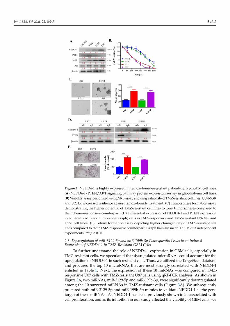
For our next experiments, we aimed to determine the role of NEDD4-1 in TMZ-resistant GBM cells. To mimic patients with higher expression of NEDD4-1 in the clinicalsetting, we measured the endogenous expression of NEDD4-1 protein in a panel of GBMcell lines using Western blotting and selected the cell lines with the highest expression ofNEDD4-1, namely, U251 and U87MG (Figure 2A). We proceeded to establish a TMZ-resistantcell line by incubating the cell lines with a low dose of TMZ, as described in Section 4.As demonstrated in the cell viability assay in Figure 2B, U87MGR and U251R cell lineswere more resistant to TMZ treatment than U87MG and U251 cell lines. We also observedthat these TMZ-resistant cells possessed a higher tendency to form tumorspheres than theirTMZ-responsive counterparts (Figure 2C). These tumorspheres also showed an increasedendogenous expression level of NEDD4-1 compared with adherent U87MG and U251 celllines (Figure 2D). Next, we incubated 1 × 10³ U87MG, U251, U87MGR, and U251R cells insix-well plates for 14 days. The cells were then stained with crystal violet and analyzed.The results demonstrated that TMZ-resistant cell lines have induced clonogenicity potentialcompared to TMZ-responsive cells (Figure 2E). This series of experiments helped us toestablish the oncogenicity of TMZ-resistant GBM cells.
Int. J. Mol. Sci. 2021, 22, 10247 5 of 17
Figure 2. NEDD4-1 is highly expressed in temozolomide-resistant patient-derived GBM cell lines.(A) NEDD4-1/PTEN/AKT signaling pathway protein expression survey in glioblastoma cell lines.(B) Viability assay performed using SRB assay showing established TMZ-resistant cell lines, U87MGRand U251R, increased resilience against temozolomide treatment. (C) Tumorsphere formation assaydemonstrating the higher potential of TMZ-resistant cell lines to form tumorspheres compared totheir chemo-responsive counterpart. (D) Differential expression of NEDD4-1 and PTEN expressionin adherent (adh) and tumorsphere (sph) cells in TMZ-responsive and TMZ-resistant U87MG andU251 cell lines. (E) Colony formation assay depicting higher clonogenicity of TMZ-resistant celllines compared to their TMZ-responsive counterpart. Graph bars are mean± SEM of 3 independentexperiments. *** p < 0.001.
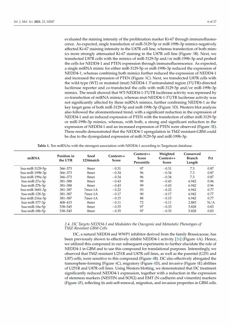
2.3. Dysregulation of miR-3129-5p and miR-199b-3p Consequently Leads to an InducedExpression of NEDD4-1 in TMZ-Resistant GBM Cells
To further understand the role of NEDD4-1 expression in GBM cells, especially inTMZ-resistant cells, we speculated that dysregulated microRNAs could account for theupregulation of NEDD4-1 in such resistant cells. Thus, we utilized the TargetScan databaseand procured the top 10 microRNAs that are most strongly correlated with NEDD4-1enlisted in Table 1. Next, the expression of these 10 miRNAs was compared in TMZ-responsive U87 cells with TMZ-resistant U87 cells using qRT-PCR analysis. As shown inFigure 3A, two miRNAs, miR-3129-5p and miR-199b-3p, were significantly downregulatedamong the 10 surveyed miRNAs in TMZ-resistant cells (Figure 3A). We subsequentlyprocured both miR-3129-5p and miR-199b-3p mimics to validate NEDD4-1 as the genetarget of these miRNAs. As NEDD4-1 has been previously shown to be associated withcell proliferation, and as its inhibition in our study affected the viability of GBM cells, we
Int. J. Mol. Sci. 2021, 22, 10247 6 of 17
evaluated the staining intensity of the proliferation marker Ki-67 through immunofluores-cence. As expected, single transfection of miR-3129-5p or miR-199b-3p mimics negativelyaffected Ki-67 staining intensity in the U87R cell line, whereas transfection of both mim-ics more strongly attenuated Ki-67 staining in the U87R cell line (Figure 3B). Next, wetransfected U87R cells with the mimics of miR-3129-5p and/or miR-199b-3p and probedthe cells for NEDD4-1 and PTEN expression through immunofluorescence. As expected,a single miRNA mimic for either miR-3129-5p or miR-199b-3p reduced the expression ofNEDD4-1, whereas combining both mimics further reduced the expression of NEDD4-1and increased the expression of PTEN (Figure 3C). Next, we transfected U87R cells withthe wild-type (WT) or mutated (mut) NEDD4-1 3′untranslated region (3′UTR)-directedluciferase reporter and co-transfected the cells with miR-3129-5p and/or miR-199b-3pmimics. The result showed that WT-NEDD4-1-3′UTR luciferase activity was repressed byco-transfection of miRNA mimics, whereas mut-NEDD4-1-3′UTR luciferase activity wasnot significantly affected by these miRNA mimics, further confirming NEDD4-1 as thekey target gene of both miR-3129-5p and miR-199b-3p (Figure 3D). Western blot analysisalso followed the aforementioned trend, with a significant reduction in the expression ofNEDD4-1 and an induced expression of PTEN with the transfection of either miR-3129-5por miR-199b-3p mimics, whereas, with both, a strong and significant reduction in theexpression of NEDD4-1 and an increased expression of PTEN were observed (Figure 3E).These results demonstrated that the NEDD4-1 upregulation in TMZ-resistant-GBM couldbe due to the dysregulated expression of miR-3129-5p and miR-199b-3p.
Table 1. Ten miRNAs with the strongest association with NEDD4-1 according to Targetscan database.
miRNA Position inthe UTR
Seed1234match
Context++Score
Context++Score
Percentile
WeightedContext++
Score
ConservedBranchLength
Pct
hsa-miR-3129-5p 366–373 8mer −0.31 97 −0.31 7.3 0.87hsa-miR-199b-3p 366–373 8mer −0.34 96 −0.34 7.3 0.87hsa-miR-199a-3p 366–373 8mer −0.34 96 −0.34 7.3 0.87hsa-miR-27a-3p 381–388 8mer −0.43 99 −0.43 6.942 0.96hsa-miR-27b-3p 381–388 8mer −0.43 99 −0.43 6.942 0.96hsa-miR-3681-3p 381–387 7mer-1A −0.22 93 −0.22 6.942 0.77hsa-miR-128-3p 381–387 7mer-1A −0.17 90 −0.17 6.942 0.77
hsa-miR-216a-3p 381–387 7mer-1A −0.15 88 −0.15 6.942 0.77hsa-miR-377-3p 408–415 8mer −0.11 72 −0.11 2.883 N/Ahsa-miR-18a-5p 538–545 8mer −0.35 97 −0.33 5.828 0.83hsa-miR-18b-5p 538–545 8mer −0.35 97 −0.33 5.828 0.83
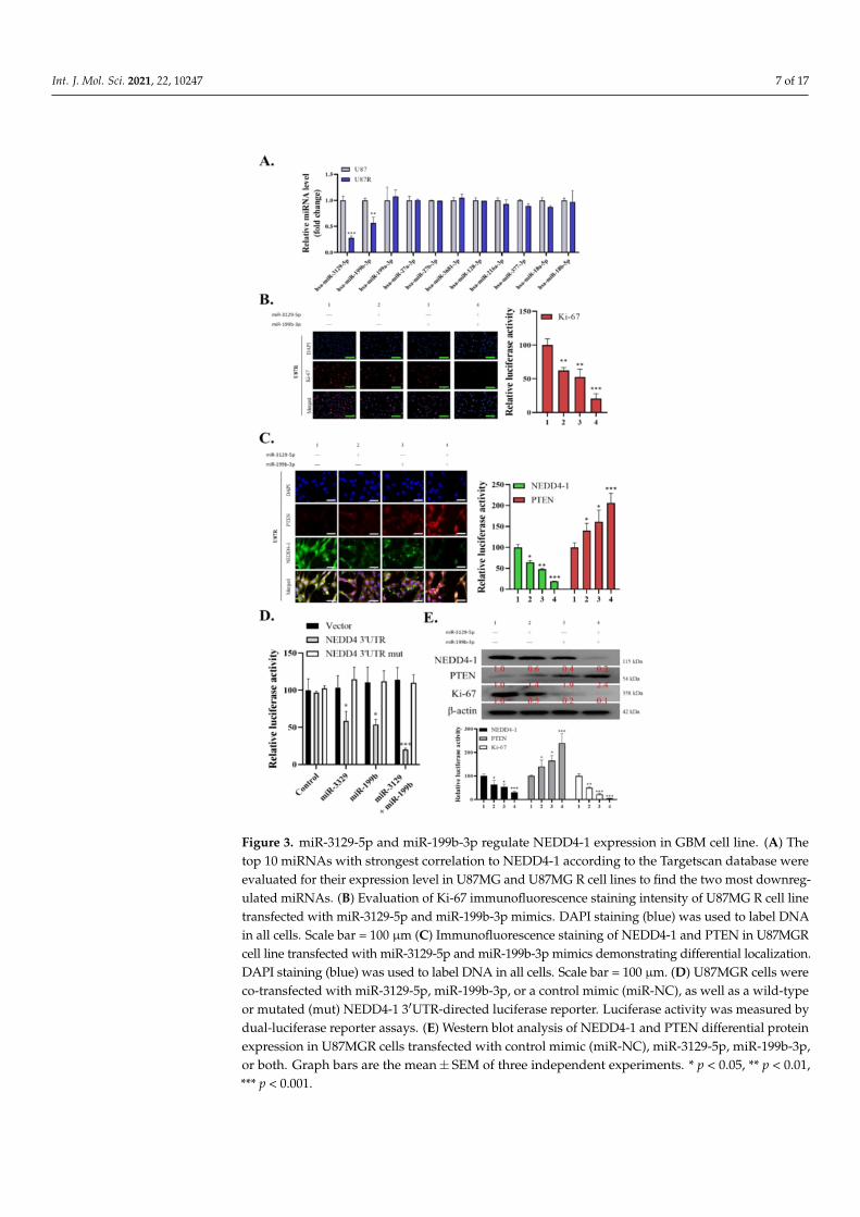
2.4. I3C Targets NEDD4-1 and Modulates the Oncogenic and Metastatic Phenotypes ofTMZ-Resistant GBM Cells
I3C, a natural NEDD4 and WWP1 inhibitor derived from the family Brassicaceae, hasbeen previously shown to effectively inhibit NEDD4-1 activity [26] (Figure 4A). Hence,we utilized this compound in our subsequent experiments to further elucidate the role ofNEDD4-1 in GBM and to use this compound for translational purposes. Interestingly, weobserved that TMZ-resistant U251R and U87R cell lines, as well as the parental (U251 andU87) cells, were sensitive to this compound (Figure 4B). I3C also effectively abrogated thetumorsphere-forming (Figure 4C), migratory (Figure 4D), and invasive (Figure 4E) abilitiesof U251R and U87R cell lines. Using Western blotting, we demonstrated that I3C treatmentsignificantly reduced NEDD4-1 expression, together with a reduction in the expressionof stemness markers (NESTIN and SOX2) and EMT (N-cadherin and vimentin) markers(Figure 4F), reflecting its anti-self-renewal, migration, and invasion properties in GBM cells.
Int. J. Mol. Sci. 2021, 22, 10247 7 of 17
Figure 3. miR-3129-5p and miR-199b-3p regulate NEDD4-1 expression in GBM cell line. (A) Thetop 10 miRNAs with strongest correlation to NEDD4-1 according to the Targetscan database wereevaluated for their expression level in U87MG and U87MG R cell lines to find the two most downreg-ulated miRNAs. (B) Evaluation of Ki-67 immunofluorescence staining intensity of U87MG R cell linetransfected with miR-3129-5p and miR-199b-3p mimics. DAPI staining (blue) was used to label DNAin all cells. Scale bar = 100 µm (C) Immunofluorescence staining of NEDD4-1 and PTEN in U87MGRcell line transfected with miR-3129-5p and miR-199b-3p mimics demonstrating differential localization.DAPI staining (blue) was used to label DNA in all cells. Scale bar = 100 µm. (D) U87MGR cells wereco-transfected with miR-3129-5p, miR-199b-3p, or a control mimic (miR-NC), as well as a wild-typeor mutated (mut) NEDD4-1 3′UTR-directed luciferase reporter. Luciferase activity was measured bydual-luciferase reporter assays. (E) Western blot analysis of NEDD4-1 and PTEN differential proteinexpression in U87MGR cells transfected with control mimic (miR-NC), miR-3129-5p, miR-199b-3p,or both. Graph bars are the mean± SEM of three independent experiments. * p < 0.05, ** p < 0.01,*** p < 0.001.
Int. J. Mol. Sci. 2021, 22, 10247 8 of 17
Figure 4. Effect of NEDD4-1 inhibitor, indole-3-carbinol (I3C), on migration, invasion, and tumor-sphere formation of temozolomide-resistant GBM cell lines. (A) Molecular structure of indole-3-carbinol (I3C). (B) SRB cell viability assay of temozolomide resistant and parental cell linestreated with I3C. (C) I3C treatment attenuated U251R and U87MGR tumorsphere-forming potential.(D) U251R and U87MGR migration ability evaluated using scratch assay. Gap closure was evaluatedafter 24 h incubation. (E) Gel-coated Transwell chambers were utilized to evaluate U251R andU87MGR invasion ability after treatment with I3C for 24 h. (F) Protein expression of NEDD4-1,NESTIN, SOX2, N-cadherin, and vimentin after treatment with I3C for 24 h, evaluated using Westernblot. Graph bars are the mean± SEM of three independent experiments. *** p < 0.001.
2.5. I3C Increased Susceptibility of TMZ-Resistant Cells to TMZ by Inhibiting NEDD4-1-InducedPTEN Ubiquitination and Subsequently Inhibiting NRF2/HO-1 Antioxidant Signaling Response
Chemotherapies commonly work by straining cancer cells with oxidative stress andeventually killing them. Resistant cells may combat this by upregulating the defensive re-sponse against ROS. Thus, we investigated the role of I3C in dismantling this defense mech-anism by inhibiting NRF2/HO-1 through NEDD4-1. We induced oxidative stress in theU87R cell line with tert-butyl hydroperoxide (TBHp) and treated these cells with/withoutI3C. TBHp is a well-known reactive oxygen species (ROC)-producing model substance foroxidative stress generation. TBHp-induced oxidative stress significantly increased NEDD4-1 expression, which led to a decreased protein level of PTEN and significantly higher AKTsignaling activity, compared with the control. As expected, the NRF2/HO-1 signaling cascadewas also strongly activated in the cells induced with TBHp. I3C treatment effectively inhibited
Int. J. Mol. Sci. 2021, 22, 10247 9 of 17
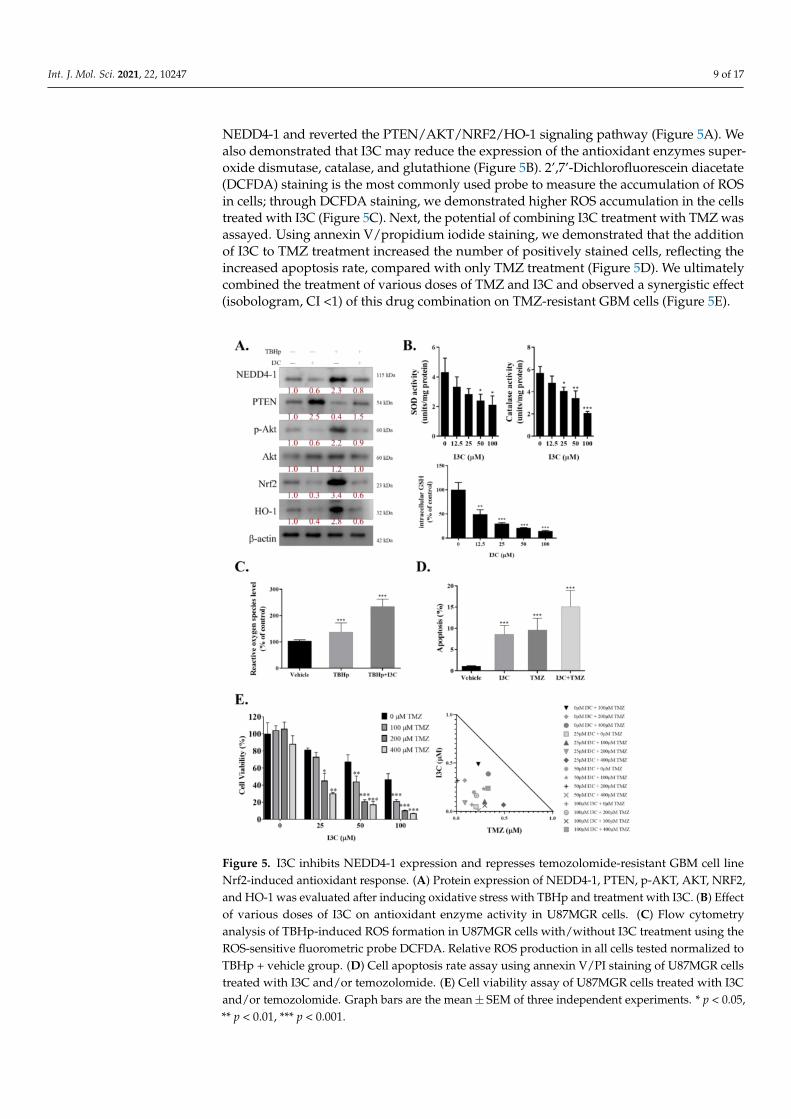
NEDD4-1 and reverted the PTEN/AKT/NRF2/HO-1 signaling pathway (Figure 5A). Wealso demonstrated that I3C may reduce the expression of the antioxidant enzymes super-oxide dismutase, catalase, and glutathione (Figure 5B). 2’,7’-Dichlorofluorescein diacetate(DCFDA) staining is the most commonly used probe to measure the accumulation of ROSin cells; through DCFDA staining, we demonstrated higher ROS accumulation in the cellstreated with I3C (Figure 5C). Next, the potential of combining I3C treatment with TMZ wasassayed. Using annexin V/propidium iodide staining, we demonstrated that the additionof I3C to TMZ treatment increased the number of positively stained cells, reflecting theincreased apoptosis rate, compared with only TMZ treatment (Figure 5D). We ultimatelycombined the treatment of various doses of TMZ and I3C and observed a synergistic effect(isobologram, CI <1) of this drug combination on TMZ-resistant GBM cells (Figure 5E).
Figure 5. I3C inhibits NEDD4-1 expression and represses temozolomide-resistant GBM cell lineNrf2-induced antioxidant response. (A) Protein expression of NEDD4-1, PTEN, p-AKT, AKT, NRF2,and HO-1 was evaluated after inducing oxidative stress with TBHp and treatment with I3C. (B) Effectof various doses of I3C on antioxidant enzyme activity in U87MGR cells. (C) Flow cytometryanalysis of TBHp-induced ROS formation in U87MGR cells with/without I3C treatment using theROS-sensitive fluorometric probe DCFDA. Relative ROS production in all cells tested normalized toTBHp + vehicle group. (D) Cell apoptosis rate assay using annexin V/PI staining of U87MGR cellstreated with I3C and/or temozolomide. (E) Cell viability assay of U87MGR cells treated with I3Cand/or temozolomide. Graph bars are the mean± SEM of three independent experiments. * p < 0.05,** p < 0.01, *** p < 0.001.
Int. J. Mol. Sci. 2021, 22, 10247 10 of 17
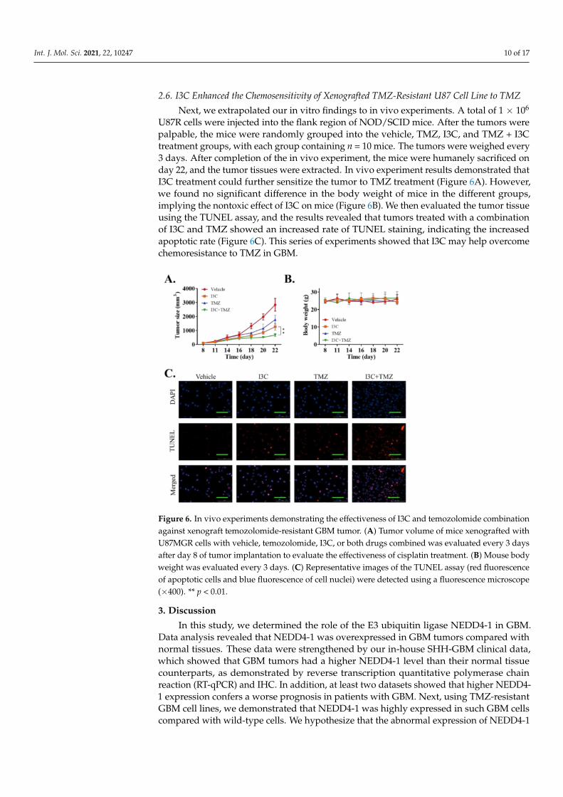
2.6. I3C Enhanced the Chemosensitivity of Xenografted TMZ-Resistant U87 Cell Line to TMZ
Next, we extrapolated our in vitro findings to in vivo experiments. A total of 1 × 10⁶
U87R cells were injected into the flank region of NOD/SCID mice. After the tumors werepalpable, the mice were randomly grouped into the vehicle, TMZ, I3C, and TMZ + I3Ctreatment groups, with each group containing n = 10 mice. The tumors were weighed every3 days. After completion of the in vivo experiment, the mice were humanely sacrificed onday 22, and the tumor tissues were extracted. In vivo experiment results demonstrated thatI3C treatment could further sensitize the tumor to TMZ treatment (Figure 6A). However,we found no significant difference in the body weight of mice in the different groups,implying the nontoxic effect of I3C on mice (Figure 6B). We then evaluated the tumor tissueusing the TUNEL assay, and the results revealed that tumors treated with a combinationof I3C and TMZ showed an increased rate of TUNEL staining, indicating the increasedapoptotic rate (Figure 6C). This series of experiments showed that I3C may help overcomechemoresistance to TMZ in GBM.
Figure 6. In vivo experiments demonstrating the effectiveness of I3C and temozolomide combinationagainst xenograft temozolomide-resistant GBM tumor. (A) Tumor volume of mice xenografted withU87MGR cells with vehicle, temozolomide, I3C, or both drugs combined was evaluated every 3 daysafter day 8 of tumor implantation to evaluate the effectiveness of cisplatin treatment. (B) Mouse bodyweight was evaluated every 3 days. (C) Representative images of the TUNEL assay (red fluorescenceof apoptotic cells and blue fluorescence of cell nuclei) were detected using a fluorescence microscope(×400). ** p < 0.01.
3. Discussion
In this study, we determined the role of the E3 ubiquitin ligase NEDD4-1 in GBM.Data analysis revealed that NEDD4-1 was overexpressed in GBM tumors compared withnormal tissues. These data were strengthened by our in-house SHH-GBM clinical data,which showed that GBM tumors had a higher NEDD4-1 level than their normal tissuecounterparts, as demonstrated by reverse transcription quantitative polymerase chainreaction (RT-qPCR) and IHC. In addition, at least two datasets showed that higher NEDD4-1 expression confers a worse prognosis in patients with GBM. Next, using TMZ-resistantGBM cell lines, we demonstrated that NEDD4-1 was highly expressed in such GBM cellscompared with wild-type cells. We hypothesize that the abnormal expression of NEDD4-1
Int. J. Mol. Sci. 2021, 22, 10247 11 of 17
in GBM is due to dysregulation of the expression of miRNAs, which is often reported inchemoresistant cells [27,28].
miRNAs are a small class of noncoding RNA molecules (21–23 nucleotide fragments)that alter the expression of target mRNA, by pairing with complementary sequences withinthe 3′UTRs of targeted transcripts. miRNA has been shown to play a role in variousbiological processes, including cell growth, differentiation, apoptosis, and proliferation [29].We generated a list of potential miRNAs that may be strongly associated with NEDD4-1 using TargetScan, a web-based database used to predict miRNA targets [30]. From10 predicted miRNAs targeting NEDD4-1, we determined the two most downregulatedmiRNAs in the TMZ-resistant GBM cell line, namely, hsa-miR-3129-5p and hsa-miR-199b-3p. These two miRNAs significantly altered the NEDD4-1 level in the U87R cell line,as validated using their mimics and by further analyzing NEDD4-1 and PTEN levelsusing a combination of immunofluorescence, luciferase reporter, and Western blot assays.This series of experiments provided evidence regarding the involvement of miRNA inregulating NEDD4-1-induced PTEN ubiquitination in chemoresistant GBM cells.
Previous studies have also demonstrated the complexity of dysregulated miRNAexpression involved in CSC characteristics, which results in GBM TMZ chemoresistance.Li et al. demonstrated that miR-186 was dysregulated in GBM-initiating cells (GICs);furthermore, the exogenous exposure of miR-186 mimic inhibited the GIC proliferationand reversed the resistance of GBM cells to cisplatin [31]. Sana et al. identified deregulatedmiRNA signatures in GIC clusters compared with non-stem-cell clusters. They foundthat these miRNAs were also associated with the poor survival of patients with GBM [32].Thus, it is not surprising that NEDD4-1 is abnormally expressed in TMZ-resistant celllines, considering that chemoresistant cells may have different biomolecular structures andfunctions, as reported in previous studies, including ours.
To subvert NEDD4-1 activity, we subsequently blocked its activity using a readilyavailable E3 ubiquitin ligase inhibitor, I3C. I3C is extracted from glucobrassicin, whichis a hydrolysis product of cruciferous vegetables such as cabbage, broccoli, and Brusselssprouts. I3C is a naturally occurring indole carbinol phytochemical implicated in variousantiproliferative pathways in leukemia, melanoma, and breast, kidney, liver, colon, andcervical cancer [26]. Through this study, we demonstrated that both TMZ-resistant celllines were sensitive to I3C treatment, and the addition of I3C attenuated the migration,invasion, and tumorsphere-forming potential of U87R and U251R cell lines. I3C treatmentsignificantly reduced NEDD4-1 expression and subsequently reduced the stemness andEMT markers of TMZ-resistant GBM cell lines. Aronchik et al. reported that I3C suppressedin vivo tumor growth and induced PTEN protein expression levels in residual tumors inTMZ-responsive PTEN-expressing melanoma xenografts developed in athymic mice [33].Our results contribute to the evidence of the role of I3C in reducing the tumorigenicity ofTMZ-resistant GBM cells by targeting NEDD4-1.
Oxidative stress has long been linked with cancer chemoresistance. Cancer cellshave a high basal level of ROS, rendering them more vulnerable than regular cells toan increase in ROS; chemoresistant cancer cells upregulate their antioxidant systems tobecome strongly adapted to intrinsic or drug-induced oxidative stress. In 2011, Olivia et al.specifically focused on the role of ROS in TMZ resistance of glioma cells. They reportedthat remodeling of the whole electron transport chain is linked to chemoresistance to TMZ,with large increases in the activity of complexes II/III and cytochrome c oxidase. They alsofound that glioma cells treated with antioxidants prevented TMZ toxicity. Furthermore,they demonstrated that TMZ-resistant mitochondrial DNA-depleted cells (rho degrees)had lower intracellular ROS levels after TMZ exposure than parental cells did [34]. In breastcancer cells, a robust proteomics approach using label-free mass spectrometry in patientswho were responsive and resistant to chemotherapy revealed oxidative stress as one of thepivotal players of breast cancer chemoresistance [35]. Similarly, we demonstrated that theTMZ-resistant cell line is more resilient against TBHp-induced oxidative stress comparedwith chemoresponsive cells. Interestingly, IC3 treatment reversed the chemoresistance
Int. J. Mol. Sci. 2021, 22, 10247 12 of 17
capacity of TMZ-resistant cells by attenuating NEDD4-1-induced degradation of PTEN,which ultimately inhibited the AKT/NRF2/HO-1 signaling pathway. Stripped of theirdefense mechanism, U87R cells were more susceptible to TMZ treatment. Our in vitroand in vivo data also showed that both IC3 and TMZ work synergistically to eliminateTMZ-resistant GBM cells.
4. Materials and Methods4.1. Clinical Samples
We analyzed the microarray gene expression dataset of patients with GBM obtainedfrom the GEO datasets through the R2 Genomics Analysis and Visualization Platformcontaining normal mixed male and female samples (Berchtold’s, GSE11882, n = 172 andHarris’s, GSE13564, n = 44), and tumor samples (Pfister, GSE36245, n = 46, Loeffler, GSE53733,n = 70, and Hegi, GSE7696, n = 84). Similarly, TCGA-GBM data, n = 173, were also applied.The R2 Genomics Analysis and Visualization Platform (https://hgserver1.amc.nl/cgi-bin/r2/main.cgi (accessed on 2 July 2020) was used to generate a Kaplan–Meier survivalcurve [36]. Parallel surgical tissues were collected and used to prepare formalin-fixedparaffin-embedded (FFPE) specimens. The China Medical University Hospital ResearchEthics Committees approved the study (project approval number CMUH-109-REC2-014).The GBM tissues and normal tissues of all mixed male and female patients were collected;tissue neighboring the tumor tissue that was more than 3 cm away was considered normal.The clinicopathological information of the patients is described in Table 2. Tissue microar-rays of GBMs were subjected to immunohistochemical (IHC) analysis after incubation withan antibody against NEDD4-1 (1:100 dilution, SC-81159; Santa Cruz Biotechnology, Inc., CA,USA) at 4 ◦C overnight. Staining using horseradish peroxidase (HRP), 3,3′-diaminobenzidine(dark brown), and hematoxylin (deep blue/purple) was performed according to the standardIHC protocol, followed by imaging and evaluation of the protein expression.
Table 2. Patients’ clinicopathological information.
Total no. Patients of Glioblastoma (n = 58) No. (%)
Age<65 40 (68.4%)≥65 18 (31.6%)
Gendermale 34 (58.6%)
female 24 (41.4%)IDH1 status
wildtype 56 (96.6%)mutation 2 (3.4%)
Treatment modalitiesChemotherapy 24 (41.4%)Radiotherapy 34 (58.6%)
4.2. Cell Culture and Generation of Temozolomide-Resistant Cell Line
LN-229, T98G, U251, and U87MG GBM cell lines were obtained from the AmericanType Culture Collection (ATCC; Manassas, VA, USA) and were maintained in an incubatorwith 5% CO2 in humidified air. The cells were cultured in Dulbecco’s modified Eagle’smedium (#12491023; GIBCO, Life Technologies Corp., Carlsbad, CA, USA) supplementedwith 10% fetal bovine serum (GIBCO, Life Technologies Corp.), penicillin (100 IU/mL), andstreptomycin (100 g/mL) (#15140122, GIBCO, Life Technologies). TMZ-resistant U87MGRand U251R cells were generated as per the protocol suggested by Akiyama et al. (2014) [37].The U87 and U251 parental cell lines, which are sensitive to TMZ, were first maintained inlow doses of TMZ (5 µM) and then successively exposed to incremental doses of TMZ (upto 150 µM). After the killing of a majority of the cells, the surviving cells were maintaineduntil a normal rate of growth was obtained. The TMZ-resistant cells were then maintained
Int. J. Mol. Sci. 2021, 22, 10247 13 of 17
at a dose of 100 µM TMZ for in vitro and in vivo experiments. The IC50 value of TMZ wasevaluated using the WST-1 assay.
4.3. Western Blotting
After all treatments, GBM cells were harvested by trypsinization and lysed to extractthe proteins using RIPA buffer (Cell Signaling Technology, Danvers, MA, USA) witha cocktail of protease inhibitors (Sigma Aldrich, St. Louis, MO, USA). Next, 10 µg ofprotein samples were separated using 10% SDS-PAGE electrophoresis and transferred topolyvinylidene fluoride (PVDF) membranes using the Bio-Rad Mini-Protein electro-transfersystem (Bio-Rad, Taipei City, Taiwan). Immunoblotting was performed after blocking with5% skimmed milk in Tris-buffered saline with Tween 20 (TBST) for 1 h and then incubatedovernight at 4 ◦C with primary antibodies against the protein of interest (SupplementaryTable S1). After incubation with the primary antibody, polyvinylidene difluoride (PVDF)membranes were washed thrice with TBST, incubated for 1 h at room temperature withan HRP-labeled secondary antibody, and rewashed with TBST. Subsequently, enhancedchemiluminescence, Western blotting reagents, and a BioSpectrum Imaging System (UVP;Upland, CA, USA) were used to detect the bands.
4.4. Transient Oligonucleotide Transfection
MiR-3129-5p and miR-199b-3p (mimics) and negative control miRNA were purchasedfrom ThermoFisher Scientific (Taipei, Taiwan) and prepared under strict adherence to thevendor’s instructions. GBM cells were transfected using Lipofectamine® 2000 (InvitrogenThermoFisher Scientific, Inc., Waltham, MA, USA).
4.5. RNA Isolation and Reverse Transcription Quantitative Polymerase Chain Reaction
Total RNA was extracted from GBM and normal brain tissues using TRIzol™ reagents(Invitrogen; Thermo-Fisher Scientific, Inc., Waltham, MA, USA). Reverse transcription quan-titative polymerase chain reaction (RT-PCR) was used to detect the expression of NEDD4-1mRNA in GBM tissues. The NEDD4-1 primer sequences used were as follows: forward,5′–CAGAAGAGGCAGCTTACAAGCC–3′; reverse, 5′–CTTCCCAACCTGGTGGTAATCC–3′. Glyceraldehyde-3-phosphate dehydrogenase (GAPDH) was considered as an internalreference to detect the NEDD4-1 mRNA expression level in the cells. All the selectedmRNAs were pre-degenerated for 5 min at 95 ◦C and 1 min at 94 ◦C for 35 cycles. Then,they were pre-degenerated for 1 min at 56 ◦C, 2 min at 72 ◦C, and 10 min at 72 ◦C. Therelative expression level of mRNA calculated through the 2−∆∆Cq method (2−∆∆Cq ≥ 2 wasregarded as high expression).
4.6. Cell Sensitivity Assay/Clonogenic Assay
The “gold standard” cellular sensitivity/clonogenic assay was used to assess thesensitivities of GBM cells after the drug treatment. The cells were subcultured, seeded intosix-well plates (2.7 × 10⁴ cells per well), and incubated at 37 ◦C for 2 days in 5% CO2. Cellswere then cultured for an additional 24 h in medium fortified with 10% serum. The treatedcells were then subcultured, reseeded at a concentration of 350 cells per well into a newsix-well plate, and incubated for an additional 10 days at 37 ◦C in a 5% CO2 humidifiedincubator. The cells were dried after being set and were stained with 0.1% crystal violet.The experiments were conducted in triplicate.
4.7. Sulforhodamine B (SRB) Assay
The cell lines were grown in DMEM with 10% fetal bovine serum and 2 mM L-glutamine;96-well microtiter plates were used to inoculate the cells. After inoculation and before theaddition of compounds to be screened, the microtiter plates were incubated for 24 h at37 ◦C under 5% CO2, 95% air, and 100% relative humidity. The cells were fixed in placeby gently adding 50 mL of cold 10% w/v trichloroacetic acid, with incubation for 60 minat 4 ◦C. Subsequently, 50 µL of 0.4% w/v SRB solution in 1% CH3COOH was added to
Int. J. Mol. Sci. 2021, 22, 10247 14 of 17
each well and incubated for 20 min at room temperature. Unbound dye was recoveredafter staining, and residual dye was removed by washing the well plates thoroughly with 1%CH3COOH and air drying. The bound stain was dissolved in a 10 mM Trizma base, and theabsorbance was measured at 515 nm on an ELISA plate reader (690 nm reference wavelength).
4.8. Immunofluorescence Assay
U251R cells were cultured on glass coverslips before being transfected, as mentionedpreviously. After incubation, the cells were fixed for 15 min at 4 ◦C with 4% formaldehyde,permeabilized for 5 min with 0.01% Triton X-100, and blocked for 30 min at room tem-perature with 1% bovine serum albumin. The cells were then incubated for 24 h at 4 ◦Cwith the primary antibodies GSK-3 (#12456, 1:100, Cell Signaling Technology, Danvers,MA, USA) and β-catenin (#8814, 1:100, Cell Signaling Technology). Subsequently, the cellswere stained with isotype-specific secondary antibody (Alexa Fluor® 594-AffiniPure DonkeyAnti-Rabbit IgG) for 1 h the following day (Jackson ImmunoResearch, West Grove, PA, USA).
4.9. Measurement of ROS Production
In a 96-well plate, the cells were plated at 20,000 cells per well in a final volume of80 µL of the medium. Dichlorodihydrofluorescein diacetate (DCFDA; 10 µL of 50 µM) wasapplied to each well and incubated for 30 min. Fluorescence was measured on a microplatefluorometer (Tecan; Seestrasse, Männedorf, Switzerland), with an excitation filter set at488 nm and an emission filter set at 530 nm.
4.10. Tumor Xenograft Study
For this study, 4–6-week-old female nonobese diabetic/severe combined immune-deficient (NOD/SCID) mice (mean weight, 17.4 ± 2.1 g) were purchased from BioLASCO(BioLASCO Taiwan, Taipei, Taiwan). The mice were inoculated subcutaneously with2 × 106 U251R cells in their hind flanks and were randomly assigned to vehicle (n = 10),TMZ (n = 10), indole-3-carbinol (I3C; n = 10), or TMZ + I3C (n = 10) groups. TMZ (2 mg/kg)treatment was initiated on day 8 when tumors started becoming palpable; TMZ wasadministered intraperitoneally (i.p.) every 72 h over the following 12 days. Using verniercalipers, tumor sizes were measured on days 6, 9, 12, 15, and 18 after GBM cell inoculation,and tumor volumes (v) were calculated as length (l) × (width (w))2 × 0.5. At the end of theexperiment on day 18, the tumor-bearing mice were carefully sacrificed, and the tumorswere extracted, examined, photographed, and measured again. All mice were housedunder specific pathogen-free conditions and used following the animal care guidelines fromthe Institutional Animal Care and Use Committee of China Medical University Hospital(CMUIACUC-2020-150). The committee approved the experimental protocol.
4.11. Statistical Analysis
Means and standard errors of the mean (SEM) were used to present all the results.For multiple comparisons or repeated measurements, Student’s t-test was used. Formultiple comparisons or repeated measurements, ANOVA or repeated ANOVA accom-panied by Tukey’s post hoc test was used. Statistical significance was described as ap-value < 0.05. GraphPad Prism 7.0 was used to conduct statistical analysis (GraphPadSoftware, San Diego, CA, USA).
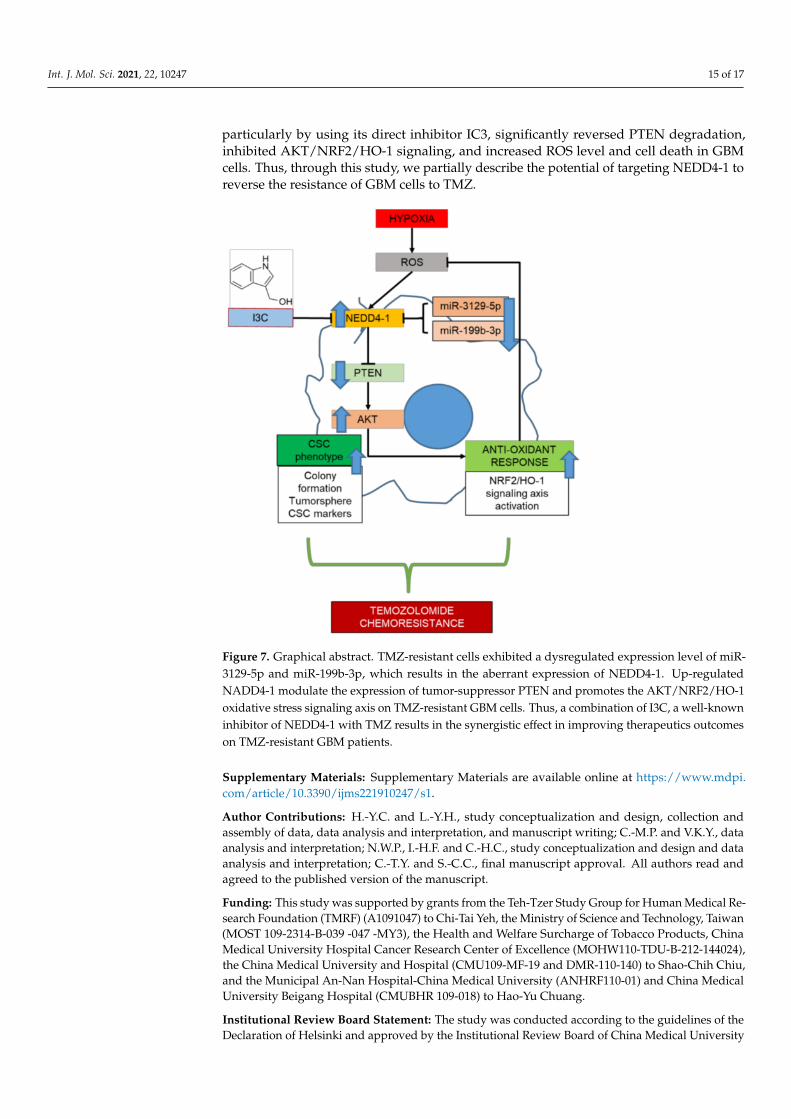
5. Conclusions
In summary, we described the critical role of NEDD4-1 in regulating the redox imbal-ance in TMZ-resistant GBM cells through the degradation of PTEN, the regulator of theAKT/NRF2/HO-1 signaling pathway. As described in our graphical abstract (Figure 7),TMZ-resistant cells underwent molecular changes that resulted in the deregulation ofcertain miRNAs, including miRNAs responsible for the expression of NEDD4-1. Theresulting overexpression of NEDD4-1 is responsible for the tumorigenicity of GBM andthe antioxidant response system that ultimately resists TMZ toxicity. Targeting NEDD4-1,
Int. J. Mol. Sci. 2021, 22, 10247 15 of 17
particularly by using its direct inhibitor IC3, significantly reversed PTEN degradation,inhibited AKT/NRF2/HO-1 signaling, and increased ROS level and cell death in GBMcells. Thus, through this study, we partially describe the potential of targeting NEDD4-1 toreverse the resistance of GBM cells to TMZ.
Figure 7. Graphical abstract. TMZ-resistant cells exhibited a dysregulated expression level of miR-3129-5p and miR-199b-3p, which results in the aberrant expression of NEDD4-1. Up-regulatedNADD4-1 modulate the expression of tumor-suppressor PTEN and promotes the AKT/NRF2/HO-1oxidative stress signaling axis on TMZ-resistant GBM cells. Thus, a combination of I3C, a well-knowninhibitor of NEDD4-1 with TMZ results in the synergistic effect in improving therapeutics outcomeson TMZ-resistant GBM patients.
Supplementary Materials: Supplementary Materials are available online at https://www.mdpi.com/article/10.3390/ijms221910247/s1.
Author Contributions: H.-Y.C. and L.-Y.H., study conceptualization and design, collection andassembly of data, data analysis and interpretation, and manuscript writing; C.-M.P. and V.K.Y., dataanalysis and interpretation; N.W.P., I.-H.F. and C.-H.C., study conceptualization and design and dataanalysis and interpretation; C.-T.Y. and S.-C.C., final manuscript approval. All authors read andagreed to the published version of the manuscript.
Funding: This study was supported by grants from the Teh-Tzer Study Group for Human Medical Re-search Foundation (TMRF) (A1091047) to Chi-Tai Yeh, the Ministry of Science and Technology, Taiwan(MOST 109-2314-B-039 -047 -MY3), the Health and Welfare Surcharge of Tobacco Products, ChinaMedical University Hospital Cancer Research Center of Excellence (MOHW110-TDU-B-212-144024),the China Medical University and Hospital (CMU109-MF-19 and DMR-110-140) to Shao-Chih Chiu,and the Municipal An-Nan Hospital-China Medical University (ANHRF110-01) and China MedicalUniversity Beigang Hospital (CMUBHR 109-018) to Hao-Yu Chuang.
Institutional Review Board Statement: The study was conducted according to the guidelines of theDeclaration of Helsinki and approved by the Institutional Review Board of China Medical University
Int. J. Mol. Sci. 2021, 22, 10247 16 of 17
Hospital Research Ethics Committees approved the study (project approval number CMUH-109-REC2-014). Parallel surgical tissues were collected and used to prepare formalin-fixed paraffin-embedded (FFPE) specimens. All mice were housed under specific pathogen-free conditions andused following the animal care guidelines from the Institutional Animal Care and Use Committee of ChinaMedical University Hospital (CMUIACUC-2020-150). The committee approved the experimental protocol.
Informed Consent Statement: Not applicable.
Data Availability Statement: The datasets used and analyzed in the current study are publiclyaccessible, as indicated in the manuscript.
Acknowledgments: The authors thank all research assistants of the Translational Cell TherapyCenter, Department of Medical Research, China Medical University Hospital for their assistance withthe flow cytometry and molecular and cell-based assays.
Conflicts of Interest: The authors declare that they have no potential financial competing intereststhat may in any way gain or lose financially from the publication of this manuscript at present or inthe future. Additionally, no non-financial competing interests are involved in the manuscript.
Abbreviations
Neuronal precursor cell-expressed developmentally downregulated 4-1 (NEDD4-1); formalin-fixedparaffin-embedded (FFPE); indole-3-carbinol (I3C); glioblastoma (GBM); reactive oxygen species (ROS);cancer stem cells (CSCs); polyvinylidene difluoride (PVDF); epithelial-to-mesenchymal transition (EMT);temozolomide (TMZ); E3 ubiquitin ligases (E3s); phosphatase and tensin homolog (PTEN).
References1. Vargas Lopez, A.J. Glioblastoma in adults: A Society for Neuro-Oncology (SNO) and European Society of Neuro-Oncology
(EANO) consensus review on current management and future directions. Neuro Oncol. 2021, 23, 502–503. [CrossRef] [PubMed]2. Zottel, A.; Jovcevska, I.; Samec, N.; Komel, R. Cytoskeletal proteins as glioblastoma biomarkers and targets for therapy:
A systematic review. Crit. Rev. Oncol. Hematol. 2021, 160, 103283. [CrossRef]3. Tome, M.E.; Frye, J.B.; Coyle, D.L.; Jacobson, E.L.; Samulitis, B.K.; Dvorak, K.; Dorr, R.T.; Briehl, M.M. Lymphoma cells with
increased anti-oxidant defenses acquire chemoresistance. Exp. Ther. Med. 2012, 3, 845–852. [CrossRef]4. Olivier, C.; Oliver, L.; Lalier, L.; Vallette, F.M. Drug Resistance in Glioblastoma: The Two Faces of Oxidative Stress. Front. Mol. Biosci.
2020, 7, 620677. [CrossRef]5. Jelic, M.D.; Mandic, A.D.; Maricic, S.M.; Srdjenovic, B.U. Oxidative stress and its role in cancer. J. Cancer Res. Ther. 2021, 17, 22–28.
[CrossRef]6. Salazar-Ramiro, A.; Ramirez-Ortega, D.; Perez de la Cruz, V.; Hernandez-Pedro, N.Y.; Gonzalez-Esquivel, D.F.; Sotelo, J.; Pineda,
B. Role of Redox Status in Development of Glioblastoma. Front. Immunol. 2016, 7, 156. [CrossRef]7. Han, S.; Shin, H.; Lee, J.K.; Liu, Z.; Rabadan, R.; Lee, J.; Shin, J.; Lee, C.; Yang, H.; Kim, D.; et al. Secretome analysis of
patient-derived GBM tumor spheres identifies midkine as a potent therapeutic target. Exp. Mol. Med. 2019, 51, 1–11. [CrossRef][PubMed]
8. Sato, A.; Okada, M.; Shibuya, K.; Watanabe, E.; Seino, S.; Narita, Y.; Shibui, S.; Kayama, T.; Kitanaka, C. Pivotal role for ROSactivation of p38 MAPK in the control of differentiation and tumor-initiating capacity of glioma-initiating cells. Stem. Cell Res.2014, 12, 119–131. [CrossRef] [PubMed]
9. Gursel, D.B.; Shin, B.J.; Burkhardt, J.K.; Kesavabhotla, K.; Schlaff, C.D.; Boockvar, J.A. Glioblastoma stem-like cells-biology andtherapeutic implications. Cancers 2011, 3, 2655–2666. [CrossRef]
10. Dey, M.; Ulasov, I.V.; Tyler, M.A.; Sonabend, A.M.; Lesniak, M.S. Cancer stem cells: The final frontier for glioma virotherapy. StemCell Rev. Rep. 2011, 7, 119–129. [CrossRef] [PubMed]
11. Kawamura, Y.; Takouda, J.; Yoshimoto, K.; Nakashima, K. New aspects of glioblastoma multiforme revealed by similaritiesbetween neural and glioblastoma stem cells. Cell Biol. Toxicol. 2018, 34, 425–440. [CrossRef] [PubMed]
12. Afify, S.M.; Oo, A.K.K.; Hassan, G.; Seno, A.; Seno, M. How can we turn the PI3K/AKT/mTOR pathway down? Insights intoinhibition and treatment of cancer. Expert Rev. Anticancer Ther. 2021, 21, 605–619. [CrossRef]
13. Li, J.; Wang, J.; Xie, D.; Pei, Q.; Wan, X.; Xing, H.R.; Ye, T. Characteristics of the PI3K/AKT and MAPK/ERK pathways involvedin the maintenance of self-renewal in lung cancer stem-like cells. Int. J. Biol. Sci. 2021, 17, 1191–1202. [CrossRef]
14. Sanches, J.G.P.; Song, B.; Zhang, Q.; Cui, X.; Yabasin, I.B.; Ntim, M.; Li, X.; He, J.; Zhang, Y.; Mao, J.; et al. The Role of KDM2Band EZH2 in Regulating the Stemness in Colorectal Cancer Through the PI3K/AKT Pathway. Front. Oncol. 2021, 11, 637298.[CrossRef]
15. He, H.; Huang, C.; Chen, Z.; Huang, H.; Wang, X.; Chen, J. An outlined review for the role of Nedd4-1 and Nedd4-2 in lungdisorders. Biomed. Pharmacother. 2020, 125, 109983. [CrossRef] [PubMed]
Int. J. Mol. Sci. 2021, 22, 10247 17 of 17
16. Sang, P.F.; Wang, H.; Wang, M.; Hu, C.; Zhang, J.S.; Li, X.J.; Zhu, F. NEDD4-1 and PTEN expression in keloid scarring. Genet. Mol. Res.2015, 14, 13467–13475. [CrossRef] [PubMed]
17. Zhang, J.; Li, X.; Zhang, Y. Correlation of NEDD4-1 and PTEN expression with the invasive capacity of pituitary adenomas. Mol.Clin. Oncol. 2017, 6, 96–100. [CrossRef]
18. Chen, C.; Matesic, L.E. The Nedd4-like family of E3 ubiquitin ligases and cancer. Cancer Metastasis Rev. 2007, 26, 587–604.[CrossRef] [PubMed]
19. Chen, C.; Seth, A.K.; Aplin, A.E. Genetic and expression aberrations of E3 ubiquitin ligases in human breast cancer. Mol. CancerRes. 2006, 4, 695–707. [CrossRef] [PubMed]
20. Jung, Y.S.; Qian, Y.; Chen, X. Pirh2 RING-finger E3 ubiquitin ligase: Its role in tumorigenesis and cancer therapy. FEBS Lett. 2012,586, 1397–1402. [CrossRef]
21. Penas, C.; Ramachandran, V.; Ayad, N.G. The APC/C Ubiquitin Ligase: From Cell Biology to Tumorigenesis. Front. Oncol. 2011,1, 60. [CrossRef]
22. Senft, D.; Qi, J.; Ronai, Z.A. Ubiquitin ligases in oncogenic transformation and cancer therapy. Nat. Rev. Cancer 2018, 18, 69–88.[CrossRef] [PubMed]
23. Samarzija, I. Post-Translational Modifications That Drive Prostate Cancer Progression. Biomolecules 2021, 11, 247. [CrossRef][PubMed]
24. Huang, X.; Chen, J.; Cao, W.; Yang, L.; Chen, Q.; He, J.; Yi, Q.; Huang, H.; Zhang, E.; Cai, Z. The many substrates and functions ofNEDD4-1. Cell Death Dis. 2019, 10, 904. [CrossRef] [PubMed]
25. Wang, Z.W.; Hu, X.; Ye, M.; Lin, M.; Chu, M.; Shen, X. NEDD4 E3 ligase: Functions and mechanism in human cancer. Semin.Cancer Biol. 2020, 67, 92–101. [CrossRef]
26. Quirit, J.G.; Lavrenov, S.N.; Poindexter, K.; Xu, J.; Kyauk, C.; Durkin, K.A.; Aronchik, I.; Tomasiak, T.; Solomatin, Y.A.;Preobrazhenskaya, M.N.; et al. Indole-3-carbinol (I3C) analogues are potent small molecule inhibitors of NEDD4-1 ubiquitinligase activity that disrupt proliferation of human melanoma cells. Biochem. Pharmacol. 2017, 127, 13–27. [CrossRef]
27. Mesrian Tanha, H.; Mojtabavi Naeini, M.; Rahgozar, S.; Moafi, A.; Honardoost, M.A. Integrative computational in-depth analysisof dysregulated miRNA-mRNA interactions in drug-resistant pediatric acute lymphoblastic leukemia cells: An attempt to obtainnew potential gene-miRNA pathways involved in response to treatment. Tumour Biol. 2016, 37, 7861–7872. [CrossRef]
28. Pogribny, I.P.; Filkowski, J.N.; Tryndyak, V.P.; Golubov, A.; Shpyleva, S.I.; Kovalchuk, O. Alterations of microRNAs and theirtargets are associated with acquired resistance of MCF-7 breast cancer cells to cisplatin. Int. J. Cancer 2010, 127, 1785–1794.[CrossRef] [PubMed]
29. Pandey, M.; Mukhopadhyay, A.; Sharawat, S.K.; Kumar, S. Role of microRNAs in regulating cell proliferation, metastasis andchemoresistance and their applications as cancer biomarkers in small cell lung cancer. Biochim. Biophys. Acta Rev. Cancer 2021,1876, 188552. [CrossRef]
30. Agarwal, V.; Bell, G.W.; Nam, J.W.; Bartel, D.P. Predicting effective microRNA target sites in mammalian mRNAs. Elife 2015,4, e05005. [CrossRef]
31. Li, J.; Song, J.; Guo, F. miR-186 reverses cisplatin resistance and inhibits the formation of the glioblastoma-initiating cell phenotypeby degrading Yin Yang 1 in glioblastoma. Int. J. Mol. Med. 2019, 43, 517–524. [CrossRef]
32. Sana, J.; Busek, P.; Fadrus, P.; Besse, A.; Radova, L.; Vecera, M.; Reguli, S.; Stollinova Sromova, L.; Hilser, M.; Lipina, R.; et al.Identification of microRNAs differentially expressed in glioblastoma stem-like cells and their association with patient survival.Sci. Rep. 2018, 8, 2836. [CrossRef] [PubMed]
33. Aronchik, I.; Kundu, A.; Quirit, J.G.; Firestone, G.L. The antiproliferative response of indole-3-carbinol in human melanomacells is triggered by an interaction with NEDD4-1 and disruption of wild-type PTEN degradation. Mol. Cancer Res. 2014, 12,1621–1634. [CrossRef]
34. Oliva, C.R.; Moellering, D.R.; Gillespie, G.Y.; Griguer, C.E. Acquisition of chemoresistance in gliomas is associated with increasedmitochondrial coupling and decreased ROS production. PLoS ONE 2011, 6, e24665. [CrossRef]
35. Falone, S.; Lisanti, M.P.; Domenicotti, C. Oxidative Stress and Reprogramming of Mitochondrial Function and Dynamics as Targets toModulate Cancer Cell Behavior and Chemoresistance. Oxid. Med. Cell. Longev. 2019, 2019, 4647807. [CrossRef] [PubMed]
36. Koster, J. R2: Genomics Analysis and Visualization Platform. Available online: http://r2.amc.nl (accessed on 2 July 2020).37. Akiyama, Y.; Ashizawa, T.; Komiyama, M.; Miyata, H.; Oshita, C.; Omiya, M.; Iizuka, A.; Kume, A.; Sugino, T.; Hayashi, N.; et al.
YKL-40 downregulation is a key factor to overcome temozolomide resistance in a glioblastoma cell line. Oncol. Rep. 2014, 32,159–166. [CrossRef]


