Embed Size (px)
Citation preview
Journal of Cerebral Blood Flow and Metabolism 9:234-242 © 1989 Raven Press, Ltd" New York
The Effect of a Low pH Saline Perfusate Upon the Integrity of the Energy-Depleted Rat Blood-Brain Barrier
J, Greenwood, A. S. Hazell, and P. J. Luthert
Department of Neuropathology, Institute of Psychiatry, DeCrespigny Park, London, United Kingdom
Summary: The effect of a low pH perfusate upon the integrity of the rat blood-brain barrier was studied using an in situ supravital brain perfusion technique in which highenergy phosphates are depleted. Control animals were perfused for 10 min with a Ringer's salt solution containing the metabolic inhibitor 2,4-dinitrophenol (DNP) and adjusted to a pH of 7.4. In two separate experimental groups the perfusate, consisting of either the same medium as the controls or with additional buffering from Tris maleate, was switched after 5 min at a pH of 7.4, to a medium adjusted to pH 5.5 with lactic acid. Following a total perfusion time of 10 min, the integrity of the bloodbrain barrier was assessed using the small molecular weight tracer [14C]mannitol. The cerebral perfusate flow rates (CPFR) after 10 min of perfusion were also deter-
Lactic acidosis has been strongly implicated in the aetiology of brain cell damage in cerebral ischaemia (Siesj6 198 1, 1985; Plum 1983; Pulsinelli and Petito 1983; Ono et aI., 1986), hypoxia (Myers 1979; Olesen 1986), trauma (Rosner and Becker 1984), and thiamine deficiency (Hakim, 1984). There is considerable evidence that when the intracellular concentration of this anaerobic metabolite rises, it becomes increasingly cytotoxic, such that in cerebral ischaemia the degree to which the tissue pH drops (Smith et aI., 1986) and the amount of cellular damage and dysfunction can be correlated with the circulating glucose concentration-hyperglycaemic animals suffering greater damage than hypoglycaemic animals (Myers and Yamaguchi 1977; Pulsinelli
Submitted August 23, 1988; accepted Oct. 17, 1988. Address correspondence and reprint requests to Dr. J. Green
wood at Department of Neuropathology, Institute of Psychiatry, DeCrespigny Park, London SE5 8AF, United Kingdom.
This work was presented in part as a preliminary communication to the Physiology Society, UK (Greenwood et al., 1986).
Abbreviations used: CPFR, cerebral perfusate flow rate; DNP, 2,4-dinitrophenol; lAP, iodoantipyrine.
234
mined in the three groups by perfusing for 40 s with [14C]iodoantipyrine. In each group, mannitol was excluded from the tissue of the brain to the same degree as has been previously reported in vivo, indicating an intact blood-brain barrier. There was also no significant pHdependent change in CPFR. Ultrastructural examination of animals that had been perfusion fixed following in situ perfusion revealed no obvious differences between the cerebral endothelium of the control and low pH perfused animals. These results demonstrate that in the absence of energy-producing metabolism a perfusate pH of 5.5 is insufficient to disrupt the blood-brain barrier. Key Words: Blood-brain barrier-Brain perfusion-Cerebral ischaemia-Energy-depleted brain-Lactic acid-Permeability.
et aI., 1982; Siemkowicz and Hansen 1978; Ginsberg et aI., 1980; Kalimo et aI., 1981a, 1981b; Rehncrona et aI., 198 1). Indeed, a problem encountered during severe incomplete ischaemia is that residual blood flow may continue to supply the tissue with sufficient glucose to fuel anaerobic glycolysis that will result in a progressive accumulation of lactate (Rehncrona et aI., 1979, 1981; Kalimo et aI., 1981a). In complete ischaemia, lactic acidosis is limited by the pre-ischaemic stores of glucose and glycogen (Ljunggren et aI., 1974a), with the degree of tissue lactate accumulation being important in post ischaemic recovery (Rehncrona et aI., 1981). The extent, however, to which the pH of the extracellular fluid has to drop before cell necrosis occurs is considerable, a reduction to below a pH of 5.3 being reported as the threshold for necrotic damage (Kraig et aI., 1987). It has further been shown that following complete ischaemia improved functional recovery can be achieved by the enhanced clearance of metabolic waste products from the vasculature (Hossmann and Olsson, 1970; Olsson and Hossmann, 197 1).

LOW pH AND BLOOD-BRAIN BARRIER 235
In addition to the generalised tissue damage found in ischaemia, lactic acidosis, and low pH are also thought to contribute specifically to endothelial cell damage and blood-brain barrier disruption (Broman and Lindberg-Broman, 1945; Klatzo, 1983; Paljarvi et aI., 1983; Kuroiwa et aI., 1985; Nagy et aI., 1985), although this has been refuted in a recent study using perfused frog pial venules in which it was found that severe hypoxia and metabolic inhibition disrupted the blood-brain barrier, but reduction in pH to 5.1 did not (Olesen 1986; Olesen and Crone 1986). The study of Nagy et aI. ( 1985) is of particular relevance to the current investigation, as he demonstrated barrier opening following intracarotid infusion of lactic acid in vivo.
The mechanism of barrier opening under these conditions is unclear, although it has been suggested that the increased permeability is either due to cell membrane damage and tight junction disruption (Nagy et aI., 1985) or to the stimulation of vesicular formation, and transfer across, the capillary endothelium (Westergaard et aI., 1976), a process that is rare in normal cerebral endothelium. The interpretation of vesicular profiles is, however, contentious as their presence does not necessarily indicate that net transfer is occurring and it does not preclude the possibility that they are in continuity with the cell surface membrane (Brightman et aI., 1983 ; Bundgaard et aI., 1979). If, on the other hand, vesicular transfer does occur, the processes of endoand exocytosis are thought to involve the utilization of metabolic energy whilst cytosolic movement might be due to Brownian motion (Cervos-Navarro et aI., 1983). In support of this theory is the suggestion that the refractory period observed between two periods of barrier opening following transient ischaemia is due to a temporary depletion of energy supplies, which inhibits the energy-requiring process of pinocytosis that leads in turn to a reduction in vascular permeability (Kuroiwa et aI., 1985).
Recently, the passive properties of the bloodbrain barrier have been studied in an energydepleted brain (Greenwood et aI., 1985; Luthert et aI., 1987), in which the rat brain is perfused in situ
with a simple nonoxygenated saline perfusate free of glucose. The addition of 2,4, -dinitrophenol (DNP) in the perfusate, as an uncoupler of oxidative phosphorylation, and the absence of added glucose and oxygen accelerates the depletion of high-energy phosphates and allows breakdown of endogenous substrates without subsequent renewal. Mter 5 min of perfusion, ATP and phosphocreatine concentrations drop below 10 and 1% of control levels, respectively (J. Greenwood et aI., in preparation), a rate of degradation similar to that reported previously following decapitation (Lowry et aI., 1964) or
total ischaemia (Ljunggren et aI., 1974a; 1974b). Under these perfusion conditions, the barrier remains intact, and adequate cerebral flow was maintained for periods of at least 12.5 min (Luthert et aI., 1987). Using this in situ perfusion technique, an attempt has been made to determine whether low pH perfusate within the luminal compartment per se disrupts the blood-brain barrier and hence to ascertain whether the mechanism of low pH endothelial disruption demonstrated in vivo (Nagy et aI., 1985) may be due to passive processes, such as tight junction disruption and cell membrane damage, or to an energy-requiring process, such as vesicle formation. Vascular permeability and cerebral perfusate flow rates (CPFR) have been used as indices of barrier integrity and adequate perfusion, with examination of the microvasculature at the light and electron microscope level.
MATERIALS AND METHODS
Surgery Experiments were carried out on male Wi star rats
weighing 360-460 g that were anaesthetised with pentobarbitone (50--{)0 mg/kg, i.p.). The surface of the skull was exposed, and a burr hole made with a dental drill, at the intersection of lambdoid and sagittal sutures, to the level of the confluence of sinuses, but without penetration of the latter. The thorax was rapidly opened, followed immediately by cutting of the right atrium to allow perfusate drainage. The left ventricle was opened to permit cannulation of the proximal ascending aorta, the cannula being tied in place. The interval between opening the thorax and successful commencement of perfusion was always less than 30 s. Opening of the confluence of sinuses then followed, allowing further efflux of perfusate, and ligation of the thoracic descending aorta was effected.
Perfusion medium and apparatus The perfusion medium consisted of either a saline Ring
er's solution (Greenwood et aI., 1985; Luthert et aI., 1987) or a Tris maleate buffer (0.05 M; KCI 4 mM; CaCl2 2.3 mM) adjusted to 300 mOs with sodium chloride. Both contained the metabolic inhibitor DNP (2 mM), and were adjusted to pH 7.4 with 1 M NaOH. For the low pH perfusates, subsequent acidification to pH 5.5 with lactic acid was carried out giving a lactate concentration of between 0.53 to 0.6 1 mM in the Ringer's perfusate and 9.2 1 to 1 1.23 mM with the Tris maleate perfusate. Control animals were perfused for 10 min with the pH 7.4 RingerDNP solution. Experimental animals were perfused with either Ringer-DNP or Tris maleate-DNP at a pH of 7.4 for 5 min and then for a further 5 min at pH 5.5. These 10 min pretest perfusions were then followed by the test perfusate which contained, in addition to the above, either e4C]mannitol (2.2 MBq/j-Lmol) or e4C]iodoantipyrine [(lAP) 2.2 MBq/j-Lmol] for quantitative estimation of blood-brain barrier permeability and CPFR, respectively. Quadruplicate samples of test perfusates were taken and prepared for measurement of radioactivity by liquid scintillation counting. The unbound dye Evans blue (MW 96 1) was added to the test perfusate in all cases as a qualitative visual indicator of barrier integrity.
J Cereb Blood Flow Metab. Vol. 9. No.2. 1989

236 J. GREENWOOD ET AL.
As previously reported (Luthert et aI., 1987), the perfusion apparatus consisted of two independent, recycling, parallel circuits connected to the aortic cannula via a three-way tap, thereby allowing rapid switching from one solution to another when required. Perfusion medium was pumped at a rate of 25 ml/min via a peristaltic pump through a water jacket, in-line filter, and bubble trap, the jacket being maintained at a temperature sufficient to ensure the head of the animal remained at approximately 37.4°C throughout the perfusion. The temperature was monitored once every minute during the perfusion by a thermistor probe positioned in the upper oesophagus of the animal. Changes in perfusate pressure during the experiments were measured using a pressure transducer (Statham P23AC) connected to a Grass Polygraph Model 5 chart recorder.
Blood-brain barrier permeability studies At the end of the 10 min pretest perfusion the test per
fusate, containing e4C]mannitol (0.15-0.20 J.LM), was pumped through the brain of each animal (at the same pH as the immediate pretest perfusate) for a period of 2.5 min (Greenwood et aI., 1985; Luthert et aI., 1987). Following this test period, a 20-s high-pressure saline washout was effected, ensuring rapid removal of radiolabel from the cerebral vasculature.
Upon termination of the perfusion, the brains were rapidly removed and, following brief washing in saline, were frozen in liquid hexane cooled over dry ice. Each brain was subdivided while still frozen into paired forebrain, cerebellum, and brainstem portions. The areas known to lack a blood-brain barrier, i.e., the choroid plexus and pineal gland, were removed from the bulk of tissue prior to it being weighed into scintillation vials. Tissue samples were dissolved in a solution of an organic base (Optisolv, Pharmacia, Milton Keyres, U.K.), with the addition of a toluene-based scintillation mixture neutralized with acetic acid, radioactivity was measured in an automatic scintillation counter (Tricarb 4430, Packard Instruments). Quench corrections when necessary were carried out using the external standard method. Barrier integrity was assessed quantitatively as a ratio RtlRp (tissue dpm per gram wet weight)/(perfusate dpm per milliliter) for the 2.5-min test perfusion.
Perfusate flow rate studies To measure the cerebral perfusate flow rate the lO-min
pretest perfusion was followed by a 40-s perfusion with the test medium containing [14C]IAP and calculated as described previously (Luthert et aI., 1987). No saline vascular washout was carried out on these animals. Subsequent removal and processing of tissue for scintillation counting was as described above for the permeability studies.
Statistical analysis Statistical comparisons between similar brain regions
in pH 7.4 and the two pH 5.5 treated groups were carried out for CPFR, RtlRp ratios, and water content data utilising an unpaired, two sided Student's t test and taking 95% as the limit of significance.
Change in perfusate pH across the cerebral
vascular bed In a series of animals (n = 6) in which bilateral ligation
of the external carotid and pterygopalatine arteries had
J Cereb Blood Flow Metab, Vol. 9, No.2, 1989
been effected, serial samples of the perfusate effluent were taken from the breached superior sagittal sinus during the 10-min perfusion in which the perfusate was changed from a pH of 7.4 to 5.5. The pH of each effluent sample was then measured using a micro-pH electrode (Amagruss pH-C-1080, Flowgen Instruments Ltd., Kent, UK).
Morphological investigations For microscopical examination of capillary morphol
ogy, animals were perfused with freshly prepared onehalf strength Karnovsky's fixative for 15 min at a rate of 12 mllmin following the lO-min pretest perfusions. The head of each animal was removed, cleared of skin and musculature, and stored in the same fixative overnight at 4°C. The brains were then removed and slices (150 J.Lm thick) were prepared using a tissue vibrotome (Vibroslice 752/M, Campden Instruments, London, UK) just rostral to the optic chiasma, at the level of the anterior commissure. These were washed in cacodylate buffer and then postfixed in I % osmium tetroxide for 1 h. Slices were resin embedded between Teflon-coated slides. Onemicron sections were cut and stained with toluidine blue for examination at the light microscope level. Ultra-thin sections were heavy metal stained and examined on a Hitachi H-600 transmission electron microscope.
RESULTS
Macroscopic examination of each brain for Evans blue leakage showed colouration of pineal gland, choroid plexus, and other circumventricular regions. Apart from such areas, the brains appeared uniformly yellow due to the partial lipid solubility of DNP. The pH 5.5 brains were more deeply stained yellow due to the increased solubility of DNP in nervous tissue at low pH (Caldwell, 1957, 1960).
Integrity of the blood-brain barrier, expressed as an RtlRp e4C]mannitol ratio, after either a to-min pretest perfusion of pH 7.4 (n = 8) or a 5-min perfusion at pH 7.4 followed by 5 min at pH 5.5 (Ringer/DNP, n = 6 and Tris maleate/DNP, n = 4), is shown in Table 1. Also shown for comparison is a similar e4C]mannitol ratio recently obtained in
vivo (Luthert et aI., 1987). There was no significant difference in RtlRp ratios between any of the four groups of animals.
The CPFR for the three brain regions studied is shown in Table 2 for the pH 7.4 and two pH 5.5 sets of animals. No significant difference in perfusate flow rates were obtained between these groups for any of the three regions examined. The different perfusion media had no effect upon reflected perfusion pressure which, as in previous studies (Luthert et aI., 1987), reached a final value of approximately 55 mm Hg at the end of each experiment. When the Ringer-DNP medium was used to perfuse the brain, the pH of the brain effluent remained consistently below 7.4, even during the 5-min perfusion with a 7.4 adjusted perfusate. When the perfusate was

LOW pH AND BLOOD-BRAIN BARRIER 237
TABLE 1. Barrier permeability expressed as the ratio of tissue radioactivity to that of the perfusate (RtlRp)
following a 2.5 min perfusion with [14C] mannitol
Rt/Rp (xW)
Cerebrum Cerebellum Brainstem
Ringer's-DNP pH 7.4 (8) 1.0 ± 0.3 2.0 ± 0.6 3.2 ± 1.2
Ringer's-DNP pH 5.5 (6) 1.0 ± 0.2 1.3 ± 0.2 1.2 ± 0.4
Tris-DNP pH 5.5 (4) 1.7 ± 0.4 1.2 ± 0.3 3.1 ± 0.6
In vivoo (6) 1.6 ± 0.4 2.2 ± 0.6 3.8 ± 1.5
The test perfusate was carried out after 10 min of pretest perfusion of differing pH and buffering capacity as described in the text. For comparison the in vivo Rt/Rp ratios for [14C]mannitol following maintenance of steady levels for 2.5 min within the circulation, are given. The values are means ± SEM with number of animals given in parentheses.
° From Luthert et at. (1987).
switched to pH 5.5, the pH of the effluent failed to drop dramatically, reaching a final pH of no less than 6.5. The pH of the cerebral effluent from the Tris maleate-DNP perfused brains remained at between 7.1 and 7.3 during the first 5 min of a pH 7.4 perfusion. On changing to the low pH the effluent pH rapidly fell to between 5.6 and 5.7 (Fig. 1).
Histological examination did not reveal any obvious changes in the appearances of vasculature exposed to the different perfusate acidities. Thick (1 flm) sections stained with Toluidine blue showed little or no astrocytic swelling in cortical regions in either the pH 7.4 or the two pH 5.5 groups.
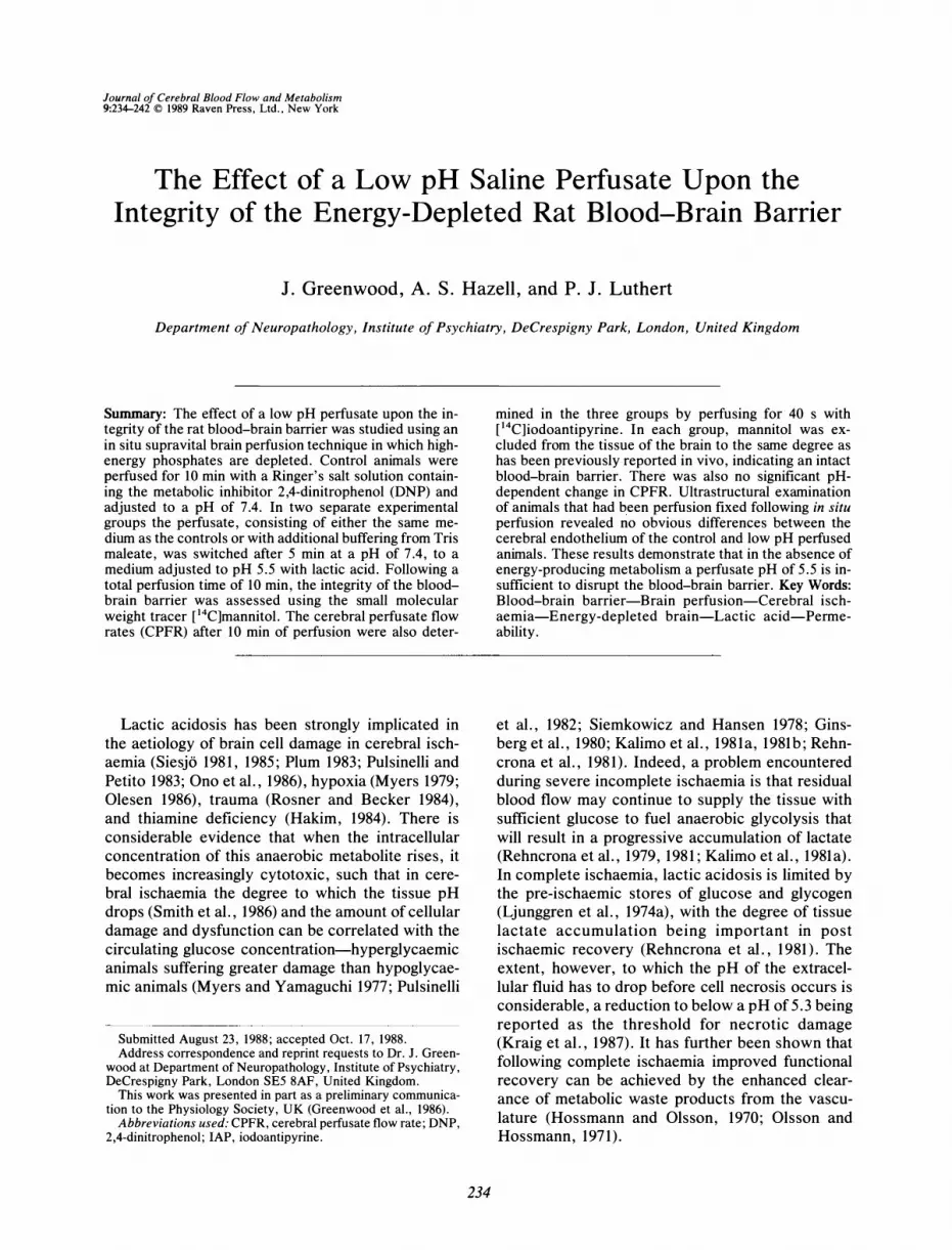
Ultrastructural examination revealed no noticeable difference in the appearance of the capillary endothelium of the three groups. The microvasculature was well preserved in both the pH 7.4 (n = 4) and the pH 5.5 (Ringer-DNP, n = 4 and Tris maleate-DNP, n = 3) groups (Figs. 2-4). Astrocyte foot processes occasionally showed signs of oedema particularly in the white matter and around larger vessels (Fig. 5). These alterations were not sufficient to cause any deformation of the capillaries, and in no case were partially or totally occluded vessels observed. The vessels in both groups were largely indistinguishable from those fixed without prior saline perfusion. No oedema was seen in the
capillary endothelial cells; there was no increase in vesicular profiles, and no obvious disruption of the tight junctions or luminal membrane was observed.
DISCUSSION
It had recently been shown that the blood-brain barrier can be opened to the high molecular weight tracer horseradish peroxidase when Ringer's, containing lactic acid, is perfused in vivo into the carotid circulation of the rat (Nagy et aI., 1985). The present study, however, shows that under conditions in which the brain is depleted of high-energy phosphates (J. Greenwood et aI., in preparation) the perfusion of a solution containing lactic acid at a pH of 5.5 for a total of 7.5 min fails to open the bloodbrain barrier to either unbound Evans blue or the small tracer e4C]mannitoi (Fig. 2). These findings are consistent with a recent report demonstrating that perfusion of frog pial vessels with a perfusate of pH 5.1 does not disrupt the vasculature (Olesen, 1986), although the same group did find vessel disruption with hypoxia and metabolic inhibition (Olesen and Crone, 1986).
The failure of luminal lactic acid and low pH to open the blood-brain barrier in this study may suggest that they are not important in ischaemic barrier disruption or that metabolic energy is required in addition. Hyperosmolar barrier opening has been shown to occur in the absence of energy-producing metabolism (Greenwood et aI., 1988), but Hossmann and Olsson ( 197 1) demonstrated suppression of barrier opening, with hypertensive and chemical insults, following a period of ischaemia during which energy depletion must have occurred. It is a reasonable supposition, therefore, that there may be more than one way in which the barrier is opened. The morphological correlates of barrier opening are discussed below. If lactic acid is not the cause of ischaemic barrier damage, perfusion of lactic acid in vivo must be disrupting the barrier in other ways, perhaps by provoking local vasodilatation, loss of autoregulation, and subsequent elevation of cerebral blood flow, a factor known to cause barrier opening (Rapoport, 1976; Kuroiwa et aI., 1985). Recently, however, it has been reported that
TABLE 2. Cerebral perfusate flow rates following the different perfusion protocols
Ringer's-DNP pH 7.4 (4) Ringer's-DNP pH 5.5 (4) Tris-DNP pH 5.5 (4)
Cerebrum
0.81 ± 0.19 0.86 ± 0.06 1.00 ± 0.09
CPFR (ml/min/g)
Cerebellum
1.19 ± 0.08 1.13 ± 0.08 1.19 ± 0.12
Brainstem
1.63 ± 0.22 1.54 ± 0.09 1.11 ± 0.09
Body weight (g)
398 ± 17 373 ± 6 393 ± 2
Values are means ± SEM with numbers of animals in parentheses.
J Cereb Blood Flow Metab, Vol. 9, No.2, 1989

238 J. GREENWOOD ET AL.
Ringer
Tris maleate
o 4 6 8 10
Perfusion time (min)
FIG. 1. Examples of the perfusate effluent pH sampled from the superior saggital sinus (see text) during a 5-min perfusion with a medium at pH 7.4 followed by a further 5-min perfusion at pH 5.5. The open circles represent the effluent pH of the Ringer's-DNP perfusate and the closed circles represent the effluent pH of the Tris-maleate-buffered perfusate.
lactic acid infused into the circulation of conscious piglets does not increase CBF (Laptook, et aI., 1988), although these differences may be due to the route of administration.
The relatively good buffering capacity of the cerebral vasculature is demonstrated by the small pH difference across the vascular bed of the pH 5.5 Ringer's perfused animal. Conversely, when Tris maleate buffer is added to the perfusate a dramatic arterio-venous difference in pH is found when the perfusate is switched from a pH of 7.4 to 5.5. What is perhaps more surprising, however, is the observation that the pH of the effluent for both the perfusates during the pH 7.4 perfusion remains below 7.4. It is possible that this may be due to the efflux of lactic acid from the tissue, although separate experiments looking at the efflux of lactic acid from perfused brains do not support this (J. Greenwood et aI., in preparation). Furthermore, if this were so
A
then one would expect a greater drop in the effluent pH of the poorly buffered Ringer's perfusate. It is clear from these experiments that, at least in the absence of energy-yielding metabolism, a pH of 5.5 within the lumen is insufficient to damage the cerebral endothelium and cause them to become disrupted.
The relationship between pH, cerebral oedema, and cerebral blood flow/CPFR is one that requires close examination. It has been shown in the past that the severity of brain oedema is likely to be proportional to the degree of tissue acidosis (Takahashi, 1966; Paljarvi et aI., 1983). In the present study, there was no significant increase in the water content of the brain following the low pH perfusions, although the values were slightly greater than those of the pH 7.4 perfused animals (Table O. Examination of the state of the microvasculature at both light and electron microscope levels showed limited perivascular swelling of astrocytic foot processes at either pH 7.4 or the two pH 5.5 groups of animals (Figs. 2-4), although this was more marked in white matter (Fig. 5). These appearances are similar to those described by Paljarvi et al. ( 1983) and Kalimo et al. ( 198 1b), who demonstrated that the microvasculature and parenchyma remained well preserved if saline was infused prior to the ischaemic period. In animals in which they infused a glucose-saline mixture, however, there was considerable astrocytic and endothelial swelling and greater lactic acidosis. In these high lactic acid preparations the preservation of the tissue was poor, with severe structural changes and vessel occlusion.
The buffering capacity of the two perfusates used
B
FIG. 2. Electron micrographs of cerebrum following perfusion with Ringer's-DNP at a pH of 7.4 for 10 min prior to fixation. (A) Parenchyma (x3,500). (8) Capillary (x11,000).
J Cereb Blood Flow Metab, Vol. 9, No.2, 1989

LOW pH AND BLOOD-BRAIN BARRIER 239
A 8
FIG. 3. Electron micrographs of cerebrum following perfusion with Ringer's-DNP at a pH of 7.4 for 5 min followed by a further 5 min at pH 5.5 prior to fixation. (A) Parenchyma (x4,900). (8) Capillary (x11,000).
in the present study differ considerably, and as a result the concentration of lactic acid required to reduce the pH to 5.5 differed by over an order of magnitude. It was only in the well-buffered Tris maleate perfusate that the concentration of lactate approached that generally found in plasma in ischaemic states; 9.2 1 to 11.23 f.LmoVml in this study compared to 10 to 15 f.LmoVml during ischaemia in
vivo (Rehncrona et aI., 1981). Indeed, it has previously been found that severe brain cell damage in experimental ischaemia only occurred when the tissue lactate rose to a level of 35 f.Lmol/g whilst at a level of 15 f.Lmol/g there was very little morphological change (Kalimo et aI., 1981a,b; Rehncrona et
A
aI., 198 1). The tissue lactic acid threshold at which morphological abnormalities occur has been put at between 16 and 20 f.Lmol/g. Unfortunately the amount of lactic acid used to acidify the KrebsRinger's bicarbonate buffer used by Nagy et ai. ( 1985) in their in vivo opening of the blood-brain barrier is not reported. The higher concentrations of lactic acid required to cause damage are consistent with the recent report by Kraig et ai. ( 1987) and Petito et ai. ( 1987), who reported that necrosis occurred only when the extracellular fluid pH dropped to below 5.3. It is also perhaps important to consider that the in vivo relationship between lactic acid concentration and pH is complex, for protons
FIG. 4. Electron micrographs of cerebrum following perfusion with Ringer's-DNP buffered with Tris-maleate and adjusted to a pH of 7.4 for 5 min followed by a further 5 min at pH 5.5 prior to fixation. (A) Parenchyma (x4,300). (8) Capillary (x 11 ,000).
J Cereb Blood Flow Metab, Vol. 9, No.2, 1989
240 J. GREENWOOD ET AL.
FIG. 5. Electron micrograph of cerebral capillary in white matter, following perfusion with Ringer's-DNP at a pH of 7.4 for 10 min prior to fixation. Mild swelling of the perivascular astrocytic foot process can be seen (x4,500).
may be produced from sources other than lactic acid, such as during the anaerobic hydrolysis of ATP (Paschen et aI., 1987). In a recent study in which lactic acid was applied directly to the spinal cord, morphological changes were found both in the parenchyma and blood vessels, the latter showing signs of capillary wall necrosis and leakage of HRP. The experimental conditions in this study were, however, extremely severe, with a pH of 1.8 and lactic acid concentrations of 6.3 M and above; both conditions are highly unlikely to be found in vivo.
A further consideration is the difference between the tissue concentration of lactic acid and the perfusion medium concentration. In the present study, lactic acid and low pH are introduced via the vasculature whilst in vivo, during ischaemia, lactate is produced intracellularly and not in the lumen. Although it is known that at the blood-brain barrier the monocarboxylic acid carrier for lactate dramatically increases its translocation at low pH values (Oldendorf et aI., 1979) it is unclear how much of the perfusate lactic acid will enter the tissue under our experimental conditions. What is known, however, is that during physiological hyperlactic acidemias in which blood levels of lactic acid can reach as high as 10 mM, it takes many hours for the lactate to equilibrate with the CSF (Pardridge, 1983), indicating a very low rate of influx across the blood-brain barrier. Indeed, the concentration of lactic acid present in the brain tissue under these conditions increase by only tenfold from control values (J. Greenwood et aI., in preparation) to levels that are believed to be insufficient to cause ul-
J Cereb Blood Flow Metab, Vol. 9, No.2, 1989
trastructural damage (Plum, 1983). Intracellular, rather than intraluminal, lactate concentration may be of prime significance in the development of cell swelling and damage, a hypothesis consistent with the results of Rehncrona et aI. ( 1981), who found identically raised blood lactate concentrations in both saline- and glucose-infused ischaemic animals, but with a significantly higher tissue concentration in the latter, the group which also showed the severe pathological change.
A pump flow rate of 25 mllmin gave CPFR values for the pH 7.4 and both the pH 5.5 groups of perfused animals that are similar to anaesthetised in
vivo CBF rates (Reid et aI., 1982; Braun et aI., 1985). There was no significant difference between the groups, suggesting that perfusate access to the cerebral vascular bed was similar in all cases. The variation in CPFR between the three brain regions examined in each group (Fig. 3) has been observed previously (Luthert et aI., 1987) and may be due to a loss of regulatory mechanisms that normally control the differential supply to different areas of the brain.
Reported endothelial cell injury following lactic acid-based perfusion (Nagy et aI., 1985), in ischaemia (Paljarvi et aI., 1983; Chiang et aI., 1968), following intracerebral lactate injections (Petito et aI., 1987), and after direct application of lactic acid to the spinal cord (Balentine and Greene, 1987) was not observed in this energy-depleted preparation. It is clear from these previous studies that only at high tissue lactate concentrations and low pH deterioration of the microvasculature occur, the threshold for damage being reported to be a tissue pH of around 6 (Mabe et aI., 1983); more recently the threshold within the extracellular fluid has been put at 5.3 (Kraig et aI., 1987; Petito et aI., 1987). Of direct relevance to the failure to open the barrier was the lack of an increase in the normal number of micropinocytotic vesicle profiles seen. It has been suggested that microvesicular transport (rarely seen in abundance in normal cerebral endothelium) is an active process and is responsible for the increase in permeability found in ischaemic brain damage (Westergaard, 1977; Kuroiwa et aI., 1985). The failure to open the barrier at low pH in the absence of energy-producing metabolism in this system could tentatively be explained by the inhibition of an active process of this nature taking place. An alternative explanation would be that under these, or any other, conditions the vasculature is simply resistant to acidity as low as pH 5.5. This does not rule out the possibility that vesicular transport does occur in ischaemic brain, but would imply that it occurs only upon restitution of blood flow and energy status.

LOW pH AND BLOOD-BRAIN BARRIER 241
In conclusion, perfusion with a low pH solution containing lactic acid is insufficient to bring about blood-brain barrier opening in the absence of energy-producing metabolism. This is despite the fact that lactic acidosis has been strongly implicated in barrier disruption in cerebral ischaemia and other pathological conditions. The absence of an increase in vascular permeability may be due to the process being one that requires the presence of metabolic energy or to the likelihood that the endothelium are resistant to external acidity of as low as pH 5.5
Acknowledgment: This work was supported by grants from the Wellcome Trust.
REFERENCES
Balentine JD, Greene WB (1987) Myelopathy induced by lactic acid. Acta Neuropathol (Ber/) 73:233-239
Braun LD, Miller LP, Pardridge WM, Oldendorf WH (1985) Kinetics of regional blood-brain barrier glucose transport and cerebral blood flow determined with the carotid injection technique in conscious rats. J Neurochem 44:911-915
Brightman MW, Zis K, Anders J (1983) Morphology of cerebral endothelium and astrocytes as determinants of the neuronal microenvironment. Acta Neuropathol (Ber/) Suppl VIII:21-33
Broman T, Lindberg-Broman AM (1945) An experimental study of disorders in the permeability of the cerebral vessels (' 'the blood-brain barrier") produced by chemical and physicochemical agents. Acta Physiol Scand 10: 102-125
Bundgaard M, Frokjaer-Jensen J, Crone C (1979) Endothelial plasmalemmal vesicles as elements in a system of branching invaginations from the cell surface. Proc Natl Acad Sci 76:6439-6442
Caldwell PC (1957) The sensitivity to pH of the inhibitory effects of dinitrophenol on squid giant axons. Biochem J 67: 1-2P
Caldwell PC (1960) The phosphorous metabolism of squid axons and its relationship to the active transport of sodium. J Physiol 152:545-560
Cervos-Navarro J, Artigas J, Mrsulja BJ (1983) Morphofunctional aspects of the normal and pathological blood-brain barrier. Acta Neuropathol (Ber/) Suppl VIII: 1-19
Chiang J, Kowada M, Ames III A, Wright RL, Majno G (1968) Cerebral ischemia. III. Vascular changes. Am J Pathol 52:455-476
Ginsberg MD, Frank AW, Budd WW (1980) Deleterious effect of glucose pretreatment on recovery from diffuse cerebral ischemia in the cat. Stroke 11:347-354
Greenwood J, Luthert PJ, Pratt OE, Lantos PL (1985) Maintenance of the integrity of the blood-brain barrier in the rat during an in situ saline-based perfusion. Neurosci Lett 56:223-227
Greenwood J, Lantos PL, Luthert PJ, Pratt OE (1986) Failure of low pH to open the blood-brain barrier of the rat in the presence of an inhibitor of energy-yielding metabolism. J Physio1 374:19P
Greenwood J, Luthert PJ, Pratt OE, Lantos PL (1988) Hyperosmolar opening of the blood-brain barrier in the energydepleted rat brain. Part 1. Permeability studies. J Cereb Blood Flow Metab 8:9-15
Hakim AM (1984) The induction and reversibility of cerebral acidosis in thiamine deficiency. Ann NeuroI16:673--679
Hossmann K-A, Olsson Y (1970) Suppression and recovery of neuronal function in transient cerebral ischemia. Brain Res 22:313-325
Hossmann K-A, Olsson Y (1971) Influence of ischemia on the passage of protein tracers across capillaries in certain bloodbrain barrier injuries. Acta Neuropathol (Ber/) 18:113-122
Kalimo H, Rehncrona S, Soderfeldt B (1981a) The role of lactic acidosis in the ischemic nerve cell injury. Acta Neuropathol (Ber/) Supp!. VII:20--22
Kalimo H, Rehncrona S, Soderfeldt B, Olsson Y, Siesj6 BK (1981b) Brain lactic acidosis and ischemic cell damage: 2. Histopathology. J Cereb Blood Flow Metab 1:313-327
Klatzo I (1983) Disturbances of the blood-brain barrier in cerebrovascular disorders. Acta Neuropathol (Berl) Supp!. VIII:81-88
Kraig RP, Petito CK, Plum F, Pulsinelli WA (1987) Hydrogen ions kill brain at concentrations reached in ischemia. J Cereb Blood Flow Metab 7:379-386
Kuroiwa T, Ting P, Martinez H, Klatzo I (1985) The biphasic opening of the blood-brain barrier to proteins following temporary middle cerebral artery occlusion. Acta Neuropathol (Ber/) 68:122-129
Laptook AR, Peterson J, Porter AM (1988) Effects of lactic acid infusions and pH on cerebral blood flow and metabolism. J Cereb Blood Flow Metab 8: 193-200
Ljunggren B, Norberg K, Siesj6 BK (1974a) Influence of tissue acidosis upon restitution of brain energy metabolism following total ischemia. Brain Res 77: 173-186
Ljunggren B, Schutz H, Siesj6 BK (l974b) Changes in energy state and acid-base parameters of the rat brain during complete compression ischemia. Brain Res 73:277-289
Luthert PJ, Greenwood J, Pratt OE, Lantos PL (1987) The effect of a metabolic inhibitor upon the properties of the cerebral vasculature during a whole-head saline perfusion of the rat. Q J Exp Physiol 72:129-141
Lowry OH, Passonneau JV, Hasselberger FX, Schulz DW (1964) Effect of ischemia on known substrates and cofactors of the glycolytic pathway in brain. J Bioi Chem 239: 18-30
Mabe H, Blomqvist P, Siesj6 BK (1983) Intracellular pH in the brain following transient ischemia. J Cereb Blood Flow Metab 3:109-114
Myers RE (1979) Lactic acid accumulation as cause of brain edema and cerebral necrosis resulting from oxygen deprivation. In: Advances in Perinatal Neurology. Vo!. 1 (Korobkin R, Guilleminault C, eds) New York, Spectrum, pp 85-114
Myers RE, Yamaguchi S (1977) Nervous system effects of cardiac arrest in monkeys. Arch Neurol 34:65-74
Nagy Z, Szabo M, Huttner I (1985) Blood-brain barrier impairment by low pH buffer perfusion via the internal carotid artery in the rat. Acta Neuropathol (Ber/) 68:160--163
Oldendorf WH, Braun L, Cornford E (1979) pH dependence of blood-brain barrier permeability to lactate and nicotine. Stroke 10:577-581
Olesen S-R (1986) Rapid increase in blood-brain barrier permeability during severe hypoxia and metabolic inhibition. Brain Res 368:24-29
Olesen S-R, Crone C (1986) Substances that rapidly augment ionic conductance of cerebral microvascular walls. Acta Physiol Scand 127:233-241
Olsson Y, Hossmann K-A (1971) The effect of intravascular saline perfusion on the sequelae of transient cerebral ischemia. Light and electron microscopical observations. Acta Neuropathol (Ber/) 17:68-79
Ono M, Obrenovitch TP, Hartell N, Symon L (1986) Extracellular pH and potassium activity in cerebral cortex of rats during anoxia and subsequent recovery period. In: Pharmacology of Cerebral Ischemia (Krieglstein J, ed), Amsterdam, Elsevier, pp 275-279
Paljarvi L, Rehncrona S, Soderfeldt B, Olsson Y, Kalimo H (1983) Brain lactic acidosis and ischemic cell damage: Quantitative ultrastructural changes in capillaries of rat cerebral cortex. Acta Neuropathol (Ber/) 60:232-240
Pardridge WM (1983) Brain metabolism: A perspective from the blood-brain barrier. Physiol Rev 63:1481-1535
Paschen W, Djuricic B, Mies G, Schmidt-Kastner R, Linn F (1987) Lactate and pH in the brain: Association and dis so-
J Cereb Blood Flow Metab, Vol. 9, No.2, 1989
242 J. GREENWOOD ET AL.
ciation in different pathophysiological states. J Neurochem 48: 154-159
Petito CK, Kraig RP, Pulsinelli W A (1987) Light and electron microscope evaluation of hydrogen ion-induced brain necrosis. J Cereb Blood Flow Metab 7:625...{i32
Plum F (1983) What causes infarction in ischemic brain? The Robert Wartenberg Lecture. Neurology (Ny) 33:222-233
Pulsinelli WA, Petito CK (1983) The neurotoxicity of hydrogen ions. Stroke 14: 130
Pulsinelli WA, Waldman S, Rawlinson D, Plum F (1982) Moderate hyperglycemia augments ischemic brain damage: a neuropathologic study in the rat. Neurology (Ny) 32: 1239-1246
Rapoport SI (1976) Opening of the blood-brain barrier by acute hypertension. Exp Neurol 52:467-479
Rehncrona S, Mela L, Siesj6 BK (1979) Recovery of brain mitochondrial function in the rat after complete and incomplete cerebral ischemia. Stroke 10:437-446
Rehncrona S, Rosen 1, Siesj6 BK (1981) Brain lactic acidosis and ischemic cell damage: I. Biochemistry and neurophysiology. J Cereb Blood Flow Metab 1:297-311
Reid AC, Teasdale GM, McCulloch J (1982) The effects of dexamethasone administration and withdrawal on water permeability across the blood-brain barrier. Ann Neurol 13:28-31
Rosner MJ, Becker DP (1984) Experimental brain injury: suc-
J Cereb Blood Flow Metab, Vol. 9, No.2, 1989
cessful therapy with the weak base, trimethamine. With an overview of CNS acidosis. J Neurosurg 60:961-971
Siemkowicz E, Hansen A (1978) Clinical restitution following cerebral ischemia in hypo-, normo-, and hyperglycemic rats. Acta Neurol Scand 58:1-8
Siesjo BK (1981) Cell damage in the brain: a speculative synthesis. J Cereb Blood Flow Metab 1:155-185
Siesj6 BK (1985) Acid-base homeostasis in the brain: physiology, chemistry, and neurochemical pathology. In: Progress in Brain Research, Vol 63 (Kogure K, Hossmann K-A, Siesj6 BK, Welsh FA, eds), Amsterdam, Elsevier, pp 121-154
Smith M-L, von Hanwehr R, Siesj6 BK (1986) Changes in extraand intracellular pH in the brain during and following ischemia in hyperglycemic and in moderately hypoglycemic rats. J Cereb Blood Flow Metab 6:574-583
Takahashi K (1966) Relationship between acidity and swelling in the brain. Tohoku J Exp Med 90:261-268
Westergaard E (1977) The blood-brain barrier to horseradish peroxidase under normal and experimental conditions. Acta Neuropathol (Berl) 39: 181-187
Westergaard E, Go G, Klatzo 1, Spatz M (1976) Increased permeability of cerebral vessels to horseradish peroxidase induced by ischemia in Mongolian gerbils. Acta Neuropathol (Berl) 35:307-325