Embed Size (px)
Citation preview

The effects of dietary Microcystis aeruginosa andmicrocystin on the copepods of the upper San FranciscoEstuary
KEMAL A. GER*, SWEE J . TEH †, DOLORES V. BAXA †, SARAH LESMEISTER † AND CHARLES
R. GOLDMAN*
*Department of Environmental Science and Policy, University of California, Davis, CA, U.S.A.†Department of Anatomy, Physiology and Cell Biology, School of Veterinary Medicine, University of California, Davis, CA,
U.S.A.
SUMMARY
1. Increasing blooms of Microcystis aeruginosa have unknown impacts on the copepods
Eurytemora affinis and Pseudodiaptomus forbesi, which are the dominant zooplankters and
key prey species for endangered larval fish in the upper San Francisco Estuary.
2. Laboratory feeding experiments were designed to measure the effect of Microcystis on
copepod survival and to distinguish the effects of toxicity and nutrition. In a series of
survival tests, copepods were fed a mixed diet of algae plus one of two strains of
Microcystis, either producing (MC+) or lacking microcystin (MC)).
3. Microcystis significantly reduced survival even when it was a small proportion of the
diet, indicating that toxicity was the major cause of mortality. Contrary to expectation,
however, the MC+ strain did not result in higher mortality, suggesting that non-MC
metabolites of Microcystis can be toxic to copepods.
4. Across treatments, survival of P. forbesi was greater than that of E. affinis, although the
two copepods responded differently to both the ratio and the strain of Microcystis in
their food. Survival of P. forbesi was greater on the MC+ strain and was inversely
proportional to the ratio of dietary Microcystis (MC+ or MC)). In contrast, survival of
E. affinis declined similarly across treatments and was not related to the proportion or
strain of dietary Microcystis. Results indicate that the copepod P. forbesi can coexist with
Microcystis while the other copepod E. affinis cannot.
5. Regardless of species, dietary Microcystis caused significant mortality to copepods, and
it may cause adverse impacts to the potentially food-limited zooplankton community of
the San Francisco Estuary. These impacts may not be related to the cellular MC
concentration because Microcystis contains other metabolites that negatively affect
copepods.
Keywords: copepod, harmful algal bloom, microcystin, Microcystis, San Francisco Estuary
Introduction
Global increases in the extent of bloom-forming
cyanobacteria have attracted growing attention from
both management and scientific interests because of
potential negative impacts on water quality and food
web structure (Kirk & Gilbert, 1992; Chorus &
Correspondence: Swee J. Teh, Department of Anatomy, Physi-
ology, and Cell Biology, Aquatic Toxicology Program, School of
Veterinary Medicine, 1321 Haring Hall, One Shields Avenue,
University of California, Davis, CA 95616, U.S.A.
E-mail: [email protected]
Freshwater Biology (2010) 55, 1548–1559 doi:10.1111/j.1365-2427.2009.02367.x
1548 � 2010 Blackwell Publishing Ltd

Bartram, 1999). Microcystis is a common bloom-form-
ing cyanobacterium that can dominate nutrient-
enriched freshwaters including upper reaches of
estuaries, such as the San Francisco Estuary (Paerl,
1988; Lehman et al., 2008).
In blooms, Microcystis can change the food web
because it is a poor quality food for zooplankton,
reducing their fitness and restricting the trophic
transfer of carbon (Paerl, 1988; Christoffersen, 1996;
Chorus & Bartram, 1999). Negative impacts on zoo-
plankton occur in response to a combination of factors
including: (i) secondary metabolites with toxic effects,
such as microcystins (MCs), (ii) nutritional inade-
quacy and (iii) poor digestibility or feeding inhibition
(Fulton & Paerl, 1987; DeMott & Moxter, 1991;
Lurling, 2003). However, not all zooplankton are
affected equally (Christoffersen, 1996; Wilson, Sarn-
elle & Tillmanns, 2006). Species-specific adaptations in
zooplankton lead to variation in the observed
responses to Microcystis blooms (Kirk & Gilbert,
1992; Rohrlack et al., 1999; Gustafsson & Hansson,
2004). Although many studies have evaluated the
interaction between zooplankton and Microcystis,
most focus on cladocerans while impacts to copepods
remain poorly understood (reviewed in Wilson et al.,
2006).
Controlled manipulations of diets in the laboratory
can provide valuable evidence of the mechanisms
involved. A major problem in predicting the impacts
of Microcystis, however, has been because of the use of
oversimplified experimental conditions, such as pure
or single strain diets (presence ⁄absence experiments),
a disproportionate focus on daphniids as the test
organism, and species ⁄strain-specific responses, pre-
venting the interpretation of impacts on other zoo-
plankton or under field conditions (reviewed in
Wilson et al., 2006). As a result, the effect of Microcystis
blooms on copepods and the dynamics of food webs
in estuaries dominated by copepods are poorly
understood.
Copepods are an important link between primary
production and fish, and commonly dominate the
mesozooplankton of estuaries (Turriff, Runge &
Cembella, 1995; Sommer et al., 2007). Calanoid cope-
pods, particularly Eurytemora affinis (Poppe, 1880) and
Pseudodiaptomus forbesi (Poppe & Richard, 1890), are
the principal food source of the endangered larval and
pelagic fish in the San Francisco Estuary, where the
abundance of pelagic organisms, including copepods
and nutritious phytoplankton, has declined to values
not seen before (Sommer et al., 2007). Increasing
blooms of Microcystis in the freshwater and low
salinity zone of the estuary raise concerns about the
further depletion of high-quality fish food (Muller-
Solger, Jassby & Muller-Navarra, 2002; Lehman et al.,
2005, 2008). Understanding the impacts of Microcystis
on the dominant copepods of the San Francisco
Estuary has become highly significant in the manage-
ment of its declining fish populations (Sommer et al.,
2007).
While a pure diet of Microcystis reduces fitness and
survival in zooplankton, it is not likely to occur in
nature (DeMott, Zhang & Carmichael, 1991; Kumar,
2003; Wilson et al., 2006). Unlike daphniids, copepods
can ingest nutritious particles selectively and avoid
those that are unwanted or toxic (DeMott et al., 1991;
Kleppel, 1993; Burns & Hegarty, 1994; Engstrom et al.,
2000; Panosso et al., 2003). Even daphniids show
different responses to Microcystis when offered in
mixed versus pure diets (Rohrlack et al., 1999; Lurling,
2003). Therefore, in studying interactions between
zooplankton and Microcystis, we need to experiment
with diets consisting of mixtures of alternative, more
palatable, food (Wilson et al., 2006).
Microcystis toxicity comes from well-known toxins,
such as MC, but also from other, poorly understood and
often unidentified, secondary metabolites (Lurling,
2003; Rohrlack et al., 2004; Wiegand & Pflugmacher,
2005; Wilson et al., 2006). Though the significance of
metabolites other than MC is becoming clear, the
relative toxicity to zooplankton of MC versus non-MC
metabolites remains unresolved (DeMott et al., 1991;
Kurmayer & Juttner, 1999; Lurling, 2003; Rohrlack et al.,
2004). It is possible to quantify the effect of MC in the
zooplankton diet using Microcystis strains of identical
morphology (assuming similar nutritional profile) but
different MC cellular content. This approach has been
used successfully to distinguish the toxicity of MC
versus non-MC metabolites in daphniids (Rohrlack
et al., 1999; Lurling, 2003). When offered in a mixed diet,
strains of Microcystis containing (MC+) and lacking
(MC)) both caused mortality, showing the toxic effects
of non-MC metabolites on daphniids (Lurling, 2003).
The current study was designed to compare the effects
of Microcystis and its toxic metabolites on the dominant
copepods of the San Francisco Estuary.
Our objective was to evaluate the impact of Micro-
cystis in the diet on the survival of the dominant
Dietary effects of Microcystis on copepods 1549
� 2010 Blackwell Publishing Ltd, Freshwater Biology, 55, 1548–1559

copepods. To distinguish between the effects of toxic
metabolites and nutritional deficiency of Microcystis
on copepods, mixed diets containing nutritious algae
and Microcystis (MC+ or MC)) were offered to
copepods in laboratory survival tests. Experiments
were designed to compare the survival of E. affinis
and P. forbesi as a function of dietary Microcystis (MC+
versus MC)) and food quantity. We hypothesised that
(i) both strains of Microcystis would negatively affect
survival, though the response might be species spe-
cific, (ii) survival with the MC+ strain in the diet
would be lowest and (iii) survival would be inversely
proportional to the proportion of Microcystis in the
diet.
Methods
Microcystis aeruginosa strains
Cultures of two different strains of M. aeruginosa
(Kutzing, 1846) were maintained for the feeding
experiments in a modified ASM-1 medium (Reynolds
& Jaworski, 1978). MC producing (MC+, UTEX 2385)
and MC absent (MC), UTEX 2386) strains of Micro-
cystis were obtained from the University of Texas
Culture Collection (Austin, TX, U.S.A.). Exponentially
growing axenic batch cultures were maintained by
dilutionwithfreshlypreparedculturedmediumtokeep
cell density between 1 · 106 and 3 · 106 cells mL)1, in
gently bubbled 500-mL glass flasks, under a 16 : 8 L : D
cycle under 30 lmol quanta m)2 s)1, in a temperature-
controlled room (22 ± 1 �C). Under these conditions,
both strains were uni- and bi-cellular, the average
growth rate of Microcystis was 0.32 (±0.04) day)1 and
cell diameter was approximately 4 lm (±0.7) for both
strains. We assumed that the only difference between
the two strains used was MC content and that each
strain had a comparable nutritional profile and
digestibility.
Instant algae (IA)
Commercially available and highly nutritious pure
phytoplankton food composed of Nannochloropsis and
Pavlova, rich in fatty acids, were kept fresh at 4 �C
(Reed Mariculture, Campbell, CA, U.S.A.). Because IA
is in a fresh but non-living state, cells have a constant
density. Cell diameter was approximately 2 lm for
Nannochloropsis and 4 lm for Pavlova.
Copepod cultures
Copepods were collected from Rio Vista and Suisun
Marsh in the San Francisco Estuary using a 174-lm
zooplankton tow net. Pseudodiaptomus forbesi was
collected in June 2007 and E. affinis in April 2007
(respective periods of dominance), and returned to
the laboratory within 2 h of collection. Gravid females
were selected and rinsed three times with salinity-
adjusted culture medium before transferring them to
the culture. We used reconstituted water (modified by
NaCl to increase salinity) as the culture medium
(Horning & Weber, 1985). Cultures were grown in
aerated 4-L beakers placed in an environmental water
bath, at 24 ± 1 �C for P. forbesi and 20 ± 1 �C for
E. affinis. Water quality in beakers, including dis-
solved oxygen (>8 mg L)1), pH (8.2 ± 0.1), water
hardness (100 mg L)1), salinity (5.0 ppt) and ammo-
nia (<1 lg L)1), was monitored weekly (Hach, Love-
land, CO, U.S.A.). For optimal culture and population
growth under laboratory conditions, a previously
determined amount of the IA mixture (an equal
biovolume of Nannochloropsis and Pavlova) was given
as food at 400 and 500 lg C L)1 day)1 for E. affinis and
P. forbesi, respectively. Approximately, 60% of the total
culture medium was replaced weekly with aerated and
temperature-adjusted medium. Copepod culture den-
sity was monitored weekly, maintained between 50
and 100 adults L)1, and diluted as necessary to ensure
exponential population growth and minimise over-
crowding. The systems were maintained under a
natural photoperiod (16L : 8D) and covered with a
semitransparent black tarpaulin. The brood cultures
were acclimated to these conditions for a minimum of
4 months before the feeding experiments.
Cell size, density and algal biomass determination
Algal biomass concentration (lg C mL)1) for the IA
mixture and Microcystis was determined by a calibration
curve of total carbon and biovolume. A serial dilution of
algal biovolumes were filtered on pre-combusted
25-mm GF ⁄C (Whatman, Kent, U.K.), desiccated at
50 �C, and stored at )80 �C until the total carbon was
measured by a gas chromatogram (UC Davis Stable
Isotope Facility, U.S.A.). Microcystis cell size and density
was determined using an inverted compound epifluo-
rescent microscope (2007 Zeiss Axio Observer A1,
Thornwood, NY, U.S.A), counted in a Haemocytometer
1550 K. A. Ger et al.
� 2010 Blackwell Publishing Ltd, Freshwater Biology, 55, 1548–1559

(Hausler Scientific, Horsham, PA, U.S.A.) and
calibrated against light absorbance at 800 nm.
Survival test
We used a series of survival tests in which dietary
treatments included Microcystis while controls did not.
The treatment diets consisted of a Microcystis – IA
mixture, with the proportion of Microcystis at 0, 10, 25,
50 or 100% of total food (by carbon), using either the
MC+ or MC) strain of Microcystis, plus IA, to a total
food concentration of 400 (E. affinis) and 500 (P. forbesi)
lg C L)1 day)1. The control diets were comprised of
IA only at the same amount as in the associated
treatment of IA and Microcystis mixture, which corre-
sponded to 100, 90, 75, 50 and 0% (starvation) of the
total food. This design allowed the distinction of
impacts because of food quantity (total nutrition
available) versus quality (MC and non-MC toxicity)
by comparing survival over a range of a given diets
with and without Microcystis. This design also enabled
the assessment of the impact of alternative food
sources when Microcystis was present in food.
Experiments took place in triplicates over 11 days
in 1-L glass beakers, starting with 10 copepods per
beaker and maintained under identical conditions
described for the brood cultures. Animals were
obtained by growing a new cohort of adults from
nauplii in the brood cultures. Experimental copepods
were then collected from the pool of >400 copepods in
the new cohort by gently filtering in a Petri dish for
each set of experiments. To minimise the chance of
age-related mortality and stress over the 11 days, only
active C5 copepodites and young adults were ran-
domly selected. Survival and activity were monitored
daily using a light table and a dissecting microscope
when necessary. Copepods were considered dead
only if they showed no sign of movement after
disturbance under a dissecting microscope; dead
copepods were removed as soon as recorded.
MC verification of strains and treatments
Microcystis cultures were sampled for MC analysis
during the experiment, to ensure the MC+ and MC)strains were uncontaminated and to measure MC
concentration of the MC+ strain culture. In addition,
the MC+ experimental beakers were sub-sampled at
day 7 of the survival test for total and dissolved MC
analysis to ensure that mortality was not because of
any dissolved MCs. A commercially available MC –
specific competitive ELISA assay with a detection
range of 0.16–2.5 lg L)1 MC-LR was used for MC
analysis (Envirologix, Portland, ME, U.S.A.). Micro-
cystis cultures were sampled and kept frozen until
analysis, upon which they were thawed to room
temperature, sonicated, diluted and measured for MC
concentration using a microplate reader at 450 nm.
Dissolved MC in treatments was measured 7 days
during the experiment, from whole water samples by
separating algal cells on a GF ⁄F glass fibre filter
(Whatman Corp) and comparing MC concentration in
the filtrate with an unfiltered sample. Additionally, a
standard polymerase chain reaction (PCR) using
published primers designed for the conserved Micro-
cystis-specific 16S ribosomal DNA (rDNA) region and
amplification of the MIC and MC synthetase genes
mcyB and mcyD was used to verify the identity of the
two strains of Microcystis. Ability to produce MC is
based on the presence or absence of the synthetase
genes (Ouellette & Wilhelm, 2003). Conventional PCR
was conducted to verify the presence of Microcystis
and the MC synthetase genes mcyB and D using
specific PCR primer sets targeting 16S rDNA and
conditions described in Ouellette & Wilhelm (2003).
The Microcystis strains were processed for genomic
DNA extraction using a modified phenol–chloroform
procedure (Sambrook & Russell, 2001).
Qualitative confirmation of Microcystis ingestion
To verify ingestion, the presence of Microcystis cells in
copepod guts was determined in a separate feeding
experiment. Copepods were starved for 5 h to evac-
uate gut contents before the experiment, which took
place under the same conditions as the survival tests.
Copepod density was kept at 10 adults in a 1-L beaker
with clean media, and then fed the same treatment
diets (MC+ and MC)) described earlier. Following a
30-min feeding period, animals were gently filtered,
rinsed three times to remove any attached Microcystis
cells and narcotised immediately with carbonated
water. For each treatment diet, 10 copepods were
dissected to expose the gut under a dissecting micro-
scope. Guts were observed for phycocyanin fluores-
cence (a pigment specific to cyanobacteria), as a
qualitative indication of Microcystis cells in copepod
guts, using an epifluorescent microscope.
Dietary effects of Microcystis on copepods 1551
� 2010 Blackwell Publishing Ltd, Freshwater Biology, 55, 1548–1559

Statistical analysis
The effect of diet type on the survival of each copepod
species was evaluated by a parametric survival anal-
ysis which modelled survival with a Weibull distribu-
tion curve (JMP version 7.0; SAS, Cary, NC, U.S.A).
Individual probability of mortality or survival (expec-
ted proportion of the population dead or surviving)
for each treatment and species was calculated by a test
of deviance based on the mean number of surviving
copepods on the last day of the experiment. Then, the
effect of diet type (e.g. Microcystis versus no Microcys-
tis) and copepod species on survival was calculated to
compare differences in the effects of the treatments and
controls. Finally, the Microcystis only treatments were
further broken down to strain (MC+ or MC)) and
ratio of dietary Microcystis (% Microcystis) to analyse
differences in the effect of these parameters on cope-
pod survival. Time to 50% mortality was analysed by
linear regression of individual replicate survival res-
ponses and calculating when 50% mortality occurred
on average per treatment. All differences noted are
significant at P = 0.05 unless otherwise mentioned.
Results
Survival test
Both dietary composition and quantity had very
pronounced effects on copepod survival. The pres-
ence and proportion of Microcystis drastically reduced
survival in all treatments for either copepod species,
regardless of Microcystis strain and MC content
(Fig. 1). In contrast, survival with the control diets
was significantly higher than corresponding Microcys-
tis treatments (Fig. 1; P < 0.001).
Reducing the concentration of IA in the controls
without Microcystis reduced survival. At the end of
the experiment, 100% IA had the highest survival
(85% ± 3.4), while survival in the 50% IA treatment
was lower (75% ± 2.3 for E. affinis, 60% ± 8.2 for
P. forbesi) (Fig. 1). Starved copepods (i.e. those fed 0%
IA and 0% Microcystis) survived for several days,
reaching 100% mortality after 7–9 days for both
species, though P. forbesi had slightly longer average
persistence than E. affinis.
Compared to E. affinis, P. forbesi was more sensitive
to differences between the Microcystis strains (MC+
versus MC)) and had a higher survival rate on a MC+
diet (Fig. 1). Differences in survival of P. forbesi
between the two different Microcystis strains took
several days to emerge. Both strains (MC+ or MC))
caused comparable mortality until day 6, and thereaf-
ter P. forbesi mortality on the MC) diet was greater
than that on the MC+ diet (Fig. 1). Towards the end of
the experiment, P. forbesi mortality began to decline in
most of the MC+ diets, but not in the MC) diets (Fig. 1).
For treatments with <50% Microcystis, the MC) diet
killed more P. forbesi than the MC+ diet, while these
diets caused similar mortality for E. affinis (Fig. 1).
Survival analysis
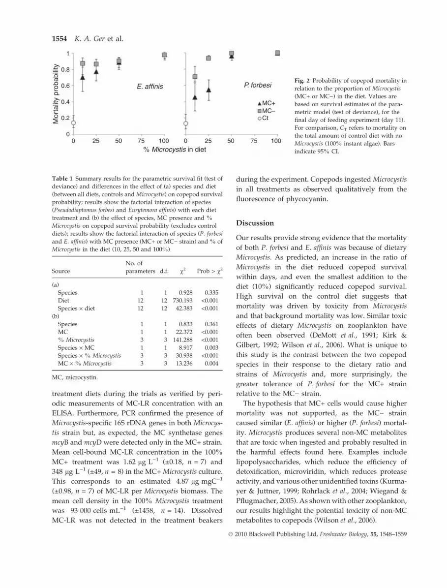
The increased proportion of Microcystis in the diet
caused highermortality andincreased the probability of
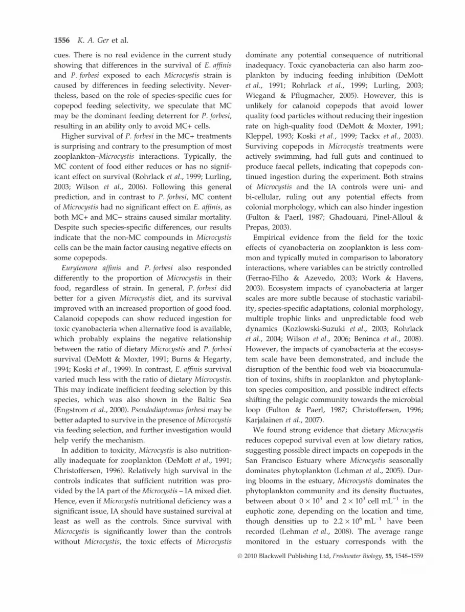
mortality (Fig. 2). The effect test showed that diet type
had a significant effect on survival and that differences
in survival were not because of copepod species
(Table 1a). When comparing results among the treat-
ments (Table 1b),differences insurvivalwerecausedby
the Microcystis strains (MC) and the ratio of Microcystis
in food (% Microcystis). There was a significant differ-
ence in how each species responded to the MC+ versus
the MC) strains, as well as the ratio of Microcystis in
food, as shown by the interaction terms (species · MC)
and (species · %Microcystis), respectively, in Table 1b.
Survival ⁄mortality probability tests confirmed that
copepods responded differently, both to the individ-
ual strains (MC+ versus MC)) and to the ratio of
Microcystis in their food. Pseudodiaptomus forbesi was
more likely to survive on the MC+ diet compared to
an MC) diet when the diet contained <50% Micro-
cystis (Fig. 2). In contrast, differences in the Microcystis
strain had no significant effect on E. affinis survival at
the end of the experiment. Additionally, P. forbesi was
more sensitive to changes in the Microcystis content of
its diet, as increased Microcystis resulted in higher
mortality. In contrast, increasing Microcystis did not
significantly change the probability of mortality for
E. affinis (Fig. 2). Rather, the presence of Microcystis
was more important in reducing E. affinis survival
probability by the end of the experiment.
Mean survival time
The time to 50% mortality (TL 50) was inversely
proportional to the % Microcystis in diets, but varied
significantly with copepod species and Microcystis
1552 K. A. Ger et al.
� 2010 Blackwell Publishing Ltd, Freshwater Biology, 55, 1548–1559

strain (Fig. 3). Among the two copepod species, TL-50
was similar when the ratio of dietary Microcystis was
high (50 and 100%), but diverged at lower ratios of
dietary Microcystis (10 and 25%) (Fig. 3). Two patterns
emerged at these lower Microcystis treatments. First, on
average, P. forbesi lived longer than E. affinis when
Microcystis was in the diet. Second, the variation in
Microcystis strains caused a significant difference in
P. forbesi survival time compared to E. affinis (Fig. 3).
The IA control diets (50, 75, 90 and 100%) all had TL-50
>15 days. Starved copepods had a mean survival time
of 3.99 ± 0.04 (E. affinis) and 4.23 ± 0.6 (P. forbesi) days.
Verification of Microcystis strains, dissolved MC and
ingestion
Cross-contamination between the MC+ and MC)strains of Microcystis was not observed in the
100
80
60
40
20
0
100
80
60
40
20
0
100
80
60
40
20
0
100
80
60
4010%MC+
25%MC+25%MC–C
50%MC+50%MC–C
100%MC+
100%MC–
Starvation
10%MC–CCt
20
00 1 2 3 4 5 6 7 8 9 10 11 0
Days
% s
urvi
val
10% Microcystis
25% Microcystis
50% Microcystis
100% MicrocystisE. affinis P. forbesi
1 2 3 4 5 6 7 8 9 10 11
Fig. 1 Survival of Eurytemora affinis and
Pseudodiaptomus forbesi on diets containing
a mixture of instant algae (IA) and
Microcystis (MC+ or MC)), at various
proportions of the total food, compared to
survival on corresponding control diets
containing only IA at the same IA con-
centration as in the corresponding mixture
of IA and Microcystis. CT shows survival
on the total amount of control diet (100%
IA). Each line represents the mean of a
minimum of three replicates (±SE).
Dietary effects of Microcystis on copepods 1553
� 2010 Blackwell Publishing Ltd, Freshwater Biology, 55, 1548–1559
treatment diets during the trials as verified by peri-
odic measurements of MC-LR concentration with an
ELISA. Furthermore, PCR confirmed the presence of
Microcystis-specific 16S rDNA genes in both Microcys-
tis strain but, as expected, the MC synthetase genes
mcyB and mcyD were detected only in the MC+ strain.
Mean cell-bound MC-LR concentration in the 100%
MC+ treatment was 1.62 lg L)1 (±0.18, n = 7) and
348 lg L)1 (±49, n = 8) in the MC+ Microcystis culture.
This corresponds to an estimated 4.87 lg mgC)1
(±0.98, n = 7) of MC-LR per Microcystis biomass. The
mean cell density in the 100% Microcystis treatment
was 93 000 cells mL)1 (±1458, n = 14). Dissolved
MC-LR was not detected in the treatment beakers
during the experiment. Copepods ingested Microcystis
in all treatments as observed qualitatively from the
fluorescence of phycocyanin.
Discussion
Our results provide strong evidence that the mortality
of both P. forbesi and E. affinis was because of dietary
Microcystis. As predicted, an increase in the ratio of
Microcystis in the diet reduced copepod survival
within days, and even the smallest addition to the
diet (10%) significantly reduced copepod survival.
High survival on the control diet suggests that
mortality was driven by toxicity from Microcystis
and that background mortality was low. Similar toxic
effects of dietary Microcystis on zooplankton have
often been observed (DeMott et al., 1991; Kirk &
Gilbert, 1992; Wilson et al., 2006). What is unique to
this study is the contrast between the two copepod
species in their response to the dietary ratio and
strains of Microcystis and, more surprisingly, the
greater tolerance of P. forbesi for the MC+ strain
relative to the MC) strain.
The hypothesis that MC+ cells would cause higher
mortality was not supported, as the MC) strain
caused similar (E. affinis) or higher (P. forbesi) mortal-
ity. Microcystis produces several non-MC metabolites
that are toxic when ingested and probably resulted in
the harmful effects found here. Examples include
lipopolysaccharides, which reduce the efficiency of
detoxification, microviridin, which reduces protease
activity, and various other unidentified toxins (Kurma-
yer & Juttner, 1999; Rohrlack et al., 2004; Wiegand &
Pflugmacher, 2005). As shown with other zooplankton,
our results highlight the potential toxicity of non-MC
metabolites to copepods (Wilson et al., 2006).
00
0.2
0.4
0.6
0.8
1
25 50 75 100% Microcystis in diet
E. affinis P. forbesi
MC+MC–CtM
orta
lity
prob
abili
ty
0 25 50 75 100
Fig. 2 Probability of copepod mortality in
relation to the proportion of Microcystis
(MC+ or MC)) in the diet. Values are
based on survival estimates of the para-
metric model (test of deviance), for the
final day of feeding experiment (day 11).
For comparison, CT refers to mortality on
the total amount of control diet with no
Microcystis (100% instant algae). Bars
indicate 95% CI.
Table 1 Summary results for the parametric survival fit (test of
deviance) and differences in the effect of (a) species and diet
(between all diets, controls and Microcystis) on copepod survival
probability; results show the factorial interaction of species
(Pseudodiaptomus forbesi and Eurytemora affinis) with each diet
treatment and (b) the effect of species, MC presence and %
Microcystis on copepod survival probability (excludes control
diets); results show the factorial interaction of species (P. forbesi
and E. affinis) with MC presence (MC+ or MC) strain) and % of
Microcystis in the diet (10, 25, 50 and 100%)
Source
No. of
parameters d.f. v2 Prob > v2
(a)
Species 1 1 0.928 0.335
Diet 12 12 730.193 <0.001
Species · diet 12 12 42.383 <0.001
(b)
Species 1 1 0.833 0.361
MC 1 1 22.372 <0.001
% Microcystis 3 3 141.288 <0.001
Species · MC 1 1 8.917 0.003
Species · % Microcystis 3 3 30.938 <0.001
MC · % Microcystis 3 3 13.236 0.004
MC, microcystin.
1554 K. A. Ger et al.
� 2010 Blackwell Publishing Ltd, Freshwater Biology, 55, 1548–1559

Declining mortality of P. forbesi following sustained
exposure to the MC+ Microcystis suggests that pro-
longed exposure may prompt an adaptation to this
strain, either via physiological tolerance or a change in
feeding behaviour. Species-specific tolerance has eco-
logical significance, since it may explain how some
zooplankton can dominate during blooms of toxic
algae (Kirk & Gilbert, 1992; Work & Havens, 2003;
Karjalainen et al., 2007).
Previous exposure to Microcystis over evolutionary
timescales may improve physiological tolerance (i.e.
detoxification) in zooplankton (DeMott et al., 1991),
which may be naturally higher for some copepod
species (Kumar, 2003). While increased tolerance of
Microcystis in daphniids can also be triggered by
short-term exposure, this ability varies among species
and the relationship between evolutionary exposure
(genotypic effects) and improved detoxification
remains unknown (Gustafsson & Hansson, 2004;
Sarnelle & Wilson, 2005). However, detoxification is
a process that follows ingestion and probably not the
mechanism by which P. forbesi was better able to
tolerate MC+ Microcystis. For MC detoxification to
occur, P. forbesi in the MC+ treatments would first
have to ingest the Microcystis cells, thereby ingesting
both MC and the non-MC metabolites. In such a case,
we would expect to see higher survival for the
copepods in the MC) treatments (relative to MC+),
since MC detoxification causes added stress to zoo-
plankton (Pflugmacher et al., 1998).
Nevertheless, higher mortality in the MC) treat-
ments confirms that the ingestion of non-MC metab-
olites is indeed lethal, excluding physiological
tolerance of MC as the main cause for coexistence
with MC+ Microcystis. A more likely mechanism is
selective feeding, which is a common adaptation in
copepods exposed to toxic cyanobacteria for extended
periods (DeMott & Moxter, 1991; Kleppel, 1993; Koski,
Engstrom & Viitasalo, 1999). Incidental mortality later
in the experiment may be explained by accidental
ingestion, as copepods can passively ingest Microcystis
cells despite selective feeding (Panosso et al., 2003).
High mortality at the onset of exposure to the MC+
diet suggests an acclimation phase when P. forbesi
optimises MC avoidance and subsequently improves
its ability to survive in the presence of Microcystis by
feeding selectively on the more palatable food parti-
cles.
Assuming that P. forbesi can reject harmful particles
such as MC+ cells, why does it not also reject the fatal
MC) cells with comparable success? Zooplankton
have developed different means of detecting and
avoiding cyanobacterial toxins, which are considered
as evolved defences against zooplankton grazing
(DeMott & Moxter, 1991; DeMott et al., 1991).
Although MCs have been shown to deter feeding in
daphniids, some copepods rely on more general and
unidentified lipophylic toxins to detect and avoid
ingesting harmful cyanobacteria (Kurmayer & Juttner,
1999; Rohrlack et al., 1999). Comparing different cal-
anoid copepods exposed to the cyanobacteria Nodu-
laria, the hepatotoxin nodularin was the grazing
deterrent for E. affinis, which selectively fed on nod-
ularin-free cyanobacteria (Engstrom et al., 2000). In
contrast, Acartia grazing was deterred by a more
general cyanobacterial cue other than nodularin, as it
selected against Nodularia regardless of its nodularin
content. This is consistent with our results and shows
that deterrence of copepod grazing on cyanobacteria is
commonly mediated by species-specific chemosensory
00
2
4
6
8
10
12
14
16
25 50 75
E. affinis P. forbesi
MC+
MC–
100% Microcystis in diet
TL-
50 (
days
)
0 25 50 75 100
Fig. 3 Mean time to mortality (TL-50) of
each treatment and copepod species as a
function of the ratio of Microcystis (MC+
or MC)) in diet (±SE).
Dietary effects of Microcystis on copepods 1555
� 2010 Blackwell Publishing Ltd, Freshwater Biology, 55, 1548–1559
cues. There is no real evidence in the current study
showing that differences in the survival of E. affinis
and P. forbesi exposed to each Microcystis strain is
caused by differences in feeding selectivity. Never-
theless, based on the role of species-specific cues for
copepod feeding selectivity, we speculate that MC
may be the dominant feeding deterrent for P. forbesi,
resulting in an ability only to avoid MC+ cells.
Higher survival of P. forbesi in the MC+ treatments
is surprising and contrary to the presumption of most
zooplankton–Microcystis interactions. Typically, the
MC content of food either reduces or has no signif-
icant effect on survival (Rohrlack et al., 1999; Lurling,
2003; Wilson et al., 2006). Following this general
prediction, and in contrast to P. forbesi, MC content
of Microcystis had no significant effect on E. affinis, as
both MC+ and MC) strains caused similar mortality.
Despite such species-specific differences, our results
indicate that the non-MC compounds in Microcystis
cells can be the main factor causing negative effects on
some copepods.
Eurytemora affinis and P. forbesi also responded
differently to the proportion of Microcystis in their
food, regardless of strain. In general, P. forbesi did
better for a given Microcystis diet, and its survival
improved with an increased proportion of good food.
Calanoid copepods can show reduced ingestion for
toxic cyanobacteria when alternative food is available,
which probably explains the negative relationship
between the ratio of dietary Microcystis and P. forbesi
survival (DeMott & Moxter, 1991; Burns & Hegarty,
1994; Koski et al., 1999). In contrast, E. affinis survival
varied much less with the ratio of dietary Microcystis.
This may indicate inefficient feeding selection by this
species, which was also shown in the Baltic Sea
(Engstrom et al., 2000). Pseudodiaptomus forbesi may be
better adapted to survive in the presence of Microcystis
via feeding selection, and further investigation would
help verify the mechanism.
In addition to toxicity, Microcystis is also nutrition-
ally inadequate for zooplankton (DeMott et al., 1991;
Christoffersen, 1996). Relatively high survival in the
controls indicates that sufficient nutrition was pro-
vided by the IA part of the Microcystis – IA mixed diet.
Hence, even if Microcystis nutritional deficiency was a
significant issue, IA should have sustained survival at
least as well as the controls. Since survival with
Microcystis is significantly lower than the controls
without Microcystis, the toxic effects of Microcystis
dominate any potential consequence of nutritional
inadequacy. Toxic cyanobacteria can also harm zoo-
plankton by inducing feeding inhibition (DeMott
et al., 1991; Rohrlack et al., 1999; Lurling, 2003;
Wiegand & Pflugmacher, 2005). However, this is
unlikely for calanoid copepods that avoid lower
quality food particles without reducing their ingestion
rate on high-quality food (DeMott & Moxter, 1991;
Kleppel, 1993; Koski et al., 1999; Tackx et al., 2003).
Surviving copepods in Microcystis treatments were
actively swimming, had full guts and continued to
produce faecal pellets, indicating that copepods con-
tinued ingestion during the experiment. Both strains
of Microcystis and the IA controls were uni- and
bi-cellular, ruling out any potential effects from
colonial morphology, which can also hinder ingestion
(Fulton & Paerl, 1987; Ghadouani, Pinel-Alloul &
Prepas, 2003).
Empirical evidence from the field for the toxic
effects of cyanobacteria on zooplankton is less com-
mon and typically muted in comparison to laboratory
interactions, where variables can be strictly controlled
(Ferrao-Filho & Azevedo, 2003; Work & Havens,
2003). Ecosystem impacts of cyanobacteria at larger
scales are more subtle because of stochastic variabil-
ity, species-specific adaptations, colonial morphology,
multiple trophic links and unpredictable food web
dynamics (Kozlowski-Suzuki et al., 2003; Rohrlack
et al., 2004; Wilson et al., 2006; Beninca et al., 2008).
However, the impacts of cyanobacteria at the ecosys-
tem scale have been demonstrated, and include the
disruption of the benthic food web via bioaccumula-
tion of toxins, shifts in zooplankton and phytoplank-
ton species composition, and possible indirect effects
shifting the pelagic community towards the microbial
loop (Fulton & Paerl, 1987; Christoffersen, 1996;
Karjalainen et al., 2007).
We found strong evidence that dietary Microcystis
reduces copepod survival even at low dietary ratios,
suggesting possible direct impacts on copepods in the
San Francisco Estuary where Microcystis seasonally
dominates phytoplankton (Lehman et al., 2005). Dur-
ing blooms in the estuary, Microcystis dominates the
phytoplankton community and its density fluctuates,
between about 0 · 103 and 2 · 103 cell mL)1 in the
euphotic zone, depending on the location and time,
though densities up to 2.2 · 106 mL)1 have been
recorded (Lehman et al., 2008). The average range
monitored in the estuary corresponds with the
1556 K. A. Ger et al.
� 2010 Blackwell Publishing Ltd, Freshwater Biology, 55, 1548–1559

treatment diets in this study, at least in terms of cell
density, as the highest concentration of Microcystis fed
to copepods was 9.3 · 103 mL)1. During blooms,
persistent concentrations of Microcystis between 103
and 106 cell mL)1 are often observed in eutrophic
waters globally, making the results of this study
relevant to the zooplankton community of the San
Francisco Estuary as well as other regions (Chorus &
Bartram, 1999; Rinta-Kanto et al., 2005; Costa et al.,
2006).
Nevertheless, as mentioned previously, Microcystis
impacts measured in the laboratory may not always
represent natural processes in the field. Depending on
the intensity of the bloom and the availability of
alternative food, including microbial sources such as
ciliates, selectively feeding copepods may or may not
persist in the presence of Microcystis (DeMott &
Moxter, 1991; Work & Havens, 2003). Zooplankton
in the San Francisco Estuary can be limited by low
phytoplankton abundances (Jassby, Cloern & Cole,
2002; Muller-Solger et al., 2002), and such food limi-
tation can intensify the negative impacts of cyanobac-
teria on copepods by reducing the effectiveness of
selective feeding (DeMott & Moxter, 1991; Engstrom
et al., 2000). Conversely, natural blooms of Microcystis,
including in the estuary, are typically dominated by
colonial forms (Lehman et al., 2005; Wilson et al.,
2006), and copepods are probably more effective at
selective feeding in the presence of colonies, when the
particle size is larger (Tackx et al., 2003). However,
copepods in the San Francisco Estuary accumulate
MC during Microcystis blooms, suggesting that inges-
tion in the field occurs, despite the dominance of
colonies (Lehman et al., 2008). For these reasons,
although we found negative impacts of Microcystis
on copepods, prediction of the ecological impacts
must also be informed by coupled laboratory and
in-situ field observations of ingestion and food limi-
tation and evaluation of the ability of copepod species
to detect and avoid harmful cells.
Acknowledgments
We greatly appreciate Ida Flores for assistance with
culturing copepods and survival experiments, Dr
Tomofuri Kurobe for help with development of the
PCR, Dr Monika Winder for assistance with epifluo-
rescence microscopy, Dr Emilio Laca for help with
statistical analysis and the suggestions of the two
anonymous reviewers, which improved the manu-
script considerably. Funding of this study is sup-
ported by Dr Swee Teh’s Aquatic Toxicology Program
fund and partially by the California Interagency
Ecological Program Pelagic Organisms Decline (IEP-
POD), CALFED (project# PO685515) and California
Department of Water Resources (Contract Nos.
4600007499 and 4600008137).
References
Beninca E., Huisman J., Heerkloss R., Johnk K.D.,Branco P.,
Van Nes E.H., Scheffer M. & Ellner S.P. (2008) Chaos in
a long-term experiment with a plankton community.
Nature, 452, 822–826.
Burns C.W. & Hegarty B. (1994) Diet selection by
copepods in the presence of cyanobacteria. Journal of
Plankton Research, 16, 1671–1690.
Chorus I. & Bartram J. (1999) Toxic Cyanobacteria in Water:
A Guide to Their Public Health Consequences, Monitoring,
and Management. E & FN Spon, London, U.K.
Christoffersen K. (1996) Ecological implications of
cyanobacterial toxins in aquatic food webs. Phycologia,
35, 42–50.
Costa I.A.S., Azavedo S.M.F.O., Senna P.A.C., Bernardo
R.R., Costa S.M. & Chellappa N.T. (2006) Occurrence of
toxin producing cyanobacteria in a Brazilian semiarid
reservoir. Brazilian Journal of Biology, 66, 211–219.
DeMott W.R. & Moxter F. (1991) Foraging on cyanobac-
teria by copepods: responses to chemical defenses and
resource abundance? Ecology, 75, 1820–1834.
DeMott W.R., Zhang Q.X. & Carmichael W. (1991) Effects
of toxic cyanobacteria and purified toxins on the
survival and feeding of a copepod and three species
of Daphnia. Limnology and Oceanography, 36, 1346–
1357.
Engstrom J., Koski M., Viitasolo M., Reinikainen M.,
Repka S. & Sivonen K. (2000) Feeding interactions of
the copepods Eurytemora affinis and Acartia bifilosa with
the cyanobacteria Nodularia. Journal of Plankton
Research, 22, 1403–1409.
Ferrao-Filho A.S. & Azevedo S.M. (2003) Effects of
unicellular and colonial forms of toxic Microcystis
aeruginosa from laboratory cultures and natural popu-
lations on tropical cladocerans. Aquatic Ecology, 37, 23–
35.
Fulton R.S. & Paerl H.W. (1987) Toxic and inhibitory
effects of the blue-green alga Microcystis aeruginosa on
herbivorous zooplankton. Journal of Plankton Research,
9, 837–855.
Ghadouani A., Pinel-Alloul B. & Prepas E.E. (2003)
Effects of experimentally induced cyanobacterial
Dietary effects of Microcystis on copepods 1557
� 2010 Blackwell Publishing Ltd, Freshwater Biology, 55, 1548–1559

blooms on crustacean zooplankton communities.
Freshwater Biology, 48, 363–381.
Gustafsson S. & Hansson L.-A. (2004) Development of
tolerance against toxic cyanobacteria in Daphnia.
Aquatic Ecology, 38, 37–44.
Horning W.B. & Weber C.I. (1985) Short-term Methods for
Estimating the Chronic Toxicity of Effluents and Receiving
Waters to Freshwater Organisms. pp. 58–75.
EPA ⁄600 ⁄ 4 ⁄85 ⁄ 014, USEPA, Cincinnati, OH, U.S.A.
Jassby A.D., Cloern J.E. & Cole B.E. (2002) Annual
primary production: patterns and mechanisms of
change in a nutrient rich tidal ecosystem. Limnology
and Oceanography, 47, 698–712.
Karjalainen M., Engstrom-Orst J., Korpinen S., Peltonen
H., Paakonen J.-P., Ronkkonen S., Suikkanen S. &
Viitasalo M. (2007) Ecosystem consequences of cyano-
bacteria in the Northern Baltic Sea. Ambio, 36, 195–202.
Kirk K.L. & Gilbert J.J. (1992) Variation in herbivore
response to chemical defenses: zooplankton foraging
on toxic cyanobacteria. Ecology, 73, 2208–2217.
Kleppel G.S. (1993) On the diets of calanoid copepods.
Marine Ecology Progress Series, 99, 183–195.
Koski M., Engstrom J. & Viitasalo M. (1999) Reproduc-
tion and survival of the calanoid copepod Eurytemora
affinis fed with toxic and non-toxic cyanobacteria.
Marine Ecology Progress Series, 186, 187–197.
Kozlowski-Suzuki B., Karjalainen M., Lehtiniemi M.,
Engstrom-Ost J., Koski M. & Carlsson P. (2003)
Feeding, reproduction and toxin accumulation by the
copepods Acartia bifilosa and Eurytemora affinis in the
presence of the toxic cyanobacterium Nodularia spumi-
gena. Marine Ecology Progress Series, 249, 237–249.
Kumar R. (2003) Effect of different food types on
developmental rates and demographic parameters of
Phyllodiaptomus blanci (Copepoda:Calanoida). Archiv
fur Hydrobiologie, 157, 351–377.
Kurmayer R. & Juttner F. (1999) Strategies for the
co-existence of zooplankton with the toxic cyanobac-
terium Planktothrix rubescens in Lake Zurich. Journal of
Plankton Research, 21, 659–683.
Lehman P.W., Boyer G., Hall C., Waller S. & Gehrts K.
(2005) Distribution and toxicity of a new colonial
Microcystis aeruginosa in the San Francisco Bay Estuary,
California. Hydrobiologia, 541, 87–99.
Lehman P.W., Boyer G., Satchwell M. & Waller S. (2008)
The influence of environmental conditions on the
seasonal variation of Microcystis cell density and
microcystins concentration in San Francisco Estuary.
Hydrobiologia, 600, 187–204.
Lurling M. (2003) Daphnia growth on microcystin-pro-
ducing and microcystin-free Microcystis aeruginosa in
different mixtures with the green alga Scenedesmus
obliqus. Limnology and Oceanography, 48, 2214–2220.
Muller-Solger A., Jassby A.D. & Muller-Navarra D. (2002)
Nutritional quality of food resources for zooplankton
(Daphnia) in a tidal freshwater system (Sacramento-San
Joaquin River Delta). Limnology and Oceanography, 47,
1468–1476.
Ouellette J.A. & Wilhelm S. (2003) Toxic cyanobacteria:
the evolving molecular toolbox. Frontiers in Ecology and
the Environment, 1, 359–366.
Paerl H.W. (1988) Nuisance phytoplankton blooms in
coastal, estuarine, and inland waters. Limnology and
Oceanography, 33, 823–847.
Panosso R., Carlsson P., Kozlowsky-Suzuki B., Azevedo
S.M.F.O. & Graneli E. (2003) Effect of grazing by a
neotropical copepod Notodiaptomus, on a natural cyano-
bacterial assemblage of toxic and non-toxic cyano-
bacterial strains. Journal of Plankton Research, 25,
1169–1175.
Pflugmacher S., Wiegand C., Oberemm A., Beattie K.A.,
Krause E., Codd G. & Steinberg C.E.W. (1998)
Identification of an enzymatically formed glutathione
conjugate of the cyanobacterial hepatotoxin microcys-
tin-LR: The first step of detoxification. Biochimica et
Biophysica Acta, 1425, 527–533.
Reynolds C.S. & Jaworski G.H.M. (1978) Enumeration of
natural Microcystis populations. British Phycological
Journal, 13, 269–277.
Rinta-Kanto J.M., Ouellette A.J.A., Boyer G.L., Twiss
M.R., Bridgeman T.B. & Wilhelm S.W. (2005) Quanti-
fication of toxic Microcystis spp. during the 2003 and
2004 blooms in Western Lake Erie using quantitative
real-time PCR. Environmental Science and Technology, 39,
4198–4205.
Rohrlack T., Dittman E., Borner T. & Christoffersen K.
(1999) Effects of cell-bound microcystins on survival
and feeding of Daphnia spp. Applied and Environmental
Microbiology, 67, 3523–3529.
Rohrlack T., Christoffersen K., Kaerbernick M. & Neilan
B.A. (2004) Cyanobacterial protease inhibitor Micro-
viridin J causes a lethal molting disruption in Daphnia
pulicaria. Applied and Environmental Microbiology, 70,
5047–5050.
Sambrook J. & Russell D.W. (2001) Molecular Cloning: A
Laboratory Manual, 3rd edn. Cold Spring Harbor
Laboratory Press, Cold Spring Harbor, NY, USA.
Sarnelle O. & Wilson A.E. (2005) Local adaptation of
Daphnia pulicaria to toxic cyanobacteria. Limnology and
Oceanography, 50, 284–289.
Sommer T., Armor C., Baxter R. et al. (2007) The collapse
of pelagic fishes in the upper San Francisco Estuary.
Fisheries, 32, 270–277.
Tackx M.L.M., Herman P.J.M., Gasparini S., Irigoien X.,
Billiones R. & Daro M.H. (2003) Selective feeding of
Eurytemora affinis (Copepoda, Calanoida) in temperate
1558 K. A. Ger et al.
� 2010 Blackwell Publishing Ltd, Freshwater Biology, 55, 1548–1559

estuaries: model and field observations. Estuarine and
Coastal Shelf Science, 56, 305–311.
Turriff N., Runge J.A. & Cembella A.D. (1995) Toxin
accumulation and feeding behaviour of the planktonic
copepod Calanus finmarchicus exposed to the red-tide
dinoflagellate Alexandrium excavatum. Marine Biology,
123, 55–64.
Wiegand C. & Pflugmacher S. (2005) Ecotoxicological
effects of selected cyanobacterial metabolites: a short
review. Toxicology and Applied Pharmacology, 203, 201–
218.
Wilson A.E., Sarnelle O. & Tillmanns A.R. (2006) Effects
of cyanobacterial toxicity and morphology on the
population growth of freshwater zooplankton: meta-
analyses of laboratory experiments. Limnology and
Oceanography, 51, 1915–1924.
Work K.A. & Havens K.A. (2003) Zooplankton grazing
on bacteria and cyanobacteria in a eutrophic lake.
Journal of Plankton Research, 25, 1301–1307.
(Manuscript accepted 11 November 2009)
Dietary effects of Microcystis on copepods 1559
� 2010 Blackwell Publishing Ltd, Freshwater Biology, 55, 1548–1559


