Embed Size (px)
Citation preview
University of New MexicoUNM Digital Repository
Undergraduate Medical Student Research Health Sciences Center Student Scholarship
9-2-2009
The effects of glucose on proteasome activities inneuronal cells under in vitro ischemia conditionsGrace Xu
Follow this and additional works at: https://digitalrepository.unm.edu/ume-research-papers
This Presentation is brought to you for free and open access by the Health Sciences Center Student Scholarship at UNM Digital Repository. It has beenaccepted for inclusion in Undergraduate Medical Student Research by an authorized administrator of UNM Digital Repository. For more information,please contact [email protected].
Recommended CitationXu, Grace. "The effects of glucose on proteasome activities in neuronal cells under in vitro ischemia conditions." (2009).https://digitalrepository.unm.edu/ume-research-papers/93
1
Medical Student Research Project Report The effects of glucose on proteasome activities in neuronal cells under in vitro ischemia conditions Student Grace Xu, Class of 2009 School of medicine, University of New Mexico Mentor Honglian Shi, PhD College of Pharmacy University of New Mexico Co-mentor Andy Hu, PhD Dept Biochem Mol Biol SOM, University of New Mexico
2
Abstract
Reducing the volume of brain damage after stroke has been a focus of recent research due
to its effect on recovery time and quality of life. Changes in proteasomal activities are
related to ischemic damage of brain. Proteasome inhibition has been suggested as a
potential treatment option for stroke. However, the mechanism of alteration of
proteasomal activities in neurons under ischemic conditions is not known. In study, we
investigated the role of glucose in regulating proteasomal activities in neuronal cells
under ischemic condition. We found that glucose concentration had remarkable
differential effect on both 20S and 26S proteasomal activities in SH-SY5Y cells under
hypoxic exposures. Further investigation revealed that reactive oxygen species caused
increase or decrease of 20S and 26S activities, dependent on the level of ROS. Finally,
antioxidant treatment confirmed that ROS was responsible, at least in part, for the
changes of proteasomal activities in ischemic neurons.
3
Introduction
According to American Stroke Association, there are 700,000 Americans suffering new
or recurrent stroke each year, and stroke is the 3rd leading cause of death after heart
disease and cancer in the U.S. In 2006, fifty eight billion dollars were spent on stroke
related medical care and disability. A significant amount of recent research on stroke
focuses on reducing the volume of brain damage that is directly related the recovery time
and quality of life. Changes in proteasomal activities are related to ischemic damage of
brain. It has been reported that treatment with proteasome inhibitor reduces effectively
neuronal and astrocytic degeneration, cortical infarct volume, infarct neutrophil
infiltration (Wojcik and Di Napoli, 2004). And thus, proteasome inhibition has been
suggested as a potential treatment option for stroke (Phillips et al., 2000). However, the
mechanism of alteration of proteasomal activities in neurons under ischemic conditions is
not known. Results from non-neuronal cells have shown that reactive oxygen species
(ROS) plays a critical role in regulating proteasomal activities. Ischemia causes
abnormal ROS metabolism, due to the interruption of nutrients and oxygen supplies and
their abnormal metabolisms. Glucose is one of the major nutrients in blood. It is not
only a major energy supplier, but also a major reducing agent provider through the
pentose phosphate pathways in cells. It has been shown that glucose quenches ROS in
neurons under ischemic conditions (Shi and Liu, 2006). Based on these findings, we
hypothesize that glucose plays a critical role in regulating proteasomal pathways through
reducing ROS levels in neuronal cells under ischemic conditions. This study will
provide experimental evidence to shed new light on the mechanism of proteasomal
4
activity changes in neurons under ischemic conditions, and thus provide assistance in
designing potential safe and effective stroke treatments.
Material and Method Cell culture and treatments: SH-SY5Y Cells was chosen for its similar property to
neuronal cells. SH-SY5Y cells were cultured in Dulbecco’s Modified Eagle’s Medium
(DMEM) with 10% fetal bovine serum (FBS) and antibiotics (penicillin-streptomycin
1:100) at 37 °C in a humidified incubator gassed with 95% air and 5% CO2. Medium
was changed to DMEM without FBS and without antibiotics at 80% confluence. For
glucose and hypoxic treatments, cells were gently washed twice with pre-warmed
phosphate buffered saline (PBS, pH 7.4), and then placed in DMEM with a range of
glucose concentrations (0, 2.5, 5.5,10, and 25 mM), which had been gassed with nitrogen
for 10 min. The range of glucose concentration is chosen according to Shi et al (Shi and
Liu, 2006). Cells were incubated at 37 °C for 3 hrs in a humidified hypoxia chamber
(Billups-Rothenberg Inc., Del Mar, CA) with 1% O2, 5% CO2 and balance nitrogen. For
ROS or antioxidants treatment, cells are handled as same method as glucose treatment.
Proteasome activity assay: 26S proteasome functions were measured by their abilities to
cleave a substrate (SucLLVY-AMC) as described by Fekete et al (Fekete et al., 2005).
Briefly, one million cells were washed twice with phosphate buffer (pH 7.4) and then
lysed by repeated freeze–thaw cycles in 0.25 M sucrose, 25 mM HEPES (pH 7.8), 10
mM MgCl2, 1 mM EDTA, and 1 mM dithiothreitol. Lysates were centrifuged at 14,000g
for 30 min. Protein levels were determined using a Bio-Rad reagent. Cell lysate (10 μg
protein) was diluted with buffer I (50 mM Tris, pH 7.4, 2 mM DTT, 5 mM MgCl2, 2 mM
5
ATP) to a final volume of 100 µl and the fluorogenic proteasome substrate SucLLVY-
AMC (80 µM in 1% DMSO, chymotrypsin-like, Sigma) was added. The mixture was
incubated at 37°C for 1 h. The reaction was stopped by adding an equal volume of ice-
cold ethanol and 10 vol of 0.125 M sodium borate (pH 9.0). All the procedure described
above was carried out under a low O2 condition by using a hypoxia chamber. Free AMC
was detected with a fluorescence plate reader (Wallac 1420 victor2) at ex 380 nm and em
440 nm using free AMC as a standard. To access 20S function, buffer I was replaced by
an ATP-free buffer containing SDS (20 mM HEPES, pH 7.8; 0.5 mM EDTA, 0.03%
SDS).
Data analysis
Each datum point was repeated 4-8 times. Results were expressed as means ± SE. The
statistical analysis will be evaluated by unpaired Student’s t-test, with the level of
significance chosen at p<0.05.
Results
To investigate the effects of glucose on proteasomal activities, we studied 20S and 26S
proteasomal activities in SH-SY5Y cells after in vitro ischemic treatments. The cells
were exposed to different glucose levels (0, 2.5, 5.5,10, and 25 mM) and low oxygen
(1%) 3 hours (hrs). After the treatments, cells were collected, and their proteasomal
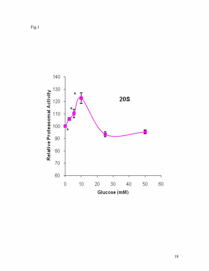
activities were analyzed. As shown in Fig. 1, the 20S proteasomal activity was glucose
concentration dependent in the hypoxic cells. In range of 0-10 mM, glucose increased
the proteasomal activity. When glucose concentration was greater than 10 mM, the
6
proteasomal activity decreased. The activity at 25 mM glucose was significant lower
than these at 0, 5.5, and 10 mM. In addition, it seemed that the activity increased again
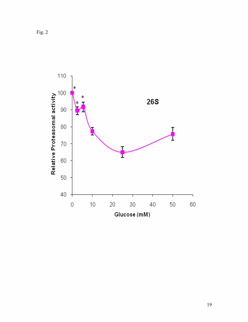
when the glucose concentration was greater than 25 mM. Fig. 2. shows the effect of
glucose on the 26S proteasomal activities in the hypoxic cells. Glucose at the range of 0-
25 mM primarily decreased the 26S activities concentration dependently. In the presence
of glucose concentration greater than 25 mM, the activity increased. The results revealed
that glucose had great effect on both 20S and 26S activities in the hypoxic cells and that
cells treated with glucose at 25 mM had the lowest activities of 20S and 26S
proteasomes.
To confirm that reactive oxygen species (ROS) was involved in the glucose-mediated
proteasomal activity changes, we carried out experiments to study the effect of ROS on
the activity of proteasomes. ROS refers to a group of oxygen centered reactive species.
Superoxide anion radical and H2O2 are two very important ROS in causing oxidative
damage and altering signal transduction under oxidative stress such as pathological
condition of ischemia. We tested the effects of superoxide and H2O2 on the activity of
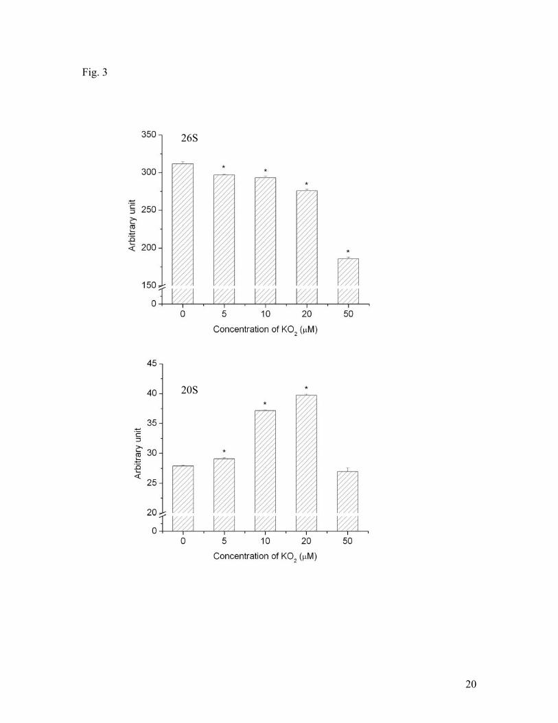
20S and 26S proteasomes in SH-SY5Y cells under hypoxic conditions. Fig. 3 shows that
superoxide decreased 26S activity under hypoxic conditions concentration-dependently.
In the presence of 50 μM KO2, the 26S activity decreased about 50%. In contrast, the
effect of superoxide on 20S activity was different from that of 26S activity. KO2
increased 20S activity in a concentration up to 20 μM. When KO2 concentration was
greater than 20 μM, it reduced 20S activity. KO2 at 50 μM only slightly reduced 20S
activity without significance, compared to the control group (in the absence of KO2).
7
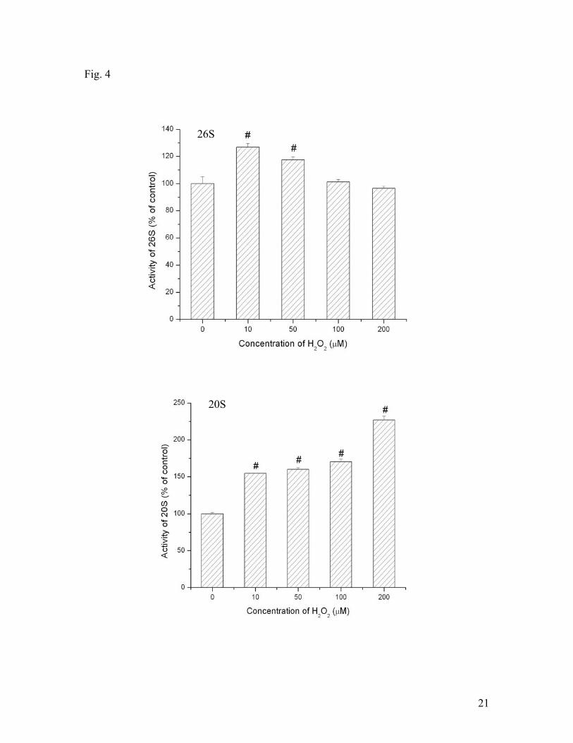
H2O2 also showed ability to affect 20S and 26S activities concentration dependently (Fig.
4). In a smaller dose (smaller than 50 μM), H2O2 elevated 26S activity. As the
concentration of H2O2 increases (greater than 50 μM), it seemed to inhibit the activity.
On the other hand, H2O2 elevated 20S activity during the range of 0-200 μM. These
results demonstrated that superoxide and H2O2 had great effect on 20S and 26S activities
in hypoxic SH-SY5Y cells although their effects were concentration dependent and
differential dependent on the species of proteasomes.
The above results indicated that ROS could increase or inhibit 20S and 26S activities in
hypoxic SH-SY5Y cells. However, there was no direct link that ROS was responsible for
glucose-mediated proteasomal activity changes in hypoxic cells. To further confirm that
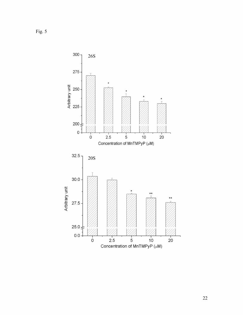
ROS was involved in proteasomal activity changes, we used specific antioxidants as ROS
scavenger to reduce the levels ROS in hypoxic cells. A membrane permeable SOD mimic
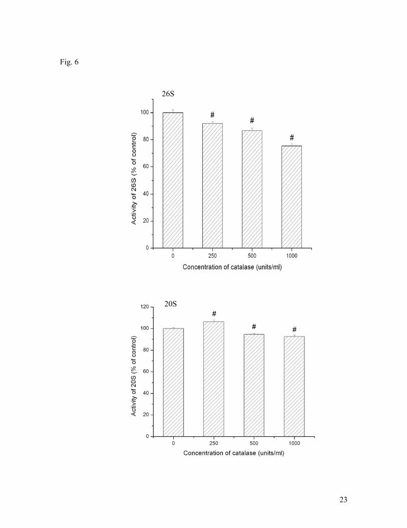
MnTmPyp was used to suppress O2•-. Catalase was used to suppress the generation of
H2O2. In these experiments, cells were pre-incubated with these antioxidants 30 min
before in vitro hypoxic treatments. Fig. 5 shows the effect of MnTmPyp on 26S and 20S
activities. In the concentration range of 0-20 μM , MnTmPyp decreased both 26S and
20S activities. Catalase also decreased the activity of 26S (Fig. 6). However, the effect
of catalase on 20S was a different from that on 26S. It increased the activity at 250
units/ml but decreased the activity when its concentration was greater than 250 units/ml.
These results confirmed that ROS were involved in the alteration of proteasomal
activities in ischemic SH-SY5Y cells.
8
Discussion
Proteasomes are large multicatalytic proteinase complexes in cytosol and nucleus of
eukaryotic cells. The proteasome complexes exist in two forms, 26S, which mainly
mediates the ubiquitin-dependent degradation of cellular proteins, and 20S, which is
ATP- and ubiquitin-independent. The two different complexes usually have different
protein substrate specificities. The 26S pathway is greatly dependent on the covalent
attachment of ubiquitin to proteins, which is known as ubiquitination, to form conjugates.
The ubiquitin-proteasome system has a central role in the selective degradation of
intracellular proteins, which are critical in regulating critical cell processes including
differentiation, cytokine-induced gene expression, apoptosis, and the stress response
(Brahimi-Horn and Pouyssegur, 2005; Ciechanover, 2006; Ding et al., 2006; Wilkinson,
1995). In addition, the ubiquitin-proteasome system component are among the most
abundant proteins of central nervous system. Cellular oxidized proteins are degraded
primarily through ubiquitin-independent 20S proteasome pathway (Ding et al., 2006).
Proteasome activities under ischemic condition have been related to activation of
proinflammatory protein through nuclear factor-kappa B (NF-kB) (Palombella et al.,
1994). The involvement of proteasome in the degradation of hypoxia-inducible factor 1
has been proved to be critical to the progression and extension of cerebral injuries during
stroke. More significantly, it has been reported that treatment with proteasome inhibitor
reduces effectively neuronal and astrocytic degeneration, cortical infarct volume, infarct
neutrophil infiltration (Wojcik and Di Napoli, 2004). Thus, proteasome inhibition has
been suggested as a potential treatment option for stroke (Phillips et al., 2000).
9
Glucose, an essential nutrient for brain function has been proved to play an important role
in ischemic injury. Both glucose deprivation and exposure to elevated glucose levels have
been shown to be pathogenic to cells. Besides as an energy source, glucose is also a
source providing reductants in cells and maintains cellular redox status. Glucose is a
main substrate to produce the principal intracellular reductant NADPH by the pentose
phosphate pathways (PPP) (Averill-Bates and Przybytkowski, 1994; Przybytkowski and
Averill-Bates, 1996). Glutathione (GSH) can be regenerated from glutathione disulfide
(GSSG) by glutathione reductase, which uses NADPH as an electron donor. The
pathways have been suggested as the major source of NADPH production for the
maintenance of GSH level, which maintains a cellular reduced milieu, in the brain (Ben-
Yoseph et al., 1994; Hotta, 1962; Hotta and Seventko, 1968). Consequently, it would be
expected that lack of glucose results in a condition of metabolic oxidative stress
characterized by increased pro-oxidant production and increased levels of GSSG because
of interfering with NADPH production by the PPP pathway. In fact, it has been
demonstrated that glucose greatly affect the ROS levels in hypoxic neurons (Shi, 2006).
The published results showed that the presence of glucose decreased the level of ROS
and cells incubated with glucose at 25 mM had the lowest levels of ROS.
ROS have been suggested to be able to affect both 26S and 20S proteasome pathways.
For example, O2•- may play a role like O2 in activating prolyl hydroxylase under hypoxia,
thus increasing the activity of ubiquitin-proteasomal pathways as a whole (Callapina et
al., 2005; Haddad, 2002), although the 26S may be vulnerable to ROS exposure. On the
other hand, ROS may facilitate protein degradation through 20S pathway because
10
oxidized proteins are preferred substrates of 20S proteasome (Grune et al., 1996, 1997;
Grune et al., 1995; Levine et al., 1996) and ROS rapidly upregulate 20S proteasome
activity (Ullrich et al., 1999). However, it is not completely known whether ROS affect
the proteasomal activities in cells under hypoxic conditions, especially in neuronal cell.
The results in this report clearly demonstrate that ROS affect the activity of proteasomal
activities in SH-SY5Y neuronal cells under hypoxic exposures. As shown in Fig. 1 and
2, cells treated hypoxia and glucose at 25 mM had the lowest 20S and 26S proteasomal
activities. As discussed previously, glucose at 25 mM induced the lowest level of ROS in
hypoxic neurons. This indicates that ROS may be was responsible for the elevation of
proteasomal activities in ischemic neurons (in the absence or low levels of glucose).
Indeed, our results from experiments with ROS and antioxidant treatments confirmed that
both superoxide and hydrogen peroxide could increase 20S and 26S activities in hypoxic
neurons although there was a concentration range for this specific effect.
Cellular injury induced by both low and high glucose has been linked to the generation of
reactive oxygen species (ROS) and increased oxidative stress. It has been reported that
glucose deprivation causes perturbations in cellular sensitivity to oxidative stress, which
is mediated by increased generation of pro-oxidants and decreased scavenging of free
radicals. Formation of hydrogen peroxide (Ho et al., 2000) as well as more general
measurements of ROS generation (Sharpe et al., 1998) have been observed under high
glucose conditions and are suggested to be responsible for high glucose-induced cell
injury and/or death in non-neuronal cells. The previous study in Dr. Shi’s lab has proven
that the optimal glucose concentration increase cellular GSH/GSSG ratio which is the
11
main criteria of cellular redox status in neuronal cell. The study of the effect of glucose
on proteasome activities will not only help us to understand the mechanism of ROS on
ischemic injury but also illustrate the interaction of nutrient and oxygen under ischemia
condition.
12
Reference:
Astrup, J., Siesjo, B.K., and Symon, L. (1981). Thresholds in cerebral ischemia - the
ischemic penumbra. Stroke 12, 723-725.
Averill-Bates, D.A., and Przybytkowski, E. (1994). The role of glucose in cellular
defences against cytotoxicity of hydrogen peroxide in Chinese hamster ovary cells. Arch.
Biochem. Biophys. 312, 52-58.
Ben-Yoseph, O., Boxer, P.A., and Ross, B.D. (1994). Oxidative stress in the central
nervous system: monitoring the metabolic response using the pentose phosphate pathway.
Dev. Neurosci. 16, 328-336.
Brahimi-Horn, C., and Pouyssegur, J. (2005). When hypoxia signalling meets the
ubiquitin-proteasomal pathway, new targets for cancer therapy. Crit. Rev. Oncol.
Hematol. 53, 115-123.
Callapina, M., Zhou, J., Schmid, T., Kohl, R., and Brune, B. (2005). NO restores HIF-
1alpha hydroxylation during hypoxia: role of reactive oxygen species. Free Radic. Biol.
Med. 39, 925-936.
Ciechanover, A. (2006). The ubiquitin proteolytic system: from a vague idea, through
basic mechanisms, and onto human diseases and drug targeting. Neurology 66, S7-19.
Ding, Q., Dimayuga, E., and Keller, J.N. (2006). Proteasome regulation of oxidative
stress in aging and age-related diseases of the CNS. Antioxid. Redox Signal 8, 163-172.
Fekete, M.R., McBride, W.H., and Pajonk, F. (2005). Anthracyclines, proteasome
activity and multi-drug-resistance. BMC Cancer 5, 114.
13
Furlan, M., Marchal, G., Viader, F., Derlon, J.M., and Baron, J.C. (1996). Spontaneous
neurological recovery after stroke and the fate of the ischemic penumbra. Ann. Neurol.
40, 216-226.
Goldberg, M.P., and Choi, D.W. (1993). Combined oxygen and glucose deprivation in
cortical cell culture: calcium-dependent and calcium-independent mechanisms of
neuronal injury. J. Neurosci. 13, 3510-3524.
Grune, T., Reinheckel, T., and Davies, K.J. (1996). Degradation of oxidized proteins in
K562 human hematopoietic cells by proteasome. J. Biol. Chem. 271, 15504-15509.
Grune, T., Reinheckel, T., and Davies, K.J. (1997). Degradation of oxidized proteins in
mammalian cells. FASEB J. 11, 526-534.
Grune, T., Reinheckel, T., Joshi, M., and Davies, K.J. (1995). Proteolysis in cultured liver
epithelial cells during oxidative stress. Role of the multicatalytic proteinase complex,
proteasome. J. Biol. Chem. 270, 2344-2351.
Haddad, J.J. (2002). Antioxidant and prooxidant mechanisms in the regulation of
redox(y)-sensitive transcription factors. Cell Signal. 14, 879-897.
Ho, F.M., Liu, S.H., Liau, C.S., Huang, P.J., and Lin-Shiau, S.Y. (2000). High glucose-
induced apoptosis in human endothelial cells is mediated by sequential activations of c-
Jun NH(2)-terminal kinase and caspase-3. Circulation 101, 2618-2624.
Hotta, S.S. (1962). Glucose metabolism in brain tissue: the hexosemonophosphate shunt
and its role in glutathione reduction. J. Neurochem. 9, 43-51.
Hotta, S.S., and Seventko, J.M., Jr. (1968). The hexosemonophosphate shunt and
glutathione reduction in guinea pig brain tissue: changes caused by chlorpromazine,
amytal, and malonate. Arch. Biochem. Biophys. 123, 104-108.
14
Levine, R.L., Mosoni, L., Berlett, B.S., and Stadtman, E.R. (1996). Methionine residues
as endogenous antioxidants in proteins. Proc. Natl. Acad. Sci. USA 93, 15036-15040.
Levison, S.W., Ducceschi, M.H., Young, G.M., and Wood, T.L. (1996). Acute exposure
to CNTF in vivo induces multiple components of reactive gliosis. Exp. Neurol. 141, 256-
268.
Liu, S., Liu, M., Peterson, S., Miyake, M., Vallyathan, V., and Liu, K.J. (2003). Hydroxyl
radical formation is greater in striatal core than in penumbra in a rat model of ischemic
stroke. J. Neurosci. Res. 71, 882-888.
Palombella, V.J., Rando, O.J., Goldberg, A.L., and Maniatis, T. (1994). The ubiquitin-
proteasome pathway is required for processing the NF-kappa B1 precursor protein and
the activation of NF-kappa B. Cell 78, 773-785.
Phillips, J.B., Williams, A.J., Adams, J., Elliott, P.J., and Tortella, F.C. (2000).
Proteasome inhibitor PS519 reduces infarction and attenuates leukocyte infiltration in a
rat model of focal cerebral ischemia. Stroke 31, 1686-1693.
Przybytkowski, E., and Averill-Bates, D.A. (1996). Correlation between glutathione and
stimulation of the pentose phosphate cycle in situ in Chinese hamster ovary cells exposed
to hydrogen peroxide. Arch. Biochem. Biophys. 325, 91-98.
Sharpe, P.C., Liu, W.H., Yue, K.K., McMaster, D., Catherwood, M.A., McGinty, A.M.,
and Trimble, E.R. (1998). Glucose-induced oxidative stress in vascular contractile cells:
comparison of aortic smooth muscle cells and retinal pericytes. Diabetes 47, 801-809.
Shi, H., and Liu, K. (2006). Effects of glucose concentration on redox status in rat
primary cortical neurons under hypoxia. Neurosci. Lett. 410, 57-61.
15
Ullrich, O., Reinheckel, T., Sitte, N., Hass, R., Grune, T., and Davies, K.J. (1999). Poly-
ADP ribose polymerase activates nuclear proteasome to degrade oxidatively damaged
histones. Proc Natl Acad Sci USA 96, 6223-6228.
Wilkinson, K.D. (1995). Roles of ubiquitinylation in proteolysis and cellular regulation.
Annu Rev Nutr 15, 161-189.
Wojcik, C., and Di Napoli, M. (2004). Ubiquitin-proteasome system and proteasome
inhibition: new strategies in stroke therapy. Stroke 35, 1506-1518.
16
Figure Legend
Fig. 1. Effect of glucose on 20S proteasomal activity under hypoxic conditions. SH-
SY5Y cells were exposed to hypoxia (1% O2) 3 hrs. The proteasomal activities were
expressed as the percentage of the value obtained in the presence of 0 mM glucose. Data
are shown as means ± SE, n=8. *p<0.05, compared to glucose at 25 mM.
Fig. 2. Effect of glucose on 26S proteasomal activity under hypoxic conditions. SH-
SY5Y cells were exposed to hypoxia (1% O2) 3 hrs. The proteasomal activities were
expressed as the percentage of the value obtained in the presence of 0 mM glucose. Data
are shown as means ± SE, n=8. *p<0.05, compared to glucose at 25 mM.
Fig. 3. Effects of superoxide anion radical on 20S and 26S proteasomal activities under
hypoxic conditions. KO2 was used as a source of superoxide anion radicals. SH-SY5Y
cells were treated with different concentrations of KO2 and hypoxia for 3 hrs. Data are
shown as means ± SE, n=3-4. *p<0.05, compared to the group without KO2 treatment.
Fig. 4. Effects of hydrogen peroxide on 20S and 26S proteasomal activities under
hypoxic conditions. SH-SY5Y cells were treated with different concentrations of H2O2
and hypoxia for 3 hrs. Data are shown as means ± SE, n=3-4. *p<0.05, compared to the
group without H2O2 treatment.
Fig. 5. Effects of antioxidant MnTMPyP on 20S and 26S proteasomal activities under
hypoxic conditions. SH-SY5Y cells were treated with different concentrations of
17
MnTMPyP and hypoxia for 3 hrs. Data are shown as means ± SE, n=3-4. *p<0.05,
compared to the group without MnTMPyP treatment.3
Fig. 6. Effects of antioxidant catalase on 20S and 26S proteasomal activities under
hypoxic conditions. SH-SY5Y cells were treated with different concentrations of
catalase and hypoxia for 3 hrs. Data are shown as means ± SE, n=3-4. *p<0.05,
compared to the group without catalase treatment.
18
Fig.1
19
Fig. 2
20
Fig. 3
26S
20S
21
Fig. 4
26S
20S
22
Fig. 5
26S
20S
23
Fig. 6
26S
20S


