Embed Size (px)
Citation preview
1999 5: 646-655RNA M Sohail, S Akhtar and E M Southern complementary oligonucleotides.The folding of large RNAs studied by hybridization to arrays of
References http://rnajournal.cshlp.org/content/5/5/646#related-urls
Article cited in:
serviceEmail alerting
click heretop right corner of the article orReceive free email alerts when new articles cite this article - sign up in the box at the
http://rnajournal.cshlp.org/subscriptions go to: RNATo subscribe to
© 1999 RNA Society
Cold Spring Harbor Laboratory Press on July 15, 2011 - Published by rnajournal.cshlp.orgDownloaded from
The folding of large RNAs studiedby hybridization to arrays ofcomplementary oligonucleotides
MUHAMMAD SOHAIL, 1 SAGHIR AKHTAR, 2 and EDWIN M. SOUTHERN 1
1Department of Biochemistry, University of Oxford, South Parks Road, Oxford, OX1 3QU, England, United Kingdom2Department of Pharmaceutical and Biological Sciences, Aston University, Aston Triangle,Birmingham B4 7ET, England, United Kingdom
ABSTRACT
Folding pathways of large RNAs are poorly understood. We have addressed this question by hybridizing in vitrotranscripts, which varied in size, to an array of antisense oligonucleotides. All transcripts included a commonsequence and all but one shared the same start-point; the other had a small deletion of the 5 9 end. Minimal free energycalculations predicted quite different folds for these transcripts. However, hybridization to the array showed predom-inant features that were shared by transcripts of all lengths, though some oligonucleotides that hybridized stronglyto the short transcripts gave weak interaction with longer transcripts. A full-length RNA fragment that had beendenatured by heating and allowed to cool slowly gave the same hybridization result as a shorter transcript. Takentogether, these results support theories that RNA folding creates local stable states that are trapped early in thetranscription or folding process. As the transcript elongates, interactions are added between regions that are tran-scribed early and those transcribed late. The method here described helps in identifying regions in the transcripts thattake part in long-range interactions.
Keywords: antisense oligonucleotides; mfold; RNA folding; scanning arrays
INTRODUCTION
Secondary folding of an RNA is a major determinant ofits higher order conformation and its interaction with avariety of other molecules; it is important to structure–function relationship (Brion & Westhof, 1997)+ Thereare two conflicting views on the secondary folding oflarge RNAs into their final biologically active state+ Thegenerally held view is that most secondary interactionsin large RNAs are local (e+g+, Moras, 1997)+ It is as-sumed that local folding is established as the moleculeis transcribed, and some early studies showed sequen-tial folding of small RNAs (Boyle et al+, 1980; Kramer &Mills, 1981), but there is no similar evidence for largeRNAs+ The alternative view is that RNAs exist in aglobal minimal free energy state that would have to beestablished after synthesis was complete+ Though theirlimitations are well known, researchers tend to use theenergy minimization computer programs (e+g+, mfold;Zuker, 1989) because they can be easily applied to
molecules up to 2–3 kb+ Such predictions allow forinteractions between distant sequences giving no pref-erence to any local folding that may have occurredduring synthesis+While the ability of these programs topredict correct secondary structure of large RNAs isdisputed by some (Milner et al+, 1997; Schuster et al+,1997; Ho et al+, 1998) others believe them to be mostlycorrect in their predictions (Tinoco & Kieft, 1997)+
The uncertainty and difficulty of predicting secondarystructure has led to the use of empirical methods todeduce information about complex RNAs+ The tertiarystructures of several small- and medium-sized RNAshave been resolved to atomic level by X-ray crystal-lography (Kim et al+, 1974; Robertus et al+, 1974; Scottet al+, 1995; Cate et al+, 1996)+ However, large RNAs(e+g+, mRNAs) are difficult to crystallize (Moras, 1997)and thus cannot be analyzed by this method+ Ap-proaches to determine secondary structure of RNAsinclude biochemical and chemical fingerprinting (e+g+,Lauber et al+, 1997) and phylogenetic analysis of largegroups of functional RNAs (Michel & Westhof, 1990)+The biochemical and chemical cleavage methods,whichidentify single- and double-stranded regions, generatelimited information and are more suited to small RNAs+
Reprint requests to: Muhammad Sohail, Department of Biochem-istry, University of Oxford, South Parks Road, Oxford, OX1 3QU,England, United Kingdom; e-mail: msohail@bioch+ox+ac+uk+
RNA (1999), 5:646–655+ Cambridge University Press+ Printed in the USA+Copyright © 1999 RNA Society+
646
Cold Spring Harbor Laboratory Press on July 15, 2011 - Published by rnajournal.cshlp.orgDownloaded from
The phylogenetic approach, which assumes that se-quences with similar biological function acquire homol-ogous secondary structure has proved to be the mostsuccessful among predictive methods for deriving RNAsecondary structure+However, it cannot be applied whenthe number of sequences is small and is unlikely to beuseful if the folded structure has no functional signifi-cance, as is likely to be the case for most mRNAs+Therefore, there is a constant need to develop newapproaches to analyze the structure of large RNAs atall levels+
We describe an analysis of the folding pathway of anmRNA by hybridizing transcripts of increasing length toan array of complementary oligonucleotides,which sug-gests that much of the local folding established early intranscription persists, but is detectably modified by in-teractions with regions transcribed later+
RESULTS
Hybridization of transcripts of variouslengths to the scanning array
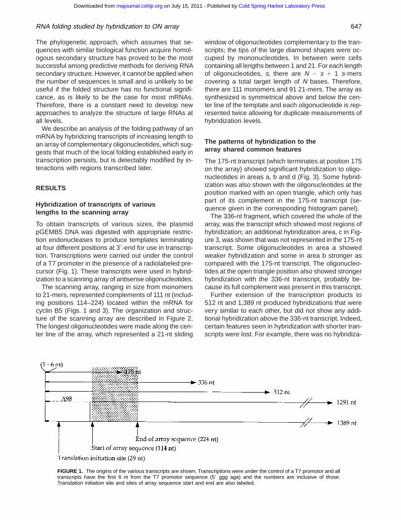
To obtain transcripts of various sizes, the plasmidpGEMB5 DNA was digested with appropriate restric-tion endonucleases to produce templates terminatingat four different positions at 39-end for use in transcrip-tion+ Transcriptions were carried out under the controlof a T7 promoter in the presence of a radiolabeled pre-cursor (Fig+ 1)+ These transcripts were used in hybrid-ization to a scanning array of antisense oligonucleotides+
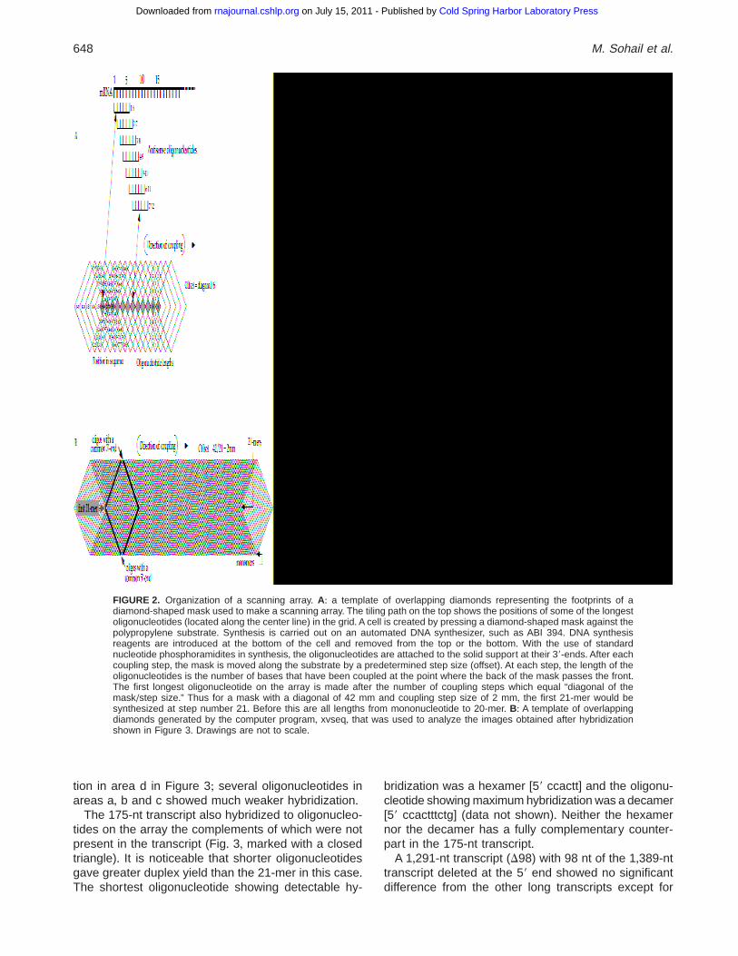
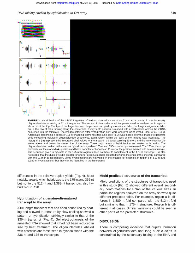
The scanning array, ranging in size from monomersto 21-mers, represented complements of 111 nt (includ-ing positions 114–224) located within the mRNA forcyclin B5 (Figs+ 1 and 3)+ The organization and struc-ture of the scanning array are described in Figure 2+The longest oligonucleotides were made along the cen-ter line of the array, which represented a 21-nt sliding
window of oligonucleotides complementary to the tran-scripts; the tips of the large diamond shapes were oc-cupied by mononucleotides+ In between were cellscontaining all lengths between 1 and 21+ For each lengthof oligonucleotides, s, there are N 2 s 1 1 s-merscovering a total target length of N bases+ Therefore,there are 111 monomers and 91 21-mers+ The array assynthesized is symmetrical above and below the cen-ter line of the template and each oligonucleotide is rep-resented twice allowing for duplicate measurements ofhybridization levels+
The patterns of hybridization to thearray shared common features
The 175-nt transcript (which terminates at position 175on the array) showed significant hybridization to oligo-nucleotides in areas a, b and d (Fig+ 3)+ Some hybrid-ization was also shown with the oligonucleotides at theposition marked with an open triangle, which only haspart of its complement in the 175-nt transcript (se-quence given in the corresponding histogram panel)+
The 336-nt fragment, which covered the whole of thearray, was the transcript which showed most regions ofhybridization; an additional hybridization area, c in Fig-ure 3,was shown that was not represented in the 175-nttranscript+ Some oligonucleotides in area a showedweaker hybridization and some in area b stronger ascompared with the 175-nt transcript+ The oligonucleo-tides at the open triangle position also showed strongerhybridization with the 336-nt transcript, probably be-cause its full complement was present in this transcript+
Further extension of the transcription products to512 nt and 1,389 nt produced hybridizations that werevery similar to each other, but did not show any addi-tional hybridization above the 336-nt transcript+ Indeed,certain features seen in hybridization with shorter tran-scripts were lost+ For example, there was no hybridiza-
FIGURE 1. The origins of the various transcripts are shown+ Transcriptions were under the control of a T7 promotor and alltranscripts have the first 6 nt from the T7 promotor sequence (59 ggg aga) and the numbers are inclusive of those+Translation initiation site and sites of array sequence start and end are also labeled+
RNA folding studied by hybridization to ON array 647
Cold Spring Harbor Laboratory Press on July 15, 2011 - Published by rnajournal.cshlp.orgDownloaded from
tion in area d in Figure 3; several oligonucleotides inareas a, b and c showed much weaker hybridization+
The 175-nt transcript also hybridized to oligonucleo-tides on the array the complements of which were notpresent in the transcript (Fig+ 3, marked with a closedtriangle)+ It is noticeable that shorter oligonucleotidesgave greater duplex yield than the 21-mer in this case+The shortest oligonucleotide showing detectable hy-
bridization was a hexamer [59 ccactt] and the oligonu-cleotide showing maximum hybridization was a decamer[59 ccactttctg] (data not shown)+ Neither the hexamernor the decamer has a fully complementary counter-part in the 175-nt transcript+
A 1,291-nt transcript (D98) with 98 nt of the 1,389-nttranscript deleted at the 59 end showed no significantdifference from the other long transcripts except for
FIGURE 2. Organization of a scanning array+ A: a template of overlapping diamonds representing the footprints of adiamond-shaped mask used to make a scanning array+ The tiling path on the top shows the positions of some of the longestoligonucleotides (located along the center line) in the grid+A cell is created by pressing a diamond-shaped mask against thepolypropylene substrate+ Synthesis is carried out on an automated DNA synthesizer, such as ABI 394+ DNA synthesisreagents are introduced at the bottom of the cell and removed from the top or the bottom+ With the use of standardnucleotide phosphoramidites in synthesis, the oligonucleotides are attached to the solid support at their 39-ends+ After eachcoupling step, the mask is moved along the substrate by a predetermined step size (offset)+ At each step, the length of theoligonucleotides is the number of bases that have been coupled at the point where the back of the mask passes the front+The first longest oligonucleotide on the array is made after the number of coupling steps which equal “diagonal of themask/step size+” Thus for a mask with a diagonal of 42 mm and coupling step size of 2 mm, the first 21-mer would besynthesized at step number 21+ Before this are all lengths from mononucleotide to 20-mer+ B: A template of overlappingdiamonds generated by the computer program, xvseq, that was used to analyze the images obtained after hybridizationshown in Figure 3+ Drawings are not to scale+
648 M. Sohail et al.
Cold Spring Harbor Laboratory Press on July 15, 2011 - Published by rnajournal.cshlp.orgDownloaded from
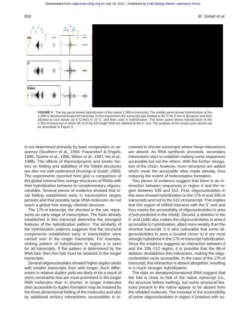
differences in the relative duplex yields (Fig+ 4)+ Mostnotably, area d,which hybridizes to the 175-nt and 336-ntbut not to the 512-nt and 1,389-nt transcripts, also hy-bridized to D98+
Hybridization of a denatured/renaturedtranscript to the array
A full length transcript that had been denatured by heat-ing and allowed to renature by slow cooling showed apattern of hybridization strikingly similar to that of the336-nt transcript (Fig+ 4)+ Gel electrophoresis of theannealed RNA showed that it had not been reduced insize by heat treatment+ The oligonucleotides labeledwith asterisks are those seen in hybridizations with the336-nt and 175-nt transcripts+
Mfold-predicted structures of the transcripts
Mfold predictions of the structures of transcripts usedin this study (Fig+ 5) showed different overall second-ary conformations for RNAs of the various sizes+ Inparticular, regions analyzed on the array showed quitedifferent predicted folds+ For example, region a is dif-ferent in 1,389-nt fold compared with the 512-nt foldbut similar to that in 175-nt structure+ Region b is dif-ferent in all cases+ Similar variations could be seen inother parts of the predicted structures+
DISCUSSION
There is compelling evidence that duplex formationbetween oligonucleotides and long nucleic acids isconstrained by the secondary folding of the RNA and
FIGURE 3. Hybridization of the mRNA fragments of various sizes with a common 59 end to an array of complementaryoligonucleotides scanning a 111-nt sequence+ The series of diamond-shaped templates used to analyze the images isshown in at the top+ The tips of the large diamond shapes are occupied by mononucleotides; the longest oligonucleotidesare in the row of cells running along the center line+ Every tenth position is marked with a vertical line across the mRNAsequence into the template+ The images obtained after hybridization (left) were analyzed using xvseq (Elder et al+, 1999)+A template comprising a series of 111 overlapping diamonds (top; also see Fig+ 2) was placed over the images to generatecells containing individual oligonucleotide sequences+ Each region within the cells of the images was integrated+ Thehistograms (right) present the integrated pixel values for the areas on the array carrying 21-mers and the two values for theareas above and below the center line of the array+ Three major areas of hybridization are marked a, b, and c+ Theoligonucleotides marked with asterisks hybridized only when 175-nt and 336-nt transcripts were used+ The 175-nt transcriptterminates at the marked (☛) position and has a complement of only an 11-mer at the position marked with an open triangle+The sequence given in brackets in the 175-nt histograms does not have its complement in the 175-nt transcript+ It is alsonoticeable that the duplex yield is greater for shorter oligonucleotides (situated towards the ends of the chevron) comparedwith the 21-mer at this position+ Some hybridizations are not visible in the images (for example, in region c of 512-nt and1,389-nt hybridizations) but they can be identified in the histograms+
RNA folding studied by hybridization to ON array 649
Cold Spring Harbor Laboratory Press on July 15, 2011 - Published by rnajournal.cshlp.orgDownloaded from
is not determined primarily by base composition or se-quence (Southern et al+, 1994; Frauendorf & Engels,1996; Toulme et al+, 1996; Milner et al+, 1997; Ho et al+,1998)+ The effects of thermodynamic and kinetic fac-tors on folding and stabilities of the folded structuresare also not well understood (Konings & Gutell, 1995)+The experiments reported here give a comparison ofthe global minimal free energy structures of RNAs withtheir hybridization behavior to complementary oligonu-cleotides+ Several pieces of evidence showed that lo-cal folding established early in transcription largelypersists and that possibly large RNA molecules do notreach a global free energy minimal structure+
The 175-nt transcript, the shortest in the set, repre-sents an early stage of transcription+ The folds alreadyestablished in this transcript determine the strongestfeatures of the hybridization pattern+ The similarity ofthe hybridization patterns suggests that the structuralcomponents established early in transcription werecarried over in the longer transcripts+ For example,striking pattern of hybridization in region a is seenfor all transcripts+ If the pattern is determined by theRNA fold, then this fold must be retained in the longertranscripts+
Several oligonucleotides showed higher duplex yieldswith smaller transcripts than with longer+ Such differ-ences in relative duplex yield are likely to be a result ofsteric constraints that are more prominent in the longerRNA molecules than in shorter+ In longer moleculessites accessible to duplex formation may be masked bythe three-dimensional folding of the molecules and alsoby additional tertiary interactions; accessibility is in-
creased in shorter transcripts where these interactionsare absent+ As RNA synthesis proceeds, secondaryinteractions start to establish making some sequencesaccessible but not the others+ With the further elonga-tion of the chain, however, more structures are addedwhich mask the accessible sites made already, thusreducing the extent of heteroduplex formation+
Two pieces of evidence suggest that there is an in-teraction between sequences in region d and the re-gion between 336 and 512+ First, oligonucleotides inthis area showed hybridization to the 175-nt and 336-nttranscripts and not to the 512-nt transcript+ This impliesthat this region of mRNA interacts with the 59 end andthus masks the accessibility of oligonucleotides in aread (not predicted in the mfold)+ Second, a deletion in the59 end (D98) also makes the oligonucleotides in area daccessible to hybridization, albeit more weakly than theshortest transcript+ It is also noticeable that some oli-gonucleotides in area a located closer to d are moststrongly hybridized in the 175-nt transcript hybridization+Since the evidence suggests an interaction between dand the 336–512 region, it is possible that the 98-ntdeletion destabilizes this interaction, making the oligo-nucleotides more accessible+ In the case of the 175-nttranscript, this interaction is absent altogether, resultingin a much stronger hybridization+
The data on denatured/renatured RNA suggest thatthe fold is close to that of the native transcript (i+e+,the structure before melting), but some structural fea-tures present in the native appear to be absent fromthe refolded molecule+ The increase in the accessibilityof some oligonucleotides in region d (marked with as-
FIGURE 4. The top panel shows hybridization of the native 1,389-nt transcript+ The middle panel shows hybridization of the1,389-nt denatured/renatured transcript+ In this experiment the transcript was heated to 85 8C for 5 min to denature and thenallowed to cool slowly (at 6 8C/min) to 22 8C+ and then used in hybridization+ The lower panel shows hybridization of the1,291-nt transcript in which 98 nt of the full length RNA are deleted at the 59 end+ The analysis of the arrays was carried outas described in Figure 2+
650 M. Sohail et al.
Cold Spring Harbor Laboratory Press on July 15, 2011 - Published by rnajournal.cshlp.orgDownloaded from
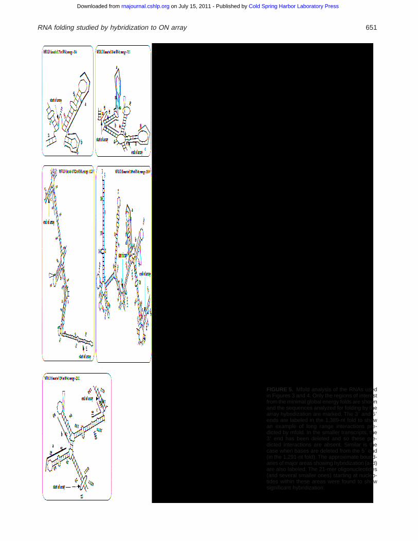
FIGURE 5. Mfold analysis of the RNAs usedin Figures 3 and 4+ Only the regions of interestfrom the minimal global energy folds are shownand the sequences analyzed for folding by thearray hybridization are marked+ The 39 and 59ends are labeled in the 1,389-nt fold to showan example of long range interactions pre-dicted by mfold+ In the smaller transcripts, the39 end has been deleted and so these pre-dicted interactions are absent+ Similar is thecase when bases are deleted from the 59 end(in the 1,291-nt fold)+ The approximate bound-aries of major areas showing hybridization (a–d)are also labeled+ The 21-mer oligonucleotides(and several smaller ones) starting at nucleo-tides within these areas were found to showsignificant hybridization+
RNA folding studied by hybridization to ON array 651
Cold Spring Harbor Laboratory Press on July 15, 2011 - Published by rnajournal.cshlp.orgDownloaded from
terisks), which is most likely to interact with a distantregion between nucleotide positions 336 and 512, in-dicates that their native interaction failed to reestablishduring renaturation+ It is thus likely that short-rangeinteractions are established first before procession todistant interactions (also see Zarrinker & Williamson,1994; Batey & Doudna, 1998)+
The 175-nt transcript hybridized to oligonucleotideswhose complement was absent from the transcript+ Al-though, it is not possible to predict which duplexes resultfrom Watson–Crick pairing, this particular hybridizationis more likely to have resulted from a non-Watson–Crick interaction between RNA and the antisense oli-gonucleotides+Another example of a shorter sequence(a dinucleotide, gg) producing stronger hybridization toa complementary target than many fully complemen-tary oligonucleotides has also been reported (South-ern et al+, 1994)+ These types of interactions can be aproblem in methods which rely on sequence-specifichybridization (e+g+, Wodicka et al+, 1997; Drmanacet al+, 1998; de Saizieu et al+, 1998)+ Several proce-dures that involve the use of fragmented RNA (e+g+, fortranscript imaging: Wodicka et al+, 1997; de Saizieuet al+, 1998) can also produce nonspecific hetero-duplexes+ Such interactions appear to be rare (onlyone example could be detected in this study) and dif-ficult to identify in these protocols; the data thus ob-tained must be analyzed with caution+
Mfold predicted structures and implicationof the hybridization results
The large differences seen in the mfold-predicted struc-tures would be expected to have profound effects onthe accessibility to duplex formation by oligonucleo-tides and lead to qualitatively different patterns ratherthan the variation in duplex yield observed+
Several thermodynamic-based methods were de-vised in the 1980s to predict the secondary structure ofRNAs (e+g+,Nussinov & Jacobson, 1980;Dumas & Ninio,1982) and were improved subsequently+ These meth-ods were mostly based on the known structure of tRNAsand thermodynamic studies of small RNA fragments insolution (e+g+, Tinoco et al+, 1973)+ tRNAs are compar-atively small and probably exist in the minimal globalfree energy state and so their secondary structure canbe accurately predicted by computational methods+ Thealgorithms devised by Zuker and colleagues (e+g+, Zuker,1989) became widely used (e+g+, mfold) because oftheir ability to calculate the free energy of folds of lon-ger sequences+
For long RNA molecules, energy calculations oftenreturn a number of different structures with similar freeenergies: there is difficulty in choosing the correct one+We analyzed the full length B5 transcript to see if thefree energy of the most stable fold differed significantly
from that of the fold in which the 59 end was con-strained to its most stable fold+ The minimum free en-ergy of the full transcript was 2250+9 kcal/mol and thesum of the free energies of the two parts folded sep-arately was 2248+2 kcal/mol, showing that there waslittle difference between them+ This result reempha-sizes the problem of using global free energy calcula-tions+ Our results further indicate that the predictedstructures have limited usage in biological applicationsuch as designing antisense oligonucleotides+ Ho et al+(1998) have also shown that accessible sites cannotbe mapped on predicted structures and that there is noobvious structural difference between accessible andinaccessible regions on the computer folded struc-tures+ Gaspin & Westhof (1995) have devised an ap-proach according to the RNA hierarchical folding viewthat allows for the dynamic incorporation of folding con-straints, enabling the user to participate in the compu-tational folding of RNA+ They predicted the secondarystructure of Group I intron Td and RNAseP of Esche-richia coli with this method, and the results were similarto those predicted by phylogenetic comparison+Despiteits demonstrated usefulness, such an approach re-quires substantial experimental data for input that maynot be available for some biological applications, inwhich case empirical approaches become more useful+
Our results suggest ways in which hybridization toarrays may be used in predicting the secondary inter-actions in RNA molecules+ First, the hybridization datacan help in testing short range interactions+ Studies oftRNA (K+U+ Mir & E+M+ Southern, submitted) suggestthat strong interactions of oligonucleotides with the tar-get, which are easily identified on the arrays, indicatecertain stem-loop structures, and may point to stackinterfaces+ The 59 regions of B5 mRNA that hybridizestrongly to oligonucleotides, shown against the pre-dicted secondary structure of lowest free energy(Fig+ 6), conform with the partial rules derived from theanalysis of tRNA+ Second, long-range interactions areindicated by the loss of hybridization that results fromextending the transcript+ The sequences of the oligo-nucleotides whose hybridization is blocked by the sec-ondary interactions can be read directly from the arrayto locate the interacting regions precisely+
Analysis of the shortest sequences within the 21-mers showing detectable hybridization revealed that inmost cases the optimal binding was achieved by se-quences shorter than 21 nt+ Further addition of bases(at 39 end) produced only a small increase in their bind-ing, as indicated by the hybridization intensity values+For example in the case of the 21-mer oligonucleotide1, optimal binding was achieved by a 14-mer and fur-ther addition of bases at its 39 end produced only asmall increase in its binding affinity+ It is striking thatseveral oligonucleotides showing optimal hybridizationwithin each area (a, b, or c) terminate at a commonbase at their 39 end (e+g+, 1 and 2 and 3, 4, and 5)+
652 M. Sohail et al.
Cold Spring Harbor Laboratory Press on July 15, 2011 - Published by rnajournal.cshlp.orgDownloaded from
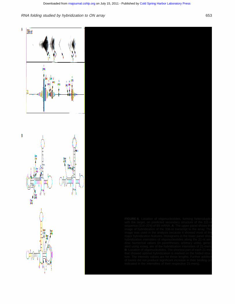
FIGURE 6. Location of oligonucleotides, forming heteroduplexwith the target, on predicted secondary structure of the 111-ntsequence (114–224) of B5 mRNA+ A: The upper panel shows theimage of hybridization of the 336-nt transcript to the array+ Thisimage was used in the analysis because it showed most of themajor hybridization features+ Histograms in the lower panel showhybridization intensities of oligonucleotides along the 21-nt win-dow+ Numerical values (in parentheses; arbitrary units), gener-ated using xvseq, are of the hybridization intensities of 21-mers+B: Location of oligonucleotides+ The shortest part of each 21-merthat showed optimal hybridization is marked on the folded struc-ture+ The intensity values are for these lengths+ Further additionof bases did not produce significant increase in their binding (asindicated in the intensities of their respective 21-mers)+
RNA folding studied by hybridization to ON array 653
Cold Spring Harbor Laboratory Press on July 15, 2011 - Published by rnajournal.cshlp.orgDownloaded from
MATERIALS AND METHODS
Synthesis of primers and DNA amplification
PCR primers were synthesized on an Applied Biosystem’s392 DNA/RNA synthesizer+The primers D98 (59 taatacgactcactatagggagaaagtacatggacatagacc) and 3END (59 cccgggcgagctcggaa) were used on the plasmid pGEMB5 (carrying genefor Xenopus laevis Cyclin B5; a gift from Dr+ Tim Hunt) toobtain a PCR product with a deletion of 98 bp at the 59 endof the full-length template for use in in vitro transcription+ DNAamplification was carried out in a PTC-200 DNA Engine (MJResearch) using 30 cycles of 94 8C for 1 min, 59 8C for 1 minand 73 8C for 2 min with a final extension step of 10 min at73 8C+Amplitaq DNA polymerase (Perkin-Elmer) with the rec-ommended buffer was used in amplification+ The reactionswere purified using Qiagen PCR Purification Kit (Qiagen) ac-cording to the manufacturer’s instructions+
Preparation of labeled RNA transcripts
Plasmid pGEMB5 was used to obtain full length B5 tran-scripts and was digested with restriction endonucleases atappropriate sites to obtain DNA templates for use in tran-scription to obtain transcripts of 175-nt, 336-nt, 512-nt, and1,389-nt lengths+ A transcript with a 98-nt deletion at the 59end was obtained from the PCR product obtained with prim-ers D98 (which has the T7 promotor at its 59 end) and 3END+In vitro transcriptions were carried out with T7 RNA polymer-ase (Promega) using standard protocol in the presence of[a-32P] UTP (Amersham; 3,000 Ci/mmol)+ The reactions wereat 30 8C for 90 min and desalted by Sephadex-G25 (Phar-macia) column chromatography+ Transcripts were analyzedon a 5% denaturing polyacrylamide gel+
Fabrication of arrays
The oligonucleotide array (complement of 111 nt includingpositions 114–224 in the Cyclin B5 mRNA) was prepareddirectly onto the surface of aminated polypropylene (Matsonet al+, 1994) on an adapted Applied Biosystem’s DNA syn-thesizer (ABI 381 A) using a 42-mm diamond-shaped maskas described previously (Southern et al+, 1994)+ Standardphosphoramidites were used in the synthesis+ The offset be-tween each coupling step was 2 mm and thus the longestoligonucleotides produced were 21-mers+ The arrays weredeprotected in 30% ammonia solution at 55 8C for 16 h in aclosed chamber (Southern et al+, 1994)+
Hybridization conditions and analysis
Hybridizations were performed in 1 M NaCl in the the pres-ence of 10 mM Tris, pH 7+4, 1 mM EDTA, and 0+01% SDS at22 8C for 3 h+Approximately, 50–60 fmol of the transcript wasdiluted in 12 mL hybridization solution and hybridizations werecarried out in a rolling hybridization chamber at 3–4 rpm+ Thearray was then washed in the hybridization solution brieflyand dried in layers of Wattman paper+ Autoradiography andanalysis were carried out essentially as described in Elderet al+ (1999)+ The array was stripped in 100 mM sodium
carbonate/bicarbonate buffer (pH 10) containing 0+01% SDSfor 2–3 min at 90–95 8C and reused+
Minimal free energy folding of mRNAs
The minimal free energy structures of mRNAs were predictedusing the program mfold (Genetics Computer Group, Madi-son,WI) according to Zuker’s latest energy minimization cal-culations, which are provided on the University of OxfordMolecular Biology Data Center UNIX+ The results were printedas “squiggles” output+
ACKNOWLEDGMENTS
The authors thank Tim Hunt, Helfrid Hochegger, AndreaKlotzbücher and Réné Le Guelle for providing the sequenceof cyclin B5 and the plasmid pGEMB5+
Received December 16, 1998; returned for revisionJanuary 20, 1999; revised manuscript receivedFebruary 24, 1999
REFERENCES
Batey RT, Doudna JA+ 1998+ The parallel universe of RNA folding.Nature Struct Biol 5:337–340+
Boyle J, Robillard GT, Kim SH+ 1980+ Sequential folding of transferRNA:A nuclear magnetic resonance study of successively longertRNA fragments with a common 59 end+ J Mol Biol 139:601–625+
Brion P, Westhof E+ 1997+ Hierarchy and dynamics of RNA folding+Annu Rev Biophys Biomol Struct 26:113–137+
Cate JH, Gooding AR, Podell E, Zhou K, Golden BL, Kundrot CE,Cech TR, Doudna JA+ 1996+ Crystal structure of a Group I ribo-zyme domain: Principles of RNA packing+ Science 273:1678–1685+
de Saizieu A, Certa U,Warrington J, Gray C, Keck W, Mous J+ 1998+Bacterial transcript imaging by hybridization of total RNA to oli-gonucleotide arrays+ Nature Biotech 16:45–48+
Drmanac S, Kita D, Labat I, Brian H, Schmidt C, Burczak JD,DrmanacR+ 1998+ Accurate sequencing by hybridization for DNA diagnos-tics and individual genomics+ Nature Biotech 16:54–58+
Dumas J-P, Ninio J+ 1982+ Efficient algorithms for folding and com-paring nucleic acid sequences+ Nucleic Acids Res 10:197–206+
Elder KJ, Johnson M, Milner N, Mir KU, Sohail M, Southern EM+1999+ Antisense oligonucleotide scanning arrays+ In: Schena M,ed+ DNA microarrays: A practical approach+ Oxford, United King-dom: IRL Press+ In press+
Frauendorf A, Engels JW+ 1996+ Interaction of linear and folded mod-ified antisense oligonucleotides with sequences containing sec-ondary structure elements+ Bioorg Med Chem Lett 4:1019–1024+
Gaspin C, Westhof E+ 1995+ An interactive framework for RNA sec-ondary structure prediction with a dynamical treatment of con-straints+ J Mol Biol 254:163–174+
Ho SP, Bao Y, Lesher T, Malhotra R, Ma LY, Fluharty SJ, Sakai RR+1998+ Mapping of RNA accessible sites for antisense experi-ments with oligonucleotide libraries+ Nature Biotech 16:59–63+
Kim SH, Suddath FL, Quigley GJ, McPherson A, Sussman JL,WangAJH, Seeman NC, Rich A+ 1974+ Three-dimensional tertiary struc-ture of yeast phenylalanine transfer RNA+ Science 185:435–440+
Konings DAM, Gutell RR+ 1995+ A comparison of thermodynamicfoldings with comparatively derived structures of 16S and 16S-like rRNAs+ RNA 1:559–574+
Kramer FR, Mills DR+ 1981+ Secondary structure formation duringRNA synthesis+ Nucleic Acids Res 9:5109–5124+
Lauber E, Guilley H, Richards K, Jonard G, Gilmer D+ 1997+ Confor-mation of the 39-end of the beet necrotic yellow vein benevirusRNA 3 analyzed by chemical and enzymatic probing and muta-genesis+ Nucleic Acids Res 25:4723–4729+
654 M. Sohail et al.
Cold Spring Harbor Laboratory Press on July 15, 2011 - Published by rnajournal.cshlp.orgDownloaded from
Matson RS, Rampal JB, Coassin PJ+ 1994+ Biopolymer synthesis onpolypropylene supports+ I+ Oligonucleotides+ Anal Biochem 217:306–310+
Michel F,Westhof E+ 1990+ Modelling of the three-dimensional archi-tecture of Group I catalytic introns based on comparative se-quence analysis+ J Mol Biol 216:585–610+
Milner N, Mir KU, Southern EM+ 1997+ Selecting effective antisensereagents on combinatorial oligonucleotide arrays+ Nature Biotech15:537–541+
Moras D+ 1997+ A major leap towards the tertiary structure of largeRNAs+ RNA 3:111–113+
Nussinov R, Jacobson AB+ 1980+ Fast algorithm for predicting thesecondary structure of single-stranded RNA+ Proc Natl Acad SciUSA 77:6309–6313+
Robertus JD, Lander JE, Finch JT, Rhodes D, Brown RS, Clark BFC,Klug A+ 1974+ Structure of yeast phenylalanine tRNA at 3 Å res-olution+ Nature 250:546–551+
Schuster P, Stadler PF, Renner A+ 1997+ RNA structure and folding:From conventional to new issues in structure predictions+ CurrOpin Struct Biol 7:229–235+
Scott WG, Finch JT, Klug A+ 1995+ The crystal structure of an all-RNA
hammerhead ribozyme:A proposed mechanism for RNA catalyticcleavage+ Cell 81:991–1002+
Southern EM, Case-Green SC, Elder JK, Johnson M, Mir KU, WangL, Williams JC+ 1994+ Arrays of complementary oligonucleotidesfor analyzing the hybridization behavior of nucleic acids+ NucleicAcids Res 22:1368–1373+
Tinoco I, Borer PN, Dengler B, Levine MD, Uhlenbeck OC, CrothersDM, Gralla J+ 1973+ Improved estimation of secondary structurein ribonucleic acids+ Nature New Biol 246:40–41+
Tinoco I, Kieft JS+ 1997+ The ion core in RNA folding+ Nature StructBiol 4:509–512+
Toulme JJ, Le Tinevez R, Brossalina E+ 1996+ Targeting RNA struc-ture by antisense oligonucleotides+ Biochimie 78:663–673+
Wodicka L, Dong H, Mittmann M, Ho M-H, Lockhart DJ+ 1997+Genome-wide expression monitoring in Saccharomyces cerevi-siae+ Nature Biotech 15:1359–1367+
Zarrinker PP, Williamson JR+ 1994+ Kinetic intermediates in RNAfolding+ Science 265:918–924+
Zuker M+ 1989+ On finding all suboptimal foldings of an RNA mol-ecule+ Science 244:48–52+
RNA folding studied by hybridization to ON array 655
Cold Spring Harbor Laboratory Press on July 15, 2011 - Published by rnajournal.cshlp.orgDownloaded from


![[Non-coding small RNAs and spermatogenesis]](https://img.pdfslide.net/doc/110x75/63350c442670d310da0eebd9/non-coding-small-rnas-and-spermatogenesis.jpg)


