Embed Size (px)
Citation preview
359
REVIEW
The hepatocyte growth factor system as a regulator of female and malegonadal function
Rob Zachow and Mehmet Uzumcu1
Department of Physiology and Biophysics, Robert Wood Johnson Medical School-UMDNJ, 675 Hoes Lane West, Piscataway, New Jersey 08854, USA1Department of Animal Sciences, School of Environmental and Biological Sciences, Rutgers, The State University of New Jersey, 84 Lipman Drive,
New Brunswick, New Jersey 08901-8525, USA
(Correspondence should be addressed to R Zachow; Email: [email protected])
Abstract
Thehepatocyte growth factor (HGF) systemcomprisesHGF, its
receptor (the c-met tyrosine kinase), HGF activator (HGFA)
protein, and HGFA inhibitor (HAI). The components of the
HGF system have been identified in a plethora of tissues to
include the ovary and testis. In its traditional context, the HGF
system works via paracrine- and autocrine-mediated feedback
in which HGF (of mesenchymal origin) binds and activates
c-met (within epithelial cells); target cells then respond to HGF
via any number of morphogenic and functional changes. The
concomitant presence of HGFA and HAI suggests that HGF
bioactivity can be locally modulated. A number of studies have
collectively shown that the mammalian ovary and testis contain
HGF, c-met, and HGFA; very little is currently known
regarding HAI within the gonad. Within the ovary, HGF
controls numerous key functionswhich collectively regulate the
growth and differentiation of ovarian follicles; these include
Journal of Endocrinology (2007) 195, 359–3710022–0795/07/0195–359 q 2007 Society for Endocrinology Printed in Great
cell growth, steroidogenesis, and apoptosis within theca cells
and/or granulosa cells. Comparatively, less is known about the
function of HGF within the testicular Leydig and Sertoli cells,
but evidence is emerging that HGF may regulate somatic cell
function, including Leydig cell steroidogenesis. Changes in the
cellular origin of HGF and c-met during fetal and postnatal
testicular development suggest that HGF, in collaboration with
other growth factors, may regulate important aspects of
testicular cell morphogenesis and differentiation which enable
male sexual viability. Likewise, experimental evidence showing
that HGF can modulate many vital processes which enable
ovarian follicle growth, differentiation, and function indicate the
importance of HGF in female reproduction. This review
presentswhat is currently known regarding the expressionof the
HGF system and its function within the ovary and testis.
Journal of Endocrinology (2007) 195, 359–371
The hepatocyte growth factor (HGF) system
HGF is an 87 kDa growth factor first identified and characterized
due to its angiogenic and proliferative effects in primary epithelial
cell cultures (Bussolino et al. 1992). This pioneered a number of
studies leading to the localization of HGF within mesenchymal
cell populations. As such, one key function ofHGF is its action as
a paracrine factor. In this context, HGF is secreted from
mesenchymal cells and targets neighboring epithelial cells which
express theHGFreceptor, c-met (Gheradi&Stoker 1991).Thus,
at the tissue level, the HGF/c-met system represents a paracrine
control mechanism for the local regulation of tissue function.
HGF is synthesized as an inactive 97 kDa single chain
precursor, and is processed to yield its bioactive disulfide-linked
heterodimer (Miyazawa et al. 1989, Nakamura et al. 1989).
Another component of the HGF system is HGF activator
(HGFA) protein. Thismolecule is a serine proteasewhich cleaves
the immatureHGFprecursor molecule to formmature bioactive
HGF (Miyazawa et al. 1993). Thus, the concomitant presence of
both HGF and HGFA within a tissue microenvironment is
presumed to exert some level of control upon HGF bioactivity.
Relatively little is known about the regulation of HGFA
expression; however, the presence of HGFA has been established
within the rat ovary (discussed below; Uzumcu et al. 2006).
The most recent addition of the HGF system to be
identified is HGFA inhibitor protein (HAI; Shimomura et al.
1997). This protein is a Kunitz-type serine protease inhibitor,
which, as the name implies, blocks the activation of HGFA.
One study to date has demonstrated the presence of the
mRNAs encoding HAI-1 and HAI-2 within the human testis
(Yamauchi et al. 2002). The function of HAI has not been
reported within the testis, and neither the expression nor
functions of HAI have been described within the ovary.
The HGF receptor, identified as the p190 c-met proto-
oncogene, is a member of the receptor tyrosine kinase family
(Bottaro et al. 1991). In its mature bioactive form, plasma
DOI: 10.1677/JOE-07-0466Britain Online version via http://www.endocrinology-journals.org
R ZACHOW and M UZUMCU . HGF and gonadal function360
membrane-bound c-met comprises disulfide-linked a and bchains; with a protein tyrosine kinase (PTK) domain (residues
Y1349 and Y1356) nested within carboxyl terminus of the bchain (Gonzatti-Haces et al. 1988, Giordano et al. 1989, Peruzzi
&Bottaro2006).Upon activationbyHGF, c-metPTKactivity is
coupled to the recruitment of several adaptor proteins including
the non-receptor PTK Src, the adaptor protein SHC, growth
factor receptor-bound protein 2 (Grb2), and Grb-2-associated
binder (Gab1) (Peruzzi & Bottaro 2006), followed by
mobilization of numerous signaling cascades. Examples of
HGF-induced signaling motifs include those mediated by cyclic
nucleotides, phosphosinositide-3 kinase (PI-3K)/protein kinase
B (Akt), mitogen-activated protein kinases, calcium–phospho-
lipids/protein kinase C, and Janus kinase/signal transducer and
activator of transcription (Baffy et al. 1992, Lail-Trecker et al.
1998, Liu 1999, Zachow & Woolery 2002, Makondo et al.
2004). Pathways coupled to the regulation of cell cycle
progression, morphogenesis, and cell survival are among those
under the control of HGF/c-met-dependent signaling (Peruzzi
& Bottaro 2006).
Ovarian function
Folliculogenesis
Folliculogenesis is a dynamic process during which follicular
granulosa cells (GCs) and theca cells proliferate and differentiate
to produce factors (e.g., steroid and peptide hormones and
growth factors) which, through autocrine, paracrine, and
endocrine pathways, support oocyte maturation and enable
ovulation. In a general sense, folliculogenesis is dependent upon
the pituitary gonadotropins: follicle-stimulating hormone
(FSH) and luteinizing hormone (LH). Within preovulatory
follicles, LH stimulates the production and secretion of the
aromatizable androgens (androstenedione and testosterone) in
theca cells. Whereas FSH targets GC, and upregulates the
expression and activityof cytochromeP450 aromatase (CYP19)
enabling the aromatization of theca cell androgens into
estradiol-17b (E2). Ovarian androgens and E2 are necessary for
normal reproductive function; importantly, aberrant levels of
ovarian steroids have been coupled to infertility and certain
reproductive pathologies such as polycystic ovary syndrome
(Erickson 1994, Ehrmann 2005). Hence, tight regulation of the
production of androgens and E2 is important for controlling
both normal and pathologic intra- and extraovarian processes.
Decades of research have shown that several intraovarian
growth factors work with the gonadotropins and facilitate
folliculogenesis through an array of autocrine and paracrine
mechanisms which control follicle growth and steroidogenesis
(Webb et al. 2004, Barnett et al. 2006, Knight &Glister 2006). It
is important to realize that a numberof growth factors have been
localized within growing follicles, and many of these work
through the activation of receptor PTK, with HGF being one
such factor. Some other examples of these which have been
shown to regulate important aspects of follicle cell function are
Journal of Endocrinology (2007) 195, 359–371
insulin and insulin-like growth factor-I (IGF-I), epidermal
growth factor, transforming growth factor-a (TGF-a), vascularendothelial growth factor (VEGF), basic fibroblast growth factor
(FGF), neurotrophins, and kit ligand (reviewed in Fortune
2003, Skinner 2005). Interestingly, these receptor PTKs often
induce signalingmechanisms common toone another and result
in overlapping cell responses. Of course, there are many other
growth factors and cytokines (e.g., TGF-b and its family
members, tumor necrosis factor-a, interleukins, and others)
using non-PTK mechanisms of action; each of which also
mobilize some similar signaling cascades (Magoffin 1991,
Terranova 1991). Such observations support the belief that an
intricateweb of cooperation occurs between the gonadotropins
and the numerous growth factor/cytokine-dependent control
mechanisms which mediate follicle cell differentiation and
function. In this review, we suggest that the mesenchymal–
epithelial paracrine mechanism, an example of which is
mediated by HGF, is one example of the many important
components which guide follicle development and ovulation.
Interestingly, of the vast number of follicles that are recruited
from the non-growing ovarian follicular reserve to develop
during each reproductive cycle (e.g., the menstrual cycle in
primates and the estrus cycle in rodents), few (arguably!1%) of
these follicles will ever fully mature and ovulate. Instead, all but
the so-called selected follicles (i.e., those follicles which will
ovulate) die by the process known as atresia. Apoptosis,
especially within GC, has been shown to be a predominant
cellular mechanism that leads to follicular atresia (Hussein 2005).
Based upon a plethora of in vivo and in vitro studies using animal
models, FSH is required to maintain follicle (e.g., GC) viability.
It is important to consider that, although FSH is a key
folliculogenic hormone, the modulation of FSH bioactivity by
intraovarian growth factors also regulates steroidogenesis, and
can prevent or promote apoptosis in GC (Hsueh et al. 1996,
Kaipia &Hsueh 1997). It seems that gonadotropin action in the
ovary is primarily mediated by local growth factors (Hillier
2001, Conti et al. 2006). Using transgenic mice to create
selective gene knockouts, it has also been shown that certain
intraovarian growth factors are in fact vital for the process of
folliculogenesis (Burns & Matzuk 2002, Roy &Matzuk 2006).
The HGF system and folliculogenesis
During the female reproductive lifespan, follicles are recruited
from a latent resting pool of primordial follicles into a growing
cohort (often referred to as the primordial to primary follicle
transition). Throughout the growth phase, follicles pass through
several stages of growth and differentiation which involves the
exquisite temporal regulationof cell proliferation,morphogenic
and functional differentiation, angiogenesis, apoptosis, and
motogenesis. Ovulation of the selected follicle results in its
rupture with concomitant damage to the ovarian surface
epithelium; this is followed by epithelial repair and terminal
differentiation of the remnant ovulated follicle into the corpus
luteum. As such, the complete course of folliculogenesis
(including ovulation and luteogenesis) represents a unique
www.endocrinology-journals.org
HGF and gonadal function . R ZACHOW and M UZUMCU 361
example of ongoing tissue growth with angiogenesis, insult,
repair, and degeneration in adult mammals. When considering
this, a remarkable comparison can be made between this vital
and healthy event and oncogenesis, the latter involving one or
more of the aforementioned processes escaping from normal
control.Many studies have documented thatmutations inHGF,
c-met, or overexpression of HGF and/or c-met occur in
numerous cancers (e.g., breast, prostate, ovarian surface
epithelial, non-small cell lung, and others; Maulik et al. 2002);
moreover, HGF has been shown to regulate many processes
involved in tumorigenesis (e.g., cell cycle control, invasion,
motility, apoptosis, angiogenesis, and branching morpho-
genesis; Maulik et al. 2002). Although a description of the
involvement of the HGF/c-met system in oncogenesis is
beyond the scope of this review, the fact that HGF can mediate
many of these mechanisms in (healthy) growing follicles, as well
as tumor cells, is compelling. Future studies which reveal how
the normal and pathologic processes which are regulated by
HGF compare at the molecular level are likely to be beneficial
for furthering our understandingof how timed cycles of growth,
differentiation, injury, and repair switch from normal to an
oncogenic course. The remaining discussion of ovarian
function describes what is currently known about the role of
the HGF/c-met system during normal follicle development.
Both HGF and c-met are expressed within mammalian
ovaries as has been shown in the human, rat, pig, cow, and
mouse (Liu et al. 1994, Parrott et al. 1994, Parrott & Skinner
1998a, Osuga et al. 1999, Shimizu et al. 2003). Importantly,
intraovarian HGF is an autocrine/paracrine regulator of GC
and theca cell growth and steroidogenesis (Zachow et al.
1997, 2000, Lail-Trecker et al. 1998, Parrott & Skinner 1998b,
Zachow & Woolery 2002).
The cellular localization of HGF within the mammalian
ovary was first demonstrated by Parrott et al. (1994). This
study showed that HGF mRNA and protein were present
within bovine theca cells; whereas, HGF was not detected
within bovine GC. Moreover, in bovine GC in vitro, HGF
induced cell proliferation, but did not induce theca cell
growth (Parrott et al. 1994). This established the basic
mesenchymal–epithelial cell model for HGF bioactivity
(described above) within the mammalian ovary. Subsequent
studies demonstrated that in addition to theca cells, HGF 1) is
expressed within GC and 2) can modulate both theca cell and
GC function, as described below.
Theca cells
The steroid modulatory effects of HGF were first
demonstrated using primary cultures of theca–interstitial
cells from sexually immature rats. In rat theca cells in vitro,
HGF suppressed LH-dependent androsterone secretion,
while also blocking the steady-state level of CYP17
expression (Zachow et al. 1997). Basal and LH-induced
progesterone production were stimulated in the presence
of HGF; but neither the expression of CYP11A nor
3b-hydroxysteroid dehydrogenase was affected by HGF
www.endocrinology-journals.org
(Zachow et al. 1997). Although steroidogenesis was
modulated by HGF, DNA content in rat theca cell cultures
was not altered, suggesting that HGF did not stimulate
proliferation of these cells. Using quantitative RT-PCR, this
same study also demonstrated that, in the immature rat, HGF
mRNA was expressed in both theca cells and GCs (Zachow
et al. 1997). Therefore, in contrast to what was observed in
the cow (Parrott et al. 1994), in the immature rat, HGF is
expressed within both follicle cell populations.
The direct effect of HGF on theca cell function was
questioned early on, in fact it was postulated that the
HGF-correlated regulation of theca cell steroidogenesis was
indirect; that is, due to actions of HGF in contaminating GC
within theca cell cultures, which secreted steroid regulatory
factors (Lail-Trecker et al. 1998). More recent data support
the initial model reported by Zachow et al. (1997) suggesting
a direct effect of HGF on theca cell function. Specifically, as
shown by Uzumcu et al. (2006) the in vivo priming of
immature rats with equine chorionic gonadotropin (eCG; to
induce the growth of mature preovulatory follicles) resulted in
elevated HGF and c-met immunoreactive proteins within the
theca cells of large antral follicles. Also, c-met expression has
been detected in human theca cells (Ito et al. 2001).
Furthermore, in human chorionic gonadotropin (hCG)-
primed gilts, c-met mRNA is expressed within theca cells
(Shimizu et al. 2003). Thus, experimental evidence shows that
both HGF and c-met are produced by theca cells as well as
GC. Moreover, theca cells are likely respond to direct
stimulation by HGF with changes in steroidogenic gene
expression leading to alterations in androgen and pro-
gesterone production (Zachow et al. 1997).
The effects of HGFwithin theca cells are not restricted to the
modulation of steroidogenesis. As shown by Parrot et al. in
bovine theca cells in vitro, hCG stimulated the production of
HGF (Parrott & Skinner 1998b). This initiated a paracrine
feedback loop whereby theca-derived HGF promoted the
secretionofkit ligand fromGC.Kit ligand in turn1)upregulated
the production of HGF within theca cells and 2) induced theca
cell proliferation (Parrott & Skinner 1998b, 2000). An essentially
identical HGF-kit ligand system was also demonstrated in
human theca cells and GC in vitro (Ito et al. 2001).
The presence of ovarian HGFA has been reported
(Uzumcu et al. 2006); recall that HGFA represents a potential
control mechanism for regulating local bioactive HGF
concentrations. As first published by Uzumcu et al. (2006),
in non-primed rat ovaries, a somewhat uniform HGFA signal
was observed in GC within, and theca cells surrounding,
preantral follicles; however, immunoreactive HGFA was not
detected in oocytes. The same general pattern of HGFA was
apparent within the antral follicles contained within these
ovaries. In the ovaries of eCG-primed immature rats, a more
intense signal for HGFA was present in theca cells when
compared with that detected within GC. This pattern for
HGFA was especially apparent in the theca cells associated
with large antral follicles. The GC within large antral follicles
showed no immunoreactive HGFA (Uzumcu et al. 2006).
Journal of Endocrinology (2007) 195, 359–371
R ZACHOW and M UZUMCU . HGF and gonadal function362
Granulosa cells
Expression of the HGF system The proliferation and
steroidogenic differentiation of GC into estrogenic and
ultimately progestagenic cells is vital for reproductive function.
Since the control of GC differentiation into steroidogenic cells
is critical for maintaining female reproductive viability, any
factor which modulates GC function is of potential
importance in this context. The first studies showing an effect
of HGF within the ovary were conducted by Parrott et al.
(1994) in which HGF induced proliferation of bovine GC
in vitro. This indicated that GC contain functional c-met, and
this was indeed established shortly thereafter when the
expression of c-met mRNA was identified in mouse GC
(Yang & Park 1995). Subsequently, it was demonstrated that
FSH andHGFeach reduced c-met expression in rat GC in vitro
(Zachow et al. 2000). Whereas, the cAMP analog, dibutyryl-
cAMP, increased the level of c-met mRNA in cultured rat GC
(Zachow et al. 2000). This was a compelling set of observations
since FSH stimulates a profound increase in cAMP production
in GC; hence, at a first approximation, these data showing
changes in the level of c-met expression appeared paradoxical.
One explanation for these observations is that FSHmobilizes a
variety of signaling molecules, and crosstalk between cAMP-
mediated and non-cAMP-mediated signaling cascades could
control c-met content in GC.
Further studies investigating the regulation of ovarian
c-met content were conducted by Uzumcu et al. (2006). As
reported by these authors using ovaries harvested from
immature rats, immunoreactive c-met staining was the most
intense in GC within small preantral follicles; however,
following eCG priming of immature rats (to induce the
growth of large antral follicles), there was a shift in c-met
intensity which resulted in relatively low c-met levels in GC,
but relatively high c-met content in theca cells surrounding
large antral follicles (Uzumcu et al. 2006).
The early report by Parrott et al. (1994) by-and-large
strengthened the belief that HGF worked via the basic
mesenchymal–epithelialmechanismof action thatwas proposed
following its identification in epithelial cell cultures (Bussolino
et al. 1992). However, subsequent studies challenged this model
by showing that human, rat, and pigGCnot onlyexpress c-met,
but also contain HGF (Zachow et al. 1997, Osuga et al. 1999,
Shimizu et al. 2003, Uzumcu et al. 2006). In the rat, eCG
priming induced a reduction in immunoreactive HGF in GC,
while increasing the HGF signal in theca cells of large antral
follicles (Uzumcu et al. 2006). It is therefore important to
consider that HGF is not expressed in theca cells and GC alone,
but the level of ovarian HGF is subject to hormonal regulation
in vivo (Uzumcu et al. 2006).
HGF and GC steroidogenesis Since a HGF system has
been localized within the ovary, it is not surprising that HGF
affects several key aspects of GC function as has been shown by
numerous in vitro studies. In GC obtained from human in vitro
fertilization (IVF) patients, HGF stimulated progesterone
Journal of Endocrinology (2007) 195, 359–371
production (Ito et al. 2001). In contrast, in the rat HGF did
not alter FSH-stimulated progesterone production, nor that
induced by combined treatment with FSH and IGF-I in GC
cultures (Zachow & Woolery 2002). Whereas in bovine
GC, HGF suppressed hCG-supported progesterone synthesis
(Parrott & Skinner 1998a). Therefore, depending upon the
species, and perhaps the state of follicle differentiation, HGF can
cause changes in progesterone production, and may thus exert
control over luteogenesis and/or corpus luteum function.
One hallmark feature of folliculogenesis is the steroidogenic
differentiation of theca cells and GC which appears to occur
sometime during late the preantral or early antral phases of
follicle growth.WithinGC, theca cell androgens are aromatized
into estrone (E1), which is subsequently converted into E2 by
17b-HSD type 1. Through this cooperation, the GCs within
preovulatory follicles account for the majority of the
preovulatory rise in serum E2; and these changes in serum E2lead to the pattern of gonadotropin secretion which enables
ovulation. Although the steroidogenic differentiation process is
held in check in small preantral follicles in vivo, GC harvested
from preantral follicles will produce copious amounts of E2when challengedwith FSHor cAMPanalogs (in the presence of
aromatizable androgens) in vitro (Erickson 1983). Likewise,
theca cells obtained from sexually immature rat ovaries will
secrete substantial quantities of androgen in the presence of LH
in vitro (Erickson 1983). Thus, it appears that the GC and theca
cells in immature follicles, which do not produce appreciable
amounts of androgens and E2 in vivo, have an abundant
steroidogenic capacity when removed from the in vivo milieu.
These observations have led to the development of a model
suggesting that one or more factors within the follicular
microenvironment most likely restrain E2 production in less
immature (preantral) GC by directly impairing CYP19 and/or
blocking the productionof theca cell androgens.Although some
of these factors, such as growth differentiation factor-9 and bone
morphogenetic protein-15 are produced and secreted by
oocytes (Vitt et al. 2000, Matzuk et al. 2002, McNatty et al.
2005), somatic cell-derived factors, including HGF, regulate
follicular cell proliferation, differentiation, and steroidogenesis.
This notion is supported by the fact that many intraovarian
growth factors have been identified, including HGF, which
modulate both FSH-directed E2 synthesis in GC and
LH-supported androgen production in theca cells (Magoffin
1991, Terranova 1991).
As shown by Zachow et al. in immature rat GC in vitro, HGF
blocked E2 production that was stimulated by FSH and FSH in
the presence of IGF-I (Zachow&Woolery 2002). Interestingly,
while HGF did not suppress FSH-stimulated E1 production in
rat GC, HGF did block the aromatization of androstenedione
into E1 which is promoted by FSH and IGF-I (Zachow &
Woolery 2002). These effects were correlated to changes in
CYP19 expression and activity, aswell asmodulationof the level
of 17b-HSD type 1 mRNA; indicating that, dependent upon
the hormonal milieu, HGF can block the metabolism of
androstenedione at one or more loci within the estrogenic
pathway (Parrott & Skinner 1998a, Zachow et al. 2000).
www.endocrinology-journals.org
HGF and gonadal function . R ZACHOW and M UZUMCU 363
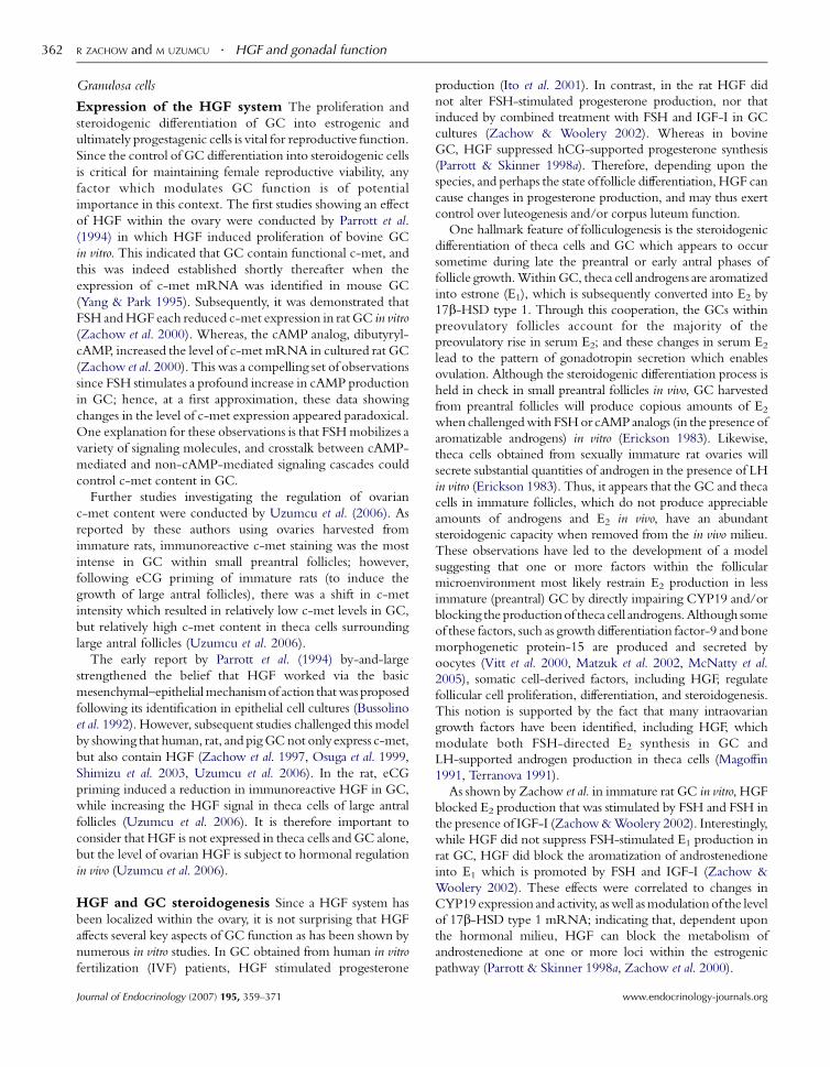
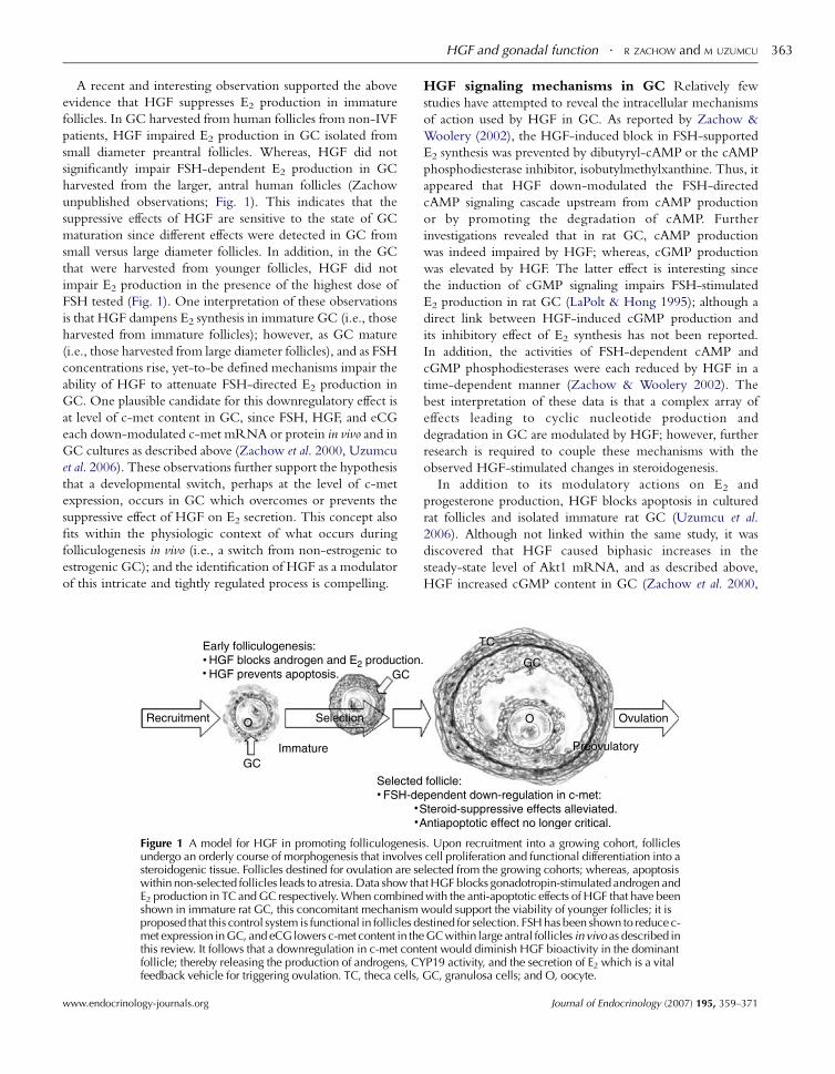
A recent and interesting observation supported the above
evidence that HGF suppresses E2 production in immature
follicles. In GC harvested from human follicles from non-IVF
patients, HGF impaired E2 production in GC isolated from
small diameter preantral follicles. Whereas, HGF did not
significantly impair FSH-dependent E2 production in GC
harvested from the larger, antral human follicles (Zachow
unpublished observations; Fig. 1). This indicates that the
suppressive effects of HGF are sensitive to the state of GC
maturation since different effects were detected in GC from
small versus large diameter follicles. In addition, in the GC
that were harvested from younger follicles, HGF did not
impair E2 production in the presence of the highest dose of
FSH tested (Fig. 1). One interpretation of these observations
is that HGF dampens E2 synthesis in immature GC (i.e., those
harvested from immature follicles); however, as GC mature
(i.e., those harvested from large diameter follicles), and as FSH
concentrations rise, yet-to-be defined mechanisms impair the
ability of HGF to attenuate FSH-directed E2 production in
GC. One plausible candidate for this downregulatory effect is
at level of c-met content in GC, since FSH, HGF, and eCG
each down-modulated c-met mRNA or protein in vivo and in
GC cultures as described above (Zachow et al. 2000, Uzumcu
et al. 2006). These observations further support the hypothesis
that a developmental switch, perhaps at the level of c-met
expression, occurs in GC which overcomes or prevents the
suppressive effect of HGF on E2 secretion. This concept also
fits within the physiologic context of what occurs during
folliculogenesis in vivo (i.e., a switch from non-estrogenic to
estrogenic GC); and the identification of HGF as a modulator
of this intricate and tightly regulated process is compelling.
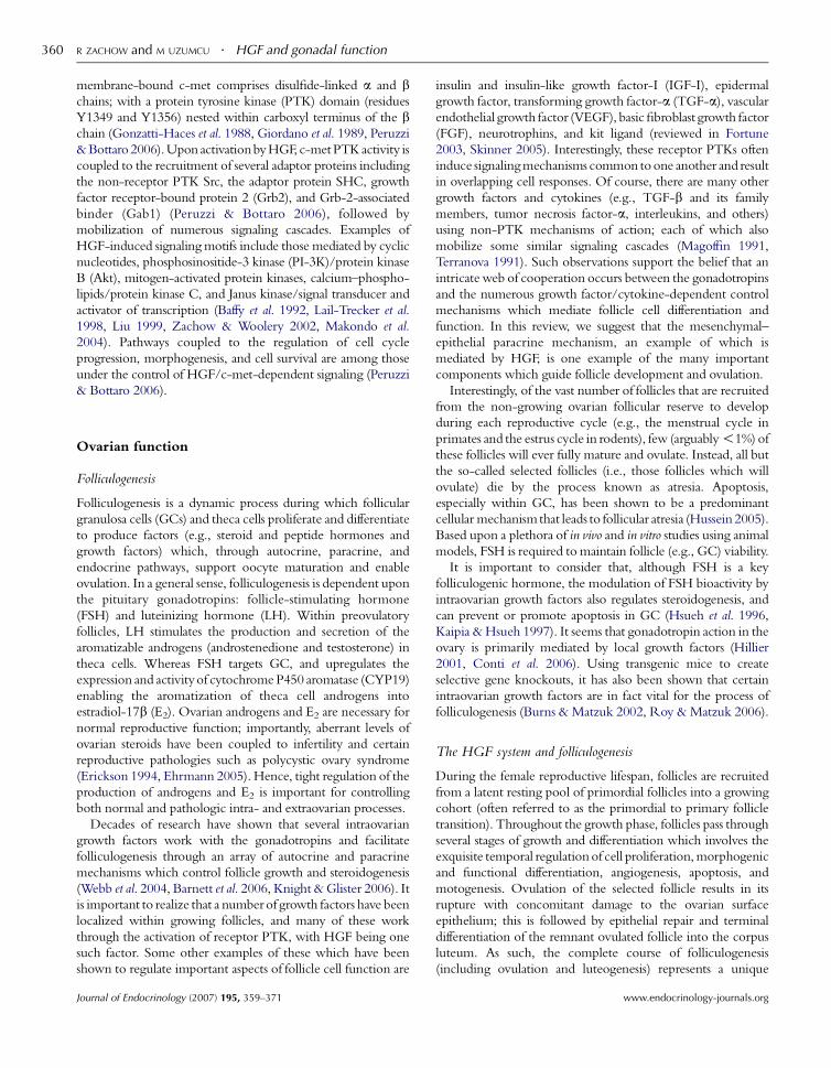
Figure 1 A model for HGF in promoting folliculogenesiundergo an orderly course of morphogenesis that involvessteroidogenic tissue. Follicles destined for ovulation are sewithin non-selected follicles leads to atresia. Data show thaE2 production in TC and GC respectively. When combinedshown in immature rat GC, this concomitant mechanismproposed that this control system is functional in follicles dmet expression in GC, and eCG lowers c-met content in thethis review. It follows that a downregulation in c-met contfollicle; thereby releasing the production of androgens, CYfeedback vehicle for triggering ovulation. TC, theca cells,
www.endocrinology-journals.org
HGF signaling mechanisms in GC Relatively few
studies have attempted to reveal the intracellular mechanisms
of action used by HGF in GC. As reported by Zachow &
Woolery (2002), the HGF-induced block in FSH-supported
E2 synthesis was prevented by dibutyryl-cAMP or the cAMP
phosphodiesterase inhibitor, isobutylmethylxanthine. Thus, it
appeared that HGF down-modulated the FSH-directed
cAMP signaling cascade upstream from cAMP production
or by promoting the degradation of cAMP. Further
investigations revealed that in rat GC, cAMP production
was indeed impaired by HGF; whereas, cGMP production
was elevated by HGF. The latter effect is interesting since
the induction of cGMP signaling impairs FSH-stimulated
E2 production in rat GC (LaPolt & Hong 1995); although a
direct link between HGF-induced cGMP production and
its inhibitory effect of E2 synthesis has not been reported.
In addition, the activities of FSH-dependent cAMP and
cGMP phosphodiesterases were each reduced by HGF in a
time-dependent manner (Zachow & Woolery 2002). The
best interpretation of these data is that a complex array of
effects leading to cyclic nucleotide production and
degradation in GC are modulated by HGF; however, further
research is required to couple these mechanisms with the
observed HGF-stimulated changes in steroidogenesis.
In addition to its modulatory actions on E2 and
progesterone production, HGF blocks apoptosis in cultured
rat follicles and isolated immature rat GC (Uzumcu et al.
2006). Although not linked within the same study, it was
discovered that HGF caused biphasic increases in the
steady-state level of Akt1 mRNA, and as described above,
HGF increased cGMP content in GC (Zachow et al. 2000,
s. Upon recruitment into a growing cohort, folliclescell proliferation and functional differentiation into alected from the growing cohorts; whereas, apoptosist HGF blocks gonadotropin-stimulated androgen andwith the anti-apoptotic effects of HGF that have been
would support the viability of younger follicles; it isestined for selection. FSH has been shown to reduce c-GC within large antral follicles in vivo as described in
ent would diminish HGF bioactivity in the dominantP19 activity, and the secretion of E2 which is a vital
GC, granulosa cells; and O, oocyte.
Journal of Endocrinology (2007) 195, 359–371
R ZACHOW and M UZUMCU . HGF and gonadal function364
Zachow & Woolery 2002). This is potentially significant in
that the activation of PI-3K/Akt- and cGMP-dependent
signaling mechanisms block apoptosis in GC (Westfall et al.
2001, Peluso & Pappalardo 2004). Collectively, these findings
imply that the anti-apoptotic effect of HGF within GC may
be mediated by the induction of PI-3K/Akt and/or cGMP-
dependent signal transduction. A review of some signaling
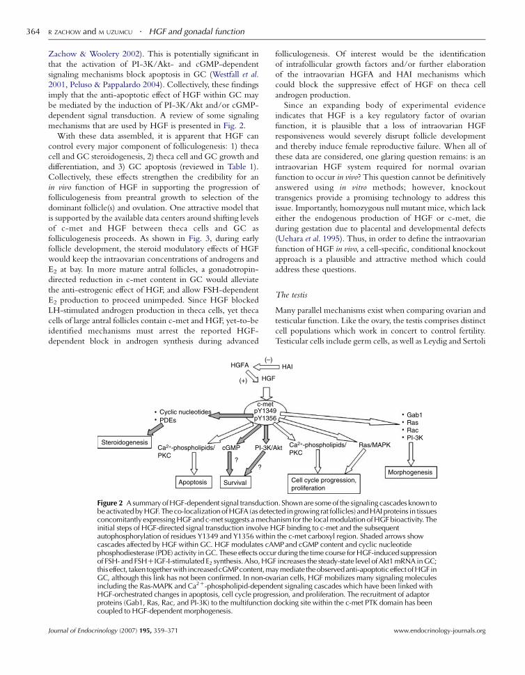
mechanisms that are used by HGF is presented in Fig. 2.
With these data assembled, it is apparent that HGF can
control every major component of folliculogenesis: 1) theca
cell and GC steroidogenesis, 2) theca cell and GC growth and
differentiation, and 3) GC apoptosis (reviewed in Table 1).
Collectively, these effects strengthen the credibility for an
in vivo function of HGF in supporting the progression of
folliculogenesis from preantral growth to selection of the
dominant follicle(s) and ovulation. One attractive model that
is supported by the available data centers around shifting levels
of c-met and HGF between theca cells and GC as
folliculogenesis proceeds. As shown in Fig. 3, during early
follicle development, the steroid modulatory effects of HGF
would keep the intraovarian concentrations of androgens and
E2 at bay. In more mature antral follicles, a gonadotropin-
directed reduction in c-met content in GC would alleviate
the anti-estrogenic effect of HGF, and allow FSH-dependent
E2 production to proceed unimpeded. Since HGF blocked
LH-stimulated androgen production in theca cells, yet theca
cells of large antral follicles contain c-met and HGF, yet-to-be
identified mechanisms must arrest the reported HGF-
dependent block in androgen synthesis during advanced
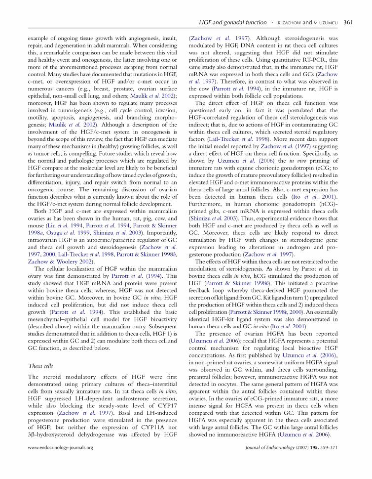
Figure 2 A summary of HGF-dependent signal transductiobe activated by HGF. The co-localization of HGFA (as deteconcomitantly expressing HGFand c-met suggests a mechinitial steps of HGF-directed signal transduction involve Hautophosphorylation of residues Y1349 and Y1356 withincascades affected by HGF within GC. HGF modulates cAphosphodiesterase (PDE) activity in GC. These effects occuof FSH- and FSHCIGF-I-stimulated E2 synthesis. Also, HGthiseffect, taken togetherwith increasedcGMPcontent,maGC, although this link has not been confirmed. In non-ovincluding the Ras-MAPK and Ca2C-phospholipid-dependHGF-orchestrated changes in apoptosis, cell cycle progreproteins (Gab1, Ras, Rac, and PI-3K) to the multifunctioncoupled to HGF-dependent morphogenesis.
Journal of Endocrinology (2007) 195, 359–371
folliculogenesis. Of interest would be the identification
of intrafollicular growth factors and/or further elaboration
of the intraovarian HGFA and HAI mechanisms which
could block the suppressive effect of HGF on theca cell
androgen production.
Since an expanding body of experimental evidence
indicates that HGF is a key regulatory factor of ovarian
function, it is plausible that a loss of intraovarian HGF
responsiveness would severely disrupt follicle development
and thereby induce female reproductive failure. When all of
these data are considered, one glaring question remains: is an
intraovarian HGF system required for normal ovarian
function to occur in vivo? This question cannot be definitively
answered using in vitro methods; however, knockout
transgenics provide a promising technology to address this
issue. Importantly, homozygous null mutant mice, which lack
either the endogenous production of HGF or c-met, die
during gestation due to placental and developmental defects
(Uehara et al. 1995). Thus, in order to define the intraovarian
function of HGF in vivo, a cell-specific, conditional knockout
approach is a plausible and attractive method which could
address these questions.
The testis
Many parallel mechanisms exist when comparing ovarian and
testicular function. Like the ovary, the testis comprises distinct
cell populations which work in concert to control fertility.
Testicular cells include germ cells, as well as Leydig and Sertoli
n. Shown are some of the signaling cascades known tocted in growing rat follicles) and HAI proteins in tissuesanism for the local modulation of HGF bioactivity. TheGF binding to c-met and the subsequentthe c-met carboxyl region. Shaded arrows show
MP and cGMP content and cyclic nucleotider during the time course for HGF-induced suppressionF increases the steady-state level of Akt1 mRNA in GC;ymediate the observedanti-apoptoticeffectofHGF in
arian cells, HGF mobilizes many signaling moleculesent signaling cascades which have been linked withssion, and proliferation. The recruitment of adaptordocking site within the c-met PTK domain has been
www.endocrinology-journals.org
Table
1Ex
pre
ssio
nan
def
fect
sof
the
hep
atocy
tegr
ow
thfa
ctor
(HG
F)sy
stem
wit
hin
thec
aan
dgr
anulo
sace
lls
Presence
ofHGF,c-met,an
dHGFA
Effect
ofHGFonsteroid
geneexpression
Effect
ofHGFon
steroidoge
nesis
Effect
ofHGFon
proliferation
Effect
ofHGFon
apoptosis
HGF-dep
enden
tsign
aling
Cell
Thec
aR
at:
tem
pora
l/eC
G-d
epen
den
tch
ange
sin
HG
F,c-
met
,H
GFA
pro
tein
s(invivo
);c-
met
mR
NA
det
ecte
d.
Hum
an:
c-m
et.
Pig
:c-
met
mR
NA
det
ecte
d.
Rat
:(K
)C
YP17
mR
NA
;N
Eon
CY
P11A
and
3b-H
SDR
at:
(K)
LH-s
tim
ula
ted
andro
ster
one,
(C)
bas
alan
dLH
-sti
mula
ted
P4
Cow
:(C
)R
at:
NE
Not
report
edN
ot
report
ed
Gra
nulo
saR
at:
tem
pora
l/eC
G-d
epen
den
tch
ange
sin
HG
F,c-
met
,H
GFA
pro
tein
sin
vivo
;c-m
etm
RN
A(K
)by
HG
Fan
dFS
H,
c-m
etm
RN
A(C
)by
db-c
AM
P.H
um
an,
cow
,ra
t,m
ouse
:c-
met
mR
NA
det
ecte
d.
Rat
:(K
)FS
H-d
epen
den
tC
YP19
and
17b-H
SD1
expre
ssio
n
Rat
:(K
)FS
H-
and
FSH
CIG
F-I-
stim
ula
ted
E 2,
NE
on
P4.H
um
an:st
imula
ted
P4,(K
)FS
H-s
tim
ula
ted
E 2in
smal
lfo
llic
les,
NE
inla
rge
foll
icle
s.C
ow
:(K
)hC
G-s
tim
ula
ted
P4
Cow
:(C
)R
at:
NE
Rat
:(K
)R
at:
(K)
FSH
-stim
u-
late
dcA
MP
conte
nt,
(C)
cGM
Pco
nte
nt;
(K)
cycl
icnucl
eo-
tide
PD
Eac
tivi
ty;
(C)
Akt
1ex
pre
ssio
n
(C),
stim
ula
ted,
incr
ease
d;
(K),
blo
cked
,re
duce
d;
NE,
no
effe
ct;
db-c
AM
P,dib
uty
ryl-
cAM
P;
PD
E,phosp
hodie
ster
ase;
eCG
,eq
uin
ech
ori
onic
gonad
otr
opin
;hC
G,
hum
anC
G.
HGF and gonadal function . R ZACHOW and M UZUMCU 365
www.endocrinology-journals.org
cells. Leydig and Sertoli cells respond to LH and FSH
respectively, to produce androgens (testosterone), E2, several
growth factors, and cytokines which control testicular
differentiation and function (Bornstein et al. 2004, Haider
2004, Rao 2005, Itman et al. 2006, Mackay & Smith 2007).
The precise function of testicular E2 remains debatable, and
has been thoroughly reviewed elsewhere (Sierens et al. 2005,
Carreau et al. 2006). However, through the collective
production of testosterone, several growth factors (including
HGF), and other proteins, Leydig and Sertoli cells (like
ovarian theca cells and GC) cooperate to support germ cell
maturation, and are thus vital for male reproductive health.
In addition to Sertoli andLeydig cells, peritubular myoid cells
represent another somatic cell type which is involved in
epithelial–mesenchymal interactions within the testis. During
embryonic gonadal development, peritubular myoid cells
differentiate from cells that migrate from neighboring
mesonephros, which is essential for normal testicular cord
formation (Tilmann & Capel 1999). During postnatal life,
peritubular myoid cells provide normal tubule morphology and
contractility, processes that are critical for spermatogenesis
(Zhang et al. 2006). Peritubular myoid cells and Sertoli cells
interact with each other through growth factor-mediated
feedback; these cells collaborate to produce extracellular matrix
components which comprise the basement membrane that
separates these two cell types (Skinner et al. 1985, Skinner 1991).
The ontogeny of HGF and c-met expression during testiculardevelopment
As with the ovary, the testis contains an endogenous HGF
system, with a pattern of expression for HGF and c-met
which is species specific. Although there are no published
reports to date showing the ontogeny of HGF and/or c-met
expression during ovarian development, changes in HGF and
c-met expression during embryonic and postnatal testicular
development have been described (Catizone et al. 1999, 2005,
Ricci et al. 1999, 2002, 2006).
HGF expression starts in the mesonephros and coelomic
epithelium of the undifferentiated male urogenital ridge as early
as 11.5 day post-coitum (dpc) in the mouse (Ricci et al. 2002),
and remains within the interstitium (presumably in peritubular
myoid cells). Although c-met is first expressed inside the cords
starting on 12.5 dpc, it is not clear at this time whether c-met is
expressed by Sertoli or germ cells within cords. However, based
on the classical paracrine model proposed for the HGF/c-met
system (suggesting that HGF is produced by mesenchymal cells
and acts on epithelial cells), Sertoli cells are the likely target for
HGF within the cords. Ricci et al. (2006) reported that in the
mouse at 13.5 and 15.5 dpc, c-met was mainly present within
the testicular cords, and by 18.5 dpc, c-met was more localized
to the interstitium (presumably fetal Leydig cells) and is no
longer expressed in the cords (Table 2).
In the rat, however, c-met mRNA and immunoreactive
protein were detected in the postnatal testis on day 10, with
expression appearing in Sertoli cells on day 25, and increasing
Journal of Endocrinology (2007) 195, 359–371
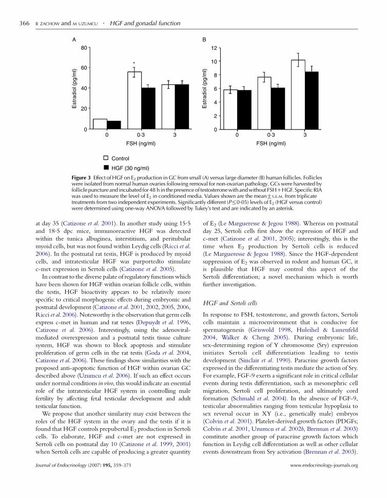
Figure 3 Effect of HGF on E2 production in GC from small (A) versus large diameter (B) human follicles. Follicleswere isolated from normal human ovaries following removal for non-ovarian pathology. GCs were harvested byfollicle puncture and incubated for 48 h in the presence of testosterone with andwithout FSHCHGF. Specific RIAwas used to measure the level of E2 in conditioned media. Values shown are the meanGS.E.M. from triplicatetreatments from two independent experiments. Significantly different (P%0.05) levels of E2 (HGF versus control)were determined using one-way ANOVA followed by Tukey’s test and are indicated by an asterisk.
R ZACHOW and M UZUMCU . HGF and gonadal function366
at day 35 (Catizone et al. 2001). In another study using 15.5and 18.5 dpc mice, immunoreactive HGF was detected
within the tunica albuginea, interstitium, and peritubular
myoid cells, but was not found within Leydig cells (Ricci et al.
2006). In the postnatal rat testis, HGF is produced by myoid
cells, and intratesticular HGF was purportedto stimulate
c-met expression in Sertoli cells (Catizone et al. 2005).
In contrast to the diverse palate of regulatory functionswhich
have been shown for HGF within ovarian follicle cells, within
the testis, HGF bioactivity appears to be relatively more
specific to critical morphogenic effects during embryonic and
postnatal development (Catizone et al. 2001, 2002, 2005, 2006,
Ricci et al. 2006). Noteworthy is the observation that germ cells
express c-met in human and rat testes (Depuydt et al. 1996,
Catizone et al. 2006). Interestingly, using the adenoviral-
mediated overexpression and a postnatal testis tissue culture
system, HGF was shown to block apoptosis and stimulate
proliferation of germ cells in the rat testis (Goda et al. 2004,
Catizone et al. 2006). These findings show similarities with the
proposed anti-apoptotic function of HGF within ovarian GC
described above (Uzumcu et al. 2006). If such an effect occurs
under normal conditions in vivo, this would indicate an essential
role of the intratesticular HGF system in controlling male
fertility by affecting fetal testicular development and adult
testicular function.
We propose that another similarity may exist between the
roles of the HGF system in the ovary and the testis if it is
found that HGF controls prepubertal E2 production in Sertoli
cells. To elaborate, HGF and c-met are not expressed in
Sertoli cells on postnatal day 10 (Catizone et al. 1999, 2001)
when Sertoli cells are capable of producing a greater quantity
Journal of Endocrinology (2007) 195, 359–371
of E2 (Le Margueresse & Jegou 1988). Whereas on postnatal
day 25, Sertoli cells first show the expression of HGF and
c-met (Catizone et al. 2001, 2005); interestingly, this is the
time when E2 production by Sertoli cells is reduced
(Le Margueresse & Jegou 1988). Since the HGF-dependent
suppression of E2 was observed in rodent and human GC, it
is plausible that HGF may control this aspect of the
Sertoli differentiation; a novel mechanism which is worth
further investigation.
HGF and Sertoli cells
In response to FSH, testosterone, and growth factors, Sertoli
cells maintain a microenvironment that is conducive for
spermatogenesis (Griswold 1998, Huleihel & Lunenfeld
2004, Walker & Cheng 2005). During embryonic life,
sex-determining region of Y chromosome (Sry) expression
initiates Sertoli cell differentiation leading to testis
development (Sinclair et al. 1990). Paracrine growth factors
expressed in the differentiating testis mediate the action of Sry.
For example, FGF-9 exerts a significant role in critical cellular
events during testis differentiation, such as mesonephric cell
migration, Sertoli cell proliferation, and ultimately cord
formation (Schmahl et al. 2004). In the absence of FGF-9,
testicular abnormalities ranging from testicular hypoplasia to
sex reversal occur in XY (i.e., genetically male) embryos
(Colvin et al. 2001). Platelet-derived growth factors (PDGFs;
Colvin et al. 2001, Uzumcu et al. 2002b, Brennan et al. 2003)
constitute another group of paracrine growth factors which
function in Leydig cell differentiation as well as other cellular
events downstream from Sry activation (Brennan et al. 2003).
www.endocrinology-journals.org
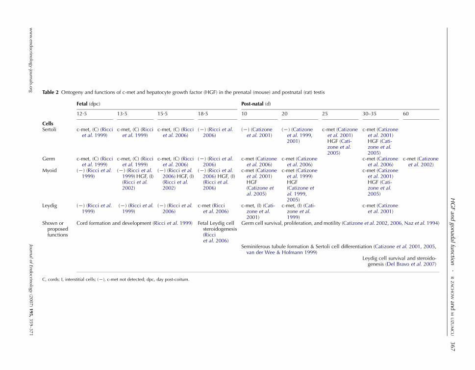
Table 2 Ontogeny and functions of c-met and hepatocyte growth factor (HGF) in the prenatal (mouse) and postnatal (rat) testis
Fetal (dpc) Post-natal (d)
12.5 13.5 15.5 18.5 10 20 25 30–35 60
CellsSertoli c-met, (C) (Ricci
et al. 1999)c-met, (C) (Ricciet al. 1999)
c-met, (C) (Ricciet al. 2006)
(K) (Ricci et al.2006)
(K) (Catizoneet al. 2001)
(K) (Catizoneet al. 1999,2001)
c-met (Catizoneet al. 2001)HGF (Cati-zone et al.2005)
c-met (Catizoneet al. 2001)HGF (Cati-zone et al.2005)
Germ c-met, (C) (Ricciet al. 1999)
c-met, (C) (Ricciet al. 1999)
c-met, (C) (Ricciet al. 2006)
(K) (Ricci et al.2006)
c-met (Catizoneet al. 2006)
c-met (Catizoneet al. 2006)
c-met (Catizoneet al. 2006)
c-met (Catizoneet al. 2002)
Myoid (K) (Ricci et al.1999)
(K) (Ricci et al.1999) HGF, (I)(Ricci et al.2002)
(K) (Ricci et al.2006) HGF, (I)(Ricci et al.2002)
(K) (Ricci et al.2006) HGF, (I)(Ricci et al.2006)
c-met (Catizoneet al. 2001)HGF(Catizone etal. 2005)
c-met (Catizoneet al. 1999)HGF(Catizone etal. 1999,2005)
c-met (Catizoneet al. 2001)HGF (Cati-zone et al.2005)
Leydig (K) (Ricci et al.1999)
(K) (Ricci et al.1999)
(K) (Ricci et al.2006)
c-met (Ricciet al. 2006)
c-met, (I) (Cati-zone et al.2001)
c-met, (I) (Cati-zone et al.1999)
c-met (Catizoneet al. 2001)
Shown orproposedfunctions
Cord formation and development (Ricci et al. 1999) Fetal Leydig cellsteroidogenesis(Ricciet al. 2006)
Germ cell survival, proliferation, and motility (Catizone et al. 2002, 2006, Naz et al. 1994)
Seminiferous tubule formation & Sertoli cell differentiation (Catizone et al. 2001, 2005,van der Wee & Hofmann 1999)
Leydig cell survival and steroido-genesis (Del Bravo et al. 2007)
C, cords; I, interstitial cells; (K), c-met not detected; dpc, day post-coitum.
HGFan
dgo
nad
alfunctio
n.
RZ
AC
HO
Wan
dM
UZ
UM
CU
367
ww
w.en
docrin
olo
gy-jo
urn
als.org
Journal
ofEn
docrin
ology
(2007)195,
359–3
71
R ZACHOW and M UZUMCU . HGF and gonadal function368
The inhibition of PDGF bioactivity or deletion of PDGFR-acauses abnormal cord formation (Uzumcu et al. 2002b,
Brennan et al. 2003).
Additional paracrine factors with important functions in the
developing testis are neurotrophins (Cupp et al. 2000, Levine
et al. 2000), TGF-b (Cupp et al. 1999), VEGF (Bott et al. 2006),kit ligand (Vincent et al. 1998), and glial cell line-derived
neurotropic factor (GDNF;Meng et al. 2000). The roles of these
growth factors in promoting testicular differentiation and
development have been extensively reviewed elsewhere
(Brennan & Capel 2004, Mackay & Smith 2007).
HGF is somewhat unique among growth factors. Unlike
the growth factors mentioned above that are mainly products
of Sertoli cells (an epithelial cell type) and act on
mesenchymal cells, HGF is primarily produced by myoid
cells (a mesenchymal cell type), especially during embryonic
development. Thus, when considering communication
between Sertoli and myoid cells, HGF represents an
interesting growth factor because it mostly acts in the
mesenchymal-to-epithelial direction. The identification of
c-met within Sertoli cells enforces the validity of this model.
The pattern of c-met expression within the cords during
embryonic life is not exactly known, but beginning sometime
between postnatal days 20–25 (in the rat), Sertoli cells contain
functional c-met protein (Catizone et al. 2001). In dissociated
rat Sertoli cells in vitro, the administration of exogenous HGF
induces the formation of structures resembling testicular
seminiferous cords (Catizone et al. 2001). This experimental
observation led to the notion that HGF may be instrumental
in testicular lumen formation in mature animals (Catizone
et al. 2001). It is also imperative to consider that testicular cord
formation is a major morphologic event during embryonic
gonad development in the male (Uzumcu et al. 2002a,b). This
process requires Sertoli cell growth and differentiation, as well
as mesonephric cell migration (reviewed in (Yao et al. 2002),
changes which may be mediated by HGF (Ricci et al. 1999).
Overall, the available evidence then suggests that HGF is one
of several growth factors (including TGF-b, FGF-9, PDGF,
neurotrophins, GDNF, VEGF, and others) with an important
role in development and function of the testis including cord
formation and Sertoli cell differentiation.
HGF and myoid cells
It seems that HGF has two different modes of action
depending upon the stage of development. Initially, during
the embryonic developmental stage, the mode of action used
by HGF is essentially paracrine. Thus, HGF from myoid cells
targets cells inside the seminiferous cords (i.e., Sertoli cells
and/or germ cells) between 12.5 and 15.5 dpc, and then acts
on fetal Leydig cells on 18.5 dpc (Ricci et al. 1999, 2002,
2006). However, a switch occurs during postnatal develop-
mental, when HGF activity becomes primarily autocrine,
with some paracrine function still operational.
Both HGF and c-met are expressed in myoid cells prior to
postnatal day 25. After day 25, the expression of HGF and
Journal of Endocrinology (2007) 195, 359–371
c-met appears in Sertoli cells, while the expression of HGF
and c-met in myoid cells is relatively subsided (Catizone et al.
1999, 2001, 2006). In addition to myoid and Sertoli cells,
c-met mRNA is also present in germ cells between postnatal
days 10 and 60 (Catizone et al. 2002, 2006), and in Leydig
cells in adolescent rats (days 30–32; Del Bravo et al. 2007).
HGF and Leydig cells
The testicular correlate of the ovarian theca cell is the Leydig
cell. As such, Leydig cells do not express FSH receptors, but
do express LH receptors, and are stimulated by LH to produce
testosterone as the predominant steroid. Beginning around
fetal day 18.5, c-met is expressed within the mouse testicular
interstitium; and organ culture of fetal day 18.5 testes revealedthat HGF stimulated basal testosterone secretion (Ricci et al.
2006). Postnatal (days 30–32) rat Leydig cells also express
immunoreactive c-met, and HGF stimulates basal testosterone
secretion and inhibits apoptosis in rat Leydig cells as well as
testicular tissue explants (Del Bravo et al. 2007).
Proposed regulatory function for HGF in the testis
What is apparent is that shifts in the expression of c-met and
HGF between Sertoli cells, germ cells, Leydig cells, and myoid
cells during fetal and postnatal testicular growth and
differentiation suggest that HGF guides changes in testicular
morphogenesis and function (including steroidogenesis) in a
developmentally regulatedmanner. This type ofmodel overlaps
with that proposed for HGF during folliculogenesis. One
hypothesiswhich canbederived fromthese data fromthe testis is
that HGF promotes Sertoli cell differentiation and cord
formation during in utero development, but then HGF control
switches to the regulation of Leydig cell steroidogenic
differentiation during the last stages of fetal development and
preceding sexual maturity. Beginning around the time of
puberty (i.e., day 25 in the rodent), Sertoli cells resume the
expression of c-met (Table 2), and once again gain HGF
responsiveness. It is possible that, in the prepubertal male, HGF
controls Sertoli cell function at multiple levels, to potentially
include the modulation of FSH-stimulated E2 production and
cell growth; this, however, remains uncertain pending future
studies. By regulating Sertoli cell function, HGF would
indirectly control sperm growth and maturation, in addition
to the direct effects of HGF on sperm function.
The stimulatory effect of HGF on steroid production in
postnatal Leydig cells has recently been reported (DelBravo et al.
2007). Based upon the most current data, HGF directly
stimulates rat Leydig cell steroidogenesis, although mature
human Leydig cells do not express c-met (Depuydt et al. 1996,
Catizone et al. 2001). Considering that any effects of HGF are
plausibly species specific, as has been documented with the
ovarian HGF system, this difference is not surprising. What has
been substantiated is that, collectively, the intratesticular HGF
system has several potentially vital roles, those being the
regulation of 1) seminiferous cord formation and development,
www.endocrinology-journals.org
HGF and gonadal function . R ZACHOW and M UZUMCU 369
2) Leydig cell steroidogenesis and survival, 3) Sertoli cell
proliferation and terminal differentiation, and 4) germ cell
function and/or survival. To date, the ability of HGF to control
testicular function in vivo (i.e., male reproductive viability) has
not been reported. Considering that the either HGF or c-met
null mutations are embryonic lethal (as described above), a
tissue-specific conditional knockout approach is needed to
establish the importance of the HGF system in regulating testis
development and function in vivo.
Concluding thoughts
Data from several laboratories have shown that HGF controls
many key functions within the ovary and testis; these range
from modulation of both steroidogenesis (Parrott et al. 1994,
Zachow et al. 1997, 2000, Ito et al. 2001, Ricci et al. 2006,
Del Bravo et al. 2007) and apoptosis (Goda et al. 2004,
Uzumcu et al. 2006, Del Bravo et al. 2007), to guiding mitosis
and morphogenesis (Parrott & Skinner 1998b, Catizone et al.
2001, 2006, Ricci et al. 2002, 1999). The breadth of the
HGF-mediated control mechanisms strongly indicates that an
intact HGF system is required for proper gonad development
and/or function; this would of course make HGF a key
regulator of female and male fertility. The next step in
determining this is to blend in vitro, molecular mechanism
studies with an elucidation of the role of HGF in regulating
reproductive function in vivo.
Acknowledgements
The authors declare that there is no conflict of interest that
would prejudice the impartiality of this scientific work.
References
Baffy G, Yang L, Michalopoulos GK & Williamson JR 1992 Hepatocyte
growth factor induces calcium mobilization and inositol phosphate
production in rat hepatocytes. Journal of Cell Physiology 153 332–339.
Barnett KR, Schilling C, Greenfeld CR, Tomic D & Flaws JA 2006 Ovarian
follicle development and transgenic mouse models. Human Reproduction
Update 12 537–555.
Bornstein SR, Rutowski H & Vrezas I 2004 Cytokines and steroidogenesis.
Molecular and Cellular Endocrinology 215 135–141.
Bott RC, McFee RM, Clopton DT, Toombs C & Cupp AS 2006 Vascular
endothelial growth factor and kinase domain region receptor are involved
in both seminiferous cord formation and vascular development during testis
morphogenesis in the rat. Biology of Reproduction 75 56–67.
Bottaro DP, Rubin JS & Faletto DL 1991 Identification of the hepatocyte
growth factor receptor as the c-met proto-oncogene product. Science 251
802–804.
Brennan J & Capel B 2004 One tissue, two fates: molecular genetic events that
underlie testis versus ovary development. Nature Review Genetics 5 509–521.
Brennan J, Tilmann C & Capel B 2003 Pdgfr-alpha mediates testis cord
organization and fetal Leydig cell development in the XY gonad. Genes and
Development 17 800–810.
Burns KH &Matzuk MM 2002 Genetic models for the study of gonadotropin
actions. Endocrinology 143 2823–2835.
www.endocrinology-journals.org
Bussolino F, DiRenzo M, Ziche M, Bocchietto E, Olivero M, Naldini L,
Gaudino G, Tomagnone L, Coffer A & Comoglio P 1992 Hepatocyte
growth factor is a potent angiogenic factor which stimulates endothelial cell
motility and growth. Journal of Cell Biology 119 629–641.
Carreau S, Dalande C, Silandre D, Bourguiba S & Lambard S 2006 Aromatase
and estrogen receptors in male reproduction. Molecular and Cellular
Endocrinology 246 65–68.
Catizone A, Ricci G, Arista V, Innocenzi A & Galdieri M 1999 Hepatocyte
growth factor and c-Met are expressed in the rat prepubertal testis.
Endocrinology 140 3106–3113.
Catizone A, Ricci G & Galdieri M 2001 Expression and a functional role of
hepatocyte growth factor receptor (c-Met) during postnatal rat testis
development. Endocrinology 142 1828–1834.
Catizone A, Ricci G & Galdieri M 2002 Functional role of hepatocyte growth
factor receptor during sperm maturation. Journal of Andrology 23 911–918.
Catizone A, Ricci G & Galdieri M 2005 HGF and post-natal testis
development. Molecular and Cellular Endocrinology 241 32–40.
Catizone A, Ricci G, Del Bravo J & Galdieri M 2006 Hepatocyte growth
factor modulates in vitro cell survival and proliferation of germ cells during
postnatal testis development. Journal of Endocrinology 189 137–146.
Colvin JS, Green RP, Schmal J, Capel B & Ornitz DM 2001 Male-to-female
sex reversal in mice lacking fibroblast growth factor 9. Cell 104 875–889.
Conti M, Hsieh M, Park YJ & Su YQ 2006 Role of the epidermal growth
factor network in ovarian follicles. Molecular Endocrinolgy 20 715–723.
Cupp AS, Kim G & Skinner MK 1999 Expression and action of transforming
growth factor b (TGFb1, TGFb2 and TGFb3) during embryonic rat testis
development. Biology of Reproduction 60 1304–1313.
Cupp AS, Kim GH & Skinner MK 2000 Expression and action of
neurotropin-3 and nerve growth factor in embryonic and early postnatal rat
testis development. Biology of Reproduction 63 1617–1628.
Del Bravo J, Catizone A, Ricci G & Galdieri M 2007 Hepatocyte growth
factor modulates rat Leydig cell functions. Journal of Andrology In Press.
Depuydt CE, Zalata A, de Potter CR, van Emmelo J & Comhaire FH 1996
The receptor encoded by human c-met oncogene is expressed in testicular
tissue and on human spermatozoa. Molecular Human Reproduction 2 2–8.
Ehrmann DA 2005 Polycystic ovary syndrome.New England Journal of Medicine
325 1223–1236.
Erickson GF 1983 Primary cultures of ovarian cells in serum-free medium as
models of hormone-dependent differentiation. Molecular and Cellular
Endocrinology 29 21–49.
Erickson GF 1994 Polycystic ovary syndrome: normal and abnormal
steroidogenesis. In Ovarian Endocrinopathies: Proceedings of the 8th Reinier
deGraaf Symposium, pp 103–115. Eds R Schats & J Schoemaker. New York:
Parthenon Publishing.
Fortune JE 2003 The early stages of follicular development: activation of
primordial follicles and growth of preantral follicles. Animal Reproduction
Science 78 135–163.
Gheradi E & Stoker M 1991 Hepatocyte growth factor-scatter factor:
mitogen, motogen, and Met. Cancer Cell 3 227–232.
Giordano S, Ponzetto C & DiRenzo MF 1989 Tyrosine kinase receptor
indistinguishable from the c-met protein. Nature 339 155–156.
Goda K, FujisawaM, Shirakawa T, Dobashi M, Shiota G, Zhang ZJ, Gotoh A&
Kamidono S 2004 Adenoviral-mediated HGF expression inhibits germ cell
apoptosis in rats with cryptorchidism. Journal of Gene Medicine 6 869–876.
Gonzatti-HacesM, SethA,ParkM,CopelandT,Oroszlan S&VandeWoudeGF
1988 Characterization of the tpr-met oncogene p65 and the met
protooncogene p140 protein tyrosine kinases. PNAS 85 21–25.
Griswold MD 1998 The central role of Sertoli cells in spermatogenesis.
Seminars in Cell and Developmental Biology 9 411–416.
Haider SG 2004 Cell biology of Leydig cells in the testis. International Review of
Cytology 233 181–241.
Hillier SG 2001 Gonadotropic control of ovarian follicular growth and
development. Molecular and Cellular Endocrinology 179 39–46.
Hsueh AJ, Eisenhauer K, Chun SY, Hsu SY & Billig H 1996 Gonadal cell
apoptosis. Recent Progress in Hormone Research 51 433–455.
Huleihel M & Lunenfeld E 2004 Regulation of spermatogenesis by
paracrine/autocrine testicular factors. Asian Journal of Andrology 6 259–268.
Journal of Endocrinology (2007) 195, 359–371
R ZACHOW and M UZUMCU . HGF and gonadal function370
Hussein MR 2005 Apoptosis in the ovary: molecular mechanisms. Human
Reproduction Update 11 162–177.
Itman C, Mendis S, Barakat B & Loveland KL 2006 All in the family:
TGF-beta family action in testis development. Reproduction 132 233–246.
Ito M, Harada T, Tanikawa M, Fuji A, Shiota G & Terakawa N 2001
Hepatocyte growth factor and stem cell factor involvement in paracrine
interplays of theca and granulosa cells in the human ovary. Fertility and
Sterility 75 973–979.
Kaipia A & Hsueh AJ 1997 Regulation of ovarian follicle atresia. Annual
Review of Physiology 59 349–363.
Knight PG & Glister C 2006 TGF-beta superfamily members and ovarian
follicle development. Reproduction 132 177–178.
Lail-Trecker M, Gulati R & Peluso JJ 1998 A role for hepatocyte growth
factor/scatter factor in regulating normal and neoplastic cells of
reproductive tissues. Journal of the Society for Gynecologic Investigations
5 114–121.
LaPolt PS & Hong LS 1995 Inhibitory effects of superoxide dismutase and
cyclic guanosine 3 0, 5 0-monophosphate on estrogen production in cultured
rat granulosa cells. Endocrinology 136 5533–5539.
Levine E, Cupp AS & Skinner MK 2000 Role of neurotropins in rat
embryonic testis morphogenesis (cord formation). Biology of Reproduction
62 132–142.
Liu Y 1999 Hepatocyte growth factor promotes renal epithelial cell survival by
dual mechanisms. American Journal of Physiology 277 F624–F633.
Liu Y, Lin L & Zarnegar R 1994 Modulation of hepatocyte growth factor
gene expression by estrogen in mouse ovary. Molecular and Cellular
Endocrinology 104 173–181.
Mackay S & Smith RA 2007 Effects of growth factors on testicular
morphogenesis. International Review of Cytology 260 113–173.
Magoffin DA 1991 Regulation of differentiated functions in ovarian theca
cells. Seminars in Reproductive Endocrinology 9 321–331.
MakondoK,Kimura K, KitamuraN, KitamuraT, Yamaji D, Jung BD&SaitoM
2004 Hepatocyte growth factor activates endothelial nitric oxide synthase by
Ca(2C)- and phosphoinositide 3-kinase/Akt-dependent phosphorylation in
aortic endothelial cells. Biochemistry Journal 374 63–69.
Le Margueresse B & Jegou B 1988 In vitro effects of germ cells on the secretory
activity of Sertoli cells recovered from rats of different ages. Endocrinology
122 1672–1680.
Matzuk MM, Burns KH, Viveiros MM & Eppig JJ 2002 Intercellular
communication in the mammalian ovary: oocytes carry the conversation.
Science 296 2178–2180.
Maulik G, Shrikhande A, Kijima T, Ma PC, Morrison PT & Salgia R 2002
Role of hepatocyte growth factor receptor, c-Met, in oncogenesis and
potential for therapeutic inhibition. Cytokine and Growth Factor Reviews 13
41–59.
McNatty KP, Juengel JL, Reader KL, Lun S, Myllymaa S, Lawrence SB,
Western A, Meerasahib MF, Mottershead DG, Groome NP et al. 2005
Bone morphogenetic protein 15 and growth differentiation factor 9
cooperate to regulate granulosa cell function in ruminants. Reproduction 129
481–487.
Meng X, Lindahl M, Hyvonen ME, Parvinen M, de Rooij DG, Hess MW,
Raatikainen-Ahokas A, Sainio K, Rauvala H, Lakso M et al. 2000
Regulation of cell fate decision of undifferentiated spermatogonia by
GDNF. Science 287 1489–1493.
Miyazawa K, Tsubouchi H, Naka D, Takahashi K, Okigaki M, Arakaki N,
Nakayama H, Hirono S, Sakiyama O, Takahashi K et al. 1989 Molecular
cloning and sequence analysis of cDNA for human hepatocyte growth
factor. Biochemical and Biophysical Research Communications 163 967–973.
Miyazawa K, Shimomura T, Kitamura A, Kondo J, Morimoto Y & Kitamura N
1993Molecular cloning and sequence analysis of the cDNAfor a human serine
protease responsible for activation of hepatocyte growth factor, structural
similarity of the protease precursor to blood coagulation factor-xii. Journal of
Biological Chemistry 269 10024–10028.
Nakamura T, Nishizawa T, Hagiya M, Seki T, Shimonishi M, Sugimura A,
Tashiro K & Shimizu S 1989 Molecular cloning and expression of human
hepatocyte growth factor. Nature 342 440–443.
Naz RK, Joseph A, Lee Y, Ahmad K & Bhargava MM 1994 Expression of
scatter factor/hepatocyte growth factor is regionally correlated with the
Journal of Endocrinology (2007) 195, 359–371
initiation of sperm motility in murine male genital tract: is scatter
factor/hepatocyte growth factor involved in the initiation of sperm
motility. Molecular Reproduction and Development 38 431–439.
Osuga Y, Tsutsumi O, Momoeda M, Okagaki R, Matsumi H, Hiroi H,
Suenaga A, Yano T & Taketani Y 1999 Evidence for the presence of
hepatocyte growth factor expression in human ovarian follicles. Molecular
Human Reproduction 5 703–707.
Parrott JA & Skinner MK 1998a Developmental and hormonal regulation of
hepatocyte growth factor expression and action in the bovine ovarian
follicle. Biology of Reproduction 59 553–560.
Parrott JA & Skinner MK 1998b Theca cell-granulosa cell interactions involve
a positive feedback loop among keratinocyte growth factor, hepatocyte
growth factor, and kit ligand during ovarian follicular development.
Endocrinology 139 2240–2245.
Parrott JA & Skinner MK 2000 Kit ligand actions on ovarian stromal cells:
effects on theca cell recruitment and steroid production. Molecular
Reproduction and Development 55 55–64.
Parrott JA, Vigne JL, Chu BZ & Skinner MK 1994 Mesenchymal epithelial
interactions within the ovarian follicle involve keratinocyte and hepatocyte
growth factor production by theca cells and their action on granulosa cells.
Endocrinology 135 569–575.
Peluso JJ & Pappalardo A 2004 Progesterone regulates granulosa cell viability
through a protein kinase G-dependent mechanism that may involve 14-3-3-s.
Biology of Reproduction 71 1870–1878.
Peruzzi B & Bottaro DP 2006 Targeting the c-Met signaling pathway in
cancer. Clinical Cancer Research 12 3657–3660.
Rao AJ 2005 Hormonal regulation of Leydig cell proliferation and
differentiation in rodent testis: a dynamic interplay between gonadotropins
and testicular factors. Reproductive Biomedicine Online 11 507–518.
Ricci G, Catizone A, Innocenzi A & Galdieri M 1999 Hepatocyte growth
factor (HGF) receptor expression and role of HGF during embryonic
mouse testis development. Developmental Biology 216 340–347.
Ricci G, Catizone A & Galdieri M 2002 Pleiotropic activity of hepatocyte
growth factor during embryonic mouse testis development. Mechanism of
Development 118 19–28.
Ricci G, Catizone A & Galdieri M 2006 Expression and functional role of
hepatocyte growth factor and its receptor (c-met) during fetal mouse tesis
development. Journal of Endocrinology 191 559–570.
Roy A & Matzuk MM 2006 Deconstructing mammalian reproduction: using
knockouts to define fertility pathways. Reproduction 131 207–219.
Schmahl J, Kim Y, Colvin JS, Ornitz DM & Capel B 2004 Fgf-9 induces
proliferation and nuclear localization of FGFR2 in Sertoli precursors during
male sex determination. Development 131 3627–3636.
Shimizu T, Iijima K, Sasada H & Sato E 2003 Messenger ribonucleic acid
expressions of hepatocyte growth factor, angiopoietins and their receptors
during follicular development in gilts. Journal of Reproduction and
Development 49 203–211.
Shimomura T, DendaK, Kitamura A,Kawaguchi T, KitoM,Kondo J, Kagaya S,
Qin L, Takata H, Miyazawa K et al. 1997 Hepatocyte growth factor activator
inhibitor, a novel Kunitz-type serine protease inhibitor. Journal of Biological
Chemistry 272 6370–6376.
Sierens JE, Sneddon SF, Collins F, Millar MR& Saunders PTK 2005 Estrogens
in testis biology. Annals of New York Academy of Sciences 1061 65–76.
Sinclair AH,Berta P, PalmerMS,Hawkins JR,GriffithsBL, SmithMJ, Foster JW,
Frischauf AM, Lovell-Badge R & Goodfellow PN 1990 A gene from the
human sex-determining region encodes a protein with homology to a
conserved DNA-binding motif. Nature 346 240–244.
Skinner MK 1991 Cell–cell interactions in the testis. Endocrine Reviews 12
45–77.
Skinner MK 2005 Regulation of primordial follicle assembly and
development. Human Reproduction Update 11 461–471.
Skinner MK, Tung PS & Fritz IB 1985 Cooperativity between Sertoli cells
and testicular peritubular cells in the production and deposition of
extracellular matrix components. Journal of Cell Biology 100 1941–1947.
Terranova PF 1991 Regulation of the granulosa cell: growth factor
interactions. Seminars in Reproductive Endocrinology 9 313–320.
www.endocrinology-journals.org
HGF and gonadal function . R ZACHOW and M UZUMCU 371
Tilmann C & Capel B 1999 Mesonephric cell migration induces testis cord
formation and Sertoli cell differentiation in the mammalian gonad.
Development 126 2883–2890.
Uehara Y, Minowa O,Mori C, Shiota K, Kuno J, Noda T & Kitamura N 1995
Placental defect and embryonic lethality in mice lacking hepatocyte growth
factor/scatter factor. Nature 373 702–705.
Uzumcu M, Dirks KA & Skinner MK 2002a Inhibition of platelet-derived
growth factor actions in the embryonic testis influences normal cord
development and morphology. Biology of Reproduction 66 745–753.
UzumcuM,Westfall SD, Dirks KA& Skinner MK 2002b Embryonic testis cord
formation and mesonephric cell migration requires the phosphotidylinositol
3-kinase signaling pathway. Biology of Reproduction 67 1927–1935.
Uzumcu M, Pan Z, Chu Y, Kuhn PE & ZachowRJ 2006 Immunolocalization
of the hepatocyte growth factor (HGF) system in the rat ovary and the
antiapoptotic effect of HGF in rat ovarian granulosa cells in vitro.
Reproduction 132 291–299.
Vincent S, Segretain D, Nishikawa S, Nishikawa SI, Sage J, Cuzin F &
Rassoulzadegan M 1998 Stage-specific expression of the Kit receptor and
its ligand (KL) during male gametogenesis in the mouse: a Kit-KL
interaction critical for meiosis. Development 125 4585–4593.
Vitt UA, Hayashi M, Klein C & Hsueh AJW 2000 Growth differentiation
factor-9 stimulates proliferation but suppresses the follicle-stimulating
hormone-induced differentiation of cultured granulosa cells from small
antral and preovulatory rat follicles. Biology of Reproduction 62 370–377.
Walker WH & Cheng J 2005 FSH and testosterone signaling in Sertoli cells.
Reproduction 130 15–28.
Webb R, Garnsworthy PC, Gong JG & Armstrong DG 2004 Control of
follicular growth: local interactions and nutritional influences. Journal of
Animal Science 82 E63–E74.
Van der Wee K &Hoffman MC 1999 An in vitro tubule assay identifies HGF as
a morphogen for the formation of seminiferous tubules in the postnatal
mouse testis. Experimental Cell Research 252 175–185.
Westfall SD, Hendry IR, Obholz KL & Davis JS 2001 Putative role of
phosphatidylinositol 3-kinase-Akt signaling pathway in the survival of
granulosa cells. Endocrine 12 315–321.
www.endocrinology-journals.org
Yamauchi M, Itoh H, Naganuma S, Koono M, Hasui Y, Osada Y & Kataoka H
2002 Expression of hepatocyte growth factor activator inhibitor type 2
(HAI-2) in human testis: identification of a distinct transcription start site for
the HAI-2 gene in the testis. Biological Chemistry 383 1953–1957.
Yang XM & Park M 1995 Expression of the hepatocyte growth factor/scatter
factor receptor tyrosine kinase is localized to epithelia in the adult mouse.
Laboratory Investigation 73 483–491.
Yao HH, Tilmann C, Zhao GQ & Capel B 2002 The battle of the sexes:
opposing pathways in sex determination. Novartis Foundation Symposium
187–198.
Zachow RJ & Woolery JK 2002 The effect of hepatocyte growth factor on
cyclic nucleotide-dependent signaling and steroidogenesis in rat granulosa
cells in vitro. Biology of Reproduction 67 454–459.
Zachow RJ, Weitsman SR & Magoffin DA 1997 Hepatocyte growth factor
regulates ovarian theca-interstitial cell differentiation and androgen
production. Endocrinology 138 691–697.
ZachowRJ, Ramski BE & Lee H 2000Modulation of estrogen production and
17b-hydroxysteroid dehydrogenase-type 1, cytochrome P450 aromatase,
c-Met, and protein kinase Ba messenger ribonucleic content in rat ovarian
granulosa cells by hepatocyte growth factor and follicle-stimulating hormone.
Biology of Reproduction 62 1851–1857.
ZhangC, Yeh S, ChenYT,WuCC,ChuangKH, LinHY,WangRS, ChangYJ,
Mendis-Handagama C, Hu L et al. 2006 Oligozoospermia with normal
fertility inmalemice lacking the androgen receptor in testis peritubular myoid
cells. PNAS 103 17718–17723.
Received in final form 11 September 2007Accepted 13 September 2007Made available online as an Accepted Preprint13 September 2007
Journal of Endocrinology (2007) 195, 359–371


