Embed Size (px)
Citation preview
RESEARCH ARTICLE Open Access
The human G93A-SOD1 mutation in a pre-symptomatic rat model of amyotrophic lateralsclerosis increases the vulnerability to a mildspinal cord compressionNatasa Jokic†, Ping K Yip†, Adina Michael-Titus, John V Priestley, Andrea Malaspina*
Abstract
Background: Traumatic injuries can undermine neurological functions and act as risk factors for the developmentof irreversible and fatal neurodegenerative disorders like amyotrophic lateral sclerosis (ALS). In this study, we haveinvestigated how a mutation of the superoxide dismutase 1 (SOD1) gene, linked to the development of ALS,modifies the acute response to a gentle mechanical compression of the spinal cord. In a 7-day post-injury timeperiod, we have performed a comparative ontological analysis of the gene expression profiles of injured spinalcords obtained from pre-symptomatic rats over-expressing the G93A-SOD1 gene mutation and from wild type(WT) littermates.
Results: The steady post-injury functional recovery observed in WT rats was accompanied by the early activation atthe epicenter of injury of several growth-promoting signals and by the down-regulation of intermediateneurofilaments and of genes involved in the regulation of ion currents at the 7 day post-injury time point. Thepoor functional recovery observed in G93A-SOD1 transgenic animals was accompanied by the induction of fewerpro-survival signals, by an early activation of inflammatory markers, of several pro-apoptotic genes involved incytochrome-C release and by the persistent up-regulation of the heavy neurofilament subunits and of genesinvolved in membrane excitability. These molecular changes occurred along with a pronounced atrophy of spinalcord motor neurones in the G93A-SOD1 rats compared to WT littermates after compression injury.
Conclusions: In an experimental paradigm of mild mechanical trauma which causes no major tissue damage, theG93A-SOD1 gene mutation alters the balance between pro-apoptotic and pro-survival molecular signals in thespinal cord tissue from the pre-symptomatic rat, leading to a premature activation of molecular pathwaysimplicated in the natural development of ALS.
BackgroundMutations of the superoxide dismutase 1 (SOD1) genecause degeneration primarily at the level of the spinalcord motor neurone pool and this detrimental effectmay be accelerated by environmental stressors. SOD1gene mutations have been found in approximately 20%of individuals with the inherited form of amyotrophiclateral sclerosis (ALS) motor neuron disease (MND), a
rapidly progressive and fatal neurological disorder [1].Neurodegeneration in ALS may result from genetic fac-tors predisposing motor and glial cells to a higher levelof vulnerability to different types of injuries, likemechanical stress [2-5]. Retrospective studies haveshown how spondylotic myelopathy, a history of bonefracture and of surgical treatment are significantly morerepresented among ALS individuals compared to thegeneral population [3]. Likewise, ALS appears to be 5 to6 folds more prevalent in Italian professional footballers[3,5]. If mechanical stress plays a part in the unravellingof ALS as suggested by these epidemiological observa-tions, it would be important to understand how the
* Correspondence: [email protected]† Contributed equallyCentre for Neuroscience and Trauma, Blizard Institute of Cell and MolecularScience, Barts and The London School of Medicine and Dentistry, QueenMary University of London
Jokic et al. BMC Genomics 2010, 11:633http://www.biomedcentral.com/1471-2164/11/633
© 2010 Jokic et al; licensee BioMed Central Ltd. This is an Open Access article distributed under the terms of the Creative CommonsAttribution License (http://creativecommons.org/licenses/by/2.0), which permits unrestricted use, distribution, and reproduction inany medium, provided the original work is properly cited.

cascade of stress-induced molecular events precipitatesor simply accelerates the development of ALS in geneti-cally susceptible individuals.We have recently shown that a mild compression
spinal cord injury (SCI) and the over-expression of theG93A-SOD1 gene in the rat can induce the time-depen-dent transcriptional regulation of the same molecularresponses modulating oxidative stress, apoptosis, inflam-mation, membrane ion transport and the neurofilamentshomeostasis [6]. These molecular pathways have alreadybeen extensively investigated in these animal models ofspinal cord degeneration [7-14]. The detrimental effectof neurofilaments aggregation on axonal transport iswidely acknowledged [13], whilst the identification ofmotif deletions of the heavy neurofilament subunit gene[(Nfh); [15]] and of an increase in the levels of Nfh inthe cerebrospinal fluid from ALS patients support thehypothesis that these cytoskeletal proteins are central inthe pathogenesis of ALS [15,16].High-penetrance genetic defects like mutations of the
SOD1 gene may not only lead to a neurodegenerative dis-order but could also increase the nervous tissue suscept-ibility to trauma in a pre-symptomatic stage. G93A-SOD1toxicity has already been shown to increase the vulnerabil-ity of motor neurons and muscles to sciatic nerve injury,by reducing the post-injury motor unit survival whilstimpairing the muscle contractile characteristics [17].In this study, we have investigated by large-scale gene
expression analysis how the G93A-SOD1 gene mutationmodifies the acute molecular response to a mild compres-sion injury in a pre-symptomatic rat. The low-intensitymechanical stress employed in this study causes no majortissue damage and healthy rats normally undergo full loco-motor recovery within a short-time from injury [18,19].We have also evaluated the post-injury expression profileof Nfh in the G93A-SOD1 transgenic rats and in theirwild type (WT) littermates, together with the determina-tion of spare white matter, of macrophage infiltration, ofmicroglial activation and of the number and size of motorcells in the spinal cord segment immediately caudal to thesite of injury. In the same one-week post-injury time-frame, we have compared the recovery of locomotor func-tions of the G93A-SOD1 rats to the one observed in WTrats after compression SCI. This study provides furtherinsight into the toxic effects induced by a SOD1 genemutation, showing how the molecular response to amechanical trauma can be profoundly modified by thisgenetic defect in an otherwise pre-symptomatic rat.
ResultsThe G93A-SOD1 rats display a poor locomotor recoveryin the first week following mild spinal cord compressionWe have previously reported the significant locomotorimprovement that WT animals undergo in a 7 day time-
period following compression spinal cord injury (SCI)[6]. Prior to surgery, the mean baseline locomotor per-formances measured using the BBB open-field locomo-tor rating scale as previously reported [20], were foundnot to be significantly different between the G93A-SOD1 and the WT groups [both above 21; normal pre-sence of movements]. However, comparative BBB scor-ing of WT and G93A-SOD1 rats in the 1-weekobservation period following compression SCI showedsignificant differences (Figure 1). The G93A-SOD1 ani-mals appear slightly less impaired at 4 hours and at 1day from compression SCI than their WT littermates,although this difference was not statistically significant.The G93A-SOD1 animals show only a marginalimprovement after compression SCI throughout the per-iod of observation, whilst WT rats display a locomotorrecovery between the first and the second day aftercompression SCI, followed by a much higher level ofrecovery throughout the 7 day period compared to theG93A-SOD1 rats (the difference in rated locomotorfunctions is statistically significant from the 3rd day, P <0.05; Figure 1). The BBB scale describes changes in thearticulation of movements of the hip, knee, ankle, tailand posterior feet position. At the 7 day post-injurytime period, the WT animals achieved a mean BBBlocomotor rating score of 13 which is classified as “...constant weight support in the plantar step, constanthindlimbs and forelimbs coordination”. At the 7 daypost-injury time period, the G93A-SOD1 rats achieved amean BBB locomotor rating score of 8, which is cate-gorized as “... frequent or consistent weight support inthe dorsal step and no support in plantar step, softmovements without supporting the body weight” [20].The molecular basis of genetic vulnerability of G93A-SOD1 to trauma was further investigated.
Comparative analysis of the gene expression changes inG93A-SOD1 and in wild type spinal cords following mildcompression spinal cord injuryWe have used an Illumina Bead-chip platform to com-pare the post-injury gene expression profile in spinalcord from G93A-SOD1 transgenic rats to the geneticallyhomogeneous age-matched wild type (WT) littermatessubjected to the same mild compression SCI procedure.We have pooled spinal cord samples containing the epi-centers of injury obtained from WT or G93A-SOD1transgenic rats sacrificed at different time points fromthe compression SCI (on average, 5 spinal cord samplesin each pool; see Methods). We have focussed our stu-dies on the first 7 days after injury, since post-traumaticcell loss by necrosis and apoptosis involving large ven-tral horn motor neurones and glial cells is at its highestlevels in this time-window [9]. The data obtained fromthe gene expression analysis have been submitted to
Jokic et al. BMC Genomics 2010, 11:633http://www.biomedcentral.com/1471-2164/11/633
Page 2 of 24

gene expression omnibus (GEO), and can be found inthis repository with the series entry GSE22161 http://www.ncbi.nlm.nih.gov/geo/query/acc.cgi?acc=GSE22161.Supplementary files can be retrieved using appropriatelinks available from the same database series entry.The number of differentially regulated genes in injured
WT and G93A-SOD1 spinal cords compared to geneti-cally-matched uninjured tissues is reported in Figure2A, for each of the chosen post-injury time-point. Atthe 30 minutes, 24 hour and 7 days time points, thetotal number of differentially regulated genes after com-pression injury was higher in WT than in G93A-SOD1spinal cord. Comparison of down-regulated versus up-regulated genes (Figure 2B) revealed that the increasednumber of differentially regulated genes in the WTspinal cord at the 7 day time-point compared to G93A-
SOD1 spinal cord was due to down-regulated genes.The spinal cords from the G93A-SOD1 transgenic ratsshowed a larger number of up-regulated genes com-pared to WT spinal cords at the 4 hour post-injury timepoint. Figure 2A and 2B also show that sham surgery inG93A-SOD1 and WT animals induced the differentialregulation of a number of gene candidates, particularlyin G93A-SOD1 spinal cord at 30 minutes from com-pression SCI (when compared to genetically and age-matched naïve animals). The correlation analysis (Bead-Studio-3 scatter plot) between the WT and the G93A-SOD1 spinal cord expression profiles after injury andgenetically age-matched naïve tissues corresponds to thedifferences seen in the raw number of differentiallyregulated genes reported above. Whilst r2 values for thecorrelation between SCI and naïve tissues are
Figure 1 Temporal pattern of recovery of locomotor functions after mild compression SCI in the G93A-SOD1 rats and in the WTlittermates. WT animals show a marked functional recovery between 1 and 2 days, followed by a steady locomotor improvement for theremaining period of observation. WT rats display an improved motor function compared to G93A-SOD1 rats from day 2 to day 7 of the post-injury experimental period (the difference between the WT and G93A-SOD1 locomotor functions is statistically significant only from the day 3, *P < 0.05), whereas G93A-SOD1 rats only appeared to perform slightly better, but non-significantly than the WT littermates at 4 hours and at 1day post-injury. BBB scoring prior to injury of the G93A-SOD1 and the WT rats showed no locomotor dysfunction and no differences in BBBfunctional scores between the two genetic types (BBB score >21; normal locomotor functions). Error bars represent SEM. N: 5 animals per groupwere used.
Jokic et al. BMC Genomics 2010, 11:633http://www.biomedcentral.com/1471-2164/11/633
Page 3 of 24

Figure 2 Number of differentially regulated genes at the site of compression spinal cord injury in the G93A-SOD1 rats and in the WTlittermates. A. Graph displaying the total number of differentially regulated genes in the 10 week old WT and G93A-SOD1 spinal cord samplesat the site of compression SCI. The largest difference in the number of differentially expressed gene candidates between the WT and the G93A-SOD1 spinal cord tissues is seen at the 7-day post-injury time point. B. Break-down of up-regulated versus down-regulated genes in the WT andG93A-SOD1 spinal cords at different time points from compression injury. The most remarkable difference between the two genetic types is thedown-regulation of 135 genes in WT injured spinal cord tissue at 7 days post-injury, whilst G93A-SOD1 injured tissue down-regulate only 10genes. Control: naïve and genetically-matched spinal cord samples (from 10 week old pre-symptomatic rats) were used as reference tissues inthe differential gene expression analysis. Five animals per a group were used.
Jokic et al. BMC Genomics 2010, 11:633http://www.biomedcentral.com/1471-2164/11/633
Page 4 of 24

comparable for G93A-SOD1 and WT spinal cords at 4hours and 24 hours after injury (4 hours WT, r2: 0.9414;4 hours G93A-SOD1, r2: 0.9389; 24 hours WT, r2:0.8588; 24 hours G93A-SOD1, r2: 0.8837), the correla-tion coefficients diverge significantly at the 7-day timepoint, with a r2 of 0.889 for the G93A-SOD1 spinal cordand a r2 of 0.809 for the WT spinal cord. A lower corre-lation coefficient indicates a higher level of differentialregulation of the Bead-array 22,418 test-genes in thespinal cord samples under investigation. The r2 value ofthe technical replica experiment performed probing twoseparate bead-arrays with the same RNA sample was0.9876.We have used High Throughput GO-Miner as pre-
viously reported [6,21] to obtain an integrated ontologi-cal analysis of the gene expression datasets of injuredG93A-SOD1 and WT spinal cords. This program identi-fies gene categories within the Gene Ontology (GO)database that are enriched with the differentiallyregulated genes under investigation and display a signifi-cant false discovery rate (FDR < 0.05), a value whichindicates the statistical significance of the identifiedgene category by eliminating probe signals detected onlyby chance [21]. Of the 150 gene categories computed byHigh Throughput GO-Miner, 36 with a broad biologicalsignificance and not related to any specific functional orbiological mechanism (e.g. GO:0007610_behavior;GO:0019725_cell_homeostasis; GO:0009893_positive_regulation_of_metabolic_process; GO:0042592_homeos-tatic_process) were excluded from further analysis, aswere 66 gene categories showing the same temporal dis-tribution and level of FDR significance in G93A-SOD1and in WT spinal cord. The time dependent expressionof the remaining 48 gene categories has been graphicallydisplayed in a heat-chart, where the FDR value for eachgene category at a specific time point is representedwith a colour-code (Figure 3). The selected gene cate-gories are grouped into functional headings, accordingto their “parent-child” relationship and biological affi-nity. Gene categories found to be up-regulated inG93A-SOD1 spinal cord, those up-regulated in WTspinal cord, those down-regulated in G93A-SOD1 andthose down-regulated in WT spinal cord are reported inFigure 3A, B, C and 3D respectively. Those gene cate-gories up-regulated predominantly in G93A-SOD1spinal cord seem to be mostly detected at 4 hours post-injury (Figure 2A), whereas those differentially regulatedin WT spinal cord are identifiable mainly at the 24 hourand 7 day time points (Figure 3B, D). The key changesshown in Figure 3 will now be briefly reviewed:The 30 min time pointOnly four gene categories are differentially regulated atthe 30 minutes post-injury time point, all of which areup-regulated in the G93A-SOD1 spinal cord and mainly
involved in transcriptional mechanisms and in cellmotility.The 4 hour time pointAmong those gene categories activated only in G93A-SOD1 spinal cord at this time point, some supervise thedevelopment of neuronal and myeloid cells, the differen-tiation of lymphoid organs and the regulation of thesynaptic plasticity (Figure 3A). At this time point, genecandidates with a pro-apoptotic effect and involved inthe release of cytochrome C from mitochondria, becomesignificantly up-regulated in G93A-SOD1 spinal cord,together with other genes involved in macrophagemigration, in the interleukin-6 and TNF-alpha molecularcascades and in the regulation of T-cell proliferation andcytokine production. The only gene category selectivelyregulated in WT spinal cord at the 4 hour time-pointcontains gene candidates involved in angiogenesis(GO:0001525; Figure 3B).The 24 hour time pointAs previously reported [6], WT spinal cord presents, atthis post-injury time point, gene expression changeswith a likely pro-survival effect, such as the up-regula-tion of genes involved in lipid transport (Figure 3B).Other molecular responses that may be detrimental tospinal cord functional recovery are also prominent,including the activation of macrophage and interleukin-6 molecular pathways (Figure 3B). Unlike G93A-SOD1spinal cord tissue, WT spinal cord up-regulates genecandidates involved in cell cycle control as previouslyreported in a rat model of mild spinal cord injury [8,22].The 7 day time pointThis time-point is characterised by further up-regulationof pro-survival signals in WT spinal cord, includinggene candidates involved in cell adhesion and in the for-mation of the extra-cellular matrix. As reported above,the 7-day time-point after compression SCI displays themost remarkable difference between G93A-SOD1 andWT spinal cord in the post-injury period in study(Figure 2A, B). This is represented by the down-regula-tion only in WT spinal cord of 135 genes (Figure 2B),which are part of 9 gene categories according to ourHigh Throughput GO-Miner analysis (Figure 3D). Thesedown-regulated genes supervise the homeostasis ofintermediate neurofilaments, cholesterol and isoprenoidmetabolism, striated muscle contraction, ion and neuro-transmitter transport, and cell-cell signalling.More detailed analysis of the Bead-array results indi-
cates that the genes included in the intermediate neuro-filaments GO category (GO:0045104), and particularlythe neurofilament heavy chain (Nfh), show a significantdrop in expression in WT spinal cord at 7 days frominjury, whilst its expression transiently increasesbetween 4 and 24 hours from compression SCI (Figure3D; 4A). G93A-SOD1 spinal cord shows a similar
Jokic et al. BMC Genomics 2010, 11:633http://www.biomedcentral.com/1471-2164/11/633
Page 5 of 24

Figure 3 Comparative gene expression analysis by High-Throughput GoMiner of spinal cord samples from G93A-SOD1 and WT ratssubjected to a mild compression SCI. The heat chart displays the Gene ontology (GO) categories (grouped into main functional headings)computed by High Throughput GoMiner computational analysis of the differentially expressed genes identified in spinal cord from WT and G93A-SOD1 rats after compression SCI, using naive (10 week old) spinal cord tissue from rats of the same genetic type as reference. High ThroughputGoMiner defines the biological significance of the gene expression changes according to a multilayered process of statistical processing. GOcategories are selected on the basis of their high level of enrichment of the differentially expressed genes, using a false discovery rate correction(FDR) cut off of <0.05. FDR introduces a multiple comparisons correction, allowing the exclusion of those gene categories that would appearenriched simply by chance. Functionally similar GO categories are grouped within the same heading and reported as up-regulated predominantlyin G93A-SOD1 spinal cord (A, ↑G93A-SOD1), in WT spinal cord (B. ↑WT), down-regulated in G93A-SOD1 spinal cord (C. ↓G93A-SOD1) and in WTspinal cord (D. ↓WT). The various levels of significance of differential regulation for each gene category are represented in the heat-chart withdifferent colour codes (the correlation between colour codes and FDR values is reported at the bottom of the heat-chart). G93A-SOD1/WT column:Comparison between the gene expression profiles of spinal cord samples from G93A-SOD1 and WT naïve rats (10 week of age).
Jokic et al. BMC Genomics 2010, 11:633http://www.biomedcentral.com/1471-2164/11/633
Page 6 of 24

Figure 4 Differential regulation in injured spinal cord tissue from WT and G93A-SOD1 rats of the intermediate neurofilament heavychain and of genes involved in retinol metabolism according to Bead-array analysis. A. Profile of post-injury differential expression of theneurofilament heavy chain (Nfh) in the WT and in the G93A-SOD1 spinal cords (intermediate neurofilaments gene category, GO:0045104),according to the Bead-array gene expression analysis. Note the higher level of expression of the neurofilament in G93A-SOD1 spinal cord at allthe post-injury time points and its significant level of down-regulation in WT spinal cord at 7 days from injury compared to naive tissue. B. Thegraph reports the 7 day post-injury up-regulation in WT and G93A-SOD1 injured spinal cord of genes included in the retinol metabolism genecategory (GO:0042572), including retinol binding protein 1 (RBP1) and cellular retinoic acid binding protein 2 (CRABP2). The up-regulation isexpressed as the ratio of intensities between injured and naive tissues. Two additional genes within the GO:0042572 gene category, the alcoholdehydrogenase 1 (ADH1) and aldehyde dehydrogenase 1, member A2 (ALDH1a2) are up-regulated only in the G93A-SOD1 spinal cord at 7 daysfrom injury.
Jokic et al. BMC Genomics 2010, 11:633http://www.biomedcentral.com/1471-2164/11/633
Page 7 of 24

pattern of temporal expression change for Nfh, but thisintermediate neurofilament maintains a higher level ofexpression compared to WT spinal cord and no signifi-cant drop in expression between 24 hours and 7 days(Figure 4A; FDR < 0.05). The SCI-induced differentialregulation of Nfh has been further characterised by real-time RT-PCR and immunohistochemistry as reportedbelow. Another relevant feature of the post-injury spinalcord molecular profile at the 7-day time-point is the up-regulation of a gene category involved in retinoid meta-bolism (GO:0042572; Figure 3A, B). This gene categorycontains 34 genes which control retinol metabolism andthe homeostasis of vitamin A, 12 of which are reportedin the Bead-array utilized in this study. The post-injuryup-regulation of GO:0042572 is shown in both spinalcord genetic types in this study, (Figure 3A, B) at 7 dayspost-injury, with a higher level of statistical significancefor the G93A-SOD1 spinal cord (FDR:0.003) comparedto WT spinal cord (FDR:0.04). Our Bead-array analysisshows that retinol binding protein 1 (RBP1) and cellularretinoic acid binding protein 2 (CRABP2) display a sig-nificant level of up-regulation at 7 days compared tonaïve and genetically matched spinal cord tissue in bothWT and G93A-SOD1 spinal cord, whilst only G93A-SOD1 spinal cord shows up-regulation of Alcohol dehy-drogenase 1 (ADH1) and of Aldehyde dehydrogenase 1,member A2 (ALDH1a2) (Figure 4B).Gene expression changes after laminectomyOur gene expression study indicates that sham-operation induces the differential regulation of a smallernumber of genes in the spinal cord at both the 30 min-utes and at the 4 hours post-injury time points, whencompared to the amount of gene expression changesgenerated by mild compression SCI (Figure 2A, B). HighThroughput Go-Miner computation of the gene expres-sion changes induced by laminectomy in these spinalcord tissues at the 30 minutes and at the 4 hours post-injury time points has disclosed a total of 126 gene cate-gories. Like for the compression SCI experiment, 33general gene categories were excluded from further ana-lysis, whilst 60 gene categories appeared to have thesame pattern of expression and level of significance inG93A-SOD1 and in WT spinal cord. Figure 5 shows theremaining 33 gene categories with a FDR < 0.05 ineither G93A-SOD1 or WT spinal cord, at 30 minutesand 4 hours from laminectomy. Most of the differen-tially expressed gene categories are up-regulated in theG93A-SOD1 spinal cord, 4 hours after laminectomy.The genes included in these categories encompass awide range of functional processes including nitric oxidemetabolism, cell adhesion, ion homeostasis, transcrip-tion, apoptosis, mitochondrion organization and biogen-esis, and interleukin-6 biosynthesis (Figure 5A). Eightgene categories containing a wide range of genes related
to the muscle cytoskeletal structure (e.g. myofibril, sar-comere, troponin complex, actin filaments, muscle con-traction machinery) are significantly down-regulatedonly in WT spinal cord 30 minutes after laminectomy(Figure 5).Direct comparison of G93A-SOD1 vs WT spinal cord tissueUsing BeadStudio-3, we have been able to directly com-pare the expression profiles of G93A-SOD1 and WTspinal cord tissues harvested at the selected time-pointsafter compression SCI. We have identified 33 gene can-didates with a significant level of differential regulation.Further evaluation of the nature of the differential regu-lation of these gene candidates in each tissue type wasperformed using genetically and age-matched naïve tis-sue as reference. The differentially regulated genes arereported in Figure 6, along with their reference GOcategories. In each tissue type, genes are reported as up-regulated or down-regulated (compared to genetically-matched naïve tissue). In bold are those genes whoseexpression changes in both the WT and the G93A-SOD1 spinal cord, and those gene categories that havebeen selected by the High Throughput GO-Miner analy-sis (reported in Figure 3) and that contain these differ-entially regulated genes.At 7 days after injury, the hyperpolarization-activated
cyclic nucleotide-gated potassium channel 2 (Hcn2)appears to have one of the highest levels of differentialregulation when directly comparing G93A-SOD1 to WTinjured tissues. This is the result of its up-regulation inG93A-SOD1 spinal cord and of its remarkable down-regulation in WT spinal cord (Figure 6). Hcn2 is repre-sented in a number of “parent-child” gene categoriesaccording to the GO database, which contain function-ally related gene candidates involved in cell-cell signal-ling (GO:0019226) and ion transport (GO: 0006813).Other genes involved in membrane ion exchange andsynaptic transmission show the same temporal patternof down-regulation in WT spinal cord (though remain-ing unchanged in G93A-SOD1 spinal cord), includingthe voltage-gated sodium channel type IV beta subunit(SCN4B) and the ATPase Ca(2+) transporting plasmamembrane 3 (Atp2b3; Figure 6). The same pattern ofover-expression in G93A-SOD1 spinal cord and ofdown-regulation in WT spinal cord has been identifiedfor the microtubule-associated protein B 1 (Mapb1) at24 hours and for the microtubule-associated serine/threonine kinase family 1 (Mast1) at 7 days from com-pression SCI (Figure 6). Mapb1 has been reported tobind to microtubules promoting their stabilizationwithin axons, and to facilitate the interplay between themicrotubule system and microfilaments. Microtubulesplay a dominant role in the central growth cone and areessential for the maintenance and the elongation of theaxon. Mast1 becomes up-regulated in the G93A-SOD1
Jokic et al. BMC Genomics 2010, 11:633http://www.biomedcentral.com/1471-2164/11/633
Page 8 of 24

Figure 5 High throughput GoMiner ontological analysis of the differentially regulated genes in WT and G93A-SOD1 spinal cord at 30minutes and 4 hours after laminectomy. The heat chart displays headings containing clusters of functionally related Gene ontology (GO)categories, computed using High Throughput GoMiner. Those gene categories found to be up-regulated in WT and G93A-SOD1 spinal cord tissueafter laminectomy compared to naive and genetically matched spinal cord tissues are displayed in Figure 5A, whilst the gene categories whichbecome down-regulated are reported in Figure 5B. At 4 hours from laminectomy, a large number of gene categories are over-expressed only inG93A-SOD1 spinal cord. At the 30 minutes time point, WT spinal cord gene expression differs from G93A-SOD1 tissue for the down-regulation ofa number of cytoskeletal gene categories. 10 week old genetically age-matched spinal cord tissues were used as reference. Other: categorieswhich include genes involved in the regulation of the action potential, of the DNA damage response and of signal transduction.
Jokic et al. BMC Genomics 2010, 11:633http://www.biomedcentral.com/1471-2164/11/633
Page 9 of 24

Figure 6 Gene candidates showing a significant differential regulation comparing the G93A-SOD1 to the WT spinal cord after mildcompression SCI. 31 gene candidates have been found to have a significant level of differential expression comparing G93A-SOD1 and WTnaïve spinal cord tissues at 10 weeks of age (*) and the same tissues at different time points after mild compression SCI (Bead-array analysis).Each gene’s differential regulation is further characterised by comparing the injured spinal cord tissue with genetically-matched naive spinal cordtissue and reported as fold changes of the ratio injured/naïve tissues. The genes are sub-divided according to whether their expression changeoccurs in WT spinal cord, in G93A-SOD1 spinal cord or in both (genes reported in bold) and to whether their expression increases (↑) ordecreases (↓) compared to naïve genetically-matched spinal cord tissues. The majority of genes showing differential expression after SCI areidentified in the 24 hours and in the 7 days time points. Mapb1, Hcn2 and Mast1 (reported in bold) are differentially regulated in both WT andG93A-SOD1 spinal cords and the differential regulation seems to go in the opposite way for each gene candidate in the two tissues in studywhen genetically age-matched naive tissue is used as reference. The GO categories which include the differentially regulated genes are alsoreported (in bold those already identified by GoMiner ontological analysis, Figure 2). *: Nfh expression appears to decrease significantly in the WTspinal cord at 24 hours and 7 days from compression SCI, compared to age-matched and genetically-matched naïve spinal cord.
Jokic et al. BMC Genomics 2010, 11:633http://www.biomedcentral.com/1471-2164/11/633
Page 10 of 24

spinal cord at 7 days from injury, whilst its expressiondiminishes in WT tissue at the same post-injury timepoint. MAST1 is part of the cell’s scaffolding kinaseactivity, with a selective expression in oligodendrocytesand in white matter containing brain regions. Amongcytoskeletal gene candidates that behave differently inthe two genetic types of injured spinal cord, we haveidentified the myelin protein zero (mpz, P0) and Nfh.Mpz, an important constituent of the myelin sheet,undergoes a significant up-regulation in injured G93A-SOD1 tissue a 7 days from compression SCI, whilst thedifferential regulation of Nfh (GO:0045104) at 24 hoursand at 7 days post-injury, is the result of a significantdown-regulation only in WT spinal cord (Figures 3D, 6).Among those genes undergoing a selective up-regula-
tion in the G93A-SOD1 spinal cord and remainingunchanged in the WT tissue (Figure 6), we have identi-fied alpha 1 defensin (Defa) and the tissue inhibitor ofmetalloproteinase 2 (Timp2). Defa is known to haveantimicrobial properties and to be localised in the gran-ules of neutrophils from where it can be secreted.Timp2 belongs to a group of matrix inhibitors of metal-loproteinases, whose potential tissue repair-promotingeffect has been recently exploited as a therapeutic targetin acute brain injuries [23]. Furthermore S100A8 andcalcium binding protein 7 (Capb7), both up-regulated inthe G93A-SOD1 spinal cord, are predominantlyexpressed in glial cells.As previously reported [6], among those gene candi-
dates down-regulated in the G93A-SOD1 uninjuredspinal cord (10 weeks of age), we have identified constitu-ents of collagen, components of the muscle contractilemachinery like the human troponin I fast-twitch isoform2 and 3 (Tnni2/3) genes, the myosin light chain 2 and 6(Myl2; Myl6), the actin - alpha skeletal muscle 1 (Acta1),the actinin – alpha 3 (Actn3), and the creatine kinase –muscle type (Ckm; GO: 0006941; Figures 3C, 6). Themitochondrial ribosomal protein L18 (Mrpl18) is alsodown-regulated in the G93A-SOD1 spinal cord at 4hours and 7 days from compression SCI.Real-time RT-PCRwe have used real-time RT-PCR to confirm the differen-tial regulation of Hcn2, Nfh, Timp2, S100A8 and Map1b,previously shown by Bead-array analysis. Figure 7 showshow both real-time RT-PCR and Bead-array analysis dis-play a significant up-regulation in G93A-SOD1 spinalcord of the selected gene candidates, although the degreeof differential regulation varies between the two techni-ques. Map1b differential regulation at 24 hours frominjury, as demonstrated using real-time RT-PCR andBead-array analysis, results from its up-regulation inG93A-SOD1 spinal cord and from its down-regulation inWT spinal cord (Figures 6, 7). The high G93A-SOD1 toWT Nfh expression ratio at 24 hours (and to a lesser
extend at 7 days from injury) is the result of a significantdown-regulation of this gene in WT injured spinal cordas already reported (Figures 6, 7).
Expression of Nfh and of synaptophysinTo confirm that the spinal cord regulation of Nfhexpression varies between WT and G93A-SOD1 animalsat different time-points from injury (as shown usingdirect differential gene expression analysis by Bead-arrayand real-time RT-PCR, Figures 6, 7) we have carried outimmunostaining for Nfh in comparable sections ofspinal cord caudal to the epicenter of compression SCI,from WT and G93A-SOD1 rats (Figure 8A-B). Nfhexpression by immunostaining appeared tendentiallyhigher in the G93A-SOD1 spinal cord at all the post-injury time points considered when compared to theWT spinal cord, although only the 24 hour time pointdisplayed a statistically significant difference (Figure 8E;p < 0.03). This significant difference in Nfh proteinexpression levels between the two groups at 24 h post-injury was further confirmed using Western blotting(Figure 8G-H).We have also performed immunostaining of the similar
sections for synaptophysin (SYN), a membrane glycopro-tein of the pre-synaptic vesicles in neuron, whose expres-sion does not seem to be altered following compressionSCI according to our gene expression analysis(Figure 8C-D). Both Nfh and SYN have already beendescribed to become differentially regulated in neurologi-cal structures subjected to mechanical injury [24,23]. SYNimmunostaining shows selective staining of synaptic bou-tons, with no statistically significant difference betweenWT and G93A-SOD1 tissues of the protein expression ofthis gene at the post-injury time points considered(Figure 8F). In addition, the staining does not reveal anysignificant difference in SYN regional distribution in theG93A-SOD1 compared to the WT spinal cord. Both theseproteins have been previously demonstrated to becomedifferentially regulated in neurological structures subjectedto mechanical injury [24,25].
Histopathological assessment of the acute effects of mildcompression spinal cord injuryWe have used a luxol fast blue staining to examinespared spinal cord white matter following compression.The white matter appears to be mostly intact in thespinal cord segment caudal to the compression site (Fig-ure 9). The comparison between the WT and theG93A-SOD1 rats showed that the area of spared whitematter was not significantly different between the twogroups (P = 0.30). Previous studies have identifiedincreases in ED1 immunopositive macrophage infiltra-tion, OX42 immunopositive activated microglia andGFAP immunopositive astrocytes in the spinal cord
Jokic et al. BMC Genomics 2010, 11:633http://www.biomedcentral.com/1471-2164/11/633
Page 11 of 24

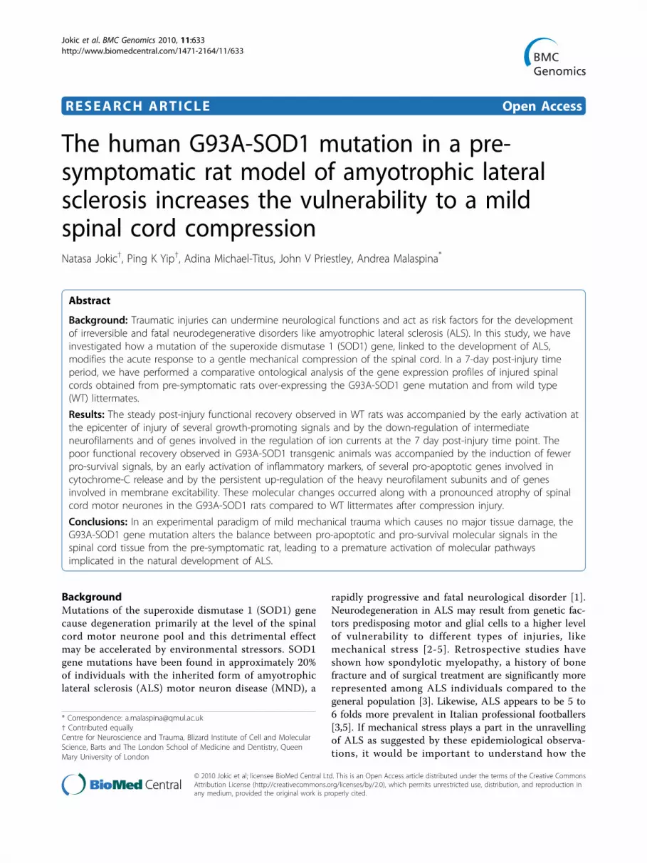
following injury [26,27]. At 7 days after spinal cordcompression in the WT and in the G93A-SOD1 rats,there was an increase in the number of ED1-, OX42-and GFAP- immunopositive cells in the spinal cord cau-dal to the compression site (Figure 10-11. However, nosignificant difference was observed between the WT andthe G93A-SOD1 rats (ED1, P = 0.08; OX42, P = 0.76;GFAP, P = 0.31).
Reduced motor neurones size in the G93A-SOD1 spinalcord following compression spinal cord injuryTo examine motor neurones, Nissl staining was usedsince the motor neurone marker choline acetyltransferasehas been shown to be significantly reduced followingspinal cord injury [28]. Motor neurone morphology canbe clearly seen in the ventral horn of a spinal cord regioncaudal to the injury epicenter 7 days after mild compres-sion SCI (Figure 11A-F). At this post-injury time point,the number of motor neurones in the ventral part of thespinal cord of WT rats did not differ from the number of
motor neurones in the G93A-SOD1 rats (Figure 11H).However, motor neurones in the anterior horns from theG93A-SOD1 rats were significantly smaller in soma size(Kolmogorov-Smirnov test, p < 0.05) compared to thosefrom WT littermates (Figure 11I).
DiscussionThis study shows how the over-expression of the G93A-SOD1 gene mutation modifies the molecular response toa mild mechanical compression in the spinal cord tissuefrom a pre-symptomatic rat. Using a histopathologicalassessment of the compression injury, we have found nosignificant differences between the WT and the G93A-SOD1 animals in the post-injury immunoresponse or inthe amount of spared white matter following the trauma(Figures 10-11). Hence, despite delivering comparablespinal cord compression injuries to pre-symptomatic WTand G93A-SOD1 rats, G93A-SOD1 rats displayed a sig-nificantly lower level of post-injury locomotor recoverythan their WT littermates. We observe that the
Figure 7 Histogram representing the G93A-SOD1 versus WT ratios of the spinal cord intensity values detected for Nfh, Map1b,S100A8, Timp2 and Hcn2 at 24 hours and at 7 days from compression injury, as measured by Bead-array and real-time RT-PCRanalysis. The G93A-SOD1 vs WT expression ratios obtained using Bead-array (red code) and real time RT-PCR (blue code) analyses for Nfh,Map1b, S100A8, Timp2 and Hcn2 at two time points after compression SCI are reported. The two techniques of gene expression analysis confirmindependently the same type of differential regulation for the selected gene candidates. S100A8 real-time RT-PCR confirms both the significantup-regulation in the G93A-SOD1 spinal cord detected by Bead-array analysis 7 days from compression SCI. Map1b RT-PCR has been included inorder to validate the sensitivity of the real time RT-PCR and Bead array analyses for expression changes close to two-folds.
Jokic et al. BMC Genomics 2010, 11:633http://www.biomedcentral.com/1471-2164/11/633
Page 12 of 24

Figure 8 Expression of neurofilament heavy chain and synaptophysin in ventral spinal cord at 24 hours after compression SCI.Neurofilament heavy chain (Nfh; clone N52) staining of spinal cord sections is clearly visible in motor neurons (arrows) and adjacent axons inboth WT (A) and G93A-SOD1 (B) which is significantly stronger in the G93A-SOD1 (* P = 0.03; E). Scale bar = 100 μm. Transverse sections aretaken within a segment 10 mm caudal to the injury epicenter. Synaptophysin (SYN) immunoreactive synaptic boutons (arrows) can be seensurrounding unstained motor neuron cell bodies in both WT (C) and G93A-SOD1 (D) spinal cord, with no difference in SYN distribution betweenthe two genetic types. The intensity of staining is non-significantly different between the G93A-SOD1 and wild type spinal cords (F). Scale bar =25 μm. SCI-SOD1: spinal cord tissue from rats over-expressing the G93A-SOD1 gene mutation. SCI-WT: spinal cord tissue from WT rats. Westernblot of spinal cord rostral to the injury epicenter obtained from WT and G93A-SOD1 rats. Three spinal cord samples for both WT and G93A-SOD1were used (G). A significant increase in Nfh (clone 52) expression levels can be detected in G93A-SOD1 rats compared to WT rats with b-actin asthe internal control (* P = 0.041; H).
Jokic et al. BMC Genomics 2010, 11:633http://www.biomedcentral.com/1471-2164/11/633
Page 13 of 24

significant differences in the post-injury locomotor recov-ery between the WT and the G93A-SOD1 rats are notdue to differences in the induction of the injury, butrather to an intrinsically different molecular response totrauma in the two genetic variants of rats. A significantlylow level of functional recovery has also been reported inG93A-SOD1 transgenic mice after sciatic nerve injury,
with an acceleration of the disease progression so that 90day old mice show deficits that are normally only seen atthe end stage in uninjured G93A-SOD1 mice [17].Whilst mutations of the SOD1 gene account only for
approximately 2% of the whole ALS population, othernot yet identified predisposing or causative genetic fac-tors may act along the same lines, reducing the neurore-generative potential and escalating the level of disabilityafter trauma. These observations may partially explainthe observed higher occurrence of ALS cases amongindividuals that have been exposed to repetitivemechanical trauma or that have been subjected to otherforms of mechanical stress or surgical procedures[2,3,5].For the purpose of our study, we have employed a well-
characterised experimental platform of mechanical stress,which is known to cause only a mild damage to the spinalcord and to be associated with a good outcome in termsof post-traumatic locomotor recovery in rodents[29,19,30]. Other forms of injury cause a significant levelof tissue destruction and a more florid profile of geneexpression changes, masking subtle transcriptionalchanges that may be induced by the SOD1 gene mutationin an otherwise macroscopically healthy spinal cord.Under the experimental conditions of mild SCI, theG93A-SOD1 spinal cord displays a higher number of up-regulated genes compared to WT tissue at the 4 hourpost-injury time point (Figure 2A-B). These include abroad range of genes involved in the modulation ofmacrophages, Toll-response, T and B-cell proliferationand in the production of interleukin-6 and TNF-alpha,featuring an early inflammatory response that surfacesonly at a later time point in WT spinal cord (Figure3A-B). Other gene expression changes unique to theG93A-SOD1 animal are detected in two later timepoints within the experimental period. Following the4-hour time point, the G93A-SOD1 spinal cord displayscharacteristic apoptotic signals, like the activation ofgenes regulating cytochrome C release from the mito-chondria. Cytochrome C release is known to occurwhen the inner mitochondrial membrane becomesexcessively permeable to ions, leading to energy failureand apoptosis [31,12]. The inflammatory milieu and theactivation of cell-death signals in the spinal cord at thisstage of the animal recovery may reduce the tissuerepair potential contributing to the observed poor func-tional recovery observed in the G93A-SOD1 rat.The post-injury molecular profile of the G93A-SOD1
spinal cord does not show the down-regulation of alarge number of genes 7 days from compression SCIdemonstrated in WT rats compared to naïve controls(Figures 2A-B; 3D). For example, those genes thatcontrol cholesterol and isoprenoid biosynthesis remainup-regulated in the G93A-SOD1 spinal cord at this
Figure 9 Spared white matter in spinal cord caudal tocompression epicenter. Spared white matter stained with luxolfast blue can be seen clearly in WT (A) and G93A-SOD1 (B) insections caudal to the compression epicenter 7 days aftercompression SCI. Morphometric analysis of the cross sectional areasof luxol fast blue stained white matter in WT and G93A-SOD1showed no significant difference between groups (P = 0.30, C). ns =non-significant. Scale bar = 200 μm.
Jokic et al. BMC Genomics 2010, 11:633http://www.biomedcentral.com/1471-2164/11/633
Page 14 of 24
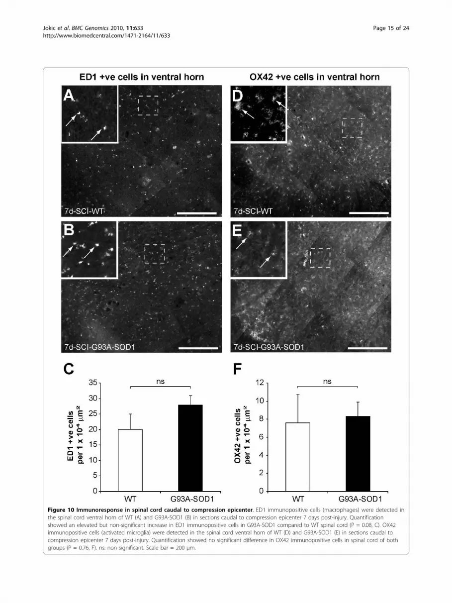
Figure 10 Immunoresponse in spinal cord caudal to compression epicenter. ED1 immunopositive cells (macrophages) were detected inthe spinal cord ventral horn of WT (A) and G93A-SOD1 (B) in sections caudal to compression epicenter 7 days post-injury. Quantificationshowed an elevated but non-significant increase in ED1 immunopositive cells in G93A-SOD1 compared to WT spinal cord (P = 0.08, C). OX42immunopositive cells (activated microglia) were detected in the spinal cord ventral horn of WT (D) and G93A-SOD1 (E) in sections caudal tocompression epicenter 7 days post-injury. Quantification showed no significant difference in OX42 immunopositive cells in spinal cord of bothgroups (P = 0.76, F). ns: non-significant. Scale bar = 200 μm.
Jokic et al. BMC Genomics 2010, 11:633http://www.biomedcentral.com/1471-2164/11/633
Page 15 of 24

time-point after compression injury. Isoprenoids are bio-logical precursors of carotenes and of coenzyme-Q, reti-nol and tocopherol (vitamin E), compounds known tomodulate mitochondrial activity and oxidative stress[32,33]. A previous report has documented a state ofsystemic dyslipidemia in individuals with ALS and this
state of altered lipid homeostasis has been linked to apotential neuroprotective effect [34]. The presence of aSOD1 gene mutation or of a genetic predisposition todevelop ALS may be associated to a state of dysregula-tion of molecular pathways of cholesterol and isoprenoidbiosynthesis, both during the natural development of the
Figure 11 Changes affecting astrocytes and motor neurones in spinal cord caudal to the compression epicentre. GFAP immunopositivecells (astrocytes) were detected in the spinal cord ventral horns of the WT (A) and of the G93A-SOD1 (D) rats, in sections caudal to thecompression epicenter 7 days post-injury. Quantification showed an elevated but non-significant increase in GFAP immunopositive cells in theG93A-SOD1 compared to the WT spinal cord (P = 0.31, G). Large Nissl stained cells (motor neurones) were detected in the spinal cord ventralhorn of WT (B-C) and of G93A-SOD1 (E-F) in sections caudal to the compression epicenter 7 days post-injury. Although the number of motorneurones in the ventral horns are not significantly different between the two groups, further analysis of the motor cells soma size showed thatthe motor neurones in the WT spinal cord were larger than those identified in the G93A-SOD1 spinal cord (H-I). Kolmogorov-Smirnov testrevealed a significant leftward shift in motor neurone soma size of the G93A-SOD1 spinal cord compared to WT spinal cord (P < 0.05, I). ns: non-significant. Scale bar = 200 μm.
Jokic et al. BMC Genomics 2010, 11:633http://www.biomedcentral.com/1471-2164/11/633
Page 16 of 24

disease and in response to a stressful situation like amechanical injury.Also down-regulated in WT spinal cord at the 7-day
post-injury time point are many genes that like Hcn2control cell-cell signalling and ion/neurotransmittertransport (GO:0019226; GO:0006836, Figures 3D and 6).Previous experimental observations in animal models ofALS and brain ischemia have reported increased neuro-nal excitability in affected tissues, generated by the up-regulation of genes capable of modulating ion currents[3,13]. Hence, the acquisition of a state of tissue hyper-excitability may represent a distinguishing feature ofSOD1-induced ALS, both during the natural develop-ment of the disease and also in a pre-symptomaticphase as a result of a stressful condition.At the 7 day time-point, both the WT and the G93A-
SOD1 spinal cords up-regulate genes involved in retinol(vitamin A) metabolism including RBP1 and CRABP2,whilst only the G93A-SOD1 spinal cord tissue presentsthe additional up-regulation of ADH1 and ALDH1 (Fig-ure 4B). A link between the vitamin-A down-streameffects and the pathological changes observed in ALShas already been suggested [35-38]. Early vitamin-Adeprivation, for example, causes motor cell loss inrodents [35]. RBP1 and CRABP2 are over-expressed inspinal cord from the end-stage G93A-SOD1 rat, whilstsurviving small motor neurons show selective immunos-taining for specific retinoid receptors [36-38]. Ourobservations in the acute post-injury phase may inspirepotential neuroprotective therapeutic strategies, sinceboth the alteration of ion current regulation and theactivation of retinoid signalling can be pharmacologicallymodified.We and others have already reported the post-injury
down-regulation and/or accumulation of phosphorylatedand non-phosphorylated neurofilaments and of synapto-physins (SYN), the latter appearing to be linked to post-traumatic motor and cognitive deficits in different mod-els of neurotrauma [24,25,6,39]. Given the reportedtrauma-induced differential regulation of Nfh and SYNexpression along the motor cell/axon/synapsis axis, wehave evaluated the Nfh and SYN differential regulationin our injury model. At a RNA level, Nfh expressionappears to be higher in injured G93A-SOD1 spinal cordcompared to injured WT tissue from the 4 hour timepoint onward (Figure 4A). Post-injury protein expressionof Nfh measured by immunohistochemistry is also ten-dentially higher in the G93A-SOD1 spinal cord at allthe time points, with a significant level of differentialexpression at the 24 hour time-point, which was furtherconfirmed with Western blotting (Figures 4, 5, 6, 7, 8).This observation may indicate that neurofilamentsexpression is altered in a situation of genetic susceptibil-ity to develop ALS, both under conditions of stress and
during the natural development of the disease. Inter-mediate neurofilaments like peripherin within spheroid-like structures are already known to accumulate inaffected tissues from animal models of ALS and to pos-sibly interfere with axonal transport [13]. We did notobserve any significant differential regulation or anygenotype-specific redistribution of SYN in spinal cordafter compression SCI. Failure to detect a post-injurychange in SYN expression or re-distribution in ourstudy may be related to the relatively low force ofimpact or to the fact that changes in synaptic vesicletransportation as a consequence of a mild compressionSCI may have not yet occurred in its full scale in the 7-day time period. Another indication that SOD1 genedefects may act on the homeostasis of neurofilamentswhen nervous tissue is under stress is the up-regulationof Mapb1 and possibly of Mast1 in the post-injuryG93A-SOD1 spinal cord, both genes appearing to bedown-regulated in WT spinal cord after compressioninjury (Figure 6). Mapb1 binds to microfilaments linkingthem with the microtubule system, whilst remainingassociated to the plasma membranes. Mapb1 exerts acentral role in the process of axonal elongation andbranching [40]. The observed selective activation ofMap1b in G93A-SOD1 spinal cord could be part of acompensative neuroprotective response that countersthe intrinsic SOD1-mediated vulnerability under condi-tions of stress. This survival program may be mediatedby the activation of molecules involved in the mainte-nance of the cytoskeletal integrity as well as by the pro-duction of cholesterol and isoprenoids derivates asreported above.Microarray studies in spinal cord have shown that
sham operation alone can induce gene expressionchanges similar to those caused by mild injury, but onlyin the first few hours after trauma [8,22]. Our studyconfirms that simple laminectomy triggers the transcrip-tional regulation of a wide range of gene categories inboth the WT and the G93A-SOD1 spinal cords. How-ever, gene categories involved in transcription, differen-tiation, ion homeostasis, apoptosis, organisation ofmitochondrial function and interleukin-6 release becomeactivated only in the G93A-SOD1 spinal cord at 4 hoursfrom laminectomy. It is clear from our experiments thatboth laminectomy and mild spinal cord injury provoke amixture of pro-survival and pro-apoptotic expressionchanges, the latter being dominated by mitochondrialcell-death signals and interleukin-6-mediated inflamma-tory changes.In this study, we have attempted to correlate the poor
functional recovery observed in the G93A-SOD1 ratsduring the acute post-injury phase with changes at thelevel of individual cell types in the affected spinal cord.We have shown only a trend towards an increase in
Jokic et al. BMC Genomics 2010, 11:633http://www.biomedcentral.com/1471-2164/11/633
Page 17 of 24

microglia, macrophages and astrocytes in a cord seg-ment caudal to the injury epicenter in the G93A-SOD1spinal cord compared to WT littermates under the sameexperimental conditions, but no significant changes inthe number of these cells between the two groups. Thisobservation is in line with a recent study that has ana-lysed the effects of a longitudinal stab injury of the lum-bar spinal cord region of the same pre-symptomaticanimal model of ALS utilised in our study [41]. In thisexperimental paradigm of more invasive spinal cordinjury, the level of host glial activation and the motorcell numbers at the site of the lesion in a 2-week post-injury period were not significantly different in theinjured G93A-SOD1 rats compared to the injured WTlittermates [41]. Our study demonstrates that the size ofthe anterior horns motor cells in the G93A-SOD1 ani-mals after compression SCI is significantly reducedwhen compared to the WT motor cell population. Pre-vious studies on animal models of ALS have also showna loss of the largest spinal motor neurons with the dis-ease progression [42]. Various attempts to clarifywhether the late stage loss of large motor neurones isdue to a higher vulnerability of these neurones, whichmay die or atrophy earlier than the small ones orwhether motor cells never reach their maximal sizebefore disease onset have not come to any conclusion[43]. It is also not known whether the loss or the atro-phy of large motor neurons is the main determinant ofthe functional decline and ultimately of the loss ofmotor units observed in the overt phase of the disease.Clearly, the same type of uncertainties may apply to theinterpretation of our results. The presence of smallermotor neurones in the G93A-SOD1 spinal cord oneweek after compression injury may play a part in thepoor locomotor performances observed in the G93A-SOD1 rats compared to their WT littermates under tothe same experimental conditions. The overall molecularchanges we have observed in the G93-SOD1 spinal cordin the post-injury phase may also be responsible of thepoor functional recovery described in the G93-SOD1rats.Similarly to a number of published investigations, our
study has used WT littermates as control for thehuman G93A-SOD1 rats, in order to ensure the maxi-mum level of genetic homogeneity between the groupsof animals. This choice is of particular importancewhen studying changes at a genetic level [6,36,41,44-48].Previous studies have shown that the human G93A-SOD1 transgene does not affect the endogenous ratSOD1 protein levels [49]. However, it has also beenshown that the total SOD1 activity in the G93A-SOD1rats is increased to 200-300% of the control level, as theresult of the combined endogenous SOD1 activity ofthe rat and of the added mutant G93A-SOD1 activity
[49]. This observation is clearly important in the inter-pretation of our results. In future studies, it will be use-ful to include rats with elevated non-mutant SOD1activity as an extra control group, in order to normalisewhatever experimental approach to comparable levels ofSOD1 activity [50].
ConclusionsOur study demonstrates a higher level of functionalimpairment in the pre-symptomatic G93A-SOD1 ratscompared to their control WT littermates after mechan-ical injury of the spinal cord, in the acute 1-weekpost-injury phase. Comparative gene ontological analysisindicates that the molecular response to a mild com-pression SCI or to a simple surgical stress in pre-symp-tomatic G93A-SOD1 rats genetically-modified todevelop ALS is distinctively different to that seen incontrol WT littermates. The response to SCI in theG93A-SOD1 spinal cord seems to replicate some of themost crucial gene expression changes described in theaffected tissues during the natural development of ALSand to demonstrate the same changes in the morphol-ogy of the motor cell population observed in spinal cordfrom individuals and animal models of the disease in anadvanced stage. The sustained recovery after injuryobserved in WT rats may be driven, at a molecularlevel, by a lower level of pro-apoptotic activity coupledwith the significant over-expression of factors involvedin transcription, angiogenesis, lipid transport and celladhesion. The results presented here contribute to theunderstanding of how the genetic background affectsthe ability of nervous tissues to withstand a mechanicaltrauma, showing how a G93A-SOD1 gene mutation cre-ates a milieu of gene expression changes which has anunfavourable effect on functional recovery following amild compression injury to the spinal cord.
MethodsAnimalsA breeding project was initiated at Taconic USA (Taco-nic Inc., US), using 5 transgenic Sprague Dawley ratsexpressing the G93A SOD1 gene mutation and 5 wild-type (WT) littermates as breeding pairs, as previouslyreported [6]. The G93A-SOD1 breeders were replacedon a monthly basis before any sign of disease onset. Anaverage of 6 pups for each breeding pair were obtainedand stored in separate cages. Only female pups wereretained for further analysis and tail samples were takenfor genotyping. Only female rats have been used toensure consistence in rat gender with other studies ofspinal cord injury (SCI) [6,19]. Since SCI requires manualemptying of bladder to prevent urinary tract infections,we have chosen female animals because their bladdersare relatively easier to empty compared to male bladders.
Jokic et al. BMC Genomics 2010, 11:633http://www.biomedcentral.com/1471-2164/11/633
Page 18 of 24

A total of 40 heterozygous rats carrying the G93A-SOD1gene mutation and 40 Wild type (WT) littermates havebeen shipped to our laboratories at the age of 6 weeksand housed in specific pathogen-free animal facilities at aroom temperature of 21°C (under a 12 h light-darkcycle). All animal procedures were conducted accordingto the Animals (Scientific Procedures) Act 1986 asapproved by the United Kingdom Home Office. TheG93A-SOD1 transgenic rat model of ALS is known todevelop initial signs of motor impairment with eitherhindlimb or forelimb distribution, at approximately17 weeks of age, and to progress to end-stage disease inapproximately 1 or 2 weeks from disease onset [6]. Wehave chosen the G93A-SOD1 rat rather than the morewidely used G93A-SOD1 mouse because of the need tooperate with an animal more suitable to well-establishedexperimental procedures for spinal cord injury.
Laminectomy and spinal cord compressionCompression SCI was performed on 27 pre-symptomaticG93A-SOD1 transgenic female rats at 10 weeks of age,an early pre-symptomatic stage for these rodent modelsof ALS. Compression SCI was also performed on 27 WTfemale age-matched littermates for reference as pre-viously reported [6]. 10 WT and 10 G93A-SOD1 femalerats were subjected only to laminectomy, at 10 weeks ofage. Surgical procedures were performed as previouslydescribed [29,6,19,30]. Following anaesthesia with a mix-ture of isoflurane (2.5%), oxygen and nitrous oxide (1:1ratio) at a flow rate of 750–1000 mL min, the skin andmuscle surrounding the vertebral column (T10-L1) wereincised and a laminectomy was performed at T12 levelwithout damaging the dura. The compression injury wasperformed by statically applying a 35g weight for 5 min,on a platform (area 2 mm × 5 mm), resting on the duraof the exposed spinal segment. Manual bladder expres-sion after surgery was performed twice daily. As pre-viously reported [6], rats were sacrificed by asphyxiationwith carbon dioxide after surgery at the following timepoints: 5 WT and 5 G93A-SOD1 rats at 30 minutes aftercompression SCI, 5 WT and 5 G93A-SOD1 rats at4 hours after compression SCI, 4 WT and 4 G93A-SOD1rats at 24 hours after compression SCI, 5 WT and 5G93A-SOD1 rats at 7 days after compression SCI, 3 WTand 3 G93A-SOD1 rats at 30 minutes after laminectomy,3 WT and 3 G93A-SOD1 rats at 4 hours after laminect-omy. In addition, 5 naïve G93A-SOD1 rats and 5 naïveWT littermates were also sacrificed at 10 weeks of age.Naïve spinal cord tissues from WT and G93A-SOD1 ani-mals were used as controls in the differential geneexpression analysis described below. Spinal cord samplesincluding the injury site and the adjacent segments (5mm rostral and caudal to the lesion epicenter), were
dissected after sacrifice, submerged into 2-methylbutaneand subsequently frozen in liquid nitrogen.
Locomotor analysis after compression SCIFollowing compression SCI, the level of functionalrecovery in G93A-SOD1 and WT female rats was evalu-ated initially at 4 hours, and then daily for a week usingthe BBB locomotor scoring system [20,51,52]. The eva-luation of the locomotor function in 24 10-week oldWT rats after mild compression SCI in a 7-day post-injury time window has previously been reported [6].The BBB scores for each animal was determined at thechosen time points by two independent blinded obser-vers and the mean BBB score per group was calculated.All G93A-SOD1 and WT rats have been independentlyscored using the BBB locomotor functional analysisprior to compression SCI in order to demonstratesimilar baseline performances.
Bead-array gene expression analysisLarge-scale gene expression analysis of the spinal cordsamples harvested from the 54 animals after mild com-pression SCI (epicenters of compression) or laminectomywas performed using the Illumina ratRef-12 v1.0 expres-sion Beadchip (Illumina, San Diego, USA; GEO GPL6101).This Bead-chip contains 12 genome-scale gene expressionmicroarrays (22,523 probes per array). RNA was extractedfrom the spinal cord samples obtained from injured anduninjured rats, and from sham operated animals subjectedto laminectomy, using the SV total RNA isolation system(Promega, UK). RNA quantification was performed usinga Nanodrop ND-1000 spectrophotometer and quality waschecked using the Agilent bioanalyser system (Agilent).For the purpose of our gene expression analysis, we havepooled the RNA samples obtained from the injury epicen-ters that have been dissected from rats of the same genetictype and sacrificed at the same post-injury time-point. Wehave obtained a total of 14 RNA-pools (4 pools containingRNAs from G93A-SOD1 cords extracted at the 30 min.(n:5), 4 hours (n:5), 24 hours (n:4) and 7 days (n:5) timepoints after compression SCI respectively, 2 pools contain-ing RNAs from G93A-SOD1 spinal cords extracted at the30 min. (n:4) and the 4 hours (n:4) time points after lami-nectomy, 2 pools containing RNAs from WT spinal cordsextracted at the 30 min. (n:4) and at the 4 hours (n:4) timepoints after laminectomy, 4 pools containing RNAs fromWT cords extracted at the 30 min (n:5), 4 hours (n:5), 24hours (n:4) and 7 days (n:5) time points after compressionSCI respectively; finally 1 pool containing G93A-SOD1(n:5) and 1 pool containing WT (n:5) spinal cordsextracted from 10 week-old naive animals (used as refer-ences in the gene expression analysis) of which we havetested the gene expression profile. The pool of RNA
Jokic et al. BMC Genomics 2010, 11:633http://www.biomedcentral.com/1471-2164/11/633
Page 19 of 24

samples from G93A-SOD1 spinal cords harvested at30 minutes from compression injury was also used for atechnical replica experiment.cRNA labeling was performed with 750 ng of pooled
RNA, using the Ambion Total Prep kit. cRNA purityand labelling was checked using the Nanodrop and Agi-lent bioanalyser. 500 ng from each of the 14 RNA poolswere hybridised simultaneously to 16 arrays (including 2arrays for the technical replica experiment) contained in2 RatRef-12 Expression BeadChips, as per Illumina pro-tocol (Illumina, San Diego, USA).Differential gene expression and correlation analysisthe Bead-chip output files were analyzed using theBeadStudio-3 Software (Illumina, San Diego, USA) aspreviously reported [6]. The expression level of eachprobe was defined by a value of intensity and by a p-value detection, which measures the probability of sig-nal recovery without specific probe target hybridiza-tion. Genes differentially expressed at the selectedtime-points after compression SCI and laminectomywere identified by comparing G93A-SOD1 and WTspinal cord tissues with genetically-matched naïve tis-sues. We have also performed a differential geneexpression analysis with a direct comparison of G93A-SOD1 and WT tissues at the selected time-points afterinjury. This approach allows the identification of genecandidates presenting different expression behavioursin the two tissues in study, but does not indicate thenature of the expression change in each spinal cordtype compared to genetically-matched un-injured(naïve) tissue. Therefore, genetically-matched naïve tis-sues were used as a reference for data interpretation.The gene expression changes identified in spinal cordfrom the naive (10-week old) G93A-SOD1 rats (usingthe spinal cord tissue from age-matched WT litter-mates as reference), have been taken into account forthe interpretation of the gene expression resultsobtained from injured spinal cord samples and fromspinal cords dissected from animals subjected tolaminectomy.Stringent selection criteria (Bead Studio-3), including
a rank invariant normalization algorithm, an IlluminaCustom error model (to compute non-specific cross-hybridization and the effects of variation arising fromnon-biological factors) and a false discovery rate (FDR)were used in the differential gene expression analysisas previously described [6]. To evaluate the reproduci-bility of our results, we have performed a correlationtest (BeadStudio scatter plot analysis) between thegene expression profiles generated from the G93A-SOD1 and the WT spinal cord tissues, with thoseobtained from naive control tissues of the correspond-ing genetic type. We have also extracted from thearray data, the intensity values of detection at the
various experimental time points after compressionSCI for Nfh and for other gene candidates involved inretinoid signaling (ReS).
Ontology analysis of the gene expression dataWe have used High-Throughput GoMiner as previouslyreported [6,21] for an ontological analysis of the geneexpression changes in spinal cord tissue from WT andG93A-SOD1 animals after compression injury and lami-nectomy. High-Throughput GoMiner provides an inte-grated biological interpretation of multiple geneexpression datasets. The program identifies those genecategories within the Gene Ontology (GO) databasewhich present a significant level of enrichment of thedifferentially regulated genes under investigation. Wehave submitted to High-Throughput GoMiner 9 text filesreporting “changed genes” found to be differentiallyregulated after compression SCI in G93A-SOD1 and inWT spinal cord pools extracted at 30 minutes, 4 hours,24 hours and 7 days from injury (tissues from geneti-cally-matched naïve rats sacrificed at the same timepoints were used as references) and in the 10-week oldG93A-SOD1 uninjured spinal cord pool compared to aWT naïve spinal cord pool from age-matched litter-mates. A set of four text files containing “changedgenes” identified in the G93A-SOD1 and in the WTspinal cord pools at 30 minutes and 4 hours after lami-nectomy were submitted separately, along with a list ofall the probes contained in the Illumina rat Beadchip.As previously reported, a flat +1 or -1 binary code wasused to express up-regulation or down-regulation of thegenes included in the 13 text files [6]. GO categorieswere identified and sorted according to a false discoveryrate (FDR; threshold of 0.05) and to the level of enrich-ment of “changed genes”. A multiple comparisons cor-rection was used to eliminate GO categories thatappeared significantly represented simply by chance.The FDR value for a specific GO category identified in atext file was automatically computed for all the remain-ing text files, allowing an estimation of the relativeimportance of each gene category across all time pointsand experimental groups. Visual integration of theresults was obtained using clustered image maps and anExcel drawing platform and the results have been dis-played as heat maps as previously reported [6].
cDNA synthesis and real time RT-PCRRNA from the epicenters of injury in the thoracic spinalcord samples obtained from rats of the same genetictype sacrificed at the same time point from compressioninjury were pooled together (5 samples for each pool)and used for cDNA synthesis using the ImProm-IIReverse Transcription System (Promega, Madison, Wis-consin). 2.5 μg of total RNA from each pool was treated
Jokic et al. BMC Genomics 2010, 11:633http://www.biomedcentral.com/1471-2164/11/633
Page 20 of 24

with RNase-free DNase and then heated to 75°C for10 minutes for DNase inactivation and RNA denatura-tion. Reverse-transcription was carried out with 1 μlrandom primers (150 ng/μl) and with 1 μg of RNA. Amixture of 4 μl 5 × reaction buffer, 2 μl of 25 mMMgCl2, 1 μl of 10 mM dNTP mix, 0.5 μl recombinantRNasin ribonuclease inhibitor (40 U/μl) and 1 μlImProm-II™ Reverse Transcriptase (Promega, Madison,Wisconsin) was added to each sample and double dis-tilled H2O up to a total of 15 μl. The samples wereincubated at 25°C for 5 minutes, then at 55°C for 60minutes and finally at 70°C for 15 minutes. Negativecontrol had no ImProm-II™ Reverse Transcriptase.Real time RT-PCRReal time RT-PCR was performed with SYBR Green(ABgene, Epsom, UK), using a Rotor-Gene 3000system (Corbett Research, Sydney, Australia). Primers(Invitrogen, Paisley, UK) were designed and used in theamplification process for the following genes: the neuro-filament heavy chain (Nfh F: AGA GGC AGA AGAGGG AGG AG - Nfh R: TGA CCT CAG CTG GTGACT TG); the microtubule-associated protein 1B(Map1b 1 F: CCT GCC AAA GAA CTT GAA GC -Map1b 1 R: CCT TTG CTG ACT TCC GTC TC);S100A8 protein (S100A8 1 F: GGC AAC TGA ACTGGA GAA GG - S100A8 1 R: ACC CTT ATC ACCAAC GCA AG); nucleotide-gated potassium channel 2(Hcn2 F: CTG CGT GAG GAG ATT GTG AA - Hcn2R: TTT GAG CTT TGT CAG CAT GG); the tissueinhibitor of metalloproteinase 2 (Timp2 1 F: CAA GTTCTT TGC CTG CAT CA - Timp2 1 R: TCC AGGAAG GGA TGT CAA AG). Amplification conditionswere as follows: 95°C for 15 min, followed by 40 cyclesof 95°C for 10 s; 60°C for 15 s; 72°C for 20 s. Each realtime PCR experiment was performed in triplicate, usingcDNA templates originated from 2.5 μg of RNA. Theexpression of the test genes was evaluated followingnormalization to the level of ribosomal 18 S containedin each sample. Due to the 18 S abundance in the tis-sues, the 18 S cDNAs were diluted 100-fold before RNAmeasurement. A standard curve was generated by real-time RT-PCR analysis from triplicates of five ten-folddilutions of cDNA generated from 1 ng of spleen RNA.Real time PCR runs showing mRNA expression signalsfor the negative control samples were discarded andnew cDNA was generated for a re-run.
ImmunohistochemistryThe protein expression of the neurofilament heavy chain(Nfh) and of synaptophysin (SYN) in spinal cord aftercompression SCI has been studied by immunohisto-chemistry. Spinal cord samples immediately caudal tothe site of compression SCI were harvested from 3 WTand 3 G93A-SOD1 rats at 30 min, 4 h, 24 h and 7 days
after compression SCI and from naïve 10-week old rats,for immunostaining. Tissue samples were frozen inliquid nitrogen and stored at -80°C. Fresh frozen tissueswere mounted on tissue holders and 15 μm sectionswere cut on a cryostat. Slides with fresh frozen tissuewere post-fixed with 100% ethanol for 10 minutes, fol-lowed by 3 washes for 10 minutes in phosphate bufferedsaline (PBS). Endogenous peroxidases were inhibited bytreating slides with 0.3% H2O2 for 10 minutes. After 3 ×PBS washes for 10 minutes, non-specific antibody siteswere blocked with 10% donkey serum in 0.3% triton/0.1% sodium azide PBS solution for 2 h. Primary anti-mouse antibodies for Nfh (clone N52, 1:20,000, Sigma)and SYN (1:5,000, Sigma) diluted in 0.3% triton/0.1%sodium azide and PBS solution were applied for 24 hfollowed by biotinylated secondary anti-mouse (for Nfh)and anti-rabbit (for SYN) antibodies at dilution 1:800 in0.3% triton/0.1% sodium azide PBS solution for 1 h.Vectastain Elite ABC Kit (Vector laboratories) was sub-sequently added for 30 minutes to form an avidin-biotincomplex. The reaction was visualised after incubationwith 3, 3’-diaminobenzidine (DAB, Sigma) and sectionswere mounted in xylene-based DPX mounting medium.The Nfh antibody (clone N52) has been previously usedin our lab and published by other authors. It has beenextensively characterised by Western blot analysis, testedfor cross-reaction with other neurofilaments, and it hasbecome an established marker for myelinated DRG neu-rons [53,51,52,27]. The synaptophysin antibody (SigmaS5768; mouse synaptosome preparation from rat retina-1:500 dilution) has also been extensively tested and itsimmunoreactivity has been quantified using the samemethod previously reported [54].Histopathological assessment of the WT and G93A-
SOD1 rat spinal cord after mild compression injuryusing luxol fast blue staining and inflammatory markerswas performed as previously described [26-29,54-56].We have used frozen 20 μm transverse spinal cord sec-tions caudal to the compression site from animals sacri-ficed 7 days after compression injury. Sections weredehydrated in a graded ethanol series (5 min each) andthen incubated overnight in 0.1% luxol fast blue solutionat 37°C. Differentiation of slides was carried out with0.05% lithium carbonate to distinguish white and graymatter staining and then dehydrated in a graded ethanolseries. Slides were dipped in xylene and mounted inDPX mounting medium. Slides with fresh frozen tissuefor immunohistochemistry were post-fixed with 4% par-aformaldehyde for 1 h, followed by 3 washes for 5 min-utes in PBS. Immunofluorescent staining with mouseanti- ED1 (1:400, Chemicon), mouse anti-OX42 (1:200,Serotec) and rabbit anti-GFAP (1:1000, Dako) was car-ried out overnight at room temperature. After 3 × PBSwashes for 5 minutes, secondary donkey anti-mouse
Jokic et al. BMC Genomics 2010, 11:633http://www.biomedcentral.com/1471-2164/11/633
Page 21 of 24

AlexaFluor 488 or goat anti-rabbit AlexaFluor 568(1:1000, Invitrogen) was applied for 3 h at room tem-perature. Nissl staining of motor neurones was carriedout using Neurotrace fluorescent Nissl stain (1:100, Invi-trogen) according to manufacturer’s instruction. After afurther 3 × PBS washes for 5 minutes, sections weremounted in Vectashield mounting medium containingwith or without DAPI (Vector Labs).A summary of the specifications of the antibodies used
for the immunohistochemical analyses is presented inTable 1.
Western blottingA spinal cord segment rostral to the compression site wasfreshly collected from 3 G93A-SOD1 and from 3 WT age-matched rats 24 hours from compression SCI and stored at-80°C until processed. The spinal cord tissues were pro-cessed for Western blot as previously described [57]. Briefly,spinal cord tissues were homogenized in 1 ml of ice-coldlysis buffer (20 mM HEPES pH 7.4, 100 nM NaCl, 100 mMNaF, 1 mM Na3VO4, 5 mM EDTA, 1% Nonidet P-40 and 1× protease inhibitor cocktail; Roche). The lysates wererotated for 2 h at 4°C then centrifuged at 13,500 g for 15min at 4°C. Total protein concentration was determined forthe collected supernatant using a bicinchoninic acid protein
assay kit (Pierce). Fifteen micrograms of total protein wereelectrophoresed on 12% acrylamide gel before transfer ontoHybond P membranes (Amersham) and incubated over-night at 4°C with mouse anti-NF200 (clone N52, 1:1,500)and mouse anti-b-actin (1:5,000). Although both antibodieswere raised in mouse, the difference in molecular size(NF200 is 200 KDa and b-actin is 42 KDa) allows for simul-taneous probing on the same blot. Visualisation was per-formed using the secondary antibody goat anti-mouseIRdye-680CW (LI-COR Biosciences). Fluorescent blot wasimaged on the Odyssey Infrared Imaging System (LI-CORBiosciences). The bands were quantified using AxioVision4.6 program and the results were expressed as a ratio withits corresponding internal control b-actin.Histological analysisThe intensity of Nfh staining in motor neurons (10 –15 per section) was measured at 20 × magnificationusing Scion Image (release alpha 4.0.3.2). The averageintensity of Nfh staining (gray level) of motor neuronswas measured and the average background staining inten-sity was subtracted. Analysis of SYN-immunoreactivesynaptic boutons was performed at 40× magnification.The average intensity (gray levels) of SYN-stained nerveterminals and subtracted the average staining intensity ofthe background was determined. The analysis of ED1,
Table 1 Main characteristics of the antibodies used for the experiments detailed in the manuscript
Manufacturer Product &Lot No.
Hostspecies
Immunogen Dilution used
Primary antibodies
Neurofilament (cloneN52)
Sigma N0142 &65K4804
mouse C-terminal segment of enzymaticallydephosphorylated pig neurofilament 200
1:20,000 usingimmunoperoxidase staining1:1500 using Western blotting
Synaptophysin (cloneSVP-38)
Sigma S5768 &90K4844
mouse Rat retina synaptosome 1:5000 using immunoperoxidasestaining
Rat CD68-macrophages/
monocytes (cloneED-1)
Chemicon MAB1435 &0606032963
mouse A single chain glycoprotein of 90-100 KDa that isexpressed on the lysosomal membrane of myeloid
cells
1:400 using immunofluorescentstaining
Rat CD11b; RPE(clone OX-42)
Serotec MCA275EL& 0105
mouse Rat peritoneal macrophages 1:200 using immunofluorescentstaining
Glial fibrillary acidprotein (clone SVP-
38)
Dako Z0334 &00045904
rabbit Cow spinal cord 1:1000 using Immunofluorescentstaining
b-actin (clone AC-15) Sigma A1978 &118K4827
mouse Fusion of mouse myeloma cells and splenocytesimmunized with a synthetic b-cytoplasmic actin N-
terminal peptide
1:5000 using Western blotting
NeuroTrace(fluorescent Nissl
stain)
MolecularProbes
N21480 &4971-21
- - 1:100 according tomanufacturer’s instruction
Secondary antibodies
Alexa Fluor 488 Invitrogen A21202 &536050
donkey Anti-mouse IgG (H+L) 1:1000 using immunofluorescentstaining
Alexa Fluor 568 Invitrogen A10042 &538952
donkey Anti-rabbit IgG (H+L) 1:1000 using immunofluorescentstaining
Alexa Fluor 680 Invitrogen A21057 &65E2-1
goat Anti-mouse IgG (H+L) 1:15,000 using Western blotting
Jokic et al. BMC Genomics 2010, 11:633http://www.biomedcentral.com/1471-2164/11/633
Page 22 of 24

OX42 and GFAP immunostaining was determined simi-larly to previously described [28,54]. Spinal cord sectionswere captured with the same intensity and settings usingthe Zeiss Axioplan 2 fluorescence microscope. The num-ber of positively stained ED1, OX42 and GFAP cells wasdetermined at 20 × magnification in a 1 × 104 μm2 boxplace over the ventral horn using the Axiovision 4.6program.To determine the amount of spared white matter cau-
dal to the injury, the area of white matter positivelystained with luxol fast blue was measured using theAxiovision 4.6 program. All analyses for immunohisto-chemical staining were performed in the ventral spinalcord of 6-8 non-adjacent sections of a spinal cord seg-ment caudal to the epicenter of injury from 3 WT and3 G93A-SOD1 animals.
Motor neurone analysis: numbers and cell sizeThe count of motor neurones and the analysis of themotor neurones cell size was carried out as previouslydescribed [57]. Cell area of Nissl labelled motor neu-rones with visible DAPI positive nuclei in the spinalcord ventral horn was acquired using the AxioVisionV4.6 program. Size and frequency distributions of motorneurones were determined for each rat and a mean dis-tribution calculated for both G93A-SOD1 and WT rats.At least six spinal cord sections were analysed andquantified per rat (n = 3-4 per group). Statistical analysiswas carried out using a two-sample Kolmogorov-Smir-nov test, performed against a significant threshold of0.05 to correct for multiple testing.
Statistical analysisThe data were presented as means and standard errorsof the means (SEM). The behavioural data wereanalysed with two-way ANOVA followed by Tukey’spost-hoc test. Histological and Western blot data wereanalysed with Student’s t-test. P < 0.05 was consideredstatistically significant.
AcknowledgementsWe thank Sharon Averill and Wenlong Huang for the excellent technicalassistance. We are grateful to The Royal London Hospital CharitableFoundation and to the Motor Neuron Disease Association UK for its financialsupport to the ongoing research projects on ALS.
Authors’ contributionsNJ has carried out all the surgical procedures of sham operation and spinalcord compression. She has performed all the functional analyses andprocessed all the tissue samples for the Bead-Array analysis and RT-PCRdescribed in the study. PKY and NJ have carried out all the histological,immunohistochemical work, along with cell count and analysis of theimages. AMT and JVP participated in the design of the study and havesupervised the direction of the experiments and the statistical analyses. AMhas performed all the gene expression data analyses and interpretationusing the bioinformatic tools reported in the manuscript. AM has conceived
the study, and participated in its design and coordination. All authors haveread and approved the final manuscript.
Received: 24 February 2010 Accepted: 15 November 2010Published: 15 November 2010
References1. Rosen DR, Siddique T, Patterson D, Figlewicz DA, Sapp P, Hentati A,
Donaldson D, Goto J, O’Regan JP, Deng HX: Mutations in Cu/Znsuperoxide dismutase gene are associated with familial amyotrophiclateral sclerosis. Nature 1993, 362:59-62.
2. Chio A, Benzi G, Dossena M, Mutani R, Mora G: Severely increased risk ofamyotrophic lateral sclerosis among Italian professional football players.Brain 2005, 128:472-476.
3. Kihira T, Kanno S, Miwa H, Okamoto K, Kondo T: The role of exogenousrisk factors in amyotrophic lateral sclerosis in Wakayama, Japan.Amyotroph Lateral Scler 2007, 8:150-156.
4. Pasinelli P, Brown RH: Molecular biology of amyotrophic lateral sclerosis:insights from genetics. Nat Rev Neurosci 2006, 7:710-723.
5. Valenti M, Pontieri FE, Conti F, Altobelli E, Manzoni T, Frati L: Amyotrophiclateral sclerosis and sports: a case-control study. Eur J Neurol 2005,12:223-225.
6. Malaspina A, Jokic N, Huang WL, Priestley JV: Comparative analysis of thetime-dependent functional and molecular changes in spinal corddegeneration induced by the G93A SOD1 gene mutation and bymechanical compression. BMC Genomics 2008, 9:500.
7. Dangond F, Hwang D, Camelo S, Pasinelli P, Frosch MP, Stephanopoulos G,Stephanopoulos G, Brown RH Jr, Gullans SR: Molecular signature of late-stage human ALS revealed by expression profiling of postmortem spinalcord gray matter. Physiol Genomics 2004, 16:229-239.
8. De BA, Knoblach SM, Di GS, Fan C, Molon A, Hoffman EP, Faden AI: Geneexpression profiling of experimental traumatic spinal cord injury as afunction of distance from impact site and injury severity. PhysiolGenomics 2005, 22:368-381.
9. Grossman SD, Rosenberg LJ, Wrathall JR: Temporal-spatial pattern of acuteneuronal and glial loss after spinal cord contusion. Exp Neurol 2001,168:273-282.
10. Kiernan MC, Bostock H: Effects of membrane polarization and ischaemiaon the excitability properties of human motor axons. Brain 2000, , 123 Pt12: 2542-2551.
11. Malaspina A, Kaushik N, de BJ: Differential expression of 14 genes inamyotrophic lateral sclerosis spinal cord detected using gridded cDNAarrays. J Neurochem 2001, 77:132-145.
12. McEwen ML, Springer JE: A mapping study of caspase-3 activationfollowing acute spinal cord contusion in rats. J Histochem Cytochem 2005,53:809-819.
13. Millecamps S, Robertson J, Lariviere R, Mallet J, Julien JP: Defective axonaltransport of neurofilament proteins in neurons overexpressingperipherin. J Neurochem 2006, 98:926-938.
14. Vucic S, Kiernan MC: Novel threshold tracking techniques suggest thatcortical hyperexcitability is an early feature of motor neuron disease.Brain 2006, 129:2436-2446.
15. Al-Chalabi A, Andersen PM, Nilsson P, Chioza B, Andersson JL, Russ C, Shaw CE,Powell JF, Leigh PN: Deletions of the heavy neurofilament subunit tail inamyotrophic lateral sclerosis. Hum Mol Genet 1999, 8:157-164.
16. Brettschneider J, Petzold A, Sussmuth SD, Ludolph AC, Tumani H: Axonaldamage markers in cerebrospinal fluid are increased in ALS. Neurology2006, 66:852-856.
17. Sharp PS, Dick JR, Greensmith L: The effect of peripheral nerve injury ondisease progression in the SOD1(G93A) mouse model of amyotrophiclateral sclerosis. Neuroscience 2005, 130:897-910.
18. Farooque M, Zhang Y, Holtz A, Olsson Y: Exudation of fibronectin andalbumin after spinal cord injury in rats. Acta Neuropathol 1992,84:613-620.
19. Nystrom B, Berglund JE, Bergquist E: Methodological analysis of anexperimental spinal cord compression model in the rat. Acta NeurolScand 1988, 78:460-466.
20. Basso DM, Beattie MS, Bresnahan JC: Graded histological and locomotoroutcomes after spinal cord contusion using the NYU weight-drop deviceversus transection. Exp Neurol 1996, 139:244-256.
Jokic et al. BMC Genomics 2010, 11:633http://www.biomedcentral.com/1471-2164/11/633
Page 23 of 24

21. Zeeberg BR, Qin H, Narasimhan S, Sunshine M, Cao H, Kane DW, Reimers M,Stephens RM, Bryant D, Burt SK, Elnekave E, Hari DM, Wynn TA,Cunningham-Rundles C, Stewart DM, Nelson D, Weinstein JN: High-Throughput GoMiner, an ‘industrial-strength’ integrative gene ontologytool for interpretation of multiple-microarray experiments, withapplication to studies of Common Variable Immune Deficiency (CVID).BMC Bioinformatics 2005, 6:168.
22. Di GS, Knoblach SM, Brandoli C, Aden SA, Hoffman EP, Faden AI: Geneprofiling in spinal cord injury shows role of cell cycle in neuronal death.Ann Neurol 2003, 53:454-468.
23. Sifringer M, Stefovska V, Zentner I, Hansen B, Stepulak A, Knaute C,Marzahn J, Ikonomidou C: The role of matrix metalloproteinases in infanttraumatic brain injury. Neurobiol Dis 2007, 25:526-535.
24. Fitzpatrick MO, Dewar D, Teasdale GM, Graham DI: The neuronalcytoskeleton in acute brain injury. Br J Neurosurg 1998, 12:313-317.
25. Gobbel GT, Bonfield C, Carson-Walter EB, Adelson PD: Diffuse alterations insynaptic protein expression following focal traumatic brain injury in theimmature rat. Childs Nerv Syst 2007, 23:1171-1179.
26. Clark AK, Yip PK, Grist J, Gentry C, Staniland AA, Marchand F, Dehvari M,Wotherspoon G, Winter J, Ullah J, Bevan S, Malcangio M: Inhibition ofspinal microglial cathepsin S for the reversal of neuropathic pain. ProcNatl Acad Sci USA 2007, 104:10655-10660.
27. Starkey ML, Davies M, Yip PK, Carter LM, Wong DJ, McMahon SB,Bradbury EJ: Expression of the regeneration-associated protein SPRR1A inprimary sensory neurons and spinal cord of the adult mouse followingperipheral and central injury. J Comp Neurol 2009, 513:51-68.
28. Gonzalez SL, Saravia F, Gonzalez Deniselle MC, Lima AE, De Nicola AF:Glucocorticoid regulation of motoneuronal parameters in rats withspinal cord injury. Cell Mol Neurobiol 1999, 19:597-611.
29. Holtz A, Nystrom B, Gerdin B, Olsson Y: Neuropathological changes andneurological function after spinal cord compression in the rat. JNeurotrauma 1990, 7:155-167.
30. Zhang Y, Hillered L, Olsson Y, Holtz A: Time course of energy perturbationafter compression trauma to the spinal cord: an experimental study inthe rat using microdialysis. Surg Neurol 1993, 39:297-304.
31. Goldstein JC, Munoz-Pinedo C, Ricci JE, Adams SR, Kelekar A, Schuler M,Tsien RY, Green DR: Cytochrome c is released in a single step duringapoptosis. Cell Death Differ 2005, 12:453-462.
32. Graf M, Ecker D, Horowski R, Kramer B, Riederer P, Gerlach M, Hager C,Ludolph AC, Becker G, Osterhage J, Jost WH, Schrank B, Stein C, Kostopulos P,Lubik S, Wekwerth K, Dengler R, Troeger M, Wuerz A, Hoge A, Schrader C,Schimke N, Krampfl K, Petri S, Zierz S, Eger K, Neudecker S, Traufeller K,Sievert M, Neundorfer B, Hecht M: High dose vitamin E therapy inamyotrophic lateral sclerosis as add-on therapy to riluzole: results of aplacebo-controlled double-blind study. J Neural Transm 2005, 112:649-660.
33. Halliwell B: Role of free radicals in the neurodegenerative diseases: therapeuticimplications for antioxidant treatment. Drugs Aging 2001, 18:685-716.
34. Dupuis L, Corcia P, Fergani A, Gonzalez De Aguilar JL, Bonnefont-Rousselot D, Bittar R, Seilhean D, Hauw JJ, Lacomblez L, Loeffler JP,Meininger V: Dyslipidemia is a protective factor in amyotrophic lateralsclerosis. Neurology 2008, 70:1004-1009.
35. Corcoran J, So PL, Maden M: Absence of retinoids can inducemotoneuron disease in the adult rat and a retinoid defect is present inmotoneuron disease patients. J Cell Sci 2002, 115:4735-4741.
36. Jokic N, Ling YY, Ward RE, Michael-Titus AT, Priestley JV, Malaspina A: Retinoidreceptors in chronic degeneration of the spinal cord: observations in a ratmodel of amyotrophic lateral sclerosis. J Neurochem 2007, 103:1821-1833.
37. Malaspina A, Turkheimer F: A review of the functional role and of theexpression profile of retinoid signaling and of nuclear receptors inhuman spinal cord. Brain Res Bull 2007, 71:437-446.
38. Malaspina A, Michael-Titus AT: Is the modulation of retinoid and retinoid-associated signaling a future therapeutic strategy in neurological traumaand neurodegeneration? J Neurochem 2008, 104:584-595.
39. Shojo H, Kibayashi K: Changes in localization of synaptophysin followingfluid percussion injury in the rat brain. Brain Res 2006, 1078:198-211.
40. Garcia ML, Cleveland DW: Going new places using an old MAP: tau,microtubules and human neurodegenerative disease. Curr Opin Cell Biol2001, 13:41-48.
41. Suzuki M, Klein S, Wetzel EA, Meyer M, McHugh J, Tork C, Hayes A,Svendsen CN: Acute glial activation by stab injuries does not lead to
overt damage or motor neuron degeneration in the G93A mutant SOD1rat model of amyotrophic lateral sclerosis. Exp Neurol 2010, 221:346-352.
42. Zang DW, Cheema SS: Degeneration of corticospinal and bulbospinalsystems in the superoxide dismutase 1(G93A G1H) transgenic mouse modelof familial amyotrophic lateral sclerosis. Neurosci Lett 2002, 332:99-102.
43. Feeney SJ, McKelvie PA, Austin L, Jean-Francois MJ, Kapsa R, Tombs SM,Byrne E: Presymptomatic motor neuron loss and reactive astrocytosis inthe SOD1 mouse model of amyotrophic lateral sclerosis. Muscle Nerve2001, 24:1510-1519.
44. Cassina P, Cassina A, Pehar M, Castellanos R, Gandelman M, de LA,Robinson KM, Mason RP, Beckman JS, Barbeito L, Radi R: Mitochondrialdysfunction in SOD1G93A-bearing astrocytes promotes motor neurondegeneration: prevention by mitochondrial-targeted antioxidants. JNeurosci 2008, 28:4115-4122.
45. Lepore AC, Rauck B, Dejea C, Pardo AC, Rao MS, Rothstein JD, Maragakis NJ:Focal transplantation-based astrocyte replacement is neuroprotective ina model of motor neuron disease. Nat Neurosci 2008, 11:1294-1301.
46. Lepore AC, Tolmie C, O’Donnell J, Wright MC, Dejea C, Rauck B, Hoke A,Ignagni AR, Onders RP, Maragakis NJ: Peripheral hyperstimulation alterssite of disease onset and course in SOD1 rats. Neurobiol Dis 2010,39:252-264.
47. Malaspina A, Ngoh SF, Ward RE, Hall JC, Tai FW, Yip PK, Jones C, Jokic N,Averill SA, Michael-Titus AT, Priestley JV: Activation transcription factor-3activation and the development of spinal cord degeneration in a ratmodel of amyotrophic lateral sclerosis. Neuroscience 2010, 169:812-827.
48. Woodruff TM, Costantini KJ, Crane JW, Atkin JD, Monk PN, Taylor SM,Noakes PG: The complement factor C5a contributes to pathology in a ratmodel of amyotrophic lateral sclerosis. J Immunol 2008, 181:8727-8734.
49. Nagai M, Aoki M, Miyoshi I, Kato M, Pasinelli P, Kasai N, Brown RH Jr,Itoyama Y: Rats expressing human cytosolic copper-zinc superoxidedismutase transgenes with amyotrophic lateral sclerosis: associatedmutations develop motor neuron disease. J Neurosci 2001, 21:9246-9254.
50. Sugawara T, Noshita N, Lewen A, Gasche Y, Ferrand-Drake M, Fujimura M,Morita-Fujimura Y, Chan PH: Overexpression of copper/zinc superoxidedismutase in transgenic rats protects vulnerable neurons againstischemic damage by blocking the mitochondrial pathway of caspaseactivation. J Neurosci 2002, 22:209-217.
51. Hahn CM, Kleinholz H, Koester MP, Grieser S, Thelen K, Pollerberg GE: Roleof cyclin-dependent kinase 5 and its activator P35 in local axon andgrowth cone stabilization. Neuroscience 2005, 134:449-465.
52. Huang WL, King VR, Curran OE, Dyall SC, Ward RE, Lal N, Priestley JV,Michael-Titus AT: A combination of intravenous and dietarydocosahexaenoic acid significantly improves outcome after spinal cordinjury. Brain 2007, 130:3004-3019.
53. Debus E, Weber K, Osborn M: Monoclonal antibodies specific for glialfibrillary acidic (GFA) protein and for each of the neurofilament tripletpolypeptides. Differentiation 1983, 25:193-203.
54. Ruscheweyh R, Forsthuber L, Schoffnegger D, Sandkuhler J: Modification ofclassical neurochemical markers in identified primary afferent neuronswith Abeta-, Adelta-, and C-fibers after chronic constriction injury inmice. J Comp Neurol 2007, 502:325-336.
55. Saganova K, Orendacova J, Cizkova D, Vanicky I: Limited minocyclineneuroprotection after balloon-compression spinal cord injury in the rat.Neurosci Lett 2008, 433:246-249.
56. Zelano J, Berg A, Thams S, Hailer NP, Cullheim S: SynCAM1 expressioncorrelates with restoration of central synapses on spinal motoneuronsafter two different models of peripheral nerve injury. J Comp Neurol2009, 517:670-682.
57. Yip PK, Wong LF, Sears TA, Yanez-Munoz RJ, McMahon SB: Corticaloverexpression of neuronal calcium sensor-1 induces functionalplasticity in spinal cord following unilateral pyramidal tract injury in rat.PLoS Biol 2010, 8:e1000399.
doi:10.1186/1471-2164-11-633Cite this article as: Jokic et al.: The human G93A-SOD1 mutation in apre-symptomatic rat model of amyotrophic lateral sclerosis increasesthe vulnerability to a mild spinal cord compression. BMC Genomics 201011:633.
Jokic et al. BMC Genomics 2010, 11:633http://www.biomedcentral.com/1471-2164/11/633
Page 24 of 24


