Embed Size (px)
Citation preview

PRIMARY RESEARCH Open Access
The in vitro influences of epidermal growth factorand heregulin-β1 on the efficacy of trastuzumabused in Her-2 positive breast adenocarcinomaTracey Hurrell* and Kim Outhoff
Abstract
Background: Human epidermal growth factor receptor-2 (Her-2) is over expressed in approximately 25-30% of allprimary breast tumors resulting in a distinctive breast cancer subtype associated with a poor prognosis and adecrease in overall survival. Trastuzumab (Herceptin®), an anti-Her-2 monoclonal antibody, has dramatically alteredthe prognosis of Her-2 positive breast cancer. Trastuzumab is, however, associated with primary and acquiredresistance.
Aim and methods: To investigate the in-vitro effects of trastuzumab on cell viability (tetrazolium conversion assay),cell cycling (propidium iodide staining), apoptosis (executioner caspases and annexin-V) and relative surface Her-2receptor expression (anti-Her-2 affibody molecule) in Her-2-positive (SK-Br-3) and oestrogen receptor positive(MCF-7) breast adenocarcinoma cells and to determine potential augmentation of these effects by twoendogenous ligands, epidermal growth factor (EGF) and heregulin-β1 (HRG- β1).Results: Cell viability was decreased in SK-Br-3 cells by exposure to trastuzumab. This was associated with G1accumulation and decreased relative surface Her-2 receptor density, supporting the cytostatic nature oftrastuzumab in vitro. SK-Br-3 cells exposed to EGF and heregulin-β1 produced differential cell responses alone andin combination with trastuzumab, in some instances augmenting cell viability and cell cycling. Relative surfaceHer-2 receptor density was reduced substantially by trastuzumab, EGF and heregulin-β1. These reductions wereamplified when ligands were used in combination with trastuzumab.
Conclusion: Cell type specific interactions of endogenous ligands appear to be dependent on absoluteHer-receptor expression and cross activation of signaling pathways. This supports the notion that receptor densityof Her-family members and multiplicity of growth ligands are of mutual importance in breast cancer cellproliferation and therefore also in resistance associated with trastuzumab.
Keywords: Her-2 receptors, Trastuzumab, EGF, Heregulin-β1, SK-Br-3 cells
BackgroundThe evolutionary ancient human epidermal growth fac-tor receptor (Her) family is composed of the structurallyrelated tyrosine kinases Her-1 (EGFR), Her-2, Her-3 andHer-4, which play a fundamental role in regulating cellfunctions including proliferation, adhesion, motility andsurvival [1-3]. Her-family receptors govern different func-tions and have different properties. Furthermore, Her-1,Her-3 and Her-4 possess the affinity for binding multiple
ligands [4,5]. Ligands for Her-1 include EGF, transforminggrowth factor-α, amphiregulin, betacellulin, epigen, epiregulinand heparin binding EGF-like growth factor while ligandsfor Her-3 and Her-4 include the isoforms of four structur-ally related heregulins [5].In contrast with the others, Her-2 receptors have no
known ligands and are thus designated as orphan receptors[6-8]. Rather, activation of these receptors is achieved bythe formation of interactive dimers, either spontaneouslywith Her-2 receptors or with other ligand-activated familymembers. Her-3 receptors are incapable of intrinsic kinaseactivity [9]; thus ordinarily, neither Her-2 nor Her-3 are
* Correspondence: [email protected] of Pharmacology, Faculty of Health Sciences, School ofMedicine, University of Pretoria, Private Bag X323, Pretoria 0007, South Africa
© 2013 Hurrell and Outhoff; licensee BioMed Central Ltd. This is an open access article distributed under the terms of theCreative Commons Attribution License (http://creativecommons.org/licenses/by/2.0), which permits unrestricted use,distribution, and reproduction in any medium, provided the original work is properly cited.
Hurrell and Outhoff Cancer Cell International 2013, 13:97http://www.cancerci.com/content/13/1/97

capable of linear signaling in isolation, which impliesthat they have strongly interdependent signaling char-acteristics [5].Modularity, redundancy and the capacity for combination
interactions are important in signal diversification ofthe Her-signaling network [6]. These horizontal networkscan be detrimental when over expressed receptors spon-taneously homo-dimerize or bias the dimer types formed[10]. Equally, over-production of endogenous ligandssuch as EGF and heregulin-β1 may over-activate thesenetworks as part of the carcinogenesis process [11,12].Approximately 25-30% of all primary breast tumors overexpress Her-2 receptors, [11] which makes the Her-2receptor a clinically relevant molecular constituent ofbreast cancer associated with a poor prognosis and adecrease in overall survival [13].Trastuzumab (Herceptin®; Genentech Inc, South San
Francisco, CA), is a recombinant, DNA derived, human-ized, anti-Her-2 monoclonal antibody which selectivelytargets subdomain IV of the extracellular domain overexpressed Her-2 receptors and is licensed as a therapyfor Her-2 positive breast cancer.Trastuzumab, while governed by strict eligibility criteria,
has become an integral component of treatment regi-mens and has dramatically altered the natural progressionof this breast cancer subtype [14,15]. Initially, studiesreported that in trastuzumab-treated patients, Her-2status remained stable over time. However, discordancesbetween primary and metastatic sites are now reportedto reach up to 30%. This dynamic receptor expressionconfounds stratification of patients into appropriatetherapeutic categories [16].Furthermore, despite having enhanced selectivity for
Her-2 over expressing cancerous cells, [17] trastuzumabefficacy is hampered by significant variations in responseand the development of resistance [18,19]. Retrospectiveanalyses with the paradigm of targeted therapies in othercancer subtypes (such as non-small-cell lung cancer)suggest that only 30-40% of patients derive substantialclinical benefit from molecular targeted therapies andthat long term disease remission is not achieved in upto 80% of cancer patients [20,21].Recent data from the Hermine observational study,
designed to evaluate outcomes in patients with meta-static breast cancer receiving trastuzumab in routineclinical practice, illustrated that while survival benefitwas evident for patients on first line trastuzumab treatment,177 (80.09%) patients from the cohort experienced diseaseprogression within the follow up period [22].The aim of this study, therefore, was to investigate the
ability of the endogenous Her-receptor activating ligandsEGF (Her-1 ligand) [5] and heregulin- β1 (Her-3 and Her-4ligand) [23,24] to influence the efficacy of trastuzumabin Her-2 positive breast adenocarcinoma in order to
gain a clearer understanding of what may occur whenthe biological context is altered.
Results and discussionTight integration and redundancy of the Her-receptorsystem creates contemporary challenges for targetedtherapies such as trastuzumab, including acquired andde novo resistance [25] associated with, among others,truncated Her-2 (p95-Her-2) forms, repackaging of in-tegral cell survival proteins and up regulation of alter-native signalling pathways [19,26]. Here we discuss theability of two endogenous molecules to alter in vitrocharacteristics of trastuzumab and assess the implicationsfor targeted therapy.The greater the dependence of cells on Her-2 receptor
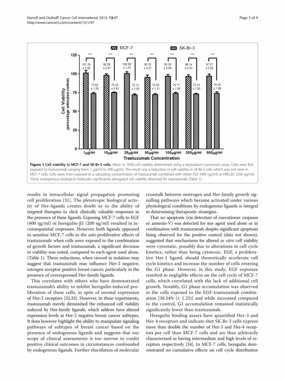
mediated growth and transformation, the greater theanti-proliferative effect of trastuzumab [27]. Not sur-prisingly, trastuzumab was unable to affect cell viabilityin MCF-7 cells. Trastuzumab (1–500 μg/ml) induced sub-stantial anti-proliferative effects in SK-Br-3 cells however(Figure 1), which suggests an extensive reliance of thesecells on Her-2 receptors for propagation of cell growth[28]. However, there was no difference in cell viability atdifferent concentrations of trastuzumab. It was speculatedthat at higher concentrations, the binding domains weresaturated or that Her-2 receptors were internalised anddegraded or unable to recycle back to the surface forfurther trastuzumab binding. Saturating concentrations oftrastuzumab (50 – 100 μg/ml) were used for further studiesto ensure continuous effects on the seeded cell populationas well as on cell progeny.Trastuzumab influenced cell cycle kinetics by inducing
significant G1 accumulation in MCF-7 at 72 hours only(Figures 2 and 3) and in SK-Br-3 cells at 24, 48 (datanot shown) and 72 hours (Figure 2). This is consistentwith other data where trastuzumab has been found tointerfere with Her-2 receptor signaling and consequentlyinhibit G1-S phase transition [29,30].While targeted therapies have achieved great com-
mendation for their selectivity and specificity, efficacyremains dependent on the continual and persistent pres-ence of targets. Unlike MCF-7 cells, which were imperviousto the effects of trastuzumab, SK-Br-3 cells demonstrateda significant decrease in Her-2 receptors when exposed totrastuzumab from as early as 6 hours [68.69% ± (1.14)].Near identical decreases were obtained at 12, 24 (Figure 4)and 48 hours suggesting that cells were not re-accumulatingon the surface within the period of time assessed. Takentogether, the dose-independent decrease in cell viability andconsistent G1 accumulation in SK-Br-3 cells may have beendue to the reduction in Her-2 receptors, a threshold abovewhich is required for further trastuzumab efficacy.Binding of Her-receptor ligands results in the formation
of dimer complexes. Subsequent auto-phosphorylation
Hurrell and Outhoff Cancer Cell International 2013, 13:97 Page 2 of 9http://www.cancerci.com/content/13/1/97
results in intracellular signal propagation promotingcell proliferation [31]. The pleiotropic biological activ-ity of Her-ligands creates doubt as to the ability oftargeted therapies to elicit clinically valuable responses inthe presence of these ligands. Exposing MCF-7 cells to EGF(400 ηg/ml) or heregulin-β1 (200 ηg/ml) resulted in in-consequential responses. However, both ligands appearedto sensitize MCF-7 cells to the anti-proliferative effects oftrastuzumab: when cells were exposed to the combinationof growth factors and trastuzumab, a significant decreasein viability was noted, compared to each agent used alone.(Table 1). These reductions, when viewed in isolation maysuggest that trastuzumab may influence Her-2 negative,estrogen receptor positive breast cancer, particularly in thepresence of overexpressed Her-family ligands.This correlates with others who have demonstrated
trastuzumab’s ability to inhibit heregulin-induced pro-liferation of these cells, in spite of normal expressionof Her-2 receptors [32,33]. However, in these experiments,trastuzumab merely diminished the enhanced cell viabilityinduced by Her-family ligands, which seldom have alteredexpression levels in Her-2 negative breast cancer subtypes.It does however highlight the ability to manipulate signalingpathways of subtypes of breast cancer based on thepresence of endogenous ligands and suggests that ourscope of clinical assessments is too narrow to conferpositive clinical outcomes in circumstances confoundedby endogenous ligands. Further elucidation of molecular
crosstalk between oestrogen and Her-family growth sig-nalling pathways which become activated under variousphysiological conditions by endogenous ligands is integralin determining therapeutic strategies.That no apoptosis (via detection of executioner caspases
or annexin-V) was detected for any agent used alone or incombination with trastuzumab despite significant apoptosisbeing observed for the positive control (data not shown),suggested that mechanisms for altered in vitro cell viabilitywere cytostatic, possibly due to alterations in cell cyclekinetics, rather than being cytotoxic. EGF, a prolifera-tive Her-1 ligand, should theoretically accelerate cellcycle kinetics and increase the number of cells enteringthe G1 phase. However, in this study, EGF exposureresulted in negligible effects on the cell cycle of MCF-7cells, which correlated with the lack of additional cellgrowth. Notably, G1 phase accumulation was observedin the cells exposed to the EGF-trastuzumab combin-ation [38.54% (± 1.25)] and while increased comparedto the control, G1 accumulation remained statisticallysignificantly lower than trastuzumab.Heregulin binding assays have quantified Her-3 and
Her-4 receptors and indicate that SK-Br-3 cells expressmore than double the number of Her-3 and Her-4 recep-tors per cell than MCF-7 cells and are thus arbitrarilycharacterized as having intermediate and high levels of re-ceptors respectively [34]. In MCF-7 cells, heregulin dem-onstrated no cumulative effects on cell cycle distribution
Figure 1 Cell viability in MCF-7 and SK-Br-3 cells. Mean (± SEM) cell viability determined using a tetrazolium conversion assay. Cells were firstexposed to trastuzumab ranging from 1 μg/ml to 500 μg/ml. The result was a reduction in cell viability in SK-Br-3 cells which was not seen inMCF-7 cells. Cells were then exposed to a saturating concentration of trastuzumab combined with either EGF (400 ηg/ml) or HRG-β1 (200 ηg/ml).These endogenous biological molecules significantly abrogated cell viability observed for trastuzumab (Table 1).
Hurrell and Outhoff Cancer Cell International 2013, 13:97 Page 3 of 9http://www.cancerci.com/content/13/1/97

A B
Figure 3 Column graphs of cell cycle analysis. Following exposure for 72 hours to trastuzumab, heregulin-β1 or the heregulin-β1-trastuzumabcombination histograms were analyzed with deconvolution software and expressed as column graphs. A) Significant alterations within the G1phase were observed in MCF-7 cells exposed to trastuzumab and the heregulin-β1-trastuzumab combination; B) Significant alterations in theS-phase were observed in SK-Br-3 cells exposed to heregulin-β1 compared to the untreated control. [EGF did not perpetuate noteworthyaccumulation or alterations in cell cycle kinetics].
G2
Sub-G1
S
G1
BA
C D
DNA Content
Eve
nt
cou
nt
Figure 2 Flow cytometry histograms analysed with deconvolution software. Gaussian curves were utilised to divide histograms into G1phase (left), S phase (centre) and G2 phase (right) A) Untreated MCF-7 cells in DMEM with 10% FCS; B) Trastuzumab treated MCF-7 cells after72 hours illustrating G1 accumulation; C) Untreated SK-Br-3 cells in RPMI with 10% FCS; D) Trastuzumab treated SK-Br-3 cells after 24 hoursillustrating G1 accumulation.
Hurrell and Outhoff Cancer Cell International 2013, 13:97 Page 4 of 9http://www.cancerci.com/content/13/1/97

Geometric meanUnstained (black) 9.11Untreated (green) 82.73Geldanamycin (red) 13.47Trastuzumab (blue) 43.39Heregulin-β1 (pink) 25.88Heregulin-β1 plus trastuzumab (purple)
18.57
75 75
75
BA
C
D
FL1 Log: Anti-Her-2 affibody molecule (FITC)
Eve
nt
cou
nt
Figure 4 Overlay plots and column graphs comparing the relative Her-2 receptor density in SK-Br-3 cells. A) Overlay plots of theunstained (black), stained (green) and the positive control, geldanamycin (red) with the associated geometric mean; B) Overlay plots of theunstained (black), stained (green) and cells exposed to trastuzumab (blue) with the associated geometric mean; C) Overlay plots of the unstained(black), stained (green) and cells exposed to heregulin-β1 (pink) or the heregulin-β1-trastuzumab combination (purple) with the associated geometricmean; D) Fluorescence (x-mean) intensity of triplicate runs, expressed as a percentage of untreated control (standardised to 100%). Concurrent heregulin-β1 abrogated relative Her-2 receptor density observed for trastuzumab. Trastuzumab, heregulin-β1 and the heregulin-β1-trastuzumab combinationillustrated a progressive decrease in Her-2 receptor density at 24 hours. Similar significance trends were observed in SK-Br-3 cells exposed to EGF [56.82%(± 2.82)] and the EGF-trastuzumab combination [45.01% (± 1.06)].
Hurrell and Outhoff Cancer Cell International 2013, 13:97 Page 5 of 9http://www.cancerci.com/content/13/1/97

(Figure 3). However, at 24 hours, a slight G2 phase increasewas noted, suggesting accelerated cell cycle kinetics toaccompany the increased cell viability.Her-2 monoclonal antibodies are capable of inhibiting
heregulin-induced activation of PI3-kinase and downstreamtargets (Akt) in MCF-7 cells [23]. This inhibition couldbe the mechanism by which trastuzumab abrogatedheregulin-induced mitogenic proliferation in our cells.The accompanying G1 accumulation in cells exposedto a combination of heregulin and trastuzumab, whichmimicked that of trastuzumab, may also be attributedto inhibition of this pathway. Although we were unable toobserve alterations in Her-2 receptor density, perhaps dueto the low constitutive Her-2 receptor number of MCF-7cells, the presence of these endogenous ligands appeared toinfluence the mechanisms that we and others have assessedfor trastuzumab in MCF-7 cells.Although mRNA can be a deceptive framework for
referencing ligand efficacy, quantitative analysis showsthat SK-Br-3 cells express approximately ten-times moreHer-1 protein than MCF-7 cells [35] which implies agreater potential for response to EGF. However, EGFresulted in a surprising decrease in cell viability of SK-Br-3cells. This was counteracted by concurrent trastuzumabexposure in favor of increased cell viability (Table 1).Synergistic effects of over expressing receptors within cel-lular transformation have been suggested [24]. However,sustained activation of Her-1 and Her-2 receptors mayactivate pathways, paradoxically leading to cell death evenin the presence of proliferative ligands [36,37].EGF exposure resulted in a substantial decrease in
Her-2 receptor density, implying that Her-2 signalingwas reduced. When combined with trastuzumab, thedecline in Her-2 receptor density was even greater.Tikhomirov et al. noted that rapid internalization of Her-1and subsequent lysosomal degradation occurs after ligandbinding [37]; Her-2 receptors may be internalized as part ofthis interactive dimer complex. Here the authors suggest
that reduction of Her-2 receptors by trastuzumab may alterthe balance of Her-1:Her-2 co-expression, and in doing so,EGF potentiates less of an anti-proliferative effect. Thus inselective circumstances, where multiple Her-receptors areover-expressed, trastuzumab may alter the co-expressionratio in favor of synergistic proliferative effects.Heregulin-β1-induced propagation of cell growth of SK-
Br-3 cells (Table 1) was consistent with the fact that thesecells also express Her-3 receptors, and was accompaniedby an increased number of cells in the S-phase of the cellcycle at 72 hours (Figure 3). Her-2:Her-3 heterodimers areconsidered the most potent signal transduction complexwith the highest mitogenic potential [5,17]. Increasedcell growth by heregulin-β1 could also be attributed to po-tentiation of already increased basal Akt activity asso-ciated with Her-2 receptor over-expression [23]. Thus,as a potent stimulator, heregulin-β1 may be capable ofcompensating for the inhibition ordinarily elicited bytrastuzumab [38]. This was corroborated when concurrentexposure with trastuzumab did not affect the cell growthpotential of heregulin-β1. Once again, the potency ofthe Her-2:Her-3 dimer pair could help explain whytrastuzumab had a negligible effect on these cells whencombined with heregulin-β1.Although heregulin-β1 propagated cell growth, sub-
stantial reductions in Her-2 receptors were observedwhen SK-Br-3 cells were exposed to the ligand. An evengreater reduction of Her-2 receptors was observed whenheregulin-β1 was used in combination with trastuzumab(Figure 4). This could have serious implications fortrastuzumab efficacy, as the target is diminished. Daly et al.previously also observed a decrease in Her-2 receptors inresponse to heregulin-β1 after 48 hours in these cells [39].Reduction in surface Her-2 receptors by EGF and
heregulin-β1 implicates both growth ligands in limitingtrastuzumab efficacy and ultimately resulting in diseaseprogression. If this remains true for additional Her-familyligands, assessing expression levels of ligands will have
Table 1 Cell viability in MCF-7 and SK-Br-3 cells
MCF-7 SK-Br-3
Trastuzumab (50 μg/ml) 95.35% (± 2.27) 72.58% (± 1.31)
(EGF + T50: P < 0.001)
EGF (400 ηg/ml) 101.70% (± 1.84) 81.43% (± 2.46)
(EGF + T50: P < 0.001)
EGF (400 ηg/ml) + Trastuzumab (50 μg/ml) 89.52% (± 1.95) 89.40% (± 2.20)
(Trastuzumab50: P < 0.05)
HRG (200 ηg/ml) 107.01% (± 2.42) 115.40% (± 2.08)
(Trastuzumab50: P < 0.001) (Trastuzumab50: P < 0.001)
HRG (200 ηg/ml) + Trastuzumab (50 μg/ml) 93.76% (±2.52) 114.50% (± 1.98)
(HRG: P < 0.001) (Trastuzumab50: P < 0.001)
Mean (± SEM) cell viability determined using a tetrazolium conversion assay.
Hurrell and Outhoff Cancer Cell International 2013, 13:97 Page 6 of 9http://www.cancerci.com/content/13/1/97

serious therapeutic implications in the clinical paradigmof trastuzumab eligibility.In our experiments, Her-2 receptors were assessed
using whole cells because trastuzumab efficacy is solelydependent on the accessibility of receptors on the cellsurface. Receptor analysis using lysed cells potentiallyalso detects ubiquitin-targeted receptors which are not yetdegraded, or internalised receptors capable of recycling; thismay skew data in favour of a higher receptor density. EGFand heregulin-β1 appeared to promote Her-2 receptor in-ternalization in the form of heterodimers which augmentedthe reduction of Her-2 receptor in trastuzumab exposedcells. However, the fate of receptors upon internalizationwas not elucidated as it was beyond the scope of this ex-periment. While the overall number of receptors withina cell may not change in response to trastuzumab, theability of receptors to be restored to the cell surface uponremoval of trastuzumab or ligands remains inconclusive.Further complicating this research is the ability of factorssuch as fetal calf serum (FCS) and seeding density toprovide differential responses in endpoint experimentsdependent on the heregulin isoforms used [39]. Whileheregulin-β1 was used in these experiments the results areintriguing and open avenues for the study of alternativeisoforms. Understanding and monitoring the molecularsubtypes of cancer and divergence of signaling pathwaysin response to targeted therapy is now required to ensurethe therapeutic benefit of trastuzumab.While the use of cytotoxic-conjugated-antibodies is being
researched extensively, antibody-dependent cell-mediatedcytotoxicity (ADCC) was not assessed in this study. Thisis due to the fact that efficacy of trastuzumab in a clinicalcontext is highly dependent on the maintenance of a con-tinuous and consistent receptor threshold. If this is notdone, as was seen over the time points assessed here thenADCC would be negligible due to the absence of sufficienttarget receptors.
ConclusionCell type specific interactions of endogenous ligands appearto be dependent on the absolute Her-receptor expressionand cross activation of signaling pathways. While athreshold of surface Her-2 receptors is required to en-sure trastuzumab binding, efficacy may be dependenton co-receptor expression ratios, as opposed to onlyHer-2 receptors, as a prognostic indicator and markerfor targeted therapy. In this study, Her-2 receptor statuswas greatly influenced by endogenous growth factors. Thereduction in surface Her-2 receptors diminished the avail-ability of targets for trastuzumab and thereby differentiallyaltered the efficacy parameters assessed for trastuzumab.It remains unclear whether these alterations in receptorabundance are maintained for sufficient periods to be clin-ically relevant. However, this data supports the notion that
receptor density of Her-family members and multiplicityof growth ligands are of mutual importance in prolifera-tion. This may provide insight into the role of endogenousHer-ligands in the emergence of resistance and have far-reaching consequences for targeted therapy and long-termdisease remission.
MethodsThis study was approved by the Faculty of Health ScienceStudent Research Ethics Committee of the Universityof Pretoria.
Human breast adenocarcinoma cellsAdherent breast adenocarcinoma cell lines included estrogenreceptor positive MCF-7 cells (ATCC Number HTB-20TM)which express physiologically normal levels of Her-2 recep-tors, and SK-Br-3 cells (ATCC Number HTB-30TM) whichover-express Her-2 receptors, obtained from American TypeCulture Collection (ATCC; Manassas, USA). MCF-7 cellswere deemed an ideal control as these cells express estrogenreceptors, enabling the potential for receptor crosstalk, whileboth MCF-7 and SK-Br-3 cells express all members of theHer-family of receptor tyrosine kinases to varying degrees.MCF-7 and SK-Br-3 cells were maintained in DMEM andRMPI 1640 medium (Sigma-Aldrich; St Louis, USA) re-spectively, supplemented with 10% heat-inactivated fetal calfserum (FCS) (PAA Laboratories; Pasching, Austria) and 1%penicillin-streptomycin (BioWhittaker; Walkersville, USA).Cells were seeded at 1×10⁴ cells per well or at 1.5×10⁵
cells per 25 cm2 flask and maintained in a humidifiedatmosphere containing 5% CO2 at 37°C.
Trastuzumab and growth factorsTrastuzumab (Herceptin®) was kindly donated by RochePharmaceuticals (Johannesburg, South Africa). Recombin-ant human epidermal growth factor and heregulin-β1were obtained as lyophilized powder from Sigma-Aldrich(St Louis, USA). Growth factors were reconstituted infilter sterilized (0.22 μM) dH2O while trastuzumab wasreconstituted in bacteriostatic water as per manufacturer’sinstructions. Cells were exposed to saturating concentra-tions of EGF (400 ηg/ml) or heregulin-β1 (200 ηg/ml) todetermine baseline effects. Additionally, cells were exposedto trastuzumab (1–500 μg/ml), to determine a dose re-sponse and a saturating concentration of trastuzumabthen combined with either EGF or heregulin-β1.
Tetrazolium conversion assayCell viability was determined using a quantitative-colorimetric conversion assay of 3-[4, 5-dimethylthiazol-2-yl]-2, 5-diphenyl tetrazolium bromide (MTT) afterexposure for 96 hours. Cells were incubated with 20 μlMTT solution (5 mg/ml) (Sigma-Aldrich; St Louis, USA)for 3 hours, washed with phosphate buffered saline
Hurrell and Outhoff Cancer Cell International 2013, 13:97 Page 7 of 9http://www.cancerci.com/content/13/1/97

(PBS) (BD Bioscience; Sparks, USA) and the formazanproduct solubilized in dimethyl sulphoxide (MerckChemicals; Darmstadt, Germany). Plates were readspectophotometrically using an ELx800uv universalmicroplate reader (Bio-TEK Instruments, INC) with a dualwavelength of 570 nm and 630 nm. Cell-free medium anddrug controls were included to discern reactivity of MTTwith extraneous variables other than cells.
Cell cycle analysisThe cell cycle was analyzed using interchelating, fluorescentpropidium iodide with flow cytometric detection at 24,48 and 72 hours. Decanted medium and trypsinizedcells were washed (1% FCS in PBS), fixed in 70% ethanol(Merck Chemicals; Darmstadt, Germany) and stored over-night at 4°C. Cell pellet was re-suspended in 1 ml ofpropidium iodide (40 μg/ml) (Sigma-Aldrich; St Louis, USA)staining solution containing Triton-X (Sigma-Aldrich; StLouis, USA), a non-ionic surfactant (0.1% v/v), and DNasefree RNase (100 μg/ml) (Sigma-Aldrich; St Louis, USA).Samples were incubated at 37°C for 40 minutes and ana-lyzed using a Cytomics FC500 (Beckman Coulter).
Executioner caspase assayCells were exposed to trastuzumab, growth factors or thepositive control [staurosporine, 10.7 μM (Sigma-Aldrich;St Louis, USA)] between 4 and 30 hours. Cell lysis buffer[10 mM 4-(2-hydroxyethyl)-1-piperazineethanesulfonicacid (HEPES), 1 mM phenylmethylsulfonyl fluoride(PMSF), 5 mM 3-[(3-cholamidopropyl)dimethylammonio]-1-propanesulfonate (CHAPS) (Merck Chemicals; Darmstadt,Germany), 2 mM ethlene-diamine-tetra-acetic acid (EDTA),and 5 mM β-mercaptoethanol (Labchem; Johannesburg,RSA)] was added and plates incubated for 40 minutes on icefollowed by addition of 125 μl assay buffer [20 mM HEPES,2 mM EDTA and 5 mM β-mercaptoethanol] containing5 μM of Ac-DEVD-AMC substrate (Sigma-Aldrich; StLouis, USA). Plates were incubated overnight at 37°C toallow Ac-DEVD-AMC substrate hydrolysis by activatedexecutioner caspases-3 and −7, and the released fluor-escent product was read using a FLUOstar OPTIMA(BMG LABTECH) with excitation-emission wavelengths of350 nm and 450 nm respectively.
Apoptosis-necrosisLater apoptotic hallmarks were assessed after exposurefor 48 or 72 hours using FITC conjugated annexin-V,counterstained with propidium iodide to assess membraneintegrity. Cells were washed and re-suspended in 100 μl ofannexin-V binding buffer [10 mM HEPES, 150 mM NaCl,5 mM KCL, 1.8 mM CaCl2 and 1 mM MgCl2 (MerckChemicals; Darmstadt, Germany)] followed by staining with2.5 μl of annexin-V-FITC (Sigma-Aldrich; St Louis, USA).Samples were incubated in the dark for 10 minutes and
3 μl of propidium iodide (3 mM) (Sigma-Aldrich; StLouis, USA) was added shortly prior to acquisition using aCytomics FC500 (Beckman Coulter).
Relative Her-2 receptor densityRelative Her-2 receptor density was determined after cellswere exposed to trastuzumab, growth factors or the posi-tive control [geldanamycin, 0.35 μM (Tocris Bioscience;Ellisville, USA)] for 12, 24 and 48 hours, using an anti-Her-2-FITC affibody molecule (Abcam; Cambridge, UK)which is a protein composed of a three-helix bundled atthe unique C-terminal cysteine which does not competefor Her-2 binding with trastuzumab [40]. Samples werewashed twice (1% FCS in PBS) and the single cell suspen-sion labeled with the affibody molecule (final concentra-tion: 3.7 μg/ml) and incubated on ice for 30 minutes. Cellswere washed prior to acquisition using a Cytomics FC500(Beckman Coulter). The mean-fluorescence intensity wasexpressed relative to the untreated control.
StatisticsA minimum of three independent inter-day repeats wereconducted with a minimum of three intra-day repeatswhere required. Cell cycle histograms were analysedusing deconvolution software, uncovering the underlyingdistribution by fitting Gaussian curves using WinCycleMultiCycle V3.0 for Windows (WinCycle Software;Washington, USA). Histrograms for relative Her-2 re-ceptor density were analysed using CyflogicTM software(CyFlo, Ltd, Finland) and the x-mean and geometric meandetermined. Non-parametric, Kruskal-Wallis, test wasconducted to compare the mean values obtained fortrastuzumab versus EGF or heregulin-β1 alone versus thetrastuzumab-growth factor combinations. Dunn’s mul-tiple comparison test was used for post-hoc analysis withsignificance set at p-value < 0.05. Statistical analysis wasconducted using GraphPad Prism version 5.0 for Windows(GraphPad Software; San Diego, California, USA).
AbbreviationsADCC: Antibody-dependent cell-mediated cytotoxicity; Akt: Protein kinase B;CHAPS: 3-[(3-cholamidopropyl)dimethylammonio]-1-propanesulfonate;EDTA: Ethlene-diamine-tetra-acetic acid; EGF: Epidermal growth factor;FCS: Fetal calf serum; HEPES: 4-(2-hydroxyethyl)-1-piperazineethanesulfonicacid; Her: Human epidermal growth factor receptor; HRG-β1: Heregulin-β1;MTT: 3-[4, 5-dimethylthiazol-2-yl]-2, 5-diphenyl tetrazolium bromide;PBS: Phosphate buffered saline; PMSF: Phenylmethylsulfonyl fluoride;TTP: Time to progression.
Competing interestsThe authors declare that they have no conflict of interest, financial ornon-financial, with respect to this research.
Authors’ contributionsKO conceived the study, participated in its design and coordination,acquired funding for the study and assisted in drafting and critically revisingthe manuscript. TH carried out the experimental procedures, performed theanalysis and assisted in drafting the manuscript. All authors read andapproved the final manuscript.
Hurrell and Outhoff Cancer Cell International 2013, 13:97 Page 8 of 9http://www.cancerci.com/content/13/1/97

AcknowledgementsThe authors would like to acknowledge Roche Pharmaceuticals for thedonation of trastuzumab and the Cancer Association of South Africa(CANSA) and the Research and Development Programme (RDP), University ofPretoria, for providing funding. Furthermore, the authors would like toacknowledge Dr AD Cromarty and Dr JJ van Tonder for their technicalsupport and guidance.
Financial supportCancer Association of South Africa (CANSA)Research Development Programme, University of Pretoria.
Received: 4 April 2013 Accepted: 10 October 2013Published: 11 October 2013
References1. Leahy BDJ: Structure and function of the epidermal growth factor
(EGF/ErbB) family of receptors. Adv Protein Chem 2004, 68:1–27.2. Hudis CA: Trastuzumab–mechanism of action and use in clinical practice.
N Engl J Med 2007, 357:39–51.3. Ross JS, Slodkowska EA, Symmans WF, Pusztai L, Ravdin PM, Hortobagyi GN:
The HER-2 receptor and breast cancer: ten years of targeted anti-HER-2therapy and personalized medicine. Oncologist 2009, 14(4):320–368.
4. Harris AL, Nicholon S: Epidermal growth factor receptors in human breastcancer. Cancer Res 1988, 40:93–118.
5. Burgess AW, Cho H, Eigenbrot C, Ferguson KM, Garrett TPJ, Leahy DJ,Lemmon MA, Sliwkowski MX, Ward CW, Yokoyama S: An open-and-shutcase? Recent insights into the activation of EGF/ErbB receptors.Mol Cell 2003, 12:541–552.
6. Citri A, Yarden Y: EGF-ERBB signalling: towards the systems level. NatureReviews. Mol Cell Bio 2006, 7(7):505–516.
7. Klapper L, Glathe S, Vaisman N, Hynes N, Andrews G, Sela M, Yarden Y: TheErbB-2 /HER2 oncoprotein of human carcinomas may function solely asa shared coreceptor for multiple stroma-derived growth factors.Proc Natl Acad Sci U S A 1999, 96:4995–5000.
8. Citri A, Skaria KB, Yarden Y: The deaf and the dumb: the biology of ErbB-2and ErbB-3. Exp Cell Res 2003, 284:54–65.
9. Guy PM, Platko JV, Cantleyt LC, Cerione RA, Carrawat KL: Insect cell-expressedp180erbb3 possesses an impared tyrosine kinase activity. Proc Natl Acad SciU S A 1994, 91:8132–8136.
10. Yarden Y, Sliwkowski MX: Untangling the ErbB signalling network. NatureReviews. Mol Cell Bio 2001, 2:127–137.
11. Burstein HJ: The distinctive nature of HER2-positive breast cancers.N Engl J Med 2005, 353(16):1652–1654.
12. Lichtenstein AV: Cancer: shift of the paradigm. Med Hypotheses 2008,71(6):839–850.
13. Hutchinson JN, Muller WJ: Transgenic mouse models of human breastcancer. Oncogene 2000, 19(53):6130–6137.
14. Perez EA, Suman VJ, Davidson NE, Sledge GW, Kaufman PA, Hudis CA,Martino S, Gralow JR, Dakhil SR, Ingle JN, Winer EP, Gelmon KA, Gersh J,Jaffe AS, Rodeheffer RJ: Cardiac safety analysis of doxorubicin andcyclophosphamide followed by paclitaxel with or without trastuzumabin the north central cancer treatment group N9831 adjuvant breastcancer trial. J Clin Oncol 2008, 26(8):1231–1238.
15. Kruser TJ, Wheeler DL: Mechanisms of resistance to HER family targetingantibodies. Exp Cell Res 2010, 316(7):1083–1100.
16. Lower EE, Glass E, Blau R, Harman S: HER-2/neu expression in primary andmetastatic breast cancer. Breast Cancer Res Treat 2009, 113(2):301–306.
17. Tai W, Mahato R, Cheng K: The role of HER2 in cancer therapy andtargeted drug delivery. J Control Release 2010, 146(3):264–275.
18. Friedländer E, Barok M, Szöllosi J, Vereb G: ErbB-directed immunotherapy:antibodies in current practice and promising new agents. Immunol Lett2008, 116(2):126–140.
19. Callahan R, Hurvitz S: Human epidermal growth factor receptor-2-positivebreast cancer: current management of early, advanced, and recurrentdisease. Curr Opin Obstet Gynecol 2011, 23(1):37–43.
20. Coate LE, John T, Tsao M, Shepherd FA: Molecular predictive and prognosticmarkers in non-small-cell lung cancer. Lancet Oncol 2009, 10(10):1001–1110.
21. Mountzios G, Sanoudou D, Syrigos KN: Clinical pharmacogenetics inoncology: the paradigm of molecular targeted therapies. Curr Pharm Des2010, 16(20):2184–2193.
22. Extra JM, Antoine EC, Vincent-Salomon A, Delozier T, Kerbrat P, Bethune-Volters A,Guastalla JP, Spielmann M, Mauriac L, Misset JL, Serin D, Campone M, Hebert C,Remblier C, Bergougnoux L, Campana F, Namer M: Efficacy of Trastuzumab inroutine clinical practice and after progression for metastatic breast cancerpatients: the observational hermine study. Oncologist 2010, 15:799–809.
23. Liu W, Li J, Roth RA: Heregulin regulation of Akt/protein kinase B inbreast cancer cells. Biochem Biophys Res Commun 1999, 261(3):897–903.
24. Koutras AK, Evans TRJ: The epidermal growth factor receptor family inbreast cancer. OncoTargets Ther 2008, 1:5–19.
25. Sahin Ö, Fröhlich H, Löbke C, Korf U, Burmester S, Majety M, Mattern J,Schupp I, Chaouiya C, Thieffry D, Poustka A, Wiemann S, Beissbarth T, Arlt D:Modeling ERBB receptor-regulated G1/S transition to find novel targetsfor de novo trastuzumab resistance. BMC Syst Biol 2009, 3(1):1–20.
26. Guarneri V, Barbieri E, Dieci MV, Piacentini F, Conte P: Anti-HER2neoadjuvant and adjuvant therapies in HER2 positive breast cancer.Cancer Treat Rev 2010, 36(Suppl 3):62–66.
27. Hudziak RM, Lewis GD, Winget M, Fendly BM, Shepard HM, Ullrich A:p185HER2 monoclonal antibody has antiproliferative effects in vitroand sensitizes human breast tumor cells to tumor necrosis factor.Mol Cell Bio 1989, 9(3):1165–1172.
28. Zhu H, Zhang G, Wang Y, Xu N, He S, Zhang W, Chen M, Lui M, Quan L, BaiJ, Xu N: Inhibition of ErbB2 by Herceptin reduces survivin expression viathe ErbB2-β-catenin/TCF4-survivin pathway in ErbB2-overexpressedbreast cancer cells. Cancer Sci 2010, 101(5):1156–1162.
29. Lane HA, Motoyama AB, Beuvink I, Hynes NE: Modulation of p27/Cdk2complex formation through 4D5-mediated inhibition of HER2 receptorsignaling. Ann Oncol 2001, 12(Suppl 3):21–22.
30. Mohsin SK, Weiss HL, Gutierrez MC, Chamness GC, Schiff R, DiGiovanna MP,Wang C, Hilsenbeck SG, Osborne CK, Allred DC, Elledge R, Chang JC:Neoadjuvant trastuzumab induces apoptosis in primary breast cancers.J Clin Oncol 2005, 23(11):2460–2468.
31. Sako Y, Minoguchi S, Yanagida T: Single-molecule imaging of EGFRsignalling on the surface of living cells. Nat Cell Biol 2000, 2:168–172.
32. Yuste L, Montero JC, Esparís-Ogando A, Pandiella A: Activation of ErbB2 byoverexpression or by transmembrane neuregulin results in differentialsignaling and sensitivity to Herceptin. Cancer Res 2005, 65(15):6801–6810.
33. Menendez JA, Mehmi I, Lupu R: Trastuzumab in combination withheregulin-activated Her-2 (erbB-2) triggers a receptor-enhancedchemosensitivity effect in the absence of Her-2 overexpression.J Clin Oncol 2006, 24(23):3735–3745.
34. Lewis GD, Lofgren JA, McMurtrey AE, Nuijens A, Fendly BM, Bauer KD, SliwkowskiMX: Growth regulation of human breast and ovarian tumor cells byheregulin: evidence for the requirement of ErbB2 as a critical component inmediating heregulin responsiveness. Cancer Res 1997, 56:1457–1465.
35. Konecny GE, Pegram MD, Venkatesan N, Finn R, Yang G, Rahmeh M, UntchM, Rusnak DW, Spehar G, Mullin RJ, Keith BR, Gilmer TM, Berger M, PodratzKC, Slamon DJ: Activity of the dual kinase inhibitor Lapatinib (GW572016)against HER-2-overexpressing and trastuzumab-treated breast cancercells. Cancer Res 2006, 66(3):1630–1639.
36. Tikhomirov O, Carpenter G: Bax activation and translocation tomitochondria mediate EGF-induced programmed cell death. J Cell Sci2005, 118(24):5681–5690.
37. Tikhomirov O, Carpenter G: Ligand-induced, p38-dependent apoptosis incells expressing high levels of epidermal growth factor receptor andErbB-2. J Biol Chem 2004, 279(13):12988–12996.
38. Diermeier S, Horváth G, Knuechel-Clarke R, Hofstaedter F, Szöllosi J, BrockhoffG: Epidermal growth factor receptor coexpression modulates susceptibilityto Herceptin in HER2/neu overexpressing breast cancer cells via specificerbB-receptor interaction and activation. Exp Cell Res 2005, 304(2):604–619.
39. Daly JM, Jannot CB, Beerli RR, Graus-Porta D, Maurer FG, Hynes NE: Neudifferentiation factor induces ErbB2 down-regulation and apoptosis ofErbB2-overexpressing breast tumor cells. Cancer Res 1997, 57:3804–3811.
40. Lee SB, Hassan M, Fisher R, Kramer-Marek G, Gandjbakhche A, Capala J:Affibody molecules for in vivo characterization of HER2-positive tumorsby near-infrared imaging. Clin Cancer Res 2008, 14(12):3840–3849.
doi:10.1186/1475-2867-13-97Cite this article as: Hurrell and Outhoff: The in vitro influences ofepidermal growth factor and heregulin-β1 on the efficacy oftrastuzumab used in Her-2 positive breast adenocarcinoma. Cancer CellInternational 2013 13:97.
Hurrell and Outhoff Cancer Cell International 2013, 13:97 Page 9 of 9http://www.cancerci.com/content/13/1/97


