Embed Size (px)
Citation preview
Molecular Biology of the CellVol. 8, 2077–2088, October 1997
The Involvement of the Intermediate Chain ofCytoplasmic Dynein in Binding the Motor Complexto Membranous Organelles of Xenopus OocytesWalter Steffen,*† Sher Karki,‡ Kevin T. Vaughan,§ Richard B. Vallee,§Erika L.F. Holzbaur,| Dieter G. Weiss,¶ and Sergei A. Kuznetsov¶
†Institute of Biochemistry and Molecular Cell Biology, Biocenter, University of Vienna, A-1030Vienna, Austria; ‡Cell Biology Graduate Group, University of Pennsylvania, Philadelphia,Pennsylvania 19104; §Cell Biology Group, Worcester Foundation for Biomedical Research,Shrewsbury, Massachusetts 01545; \Department of Animal Biology, University of Pennsylvania Schoolof Veterinary Medicine, Philadelphia, Pennsylvania 19104; and ¶Institute of Zoology, University ofRostock, D-18051 Rostock, Germany
Submitted May 12, 1997; Accepted July 22, 1997Monitoring Editor: J. Richard McIntosh
Cytoplasmic dynein is one of the major motor proteins involved in intracellular trans-port. It is a protein complex consisting of four subunit classes: heavy chains, intermediatechains (ICs), light intermediate chains, and light chains. In a previous study, we hadgenerated new monoclonal antibodies to the ICs and mapped the ICs to the base of themotor. Because the ICs have been implicated in targeting the motor to cargo, we testedwhether these new antibodies to the intermediate chain could block the function ofcytoplasmic dynein. When cytoplasmic extracts of Xenopus oocytes were incubated witheither one of the monoclonal antibodies (m74–1, m74–2), neither organelle movementnor network formation was observed. Network formation and membrane transport wasblocked at an antibody concentration as low as 15 mg/ml. In contrast to these observa-tions, no effect was observed on organelle movement and tubular network formation inthe presence of a control antibody at concentrations as high as 0.5 mg/ml. Afterincubating cytoplasmic extracts or isolated membranes with the monoclonal antibodiesm74–1 and m74–2, the dynein IC polypeptide was no longer detectable in the membranefraction by SDS-PAGE immunoblot, indicating a loss of cytoplasmic dynein from themembrane. We used a panel of dynein IC truncation mutants and mapped the epitopesof both antibodies to the N-terminal coiled-coil domain, in close proximity to thep150Glued binding domain. In an IC affinity column binding assay, both antibodiesinhibited the IC–p150Glued interaction. Thus these findings demonstrate that direct IC–p150Glued interaction is required for the proper attachment of cytoplasmic dynein tomembranes.
INTRODUCTION
Microtubule-based motor proteins provide the ma-chinery for most membrane trafficking within the cy-toplasm of higher eukaryotes, and the microtubule
cytoskeleton provides the polarized framework onwhich an oriented transport can take place. Orientedtransport is achieved by two classes of motor proteinswith opposite directionality, kinesins and cytoplasmicdyneins. Cytoplasmic dynein is a minus-end–directedmotor complex that is responsible for centripetaltransport. Cytoplasmic dynein has been colocalizedwith components of the Golgi apparatus (Corthesy-Theulaz et al., 1992; Fath et al., 1994; Vaisberg et al.,
* Corresponding author: Institute of Biochemistry and MolecularCell Biology, Biocenter, University of Vienna, Dr. Bohrgasse 9,A-1030 Vienna, Austria.
© 1997 by The American Society for Cell Biology 2077
1996), endocytic vesicles (Lin and Collins, 1992), lyso-somes (Lin and Collins, 1992), and mitochondria (Hi-rokawa et al., 1990). Furthermore, Aniento et al. (1993)have demonstrated that cytoplasmic dynein is essen-tial for the fusion of endocytic vesicles.
The extension of tubular networks of the endoplas-mic reticulum (ER) is another process that most likelywill depend on microtubular motors (Lee and Chen,1988; Lee et al., 1989; Terasaki and Reese, 1994). TheER, which is linked to microtubules via cross-bridges(Franke, 1971), represents the largest membrane sys-tem within a cell. Experiments examining recoveryfrom nocodazole treatment in cultured cells havedemonstrated that the extension of the ER tubules ispreceded by the polymerization of microtubules (Leeet al., 1989). The extension of the ER networks is ori-ented toward the cell periphery along microtubulartracks (Terasaki and Reese, 1994) and it is believed tobe caused by the plus-end–directed motor kinesin(Dabora and Sheetz, 1988; Vale and Hotani, 1988;Toyoshima et al., 1992). ER network formation has alsobeen reconstituted in vitro in cytoplasmic extracts ofcultured cells (Dabora and Sheetz, 1988) and of Xeno-pus oocytes (Allan and Vale, 1991). Interestingly, in thecase of Xenopus oocytes, Allan and Vale (1991, 1994)have demonstrated that cytoplasmic dynein but notkinesin is involved in the formation and maintenanceof ER networks (Allan and Vale, 1994; Allan, 1995) invitro. It is currently not understood why Xenopus ERshould move exclusively by cytoplasmic dynein. Thelarge size of the Xenopus oocyte may, however, requirean egg-specific function (Allan, 1995). Cytoplasmicextracts of Xenopus oocytes provide, therefore, an ex-cellent in vitro system to study the function of cyto-plasmic dynein.
Cytoplasmic dynein is a large complex consisting offour subunit classes: heavy chains, intermediatechains (ICs), light intermediate chains, and lightchains (Paschal et al., 1987). Although cytoplasmic dy-nein interacts with microtubules dynamically via thecatalytic heavy chains, it had been postulated that ICslink the motor complex to membranous organelles(King et al., 1991; Paschal et al. 1992). In a previousstudy, we have demonstrated that the ICs are local-ized at the base of the motor complex (Steffen et al.,1996). The position of the ICs is, therefore, in completeagreement with them being responsible for dockingthe motor protein to cargo.
Dynein-dependent vesicle transport in vitro de-pends on the presence of dynactin (Gill et al., 1991).More recent work suggests that dynactin might func-tion as the cytoplasmic dynein “receptor” (Vaughanand Vallee, 1995; Echeverri et al., 1996). By using blotoverlay and immunoprecipitation assays and im-muno-affinity column chromatography, it was foundthat dynein IC interacted with dynactin and the inter-action was mediated via the p150Glued subunit (Karki
and Holzbaur, 1995; Vaughan and Vallee, 1995). Todetermine whether the ICs play an important part inthe function of cytoplasmic dynein, we investigatedthe formation of membrane networks in cytoplasmicextracts of Xenopus eggs by using our monoclonalantibodies (m74–1 and m74–2) specific for the 74-kDadynein IC. We demonstrate that IC-specific antibodiesprevented the interaction between dynactin and dy-nein IC in vitro and disrupted the dynein–membraneinteraction. Our data, therefore, indicate that the IC–p150Glued interaction plays a critical role in binding themotor complex to membranous organelles.
MATERIALS AND METHODS
AntibodiesMonoclonal antibodies m74–1 and m74–2 specific for dynein IC thathad been characterized previously (Steffen et al., 1996) were ob-tained from cell culture supernatant. Monoclonal cell lines werecultured in Ultrodoma medium (BioWittaker, Bender, Vienna, Aus-tria) containing 2% fetal calf serum depleted of IgGs at 5% CO2.Antibodies were isolated from cell culture supernatant by protein Gcolumn chromatography. Purified antibodies were dialyzed intophosphate-buffered saline (PBS: 10 mM sodium phosphate, 150 mMNaCl, 2.7 mM KCl, pH 7.4), concentrated to about 10 mg/ml withCentricon 10 filters (Amicon, Witten, Germany), and stored in ali-quots at 280°C. Fab fragments were generated according to Mage(1980) by incubating protein G-purified antibody with 50 mM cys-teine, 1 mM EDTA, and 3 U/ml papain attached to agarose beadsfor 90 min at 37°C. The completion of the digest was monitored bySDS-PAGE.
Monoclonal antibody LEP100 developed by Dr. D.M. Fambroughwas obtained from the Developmental Studies Hybridoma Bank(maintained by the Department of Pharmacology and MolecularSciences, Johns Hopkins University School of Medicine, Baltimore,MD, and the Department of Biological Science, University of Iowa,Iowa City, IA, under contract N01-HD-6–2915 from the NationalInstitute of Child Health and Human Development). Monoclonalantibody m150–1 specific for p150Glued was raised against ATP-extracted microtubule-associated proteins from bovine brain as de-scribed earlier (Steffen et al., 1996). An affinity-purified polyclonalantibody raised against bacterially expressed heavy chain fromDictyostelium was obtained from E. Vaisberg (Vaisberg et al., 1993).
Isolation of Xenopus Egg CytosolInterphase cytoplasmic extract was isolated from Xenopus oocytesaccording to Allan (1993) and Murray (1991). The eggs were washedin MMR/4 buffer [MMR buffer: 100 mM NaCl, 2 mM KCl, 1 mMMgSO4, 2 mM CaCl2, 5 mM N-(2-hydroxyethyl)piperazine-N9-(2-ethanesulfonic acid) (HEPES), 0.1 mM EDTA, pH 7.8]. The jellycoats were removed with freshly prepared 2% cysteine (pH 7.8). Theeggs were washed again three times with MMR/4 buffer. Interphaseeggs were obtained by activation in the presence of 0.4 mg/mlionophore A23187 for 5 min, washing in MMR/4 buffer containing100 mg/ml cycloheximide, and incubation at room temperature for20 min. The eggs were washed twice in ice-cold acetate buffer [100mM potassium acetate, 2.5 mM magnesium acetate, 5 mM ethyleneglycol-bis(b-aminoethyl ether)-N,N,N9,N9-tetraacetic acid (EGTA),10 mM HEPES, 50 mM sucrose, 1 mM dithiothreitol (DTT), pH 7.2].Cytochalasin B (50 mg/ml) and protease inhibitors (10 mg/ml leu-peptin, 1 mg/ml pepstatin, 1 mM phenylmethylsulfonyl fluoride, 10mg/ml Na-p-tosyl-l-arginine methyl ester) were added to the lastwashing step. Eggs were settled by centrifugation at 150 3 g for 1min followed by a centrifugation at 600 3 g for 30 s. Excess bufferwas removed and eggs were crushed by centrifugation at 10,000
W. Steffen et al.
Molecular Biology of the Cell2078
rpm in a Beckman SW60 rotor for 20 min. Cytosol was removed,0.05 volume of the energy mixture (150 mM creatine phosphate, 20mM Mg-ATP, 2 mM EGTA), protease inhibitors, and cytochalasin B(50 mg/ml) were added. The cytosol was frozen as 50- to 100-mlaliquots in liquid N2 and stored at 280°C.
Fractionation of Cytoplasm and SDS-PAGECytoplasmic extract was fractionated by flotation through a sucrosedensity gradient. Twenty microliters of cytosol was mixed with 0.5ml of 60% sucrose in acetate buffer, placed at the bottom of a stepgradient (0.5 ml of 58% sucrose, 1 ml of 50% sucrose, and 0.5 ml of15% sucrose in acetate buffer) in a Beckman TLS-55 centrifuge tube,and centrifuged at 55,000 rpm for 30 min at 4°C. The membranefraction at the 15–50% sucrose interface was removed from the topwith a hypodermic needle and a peristaltic pump. The membranefractions were precipitated with ethanol or pelleted at 100,000 3 gand then analyzed by SDS-PAGE and immunoblot. Electrotransferto nitrocellulose was carried out according to Towbin et al. (1979) at70 V for 3 h by using a Tris(hydroxymethyl)aminomethane (Tris)/glycine buffer containing 25 mM Tris, 192 mM glycine, and 10%methanol. The antigens were detected with a goat anti-mouse and agoat anti-rabbit peroxidase-conjugated antibody (Bio-Rad, Rich-mond, CA) and visualized using a luminescence assay (Pierce,Rockford, IL). In some experiments cytoplasmic extracts were incu-bated for 3 h on ice with 0.5 mg/ml antibody prior to flotation in thepresence or absence of protein kinase inhibitors (150 mM H7, 10 mMstaurosporin) and/or phosphatase inhibitors (1 mM calyculin, 10mM microcystein, 1 mM okadaic acid).
Quantification of SDS-PAGE and SDS-PAGEImmunoblotMembrane fractions were isolated from equal volumes of cytoplas-mic extracts. The relative proportion of the membrane fraction wasdetermined by gel densitometry using a flat bed scanner HP ScanjetIIp (Hewlett-Packard, Vienna, Austria) and the NIH Image software(National Institutes of Health, Bethesda, MD). The SDS-PAGE im-munoblots were normalized to the major membrane proteins oncorresponding SDS-PAGE gels to compensate for slight variationsin loading between lanes. Also, where equal loading was necessary,the protein loading was adjusted prior to SDS-PAGE immunoblot.
Isolation and Purification of Cytoplasmic Dyneinand TubulinCytoplasmic dynein was isolated via its affinity to taxol-stabilizedmicrotubules and purified by sucrose density gradient (5–25%)centrifugation (Paschal et al. 1987). Taxol was kindly provided to usby Jill Johnson (National Cancer Institute, Bethesda, MD). Brainstem (100–150 g) from bovine brain was homogenized in an equalvolume of PEM buffer [100 mM piperazine-N,N9-bis(2-ethanesul-fonic acid), pH 7.3, 1 mM MgCl2, 1 mM EGTA, 0.5 mM DTT, 10mg/ml leupeptin, 1 mg/ml pepstatin, 1 mM PMSF, 10 mg/ml Na-p-tosyl-l-arginine methyl ester] and then centrifuged at 10,000 3 g for15 min and 80,000 3 g for 30 min at 4°C. Microtubules werepolymerized at 37°C in the presence of 1 U/ml hexokinase and 10mg/ml glucose by adding 10 mM taxol and then centrifugedthrough a 10% sucrose cushion in PEM at 80,000 3 g for 40 min at25°C. Dynein was released by resuspending taxol-stabilized micro-tubules in 0.05 tissue volume of extraction buffer (PEM supple-mented with 20 mM Mg-ATP and 5 mM taxol) followed by anincubation at room temperature for 30 min. Released microtubule-associated proteins were separated from microtubules by centrifu-gation at 100,000 3 g for 30 min at 25°C. The supernatant was thencentrifuged through a 5–25% continuous sucrose density gradient ina Beckman SW-28 rotor at 100,000 3 g (27,000 rpm) for 18 to 19 h. Upto 0.1 mM Mg-ATP was included in the sucrose gradient to enhance
the stability of cytoplasmic dynein. Fractions of about 0.8 ml werecollected from the bottom of the gradient.
Taxol-stabilized microtubules obtained as leftover from the dy-nein preparation were used as a source of tubulin. Microtubuleswere depolymerized by resuspending and incubating them in PEMbuffer containing a final concentration of 300 mM NaCl and 3 mMCaCl2 for 90 min on ice according to Collins (1991). The depolymer-ized microtubules were centrifuged at 100,000 3 g for 60 min. Thesupernatant was adjusted to 250 mM NaCl and 0.2 mM GTP andthen further purified by DEAE-Sephadex column chromatography.DEAE-Sephadex A-50 was equilibrated in PEM buffer containing250 mM NaCl. Tubulin was bound to the column, washed with 250mM NaCl in PEM buffer, eluted with 500 mM NaCl in PEM buffer,and then dialyzed against PEM containing 0.1 mM GTP and 0.5 mMDTT. Aliquots were frozen in liquid N2 and stored at 280°C. Thepurity of cytoplasmic dynein and tubulin was examined by SDS-PAGE.
Video MicroscopyMicroscope flow chambers were built as described by Blocker et al.(1996). An 11-mm circular cover glass (Menzel, Braunschweig, Ger-many) was placed onto a glass microscope slide with two pieces ofdouble-sided tape (Scotch, 3 M, St. Paul, MN) forming a 2- to 3-mlchamber. Chambers were perfused with Xenopus cytoplasmic ex-tracts diluted 1:4 (vol/vol) with acetate buffer containing 2 mMATP. Preparations were observed with video-enhanced differentialinterference contrast (VEC-DIC) microscopy (Allen et al., 1981, 1985;Weiss et al., 1989). A Nikon Diaphot 300 inverted microscope (Ni-kon, Dusseldorf, Germany) equipped with an oil-immersion con-denser (numerical aperature 1.4), 1003 DIC PlanApo oil objective(numerical aperature 1.4), and xenon lamp (XBO 100). AHamamatsu C2400–07 Newvicon camera was used for acquiringDIC images (Hamamatsu Photonics Deutschland, Herrsching, Ger-many). The video signals were first subjected to analogue contrastenhancement (Allen et al., 1981, 1985; Weiss et al., 1989), followed byreal-time digital image processing in the following steps: subtrac-tion of an out-of-focus background (mottle pattern), accumulationor averaging of images to increase signal to noise ratio, and finallyselection of the desired range of gray levels (histogram stretching ordigital contrast enhancement; Allen and Allen, 1983; Weiss andMaile, 1993). The analogue and digital processing of VEC-DIC sig-nals was performed by using the ARGUS 20 real-time image pro-cessor (Hamamatsu Photonics Deutschland). All observations wererecorded in real time with a Panasonic AG-6730 SVHS video re-corder. Single-frame images were captured from video tape ordirectly from ARGUS 20 image processor onto a Power MacIntosh8500 (Apple Computer, Cupertino CA) equipped with a LG-3 framegrabber (Scion, Frederick, MD) using IPLab Spectrum software(Scanalytics, Vienna, VA).
Affinity Column Binding AssayThe affinity column binding assay was performed essentially asdescribed by Karki and Holzbaur (1995). Three identical columnswere constructed by packing 200 ml of CH-Sepharose 4B (Pharma-cia, Uppsala, Sweden) beads cross-linked with purified recombinantdynein IC at a concentration of 2 mg/ml. After thorough equilibra-tion of the columns with PHEM buffer [50 mM sodium piperazine-N,N9-bis(2-ethanesulfonic acid), 50 mM sodium HEPES, 1 mMEDTA, 2 mM MgCl2, pH 6.9] 2 mg bovine serum albumin (BSA) wasadded to the first column, 700 mg of antibody m74–1 was added tothe second column, and 700 mg of antibody m74–2 was added to thethird column. After washing with 10 column volumes of PHEM, 500ml of rat brain cytosol was loaded onto each column. After thoroughwashing (100 column volumes) with HEM buffer (50 mM sodiumHEPES, 1 mM EDTA, 2 mM MgCl2, pH 6.9), the columns wereeluted with 500 ml of 1 M NaCl, and the resulting fractions wereprecipitated with trichloroacetic acid. The precipitates were each
The Association of Cytoplasmic Dynein with Membranes
Vol. 8, October 1997 2079
resuspended in 30 ml of 13 Laemmli sample buffer and neutralizedwith saturated Tris. A 15-ml aliquot of the precipitate was loadedand resolved by SDS-PAGE. The proteins were transferred ontoImmobilon-P (Millipore, Bedford, MA), Coomassie-stained, andsubsequently probed with affinity-purified rabbit polyclonal anti-bodies to 150Glued and centractin (Holleran et al., 1996; Tokito et al.,1996).
Expression of Dynein IC cDNAThe coding region of IC-1a (Paschal et al., 1992) was subcloned intothe pET-14b expression vector (Novagene, Madison, WI) by usingstandard cloning techniques (Vaughan and Vallee, 1995). Trunca-tion mutants of IC-1a were prepared in pET-14b by using a combi-nation of internal restriction sites and site-directed mutagenesis(Vaughan and Vallee, 1995). After transformation into the expres-sion strain BL21(DE3) and induction with 0.4 mM isopropyl b-d-thiogalactoside, bacteria were lysed in a French press and expressedproteins were purified by using nickel-affinity chromatography.Purified recombinant polypeptides were immobilized on Immo-bilon-P (Millipore) membranes by using a slot-blot apparatus (BRL,Gaithersburg, MD). Immunoblots were performed by standardtechniques and results were visualized by using ECL (Kirkegaard &Perry, Gaithersburg, MD). A pan-IC antiserum was generatedagainst full-length IC-1a and shown to react with all truncationmutant polypeptides (Vaughan and Vallee, 1995).
RESULTS
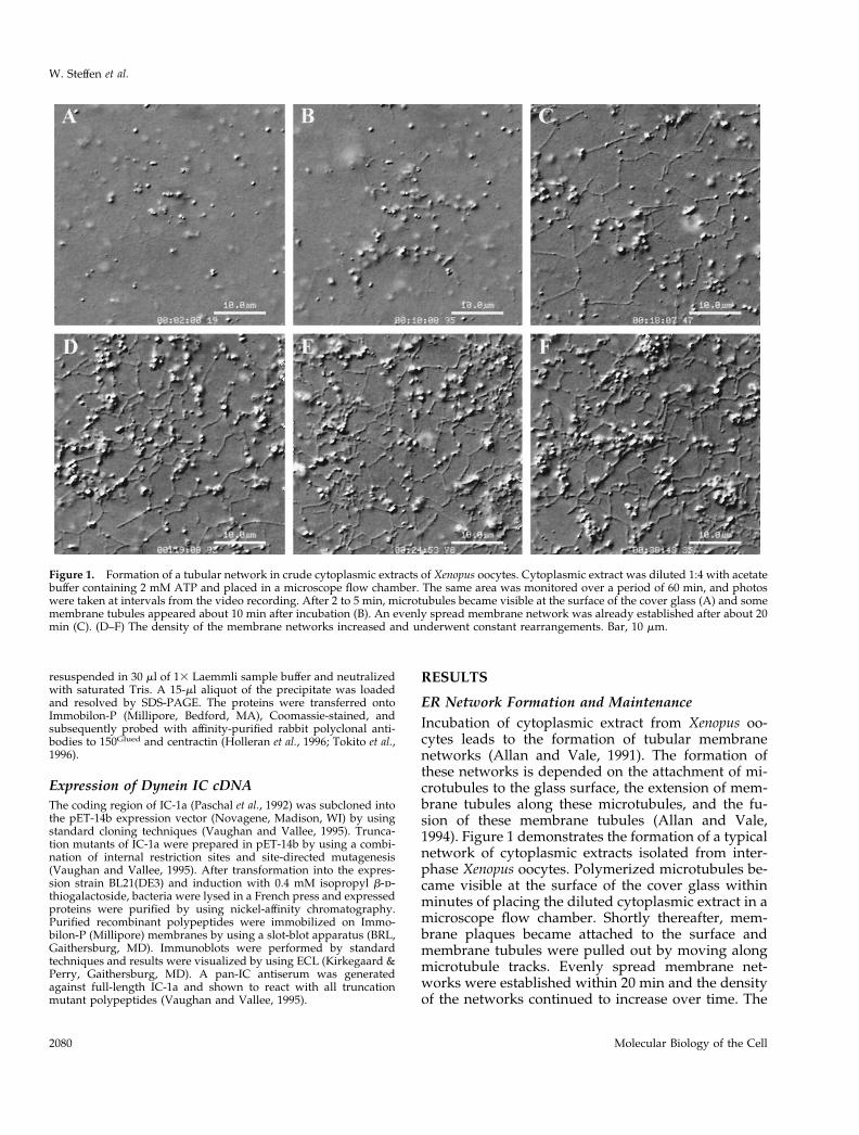
ER Network Formation and MaintenanceIncubation of cytoplasmic extract from Xenopus oo-cytes leads to the formation of tubular membranenetworks (Allan and Vale, 1991). The formation ofthese networks is depended on the attachment of mi-crotubules to the glass surface, the extension of mem-brane tubules along these microtubules, and the fu-sion of these membrane tubules (Allan and Vale,1994). Figure 1 demonstrates the formation of a typicalnetwork of cytoplasmic extracts isolated from inter-phase Xenopus oocytes. Polymerized microtubules be-came visible at the surface of the cover glass withinminutes of placing the diluted cytoplasmic extract in amicroscope flow chamber. Shortly thereafter, mem-brane plaques became attached to the surface andmembrane tubules were pulled out by moving alongmicrotubule tracks. Evenly spread membrane net-works were established within 20 min and the densityof the networks continued to increase over time. The
Figure 1. Formation of a tubular network in crude cytoplasmic extracts of Xenopus oocytes. Cytoplasmic extract was diluted 1:4 with acetatebuffer containing 2 mM ATP and placed in a microscope flow chamber. The same area was monitored over a period of 60 min, and photoswere taken at intervals from the video recording. After 2 to 5 min, microtubules became visible at the surface of the cover glass (A) and somemembrane tubules appeared about 10 min after incubation (B). An evenly spread membrane network was already established after about 20min (C). (D–F) The density of the membrane networks increased and underwent constant rearrangements. Bar, 10 mm.
W. Steffen et al.
Molecular Biology of the Cell2080
membrane networks underwent constant rearrange-ments and stayed motile for at least 3 h (period ofobservation). During the whole period, movement ofmembrane tubules along microtubules and vesiclemotility could be observed.
Importance of Cytoplasmic Dynein for ERNetwork FormationIn a previous study, the 74-kDa intermediate chain ofbovine cytoplasmic dynein was localized to the base ofthe motor complex by direct immunogold labeling(Steffen et al., 1996). To investigate the role of thedynein IC in docking the motor complex onto themembrane and thereby influencing the formation of
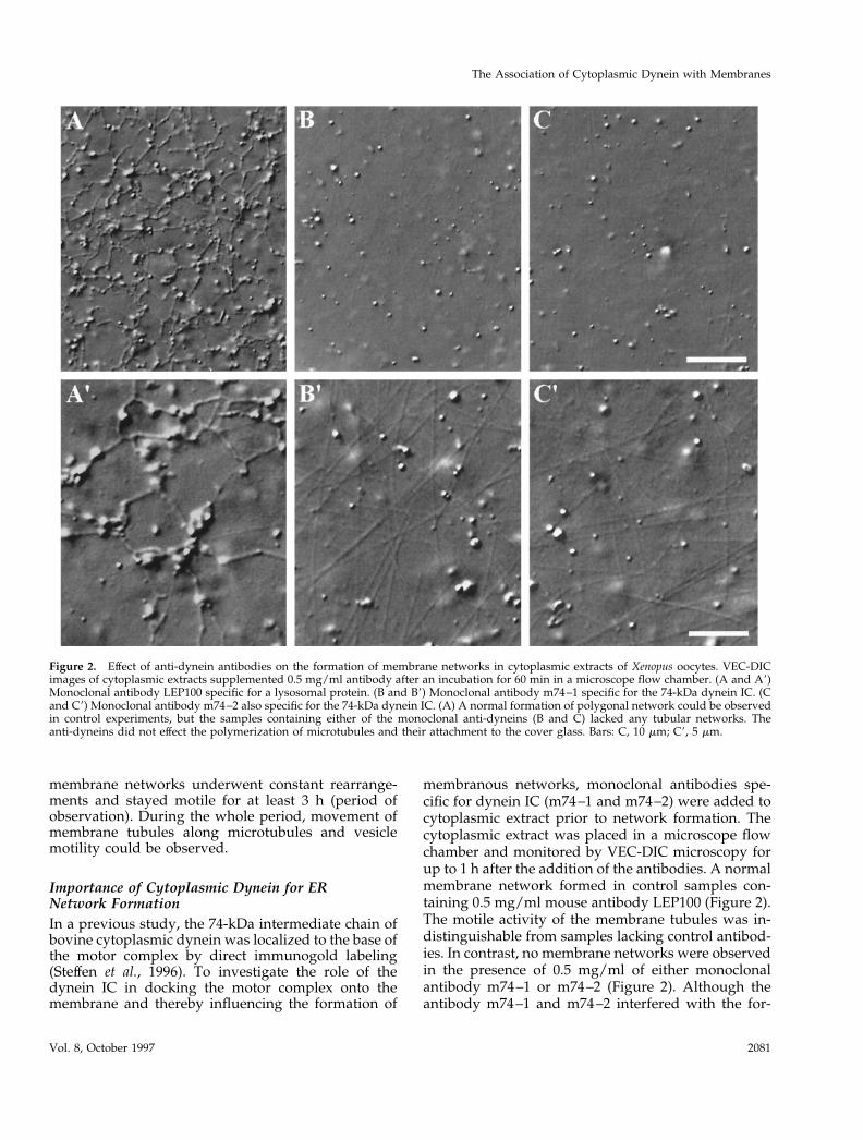
membranous networks, monoclonal antibodies spe-cific for dynein IC (m74–1 and m74–2) were added tocytoplasmic extract prior to network formation. Thecytoplasmic extract was placed in a microscope flowchamber and monitored by VEC-DIC microscopy forup to 1 h after the addition of the antibodies. A normalmembrane network formed in control samples con-taining 0.5 mg/ml mouse antibody LEP100 (Figure 2).The motile activity of the membrane tubules was in-distinguishable from samples lacking control antibod-ies. In contrast, no membrane networks were observedin the presence of 0.5 mg/ml of either monoclonalantibody m74–1 or m74–2 (Figure 2). Although theantibody m74–1 and m74–2 interfered with the for-
Figure 2. Effect of anti-dynein antibodies on the formation of membrane networks in cytoplasmic extracts of Xenopus oocytes. VEC-DICimages of cytoplasmic extracts supplemented 0.5 mg/ml antibody after an incubation for 60 min in a microscope flow chamber. (A and A9)Monoclonal antibody LEP100 specific for a lysosomal protein. (B and B9) Monoclonal antibody m74–1 specific for the 74-kDa dynein IC. (Cand C9) Monoclonal antibody m74–2 also specific for the 74-kDa dynein IC. (A) A normal formation of polygonal network could be observedin control experiments, but the samples containing either of the monoclonal anti-dyneins (B and C) lacked any tubular networks. Theanti-dyneins did not effect the polymerization of microtubules and their attachment to the cover glass. Bars: C, 10 mm; C9, 5 mm.
The Association of Cytoplasmic Dynein with Membranes
Vol. 8, October 1997 2081
mation of membrane networks, the polymerization ofmicrotubules and their attachment to the glass surfaceremained unaffected (Figure 2). Because both mono-clonal antibodies m74–1 and m74–2 appeared to haveidentical effects in blocking the formation of mem-brane networks, only m74–2 was used in the followup experiments.
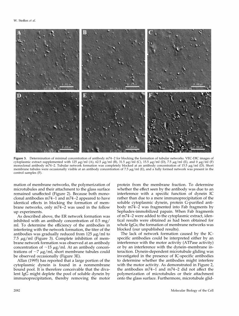
As described above, the ER network formation wasinhibited with an antibody concentration of 0.5 mg/ml. To determine the efficiency of the antibodies ininterfering with the network formation, the titer of theantibodies was gradually reduced from 125 mg/ml to7.5 mg/ml (Figure 3). Complete inhibition of mem-brane network formation was observed at an antibodyconcentration of ;15 mg/ml. At an antibody concen-trations of ;7 mg/ml, short membrane tubules couldbe observed occasionally (Figure 3E).
Allan (1995) has reported that a large portion of thecytoplasmic dynein is found in a nonmembranebound pool. It is therefore conceivable that the diva-lent IgG might deplete the pool of soluble dynein byimmunoprecipitation, thereby removing the motor
protein from the membrane fraction. To determinewhether the effect seen by the antibody was due to aninterference with a specific function of dynein ICrather than due to a mere immunoprecipitation of thesoluble cytoplasmic dynein, protein G-purified anti-body m74–2 was fragmented into Fab fragments bySephadex-immobilized papain. When Fab fragmentsof m74–2 were added to the cytoplasmic extract, iden-tical results were obtained as had been obtained forwhole IgGs; the formation of membrane networks wasblocked (our unpublished results).
The lack of network formation caused by the IC-specific antibodies could be interpreted either by aninterference with the motor activity (ATPase activity)or by an interference with the dynein–membrane in-teraction. Dynein-dependent microtubule gliding wasinvestigated in the presence of IC-specific antibodiesto determine whether the antibodies might interferewith the motor activity. As demonstrated in Figure 2,the antibodies m74–1 and m74–2 did not affect thepolymerization of microtubules or their attachmentonto the glass surface. Furthermore, microtubule glid-
Figure 3. Determination of minimal concentration of antibody m74–2 for blocking the formation of tubular networks. VEC-DIC images ofcytoplasmic extract supplemented with 125 mg/ml (A), 62.5 mg/ml (B), 31.5 mg/ml (C), 15.5 mg/ml (D), 7.5 mg/ml (E), and 0 mg/ml (F)monoclonal antibody m74–2. Tubular network formation was completely blocked at an antibody concentration of 15.5 mg/ml (D). Shortmembrane tubules were occasionally visible at an antibody concentration of 7.5 mg/ml (E), and a fully formed network was present in thecontrol samples (F).
W. Steffen et al.
Molecular Biology of the Cell2082
ing was observed as well. In cytoplasmic extracts ofXenopus oocytes, the directionality of microtubulemovement could not be determined due to the highdensity of microtubules and most importantly due thelack of an appropriate marker of microtubule polarity.To determine whether m74–2 interfered with dynein-dependent microtubule motility, microtubule glidingwas analyzed by using taxol-stabilized microtubulesand cytoplasmic dynein from bovine brain. In thisassay, the antibody m74–2 did not cause an inhibitionof microtubule gliding. Instead, the velocity of glidingmicrotubules was slightly elevated in the presence ofthe antibody. In control samples microtubules movedwith a velocity of 0.62 6 0.14 mm/sec (mean 6 SD; n 551), and they moved with a velocity of 0.82 6 0.2mm/sec (mean 6 SD; n 5 46) in samples treated withm74–2, indicating that the antibody did not block theATPase activity.
Blocking of Dynein–Membrane InteractionThe lack of network formation in the presence ofIC-specific antibodies appeared to be due to interfer-ence with the dynein–membrane interaction. To gain abetter understanding of how the antibody m74–2
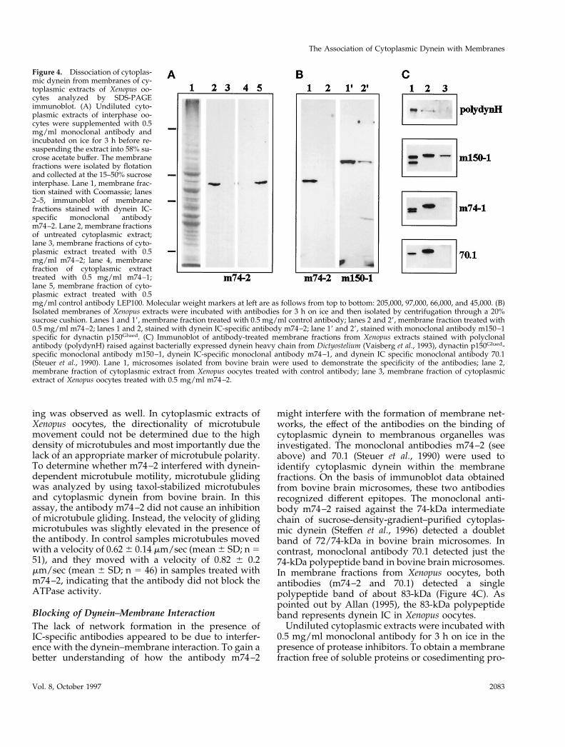
might interfere with the formation of membrane net-works, the effect of the antibodies on the binding ofcytoplasmic dynein to membranous organelles wasinvestigated. The monoclonal antibodies m74–2 (seeabove) and 70.1 (Steuer et al., 1990) were used toidentify cytoplasmic dynein within the membranefractions. On the basis of immunoblot data obtainedfrom bovine brain microsomes, these two antibodiesrecognized different epitopes. The monoclonal anti-body m74–2 raised against the 74-kDa intermediatechain of sucrose-density-gradient–purified cytoplas-mic dynein (Steffen et al., 1996) detected a doubletband of 72/74-kDa in bovine brain microsomes. Incontrast, monoclonal antibody 70.1 detected just the74-kDa polypeptide band in bovine brain microsomes.In membrane fractions from Xenopus oocytes, bothantibodies (m74–2 and 70.1) detected a singlepolypeptide band of about 83-kDa (Figure 4C). Aspointed out by Allan (1995), the 83-kDa polypeptideband represents dynein IC in Xenopus oocytes.
Undiluted cytoplasmic extracts were incubated with0.5 mg/ml monoclonal antibody for 3 h on ice in thepresence of protease inhibitors. To obtain a membranefraction free of soluble proteins or cosedimenting pro-
Figure 4. Dissociation of cytoplas-mic dynein from membranes of cy-toplasmic extracts of Xenopus oo-cytes analyzed by SDS-PAGEimmunoblot. (A) Undiluted cyto-plasmic extracts of interphase oo-cytes were supplemented with 0.5mg/ml monoclonal antibody andincubated on ice for 3 h before re-suspending the extract into 58% su-crose acetate buffer. The membranefractions were isolated by flotationand collected at the 15–50% sucroseinterphase. Lane 1, membrane frac-tion stained with Coomassie; lanes2–5, immunoblot of membranefractions stained with dynein IC-specific monoclonal antibodym74–2. Lane 2, membrane fractionsof untreated cytoplasmic extract;lane 3, membrane fractions of cyto-plasmic extract treated with 0.5mg/ml m74–2; lane 4, membranefraction of cytoplasmic extracttreated with 0.5 mg/ml m74–1;lane 5, membrane fraction of cyto-plasmic extract treated with 0.5mg/ml control antibody LEP100. Molecular weight markers at left are as follows from top to bottom: 205,000, 97,000, 66,000, and 45,000. (B)Isolated membranes of Xenopus extracts were incubated with antibodies for 3 h on ice and then isolated by centrifugation through a 20%sucrose cushion. Lanes 1 and 19, membrane fraction treated with 0.5 mg/ml control antibody; lanes 2 and 29, membrane fraction treated with0.5 mg/ml m74–2; lanes 1 and 2, stained with dynein IC-specific antibody m74–2; lane 19 and 29, stained with monoclonal antibody m150–1specific for dynactin p150Glued. (C) Immunoblot of antibody-treated membrane fractions from Xenopus extracts stained with polyclonalantibody (polydynH) raised against bacterially expressed dynein heavy chain from Dictyostelium (Vaisberg et al., 1993), dynactin p150Glued-specific monoclonal antibody m150–1, dynein IC-specific monoclonal antibody m74–1, and dynein IC specific monoclonal antibody 70.1(Steuer et al., 1990). Lane 1, microsomes isolated from bovine brain were used to demonstrate the specificity of the antibodies; lane 2,membrane fraction of cytoplasmic extract from Xenopus oocytes treated with control antibody; lane 3, membrane fraction of cytoplasmicextract of Xenopus oocytes treated with 0.5 mg/ml m74–2.
The Association of Cytoplasmic Dynein with Membranes
Vol. 8, October 1997 2083
tein complexes, the membrane fractions were isolatedby flotation through a sucrose step gradient (Niclas etal., 1996) and then analyzed by SDS-PAGE immuno-blot. Although the 83-kDa IC was observed in mem-brane fractions of untreated samples and samplestreated with a control antibody (Figure 4A, lanes 2 and5), it was no longer detectable in membrane fractionsof samples treated with antibodies m74–1 and m74–2(Figure 4B, lanes 3 and 4). Identical results were ob-tained, if the immunoblot was carried out with themonoclonal antibodies m74–1 and 70.1 (Figure 4C).The absence of the 83-kDa antigen in m74–2-treatedmembrane fractions indicated a dissociation of cyto-plasmic dynein from the membranes. To determine,therefore, whether the loss of the m74–2 antigen fromthe membrane fraction had been caused by blockingthe rebinding of cytoplasmic dynein due to the pres-ence of the monoclonal antibody m74–2, membraneswere isolated by flotation prior to incubation withm74–2. The treatment of isolated membrane fractionswith the monoclonal antibody m74–2 resulted againin a complete loss of the 83-kDa IC (Figure 4B). Im-munoblot replicas of antibody-treated membranesamples were also probed with a polyclonal antibodyagainst the heavy chain and two different monoclonalantibodies against the intermediate chain, to deter-mine whether the whole motor complex was removedfrom the membrane (Figure 4C). By using the lumi-nescence immunoblot assay, neither of these antibod-ies detected subcomponents of cytoplasmic dynein inm74–2-treated membrane fractions.
As demonstrated by video microscopy, 15 mg/mlm74–2 was sufficient to block ER network formation(Figure 3). For the network formation assay, the cyto-plasmic extract was diluted 1:5, and undiluted extractwas used for the membrane binding study. To com-pare the data from motility assay and membrane bind-ing study, we determined the molar ratio of the anti-body per 1 ml of undiluted cytoplasmic extract andfound that 0.39 nmol of m74–2 was sufficient to blockER network formation. Because the biochemical exper-iments were carried out on ice to prevent microtubulepolymerization, an ;10-fold excess of antibody wasused to disrupt the dynein–membrane interaction(Figure 4). To be able to compare both sets of experi-ment, cytoplasmic extract was incubated with 0.66nmol/ml of m74-2 and analyzed after 5, 10, and 30min of incubation. A loss of ;90% of dynein IC wasalready observed after an incubation of 10 min (ourunpublished observation), indicating a similar activityof the antibody in the membrane binding studies.
Interaction of Cytoplasmic Dynein with DynactinIt has been demonstrated that dynactin, a regulator forthe dynein-dependent membrane transport (Gill et al.,1991), can bind to dynein IC via the p150Glued subcom-
ponent (Karki and Holzbaur, 1995; Vaughan andVallee, 1995). Because the IC-specific antibody m74–2was able to influence the dynein–membrane interac-tion, the association of the regulatory component dy-nactin with the membrane fractions was also investi-gated. The monoclonal antibody m150–1 raisedagainst p150Glued from bovine brain recognized twopolypeptide bands of ;150 kDa and ;160 kDa inmicrosomes of bovine brain (Figure 4C, lane 1). Inmembrane fractions from Xenopus oocytes, the anti-body m150–1 detected only a single polypeptide bandof about 165-kDa. A significant reduction in the asso-ciation of dynactin with membrane was observed inm74–2 treated cytoplasmic extracts by using themonoclonal antibody m150–1 to detect dynactin (Fig-ure 4B, lanes 19 and 29). When compared with un-treated membranes, only about 20% of the dynactinp150Glued remained with the membranes after treat-ment with the antibody m74–2. The use of phospha-tase inhibitors and protein kinase inhibitors did notinfluence the dissociation of dynactin caused bym74–2 (our unpublished results).
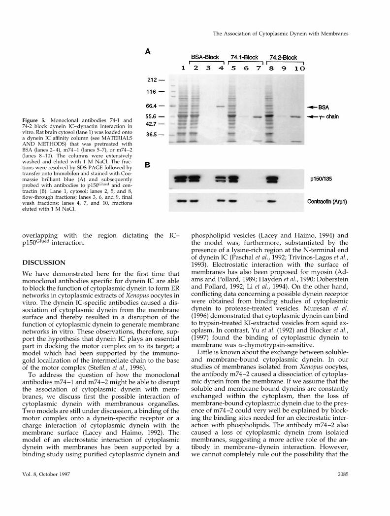
The effect of the antibody m74–2 on the dynein–dynactin interaction was further investigated by anaffinity column binding assay. Immobilized bacteriallyexpressed dynein IC was employed as affinity matrix.The column matrix was equilibrated with BSA,m74–1, or m74–2 before loading whole brain extract.The wash fractions and the eluted fractions were an-alyzed by SDS-PAGE immunoblot using antibodies todynactin p150Glued and centractin. As shown in Figure5, both antibodies m74–1 and m74–2 prevented thebinding of dynactin to the column matrix, whereas inthe presence of BSA, the dynactin p150Glued and cen-tractin were retained by the dynein IC, demonstratingthat both antibodies m74–1 and m74–2 interfered withthe dynein–dynactin interaction.
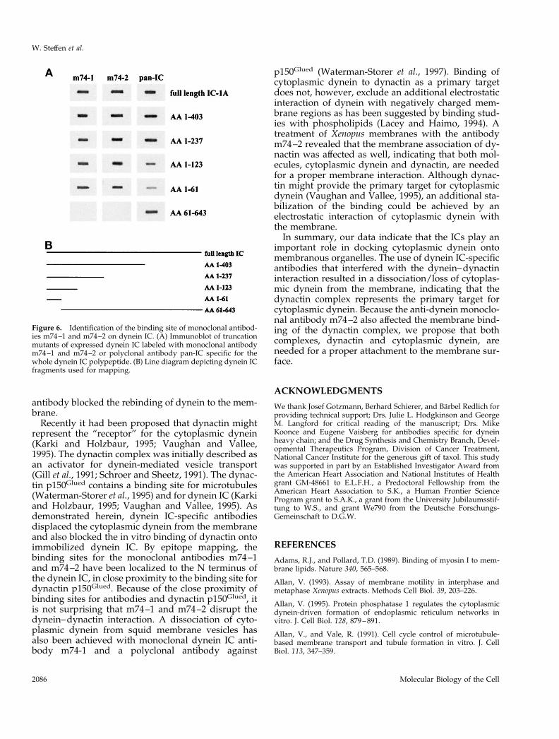
Determination of Epitopes for m74–1 and m74–2Binding studies using the IC affinity column indicatethat both antibodies interfere with the interaction ofdynein IC with dynactin p150Glued. This interactionhas been mapped to the N-terminal domain of the ICscontaining a coiled-coil structure (Vaughan andVallee, 1995). To test this possibility, we mapped theepitopes of the two monoclonal antibodies by using apanel of IC truncation mutants. A pan-IC antibodygenerated against full-length IC-1A detected all pep-tide fragments (Figure 6), as reported previously(Vaughan and Vallee, 1995). Both monoclonal antibod-ies detected all fragments down to amino acids 1–66yet failed to detect a fragment containing amino acids61–643 (Figure 6). These findings suggest that theepitopes for the monoclonal antibodies m74–1 andm74–2 lie within the N-terminal coiled-coil domain
W. Steffen et al.
Molecular Biology of the Cell2084
overlapping with the region dictating the IC–p150Glued interaction.
DISCUSSION
We have demonstrated here for the first time thatmonoclonal antibodies specific for dynein IC are ableto block the function of cytoplasmic dynein to form ERnetworks in cytoplasmic extracts of Xenopus oocytes invitro. The dynein IC-specific antibodies caused a dis-sociation of cytoplasmic dynein from the membranesurface and thereby resulted in a disruption of thefunction of cytoplasmic dynein to generate membranenetworks in vitro. These observations, therefore, sup-port the hypothesis that dynein IC plays an essentialpart in docking the motor complex on to its target; amodel which had been supported by the immuno-gold localization of the intermediate chain to the baseof the motor complex (Steffen et al., 1996).
To address the question of how the monoclonalantibodies m74–1 and m74–2 might be able to disruptthe association of cytoplasmic dynein with mem-branes, we discuss first the possible interaction ofcytoplasmic dynein with membranous organelles.Two models are still under discussion, a binding of themotor complex onto a dynein-specific receptor or acharge interaction of cytoplasmic dynein with themembrane surface (Lacey and Haimo, 1992). Themodel of an electrostatic interaction of cytoplasmicdynein with membranes has been supported by abinding study using purified cytoplasmic dynein and
phospholipid vesicles (Lacey and Haimo, 1994) andthe model was, furthermore, substantiated by thepresence of a lysine-rich region at the N-terminal endof dynein IC (Paschal et al., 1992; Trivinos-Lagos et al.,1993). Electrostatic interaction with the surface ofmembranes has also been proposed for myosin (Ad-ams and Pollard, 1989; Hayden et al., 1990; Dobersteinand Pollard, 1992; Li et al., 1994). On the other hand,conflicting data concerning a possible dynein receptorwere obtained from binding studies of cytoplasmicdynein to protease-treated vesicles. Muresan et al.(1996) demonstrated that cytoplasmic dynein can bindto trypsin-treated KI-extracted vesicles from squid ax-oplasm. In contrast, Yu et al. (1992) and Blocker et al.,(1997) found the binding of cytoplasmic dynein tomembrane was a-chymotrypsin-sensitive.
Little is known about the exchange between soluble-and membrane-bound cytoplasmic dynein. In ourstudies of membranes isolated from Xenopus oocytes,the antibody m74–2 caused a dissociation of cytoplas-mic dynein from the membrane. If we assume that thesoluble and membrane-bound dyneins are constantlyexchanged within the cytoplasm, then the loss ofmembrane-bound cytoplasmic dynein due to the pres-ence of m74–2 could very well be explained by block-ing the binding sites needed for an electrostatic inter-action with phospholipids. The antibody m74–2 alsocaused a loss of cytoplasmic dynein from isolatedmembranes, suggesting a more active role of the an-tibody in membrane–dynein interaction. However,we cannot completely rule out the possibility that the
Figure 5. Monoclonal antibodies 74-1 and74-2 block dynein IC–dynactin interaction invitro. Rat brain cytosol (lane 1) was loaded ontoa dynein IC affinity column (see MATERIALSAND METHODS) that was pretreated withBSA (lanes 2–4), m74–1 (lanes 5–7), or m74–2(lanes 8–10). The columns were extensivelywashed and eluted with 1 M NaCl. The frac-tions were resolved by SDS-PAGE followed bytransfer onto Immobilon and stained with Coo-massie brilliant blue (A) and subsequentlyprobed with antibodies to p150Glued and cen-tractin (B). Lane 1, cytosol; lanes 2, 5, and 8,flow-through fractions; lanes 3, 6, and 9, finalwash fractions; lanes 4, 7, and 10, fractionseluted with 1 M NaCl.
The Association of Cytoplasmic Dynein with Membranes
Vol. 8, October 1997 2085
antibody blocked the rebinding of dynein to the mem-brane.
Recently it had been proposed that dynactin mightrepresent the “receptor” for the cytoplasmic dynein(Karki and Holzbaur, 1995; Vaughan and Vallee,1995). The dynactin complex was initially described asan activator for dynein-mediated vesicle transport(Gill et al., 1991; Schroer and Sheetz, 1991). The dynac-tin p150Glued contains a binding site for microtubules(Waterman-Storer et al., 1995) and for dynein IC (Karkiand Holzbaur, 1995; Vaughan and Vallee, 1995). Asdemonstrated herein, dynein IC-specific antibodiesdisplaced the cytoplasmic dynein from the membraneand also blocked the in vitro binding of dynactin ontoimmobilized dynein IC. By epitope mapping, thebinding sites for the monoclonal antibodies m74–1and m74–2 have been localized to the N terminus ofthe dynein IC, in close proximity to the binding site fordynactin p150Glued. Because of the close proximity ofbinding sites for antibodies and dynactin p150Glued, itis not surprising that m74–1 and m74–2 disrupt thedynein–dynactin interaction. A dissociation of cyto-plasmic dynein from squid membrane vesicles hasalso been achieved with monoclonal dynein IC anti-body m74-1 and a polyclonal antibody against
p150Glued (Waterman-Storer et al., 1997). Binding ofcytoplasmic dynein to dynactin as a primary targetdoes not, however, exclude an additional electrostaticinteraction of dynein with negatively charged mem-brane regions as has been suggested by binding stud-ies with phospholipids (Lacey and Haimo, 1994). Atreatment of Xenopus membranes with the antibodym74–2 revealed that the membrane association of dy-nactin was affected as well, indicating that both mol-ecules, cytoplasmic dynein and dynactin, are neededfor a proper membrane interaction. Although dynac-tin might provide the primary target for cytoplasmicdynein (Vaughan and Vallee, 1995), an additional sta-bilization of the binding could be achieved by anelectrostatic interaction of cytoplasmic dynein withthe membrane.
In summary, our data indicate that the ICs play animportant role in docking cytoplasmic dynein ontomembranous organelles. The use of dynein IC-specificantibodies that interfered with the dynein–dynactininteraction resulted in a dissociation/loss of cytoplas-mic dynein from the membrane, indicating that thedynactin complex represents the primary target forcytoplasmic dynein. Because the anti-dynein monoclo-nal antibody m74–2 also affected the membrane bind-ing of the dynactin complex, we propose that bothcomplexes, dynactin and cytoplasmic dynein, areneeded for a proper attachment to the membrane sur-face.
ACKNOWLEDGMENTS
We thank Josef Gotzmann, Berhard Schierer, and Barbel Redlich forproviding technical support; Drs. Julie L. Hodgkinson and GeorgeM. Langford for critical reading of the manuscript; Drs. MikeKoonce and Eugene Vaisberg for antibodies specific for dyneinheavy chain; and the Drug Synthesis and Chemistry Branch, Devel-opmental Therapeutics Program, Division of Cancer Treatment,National Cancer Institute for the generous gift of taxol. This studywas supported in part by an Established Investigator Award fromthe American Heart Association and National Institutes of Healthgrant GM-48661 to E.L.F.H., a Predoctoral Fellowship from theAmerican Heart Association to S.K., a Human Frontier ScienceProgram grant to S.A.K., a grant from the University Jubilaumsstif-tung to W.S., and grant We790 from the Deutsche Forschungs-Gemeinschaft to D.G.W.
REFERENCES
Adams, R.J., and Pollard, T.D. (1989). Binding of myosin I to mem-brane lipids. Nature 340, 565–568.
Allan, V. (1993). Assay of membrane motility in interphase andmetaphase Xenopus extracts. Methods Cell Biol. 39, 203–226.
Allan, V. (1995). Protein phosphatase 1 regulates the cytoplasmicdynein-driven formation of endoplasmic reticulum networks invitro. J. Cell Biol. 128, 879–891.
Allan, V., and Vale, R. (1991). Cell cycle control of microtubule-based membrane transport and tubule formation in vitro. J. CellBiol. 113, 347–359.
Figure 6. Identification of the binding site of monoclonal antibod-ies m74–1 and m74–2 on dynein IC. (A) Immunoblot of truncationmutants of expressed dynein IC labeled with monoclonal antibodym74–1 and m74–2 or polyclonal antibody pan-IC specific for thewhole dynein IC polypeptide. (B) Line diagram depicting dynein ICfragments used for mapping.
W. Steffen et al.
Molecular Biology of the Cell2086
Allan, V., and Vale, R. (1994). Movement of membrane tubulesalong microtubules in vitro: evidence for specialized sites of motorattachment. J. Cell Sci. 107, 1885–1897.
Allen, R.D., and Allen, N.S. (1983). Video-enhanced microscopywith a computer frame memory. J. Microscopy 129, 3–17.
Allen, R.D., Allen, N.S., and Travis, J.L. (1981). Video-enhancedcontrast, differential interference contrast (AVEC-DIC) microscopy:a new method capable of analyzing microtubule-related motility inthe reticulopodial network of Allogromia laticollaris. Cell Motil. Cy-toskeleton 1, 291–302.
Allen, R.D., Weiss, D.G., Hayden, J.H., Brown, D.T., Fujiwake, H.,and Simpson, M. (1985). Gliding movement of and bidirectionaltransport along single native microtubules from squid axoplasm:evidence for an active role of microtubules in cytoplasmic transport.J. Cell Biol. 100, 1736–1752.
Aniento, F., Emans, N., Griffiths, G., and Gruenberg, J. (1993). Cy-toplasmic dynein-dependent vesicular transport from early to lateendosomes. J. Cell Biol. 123, 1373–1387.
Blocker, A., Severin, F.F., Habermann, A., Hyman, A.A., Griffiths,G.G., and Burkhardt, J.K. (1996). Microtubule-associated protein-dependent binding of phagosomes to microtubules. J. Biol. Chem.271, 3803–3811.
Blocker, A., Severin, F.F., Burkhardt, J.K., Bingham, J.B., Yu, H.,Olivo, J.C., Schroer, T.A., Hyman, A.A., and Griffiths, G. (1997).Molecular requirements for bi-directional movement of phago-somes along microtubules. J. Cell Biol. 137, 113–129.
Collins, C.A. (1991). Reversible assembly purification of taxol-treated microtubules. Methods Enzymol. 196, 246–253.
Corthesy-Theulaz, I., Pauloin, A., and Pfeffer, S.R. (1992). Cytoplas-mic dynein participates in the centrosomal localization of the Golgicomplex. J. Cell Biol. 118, 1333–1345.
Dabora, S., and Sheetz, M.P. (1988). The microtubule-dependentformation of a tubulovesicular network with characteristics of theER from cultured cell extracts. Cell 54, 27–35.
Doberstein, S.K., and Pollard, T.D. (1992). Localization and specific-ity of the phospholipid and actin binding sites on the tail of Acanth-amoeba myosin IC. J. Cell Biol. 117, 1241–1249.
Echeverri, C.J., Paschal, B.M., Vaughan, K.T., and Vallee, R.B. (1996).Molecular characterization of the 50-kDa subunit of dynactin re-veals function for the complex in chromosome alignment and spin-dle organization during mitosis. J. Cell Biol. 132, 617–633.
Fath, K.R., Trimbur, G.M., and Burgess, D.R. (1994). Molecularmotors are differentially distributed on Golgi membranes from po-larized epithelial cells. J. Cell Biol. 126, 661–675.
Franke, W.W. (1971). Cytoplasmic microtubules linked to endoplas-mic reticulum with cross-bridges. Exp. Cell Res. 66, 486–489.
Gill, S.R., Schroer, T.A., Szilak, I., Steuer, E.R., Sheetz, M.P., andCleveland, D.W. (1991). Dynactin, a conserved, ubiquitously ex-pressed component of an activator of vesicle motility mediated bycytoplasmic dynein. J. Cell Biol. 115, 1639–1650.
Hayden, S.M., Wolenski, J.S., and Mooseker, M.S. (1990). Binding ofbrush border myosin I to phospholipid vesicles. J. Cell Biol. 111,443–451.
Hirokawa, N., Sato-Yoshitake, R., Yoshida, T., and Kawashima, T.(1990). Brain dynein (MAP-1C) localizes on both anterogradely andretrogradely transported membranous organelles in vivo. J. CellBiol. 111, 1027–1037.
Holleran, E.A., Tokito, M.K., Karki, S., and Holzbaur, E.L.F. (1996).Centractin (ARP1) associates with spectrin revealing a potentialmechanism to link dynactin to intracellular organelles. J. Cell Biol.135, 1815–1829.
Karki, S., and Holzbaur, L.F. (1995). Affinity chromatography dem-onstrates a direct binding between cytoplasmic dynein and thedynactin complex. J. Biol. Chem. 270, 28806–28811.
King, S.M., Wilkerson, C.G., and Witman, G.B. (1991). The Mr 78,000intermediate chain of Chlamydomonas outer arm dynein interactswith a-tubulin in situ. J. Biol. Chem. 266, 8401–8407.
Lacey, M.L., and Haimo, L.T. (1992). Cytoplasmic dynein is a vesicleprotein. J. Biol. Chem. 267, 4793–4798.
Lacey, M.L., and Haimo, L.T. (1994). Cytoplamic dynein binds tophospholipid vesicles. Cell Motil. Cytoskeleton 28, 205–212.
Lee, C., and Chen, L.B. (1988). Dynamic behavior of endoplasmicreticulum in living cells. Cell 54, 37–46.
Lee, C., Ferguson, M., and Chen, L.B. (1989). Construction of theendoplasmic reticulum. J. Cell Biol. 109, 2045–2055.
Li, D.Q., Miller, M., and Chantler, P.D. (1994). Association of acellular myosin-II with anionic phospholipids and the neuronalplasma membrane. Proc. Natl. Acad. Sci. USA 91, 853–857.
Lin, S.X., and Collins, C.A. (1992). Immunolocalization of cytoplas-mic dynein in lysosomes in cultured cells. J. Cell Sci. 110, 125–137.
Mage, M.G. (1980). Preparation of Fab fragments from IgGs ofdifferent animal species. Methods Enzymol. 70, 142–150.
Muresan, V., Godek, C.P., Reese, T.S., and Schnapp, B.J. (1996).Plus-end motors override minus-end motors during transport ofsquid axon vesicles on microtubules. J. Cell Biol. 135, 383–397.
Murray, A. (1991). Cell cycle extracts. Methods Cell Biol. 36, 581–605.
Niclas, J., Allan, V.J., and Vale, R.D. (1996). Cell cycle regulation ofdynein association with membrane modulates microtubule-basedorganelle transport. J. Cell Biol. 133, 585–593.
Paschal, B.M., Mikami, A., Pfister, K.K., and Vallee, R.B. (1992).Homology of the 74-kDa cytoplasmic dynein subunit with a flagel-lar dynein polypeptide suggests an intracellular targeting function.J. Cell Biol. 118, 1133–1143.
Paschal, B.M., Shpetner, H.S., and Vallee, R.B. (1987). MAP1C is amicrotubule-associated ATPase which translocates microtubules invitro and has dynein-like properties. J. Cell Biol. 105, 1273–1282.
Schroer, T.A., and Sheetz, M.P. (1991). Two activators of microtu-bule-based vesicle transport. J. Cell Biol. 115, 1309–1318.
Steffen, W., Hodgkinson, J.L., and Wiche, G. (1996). Immunogoldlocalization of the intermediate chain within the protein complex ofcytoplasmic dynein. J. Struct. Biol. 117, 227–235.
Steffen, W., Langford, G.M., Weiss, D.G., and Kuznetsov, S.A.(1997). Inhibition of microtubule-dependent minus-end directedtransport of axoplasmic organelles by a dynein intermediate chainspecific antibody. Biol. Bull. (in press).
Steuer, E.R., Wordeman, L., Schroer, T.A., and Sheetz, M.P. (1990).Localization of cytoplasmic dynein in mitotic spindles and kineto-chores. Nature 345, 266–269.
Terasaki, M., and Reese, T.S. (1994). Interactions among endoplas-mic reticulum, microtubules, and retrograde movements of the cellsurface. Cell Motil. Cytoskeleton 29, 291–300.
Tokito, M.K., Howland, D.S., Lee, V.M., and Holzbaur, E.L. (1996).Functionally distinct isoforms of dynactin are expressed in humanneurons. Mol. Biol. Cell 7, 1167–1180.
Toyoshima, I., Yu, H., Steuer, E.R., and Sheetz, M.P. (1992). Kinectin,a major kinesin-binding protein on ER. J. Cell Biol. 118, 1121–1131.
Towbin, H., Staehelin, T., and Gordon, J. (1979). Electrophoretictransfer of proteins from polyacrylamide gels to nitrocellulosesheets: procedure and some applications. Proc. Natl. Acad. Sci. USA76, 4350–4354.
The Association of Cytoplasmic Dynein with Membranes
Vol. 8, October 1997 2087
Trivinos-Lagos, L., Collins, C.A., and Chrisholms, R.L. (1993). Clon-ing of dynein intermediate chain: multiple isoforms are expressedduring Dictyostelium development. Mol. Biol. Cell 4, 47a (Abstract).
Vaisberg, E.A, Grissom, P.M., and McIntosh, J.R. (1996). Mamma-lian cells express three distinct dynein heavy chains that are local-ized to different cytoplasmic organelles. J. Cell Biol. 133, 831–842.
Vaisberg, E.A., Koonce, M.P., and McIntosh, J.R. (1993). Cytoplas-mic dynein plays a role in mammalian mitotic spindle formation. J.Cell Biol. 123, 849–858.
Vale, R.D., and Hotani, H. (1988). Formation of membrane networksin vitro by kinesin-driven microtubule movement. J. Cell Biol. 107,2233–2242.
Vaughan, K.T., and Vallee, R.B. (1995). Cytoplasmic dynein bindsdynactin through a direct interaction between the intermediatechains and p150Glued. J. Cell Biol. 131, 1507–1517.
Waterman-Storer, C.M., Karki, S., and Holzbaur, E.L.F. (1995). Thep150Glued component of the dynactin complex binds to both micro-
tubules and the actin-related protein centractin (Arp-1). Proc. Natl.Acad. Sci. USA 92, 1634–1638.
Waterman-Storer, C.M., Kuznetsov, S.A., Karki, S., Tabb, J.S., Weiss,D.G., Langford, G.M., and Holzbaur, E.L.F. (1997). The interactionbetween cytoplasmic dynein and dynactin is required for fast ax-onal transport. Proc. Natl. Acad. Sci. (in press).
Weiss, D.G., and Maile, W. (1993). Principle, practice, and applica-tion of video-enhanced contrast microscopy. In: Electronic LightMicroscopy, ed. D.M. Shotton, New York: Wiley-Liss, 105–140.
Weiss, D.G., Maile, W., and Wick, R.A. (1989). Video microscopy. In:Light Microscopy in Biology. A Practical Approach, ed. A.J. Lacey,Oxford, United Kingdom: IRL Press at Oxford University Press,221–278.
Yu, H., Toyoshima, I., Steuer, E.R., and Sheetz, M.P. (1992). Kinesinand cytoplasmic dynein binding to brain microsomes. J. Biol. Chem.267, 20457–20464.
W. Steffen et al.
Molecular Biology of the Cell2088


