Embed Size (px)
Citation preview

The Mouse Prolactin Gene Family Locus
DUSTIN O. WIEMERS, LONG-JIANG SHAO, RUPASRI AIN, GUOLI DAI, AND MICHAEL J. SOARES
Institute of Maternal-Fetal Biology and the Departments of Molecular & Integrative Physiology and Obstetrics &Gynecology, University of Kansas Medical Center, Kansas City, Kansas 66160
In the mouse, there is a large family of paralogous genesclosely related to PRL. The objective of this report was toinvestigate the organization of the mouse PRL gene familylocus. PRL family genes reside on chromosome 13 of the mousegenome. The PRL gene family members were localized to aseries of overlapping bacterial artificial chromosome clonesand arranged based on structural relationships. Additionally,several new members of the PRL gene family were identified.Placental lactogen I (PL-I) was found to be encoded by threeclosely related (>98% exon sequence identity) contiguousgenes (termed: PL-I�, PL-I�, and PL-I�). Two previously un-identified mouse orthologs for members of the rat PRL family,
PRL-like protein-I (PLP-I) and PLP-K were discovered, aswere two new members of the PLP-C subfamily, PLP-C� andPLP-C�, and two new entirely unique members of the PRLfamily, PLP-N and PLP-O. Amino acid sequences predictedfrom the latter two genes most closely resembled proliferin-related protein. Each of the nine newly discovered genes isexpressed in trophoblast cells of the mouse placenta in a ges-tationally specific pattern. In summary, elucidation of themouse PRL gene family locus provides new insights into theexpansion of the mouse PRL family and new tools for studyingthe genetics and biology of its members. (Endocrinology 144:313–325, 2003)
PROLACTIN (PRL) IS a hormone/cytokine responsiblefor the coordination of a wide range of biological pro-
cesses in vertebrates. In the mouse, rat, cow, and likely othermammalian species, there are large families of paralogousgenes closely related to PRL (1). The proteins encoded by thePRL family genes have been given a variety of names, in-cluding placental lactogens (PLs), PRL-like proteins (PLPs),PRL-related proteins (PRPs), proliferin (PLF), and PLF-related protein (PLF-RP). Unfortunately, in some instances,the literature contains nomenclature that is confusing and/orincorrect. Members of the PRL family are expressed in cell-and temporal-specific patterns in the uteroplacental com-partment and anterior pituitary (1). An overriding themecharacteristic of the PRL family is its association with preg-nancy and regulatory mechanisms controlling viviparity.
The initial identification of a substance extracted from theanterior pituitary possessing actions on the mammary glandand the initiation of lactation and its subsequent naming asPRL occurred over 70 yr ago (2–5). The notion that there maybe other hormones related to PRL developed from studieswith the pregnant mouse and pregnant rat. Removal of theanterior pituitary during the second half of gestation in themouse or rat was consistent with continued development ofthe mammary glands and corpus luteum (another target ofPRL; Refs. 6 and 7). These studies suggested an extrapituitarysource of a PRL-like hormone and were followed by thediscovery that extracts from the placenta possessed luteo-tropic (8) and mammotropic activities (9). The isolation offunctional PRL mimics (PLs) from the rodent proceededwithin the next few years (10–12). Their relationship withPRL was apparent following the cloning and sequence anal-
yses of PRL cDNAs and the initial PL cDNAs (13–17). Ex-istence of an expanded PRL family became evident during asearch for mRNAs in 3T3 fibroblasts whose expression wasdependent upon growth factor stimulation and as a byprod-uct of cloning rat placental lactogen (PL)-II. In the first in-stance, growth factor regulated mRNAs were identified withsequence similarity to PRL (18), and in the second instanceunique cDNAs were isolated from a rat placental cDNAlibrary with sequence similarity to the PRL sequence (19, 20).Several additional members were discovered during thecharacterization of known members of the PRL family atprotein, cDNA, and genomic levels and through the use ofdegenerate probes and differential display strategies (21–32).More recent discoveries have been directly attributed tosearching mouse and rat expressed sequence tag databases(33–43).
The PRL family genes in the mouse, rat, and cow appearto be clustered on chromosomes 13 (mouse: 30, 33–35, 39, 40,44), 17 (rat: 24, 25, 27, 45–48), and 23 (cow: 49) of eachrespective species genome. Gene structure has been deter-mined for a few members of the PRL family. Two conservedexon-intron arrangements are evident: 1) a prototypical fiveexon-four intron structure found in PRL (50–52), PL-II (48,53), PLP-A (54), PLF (55), and two bovine PRL family mem-bers (56, 57); and 2) a six exon-five intron structure presentin members of the PLP-C subfamily, including PLP-C variant(v) (27), PLP-C� (30), and decidual (d)PRP (58). The latterexon/intron arrangement represents the addition of a shortexon between exons II and III of the prototypical PRL exon/intron organization. Based on amino acid similarities, theextra exon is likely present in all members of the PLP-Csubfamily, and PLF-RP. PLP-E and PLP-F may possess anonhomologous exon situated in the same region (37).
In the present study, we have used information derivedfrom the public mouse genome database as a tool for iden-tifying several new members of the mouse PRL family. We
Abbreviations: BAC, Bacterial artificial chromosome; d, decidual;G3PDH, glyceraldehyde-3-phosphate dehydrogenase; PLF, proliferin;PLF-RP, PLF-related protein; PL-I or -II, placental lactogen (I or II); PLP,PRL-like protein; PRL, prolactin; PRP, PRL-related protein; SSC, stan-dard saline citrate; v, variant.
0013-7227/03/$15.00/0 Endocrinology 144(1):313–325Printed in U.S.A. Copyright © 2003 by The Endocrine Society
doi: 10.1210/en.2002-220724
313

report the identification of nine new members of the PRLfamily and their expression patterns, and some insights intothe organization of the PRL family locus on chromosome 13.
Materials and MethodsAnimals and tissue preparation
CD-1 mice were obtained from Charles River Laboratories, Inc. (Wil-mington, MA). The animals were housed in an environmentally con-trolled facility, with lights on from 0600–2000 h, and allowed free accessto food and water. Timed pregnancies were generated and tissue dis-sections were performed as previously described (35). The presence ofa copulatory plug was designated as d 1 of pregnancy. Protocols for thecare and use of animals were approved by the University of KansasAnimal Care and Use Committee.
Conceptuses with associated uteri were removed on d 11, 13, 16, and19 of gestation. Tissues were frozen in dry ice-cooled heptane and storedat –80 C until used for in situ hybridization. Alternatively, placentaltissues were dissected free from maternal and fetal structures, imme-diately frozen in liquid nitrogen, and stored at –80 C until processed forRNA analysis.
Blastocysts were isolated by flushing uterine horns on d 4 of gestation,washed, transferred to 35-mm dishes, and cultured in Roswell ParkMemorial Institute 1640 medium in a humidified incubator at 37 C withan atmosphere of 95% air/5% CO2. The culture medium was supple-mented with antibiotics, 1 mm glutamine, 1 mm sodium pyruvate, and20% fetal bovine serum. Blastocysts hatched, attached, and demon-strated a significant outgrowth within a few days of the initiation ofculture. Outgrowths were harvested following 6 d of culture.
Cloning and characterization of the PRL family cDNAs
Genes for new members of the PRL family were identified by BLASTanalysis (59) using nucleotide sequences of previously identified mem-bers of the PRL family (Ref. 1; and Table 1) against the public mouse
genome assembly (http://mouse.ensembl.org and http://www.ncbi.nlm.nih.gov/genome/seq/MmBlast.html). Members of the PRL familywere found on several overlapping bacterial artificial chromosome(BAC) clones localized to chromosome 13 (RP23–351I6, RP23–189A16,RP23–122C3, RP23–142M3, RP23–117C5, RP23–20A22, RP23–231P12;Children’s Hospital-BACPAC Resources, Oakland, CA). Genomic se-quences possessing similarity to members of the PRL family were usedto search the mouse expressed sequence tag database (National Centerfor Biotechnology Information; Ref. 59). One cDNA (PLP-O; clone no.5280049R, BG077704) was obtained from the American Type TissueCollection (ATCC, Manassas, VA) and another cDNA (PLP-C�; RikenNo. 1600019J01, AV036496) from the Riken Institute (Ibaraki, Japan; Ref.60). The remaining cDNAs were isolated by RT-PCR from total RNAextracted with the TRIzol reagent (61) from mouse blastocyst out-growths or placental tissues. Five micrograms of total RNA and 0.5 �gof oligo-deoxythymidine were used for reverse transcription reactions.PCRs were performed using Pfu polymerase with sets of primers basedon genomic sequences (Table 2). PCR was performed for 30 cycles(denature, 95 C for 1 min; anneal, 55 C for 1 min; extension, 72 C for 1.5min). Amplified products were subcloned into pCRII-TOPO vectorflanked by SP6 and T7 promoters with the TOPO TA cloning kit (In-vitrogen, Carlsbad, CA). cDNAs were sequenced by the BiotechnologySupport Facility of the University of Kansas Medical Center. Nucleotideand amino acid sequence comparisons were performed with CLUSTALW (version 1.8; Ref. 62). The location of signal peptides was determinedby homology and the SignalP software program (version 2.0.b2; Ref. 63).
Analysis of mRNA expression
RT-PCR restriction enzyme analysis of PL-I-related genes. PL-I-relatedcDNAs were amplified from blastocyst outgrowths or placentas from d10 of gestation. Amplified products were digested with restriction en-zymes capable of differentially cutting PL-I�, PL-I�, and/or PL-I�. BsaJ1digests each of the PL-I cDNAs at nucleotide 136 and PL-I� uniquely atnucleotide 420. NlaIII digests each of the PL-I cDNAs at nucleotide 140and 740; PL-I� and PL-I� are additionally cut at nucleotide 324. Thedigested products were resolved by electrophoresis in 2% agarose gelsand ethidium bromide staining.
Northern blot analysis. Northern blots were performed as previouslydescribed (34, 64). RNA was extracted from tissues using TRIzol (61).Total RNA (20 �g) was separated on 1% formaldehyde-agarose gels andtransferred to nylon membranes. Blots were probed with [32P]-labeledcDNAs for PLP-C�, PLP-C�, PLP-C�, PLP-I, PLP-K, PLP-L, PLP-N, orPLP-O. Glyceraldehyde-3-phosphate dehydrogenase (G3PDH) cDNAwas used as an internal reference to ensure integrity of the RNA samples.
In situ hybridization. PRL family mRNAs were detected in placentaltissues using nonradioactive in situ hybridization as previously de-scribed (65). Plasmids containing cDNAs for PLP-C�, PLP-C�, PLP-I,PLP-K, PLP-L, PLP-N, and PLP-O were linearized and used as templatesto synthesize sense and antisense digoxigenin-labeled riboprobes ac-cording to the manufacturer’s instructions (Roche Molecular Biochemi-cals, Indianapolis, IN). Tissue sections were air dried and fixed in ice-cold 4% paraformaldehyde in PBS for 15 min. Prehybridization wascarried out in a humidified chamber at 50 C in 5� SSC (standard salinecitrate), 50% deionized formamide, 1� Denhardt’s reagent, 10% dextransulfate, and salmon sperm DNA (100 �g/ml). Hybridizations wereperformed in the same incubation conditions overnight. Slides werewashed in 2� SSC at room temperature for 30 min followed by treatmentwith ribonuclease A (100 ng/ml) and additional washes with 2� SSC for30 min at room temperature, followed by washes with 2� SSC for 1 hat 65 C and 0.1 � SSC for 1 h at 65 C. Tissue sections were blocked for30 min and incubated with alkaline phosphatase-conjugated antidigoxi-genin antibody (1:500) in blocking buffer (Roche Molecular Biochemi-cals) for 2 h at room temperature. Slides were then washed and detectionwas performed using nitro blue tetrazolium (250 �g/ml) and 5-bromo-4-chloro-3-indolyl-phosphate (225 �g/ml; Roche Molecular Biochemicals).
ResultsMouse PRL family gene locus
BLAST analysis of the public mouse genome assemb-ly (http://mouse.ensembl.org and http://www.ncbi.nlm.
TABLE 1. Mouse PRL gene family
PRLfamily Other names GenBank
accession no. Reference
1 PRL X02892 142 PL-I� Midpregnancy
lactogen; PL-I;PL-Im
M35662,AF525162
17; Present study
3 PL-I� AF525160 Present study4 PL-I� AF525161 Present study5 PLP-J PLP-I, Decidualin,
PrlpjAB019118 39, 40
6 PL-II PL M14647 157 PLP-I PLP-H AF525154 Present study8 PLP-B Prlpb AF015563 34, 369 DPRP D/tPRP AF015729 34, 35
10 PLP-K Prlpk AF525155 Present study11 PLP-C� Prlpc3 AF466150 Present study12 PLP-C� Prlpc2 AF158744 3213 PLP-C� AF525158 Present study14 PLP-C� Prlpc1 AF090140 3015 PLP-N AF525156 Present study16 PLP-E PLP-G, Prlpe AF020525 33, 3717 PLP-F Prlpf AF020524 33, 3718 PLP-O AF525157 Present study19 PLF-RP PRP X02594 2120 PLF-1 MRP-1 K02245 1821 PLF-2 MRP-2 K03235 6622 PLF-3 MRP-3 NM_011954 5523 PLF-4 MRP-4 AF128884 6724 PLP-M Prlpm AF234636 4025 PLP-A Prlpa AF015562 34, 3626 PLP-L AF226611 43
d/tPRP or dPRP, Decidual/trophoblast PRL-related protein; MRP,mitogen-regulated protein; PLF-RP or PRP, PLF-related protein.
314 Endocrinology, January 2003, 144(1):313–325 Wiemers et al. • PRL Gene Family
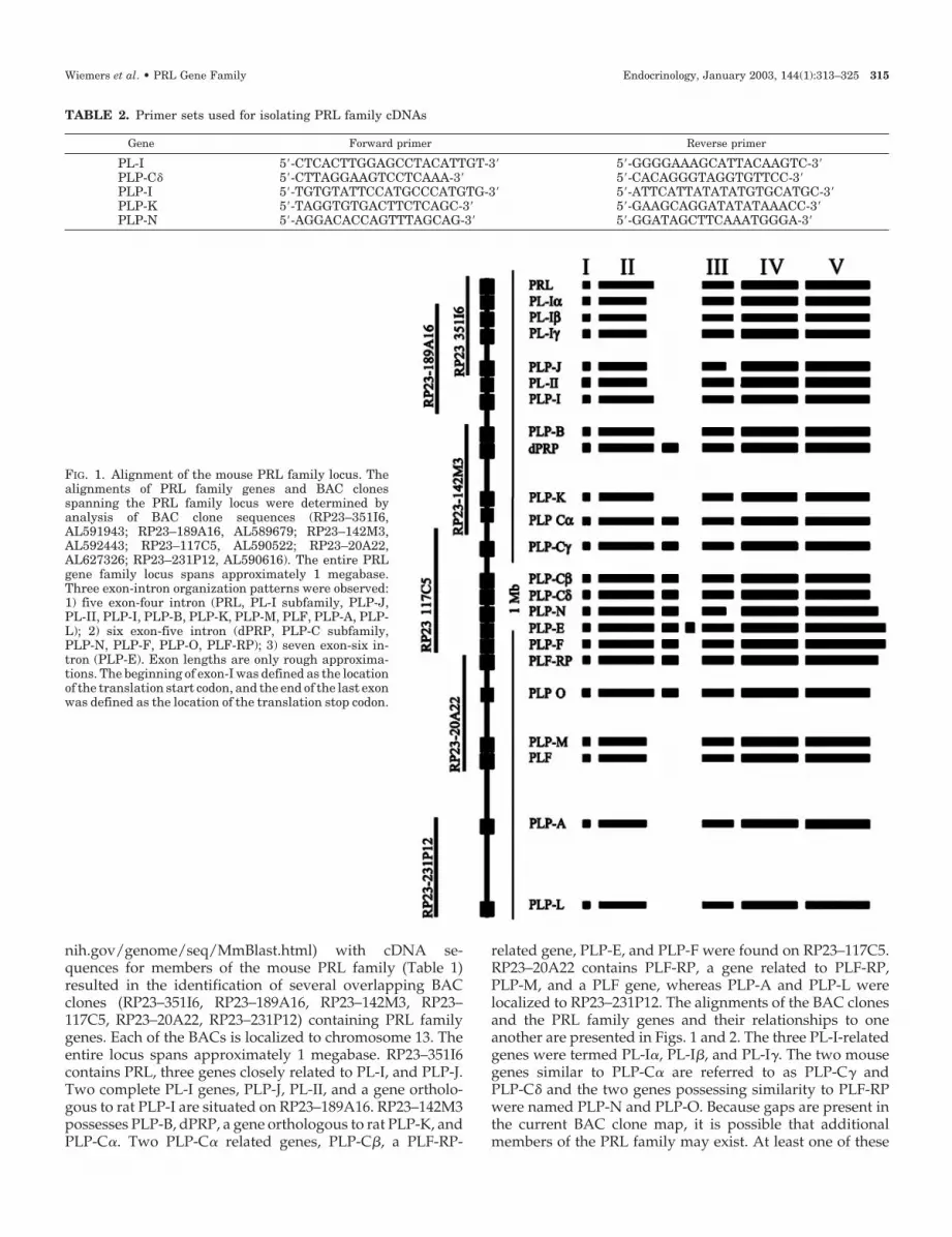
nih.gov/genome/seq/MmBlast.html) with cDNA se-quences for members of the mouse PRL family (Table 1)resulted in the identification of several overlapping BACclones (RP23–351I6, RP23–189A16, RP23–142M3, RP23–117C5, RP23–20A22, RP23–231P12) containing PRL familygenes. Each of the BACs is localized to chromosome 13. Theentire locus spans approximately 1 megabase. RP23–351I6contains PRL, three genes closely related to PL-I, and PLP-J.Two complete PL-I genes, PLP-J, PL-II, and a gene ortholo-gous to rat PLP-I are situated on RP23–189A16. RP23–142M3possesses PLP-B, dPRP, a gene orthologous to rat PLP-K, andPLP-C�. Two PLP-C� related genes, PLP-C�, a PLF-RP-
related gene, PLP-E, and PLP-F were found on RP23–117C5.RP23–20A22 contains PLF-RP, a gene related to PLF-RP,PLP-M, and a PLF gene, whereas PLP-A and PLP-L werelocalized to RP23–231P12. The alignments of the BAC clonesand the PRL family genes and their relationships to oneanother are presented in Figs. 1 and 2. The three PL-I-relatedgenes were termed PL-I�, PL-I�, and PL-I�. The two mousegenes similar to PLP-C� are referred to as PLP-C� andPLP-C� and the two genes possessing similarity to PLF-RPwere named PLP-N and PLP-O. Because gaps are present inthe current BAC clone map, it is possible that additionalmembers of the PRL family may exist. At least one of these
FIG. 1. Alignment of the mouse PRL family locus. Thealignments of PRL family genes and BAC clonesspanning the PRL family locus were determined byanalysis of BAC clone sequences (RP23–351I6,AL591943; RP23–189A16, AL589679; RP23–142M3,AL592443; RP23–117C5, AL590522; RP23–20A22,AL627326; RP23–231P12, AL590616). The entire PRLgene family locus spans approximately 1 megabase.Three exon-intron organization patterns were observed:1) five exon-four intron (PRL, PL-I subfamily, PLP-J,PL-II, PLP-I, PLP-B, PLP-K, PLP-M, PLF, PLP-A, PLP-L); 2) six exon-five intron (dPRP, PLP-C subfamily,PLP-N, PLP-F, PLP-O, PLF-RP); 3) seven exon-six in-tron (PLP-E). Exon lengths are only rough approxima-tions. The beginning of exon-I was defined as the locationof the translation start codon, and the end of the last exonwas defined as the location of the translation stop codon.
TABLE 2. Primer sets used for isolating PRL family cDNAs
Gene Forward primer Reverse primer
PL-I 5�-CTCACTTGGAGCCTACATTGT-3� 5�-GGGGAAAGCATTACAAGTC-3�PLP-C� 5�-CTTAGGAAGTCCTCAAA-3� 5�-CACAGGGTAGGTGTTCC-3�PLP-I 5�-TGTGTATTCCATGCCCATGTG-3� 5�-ATTCATTATATATGTGCATGC-3�PLP-K 5�-TAGGTGTGACTTCTCAGC-3� 5�-GAAGCAGGATATATAAACC-3�PLP-N 5�-AGGACACCAGTTTAGCAG-3� 5�-GGATAGCTTCAAATGGGA-3�
Wiemers et al. • PRL Gene Family Endocrinology, January 2003, 144(1):313–325 315

gaps is situated between BAC clones, RP23–20A22, andRP23–231P12, and near the PLF gene cluster. Thus far, wehave only identified a single PLF gene from a subfamily ofat least four highly related PLF genes. The sequence of theidentified PLF gene is very similar but different than each ofthe known sequences for PLF-1, PLF-2, PLF-3, and PLF-4 (18,55, 66, 67), suggesting the PLF cluster may consist of at leastfive related genes.
Physical relationships of members of the PRL familywithin the mouse PRL family locus were best determined bysequence and structural similarities. In general, genes withgreater sequence and structural similarity were located incloser proximity. This was best typified by the tandem align-ment of the three PL-I related genes and the tandem align-ment of the four PLP-C related genes. There are exceptionsto this rule. For example, PRL and PLP-L possess consider-able sequence similarities; however, they are situated at op-posite ends of the locus. There was also a strong trend towardclustering of genes based on their exon-intron organization.
Five exon-four intron gene clusters flanked a centrally lo-cated cluster of six/seven exon-five/six intron genes.
All PRL family genes contain five conserved exons. Asubset contains an additional exon(s) situated between exonsII and III of the conserved structure. dPRP, PLP-C-relatedgenes, PLP-N, PLP-F, PLF-RP, and PLP-O contains a singleextra exon, whereas PLP-E has two additional exons in thisregion.
PL-I subfamily: structure and expression
The mouse PL-I cDNA was cloned several years ago (17).In the original report describing the cloning of the mousePL-I cDNA, the authors noted a complex Southern blot anal-ysis and suggested that there may be multiple PL-I genes. Asrevealed from sequence analysis of two overlapping BACclones, RP23–351I6 and RP23–189A16, there are three closelyrelated PL-I genes that we have named PL-I�, PL-I�, andPL-I�. Each of the PL-I cDNAs was cloned by PCR fromeither RNA isolated from blastocyst outgrowths or fromgestation d 10 placentas and sequenced (Fig. 3). Two PL-I�,three PL-I�, and four PL-I� cDNA clones were isolated fromblastocyst outgrowths, whereas six PL-I� and one PL-I�cDNA clones were isolated from gestation d 10 placenta.PL-I�, PL-I�, and PL-I� nucleotide and predicted amino acidsequences exhibited more than 98% sequence identity whencompared with one another. The predicted amino acid se-quence of PL-I� is identical to the predicted amino acidsequence for the originally cloned PL-I cDNA except for theabsence of an amino acid in the signal peptide (Ala�20; Ref.17). PL-I� differed from PL-I� at three amino acid residues(Thr�69 vs. Ile�69; Val�119 vs. Ile�119; Ser�147 vs. Thr�147) anddiffered from PL-I� at five amino acid residues (Leu�7 vs.Phe�7; Asp�10 vs. Glu�10; Asn�22 vs. Ser�22; Thr�69 vs. Ile�69;Val�119 vs. Ile�119). PL-I� and PL-I� differed at four aminoacid residues (Leu�7 vs. Phe�7; Asp�10 vs. Glu�10; Asn�22 vs.Ser�22; Thr�147 vs. Ser�147).
The cloning results suggested that there might be differ-ential regulation of the three PL-I genes during development.Consequently, we established an RT-PCR-restriction en-zyme-based assay to distinguish the three PL-I related tran-scripts (Fig. 4). BsaJ1 and NlaIII restriction enzymes wereused to discriminate PL-I� from PL-I� and PL-I�. PL-I� wasexpressed in both blastocyst outgrowths and in d 10 mouseplacentas (Fig. 4). PL-I� and PL-I� transcripts were onlydetectable in RNA from blastocyst outgrowths (Fig. 4). Theseresults are consistent with the cloning data and reinforce thenotion that all three PL-I genes are expressed and exhibitdifferent patterns of regulation.
Identification of new PRL family genes
Searching the mouse genomic database resulted in thediscovery of several previously unidentified members of thePRL family.
PLP-I. Perusal of mouse genomic sequences revealed theexistence of a mouse ortholog for rat PLP-I on BAC clone,RP23–189A16 (38). A mouse PLP-I cDNA was cloned byRT-PCR from gestation d 19 mouse placenta, sequenced, andcompared with rat PLP-I (Fig. 5). Upon careful examination,
FIG. 2. The mouse PRL family tree. Comparisons of the 26 paralo-gous mouse PRL family cDNAs and phylogenetic tree constructionwere performed using CLUSTAL W (version 2.0) software program(62). See Table 1 for GenBank accession numbers and nomenclature.
316 Endocrinology, January 2003, 144(1):313–325 Wiemers et al. • PRL Gene Family

we noted two errors in the original published sequence forrat PLP-I (38). Two extra nucleotides were found in the 5�-region of the cDNA resulting in two separate frame shifterrors in the corresponding amino acid sequence (correctedPLP-I GenBank accession no. AF526270). Mouse PLP-I is 83%and 75% identical to rat PLP-I at nucleotide and amino acidlevels. Mouse and rat PLP-I each possess a predicted 29amino acid signal peptide; share the placement of six cysteineresidues, and two conserved putative N-linked glycosylation
sites. PLP-I most closely resembled PLP-J and was situatednear the PL genes and PLP-J gene in the PRL gene familylocus.
PLP-K. A mouse ortholog for rat PLP-K was identified basedon nucleotide and predicted amino acid sequence similaritieson BAC clone, RP23–142M3 (38, 40). A cDNA correspondingto mouse PLP-K was cloned by RT-PCR from gestation d 16mouse placentas and sequenced (Fig. 6). Mouse PLP-K is 87%
FIG. 3. Predicted amino acid sequences for mouse PL-I�, PL-I�, and PL-I�. An arrow indicates the predicted signal peptide cleavage sites. Theidentity of these sites as the cleavage sites is based on the SignalP software program and similarities with other members of the PRL family.Note similarities in the positioning of cysteine residues (shown as outlined boxes) and N-linked glycosylation sites (gray shaded boxes). Asterisksbelow the sequences denote identity.
FIG. 4. Analysis of PL-I�, PL-I�, and PL-I�expression in developing trophoblast. PL-Irelated cDNAs were amplified from blasto-cyst outgrowths or placentas from d 10 ofgestation. Amplified products were digestedwith restriction enzymes capable of differ-entially cutting PL-I�, PL-I�, and/or PL-I�.BsaJ1 digests each of the PL-I cDNAs atnucleotide 136 and PL-I� uniquely at nu-cleotide 420. NlaIII digests each of the PL-IcDNAs at nucleotide 140 and 740; PL-I� andPL-I� are additionally cut at nucleotide 324.The digested products were resolved by elec-trophoresis in 1% agarose gels and ethidiumbromide staining. Blastocyst outgrowthspreferentially expressed PL-I� and PL-I�and modest amounts of PL-I�. In contrast, d10 placenta primarily expressed PL-I� andmodest if any PL-I� and PL-I�.
Wiemers et al. • PRL Gene Family Endocrinology, January 2003, 144(1):313–325 317

and 83% identical to rat PLP-K at nucleotide and amino acidlevels, respectively. Mouse and rat PLP-K each possess apredicted 31 amino acid signal peptide and five conservedcysteine residues. Rat PLP-K contains a single putative N-linked glycosylation site, whereas mouse PLP-K lacksapparent sites for the addition of N-linked carbohydrategroups. As previously reported, PLP-K bears the greatestsimilarity to PLP-A, PLP-M, and PLF. Unlike most othermembers of the PRL family, the location of the PLP-K genewithin the mouse PRL gene family locus does not directlycorrespond with its structural similarities to its nearestneighbors.
PLP-C� and PLP-C�. Based on sequence analyses of BACclone, RP23–117C5, two new members of the mouse PLP-Csubfamily were identified and named PLP-C� and PLP-C�.The PLP-C� clone was obtained from the Riken Institute andthe PLP-C� cDNA was cloned by RT-PCR from gestation d19 mouse placentas. Both cDNAs were sequenced and theirpredicted amino acid sequences compared with amino acid
sequences for other members of the PLP-C subfamily (Fig. 7).PLP-C� and PLP-C� genes are localized to a region of the PRLfamily locus near three other related genes, dPRP, PLP-C�,and PLP-C�. Overall predicted amino acid sequence iden-tities among the PLP-C subfamily range from 51–66% (Fig.7). Each member of the subfamily contains a 28–30 aminoacid signal peptide, six conserved cysteine residues, and aputative N-linked glycosylation site. PLP-C�, PLP-C�, anddPRP each possess an additional putative N-linked glyco-sylation site.
PLP-N and PLP-O. Two additional new members of themouse PRL gene family were identified on two overlappingBAC clones, RP23–117C5 and RP23–20A22. The PLP-NcDNA was cloned by PCR from gestation d 19 mouse pla-centas, and the PLP-O cDNA was obtained from the ATCC.Both cDNAs were sequenced and relationships of their nu-cleotide and predicted amino acid sequences determined(Fig. 8). Based on amino acid sequence homologies PLP-Nand PLP-O both showed some similarities to PLF-RP (Fig. 8).
FIG. 5. Amino acid sequence comparison of mouse and rat PLP-I. The rat PLP-I sequence has been corrected from that originally published(Ref. 38; corrected rat PLP-I GenBank accession no. AF526270). An arrow indicates the predicted signal peptide cleavage sites. The identityof these sites as the cleavage sites is based on the SignalP software program and similarities with other members of the PRL family. Notesimilarities in the positioning of cysteine residues (shown as outlined boxes) and N-linked glycosylation sites (gray shaded boxes). Asterisks belowthe sequences denote identity.
FIG. 6. Amino acid sequence comparison of mouse and rat PLP-K (Ref. 38, rat PLP-K GenBank accession no. AF234635). An arrow indicatesthe predicted signal peptide cleavage sites. The identity of these sites as the cleavage sites is based on the SignalP software program andsimilarities with other members of the PRL family. Note similarities in the positioning of cysteine residues (shown as outlined boxes). Rat PLP-Kpossesses a single predicted N-linked glycosylation site (denoted as a gray shaded box), whereas mouse PLP-K lacks putative N-linkedglycosylation sites. Asterisks below the sequences denote identity.
318 Endocrinology, January 2003, 144(1):313–325 Wiemers et al. • PRL Gene Family

PLP-N and PLP-O predicted amino acid sequences are 38%and 46% identical to PLF-RP, respectively. The putative pro-teins encoded by PLP-N, PLP-O, and PLF-RP each possessesa predicted signal peptide region (29–30 amino acids) and
four conserved cysteine residues. PLP-N and PLF-RP have afifth cysteine residue situated in the middle of their predictedprotein sequences. PLP-N and PLP-O each have a singleputative N-linked glycosylation site within the amino-
FIG. 7. Amino acid sequence comparison of members of the PLP-C subfamily. An arrow indicates the predicted signal peptide cleavage sites.The identity of these sites as the cleavage sites is based on the SignalP software program and similarities with other members of the PRL family.Similarities in the positioning of cysteine residues are shown as outlined boxes. Putative N-linked glycosylation sites are denoted as gray shadedboxes. Asterisks below the sequences denote identity.
FIG. 8. Amino acid sequence comparison of PLP-N, PLP-O, and PLF-RP. An arrow indicates the predicted signal peptide cleavage sites. Theidentity of these sites as the cleavage sites is based on the SignalP software program and similarities with other members of the PRL family.Similarities in the positioning of cysteine residues are shown as outlined boxes. Putative N-linked glycosylation sites are denoted as gray shadedboxes. Asterisks below the sequences denote identity.
Wiemers et al. • PRL Gene Family Endocrinology, January 2003, 144(1):313–325 319
terminal half of the proteins, whereas PLF-RP contains threeputative sites for N-linked addition of carbohydrate withinthis region.
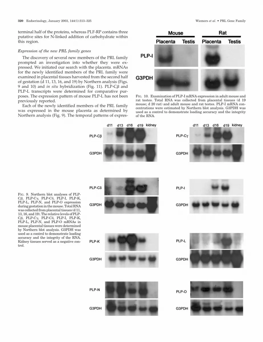
Expression of the new PRL family genes
The discovery of several new members of the PRL familyprompted an investigation into whether they were ex-pressed. We initiated our search with the placenta. mRNAsfor the newly identified members of the PRL family wereexamined in placental tissues harvested from the second halfof gestation (d 11, 13, 16, and 19) by Northern analysis (Figs.9 and 10) and in situ hybridization (Fig. 11). PLP-C� andPLP-L transcripts were determined for comparative pur-poses. The expression pattern of mouse PLP-L has not beenpreviously reported.
Each of the newly identified members of the PRL familywas expressed in the mouse placenta as determined byNorthern analysis (Fig. 9). The temporal patterns of expres-
FIG. 9. Northern blot analyses of PLP-C�, PLP-C�, PLP-C�, PLP-I, PLP-K,PLP-L, PLP-N, and PLP-O expressionduringgestation in themouse.TotalRNAwas collected from placental tissues (d 11,13, 16, and 19). The relative levels of PLP-C�, PLP-C�, PLP-C�, PLP-I, PLP-K,PLP-L, PLP-N, and PLP-O mRNAs inmouse placental tissues were determinedby Northern blot analysis. G3PDH wasused as a control to demonstrate loadingaccuracy and the integrity of the RNA.Kidney tissues served as a negative con-trol.
FIG. 10. Examination of PLP-I mRNA expression in adult mouse andrat testes. Total RNA was collected from placental tissues (d 19mouse; d 20 rat) and adult mouse and rat testes. PLP-I mRNA con-centrations were estimated by Northern blot analysis. G3PDH wasused as a control to demonstrate loading accuracy and the integrityof the RNA.
320 Endocrinology, January 2003, 144(1):313–325 Wiemers et al. • PRL Gene Family

sion differed. PLP-C�, PLP-C�, PLP-C�, PLP-I, and PLP-Ltranscripts showed increases in abundance as gestation ad-vanced, whereas PLP-N and PLP-O mRNAs were detectablethroughout the second half of gestation. None of the PRLfamily mRNAs tested were expressed in the kidney.
In an earlier report, PLP-I mRNA was detected in the rat
testis (38). Consequently, we evaluated the expression ofPLP-I in testes from adult mice and rats by Northern blot andin situ hybridization analyses with homologous probes (Fig.10). We could not detect PLP-I transcripts in either the mouseor rat testes by Northern blotting (Fig. 10) or in situ hybrid-ization (data not shown), and thus were not able to confirm
FIG. 11. Cell and tissue-specific local-ization of PLP-C�, PLP-C�, PLP-I,PLP-K, PLP-L, PLP-N, and PLP-O inmouse placental tissues. The in situ de-tection of mRNA expression was per-formed on frozen tissue sections. cDNAswere used as templates for the synthe-sis of digoxigenin-labeled sense and an-tisense RNA probes. A, Schematic dia-gram of the mature rodent placenta; theboxed area represents the histologicalsections presented in panels B–H. B,PLP-C� antisense probe on a d 19 pla-cental section; C, PLP-C� antisenseprobe on a d 19 placental section; thearrow depicts the location of migratorytrophoblast; D, PLP-I antisense probeon a d 19 placental section; E, PLP-Kantisense probe on a d 19 placental sec-tion; F, PLP-L antisense probe on a d 19placental section; G, PLP-N antisenseprobe on a d 19 placental section; thearrow depicts the location of migratorytrophoblast located in the decidua andmyometrium. H, PLP-O antisense probeon a d 13 placental section. Please notethat sense probes did not provide detect-able hybridization in any of the tissuesinvestigated. Magnification, �40.
Wiemers et al. • PRL Gene Family Endocrinology, January 2003, 144(1):313–325 321

the earlier finding (38). Thus, based on our experimentation,PLP-I does not appear to be expressed in testes from adultmice and rats.
Each of the newly identified members of the mouse PRLfamily was localized to trophoblast cells. However, they ex-hibited some prominent differences in the trophoblast lin-eage responsible for their expression. Four different tropho-blast lineages are involved in the expression of PRL familymembers: 1) trophoblast cells of the labyrinth zone; 2) spon-giotrophoblast cells of the junctional zone; 3) trophoblastgiant cells of the junctional zone; 4) migratory trophoblastcells invading the decidua and myometrium of the mesome-trial compartment (see Fig. 11A). PLP-C�, PLP-C�, PLP-I,and PLP-L mRNAs were all localized to the junctional zoneof the chorioallantoic placenta (Fig. 11, B–D, F). The expres-sion of PLP-C�, PLP-I, and PLP-L was restricted to spon-giotrophoblast cells, whereas PLP-C� was localized to spon-giotrophoblast cells, trophoblast giant cells, and migratorytrophoblast cells. PLP-K and PLP-O transcripts were de-tected in trophoblast cells of both the junctional and labyrinthzone compartments (Fig. 11, E and H). Within the junctionalzone, PLP-K expression was restricted to spongiotrophoblastcells, whereas PLP-O was expressed in both spongiotropho-blast cells and trophoblast giant cells. A similar localizationof PLP-K mRNA in the mouse placenta was previously dem-onstrated with a short mouse PLP-K probe (40). PLP-N tran-scripts were localized to spongiotrophoblast cells and tro-phoblast giant cells of the junctional zone and to migratorytrophoblast cells present in the mesometrial decidua andmyometrium (Fig. 11G). The PLP-N expressing cells in thedecidua and myometrium were cytokeratin-positive, thusconfirming their trophoblast origin (data not shown). In sum-mary, the newly discovered PRL family genes are expressedin trophoblast cells of the mouse placenta in gestationallyspecific patterns.
Discussion
Progress in sequencing human and mouse genomes hasled to the identification and characterization of gene familiesand the discovery of new genes encoding ligands and re-ceptors (68). The PRL cytokine/hormone gene family is com-pelling in that it represents an example of species-specificexpansion. In the mouse, the PRL gene family has undergonean extensive expansion, whereas in the human there appearsto be just a single member, PRL (1). Gene families arise bygene duplication and natural selection (69). To date, at least26 separate mouse genes encoding members of the PRL fam-ily have been reported. Core features shared among mem-bers of the mouse PRL family are their structural similaritieswith PRL and the localization of their genes to a 1-megabasesegment of chromosome 13.
The alignment of genes along the PRL family locus pri-marily reflects sequence conservation. Those genes possess-ing greater nucleotide identities are situated in closer prox-imity. PRL is positioned at one end of the mouse locus,whereas PLP-L is positioned at the other end of the locus. Ata nucleotide level these genes exhibit modest similarity; how-ever, the protein encoded by the PLP-L gene exhibits markedconservation with PRLs, especially more evolutionarily an-
cient PRL structures (70). Commonalities in cell-specificand/or temporal-specific aspects of gene expression do notappear to be main factors governing alignment along thelocus. It is likely that analogous PRL family loci exist on ratchromosome 17 and bovine chromosome 23. Although manymembers of the mouse and rat PRL families are orthologous,this does not seem to be the situation in the cow, whichsuggests the independent utilization of the ancestral PRLtemplate (71).
Genes within the PRL family locus were also organizedbased on their exon-intron structure. The prototypical fiveexon-four intron organization characteristic of PRL was afeature of genes clustered at both ends of the locus. Thecentral core of genes possessed an additional exon(s) situatedbetween exons II and III of the prototypical sequence. Theinsertion of the additional short exon(s) is near the locationof binding site 1 found in the amino acid sequences of ligandsinteracting with the PRL receptor (72). dPRP, PLF-RP, andPLP-E possess the extra exon(s) and do not effectively bindthe PRL receptor (73–75). Whether this is also true for theremaining members of the six/seven exon subfamily is un-known. The repeated insertion of an additional exon(s) be-tween exons II and III of the prototypical PRL gene structurein numerous members of the PRL family implies some func-tional significance. Whether the extra exon(s) encode for aphysical spacer, separating key regulatory domains or theinclusion of a biologically relevant motif is yet to be resolved.Exon III of PLP-C and PLF-RP related genes encodes for ashort exon rich in aromatic amino acids, whereas the extraexon(s) of PLP-E and PLP-F are distinct. The latter genesencode for proteins regulating hematopoiesis (75–77). Thesignificance of the additional exon(s) in the biological actionsof PLP-E and PLP-F remains to be determined.
PL-I is situated adjacent to the PRL gene and shares thesame exon-intron structure, biological targets, and receptor-signaling pathway. It is now evident in the mouse that PL-Iis actually encoded by three closely related genes, whichencode for three closely related proteins, referred to as PL-I�,PL-I�, and PL-I�. The PL-I cDNA was originally cloned fromthe midgestation mouse placenta (17) and is equivalent toPL-I�. Each PL-I gene is expressed and exhibits distinct pat-terns of activation during development. PL-I� and PL-I� areactivated most dramatically in trophoblast cells isolated fromblastocyst outgrowths, whereas PL-I� predominates in pla-centa from d 10 of gestation. These observations suggest thatdifferences may exist in the transcriptional control of thePL-I� gene vs. PL-I� and PL-I� genes. A mouse PL-I pro-moter, likely corresponding to PL-I�, has been studied in theRcho-1 trophoblast cell model (78–80). These studies haveimplicated the involvement of activator protein 1 and GATAfactors. Whether these regulators are restricted to a specificPL-I gene or whether they will be shared among all three PL-Igenes will need to be resolved. The differential PL-I geneexpression patterns suggest that there may be differences inthe biological activities of each PL-I. Furthermore, the needsof an implanting blastocyst differ markedly from events atmidgestation. PL-I� is a known PRL receptor agonist (81).The abilities of PL-I� and PL-I� proteins to activate the PRLreceptor-signaling pathway are not known. Amino acid se-quences encoded by the three PL-I genes differ at a maximum
322 Endocrinology, January 2003, 144(1):313–325 Wiemers et al. • PRL Gene Family
of only five residues in the mature proteins. These amino acidsubstitutions are, for the most part, conservative changes.Although the amino acid changes in PL-I� and PL-I� are notdramatically different from PL-I� they may impact the trans-port, stability, or actions of the ligands.
Six additional new members of the PRL gene family werediscovered via mining of the mouse genome database. Twoof the new members were orthologous to previously char-acterized members of the rat PRL family, PLP-I and PLP-K;two were classified as part of the PLP-C subfamily and weretermed PLP-C� and PLP-C�, and two exhibited structuralrelationships with PLF-RP and were named PLP-N andPLP-O. The amino acid sequence similarities and expressionpatterns may provide some clues into the biology of thesenew members of the PRL family.
PLP-I. PLP-I is a five exon-four intron gene situated near thePRL and PL genes. It has an ortholog in the rat. Earliercomments about peculiar structural features and expressionpatterns of rat PLP-I appear to be unfounded (38). Placentalexpression of PLP-I is prominent during the latter stages ofgestation. Expression is restricted to spongiotrophoblastcells, suggesting that PLP-I may be preferentially directedtoward the maternal compartment. The predicted PLP-I pro-tein has putative N-linked sites for glycosylation and exhibitssome structural relationships to classical members of the PRLfamily (i.e. those using the PRL receptor signaling pathway);however, whether it shares similar targets or modes of actionare unknown.
PLP-K. An ortholog of rat PLP-K was identified and pos-sessed a five exon-four intron structure. Mouse PLP-K is verysimilar to rat PLP-K with the notable exception of a putativeN-linked glycosylation site present in rat but not in mousePLP-K. Based on sequence comparisons, PLP-K shares somesimilarity to PLF. PLF is a known regulator of blood vesseldevelopment and uterine growth (82, 83). The actions of PLFon endothelial cells are mediated by carbohydrate structureson the PLF protein backbone (84). These carbohydrate motifsinteract with the insulin-like growth factor-II/mannose-6-phosphate receptor to stimulate angiogenesis (84). The ab-sence of N-linked glycosylation sites in mouse PLP-K maynot be consistent with a PLF-like mode of action. The ex-pression pattern of mouse PLP-K is intriguing and uniqueamong members of the PRL family. PLP-K mRNA is ex-pressed at high levels in trophoblast cells from both junc-tional and labyrinth zones of the chorioallantoic placentaduring the last week of gestation. The distribution of PLP-Kin both junctional and labyrinth zones indicates that thePLP-K protein may be targeted to both maternal and fetalcompartments.
PLP-C� and PLP-C�. Two new members of the PLP-C sub-family possessing the characteristic six exon-five intron geneorganization were found. The PLP-C subfamily appears tohave been more recently evolved. Five PLP-C family mem-bers have been identified in the mouse (dPRP, PLP-C�, PLP-C�, PLP-C�, and PLP-C�) and six in the rat (dPRP, PLP-C,PLP-Cv, PLP-C�, PLP-D, and PLP-H). dPRP and PLP-C� areorthologous in the mouse and rat; however, the remainingmouse and rat members are not orthologous. The biology of
the PLP-C subfamily is poorly understood, other than dPRP,which binds heparin and targets eosinophils (85). Both PLP-C�, and PLP-C� are predominantly expressed in the junc-tional zone during the latter third of gestation. AlthoughPLP-C� expression is restricted to spongiotrophoblast cells,PLP-C� is expressed in spongiotrophoblast and trophoblastgiant cells, and to a lesser extent in a subpopulation of tro-phoblast cells migrating into the mesometrial decidua (seeadditional discussion about migratory trophoblast cells be-low). This pattern of gene expression indicates that PLP-C�and PLP-C� are likely targeted to maternal tissues, similar tothe pattern of PLP-C� and PLP-C� and the rat members ofthe PLP-C subfamily. The species specificity of the PLP-Csubfamily of ligands may have evolved to regulate preg-nancy-dependent processes that are unique to the mouse vs.the rat.
PLP-N and PLP-O. Two genes with six exon-five intron struc-tures encoding proteins with sequence similarity to PLF-RPwere discovered. Orthologs for PLP-N and PLP-O have notbeen reported for any other species, including the rat.PLF-RP orthologs exist in the mouse, rat, and likely hamster(21, 41, 86). PLP-N is abundantly expressed in trophoblastcells of the junctional zone and trophoblast migrating into themesometrial decidua. Migratory trophoblast cells form inti-mate relationships with the vasculature supplying the cho-rioallantoic placenta and increase in numbers during thelatter stages of pregnancy (87, 88). In comparison, PLP-O isexpressed in spongiotrophoblast cells and trophoblast giantcells of the junctional zone and in labyrinthine trophoblast.The spongiotrophoblast and labyrinthine trophoblast cell ex-pression overlapped with the transcript distribution forPLP-K. In the mouse, PLF-RP targets the vasculature. It is aninhibitor of angiogenesis and a potent antagonist of PLFaction (82, 89). Both PLP-N and PLP-O are in close proximityto vasculature critical to the placenta and the fetus, respec-tively. Whether these two PLF-RP related cytokines also tar-get blood vessels needs to be evaluated.
Overview
The mouse possesses at least 26 different genes comprisingthe PRL family locus. We have considerable understandingof the biology of PRL but very little knowledge of the phys-iology of most of the other constituents of the locus. Theevolutionary survival of this expanded gene family impliesfunctional importance and relevance to speciation. Some li-gand-receptor families have undergone coordinate evolution(69). However, no evidence exists for an expanded PRL re-ceptor family. Our current information indicates that a fewmembers of the PRL family use the PRL receptor signalingsystem; however, most do not and may instead use receptorsignaling pathways characteristic of other ligands. Thus,there must be something intrinsic in the structure of theancestral PRL template that enables it to evolve into struc-tures recognized by other receptor systems. The existence ofat least 26 natural variants should facilitate understandingthe significance of key structural motifs within this family ofcytokines/hormones.
In summary, elucidation of the mouse PRL gene familylocus provides new insights into the expansion of the mouse
Wiemers et al. • PRL Gene Family Endocrinology, January 2003, 144(1):313–325 323

PRL family and new tools for studying the genetics andbiology of its members.
Acknowledgments
We would like to thank Jared T. Soares and Adam Alt for assistancewith the preparation of some of the figures.
Received July 17, 2002. Accepted September 4, 2002.Address all correspondence and requests for reprints to: Dr. Michael
J. Soares, Department of Molecular & Integrative Physiology, Universityof Kansas Medical Center, Kansas City, Kansas 66160-7401. E-mail:[email protected].
This work was supported by grants from the National Institute ofChild Health and Human Development (HD-020676, HD-029797, HD-033994, HD-037123, HD-037678, and HD-039878). R.A. was supportedby a postdoctoral fellowship from the American Heart Association. G.D.was supported by an investigator development grant from the AndrewMellon Foundation.
Paralogous refers to the relationship of homologous genes due to aduplication event; orthologous refers to the relationship of homologousgenes due to a speciation event (90, 91).
References
1. Soares MJ, Linzer DIH 2001 Rodent prolactin family and pregnancy. In:Horseman ND, ed. Prolactin. Norwell, MA: Kluwer Academic Publishers;129–167
2. Stricker P, Grueter F 1928 Action du lobe anterieur de l’hypophyse sur lamontee laiteuse. C R Soc Biol 99:1978–1980
3. Grueter F, Stricker P 1929 Uber die wirkung eines hypophysenvorderlap-penshormons auf die auslosung der milchsekretion. Klin Wochschr 8:2322–2323
4. Evans HM, Simpson ME 1929 Hyperplasia of the mammary apparatus ofadult virgin females induced by anterior hypophyseal hormones. Proc Soc ExpBiol Med 26:598
5. Riddle O, Bates RW, Dykshorn SW 1933 The preparation, identification ofprolactin—a hormone of the anterior pituitary. Am J Physiol 105:191–216
6. Pencharz RI, Long JA 1931 The effect of hypophysectomy on gestation in therat. Science 74:206
7. Selye H, Collip JB, Thomson DL 1933 Effect of hypophysectomy upon preg-nancy and lactation in mice. Proc Soc Exp Biol Med 31:82–83
8. Astwood EB, Greep RO 1938 A corpus luteum stimulating substance in therat placenta. Proc Soc Exp Biol Med 38:713–716
9. Cerruti RA, Lyons WR 1960 Mammogenic activities of the midgestationalmouse placenta. Endocrinology 67:884–887
10. Robertson MC, Friesen HG 1975 The purification and characterization of ratplacental lactogen. Endocrinology 97:621–629
11. Colosi P, Marr G, Lopez J, Haro L, Ogren L, Talamantes F 1982 Isolation andpartial characterization of mouse placental lactogen. Proc Natl Acad Sci USA79:771–775
12. Colosi P, Ogren L, Thordarson G, Talamantes F 1987 Purification and partialcharacterization of two prolactin-like glycoprotein hormone complexes fromthe midpregnant mouse conceptus. Endocrinology 120:2500–2511
13. Gubbins EJ, Maurer RA, Hartley JL, Donelson JE 1979 Construction andanalysis of recombinant DNAs containing a structural gene for rat prolactin.Nucleic Acids Res 6:915–930
14. Linzer DIH, Talamantes F 1985 Nucleotide sequence of mouse prolactin andgrowth hormone mRNAs, and expression of these mRNAs during pregnancy.J Biol Chem 260:9574–9579
15. Jackson LL, Colosi P, Talamantes F, Linzer DIH 1986 Molecular cloning ofmouse placental lactogen cDNA. Proc Natl Acad Sci USA 83:8496–8500
16. Duckworth ML, Kirk KL, Friesen HG 1986 Isolation and identification of acDNA clone of rat placental lactogen II. J Biol Chem 261:10871–10878
17. Colosi P, Talamantes F, Linzer DIH 1987 Molecular cloning and expressionof mouse placental lactogen I complementary deoxyribonucleic acid. MolEndocrinol 1:767–776
18. Linzer DIH, Nathans D 1984 Nucleotide sequence of a growth-related memberof the prolactin-growth hormone family. Proc Natl Acad Sci USA 81:4255–4259
19. Duckworth ML, Peden LM, Friesen HG 1986 Isolation of a novel prolactin-likecDNA clone from developing rat placenta. J Biol Chem 261:10879–10884
20. Duckworth ML, Peden LM, Friesen HG 1988 A third prolactin-like proteinexpressed by the developing rat placenta: complementary deoxyribonucleicacid sequence and partial structure of the gene. Mol Endocrinol 2:912–920
21. Linzer DIH, Nathans D 1985 A new member of the prolactin-growth hormonefamily expressed in mouse placenta. EMBO J 4:1419–1423
22. Deb S, Faria TN, Roby KF, Larsen D, Kwok SCM, Talamantes F, Soares MJ
1991 Identification and characterization of a new member of the placentalprolactin family: placental lactogen-I variant. J Biol Chem 266:605–1610
23. Robertson MC, Schroedter IC, Friesen HG 1991 Molecular cloning and ex-pression of rat placental lactogen-IV, a variant of rPL-I present in late gestationrat placenta. Endocrinology 129:2746–2756
24. Deb S, Roby KF, Faria TN, Szpirer C, Levan G, Kwok SCM, Soares MJ 1991Molecular cloning and characterization of prolactin-like protein-C comple-mentary deoxyribonucleic acid. J Biol Chem 266:23027–23032
25. Roby KF, Deb S, Gibori G, Szpirer C, Levan G, Kwok SCM, Soares MJ 1993Decidual prolactin-related protein. Identification, molecular cloning, and char-acterization. J Biol Chem 268:3136–3142
26. Hirosawa M, Miura R, Min K-S, Hattori N, Shiota K, Ogawa T 1994 A cDNAencoding a new member of the rat placental lactogen family, PL-I mosaic(PL-Im). Endocr J 41:387–397
27. Dai G, Liu B, Szpirer C, Levan G, Kwok SCM, Soares MJ 1996 Prolactin-likeprotein-C variant: complementary DNA, unique six exon gene structure, andtrophoblast cell-specific expression. Endocrinology 137:5009–5019
28. Iwatsuki K, Shinozaki M, Hattori N, Hirasawa K, Itagaki S-I, Shiota K,Ogawa T 1996 Molecular cloning and characterization of a new member of therat placental prolactin (PRL) family, PRL-like protein-D. Endocrinology 137:3849–3855
29. Iwatsuki K, Oda M, Sun W, Tanaka S, Ogawa T, Shiota K 1998 Molecularcloning and characterization of a new member of the rat placental prolactin(PRL) family, PRL-like protein H. Endocrinology 139:4976–4983
30. Dai G, Chapman BM, Liu B, Orwig KE, Wang D, White RA, Preuett B, SoaresMJ 1998 A new member of the mouse prolactin (PRL)-like protein-C subfamily,PRL-like protein-C�: structure and expression. Endocrinology 139:5157–5163
31. Hiraoka Y, Ogawa M, Sakai Y, Takeuchi Y, Komatsu N, Shiozawa M, TanabeK, Aiso S 1999 PLP-I: a novel prolactin-like gene in rodents. Biochim BiophysActa 1447:291–297
32. Hwang IT, Lee YH, Moon BC, Ahn KY, Lee SW, Chun JY 2000 Identificationand characterization of a new member of the placental placental-like protein-C(PLP-C) subfamily, PLP-C�. Endocrinology 141:3343–3352
33. Lin J, Poole J, Linzer DIH 1997 Two novel members of the prolactin/growthhormone family are expressed in the mouse placenta. Endocrinology 138:5535–5540
34. Lin J, Poole J, Linzer DIH 1997 Three new members of the mouse prolactin/growth hormone family are homologous to proteins expressed in the rat.Endocrinology 138:5541–5549
35. Orwig KE, Ishimura R, Muller H, Liu B, Soares MJ 1997 Identification andcharacterization of a mouse homolog for decidual/trophoblast prolactin-related protein. Endocrinology 139:5511–5517
36. Muller H, Ishimura R, Orwig KE, Liu B, Soares MJ 1998 Homologues forprolactin-like protein-A and B are present in the mouse. Biol Reprod 58:45–51
37. Muller H, Orwig KE, Soares MJ 1998 Identification of two new members ofthe mouse prolactin gene family. Biochim Biophys Acta 1396:251–258
38. Ishibashi K, Imai M 1999 Identification of four new members of the ratprolactin/growth hormone gene family. Biochem Biophys Res Commun 262:575–578
39. Toft DJ, Linzer DIH 1999 Prolactin (PRL)-like protein J, a novel member of thePRL/growth hormone family, is exclusively expressed in maternal decidua.Endocrinology 140:5095–5101
40. Dai G, Wang D, Liu B, Kasik JW, Muller H, White RA, Hummel GS, SoaresMJ 2000 Three novel paralogs of the rodent prolactin gene family. J Endocrinol166:63–75
41. Sahgal N, Knipp GT, Liu B, Chapman BM, Dai G, Soares MJ 2000 Identi-fication of two new nonclassical members of the rat prolactin family. J MolEndocrinol 24:95–108
42. Tanaka TS, Jaradat SA, Lim MK, Kargul GJ, Wang X, Grahovac MJ, PantanoS, Sano Y, Piao Y, Nagaraja R, Doi H, Wood WH, Becker KG, Ko MSH 2000Genome-wide expression profiling of mid-gestation placenta and embryo us-ing a 15,000 mouse developmental cDNA microarray. Proc Natl Acad Sci USA97:9127–9132
43. Toft DJ, Linzer DIH 2000 Identification of three prolactin-related hormonesas markers of invasive trophoblasts in the rat. Biol Reprod 63:519–525
44. Jackson-Grusby LL, Pravtcheva D, Ruddle FH, Linzer DIH 1988 Chromo-somal mapping of the prolactin/growth hormone gene family in the mouse.Endocrinology 122:2462–2466
45. Cooke NE, Szpirer C, Levan G 1986 The related genes encoding growthhormone and prolactin have been dispersed to chromosomes 10 and 17 in rat.Endocrinology 119:2451–2454
46. Cohick CB, Dai G, Xu L, Deb S, Kamei T, Levan G, Szpirer C, Szpirer J, KwokSCM, Soares MJ 1996 Placental lactogen-I variant utilizes the prolactin re-ceptor signaling pathway. Mol Cell Endocrinol 116:49–58
47. Dai G, Imagawa W, Liu B, Szpirer C, Levan G, Kwok SCM, Soares MJ 1996Rcho-1 trophoblast cell placental lactogens: complementary DNAs, heterolo-gous expression, and biological activities. Endocrinology 137:5020–5027
48. Shah P, Sun Y, Szpirer C, Duckworth ML 1998 Rat placental lactogen II gene:characterization of gene structure and placental-specific expression. Endocri-nology 139:967–973
49. Dietz AB, Georges M, Threadgill DW, Womack JE, Schuler LA 1992 Somatic
324 Endocrinology, January 2003, 144(1):313–325 Wiemers et al. • PRL Gene Family

cell mapping, polymorphism, and linkage analyis of bovine prolactin-relatedproteins and placental lactogen. Genomics 14:137–143
50. Cooke NE, Baxter JD 1982 Structural analysis of the prolactin gene suggestsa separate origin of its 5�end. Nature 297:603–606
51. Chien Y-H, Thompson EB 1980 Genomic organization of rat prolactin andgrowth hormone genes. Proc Natl Acad Sci USA 77:4583–4587
52. Maurer RA, Erwin CR, Donelson JD 1981 Analysis of 5� flanking sequences andintron-exon boundaries of the rat prolactin gene. J Biol Chem 256:10524–10528
53. Shida MM, Jackson-Grusby LL, Ross SR, Linzer DIH 1992 Placental-specificexpression from the mouse placental lactogen II gene promoter. Proc Natl AcadSci USA 89:3864–3868
54. Dai G, Chapman BM, Wang D, White RA, Preuett B, Soares MJ 1999 Pro-lactin-like protein-A gene structure and chromosomal mapping. Mamm Ge-nome 10:78–80
55. Connor AM, Waterhouse P, Khokha R, Denhardt DT 1989 Characterizationof a mouse mitogen-regulated protein/proliferin gene and its promoter: amember of the growth hormone/prolactin gene superfamily. Biochim BiophysActa 1009:75–82
56. Ebbitt DM, Hurley WL, Kessler MA, McDonald DJ, Schuler LA 1989 Char-acterization of the gene corresponding to bovine placental prolactin-relatedcDNA I: evolutionary implications. DNA 8:161–169
57. Kessler MA, Schuler LA 1991 Structure of the bovine placental lactogen geneand alternative splicing of transcripts. DNA Cell Biol 10:93–104
58. Orwig KE, Dai G, Rasmussen CA, Soares MJ 1997 Decidual/trophoblastprolactin related protein: characterization of gene structure and cell-specificexpression. Endocrinology 138:2491–2500
59. Altschul SF, Gish W, Miller W, Myers EW, Lipman DJ 1990 Basic localalignment search tool. J Mol Biol 215:403–410
60. Carninci P, Shibata Y, Hayatsu N, Sugahara Y, Shibata K, Itoh M, Konno H,Okazaki Y, Muramatsu M, Hayashizaki Y 2000 Normalization and subtrac-tion of cap-trapper-selected cDNAs to prepare full-length cDNA libraries forrapid discovery of new genes. Genome Res 10:1617–1630
61. Chomczynski P, Sacchi N 1987 Single-step method of RNA isolation by acidguanididium thiocyanate-phenol-chloroform extraction. Anal Biochem 162:156–159
62. Thompson JD, Higgins DG, Gibson TJ 1994 CLUSTAL W: improving thesensitivity of progressive multiple sequence alignment through sequenceweighting, positions-specific gap penalties and weight matrix choice. NucleicAcids Res 22:4673–4680
63. Nielsen H, Engelbrecht J, Brunak S, von Heijne G 1997 Identification ofprokaryotic and eukaryotic signal peptides and prediction of their cleavagesites. Protein Eng 10:1–6
64. Faria TN, Deb S, Kwok SCM, Talamantes F, Soares MJ 1990 Ontogeny ofplacental lactogen-I and placental lactogen-II expression in the developing ratplacenta. Dev Biol 141:279–291
65. Braissant O, Wahli W 1998 A simplified in situ hybridization protocol usingnon-radioactively labeled probes to detect abundant and rare mRNAs on tissuesections. Biochemica 1:10–16
66. Linzer DIH, Lee SJ, Ogren L, Talamantes F, Nathans D 1985 Identification ofproliferin mRNA and protein in mouse placenta. Proc Natl Acad Sci USA82:4356–4359
67. Fassett JT, Hamilton RT, Nilsen-Hamilton M 2000 Mrp4, a new mitogen-regulated protein/proliferin gene; unique in this gene family for its expressionin the adult mouse tail and ear. Endocrinology 141:1863–1871
68. Leo CP, Hsu SY, Hsueh AJ 2002 Hormonal genomics. Endocr Rev 23:369–8169. Fryxell KJ 1996 The coevolution of gene family trees. Trends Genet 12:364–36970. Wallis M 1992 The expanding growth hormone/prolactin family. J Endocrinol
9:185–188
71. Schuler LA, Kessler MA 1992 Bovine prolactin-related hormones. TrendsEndocrinol Metab 3:334–338
72. Goffin V, Shiverick KT, Kelly PA, Martial JA 1996 Sequence-function rela-tionships within the expanding family of prolactin, growth hormone, placentallactogen, and related proteins in mammals. Endocr Rev 17:385–410
73. Rasmussen CA, Hashizume K, Orwig KE, Xu L, Soares MJ 1996 Decidualprolactin-related protein: heterologous expression and characterization. En-docrinology 137:5558–5566
74. Colosi P, Swiergiel JJ, Wilder EL, Oviedo A, Linzer DIH 1988 Character-ization of proliferin-related protein. Mol Endocrinol 2:579–586
75. Lin J, Linzer DIH 1999 Induction of megakaryocyte differentiation by a novelpregnancy-specific hormone. J Biol Chem 274:21485–21489
76. Bittorf T, Jaster R, Soares MJ, Seiler J, Brock J, Friese K, Muller H 2000Induction of erythroid proliferation and differentiation by a trophoblast-specific cytokine involves activation of the JAK/STAT pathway. J MolEndocrinol 25:253–262
77. Bhattacharyya S, Lin J, Linzer DIH 2002 Reactivation of a hematopoieticendocrine program of pregnancy contributes to recovery from thrombocyto-penia. Mol Endocrinol 16:1386–1393
78. Shida MM, Ng YK, Soares MJ, Linzer DIH 1993 Trophoblast-specific tran-scription from the mouse placental lactogen-I gene promoter. Mol Endocrinol7:181–188
79. Ng YK, George KM, Engel JD, Linzer DIH 1994 GATA factor activity isrequired for the trophoblast-specific transcriptional regulation of the mouseplacental lactogen I gene. Development 120:3257–3266
80. Peters TJ, Chapman BM, Wolfe MW, Soares MJ 2000 Placental lactogen-I geneactivation in differentiating trophoblast cells: extrinsic and intrinsic regulationinvolving mitogen-activated protein kinase pathways. J Endocrinol 165:443–456
81. Colosi P, Ogren L, Southard JN, Thordarson G, Linzer DIH, Talamantes F1988 Biological, immunological, and binding properties of recombinant mouseplacental lactogen-I. Endocrinology 123:2662–2667
82. Jackson D, Volpert OV, Bouck N, Linzer DIH 1994 Stimulation and inhibitionof angiogenesis by placental proliferin and proliferin-related protein. Science266:1581–1584
83. Nelson JT, Rosenzweig N, Nilsen-Hamilton M 1995 Characterization of themitogen-regulated protein (proliferin) receptor. Endocrinology 136:283–288
84. Volpert O, Jackson D, Bouck N, Linzer DIH 1996 The insulin-like growthfactor II/mannose 6-phosphate receptor is required for proliferin-inducedangiogenesis. Endocrinology 137:3871–3876
85. Wang D, Ishimura R, Walia DS, Muller H, Dai G, Hunt JS, Lee NA, Lee JJ,Soares MJ 2000 Eosinophils are cellular targets of the novel uteroplacentalheparin-binding cytokine, decidual/trophoblast prolactin-related protein. JEndocrinol 167:15–29
86. Barnes SW, Renegar RH 1996 Identification, characterization, and expression ofa new prolactin-like molecule in the hamster placenta. Biol Reprod 55:370–378
87. Georgiades P, Ferguson-Smith AC, Burton GJ 2001 Comparative developmentalanatomy of the murine and human definitive placentae. Placenta 23:3–19
88. Pijnenborg R, Robertson WB, Brosens I, Dixon G 1981 Trophoblastic invasionand the establishment of haemochorial placentation in man and laboratoryanimals. Placenta 2:71–92
89. Bengtson NW, Linzer DIH 2000 Inhibition of tumor growth by the antian-giogenic placental hormone, proliferin-related protein. Mol Endocrinol 14:1934–1943
90. Fitch WM 2000 Homology a personal view on some of the problems. TrendsGenet 16:227–231
91. Eichler EE 2001 Recent duplication, domain accretion and the dynamic mu-tation of the human genome. Trends Genet 17:661–669
Wiemers et al. • PRL Gene Family Endocrinology, January 2003, 144(1):313–325 325


