Embed Size (px)
Citation preview
1
N. Fortunati1, M.G. Catalano2, F. Marano1, V. Mugoni1, M. Pugliese2, O. Bosco2, F.
Mainini3, G. Boccuzzi1,2
THE pan-DAC INHIBITOR LBH589 IS A MULTI-FUNCTIONAL AGENT IN
BREAST CANCER CELLS: CYTOTOXIC DRUG AND INDUCER OF SODIUM-
IODIDE SYMPORTER (NIS).
1Oncological Endocrinology, AOU San Giovanni Battista, 2Department of Clinical
Pathophysiology, University of Turin, Turin, Italy; 3Novartis Farma S.p.A. - Origgio (VA),
Italy.
Corresponding Author and Reprints Requests : Prof. Giuseppe Boccuzzi, Dept. Clinical
Phisiopathology, Via Genova 3, 10126 Torino, Italy; tel.+39-011-6705399; fax. +39-011-
6705366; e-mail:[email protected]
peer-00534548, version 1 - 10 Nov 2010
Author manuscript, published in "Breast Cancer Research and Treatment (2010) 667-675" DOI : 10.1007/s10549-010-0789-z
2
ABSTRACT
New drugs with anti-tumour activity, also able to modify the expression of selected
molecules, are under evaluation in breast cancer which is becoming resistant to conventional
treatment, or in metastatic disease. The sodium-iodide symporter (NIS), which mediates
iodide uptake into thyroid cells, and is the molecular basis of radioiodine imaging and therapy
in thyroid cancer, is also expressed in a large portion of breast tumours. Since NIS expression
in breast cancer is not sufficient for a significant iodide uptake, drugs able to induce its
expression and correct function are under evaluation. In the present study, we report for the
first time that the pan-deacetylase (DAC) inhibitor LBH589 (panobinostat) significantly
induced NIS, both as mRNA and as protein, through the increase of NIS promoter activity,
with the final consequence of obtaining a significant up-take of iodide in MCF7, T47D, and
MDA-MB231 breast cancer cells. Moreover, we observed that LBH589 causes a significant
reduction in cell viability of estrogen-sensitive and –insensitive breast cancer cells within
nanomolar range. The anti-tumour effect of LBH589 is sustained by apoptosis induction and
cell cycle arrest in G2/M. In conclusion, our data suggest that LBH589 might be a powerful
tool in the management of breast cancer due to its multiple effects and support a potential
application of LBH589 in the diagnosis and treatment of this disease.
KEYWORDS
DAC-inhibitors; breast cancer; NIS; targeted therapy; epigenetic
peer-00534548, version 1 - 10 Nov 2010
3
INTRODUCTION
Breast cancer is the most common cancer in women all over the world [1]. Even though its
management has become more and more successful over time, owing to the different
available therapeutic strategies [2], both metastatic disease and cancer which are becoming
resistant to conventional treatment are emerging problems [3, 4]. New drugs with anti-tumour
activity and/or able to modify the expression of selected molecules in these situations are
under evaluation.
The sodium/iodide symporter (NIS) has been receiving a good deal of attention, both for its
function that is requisite for potential clinical application, and for its sensitivity to a number of
agents able to induce/increase its cell expression. NIS is a transmembrane glycoprotein
mainly expressed in thyrocytes, functioning as iodide carrier [5]. The expression and correct
functioning of NIS in differentiated thyroid cancer is the molecular requisite for successful
radioiodide therapy in this neoplasm [6]. NIS is also expressed in lactating mammary glands,
favouring iodide concentration in milk, and in more than 80 % of human breast carcinomas
[7]. Although NIS expression is present and intense in a majority of invasive breast cancers,
radioiodide accumulation has been observed in only a small fraction of cases [8, 9, 10]. The
achievement of a good level of expression of NIS is regarded as a promising new strategy to
unable the use of radioiodide for imaging and treatment of breast cancer [11, 12].
In non neoplastic breast cells, estrogens, oxitocyn and prolactin, mainly acting in concert,
were reported to induce NIS [7]. In breast cancer cells, many authors reported that retinoic
acid is a powerful enhancer of NIS expression especially in association with corticosteroids
[12-14]. Furthermore, in one report it was also observed that retinoic acid and dexamethasone
treatment determined a significant 131I-induced cytotoxicity in breast cancer cells [15].
NIS expression, in both thyroid and non-thyroid cells, is also regulated by epigenetic
phenomena. NIS gene silencing can be related to the level of histone acetylation; in fact,
peer-00534548, version 1 - 10 Nov 2010

4
compounds like Trycostatin A (TSA), sodium butyrate (NaB) and valproic acid (VPA), that
belong to the decetylase inhibitor (DAC-inhibitor) group, stimulate NIS expression in thyroid
cancer cells [16-18]DAC-inhibitors [19, 20] could also be used to control NIS expression
inducing radioiodine sensitivity in different human extra-thyroid tumours such as breast
cancer. TSA and NaB greatly increased NIS
mRNA also in breast cancer MCF-7 cell line [21].
The aim of the present work was to evaluate the ability of the powerful pan-DAC inhibitor
LBH589 (panobinostat) [22] to induce the expression of functional NIS in breast cancer cells
with different estrogen sensitivity.
peer-00534548, version 1 - 10 Nov 2010

5
MATERIALS AND METHODS
Cell lines and reagents
Estrogen receptor-positive MCF-7 and T47D and estrogen receptor-negative MDA-MB 231
cells were purchased from ECACC (Salisbury, UK).
Cells were routinely maintained at 37°C, in 5% CO2 and 95 % humidity, in RPMI 1640
(Sigma, St Louis, MO, USA) with 100 IU/ml penicillin and 100 g/ml streptomycin added,
supplemented with 10% heat inactivated FCS (Euroclone, Wetherby, West York, UK).
LBH589 was provided by Novartis Pharma AG (Basel, Switzerland), prepared as a 5 mM
stock solution in DMSO and stored at -20°C.
Immunoblotting
To evaluate the effect of LBH589 on histone H4 acetylation state, breast cancer cells, after
treatment with LBH589 (5-100 nM), were harvested and lysed in the presence of 200 l RIPA
buffer (1% NP40, 0.5% desoxycholate sodium, 0.1% SDS in PBS pH 7.4, with 10 mg/ml
PMSF, 30 l/ml aprotinin and 100mM sodium orthovanadate) and incubated on ice for 30
min; cells were then centrifuged for 20 min at 15,000 x g at 4°C, and clear supernatants used.
SDS-PAGE was performed on gels, loading 40 g protein/well. Separated proteins were
electro-transferred onto PVDF membrane and probed with anti-acetyl-histone H4 antibody
(06-598, 1:1000 dilution, Upstate, Lake Placid, NY, USA). The membrane was then stripped
and re-probed with an anti-GAPDH antibody (1:10000 dilution, Sigma, Saint Louis, MI,
USA) to check protein loading. Proteins were detected with Pierce Super Signal
chemiluminescent substrate following the manufacturer’s instructions. Bands were
photographed and analyzed using Kodak 1D Image Analysis software.
peer-00534548, version 1 - 10 Nov 2010

6
Cytotoxic activity
To evaluate the LBH589 cytotoxic activity, MCF-7, T47D and MDA-MB 231 cells were
seeded at 4 x 10³ cells/well in 96-well plates (Corning, New York, NY, USA) in culture
medium plus 10% heat- inactivated FCS. After 24 hours, cells were exposed to increasing
concentration of LBH589 (1-200 nM). After 72 hours incubation cell viability was assessed
using the Cell Proliferation Reagent WST-1 (Roche Applied Science, Penzberg, Germany),
following manufacturer’s instructions. Four replicate wells were used to determine each data
point. Three response parameters, median growth inhibition (GI50), total growth inhibition
(TGI) and median lethal concentration (LC50) were calculated for each cell line. The GI50
corresponds to the concentration of the compound that inhibits 50% net cell growth; TGI
value is the concentration of the compound leading to total growth inhibition; LC50 value is
the concentration of the compound leading to 50% net cell death.
Apoptosis Detection - Cell Death Detection ELISA
For apoptosis studies, 4 x 10³ cells were seeded in 96-well plates and treated with LBH589,
using the same schedule as for the viability assay. After treatment, apoptosis was evaluated
using Cell Death Detection ELISA PLUS (Roche Applied Science, Penzberg, Germany)
following manufacturer’s instructions. Apoptosis was expressed as enrichment factor,
calculated as a fraction of the absorbance of treated cells versus untreated controls.
Cell cycle analysis
Cells were treated with LBH589 up to 72 hours. At different times (24, 48 and 72 hours), all
cells were collected, fixed in 70% ethanol for 30 minutes on ice and incubated in propidium
iodide solution (20 g/ml propidium iodide, 0.2 mg/ml RNAseA in PBS) for 1 hour at room
temperature. The cell population was analyzed by flow cytometry.
peer-00534548, version 1 - 10 Nov 2010
7
Gene expression evaluation with Real Time PCR
Cells (1x10⁶) were seeded in 75cm2 flasks and treated with LBH589 (5-100nM).
Total RNA was extracted using TRIzol Reagent (Invitrogen Ltd, Paisley, UK), as previously
described. DNase I was added to remove remaining genomic DNA. 1 g of total RNA was
reverse-transcribed with iScript cDNA Synthesis Kit (BioRad Laboratories, Inc.), following
manufacturer’s protocol.
Primers (Table 1) were designed using Beacon Designer 5.0 software according to parameters
outlined in the BioRad iCycler Manual. Specificity of primers was confirmed by BLAST
analysis. Real-time PCR was performed using a BioRad iQ iCycler Detection System
(BioRad Laboratories, Inc.) with SYBR green fluorophore. Reactions were performed in a
total volume of 25 l, including 12.5 l IQ SYBR Green Supermix (BioRad Laboratories,
Inc.), 1 l of each primer at 10 M concentration, and 5 l of the previously reverse-
transcribed cDNA template. Protocol for primer set was optimised using seven serial 5X
dilutions of template cDNA obtained from cells in basal conditions. The protocol used is as
follows: denaturation (95°C for 5 min), amplification repeated 40 times (95°C for 15 sec,
60°C for 1 min). A melting curve analysis was performed following every run to ensure a
single amplified product for every reaction. All reactions were carried out at least three times
for each sample.
Luciferase Transcription Assay
Cells (2.5 x 10⁵) were seeded in 6-well plates. 24 hours after seeding, cells were transfected
with 1μg of NIS-luc plasmid/well using Lipofectin Reagent (Invitrogen Ltd, Paisley, UK).
The plasmid, a kind gift of Prof. G. Damante (Dipartimento di Scienze & Tecnologie
peer-00534548, version 1 - 10 Nov 2010

8
Biomediche, Policlinico Universitario di Udine, Udine) contains 2.2 kb of 5' genomic
sequence for the NIS promoter and the minimal promoter is linked to the luciferase (LUC)
gene as reporter [23]. After 24 hours, cells were treated with LBH589 (10-50 nM) for a
further 24 hours. At completion, luciferase activity was assayed with the Luciferase Assay
System (Promega Corporation, Madison, WI, USA).
NIS Immunoflorescence microscopy
Cells (5 x 10⁵) were seeded in 6-well plates. 24 hours after seeding, cells were treated with 25
nM LBH589 for 48 hours. After treatment, cells were fixed in acetone/methanol (1:1) at -20
°C for 20 minutes, washed with PBS containing 0.5% Triton X-100, 0.05% NaN3 and 1%
Horse Serum and incubated with monoclonal anti-NIS antibody (1:1000) (FP5A, Thermo
Fisher Scientific, Fremont CA, USA) in PBS at 4°C overnight. Then cells were washed with
PBS containing 0.5% Triton and 0.05% NaN3 for 10 minutes for three times followed by
detection with anti-mouse Cy3-conjugated secondary antibody (1:1000), (GE Healthcare
Europe, GmbH, Milano, Italy) in PBS plus 0.5% Triton and 0.05% NaN3 for 2 hours and
nuclei counterstaining with Hoechst 33258 (500 ng/ml in DMSO) in PBS. Cells were washed
twice with distilled water and mounted with 50% glycerol-PBS media.
125I uptake by cells
Cells (2 x 10⁴) were seeded into 24-well plates and incubated with LBH589 (5-100nM) for 48
hours. 125I uptake was assayed as reported elsewhere [18]. Briefly, after aspirating drug-
containing medium, cells were washed with 1 ml HBSS (Invitrogen, Groningen, The
Netherlands). 125I uptake was initiated by adding 0.5 ml HBSS containing 2 Ci carrier-free
Na125I (PerkinElmer Life and Analytical Sciences, Inc., Boston, USA) and 30 M NaI.
Incubation was conducted for 30 min at 37°C and terminated by removing the radioactive
peer-00534548, version 1 - 10 Nov 2010

9
medium and washing the cells twice with ice-cold HBSS. Cells were then solubilized with 1
ml of absolute ethanol for 20 min, after which cell-associated iodide was measured in a -
counter.
Statistical analysis
Data are expressed throughout as means SEM, calculated from at least three different
experiments. Comparison between groups was performed with analysis of variance (one-way
ANOVA) and the threshold of significance was calculated with the Bonferroni test. Statistical
significance was set at P<0.05.
peer-00534548, version 1 - 10 Nov 2010

10
RESULTS
Anti-tumor activity of LBH589
LBH589 acts as deacetylase inhibitor (DAC inhibitor) in breast cancer cells, inducing hyper-
acetylation of histone H4, as reported in figure 1, and is a powerful cytotoxic agent causing a
significant reduction in cell viability of MCF7, T47D, and MDA-MB231 cells (figure 2).
Furthermore, in figure 2 we report the calculated values of GI50, TGI, LC50, that are all in
nanomolar range.
The mechanisms of action elicited by LBH589 to exert its cytotoxic activity in breast cancer
cells are reported in figure 3. Actually, LBH589 was able to induce apoptosis in a dose-
dependent manner (panels on the left), and a significant growth arrest in G2/M (panels on the
right). These effects were observed both in ER-positive and ER-negative breast cancer cells,
even though higher doses of LBH589 were required in MDA-MB 231.
Effect of LBH589 on NIS Expression and Function The ability of LBH589 to induce the Na+/I symporter in breast cancer cells was thoroughly
evaluated.
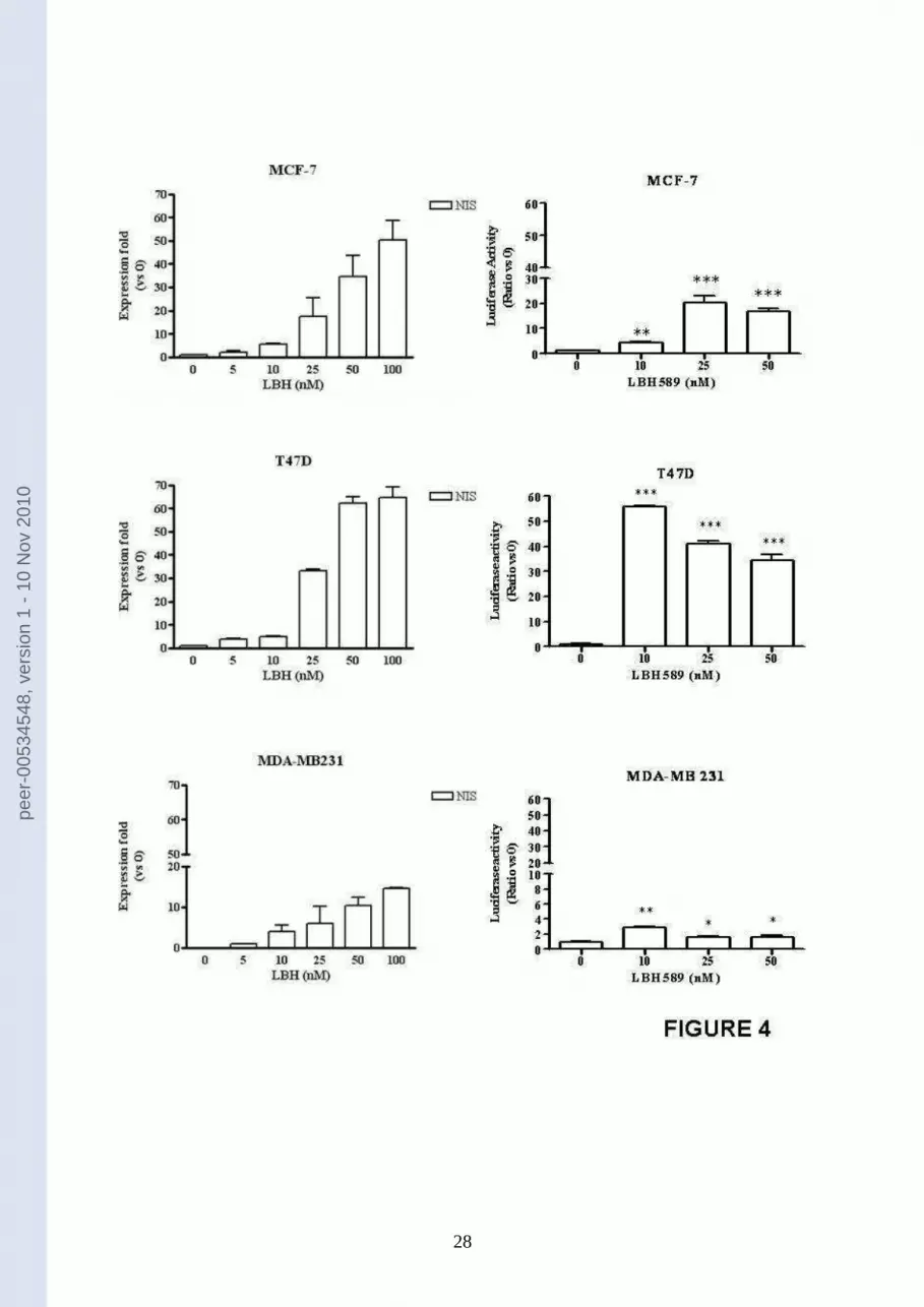
NIS was first studied as mRNA expression. As reported in figure 4 (panels on the left), the
expression of NIS mRNA was significantly induced by LBH589 in a dose-dependent manner
in all the three breast cancer cell lines. In addition, in figure 4 (panels on the right) we report
the effect of LBH589 on the activity of NIS promoter, evaluated with a luciferase assay,
demonstrating that LBH589 is effective in inducing NIS promoter activity in the cell lines
under study.
The induction of NIS gene transcription has to go together with the presence of NIS protein in
cells, and this was clearly demonstrated in MCF7, T47D, and MDA-MB231 breast cancer
peer-00534548, version 1 - 10 Nov 2010

11
cells under LBH589 treatment as shown in figure 5 (upper panels). Moreover, the symporter
induced by LBH589 confers the ability of up-taking iodide to breast cancer cells (figure 5,
lower panels).
In all these experiments, LBH589 was effective in inducing a functional NIS either in ER-
positive and ER-negative breast cancer cells, even though ER-positive cells were shown to be
more sensitive to the compound.
peer-00534548, version 1 - 10 Nov 2010

12
DISCUSSION
The present study shows the potent cytotoxic activity of LBH589 in both estrogen-sensitive
and –insensitive cells and, especially, highlights its ability to induce the sodium/iodide
symporter (NIS) in breast cancer cells that, as a consequence, become able to concentrate
radioactive iodide.
At present four different trials are recruiting patients with recurrent or metastatic breast cancer
for treatment with LBH589 both in monotherapy and in association with trastuzumab and
paclitaxel (www.clinicaltrials.gov), although knowledge concerning the precise mechanism of
LBH589 cytotoxicity in breast cancer cells is still incomplete. The primary molecular effect
of DAC-inhibitors is to alter the acetylation status of the core histone proteins, facilitating
chromatin remodelling and modification in gene expression [24]. It has been reported that 100
and 200 nM LBH589 alone or in association with 4OHTamoxifene were effective in reducing
the growth of ER-negative MDA-MB 231cells [25] and that LBH589 significantly reduced
the viability of ER-positive MCF-7 cells [26] and increased the fraction of dying cells after
radiation exposure [27]. Here we demonstrate that LBH589 alone, in nanomolar range which
is far from toxic range in humans [28], is able to reduce the viability of ER-positive MCF-7
and T47D and ER-negative MDA-MB 231 breast cancer cells. Besides this, we show that the
mechanisms underlying its effect on cell viability include both apoptosis induction and cell
cycle arrest, as already reported for other DAC-inhibitors [29].
We demonstrate in particular that LBH589 induces and enhances NIS expression in breast
cancer cells. The efficiency of NIS is at the basis of successful treatment of differentiated
thyroid cancer thus the possibility of increasing NIS expression in thyroid neoplasms which
have lost the ability to uptake and concentrate iodide is under extensive evaluation [30, 31].
Similarly, as NIS is expressed in quite a significant percentage of breast cancers [7], NIS-
mediated radionuclide imaging and targeted therapy could be useful goals in breast cancer
peer-00534548, version 1 - 10 Nov 2010

13
management. Unfortunately, although NIS expression is present and intense in the majority of
invasive breast cancers, only a small fraction of NIS-positive breast cancers are able to
concentrate iodide, due to low cell surface NIS levels and cell surface trafficking defects,
commonly found in breast tumours [32]. Therefore, research efforts should focus on strategies
to induce/increase NIS expression in breast cancer as it has to be adequately expressed in
order to get a considerable iodide uptake. A large number of agents with very different
mechanisms of action have been utilized to stimulate NIS expression, such as re-
differentiating agents [12, 15, 33-35], or compounds that are reported to physiologically
regulate NIS in breast cells [7, 36-38]. Retinoid acid, especially in the presence of other
compounds such as dexamethasone, induce NIS and iodide uptake in breast cancer cells [12,
15, 33-35] and in animal models [39]. Even though these findings suggest their potential
clinical use, as yet no clinical trials have been presently designed to this aim and additional
strategies can be foreseen.
Epigenetic modifications, such as histone deacetylation, have been considered possible causes
of NIS silencing in cancer cells. Our data clearly demonstrate that the deacetylase- inhibitor
LBH589 induced NIS, both as mRNA and as a protein, and determined a significant iodide
uptake in breast cancer cell lines. Furthermore, the observation that LBH589 strongly
activates the transcription of the construct luc-NIS that we had transfected in breast cancer
cells both ER-positive (MCF-7 and T47D) and –negative (MDA-MB 231) is consistent with a
direct effect of the drug on NIS promoter. Our data suggest that DAC-inhibitor treatment
could therefore modify the acetylation state of NIS promoter allowing its transcription and
expression as already reported for other DAC-inhibitors [19]. The effect was evident
regardless of ER status, even if both ER-positive MCF-7 and T47D cells are more sensitive
than ER-negative MDA-MB 231 cells (the increase in NIS expression in ER-positive cells
was about three times higher than in ER-negative cells). Thus, our study is the first report
peer-00534548, version 1 - 10 Nov 2010

14
concerning the possibility to induce not only NIS mRNA expression but also iodide
accumulation in breast cancer cells by using the pan-DAC inhibitor, LBH589.
In conclusion, the pan-DAC inhibitor LBH589 is a powerful anti-tumour agent and is able to
induce NIS and iodide accumulation in breast cancer cells. The effects of LBH589 are
independent from estrogen-sensitivity and ER-status of cells, making the drug of valuable
use for diagnostic and therapeutic application in both estrogen-sensitive and –insensitive
tumours.
peer-00534548, version 1 - 10 Nov 2010

15
ACKNOWLEDGMENTS
We thank Novartis Pharma AG, Basel, Switzerland for providing us LBH589; Giuseppe
Damante, Dipartimento di Scienze e Tecnologie Biomediche, Policlinico Universitario di
Udine, Udine, Italy, for providing us with the luc-NIS construct; Daniela Taverna and
Alessandra Solero, Dipartimento di Scienze Oncologiche, Università di Torino and M. B. C.,
Torino, Italy, for helping us with luciferase experiments.
This study was supported by the Project “Alfieri 2007”, Fondazione CRT, Torino, the Special
Project “Oncology”, Compagnia San Paolo, Torino, by MIUR and by Regione Piemonte to
Giuseppe Boccuzzi
peer-00534548, version 1 - 10 Nov 2010

16
REFERENCES
1. Parkin DM, Bray F, Ferlay J, Pisani P (2005) Global cancer statistics, 2002, CA
Cancer J Clin 55: 74-108.
2. Berry DA, Cronin KA, Plevritis SK, Fryback DG, Clarke L, Zelen M, Mandelblatt JS,
Yakovlev AY, Habbema JD, Feuer EJ (2005) Cancer Intervention and Surveillance
Modeling Network (CISNET) Collaborators. Effect of screening and adjuvant therapy
on mortality from breast cancer. N Engl J Med 353:1784-92.
3. Gonzalez-Angulo AM, Morales-Vasquez F, Hortobagyi GN (2007) Overview of
resistance to systemic therapy in patients with breast cancer. Adv Exp Med Biol 608:
1-22
4. Cardoso F, Bedard PL, Winer EP, Pagani O, Senkus-Konefka E, Fallowfield LJ,
Kyriakides S, Costa A, Cufer T, Albain KS, ESO-MBC Task Force (2009)
International guidelines for management of metastatic breast cancer: combination vs
sequential single-agent chemotherapy. J Natl Cancer Inst 101: 1174-81
5. Dai G, Levy O, Carrasco N (1996) Cloning and characterization of the thyroid iodide
transporter. Nature 379: 458–46
6. Reiners C, Dietlein M, Luster M (2008) Radio- iodine therapy in differentiated thyroid
cancer: indications and procedures. Best Pract Res Clin Endocrinol Metab 22: 989-
1007.
7. Tazebay UH, Wapnir IL, Levy O, Dohan O, Zuckier LS, Zhao QH, Deng HF,
Amenta PS, Fineberg S, Pestell RC, Carrasco N (2000) The mammary gland iodide
transporter is expressed during lactation and in breast cancer. Nat Med 6: 871–878.
8. Wapnir IL, van de Rijn M, Nowels K, Amenta PS, Walton K, Montgomery K, Greco
RS, Dohán O, Carrasco N (2003) Immunohistochemical profile of the sodium/iodide
peer-00534548, version 1 - 10 Nov 2010
17
symporter in thyroid, breast, and other carcinomas using high density tissue
microarrays and conventional sections. J Clin Endocrinol Metab 88: 1880-8
9. Wapnir IL, Goris M, Yudd A, Dohan O, Adelman D, Nowels K Carrasco N (2004)
The Na+/I−symporter mediates iodide uptake in breast cancer metastases and can be
selectively down-regulated in the thyroid. Clin Cancer Res 10: 4294–302
10. Moon DH, Lee SJ, Park KY, Park KK, Ahn SH, Pai MS, Chang H, Lee HK, Ahn IM
(2001) Correlation between 99mTc pertechnetate uptakes and expressions of human
sodium iodide symporter gene in breast tumor tissues. Nucl Med Biol 28: 829–34
11. Boelaert K, Franklyn JA (2003) Sodium iodide symporter: a novel strategy to target
breast, prostate, and other cancers? Lancet 361: 796-797
12. Kogai T, Kanamoto Y, Li AI, Che LH, Ohashi E, Taki K, Chandraratna RA, Saito T,
Brent GA (2005) Differential regulation of sodium/iodide symporter gene expression
by nuclear receptor ligands in MCF-7 breast cancer cells. Endocrinology 146: 3059–
69.
13. Willhauck MJ, Sharif-Samani B, Senekowitsch-Schmidtke R, Wunderlich N, Göke B,
Morris JC, Spitzweg C (2008) Functional sodium iodide symporter expression in
breast cancer xenografts in vivo after systemic treatment with retinoic acid and
dexamethasone. Breast Cancer Res Treat 109: 263-72.
14. Ohashi E, Kogai T, Kagechika H, Brent GA (2009) Activation of the PI3 kinase
pathway by retinoic acid mediates sodium/iodide symporter induction and iodide
transport in MCF-7 breast cancer cells. Cancer Res 69: 3443-50.
15. Unterholzner S, Willhauck MJ, Cengic N, Schütz M, Göke B, Morris JC, Spitzweg C
(2006) Dexamethasone stimulation of retinoic Acid- induced sodium iodide symporter
expression and cytotoxicity of 131-I in breast cancer cells. J Clin Endocrinol Metab
91: 69-78.
peer-00534548, version 1 - 10 Nov 2010

18
16. Kitazono M, Robey R, Zhan Z, Sarlis NJ, Skarulis MC, Aikou T, Bates S, Fojo T
(2001) Low concentrations of the histone deacetylase inhibitor, depsipeptide
(FR901228), increase expression of the Na(+)/I(+) symporter and iodine accumulation
in poorly differentiated thyroid carcinoma cells. J Clin Endocrinol Metab 86: 3430–
3435
17. Zarnegar R, Brunaud L, Kanauchi H, Wong M, Fung M, Ginzinger D, Duh QY, Clark
OH (2002) Increasing the effectiveness of radioactive iodine therapy in the treatment
of thyroid cancer using Trichostatin A, a histone deacetylase inhibitor. Surgery 132:
984 –990
18. Fortunati N, Catalano MG, Arena K, Brignardello E, Piovesan A ,Boccuzzi G (2004)
Valproic acid induces the expression of the Na+/I+ symporter and iodine uptake in
poorly differentiated thyroid cancer cells. J Clin Endocrinol Metab 89: 1006–1009
19. Botrugno OA, Santoro F, Minucci S (2009) Histone deacetylase inhibitors as a new
weapon in the arsenal of differentiation therapies of cancer. Cancer Lett 280: 134-44
20. Lee MJ, Kim YS, Kummar S, Giaccone G, Trepel JB (2008) Histone deacetylase
inhibitors in cancer therapy. Curr Opin Oncol 20: 639-49
21. Puppin C, D'Aurizio F, D'Elia AV, Cesaratto L, Tell G, Russo D, Filetti S, Ferretti E,
Tosi E, Mattei T, Pianta A, Pellizzari L, Damante G (2005) Effects of histone
acetylation on sodium iodide symporter promoter and expression of thyroid-specific
transcription factors. Endocrinology 146: 3967-74
22. Atadja P (2009) Development of the pan-DAC inhibitor panobinostat (LBH589):
successes and challenges. Cancer Lett 280: 233-41
23. Behr M, Schmitt TL, Espinoza CR, Loos U (1998) Cloning of a functional promoter
of the human sodium/iodide-symporter gene. Biochem J 331: 359–363
peer-00534548, version 1 - 10 Nov 2010

19
24. Bolden JE, Peart MJ, Johnstone RW (2006) Anticancer activities of histone
deacetylase inhibitors. Nat Rev Drug Discov 5: 769-784
25. Zhou Q, Atadja P, Davidson NE (2007) Histone deacetylase inhibitor LBH589
reactivates silenced estrogen receptor alpha (ER) gene expression without loss of
DNA hypermethylation. Cancer Biol Ther 6: 64-9
26. Fiskus W, Ren Y, Mohapatra A, Bali P, Mandawat A, Rao R, Herger B, Yang Y,
Atadja P, Wu J, Bhalla K (2007) Hydroxamic acid analogue histone deacetylase
inhibitors attenuate estrogen receptor-alpha levels and transcriptional activity: a result
of hyperacetylation and inhibition of chaperone function of heat shock protein 90.
Clin Cancer Res 13: 4882-90
27. Kim IA, No M, Lee JM, Shin JH, Oh JS, Choi EJ, Kim IH, Atadja P, Bernhard EJ
(2009) Epigenetic modulation of radiation response in human cancer cells with
activated EGFR or HER-2 signaling: potential role of histone deacetylase 6.
Radiother Oncol. 92:125-32
28. Giles F, Fischer T, Cortes J, Garcia-Manero G, Beck J, Ravandi F, Masson E, Rae P,
Laird G, Sharma S, Kantarjian H, Dugan M, Albitar M, Bhalla K (2006) A phase I
study of intravenous LBH589, a novel cinnamic hydroxamic acid analogue histone
deacetylase inhibitor, in patients with refractory hematologic malignancies. Clin
Cancer Res 12: 4628-4635
29. Fortunati N, Bertino S, Costantino L, Bosco O, Vercellinatto I, Catalano MG,
Boccuzzi G (2008) Valproic acid is a selective antiproliferative agent in estrogen-
sensitive breast cancer cells. Cancer Lett 259: 156-64
30. Dohán O, De la Vieja A, Paroder V, Riedel C, Artani M, Reed M, Ginter CS, Carrasco
N (2003) The sodium/iodide Symporter (NIS): characterization, regulation, and
medical significance. Endocr Rev 24: 48-77
peer-00534548, version 1 - 10 Nov 2010

20
31. Furuya F, Shimura H, Suzuki H, Taki K, Ohta K, Haraguchi K, Onaya T, Endo T,
Kobayashi T (2004) Histone deacetylase inhibitors restore radioiodide uptake and
retention in poorly differentiated and anaplastic thyroid cancer cells by expression of
the sodium/iodide symporter thyroperoxidase and thyroglobulin. Endocrinology 145:
2865–2875
32. Beyer SJ, Jimenez RE, Shapiro CL, Cho JY, Jhiang SM (2009) Do cell surface
trafficking impairments account for variable cell surface sodium iodide symporter
levels in breast cancer? Breast Cancer Res Treat 115: 205-12
33. Kogai T, Ohashi E, Jacobs MS, Sajid-Crockett S, Fisher ML, Kanamoto Y, Brent GA
(2008) Retinoic acid stimulation of the sodium/iodide symporter in MCF-7 breast
cancer cells is mediated by the insulin growth factor-I/phosphatidylinositol 3-kinase
and p38 mitogen activated protein kinase signaling pathways. J Clin Endocrinol Metab
93: 1884-92
34. Dohán O, De la Vieja A, Carrasco N (2006) Hydrocortisone and purinergic signaling
stimulate sodium/iodide symporter (NIS)-mediated iodide transport in breast cancer
cells. Mol Endocrinol 20:1121-37
35. Tanosaki S, Ikezoe T, Heaney A, Said JW, Dan K, Akashi M, Koeffler HP (2003)
Effect of ligands of nuclear hormone receptors on sodium/iodide symporter expression
and activity in breast cancer cells. Breast Cancer Res Treat 79: 335-45
36. Cho JY, Leveille R, Kao R, Rousset B, Parlow AF, Burak WE, Jr, Mazzaferri EL,
Jhiang SM (2000) Hormonal regulation of radioiodide uptake activity and NaC/IK
symporter expression in mammary glands. J Clin Endocrinol Metab 85: 2936–2943
37. Arturi F, Ferretti E, Presta I, Mattei T, Scipioni A, Scarpelli D, Bruno R, Lacroix L,
Tosi E, Gulino A, Russo D, Filetti S (2005) Regulation of iodide uptake and
peer-00534548, version 1 - 10 Nov 2010

21
sodium/iodide symporter expression in the MCF-7 human breast cancer cell line. J
Clin Endocrinol Metab. 90: 2321-6
38. Knostman KA, McCubrey JA, Morrison CD, Zhang Z, Capen CC, Jhiang SM (2007)
PI3K activation is associated with intracellular sodium/iodide symporter protein
expression in breast cancer. BMC Cancer 7:137.
39. Kogai T, Kanamoto Y, Che LH, Taki K, Moatamed F, Schultz JJ, Brent GA (2004)
Systemic retinoic acid treatment induces sodium/iodide symporter expression and
radioiodide uptake in mouse breast cancer models. Cancer Res. 64: 415-22.
peer-00534548, version 1 - 10 Nov 2010

22
LEGEND TO FIGURES
Figure 1 - LBH589 effect on histone acetylation. Acetylation of histone H4 in MCF-7,
T47D and MDA-MB231 cells incubated for 24 hours with LBH589 (5-100 nM), was assessed
by Western blotting. Equal loading and transfer were verified by re-probing the membranes
with anti-GAPDH antibody. The figure shows a typical experiment.
Figure 2 – LBH589 effect of cell viability. Breast cancer cells were incubated with LBH589
(1-200 nM) for 72 hours. After treatment, cell viability was determined by the WST-1
method, and expressed as percentage (untreated controls being 100%). Three response
parameters, median growth inhibition (GI50), total growth inhibition (TGI) and median lethal
concentration (LC50) were calculated for each cell line, and values are reported in the Table.
Figure 3 – LBH589 effect on apoptosis and cell cycle. Left Panels: ELISA detection of
DNA-histone complex in MCF-7, T47D and MDA-MB231 cells incubated for 72 hours with
LBH589 (1-200 nM). The enrichment factor was calculated as the ratio between the
absorbance measurements of treated cells and untreated controls. Results are expressed as
means ± SEM; n = 3. Significance vs untreated cells (0): * P< 0.05; ** P< 0.01; *** P<
0.001. Right Panels: Cell cycle analysis was performed by flow cytometry after 72 hour
treatment with LBH589 (5-100nM). Results are expressed as means ± SEM; n = 3.
Significance for G2/M vs untreated cells (0): *P<0.05; ** P< 0.01; ** *P< 0.001.
peer-00534548, version 1 - 10 Nov 2010

23
Figure 4 –LBH589 effect on NIS mRNA expression and NIS promoter activity. Left
Panels: NIS mRNA was evaluated with Real Time PCR in MCF-7, T47D and MDA-MB 231
cells treated with 5-100 nM LBH589. Results were normalized for three different
housekeeping genes (-actin, 2-microglobulin and L13A) and expressed as relative
expression fold vs untreated controls (0). Results are expressed as means ± SEM; n = 3. Right
Panels: breast cancer cells were transiently transfected with a reporter plasmid carrying the
luciferase gene under the control of NIS promoter (NIS-luc). The luciferase activity was
assayed before and after treatment with 10-50 nM LBH589. Data are expressed as mean ±
SEM; n=3.
Figure 5 – LBH589 effects on NIS protein expression and iodide uptake. Upper Panels:
immunofluorescence for NIS was performed on breast cancer cells: (A) untreated MCF-7
cells, (B) 25 nM LBH589 treated MCF-7 cells; (C) untreated T47D cells; (D) 25 nM LBH
589 treated T47D cells; (E) untreated MDA-MB 231 cells; (F) 25 nM LBH589 treated MDA-
MB 231 cells. Lower Panel: 125I uptake in breast cancer cells treated with 5-100 nM LBH589
was evaluated as described; data are expressed as mean ± SEM; n=3. Significance vs
untreated cells (0) was: *P<0.05; ** P< 0.01.
peer-00534548, version 1 - 10 Nov 2010
24
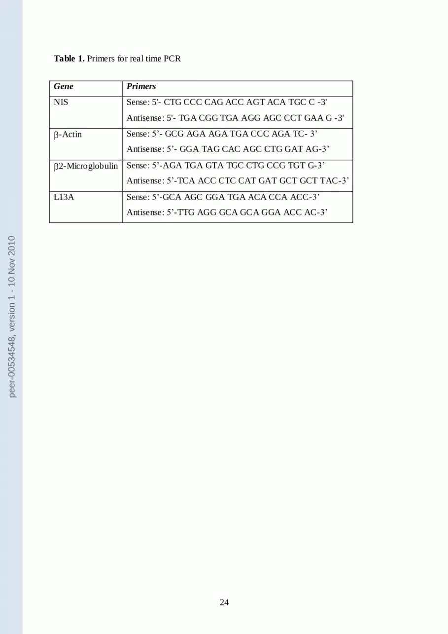
Table 1. Primers for real time PCR
Gene Primers
NIS Sense: 5'- CTG CCC CAG ACC AGT ACA TGC C -3'
Antisense: 5'- TGA CGG TGA AGG AGC CCT GAA G -3'
-Actin Sense: 5’- GCG AGA AGA TGA CCC AGA TC- 3’
Antisense: 5’- GGA TAG CAC AGC CTG GAT AG-3’
2-Microglobulin Sense: 5’-AGA TGA GTA TGC CTG CCG TGT G-3’
Antisense: 5’-TCA ACC CTC CAT GAT GCT GCT TAC-3’
L13A Sense: 5’-GCA AGC GGA TGA ACA CCA ACC-3’
Antisense: 5’-TTG AGG GCA GCA GGA ACC AC-3’
peer-00534548, version 1 - 10 Nov 2010

25
peer-00534548, version 1 - 10 Nov 2010

26
peer-00534548, version 1 - 10 Nov 2010

27
peer-00534548, version 1 - 10 Nov 2010
28
peer-00534548, version 1 - 10 Nov 2010

29
peer-00534548, version 1 - 10 Nov 2010


