Embed Size (px)
Citation preview

European Journal of Pharmacology 659 (2011) 53–60
Contents lists available at ScienceDirect
European Journal of Pharmacology
j ourna l homepage: www.e lsev ie r.com/ locate /e jphar
Pulmonary, Gastrointestinal and Urogenital Pharmacology
The protective effects of ursodeoxycholic acid on isoniazid plus rifampicin inducedliver injury in mice
Xi Chen a, Juan Xu a, Cheng Zhang b, Tao Yu b, Hua Wang b, Mei Zhao b, Zi-Hao Duan b, Ying Zhang b,Jian-Ming Xu a, De-Xiang Xu b,⁎a Department of Gastroenterology, The First Affiliated Hospital, Anhui Medical University, Hefei 230022, Chinab Department of Toxicology, Anhui Medical University, Hefei 230032, China
⁎ Corresponding author. Tel.: +86 551 5167923; fax:E-mail address: [email protected] (D.-X. Xu).
0014-2999/$ – see front matter © 2011 Elsevier B.V. Aldoi:10.1016/j.ejphar.2011.03.007
a b s t r a c t
a r t i c l e i n f oArticle history:Received 17 November 2010Received in revised form 1 March 2011Accepted 8 March 2011Available online 17 March 2011
Keywords:UDCA (Ursodeoxycholic acid)HepatotoxicityApoptosisAntioxidant
Antitubercular drugs have been known to be potentially hepatotoxic and may lead to drug-induced liverinjury. In this study, we aimed to investigate the protective effects of ursodeoxycholic acid (UDCA) on liverinjury caused by co-administration with isoniazid and rifampicin, two famous antitubercular drugs. Liverinjury was induced by co-treatment with isoniazid (75 mg/kg) and rifampicin (150 mg/kg) for one week.Mice were orally administered with UDCA (15, 50 and 150 mg/kg) 30 min before isoniazid and rifampicin. Weshow that serum alanine aminotransferase (ALT) and alkaline phosphatase (ALP) were significantly increasedin mice treated with isoniazid plus rifampicin. An obvious fatty accumulation, accompanied by mild necrosisand inflammation, was observed in liver of mice treated with rifampicin plus isoniazid. In addition, isoniazidplus rifampicin resulted in hepatic apoptosis, as determined by terminal dUTP nick-end labeling (TUNEL)staining and caspase-3 activation. Additional experiment showed that isoniazid plus rifampicin significantlyincreased the level of hepatic malondialdehyde (MDA) and caused glutathione (GSH) depletion and 3-nitrotyrosine (3-NT) residues in liver. UDCA pretreatment significantly attenuated isoniazid plus rifampicininduced oxidative stress in liver. Importantly, UDCA pretreatment significantly alleviated isoniazid plusrifampicin induced hepatic apoptosis. Moreover, UDCA-mediated anti-apoptotic effect seemed to beassociated with its regulation of Bcl-2 and Bax gene expression in liver. These findings suggest that UDCAmight protect against isoniazid and rifampicin induced liver injury through its anti-oxidative and anti-apoptotic effects.
+86 551 5161179.
l rights reserved.
© 2011 Elsevier B.V. All rights reserved.
1. Introduction
Isoniazid and rifampicin, two front-line drugs used in antituber-culosis therapy, have been known to be potentially hepatotoxic andmay lead to drug-induced liver injury (Hwang et al., 1997). A meta-analysis of studies involving the use of a multiplicity of anti-tuberculosis drug regimens predominantly in adults has shown anincidence of toxic hepatitis of 1.6% in patients with isoniazid alone,1.1% in patients with RPF alone and 2.55% in patients with isoniazidplus rifampicin (Steele et al., 1991). Isoniazid is thought to be initiatedby cytochrome P450 mediated metabolism of isoniazid to acetylhy-drazine and hydrazine that is hepatotoxic (Preziosi, 2007; Yue et al.,2004; Bhadauria et al., 2007). Rifampicin, which is generally co-administered with isoniazid in treatment of tuberculosis, is toxic tohepatocytes (Christiane and Peter, 2006). In addition, as a powerfulinducer of drug metabolizing enzymes in man and rats, rifampicin
aggravates isoniazid-induced hepatotoxicity by enhancing the pro-duction of toxic metabolites (Tasdug et al., 2007).
Reactive oxygen species play an important role in isoniazid andrifampicin induced liver injury (Chowdhury et al., 2006). According toseveral earlier studies, hepatic glutathione-S transferase activity andglutathione (GSH) level were significantly decreased in rats treatedwith isoniazid or hydrazine (Sodhi et al., 1996, 1997). Clinic datashowed that an increased level of GSH and a decreased level ofmalondialdehyde (MDA) were measured in plasma of patient treatedwith isoniazid and rifampicin, suggesting that isoniazid and rifampi-cin could result in oxidative stress (Chowdhury et al., 2001). Sinceoxidative stress has been regarded as the major mechanism ofantituberculosis drug-induced hepatotoxicity, antioxidants might beused as potential antihepatotoxic drugs against isoniazid andrifampicin caused liver injury (Sano et al., 2004).
Ursodeoxycholic acid (UDCA, 3α, 7β-dihydroxy-5β-cholanic acid)is a hydrophilic dihydroxy bile acid which was found in the bile ofblack bear (Hagey et al., 1993). UDCA has been used in Chinesetraditional medicine for the treatment of liver diseases for centuriesand was also wildly used for the treatment of various cholestaticdisorders (Paumgartner and Beuers, 2002). UDCA exerts its actions in

Table 1Effect of UDCA on serum parameters and the relative liver weight in isoniazid and rifampicin treated mice.
Groups ALT (U/l) AST (U/l) ALP (U/l) Liver/body eight (%)
Control 30.00±6.43 113.50±9.14 74.70±9.63 4.5±0.5Isoniazid+rifampicin 38.70±6.51a 124.20±12.04 90.70±12.91a 5.6±0.3a
UDCA (150 mg/kg) 28.89±5.13 108.11±10.82 66.22±10.34 4.1±0.6Isoniazid+rifampicin+UDCA (15 mg/kg) 28.90±6.45b 120.30±12.29 69.60±10.79b 5.5±0.4Isoniazid+rifampicin+UDCA (50 mg/kg) 26.90±5.89b 99.50±12.51 80.50±8.69b 5.2±0.5b
Isoniazid+rifampicin+UDCA (150 mg/kg) 29.60±6.96b 126.00±13.29 68.50±11.02b 5.1±0.5c
a Pb0.01 vs control group.b Pb0.05 vs isoniazid+rifampicin group.c Pb0.01 vs isoniazid+rifampicin group.
54 X. Chen et al. / European Journal of Pharmacology 659 (2011) 53–60
liver through multiple possibly interrelated pathways includingalterations of bile acid pool, choleresis, immune modulation andcytoprotective effects (Lazaridis et al., 2001). UDCA is an antioxidant.UDCA protects against secondary biliary cirrhosis in rats by prevent-ing mitochondrial oxidative stress (Serviddio et al., 2004). A recentstudy showed that UDCA protected against hepatotoxicity caused byamoxicillin-clavulanic acid in rats though its antioxidant properties(El-Sherbiny et al., 2009). In addition, UDCA is also an antiapoptoticagent. An earlier study showed that UDCA pretreatment preventedfetal rat liver from apoptosis induced by maternal cholestasis (Perezet al., 2005). In the present study, we aimed to investigate theprotective effect of UDCA on isoniazid and rifampicin induced liverinjury in mice. Our results indicate that UDCA pretreatment couldprotect isoniazid and RPF induced liver injury not only through itsantioxidant effect but also its antiapoptotic ability.
2. Materials and methods
2.1. Reagents
Isoniazid and rifampicin were from Sigma Chemical Co. (St. Louis,MO). 3-NT, β-actin, Bax, Bcl-2 and active caspase-3 antibodies werefrom Santa Cruz Biotechnologies (Santa Cruz, CA). TUNEL detection kitwas from Promega Corporation (Madison, WI). Chemiluminescence(ECL) detection kit was from Pierce Biotechnology (Rockford, IL). Allthe other reagents were from Sigma or indicated in the specifiedmethods.
Fig. 1. Effects of UDCA on histopathology of liver treated with rifampicin plus isoniazid. (Arifampicin plus isoniazid. (C) Liver section from mice treated with UDCA. (D) Liver section fr(E) Liver section from mice pretreated with UDCA (50 mg/kg) before rifampicin plus isonirifampicin plus isoniazid treatment. Liver sections were stained with hematoxylin and eosi
2.2. Animals and treatments
Female CD-1 mice (6–8 week-old, 24–26 g) were purchased fromBeijingVital River (Beijing, China). The animalswere allowed free accessto food andwater at all times and weremaintained on a 12 h light/darkcycle in a controlled temperature (20–25 °C) and humidity (50±5%)environment for a period of 1 week before use. In isoniazid plusrifampicin group, mice were administered with isoniazid (75 mg/kg)plus rifampicin (150 mg/kg) by gavage daily for one week. The doses ofisoniazid and rifampicin used in the present study referred to the resultsfrom preliminary study. Preliminary study showed that administrationwith 75 mg/kg isoniazid plus 150 mg/kg rifampicin daily for a weekresulted in obvious liver injuries including fatty accumulation, hepaticapoptosis and the elevation of serumALT. In UDCA pretreatment group,mice were treated with different doses of UDCA (15, 50, 150 mg/kg)30 min before isoniazid and rifampicin. In control groups, mice wereadministered with PBS or UDCA (150 mg/kg) by gavage daily for oneweek. The doses of UDCA used in the present study referred to others(Ishizaki et al., 2008). All procedures on animals followed the guidelinesfor humane treatment set by the Association of Laboratory AnimalSciences and the Center for Laboratory Animal Sciences at AnhuiMedical University (Agreement No: 20090815003).
2.3. Biochemical parameters
Serum alanine aminotransferase (ALT), aspartate aminotransfer-ase (AST) and alkaline phosphatase (ALP) were measured using
) Liver section from mice treated with saline. (B) Liver section from mice treated withom mice pretreated with UDCA (15 mg/kg) before rifampicin plus isoniazid treatment.azid treatment. (F) Liver section from mice pretreated with UDCA (150 mg/kg) beforen (original magnification ×400).
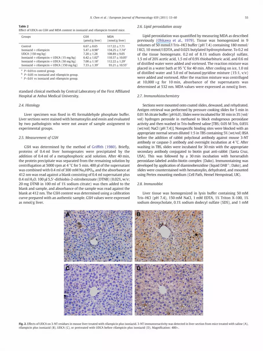
Table 2Effect of UDCA on GSH and MDA content in isoniazid and rifampicin treated mice.
Groups GSH(μmol/g liver)
MDA(nmol/g liver)
Control 6.67±0.65 117.22±7.71Isoniazid+rifampicin 5.47±0.98a 134.25±7.74a
UDCA (150 mg/kg) 7.20±1.28 108.89±9.05Isoniazid+rifampicin+UDCA (15 mg/kg) 6.42±1.02b 110.37±10.05c
Isoniazid+rifampicin+UDCA (50 mg/kg) 7.00±1.18c 112.22±1.29c
Isoniazid+rifampicin+UDCA (150 mg/kg) 7.33±1.39c 93.21±10.55c
a Pb0.01vs control group.b Pb0.05 vs isoniazid and rifampicin group.c Pb0.01 vs isoniazid and rifampicin group.
55X. Chen et al. / European Journal of Pharmacology 659 (2011) 53–60
standard clinical methods by Central Laboratory of the First AffiliatedHospital at Anhui Medical University.
2.4. Histology
Liver specimen was fixed in 4% formaldehyde phosphate buffer.Liver sectionswere stainedwith hematoxylin and eosin and evaluatedby two pathologists who were not aware of sample assignment toexperimental groups.
2.5. Measurement of GSH
GSH was determined by the method of Griffith (1980). Briefly,proteins of 0.4 ml liver homogenates were precipitated by theaddition of 0.4 ml of a metaphosphoric acid solution. After 40 min,the protein precipitate was separated from the remaining solution bycentrifugation at 5000 rpm at 4 °C for 5 min. 400 μl of the supernatantwas combinedwith 0.4 ml of 300 mMNa2HPO4, and the absorbance at412 nmwas read against a blank consisting of 0.4 ml supernatant plus0.4 ml H2O. 100 μl 5,5′-dithiobis-2-nitrobenzoate (DTNB) (0.02%, w/v;20 mg DTNB in 100 ml of 1% sodium citrate) was then added to theblank and sample, and absorbance of the sample was read against theblank at 412 nm. The GSH content was determined using a calibrationcurve prepared with an authentic sample. GSH values were expressedas nmol/g liver.
Fig. 2. Effects of UDCA on 3-NT residues in mouse liver treated with rifampicin plus isoniazidrifampicin plus isoniazid (B), UDCA (C), or pretreated with UDCA before rifampicin plus iso
2.6. Lipid peroxidation assay
Lipid peroxidation was quantified by measuring MDA as describedpreviously (Ohkawa et al., 1979). Tissue was homogenized in 9volumes of 50 mmol/l Tris–HCl buffer (pH 7.4) containing 180 mmol/l KCl, 10 mmol/l EDTA, and 0.02% butylated hydroxytoluene. To 0.2 mlof the tissue homogenate, 0.2 ml of 8.1% sodium dodecyl sulfate,1.5 ml of 20% acetic acid, 1.5 ml of 0.9% thiobarbituric acid, and 0.6 mlof distilled water were added and vortexed. The reaction mixture wasplaced in a water bath at 95 °C for 40 min. After cooling on ice, 1.0 mlof distilled water and 5.0 ml of butanol/pyridine mixture (15:1, v/v)were added and vortexed. After the reaction mixture was centrifugedat 10,000 ×g for 10 min, absorbance of the supernatants wasdetermined at 532 nm. MDA values were expressed as nmol/g liver.
2.7. Immunohistochemistry
Sections weremounted onto coated slides, dewaxed, and rehydrated.Antigen retrieval was performed by pressure cooking slides for 5 min in0.01 Mcitratebuffer (pH6.0). Slideswere incubated for30 min in3%(vol/vol) hydrogen peroxide in methanol to block endogenous peroxidaseactivity and then washed in Tris-buffered saline [TBS; 0.05 M Tris, 0.85%(wt/vol) NaCl (pH 7.4)]. Nonspecific binding sites were blocked with anappropriate normal serum diluted 1:5 in TBS containing 5% (wt/vol) BSAbefore the addition of rabbit polyclonal antibody against mouse 3-NTantibody or caspase-3 antibody and overnight incubation at 4 °C. Afterwashing in TBS, slides were incubated for 30 min with the appropriatesecondary antibody conjugated to biotin goat anti-rabbit (Santa Cruz,USA). This was followed by a 30 min incubation with horseradishperoxidase-labeled avidin-biotin complex (Dako). Immunostaining wasdeveloped by application of diaminobenzidine (liquid DAB+; Dako), andslides were counterstained with hematoxylin, dehydrated, and mountedusing Pertex mounting medium (Cell Path, Hemel Hempstead, UK).
2.8. Immunoblot
Liver tissue was homogenized in lysis buffer containing 50 mMTris–HCl (pH 7.4), 150 mM NaCl, 1 mM EDTA, 1% Triton X-100, 1%sodium deoxycholate, 0.1% sodium dodecyl sulfate (SDS), and 1 mM
. 3-NT immunoreactivity was detected in liver section frommice treated with saline (A),niazid (D). Magnification: 400×.
56 X. Chen et al. / European Journal of Pharmacology 659 (2011) 53–60
phenylmethylsulfonyl fluoride (PMSF). Samples were then centri-fuged at 15,000 ×g for 15 min. Supernatants from each sample wereadded to a gel loading buffer (100 mM Tris, pH 6.8, 20% glycerol,200 mM DTT, 4% SDS, 0.03% bromophenol blue) and boiled for 5 min.Proteins (50 μg/sample) in loading buffer were subjected to electro-phoresis in 10% SDS-polyacrylamide gel for 3 h. Protein in the gel wastransferred electrophoretically onto a polyvinylidene fluoride mem-brane (Millipore Corp., Bedford, Massachusetts, USA) and blocked in5% nonfat powdered milk in Dulbecco's PBS (DPBS) overnight at 4 °C.The membranes were then incubated for 2 h with rabbit polyclonalantibody against mouse Bax, Bcl-2 or β-actin at room temperature.After washing in DPBS containing 0.05% Tween-20 four times for10 min each, the membranes were incubated with goat anti-rabbitIgG antibody for 2 h. Themembranes were then washed for four timesin DPBS containing 0.05% Tween-20 for 10 min each, followed bysignal development using an ECL detection kit.
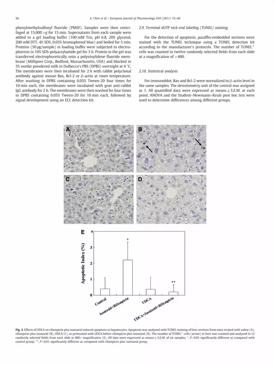
Fig. 3. Effects of UDCA on rifampicin plus isoniazid induced apoptosis in hepatocytes. Apoptorifampicin plus isoniazid (B), UDCA (C), or pretreated with UDCA before rifampicin plus isonrandomly selected fields from each slide at 400× magnification (E). All data were expressecontrol group; **, Pb0.01 significantly different as compared with rifampicin plus isoniazid
2.9. Terminal dUTP nick-end labeling (TUNEL) staining
For the detection of apoptosis, paraffin-embedded sections werestained with the TUNEL technique using a TUNEL detection kitaccording to the manufacturer's protocols. The number of TUNEL+
cells was counted in twelve randomly selected fields from each slideat a magnification of ×400.
2.10. Statistical analysis
For immunoblot, Bax and Bcl-2 were normalized to β-actin level inthe same samples. The densitometry unit of the control was assignedas 1. All quantified data were expressed as means±S.E.M. at eachpoint. ANOVA and the Student–Newmann–Keuls post hoc test wereused to determine differences among different groups.
sis was analyzed with TUNEL staining of liver sections frommice treated with saline (A),iazid (D). The number of TUNEL+ cells (arrow) in liver was counted and analyzed in 12d as means±S.E.M. of six samples. *, Pb0.01 significantly different as compared withgroup.

57X. Chen et al. / European Journal of Pharmacology 659 (2011) 53–60
3. Results
3.1. Effects of UDCA on isoniazid and rifampicin induced liver injury
As shown in Table 1, the level of serum ALT and ALP wassignificantly increased in mice administered with isoniazid andrifampicin for one week. UDCA pre-treatment significantly attenuatedisoniazid plus rifampicin induced elevation of serum ALT and ALP. Theeffects of UDCA on isoniazid plus rifampicin induced liver injury wereanalyzed. As expected, the relative liver weight was significantlyincreased in mice administered with isoniazid and rifampicin(Table 1). An obvious fatty accumulation, accompanied by mildnecrosis and inflammation, was observed in liver of mice treated withisoniazid plus rifampicin (Fig. 1B). UDCA pre-treatment significantlyalleviated isoniazid plus rifampicin induced increase in the relativeliver weight in a dose-dependent manner (Table 1). In addition, UDCA
Fig. 4. Effect of UDCA on rifampicin plus isoniazid induced caspase-3 activation. Active caspasaline (A), rifampicin plus isoniazid (B), UDCA (C), or pretreated with UDCA before rifampiccounted and analyzed in 12 randomly selected fields from each slide at 400× magnificatiosignificantly different as compared with control group; *, Pb0.01 significantly different as c
pre-treatment alleviated isoniazid plus rifampicin induced patholog-ical damage (Fig. 1D, E and F).
3.2. UDCA protects against isoniazid and rifampicin induced oxidativestress in liver
As shown in Table 2, hepatic GSH content was significantlydecreased in mice administered with isoniazid and rifampicin.Conversely, the level of MDA, an index of lipid peroxidation, wassignificantly increased in liver of mice treated with isoniazid andrifampicin (Table 2). UDCA pre-treatment significantly alleviatedisoniazid plus rifampicin induced hepatic GSH depletion. In addition,UDCA pre-treatment completely prevented isoniazid plus rifampicininduced lipid peroxidation. The effects of UDCA on the intensity of 3-NT residues in liver are presented in Fig. 2. As expected, the intensityof hepatic 3-NT staining was strengthened in mice treated with
se-3 was measured with immunohistochemistry. Liver sections from mice treated within plus isoniazid (D). The number of active caspase-3-positive cells (arrow) in liver wasn (E). All data were expressed as means±S.E.M. of six samples per group. *, Pb0.01ompared with rifampicin plus isoniazid group.

58 X. Chen et al. / European Journal of Pharmacology 659 (2011) 53–60
isoniazid and rifampicin (Fig. 2B). UDCA pre-treatment significantlyattenuated hepatic 3-NT staining (Fig. 2D).
3.3. UDCA protects against isoniazid and rifampicin induced apoptosisin liver
Hepatic apoptosis was detected by TUNEL. As shown in Fig. 3, thenumber of TUNEL+ cells was significantly increased in liver of micetreated with isoniazid and rifampicin. UDCA pre-treatment signifi-cantly attenuated isoniazid and rifampicin induced apoptosis in liver.The effects of UDCA pretreatment on isoniazid plus rifampicininduced caspase-3 activation are presented in Fig. 4. In accordancewith the results from TUNEL, the number of hepatocytes with activecaspase-3 was significantly increased in mice administered withisoniazid and rifampicin (Fig. 4B and E). UDCA pre-treatmentprotected against isoniazid and rifampicin induced caspase-3 activa-tion (Fig. 4D and E). The effects of UDCA on the expression of Bax andBcl-2 are presented in Fig. 5. As expected, the level of Bax wassignificantly increased in liver of mice treated with isoniazid andrifampicin (Fig. 5A and B). By contrast, the level of Bcl-2 wassignificantly decreased in liver of mice treated with isoniazid andrifampicin (Fig. 5A and C). UDCA pretreatment obviously protectedagainst the alteration of hepatic Bax and Bcl-2 expression (Fig. 5A, B,and C).
4. Discussion
UDCA has been suggested as an efficient therapy in various liverdiseases. First, UDCA is the currently accepted method for treating
Fig. 5. Effect of UDCA on rifampicin plus isoniazid induced expression of hepatic Bax and Bcl-Quantitative analysis of scanning densitometry on six samples per groupwas performed. Baxof the control were assigned as 1. All data were expressed as means±S.E.M. of six samplessignificantly different as compared with control group; ***, Pb0.01 significantly different as
cholestasis. According to combined analysis of the three largestrandomized clinical trials, UDCA improved clinical and biochemicalindices and prolonged survival free of liver transplantation (Pouponet al., 1997). An earlier study in rats found that UDCA prevented fromsecondary biliary cirrhosis through counteracting mitochondrialoxidative stress (Serviddio et al., 2004). Second, it has beendemonstrated that UDCA protects against drug-induced hepatotoxic-ity. A recent study showed that UDCA protected rats from liver injuryinduced by methotrexate, an immunosuppressive drug (Uraz et al.,2008). In addition, UDCA acts as effective hepatoprotective agentagainst liver dysfunction caused by the broad spectrum antibioticcombination amoxicillin-clavulanic acid (El-Sherbiny et al., 2009). Inthis study, we found that UDCA pretreatment significantly attenuatedthe elevation of serum ALT and ALP levels in mice treated withisoniazid and rifampicin. In addition, UDCA pretreatment alleviatedisoniazid plus rifampicin induced pathological damage. These resultsprovide new evidence that UDCA could protect against drug-inducedliver injury.
Numerous studies have demonstrated that oxidative damage is animportant mechanism of anti-tuberculosis drug-induced hepatotox-icity (Sodhi et al., 1996, 1997; Bhadauria et al., 2007; Chowdhury et al.,2006; Attri et al., 2000). In the present study, we found that the levelof hepatic GSH was significantly decreased in mice treated withisoniazid plus rifampicin. Conversely, the level of hepatic MDA, amarker of lipid peroxidation, was obviously increased in miceadministered with isoniazid plus rifampicin. 3-NT is a specific markerfor protein nitration. A recent study demonstrated that 3-NT couldcause protein denaturation and DNA damage, leading to cell death inliver (Oberley et al., 2008). The present study showed that the
2 in mice. The protein expression of Bax and Bcl-2 was measured using immunoblotting.and Bcl-2 were normalized to β-actin level in the same samples. The densitometry unitsper group.*, Pb0.05 significantly different as compared with control group; **, Pb0.01compared with rifampicin plus isoniazid group.

59X. Chen et al. / European Journal of Pharmacology 659 (2011) 53–60
intensity of hepatic 3-NT staining was strengthened in miceadministered with isoniazid and rifampicin. These results suggestthat oxidative stress might contribute, at least partially, to liver injuryin mice co-administered with isoniazid and rifampicin.
A recent study showed that N-acetylcysteine, a well-knownantioxidant, protected against isoniazid plus rifampicin inducedhepatotoxicity through its antioxidant effect (Attri et al., 2000).UDCA is an antioxidant. An earlier study showed that UDCA alleviatedhepatic lipid peroxidation in experimental cholestatic liver disease[Ljubuncic et al., 2000]. In addition, UDCA significantly increased thelevels of GSH and thiol-containing proteins, thereby protectinghepatocytes against oxidative injury (Mitsuyoshi et al., 1999). Arecent study demonstrated that UDCA induced the expression ofdetoxifying enzymes and antioxidative stress genes via the Nrf2transcriptional pathway (Okada et al., 2008). In the present study, wefound that UDCA pretreatment significantly attenuated hepatic lipidperoxidation and GSH depletion in mice administered with isoniazidplus rifampicin. In addition, UDCA pretreatment alleviated hepatic 3-NT residues in mice treated with isoniazid and rifampicin. Theseresults indicate that UDCA-mediated protection against isoniazid plusrifampicin induced liver damage might be associated with its strongantioxidant effect.
In the present study, we found that the number of TUNEL+ cellswas significantly increased in liver of mice co-treated with isoniazidand rifampicin. In addition, the number of hepatocytes with activecaspase-3 was significantly increased in liver of mice administeredwith isoniazid plus rifampicin, suggesting that apoptosis mightpartially contribute to isoniazid plus rifampicin induced liver injury.To investigate the molecular mechanism of isoniazid plus rifampicininduced apoptosis, the expression of hepatic Bcl-2 and Bax wasanalyzed. As expected, the expression of hepatic Bax was significantlyincreased in mice administered with isoniazid and rifampicin. Bycontrast, co-administration with isoniazid and rifampicin significantlyreduced the level of hepatic Bcl-2. UDCA has an antiapoptotic effect.An in vitro study showed that UDCA protected against Fas-ligand-induced apoptosis in mouse hepatocytes (Azzaroli et al., 2002). In thepresent study, we investigated the effects of UDCA on isoniazid plusrifampicin induced apoptosis in liver. We found that UDCA pretreat-ment significantly reduced the number of TUNEL+ cells in liver ofmice administered with isoniazid and rifampicin. In addition, UDCApretreatment obviously alleviated isoniazid and rifampicin inducedhepatic caspase-3 activation. These results indicate that UDCA-mediated protection against isoniazid plus rifampicin induced liverinjury might be associated with its strong antiapoptotic effect.
The mechanism of UDCA-mediated antiapoptotic effect remainedunclear. Indeed, UDCA stabilizes the mitochondrial membranes ofhepatocytes and protects hepatocytes from various outer injuries (Botlaet al., 1995). According to several earlier studies, UDCA preventedcytochrome c release and inhibited hepatocyte apoptosis throughmodulating mitochondrial membrane perturbation (Rodrigues et al.,1998, 1999). Another study found that UDCA protected hepatocytesfrom bile acid induced apoptosis via activation of survival pathways(Schoemaker et al., 2004). A recent studydemonstrated that p53 is a keymolecular target of UDCA in regulating apoptosis (Amaral et al., 2007).In the present study, we found that isoniazid plus rifampicin inducedup-regulation of Bax was repressed in liver of mice pretreated withUDCA. Conversely, UDCA pretreatment upregulated the expression ofhepatic Bcl-2 inmice treatedwith isoniazid and rifampicin. These resultssuggest that the UDCA pretreatment might protect against isoniazidplus rifampicin induced hepatic apoptosis through regulating theexpression of Bcl-2 family.
In summary, the results of the present study indicate that UDCApretreatment protects against isoniazid plus rifampicin induced liverinjury. UDCA-mediated protection against isoniazid plus rifampicininduced liver injury is associated not only with its antioxidant effectbut also its antiapoptotic ability.
Acknowledgments
This project was supported by National Natural Science Founda-tion of China (30371667, 30572223, 30973544, 81001480) andNatural Science Foundation of Anhui province (090413142).
References
Amaral, J.D., Castro, R.E., Solá, S., Steer, C.J., Rodrigues, C.M., 2007. p53 is a key moleculartarget of ursodeoxycholic acid in regulating apoptosis. J. Biol. Chem. 282,34250–34259.
Attri, S., Rana, S.V., Vaiphei, K., Sidhi, C.P., Katyal, R., Goel, R.C., Nain, C.K., Singh, K.,2000. Isoniazid- and rifampicin-induced oxidative hepatic injury-protection byN-acetylcysteine. Hum. Exp. Toxicol. 19, 517–522.
Azzaroli, F., Mehal, W., Soroka, C.J., Wang, L., Lee, J., Crispe, N., Boyer, J.L., 2002.Ursodeoxycholic acid diminishes Fas-ligand-inducedapoptosis inmouse hepatocytes.Hepatology 36, 49–54.
Bhadauria, S., Singh, G., Sinha, N., Srivastava, S., 2007. Isoniazid induces oxidative stress,mitochondrial dysfunction and apoptosis in Hep G2 cells. Cell. Mol. Biol. 53,102–114.
Botla, R., Spivey, J.R., Aguilar, H., Bronk, S.F., Gores, G.J., 1995. Ursodeoxycholate (UDCA)inhibits the mitochondrial membrane permeability transition induced by glyco-chenodeoxycholate: a mechanism of UDCA cytoprotection. J. Pharmacol. Exp. Ther.272, 930–938.
Chowdhury, A., Santra, A., Kundu, S., Mukherjee, A., Pandit, A., Chaudhuri, S., Dhali, G.K.,2001. Induction of oxidative stress in antitubercular drug-induced hepatotoxicity.Indian J. Gastroenterol. 20, 97–100.
Chowdhury, A., Santra, A., Bhattacharjee, K., Ghatak, S., Saha, D.R., Dhali, G.K., 2006.Mitochondrial oxidative stress and permeability transition in Isoniazid andRifampicin induced liver injury in mice. J. Hepatol. 45, 117–126.
Christiane, P.M., Peter, J.M., 2006. Hepatobiliary transporters and drug-inducedcholestasis. Hepatology 44, 778–787.
El-Sherbiny, G.A., Taye, A., Abdel-Raheem, I.T., 2009. Role of ursodeoxycholic acid inprevention of hepatotoxicity caused by amoxicillin-clavulanic acid in rats. Ann.Hepatol. 8, 134–140.
Griffith, O.W., 1980. Determination of glutathione and glutathione disulfide usingglutathione reductase and 2-vinylpyridine. Anal. Biochem. 106, 207–212.
Hagey, L.R., Crombie, D.L., Espinosa, E., Carey, M.C., Igimi, H., Hofmann, A.F., 1993.Ursodeoxycholic acid in the Ursidae: biliary bile acids of bears, pandas, and relatedcarnivores. J. Lipid Res. 34, 1911–1917.
Hwang, S.J., Wu, J.C., Lee, C.N., Yen, F.S., Lu, C.L., Lin, T.P., Lee, S.D., 1997. A prospectiveclinical study of isoniazid–rifampicin–pyrazinamide-induced liver injury in an areaendemic for hepatitis B. J. Gastroenterol. 12, 87–91.
Ishizaki, K., Iwaki, T., Kinoshita, S., Koyama, M., Fukunari, A., Tanaka, H., Tsurufuji, M.,Sakata, K., Maeda, Y., Imada, T., Chiba, K., 2008. Ursodeoxycholic acid protectsconcanavalin A-induced mouse liver injury through inhibition of intrahepatictumor necrosis factor-alpha and macrophage inflammatory protein-2 production.Eur. J. Pharmacol. 578, 57–64.
Lazaridis, K.N., Gores, G.J., Lindor, K.D., 2001. Ursodeoxycholic acid mechanisms ofaction and clinical use in hepatobiliary disorders. J. Hepatol. 35, 134–146.
Ljubuncic, P., Tanne, Z., Bomzon, A., 2000. Ursodeoxycholic acid suppresses extent oflipid peroxidation in diseased liver in experimental cholestatic liver disease. Dig.Dis. Sci. 45, 1921–1928.
Mitsuyoshi, H., Nakashima, T., Sumida, Y., Yoh, T., Nakajima, Y., Ishikawa, H., Inaba, K.,Sakamoto, Y., Okanoue, T., Kashima, K., 1999. Ursodeoxycholic acid protectshepatocytes against oxidative injury via induction of antioxidants. Biochem.Biophys. Res. Commun. 263, 537–542.
Oberley, T.D., Swanlund, J.M., Zhang, H.J., Kregel, K.C., 2008. Aging results in increasedautophagy of mitochondria and protein nitration in rat hepatocytes following heatstress. J. Histochem. Cytochem. 6, 615–627.
Ohkawa, H., Ohishi, N., Yagi, K., 1979. Assay for lipid peroxidation in animal tissues bythiobarbituric acid reaction. Anal. Biochem. 44, 276–278.
Okada, K., Shoda, J., Taguchi, K., Maher, J.M., Ishizaki, K., Inoue, Y., Ohtsuki, M., Goto, N.,Takeda, K., Utsunomiya, H., Oda, K., Warabi, E., Ishii, T., Osaka, K., Hyodo, I.,Yamamoto, M., 2008. Ursodeoxycholic acid stimulates Nrf2-mediated hepatocel-lular transport, detoxification, and antioxidative stress systems in mice. Am. J.Physiol. Gastrointest. Liver Physiol. 295, 735–747.
Paumgartner, G., Beuers, U., 2002. Ursodeoxycholic acid in cholestatic liver disease:mechanisms of action and therapeutic use revisited. Hepatology 36, 525–531.
Perez, M.J., Macias, R.I.R., Duran, C., Monte, M.J., Gonzalez-Buitrago, J.M., Marin, J.J.G.,2005. Oxidative stress and apoptosis in fetal rat liver induced by maternalcholestasis. Protective effect of ursodeoxycholic acid. J. Hepatol. 43, 324–332.
Poupon, R.E., Lindor, K.D., Cauch-Dudek, K., Dickson, E.R., Poupon, R., Heathcote, E.J.,1997. Combined analysis of randomized controlled trials of ursodeoxycholic acid inprimary biliary cirrhosis. Gastroenterology 113, 884–890.
Preziosi, P., 2007. Isoniazid: metabolic aspects and toxicological correlates. Curr. DrugMetab. 8, 839–851.
Rodrigues, C.M., Fan, G., Ma, X., Kren, B.T., Steer, C.J., 1998. A novel role forursodeoxycholic acid in inhibiting apoptosis by modulating mitochondrialmembrane perturbation. J. Clin. Invest. 101, 2790–2799.
Rodrigues, C.M., Ma, X., Linehan-Stieers, C., Fan, G., Kren, B.T., Steer, C.J., 1999.Ursodeoxycholic acid prevents cytochrome c release in apoptosis by inhibitingmitochondrial membrane depolarization and channel formation. Cell Death Differ.6, 842–854.

60 X. Chen et al. / European Journal of Pharmacology 659 (2011) 53–60
Sano, K., Tomioka, H., Sato, K., Sano, C., Kawauchi, H., Cai, S., Shimizu, T., 2004.Interaction of antimycobacterial drugs with the anti-mycobacterium aviumcomplex effects of antimicrobial effectors, reactive oxygen intermediates, reactivenitrogen intermediates, and free fatty acids produced by macrophages. Antimicrob.Agents Chemother. 48, 2132–2139.
Schoemaker, M.H., Conde de la Rosa, L., Buist-Homan, M., Vrenken, T.E., Havinga, R.,Poelstra, K., Haisma, H.J., Jansen, P.L., Moshage, H., 2004. Tauroursodeoxycholic acidprotects rat hepatocytes from bile acid-induced apoptosis via activation of survivalpathways. Hepatology 39, 1563–1573.
Serviddio, G., Pereda, J., Pallardo, F.V., Carretero, J., Borras, C., Cutrin, J., Vendemiale, G., Poli, G.,Vina, J., Sastre, J., 2004. Ursodeoxycholic acid protects against secondary biliary cirrhosisin rats by preventing mitochondrial oxidative stress. Hepatology 39, 711–720.
Sodhi, C.P., Rana, S.V., Mehta, S.K., Vaiphei, K., Attri, S., Thakur, S., Mehta, S., 1996. Studyof oxidative stress in isoniazid-induced hepatic injury in young rats with andwithout protein-energy malnutrition. J. Biochem. Toxicol. 11, 139–146.
Sodhi, C.P., Rana, S.V., Mehta, S.K., Vaiphei, K., Attari, S., Mehta, S., 1997. Study ofoxidative-stress in isoniazid–rifampicin induced hepatic injury in young rats. DrugChem. Toxicol. 20, 255–269.
Steele, M.A., Burk, R.F., DesPrez, R.M., 1991. Toxic hepatitis with isoniazid andrifampicin. A meta-analysis. Chest 99, 465–471.
Tasdug, S.A., Kaiser, P., Sharma, S.C., Johri, R.K., 2007. Potentiation of isoniazid-inducedliver toxicity by rifampicin in a combinational therapy of antitubercular drugs(rifampicin, isoniazid and pyrazinamide) in Wistar rats: a toxicity profile study.Hepatol. Res. 37, 845–853.
Uraz, S., Tahan, V., Aygun, C., Eren, F., Unluguzel, G., Yuksel, M., Senturk, O., Avsar, E.,Haklar, G., Celikel, C., Hulagu, S., Tozun, N., 2008. Role of ursodeoxycholic acid inprevention of methotrexate-induced liver toxicity. Dig. Dis. Sci. 53, 1071–1077.
Yue, J., Peng, R.X., Yang, J., Kong, R., Liu, J., 2004. CYP2E1 mediated isoniazid-inducedhepatotoxicity in rats. Acta Pharmacol. Sin. 25, 699–704.


