Embed Size (px)
Citation preview
Cardiovascular and cancer:The role of macrophages and dendritic cells in the clearance of apoptotic cells inadvanced atherosclerosis
Edward Thorp, Manikandan Subramanian, and Ira TabasDepartment of Medicine, Division of Molecular Medicine, Columbia University, New York, NY,USA
AbstractAccumulating evidence supports the notion that defective phagocytic clearance of dying cells, ordefective “efferocytosis,” is causally linked to the progression of advanced atherosclerosis. Inadvanced atherosclerotic lesions, defective efferocytosis leads to post-apoptotic necrosis,expansion of plaque necrotic cores, and susceptibility to atherothrombosis. Both macrophages andDC-like efferocytes are juxtaposed near expanding necrotic cores, where they engage apoptoticcells. In this Viewpoint, we discuss how reduced efferocytosis by macrophages and CD11cHI DC-like cells may combine to reduce overall plaque stability and therefore promote susceptibility toacute atherothrombosis.
KeywordsDC; Efferocytosis; Macrophage
Relevance of efferocytosis by macrophage and DC-like cells in advancedatherosclerosis
Atherosclerosis is the leading cause of death in industrialized societies [1]. Atherosclerosisis characterized by infiltration of leukocytes into the arterial intima, which lies between theendothelium and the media. The recruited leukocytes include phagocytes such as monocyte/macrophages and CD11c+ DC-like cells. Theses phagocytes are initially recruited inresponse to apolipoprotein B-100 lipoproteins retained in the intima [2]. Once embeddedwithin the vascular intima, macrophages internalize cholesterol-rich lipoproteins, giving riseto the characteristic foam cells of early atherogenesis. Smooth muscle cells of the medialvascular wall become activated and transmigrate towards the lumen as they act to fortify theendothelial lining by generating a fibrous cap. These reactive processes begin to degenerateas lesions mature from early stable lesions to advanced rupture-prone inflammatory plaque.In advanced atherosclerosis, inflammation fails to subside and this promotes lesiondestabilization and susceptibility to heart attack and stroke [3, 4].
Failure to resolve inflammation in atherosclerotic lesions is due to the continuing andamplified presence of the inciting process, subendothelial retention of apolipoprotein B-
© 2011 WILEY-VCH Verlag GmbH & Co. KGaA, WeinheimCorrespondence: Dr. Edward Thorp, Department of Pathology, Northwestern University, Chicago, IL, USA, Fax: +1-312-503-8240,[email protected]. Additional correspondence: Dr. Ira Tabas, Department of Medicine, Division of Molecular Medicine,Columbia University, New York, NY, 10032, USA, Fax: +1-312-503-8240, [email protected] of interest: The authors declare no financial or commercial conflict of interest.
NIH Public AccessAuthor ManuscriptEur J Immunol. Author manuscript; available in PMC 2012 March 1.
Published in final edited form as:Eur J Immunol. 2011 September ; 41(9): 2515–2518. doi:10.1002/eji.201141719.
NIH
-PA Author Manuscript
NIH
-PA Author Manuscript
NIH
-PA Author Manuscript
containing lipoproteins, and to the tandem processes of accelerated apoptosis ofmacrophages and smooth muscle cells and defective apoptotic cell (AC) clearance [5].Apoptosis in atherosclerosis is driven by a combination of factors, including cholesterol,bioactive lipids, and ER stress, and pattern-recognition receptor activation [6]. These factorssynergize to suppress adaptive cell survival pathways and reinforce intracellular signalingthat promotes programmed cell death [7]. In the intima of early lesions, clearance of ACs byphagocytes, termed “efferocytosis,” is rapid and nonphlogistic. As lesions mature, however,there is an accumulation of TUNEL-positive, non-phagocytosed ACs [8]. Non-cleared ACslose membrane integrity and become secondarily necrotic, contributing over time to anexpanding region of tissue necrosis that destabilizes plaque and is linked to occlusivethrombin deposition in the overlying arterial lumen and acute myocardial infarction [9].Evidence in humans and experimental mice suggests that one mechanism behind post-apoptotic necrosis in cardiovascular disease is defective efferocytosis [5, 8]. The reasons fordefective efferocytosis in plaque are unclear. These defects may be manifest at multiplelevels, including improper presentation of AC ligands, failure to secrete “come find me”recruitment signals, or a defect at the level of the phagocyte itself, as discussed in thisViewpoint [10].
During the maturation of atherosclerotic lesions, efferocytes are recruited by chemokinesand “come find me” signals [11]. These efferocytes include BM- and spleen-derivedmonocytes, differentiated and polarized macrophages, and CD11cHI DC-like cells [12].Though macrophages are the predominant phagocyte within the intima, recent studies alsohighlight the presence of DCs [13, 14]. The overall role of DCs in atheromata, includingtheir contribution to clearance efficiency, is less understood relative to their macrophagecounterparts. In fact, whether macrophages and DCs are indeed distinct populations withinplaque is not entirely clear [15]. Nevertheless, gene expression profiling of lesion cell typesby laser-capture micro-dissection and RT-QPCR, combined with carefulimmunohistochemistry, clearly indicate phagocyte heterogeneity [16]. Within this diversity,there are cells that exhibit phenotypic and functional traits of DCs, including the expressionof DC maturation markers and the ability to present antigen and stimulate T-cell activation[17]. In the following sections, we discuss candidate mechanisms of macrophage and DCefferocytosis in plaque and how suppression of these mechanisms could promote plaquedestabilization.
Mechanisms of macrophage efferocytosis in atheromataIn the intimal space of advanced atheromata, macrophages outnumber all other phagocytes.Therefore, the efficiency, or lack thereof, of AC clearance in atherosclerotic lesions is likelyto be affected by the integrity of macrophage-mediated clearance mechanisms. Carefulhistologic examination of human atherosclerotic plaque, combined with more recent geneticcausation tests in experimental animals, suggest that macrophage efferocytosis signalingpathways in atheromata are both required and later compromised [18]. For example, inhumans, atherosclerotic lesions contain considerable numbers of ACs that are not engulfedby nearby CD68+ phagocytes [8]. This finding is most striking when compared to non-diseased tissues, such as the tonsils and thymus, where cell turnover is relatively high, yetfree, i.e. phagocyte-unassociated, and ACs are rarely detected due to efficient clearance [19].These data are consistent with defective efferocytosis in advanced human atherosclerosis,but they do not address the critical issues of causation.
Thorp et al. Page 2
Eur J Immunol. Author manuscript; available in PMC 2012 March 1.
NIH
-PA Author Manuscript
NIH
-PA Author Manuscript
NIH
-PA Author Manuscript
What are the molecular mechanisms of macrophage efferocytosis inatheromata?
Macrophage efferocytosis in atherosclerosis requires an interplay between AC ligands,phagocyte receptors, and extracellular bridging molecules that link phagocytes to ACs [20](Fig. 1). These interactions only align after recruitment factors, called “find me” signals,attract the phagocyte to its AC prey [21]. Studies of macrophage efferocytosis in vitro thatmodel the in vivo milieu suggest that the mechanisms required for clearance in atheromatamay indeed be unique. For example, in a cell culture model of cholesterol-ladenatherosclerotic lesions, Li et al. showed that interrupting the interactions of many prototypicefferocytosis receptors, such as CD36, had minimal effects on uptake of ACs that had beenkilled by free cholesterol. On the other hand, when phagocytes were deficient for theMERTK engulfment receptor, ingestion of these cholesterol-loaded ACs was markedlysuppressed [22]. Mice deficient in MERTK show evidence of defective efferocytosis andsusceptibility to a lupus-like autoimmune syndrome [23]. In advanced atheroscleroticlesions, mice lacking MERTK also had a defect in macrophage efferocytosis and thiscorrelated with an increase in plaque inflammation and plaque necrosis [24, 25]. MERTKexpression is much more abundant in macrophages relative to their DC counterparts [26],suggesting that macrophages and, particularly macrophage MERTK, is critical for theclearance of ACs in advanced atheromata.
Mouse studies have revealed roles for several other macrophage efferocytosis receptors andtheir ligands in advanced atherosclerosis. Fazio and colleagues showed that clearance ofapoptotic macrophages was significantly reduced in Lrp1−/− lesions relative to controls.Compared with wild-type lesions, Lrp1−/− lesions exhibited larger necrotic cores with moredead cells not associated with antibody-stained macrophages [27]. Another efferocytosisreceptor, the cell-surface and protein cross-linking transglutaminase-2 (TG2), is expressed inlesional macrophages and participates in recognition and engulfment of ACs. In vivo,atherogenic Ldlr−/− mice engrafted with Tg2−/− BM cells exhibit larger aortic root lesionsand expanded necrotic cores relative to controls [28]. TG2, in cooperation with the αvβ3integrin, can engage lactadherin-opsonized ACs and promote engulfment [29]. Lactadherin(MFG-E8) is expressed in atherosclerotic lesions and promotes efferocytosis in vitro and invivo. Mallat and colleagues showed that lesions in mice lacking Mfge8 in BM were larger,more necrotic, and contained an increased amount of apoptotic cellular debris [30].
What goes wrong with macrophage efferocytosis in advancedatherosclerosis?
Studies, such as those described in the previous section, test the genetic deficiency ofcandidate efferocytosis molecules and help us understand the causal role of a molecule invivo. However, we need to elucidate how efferocytosis mechanisms are naturallycompromised in the setting of actual pathology, in this case advanced atherosclerosis. Asone example, recent studies [24, 25] indicate that the macrophage efferocytosis receptorMERTK can become inactivated under some inflammatory conditions. The possibility thatMERTK function is defective is intriguing in view of the fact that this molecule undergoescleavage by one or more plasma membrane sheddases under inflammatory conditions [31].The cleavage of MERTK suppresses efferocytosis by both destroying the receptor and bycreating soluble MER, which competes for the efferocytosis bridging molecules Gas6 andProtein S. Whether MERTK cleavage accounts for the defective efferocytosis in advancedatherosclerosis remains to be tested experimentally. In our own hands [32] and work ofothers [33], evidence for MERTK cleavage has been identified in advanced plaques. Similar
Thorp et al. Page 3
Eur J Immunol. Author manuscript; available in PMC 2012 March 1.
NIH
-PA Author Manuscript
NIH
-PA Author Manuscript
NIH
-PA Author Manuscript
mechanisms may come into play for LRP. LRP is highly expressed in intimal macrophagesand, like MERTK, can be cleaved [34, 35].
Besides post-translational mechanisms, changes in gene expression may also come into play.MFG-E8 was found to be down-regulated in splenic macrophages in a mouse model ofsepsis [36]. Concomitant with this down-regulation, which was TLR4-dependent, was adecrease in efferocytosis and an increase in apoptotic bodies in the spleen. Given thatadvanced atheromata are at a heightened state of inflammation and have functional TLR4signaling [37], a similar process might contribute to phagocytic inefficiency in advancedlesions. Finally, defective efferocytosis by F4/80-positive cells was observed inatherosclerotic lesions of genetically obese ob/obLdlr−/− mice, and this defect was reversedby feeding the mice a fish oil-rich diet [38]. These findings, together with in vitromechanistic data, suggest that in obesity and type 2 diabetes, elevated levels of saturatedfatty acids and/or decreased levels of ω-3 fatty acids contribute to decreased macrophageefferocytosis.
The case for DC efferocytosis in plaqueDCs are characterized by high expression of CD11c and MHC II and can compriseapproximately 10–30% of the intima of the atherosclerotic plaque [39]. The overall impactof these antigen-presenting cells within the complex milieu of the plaque is unknown;however, recent findings suggest that they play a role in the formation of foam cells innascent atherosclerotic lesions [14], as well as promoting Th1-driven plaque immuneresponses [40]. Recent evidence in humans indicates that the number of DCs increases withthe progression of lesions [41] and that an accumulation of mature DCs is observed inrupture-prone vulnerable plaques [41]. The nature of the stimulus that results in DCmaturation within a plaque is not known, however, maturation could be mediated byinflammatory cytokines, as well as by undigested components of necrotic cells [42].Whatever the mechanism, it is known that DCs markedly lose their efferocytic capacityupon maturation [43]. Thus, it is possible that pockets of advanced lesions with a highcontent of mature DCs are a focus for plaque necrosis, i.e. due to locally poor clearance ofACs. The mechanism of decreased efferocytosis in mature DCs is not known, although thereis a correlation in vitro with down-regulation of the efferocytosis receptors CD36 and αvintegrins [43] and the bridging molecule MFGE-8 [44]. In immature DCs in vitro, severalreceptors including AXL/TYRO [26], CD36, αvβ5 and TIM-3 [45] have been implicated inefferocytosis (Fig. 2), but the roles of these receptors in vivo is not known.
In vivo several different subsets of DCs can be identified based on cell-surface expressionmarkers. The CD8α+ DC subset selectively engulfs dying cells in culture, as well as in vivo[46] and cross-presents dead cell-associated antigens to T-cells to induce tolerance. On theother hand, CD8α− DCs are relatively poor efferocytes and so, as might be the case withmature DCs, focal areas of necrosis could develop if pockets of advanced plaquesaccumulated this DC subset. In this context, it is interesting to note that DCs expressing33D1, a selective marker for CD8α− splenic DCs, have been identified in the intima ofatherosclerotic lesions [14]. Interestingly, interactions between the CD8α+ DC subset andmacrophages may affect efferocytosis. For example, splenic marginal zone macrophagesdirect the uptake of dying cells selectively by CD8α+ DCs, thereby inducing tolerance.However, deletion of these marginal zone macrophages by experimental means results in theanomalous uptake of dying cells by the normally poorly efferocytoic CD8α− DCs, whichresults in activation of the immune response [47]. The mechanisms of these effects and theirpossible relevance to defective efferocytosis in advanced atherosclerosis remain to bedetermined. Moreover, alterations in DC biology in advanced plaques might affect lesional
Thorp et al. Page 4
Eur J Immunol. Author manuscript; available in PMC 2012 March 1.
NIH
-PA Author Manuscript
NIH
-PA Author Manuscript
NIH
-PA Author Manuscript
T cell activation [48], which in turn could affect efferocytosis by macrophages through Tcell cytokine-mediated effects [49].
Concluding remarksUnderstanding the mechanisms and roles of macrophage and DC efferocytes in advancedatherosclerotic lesions may provide unique therapeutic targets for the prevention of plaqueprogression. We imagine that the adverse effect profile of efferocytosis-enhancing therapywould be minimal, because it would be naturally buffered by cellular “don’t eat me” signals,which should prevent removal of viable cells. To achieve the steps necessary to conceive,test, and evaluate therapy of this nature, we must first elucidate which receptors andmediators are involved in efferocytosis by lesional macrophages and DCs before the defectsset in and which processes go awry as the lesions progress. Then, genetically altered modelsbased on these discoveries can evaluate causation in vivo and provide proof-of-concept forfuture drug development and testing. The challenges are significant, but as more datasupport a key role for defective efferocytosis in plaque necrosis, the gains could besubstantial.
AcknowledgmentsWork described herein was supported by NIH grants HL54591 and HL75662 (to I. T.) and 1K99HL097021-01 andan Irving Institute for Clinical and Translational Research Pilot Grant (to E. T.).
References1. Lloyd-Jones D, et al. Circulation. 2010; 121:e46. [PubMed: 20019324]2. Tabas I, et al. Circulation. 2007; 116:1832–1844. [PubMed: 17938300]3. Libby P. Nature. 2002; 420:868–874. [PubMed: 12490960]4. Tabas I. Nat. Rev. Immunol. 2010; 10:36–46. [PubMed: 19960040]5. Tabas I. Arterioscler. Thromb. Vasc. Biol. 2005; 25:2255–2264. [PubMed: 16141399]6. Tabas I. Circ. Res. 2010; 107:839–850. [PubMed: 20884885]7. Seimon T, Tabas I. J. Lipid Res. 2009; 50:S382–S387. [PubMed: 18953058]8. Schrijvers DM, et al. Arterioscler. Thromb. Vasc. Biol. 2005; 25:1256–1261. [PubMed: 15831805]9. Virmani R, et al. J. Interv. Cardiol. 2002; 15:439–446. [PubMed: 12476646]10. Vandivier RW, et al. Chest. 2006; 129:1673–1682. [PubMed: 16778289]11. Tacke F, et al. J. Clin. Invest. 2007; 117:185–194. [PubMed: 17200718]12. Libby P, et al. Circulation. 2008; 117:3168–3170. [PubMed: 18574058]13. Choi JH, et al. J. Exp. Med. 2009; 206:497–505. [PubMed: 19221394]14. Paulson KE, et al. Circ. Res. 2010; 106:383–390. [PubMed: 19893012]15. Gordon S, Taylor PR. Nat. Rev. Immunol. 2005; 5:953–964. [PubMed: 16322748]16. Trogan E, et al. Proc. Natl. Acad. Sci. USA. 2006; 103:3781–3786. [PubMed: 16537455]17. Packard RR, et al. Circ. Res. 2008; 103:965–973. [PubMed: 18832748]18. Tabas I. Nat. Rev. Immunol. 2010; 10:34–36.19. Surh CD, Sprent J. Nature. 1994; 372:100–103. [PubMed: 7969401]20. Elliott MR, Ravichandran KS. J. Cell Biol. 2010; 189:1059–1070. [PubMed: 20584912]21. Ravichandran KS. Cell. 2003; 113:817–820. [PubMed: 12837239]22. Li Y, et al. J. Biol. Chem. 2006; 281:6707–6717. [PubMed: 16380374]23. Scott RS, et al. Nature. 2001; 411:207–211. [PubMed: 11346799]24. Thorp E, et al. Arterioscler. Thromb. Vasc. Biol. 2008; 28:1421–1428. [PubMed: 18451332]25. Ait-Oufella H, et al. Arterioscler. Thromb. Vasc. Biol. 2008; 28:1429–1431. [PubMed: 18467644]26. Seitz HM, et al. J. Immunol. 2007; 178:5635–5642. [PubMed: 17442946]
Thorp et al. Page 5
Eur J Immunol. Author manuscript; available in PMC 2012 March 1.
NIH
-PA Author Manuscript
NIH
-PA Author Manuscript
NIH
-PA Author Manuscript
27. Yancey PG, et al. Arterioscler. Thromb. Vasc. Biol. 2010; 30:787–795. [PubMed: 20150557]28. Boisvert WA, et al. Arterioscler. Thromb. Vasc. Biol. 2006; 26:563–569. [PubMed: 16410462]29. Toth B, et al. J. Immunol. 2009; 182:2084–2092. [PubMed: 19201861]30. Ait-Oufella H, et al. Circulation. 2007; 115:2168–2177. [PubMed: 17420351]31. Sather S, et al. Blood. 2007; 109:1026–1033. [PubMed: 17047157]32. Thorp E, et al. Arterioscler. Thromb. Vasc. Biol. 2010; 30:e186.33. Hurtado B, et al. Thromb. Haemost. 2011; 105:873–882. [PubMed: 21384080]34. Zhang L, et al. J. Biol. Chem. 2008; 283:35507–35516. [PubMed: 18952609]35. Quinn KA, et al. J. Biol. Chem. 1997; 272:23946–23951. [PubMed: 9295345]36. Komura H, et al. J. Immunol. 2009; 182:581–587. [PubMed: 19109191]37. Curtiss LK, Tobias PSJ. Lipid Res. 2009; 50:S340–S345.38. Li S, et al. Circ. Res. 2009; 105:1072–1082. [PubMed: 19834009]39. Shaposhnik Z, et al. Arterioscler. Thromb. Vasc. Biol. 2007; 27:621–627. [PubMed: 17158354]40. Gautier EL, et al. Circulation. 2009; 119:2367–2375. [PubMed: 19380622]41. Yilmaz A, et al. Atherosclerosis. 2004; 172:85–93. [PubMed: 14709361]42. Shi Y, et al. Nature. 2003; 425:516–521. [PubMed: 14520412]43. Albert ML, et al. J. Exp. Med. 1998; 188:1359–1368. [PubMed: 9763615]44. Miksa M, et al. Int. J. Mol. Med. 2008; 22:743–748. [PubMed: 19020771]45. Nakayama M, et al. Blood. 2009; 113:3821–3830. [PubMed: 19224762]46. Iyoda T, et al. J. Exp. Med. 2002; 195:1289–1302. [PubMed: 12021309]47. Miyake Y, et al. J. Clin. Invest. 2007; 117:2268–2278. [PubMed: 17657313]48. Hansson GK, Hermansson A. Nat. Immunol. 2011; 12:204–212. [PubMed: 21321594]49. Odegaard JI, Chawla A. Annu. Rev. Pathol. 2011; 6:275–297. [PubMed: 21034223]
Abbreviations
AC apoptotic cell
LRP low density lipoprotein related protein
TG2 transglutaminase-2
Thorp et al. Page 6
Eur J Immunol. Author manuscript; available in PMC 2012 March 1.
NIH
-PA Author Manuscript
NIH
-PA Author Manuscript
NIH
-PA Author Manuscript
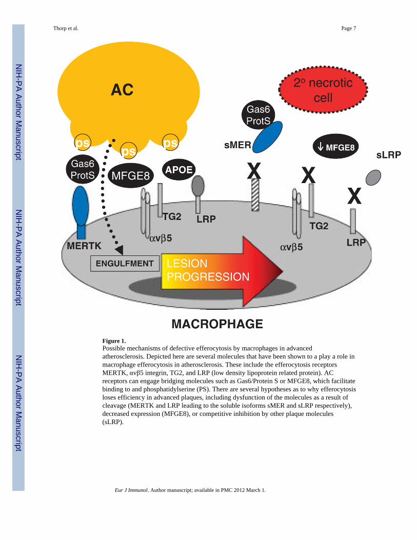
Figure 1.Possible mechanisms of defective efferocytosis by macrophages in advancedatherosclerosis. Depicted here are several molecules that have been shown to a play a role inmacrophage efferocytosis in atherosclerosis. These include the efferocytosis receptorsMERTK, αvβ5 integrin, TG2, and LRP (low density lipoprotein related protein). ACreceptors can engage bridging molecules such as Gas6/Protein S or MFGE8, which facilitatebinding to and phosphatidylserine (PS). There are several hypotheses as to why efferocytosisloses efficiency in advanced plaques, including dysfunction of the molecules as a result ofcleavage (MERTK and LRP leading to the soluble isoforms sMER and sLRP respectively),decreased expression (MFGE8), or competitive inhibition by other plaque molecules(sLRP).
Thorp et al. Page 7
Eur J Immunol. Author manuscript; available in PMC 2012 March 1.
NIH
-PA Author Manuscript
NIH
-PA Author Manuscript
NIH
-PA Author Manuscript
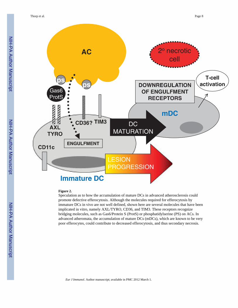
Figure 2.Speculation as to how the accumulation of mature DCs in advanced atherosclerosis couldpromote defective efferocytosis. Although the molecules required for efferocytosis byimmature DCs in vivo are not well defined, shown here are several molecules that have beenimplicated in vitro, namely AXL/TYRO, CD36, and TIM3. These receptors recognizebridging molecules, such as Gas6/Protein S (ProtS) or phosphatidylserine (PS) on ACs. Inadvanced atheromata, the accumulation of mature DCs (mDCs), which are known to be verypoor efferocytes, could contribute to decreased efferocytosis, and thus secondary necrosis.
Thorp et al. Page 8
Eur J Immunol. Author manuscript; available in PMC 2012 March 1.
NIH
-PA Author Manuscript
NIH
-PA Author Manuscript
NIH
-PA Author Manuscript


