Embed Size (px)
Citation preview

Clin Sports Med 25 (2006) 569–590
CLINICS IN SPORTS MEDICINE
The Role of Ultrasound in theEvaluation of Sports Medicine Injuriesof the Upper Extremity
Richard Kijowski, MD*, Arthur A. De Smet, MDDivision of Musculoskeletal Radiology, Department of Radiology, University of WisconsinClinical Science Center, 600 Highland Avenue, E3/311 CSC, Madison, WI 53792-3252, USA
The two major imaging modalities used for assessment of soft tissue injuriesare ultrasound and MRI. Although ultrasound of the extremities is usedonly to a limited extent in the United States, it is widely used in many
other countries for evaluation of extremity injuries.This article first considers the advantages and disadvantages of MRI and ul-
trasound. Understanding these differences will help the reader understand therole of ultrasound as compared with MRI in evaluating upper extremity in-juries. The uses of ultrasound for evaluating sports medicine injuries in specificregions of the upper extremity are then reviewed. Where the data are available,the reported accuracy of ultrasound is compared with MRI for each type ofinjury.
ADVANTAGES AND DISADVANTAGES OF MRIThe most obvious advantage of MRI is its ability to show all tissues of the ex-tremity in multiple planes; thus the examination provides detailed evaluation ofall the soft tissues, the bones, and the joints in a single study (Fig. 1). Althoughthe correct MR pulse sequences must be obtained to optimize detection of pa-thology, the physician assessing the patient can readily determine whether theMRI examination is a complete study that fully delineates the region of interest.
The major disadvantages of MRI are its higher cost and longer examinationtime when compared with ultrasound. Most MRI examinations take 30 to 45minutes to perform, whereas an ultrasound examination takes about 15 min-utes. Charges for MRI and ultrasound examinations vary by region and imag-ing center; however, the Medicare global relative value units are 13.44 for anMRI of the lower extremity without intravenous (IV) contrast and 2.31 for anultrasound of the lower extremity. In addition, MRI cannot be performed inpatients who are claustrophobic or who have certain implanted devices such
*Corresponding author. E-mail address: [email protected] (R. Kijowski).
0278-5919/06/$ – see front matter ª 2006 Elsevier Inc. All rights reserved.doi:10.1016/j.csm.2006.03.004 sportsmed.theclinics.com

570 KIJOWSKI & DE SMET
as cardiac pacemakers. Furthermore, high-quality MRI examinations are diffi-cult to obtain in obese patients and patients who have implanted metallic ortho-pedic hardware.
ADVANTAGES AND DISADVANTAGES OF ULTRASOUNDUltrasound is superior to MRI in that it provides higher resolution imaging ofsuperficial structures such as nerves and tendons (Fig. 2). Ultrasound can alsobe used to dynamically assess for tendon or nerve intermittent subluxation, andto determine the severity of a tendon, ligament, or muscle injury. Dynamic
Fig. 1. Radiographically occult scaphoid fracture. Coronal fat-saturated, T2-weighted, fastspin-echo MR image of the wrist shows a low signal intensity fracture line (arrow) within thewaist of the scaphoid that is surrounded by high signal intensity marrow edema. MRI offerscomplete assessment of all osseous, articular, and soft tissue structures, in contrast with ultra-sound, which provides smaller field of view images primarily limited to the soft tissues.
Fig. 2. Normal volar anatomy of the distal forearm. (A) Longitudinal ultrasound image of thevolar forearm shows the superficialis (arrows) and profunda (arrowheads) flexor tendons. Thetendons have the normal fibrillar pattern consisting of parallel echogenic white lines that rep-resent the tendon fascicles. (B) Transverse ultrasound image of the volar wrist shows two super-ficialis (arrowheads) and two profunda (arrows) flexor tendons. Tendons have the fine meshpatterns of echogenic white dots and lines of normal tendons.
571ULTRASOUND IN UPPER EXTREMITY INJURIES
imaging is particularly useful in evaluating whether a tendon or ligament tear ispartial or complete. It can also be useful in confirming a suspected intermittentfascial or muscle hernia. Ultrasound is also the preferred modality for imagingnon-radiopaque foreign bodies, although this is not a typical sports medicineinjury.
The major disadvantages of ultrasound are its limited field of view and lackof assessment of deep soft tissue structures and bones. Because only a smallarea of the extremity is visualized on a given image, the visualized pathologycannot be easily related to anatomical landmarks. In addition, ultrasound pro-vides only limited evaluation of intra-articular structures. Finally, if the physi-cian assessing the patient had not personally performed the examination, it isoften difficult to determine if an adequate study has been performed.
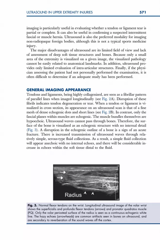
GENERAL IMAGING APPEARANCETendons and ligaments, being highly collagenized, are seen as a fibrillar patternof parallel lines when imaged longitudinally (see Fig. 2A). Disruption of thesefibrils indicates tendon degeneration or tear. When a tendon or ligament is vi-sualized in cross section, its appearance on an ultrasound scan is that of a finemesh of dense echogenic dots and short lines (see Fig. 2B). In contrast, only thefascial planes within muscles are echogenic. The muscle bundles themselves arehypoechoic. Ultrasound waves cannot pass through bones. Therefore, the sur-face of the bone is visualized as an echogenic structure with no internal detail(Fig. 3). A disruption in the echogenic outline of a bone is a sign of an acutefracture. There is increased transmission of ultrasound waves through rela-tively simple, serous-type fluid collections. As a result, a simple fluid collectionwill appear anechoic with no internal echoes, and there will be considerable in-crease in echoes within the soft tissue distal to the fluid.
Fig. 3. Normal flexor tendons on the wrist. Longitudinal ultrasound image of the volar wristshows the superficialis and profunda flexor tendons (arrows) and pronator quadratus muscle(PQ). Only the volar periosteal surface of the radius is seen as a continuous echogenic whiteline. The hazy echoes (arrowheads) are common artifacts seen in bones on ultrasound, andare secondary to reverberation of the sound waves off the cortex.

572 KIJOWSKI & DE SMET
OPTIMAL USES FOR ULTRASOUND EXAMINATION OF SPORTSMEDICINE INJURIESUltrasound is best characterized as a targeted examination to confirm or ex-clude a suspected diagnosis. Many imagers who use ultrasound to assess fortendon or ligament pathology feel that ultrasound provides higher resolutionand greater detail than MRI, especially in the small tendons of the distal ex-tremities. The dynamic real-time nature of ultrasound during provocative ma-neuvers is ideal for assessing subluxing tendons and nerves and intermittentfascia hernias. The major limitation in using ultrasound remains the difficultyin performing the technique. In addition, although ultrasound provides excel-lent delineation of the superficial soft tissues, its accuracy in detecting pathologyhas been documented in only a limited number of conditions.
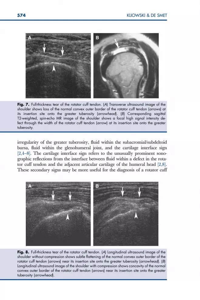
SONOGRAPHIC EVALUATION OF THE SHOULDERRotator Cuff TendonUltrasound is an effective method for evaluating the rotator cuff tendons. Onultrasound, the normal rotator cuff tendons appear as an echogenic fibrillarstructure with a convex outer border that is located between the humeralhead and subacromial/subdeltoid bursa (Fig. 4) [1–3]. The sonographic findingof a partial-thickness rotator cuff tear is a focal hypoechoic area, or more com-monly, a mixed hyperechoic and hypoechoic area within either the articular orbursal side of the tendon (Fig. 5) [1]. The main sonographic finding of a full-thickness rotator cuff tear is a focal anechoic or hypoechoic defect throughthe width of the tendon (Fig. 6). Occasionally, the overlying deltoid muscle,peribursal fat, and hypertrophied synovium within the subacromial bursadrop into the defect, which results in loss of the normal convex outer borderof the rotator cuff tendon (Fig. 7). The loss of the normal convex outer border
Fig. 4. Normal rotator cuff tendon. Longitudinal (A) and transverse (B) ultrasound imagesof the shoulder show the normal rotator cuff tendon (arrows) as an echogenic fibrillar structurewith a convex outer border that attaches to the greater tuberosity of the proximal humerus(arrowhead).

573ULTRASOUND IN UPPER EXTREMITY INJURIES
of the torn rotator tendon may only be visualized when firm pressure is appliedwith the transducer to displace the more superficially located structures fromthe defect within the tendon (Fig. 8) [2,3].
When using ultrasound to assess the integrity of the rotator cuff tendon, it isimportant to evaluate the adjacent structures of the shoulder for secondaryfindings of a rotator cuff tear. These secondary findings include cortical
Fig. 5. Partial-thickness articular surface tear of the rotator cuff tear. (A) Longitudinal ultra-sound image of the shoulder shows a focal hypoechoic area within the articular surface ofthe rotator cuff tendon (arrows) at its insertion site onto the greater tuberosity (arrowhead).(B) Corresponding coronal fat-suppressed, T2-weighted, fast spin-echo MR image of the shoul-der shows a focal area of high signal intensity within the articular surface of the rotator cufftendon (arrow) at its insertion site onto the greater tuberosity.
Fig. 6. Full-thickness tear of the rotator cuff tendon. (A) Longitudinal ultrasound image of theshoulder shows a focal anechoic defect through the width of the rotator cuff tendon (arrows) atits insertion site onto the greater tuberosity (arrowhead). (B) Corresponding coronal fat-sup-pressed, T2-weighted, fast spin-echo MR image of the shoulder shows a focal high signal in-tensity defect through the width of the rotator cuff tendon (arrow) at its insertion site onto thegreater tuberosity.
574 KIJOWSKI & DE SMET
irregularity of the greater tuberosity, fluid within the subacromial/subdeltoidbursa, fluid within the glenohumeral joint, and the cartilage interface sign[2,4–8]. The cartilage interface sign refers to the unusually prominent sono-graphic reflections from the interface between fluid within a defect in the rota-tor cuff tendon and the adjacent articular cartilage of the humeral head [2,8].These secondary signs may be more useful for the diagnosis of a rotator cuff
Fig. 7. Full-thickness tear of the rotator cuff tendon. (A) Transverse ultrasound image of theshoulder shows loss of the normal convex outer border of the rotator cuff tendon (arrows) atits insertion site onto the greater tuberosity (arrowhead). (B) Corresponding sagittalT2-weighted, spin-echo MR image of the shoulder shows a focal high signal intensity de-fect through the width of the rotator cuff tendon (arrow) at its insertion site onto the greatertuberosity.
Fig. 8. Full-thickness tear of the rotator cuff tendon. (A) Longitudinal ultrasound image of theshoulder without compression shows subtle flattening of the normal convex outer border of therotator cuff tendon (arrows) near its insertion site onto the greater tuberosity (arrowhead). (B)Longitudinal ultrasound image of the shoulder with compression shows concavity of the normalconvex outer border of the rotator cuff tendon (arrows) near its insertion site onto the greatertuberosity (arrowhead).

575ULTRASOUND IN UPPER EXTREMITY INJURIES
tear than the primary finding of abnormal echogenicity and loss of the convexouter border of the rotator cuff tendon [9].
Multiple studies have investigated the ability of ultrasound to detect tears ofthe rotator cuff tendons, with a wide range in reported accuracy. Ultrasoundfor cuff tears has a reported sensitivity of 57% to 100% and specificity of76% to 100% for the detection of full-thickness rotator cuff tears [10–19]. Ultra-sound has a reported sensitivity of 41% to 93% and a specificity of 85% to 94%for the detection of partial-thickness rotator cuff tears [1,11,12,19]. The maindifficulties in evaluating the rotator cuff tendon with ultrasound are distinguish-ing partial-thickness, articular surface tears from tendinopathy; and distinguish-ing high-grade, partial-thickness tears from full-thickness tears [20]. With theuse of modern equipment, improved scanning technique, and refined diagnos-tic criteria, however, many centers with experienced musculoskeletal sonogra-phers have reported sensitivities and specificities values exceeding 90% for thedetection of both full-thickness and partial-thickness rotator cuff tears [1,11,18].Ultrasound also has low interobserver variability in the detection and charac-terization of rotator cuff tears [21]. In addition, ultrasound can provide impor-tant information about the location, size, and degree of retraction of the tornrotator cuff tendon [11,13,22].
There has been much recent debate about what is best imaging modality forevaluating the rotator cuff tendon. A retrospective review of the literature [23],which included 38 cohort studies on ultrasound and 29 cohort studies on MRI,concluded that both imaging modalities are equally effective at detecting full-thickness rotator cuff tears. In addition, a recent prospective study on 71 con-secutive patients who had surgically confirmed tears of the rotator cuff tendon[18] found that ultrasound and MRI were equally effective at identifyingand determining the size of both full-thickness and partial-thickness tears.Thus, the decision regarding the most appropriate imaging modality for eval-uating the rotator cuff tendon need not be based upon accuracy concerns. In-stead, the decision should be based on other factors, such as the experience ofthe sonographer, patient tolerance, cost, and the need to evaluate the adjacentstructures of the shoulder.
Ultrasound is faster, cheaper, and more comfortable for the patient thanMRI. Ultrasound can also provide excellent evaluation of the rotator cuff ten-don in obese and claustrophobic patients, and in patients who have certain me-tallic implanted devices; however, MRI provides a more global evaluation ofthe shoulder, and is superior to ultrasound at detecting abnormalities of the os-seous and muscular structures, glenoid labrum, glenohumeral ligaments, andjoint capsule. Perhaps the most important factor when choosing the best imag-ing modality for evaluating the rotator cuff tendon is the experience of the so-nographer [24]. The identification and characterization of rotator cuff tears isone of the most challenging aspects of musculoskeletal ultrasound, and hasa very steep learning curve. For this reason, most general radiologists andeven most fellowship-trained musculoskeletal radiologists are more comfort-able using MRI to evaluate the rotator cuff tendon.

576 KIJOWSKI & DE SMET
Long Head of the Biceps TendonUltrasound may also be used to evaluate the long head of the biceps tendon.The normal biceps tendon is an echogenic fibrillar structure that is visualizedsonographically as it emerges from beneath the acromion and courses distallythrough the glenohumeral joint and into the bicipital groove of the prox-imal humerus (Fig. 9). The sonographic findings of tendinopathy and partial-thickness tear of the biceps tendon are similar, and include enlargement anddecreased echogenicity of the tendon and fluid within the tendon sheath.Hypervascularity of the synovium within the tendon sheath may also be notedon Doppler ultrasound (Fig. 10). The most common sonographic finding ofcomplete rupture of the biceps tendon is nonvisualization of the tendon withinthe bicipital groove; however, a completely ruptured biceps tendon that is at-tached by adhesions within the bicipital sheath may have a normal sonographicappearance within the bicipital groove. Furthermore, nonvisualization of the bi-ceps tendon within the bicipital groove may also be secondary to tendon sub-luxation or dislocation. For this reason, it is important to evaluate the entirelength of the biceps tendon, from where it emerges from beneath the acromionproximally to its musculotendinous junction distally. Displacement of the prox-imal biceps tendon anterior or medial to the lesser tuberosity indicates tendonsubluxation or dislocation. Nonvisualization of the proximal biceps tendon, es-pecially at the entrance to the bicipital groove, indicates complete tendon rup-ture [2,3,11,19].
Two preliminary studies have investigated the ability of ultrasound to eval-uate the long head of the biceps tendon [11,19]. Ultrasound has a sensitivity of63% to 100% and specificity of 100% for the detection of biceps tendon rupture,and a sensitivity of 75% to 83% and specificity of 100% for the detection of bi-ceps tendon subluxation and dislocation; These preliminary studies included
Fig. 9. Normal long head of the biceps tendon. Longitudinal (A) and transverse (B) ultrasoundimages of the shoulder shows the normal biceps tendon as an echogenic fibrillar structure (ar-rows) that is located within the bicipital groove of the proximal humerus. (arrowheads).

577ULTRASOUND IN UPPER EXTREMITY INJURIES
only a small number of patients with surgical correlation, however. Multiplelarge clinical studies with surgical correlation have documented the accuracyof MRI for the detection of biceps tendon rupture and biceps tendon subluxa-tion and dislocation [25,26]. Furthermore, many abnormalities of the bicepstendon involve the proximal intra-articular portion of the tendon, which is lo-cated beneath the acromion and is difficult to visualize on ultrasound [27]. Inview of the lower sensitivity of ultrasound, MRI remains the best imaging mo-dality for evaluating of the long head of the biceps tendon.
Glenohumeral JointUltrasound may be used to evaluate limited portions of the glenoid labrum andhumeral head in patients who have shoulder instability. The normal labrum
Fig. 10. Tendonopathy of the long head of the biceps tendon. (A) Longitudinal ultrasound im-age of the shoulder shows anechoic fluid (large arrow) within the synovial sheath surroundingthe biceps tendon (small arrows) as the tendon passes through the bicipital groove (arrow-head). (B) Longitudinal Doppler ultrasound image of the shoulder shows increased vascularity(large arrow) of the synovial sheath surrounding the biceps tendon (small arrows) as the tendonpasses through the bicipital groove (arrowhead). (C) Corresponding axial fat-suppressed, pro-ton density-weighted, fast spin-echo MR image of the shoulder shows high signal intensity fluid(arrows) surrounding the biceps tendon (arrowhead).
578 KIJOWSKI & DE SMET
appears as a triangular-shaped, homogenously hypoechoic structure that pro-jects off of the glenoid rim. A thin (less than 2 mm) hypoechoic zone may bepresent at the base of the normal labrum [28]. The sonographic findings ofa labral tear include absence or truncation of the labrum, abnormal labral mo-tion during dynamic imaging, and an enlarged (greater than 2 mm) hypoechoiczone at the base of the labrum [29–31]. An associated Hill-Sachs deformity ofthe humeral head may also be identified using ultrasound, and appears asa wedge-shaped defect of the hyperechoic bony contour of the humeralhead, at the site of attachment of the infraspinatus tendon [32–34].
Multiple preliminary studies have investigated the ability of ultrasound toevaluate patients who have shoulder instability [29–34]. Ultrasound has a sen-sitivity of 88% to 95% and specificity of 67% to 100% for the detection of labraltears, and a sensitivity of 91% to 100% and specificity of 89% to 100% for de-tection of Hill-Sachs deformities. These preliminary studies, however, includeda relatively small number of patients with surgical correlation. Furthermore, allultrasound examinations were performed by experienced musculoskeletal so-nographers. Multiple large clinical studies with surgical correlation have docu-mented the accuracy of MRI for the detection of labral tears and associatedHill-Sachs deformities [35–38]. In view of the limited visualization of intra-artic-ular structures, MRI remains the best imaging modality for evaluating patientswho have shoulder instability.
Acromioclavicular JointUltrasound may be used to evaluate the acromioclavicular joint. Ultrasoundmay identify joint space widening, thickening of the joint capsule and adjacentligaments, and intra-articular and subcutaneous hematomas in patients whohave acromioclavicular joint separation [39]. Ultrasound can detect osteo-phytes, subchondral cysts, joint space narrowing, intra-articular fluid, and dys-trophic calcification in patients who have osteoarthritis of the acromioclavicularjoint [40]. Ultrasound may also identify irregular cortical erosions within thedistal clavicle, joint space widening, intra-articular fluid, and soft tissue swellingin patients who have post-traumatic osteolysis of the distal clavicle [41].
Subacromial-Subdeltoid BursaUltrasound may be used to evaluate the subacromial/subdeltoid bursa. Thenormal subacromial-subdeltoid bursa appears as a thin hypoechoic stripe thatis separated from the underlying rotator cuff tendon and overlying deltoid mus-cle by a thin layer of echogenic peribursal fat. Fluid within the subacromial/sub-deltoid bursa tends to accumulate along the lateral edge of the greatertuberosity of the proximal humerus, which produces a typical anechoic ‘‘tear-drop’’ appearance on longitudinal ultrasound images. When fluid is identifiedwithin the subacromial/subdeltoid bursa, it is important to closely evaluate theadjacent rotator cuff tendon for evidence of a full-thickness tear; however, fluidmay also be present within the subacromial/subdeltoid bursa in patients whohave infectious and inflammatory bursitis (Fig. 11), and in patients who haveshoulder impingement syndrome [42]. One important sonographic finding in

579ULTRASOUND IN UPPER EXTREMITY INJURIES
patients who have impingement syndrome is gradual distension of the subacro-mial/subdeltoid bursa and pooling of fluid within the lateral subdeltoid portionof the bursal system with elevation of the arm [43].
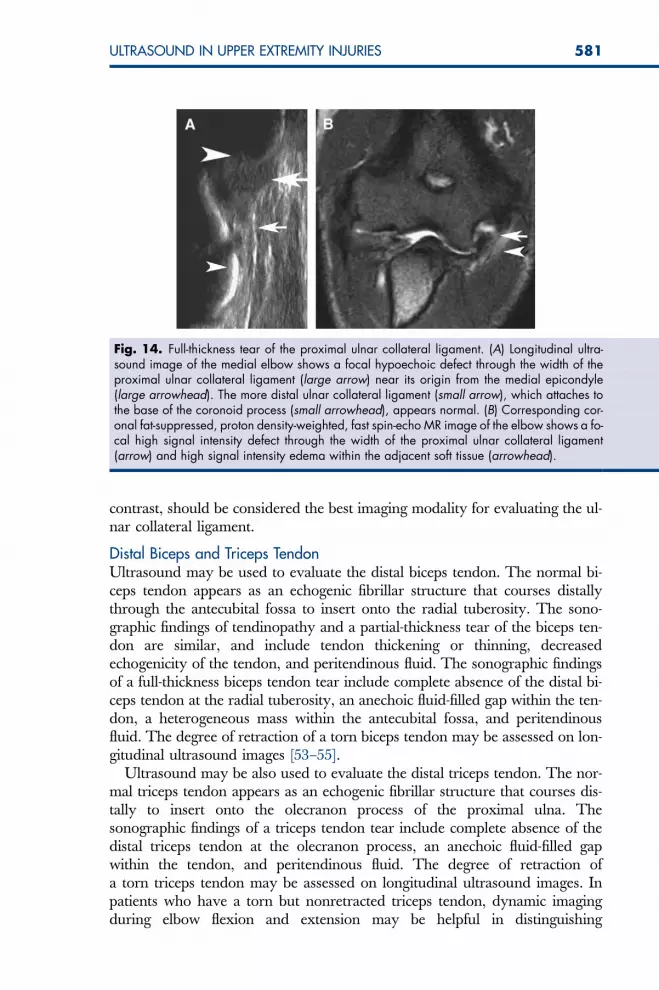
SONOGRAPHIC EVALUATION OF THE ELBOWUlnar Collateral LigamentUltrasound may be used to evaluate the ulnar collateral ligament. The normalanterior bundle of the ulnar collateral ligament appears as an echogenic fibrillarstructure that arises from the medial epicondyle and courses distally to insertonto the base of the coronoid process of the proximal ulna (Fig. 12) [44]. Ultra-sound may be useful at identifying acute partial-thickness and full-thicknesstears of the ulnar collateral ligament, and chronic ligamentous injuries second-ary to repetitive microtrauma. The sonographic findings of an acute partial-thickness tear of the ulnar collateral ligament include diffuse thickening anddecreased echogenicity of the ligament, and focal hypoechoic areas withinthe ligament (Fig. 13) [44,45]. The sonographic findings of an acute full-thick-ness tear of the ulnar collateral ligament include an anechoic or hypoechoicfluid-filled gap within the ligament, and nonvisualization of the ligament(Fig. 14) [44,45]. The sonographic findings of chronic injury to the ulnar collat-eral ligament secondary to repetitive microtrauma include diffuse thickening ofthe ligament, intraligamentous calcification, and focal hypoechoic areas withinthe ligament [46]. In patients who have both acute and chronic ulnar collateralligament injuries, dynamic imaging may demonstrate ligamentous laxity withasymmetric widening of the medial joint space with valgus stress [46–48].
Fig. 11. Septic subacromial/subdeltoid bursitis. (A) Longitudinal ultrasound image of theshoulder shows anechoic fluid within the subacromial/subdeltoid bursa (small arrows). Notethe overlying deltoid muscle (arrowheads) and the underlying rotator cuff tendon (large arrow).(B) Corresponding coronal post-contrast, fat-suppressed, T1-weighted, spin-echo MR image ofthe shoulder shows low signal intensity fluid within the subacromial/subdeltoid bursa (small ar-rows). Note the high signal intensity enhancing synovium (arrowheads) within the subacromialbursa, and the underlying low signal intensity rotator cuff tendon (large arrow).

580 KIJOWSKI & DE SMET
Clinical studies with surgical correlation that document the accuracy of ultra-sound for the detection of ulnar collateral ligament tears have yet to be per-formed. In contrast, multiple clinical studies with surgical correlation haveshown that MRI can accurately detect full-thickness and partial-thickness tearsof the ulnar collateral ligament [49–52]. Until studies are reported with surgicalcorrelation of sonographic findings, MRI, especially with intra-articular
Fig. 12. Normal ulnar collateral ligament. Longitudinal ultrasound image of the medial elbowshows the normal ulnar collateral ligament as an echogenic fibrillar structure (large arrows)coursing between the medial epicondyle of the distal humerus (small arrow) and the base ofthe coronoid process of the proximal ulna (arrowhead).
Fig. 13. Partial-thickness tear of the proximal ulnar collateral ligament. (A) Longitudinal ultra-sound image of the medial elbow shows a focal hypoechoic area within the superficial surfaceof the proximal ulnar collateral ligament (large arrow) near its origin from the medial epicon-dyle (large arrowhead). The more distal ulnar collateral ligament (small arrows), which at-taches to the base of the coronoid process (small arrowhead), appears normal. (B)Corresponding coronal fat-suppressed, T2-weighted, fast spin-echo MR image of the elbowshows diffuse high signal intensity within the intact proximal ulnar collateral ligament (arrow)and high signal intensity edema within the adjacent soft tissue (arrowhead).
581ULTRASOUND IN UPPER EXTREMITY INJURIES
contrast, should be considered the best imaging modality for evaluating the ul-nar collateral ligament.
Distal Biceps and Triceps TendonUltrasound may be used to evaluate the distal biceps tendon. The normal bi-ceps tendon appears as an echogenic fibrillar structure that courses distallythrough the antecubital fossa to insert onto the radial tuberosity. The sono-graphic findings of tendinopathy and a partial-thickness tear of the biceps ten-don are similar, and include tendon thickening or thinning, decreasedechogenicity of the tendon, and peritendinous fluid. The sonographic findingsof a full-thickness biceps tendon tear include complete absence of the distal bi-ceps tendon at the radial tuberosity, an anechoic fluid-filled gap within the ten-don, a heterogeneous mass within the antecubital fossa, and peritendinousfluid. The degree of retraction of a torn biceps tendon may be assessed on lon-gitudinal ultrasound images [53–55].
Ultrasound may be also used to evaluate the distal triceps tendon. The nor-mal triceps tendon appears as an echogenic fibrillar structure that courses dis-tally to insert onto the olecranon process of the proximal ulna. Thesonographic findings of a triceps tendon tear include complete absence of thedistal triceps tendon at the olecranon process, an anechoic fluid-filled gapwithin the tendon, and peritendinous fluid. The degree of retraction ofa torn triceps tendon may be assessed on longitudinal ultrasound images. Inpatients who have a torn but nonretracted triceps tendon, dynamic imagingduring elbow flexion and extension may be helpful in distinguishing
Fig. 14. Full-thickness tear of the proximal ulnar collateral ligament. (A) Longitudinal ultra-sound image of the medial elbow shows a focal hypoechoic defect through the width of theproximal ulnar collateral ligament (large arrow) near its origin from the medial epicondyle(large arrowhead). The more distal ulnar collateral ligament (small arrow), which attaches tothe base of the coronoid process (small arrowhead), appears normal. (B) Corresponding cor-onal fat-suppressed, proton density-weighted, fast spin-echo MR image of the elbow shows a fo-cal high signal intensity defect through the width of the proximal ulnar collateral ligament(arrow) and high signal intensity edema within the adjacent soft tissue (arrowhead).

582 KIJOWSKI & DE SMET
a partial-thickness tear, in which residual fibers remain attached to the olecra-non process, from a full-thickness tear [56,57].
Clinical studies with surgical correlation that document the accuracy of ultra-sound for the detection of biceps and triceps tendon tears have yet to be per-formed. Multiple clinical studies with surgical correlation have shown thatMRI can accurately detect full-thickness and partial-thickness tears of the bicepstendon and triceps tendon [58–62]. Until studies are reported with surgical cor-relation of sonographic findings, MRI, should be considered the best imagingmodality for evaluating patients who have distal biceps and triceps tendoninjuries.
Elbow-Common Extensor TendonUltrasound may be used to evaluate the common extensor tendon. The normalcommon extensor tendon appears as an echogenic fibrillar structure that arisesfrom the lateral epicondyle and courses distally to blend in with the extensorcarpi radialis brevis and extensor digitorum muscles (Fig. 15). The sonographicfindings of lateral epicondylitis include tendon thickening, intratendinous calci-fication, diffuse heterogeneous echotexture of the tendon with loss of the nor-mal fibrillar pattern, focal hypoechoic areas within the tendon, peritendinousedema, and cortical irregularity of the lateral epicondyle. Anechoic areas withinthe common extensor tendon may be seen in patients who have severe lateralepicondylitis, and are thought to represent partial-thickness tendon tears(Fig. 16) [63–65].
Many large clinical studies have documented the usefulness of ultrasoundfor evaluating patients who have lateral epicondylitis. Ultrasound can confirmthe presence of lateral epicondylitis and can provide information about the se-verity of the disease process. Ultrasound has a sensitivity of 64% to 88% for thedetection of lateral epicondylitis; however, the main disadvantage of using
Fig. 15. Normal common extensor tendon. Longitudinal ultrasound image of the lateral el-bow shows the normal common extensor tendon as an echogenic fibrillar structure (arrows)originating from the lateral epicondyle of the distal humerus (arrowhead).

583ULTRASOUND IN UPPER EXTREMITY INJURIES
ultrasound for the diagnosis of patients who have lateral epicondylitis is itspoor specificity, which is as low as 36% in some studies [63–65]. Multiple clin-ical studies with surgical correlation have shown that MRI can be accuratelyused to diagnose patients who have lateral epicondylitis [66–68]. Furthermore,MRI was found to be more sensitive and specific than ultrasound for the detec-tion of lateral epicondylitis in a prospective study comparing both imaging mo-dalities [65]. In light of the reported lower sensitivity and specificity ofultrasound, MRI remains the best imaging modality for evaluating the com-mon extensor tendon in patients who have lateral elbow pain.
SONOGRAPHIC EVALUATION OF THE WRIST AND HANDLigaments of the Wrist and HandUltrasound may be used to evaluate the intrinsic ligaments of the wrist. Thescapholunate and lunotriquetral ligaments are well-visualized on ultrasoundand appear as echogenic fibrillar structures connecting the proximal carpalbones [69,70]. The sonographic findings of a torn scapholunate or lunotrique-tral ligament include loss of the normal echogenic appearance of the ligament,disruption or absence of the ligament, or the presence of fluid interposed be-tween the proximal carpal bones [56,69]. In a small preliminary study using ar-thrography as the reference standard [56], ultrasound was found to havea sensitivity of 100% for the detection of scapholunate ligament tears, and a sen-sitivity of 25% for the detection of lunotriquetral ligament tears. Although thispreliminary study is encouraging, MRI remains the imaging modality of choicefor evaluating the intrinsic ligaments of the wrist [71–73].
Ultrasound may be used to evaluate the ulnar collateral ligament of thethumb. The normal ulnar collateral ligament appears as homogenous,
Fig. 16. Lateral epicondylitis. (A) Longitudinal ultrasound image of the lateral elbow showsa focal hypoechoic area within the common extensor tendon (arrows), near its origin fromthe lateral epicondyle (arrowhead). (B) Corresponding coronal fat-suppressed, proton den-sity-weighted, fast spin-echo MR image of the elbow shows diffuse high signal intensity withina thickened common extensor tendon (arrow).

584 KIJOWSKI & DE SMET
hypoechoic, bandlike structure extending from the metacarpal head to the baseof the proximal phalanx. The sonographic findings of a partial-thickness tearand a nondisplaced, full-thickness tear of the ulnar collateral ligament are iden-tical, and include thickening and heterogeneously increased echogenicity of theligament. The sonographic findings of a displaced, full-thickness tear of the ul-nar collateral ligament include nonvisualization of the ligament and identifica-tion of a hyperechoic lesion (Stener’s lesion), with a surrounding hypoechoichalo lying on the dorsal aspect of the adductor pollicis muscle [74–77].
Multiple clinical studies with surgical correlation have documented the abil-ity of both ultrasound and MRI to identify full-thickness and partial-thicknesstears of the ulnar collateral ligament of the thumb, and to distinguish betweendisplaced and nondisplaced full-thickness tears [75–84]. In a prospective studycomparing both imaging modalities [78], however, MRI was found to be moresensitive and specific than ultrasound for differentiating displaced and nondis-placed full-thickness ligament tears. Because ultrasound is less expensive thanMRI, it may be considered as a good alternative to MRI at institutions withhighly experienced musculoskeletal sonographers.
Triangular Fibrocartilage ComplexUltrasound may be used to evaluate the triangular fibrocartilage complex of thewrist. The normal triangular fibrocartilage complex appears as a homogenousechogenic, triangular structure that is located distal to the ulna and deep to theextensor carpi ulnaris tendon. The sonographic findings of a tear of the tri-angular fibrocartilage complex include loss of the normal homogenously echo-genic appearance and triangular shape of the structure, absence of portion ofthe structure, and focal or linear areas of decreased echogenicity within thestructure. In a small preliminary study using arthrography as the referencestandard [56], ultrasound was found to have 64% sensitivity for the detectionof tears of the triangular fibrocartilage complex. Although this preliminarystudy is encouraging, MRI remains the imaging modality of choice for evalu-ating of the triangular fibrocartilage complex of the wrist [85–87].
Tendons of the Wrist and HandUltrasound may be used to evaluate the flexor and extensor tendons of thewrist and hand. The normal flexor and extensor tendons appear as homoge-nous echogenic, fibrillar structures that are surrounded by a thin, barely dis-cernible hypoechoic synovial sheath (Fig. 17). The sonographic findings oftendinopathy include tendon thickening, diffuse or focal areas of decreasedechogenicity within the tendon, and anechoic fluid within the tendon sheath.The sonographic findings of partial-thickness tendon tear include a focal an-echoic or hypoechoic cleft within the tendon, and anechoic fluid within the ten-don sheath (Fig. 18). The sonographic findings of a full-thickness tendon tearinclude nonvisualization of the tendon at the site of rupture, and anechoic fluidor heterogeneous hyperechoic hematoma within the tendon sheath that sepa-rates the retracted edges of the torn tendon [88–90].

585ULTRASOUND IN UPPER EXTREMITY INJURIES
Clinical studies with surgical correlation that document the accuracy of ultra-sound for the detection of flexor and extensor tendon injuries have yet to beperformed. Multiple clinical studies with surgical correlation have shown thatMRI can accurately detect acute and chronic injuries of the flexor and extensortendons [91–96]. Until studies are reported with surgical correlation of sono-graphic findings, MRI should be considered the best imaging modality for eval-uating patients who have abnormalities of the flexor and extensor tendons ofthe wrist and hand.
Fig. 18. Tear of the extensor carpi ulnaris tendon. (A) Transverse ultrasound image of the ul-nar aspect of the wrist shows a focal hypoechoic defect (large arrow) within the extensor carpiulnaris tendon (small arrows) that is located within the ulnar styloid groove (arrowheads). (B)Corresponding axial fat-suppressed, T2-weighted, fast spin-echo MR image of the wrist showsan intermediate signal intensity defect (arrow) splitting the extensor carpi ulnaris tendon intotwo separate low signal intensity fragments (arrowheads).
Fig. 17. Normal extensor carpi ulnaris tendon. Transverse ultrasound image of the ulnaraspect of the wrist shows the normal extensor carpi ulnaris tendon (arrows) as an echogenicfibrillar structure that is located within the ulnar styloid groove (arrowhead).

586 KIJOWSKI & DE SMET
SUMMARYUltrasound examination of the extremities is a rapidly growing area for theevaluation of sports medicine injuries. It is likely that in coming years, furtherstudies with surgical correlation will document the accuracy of ultrasound. Theauthors suspect that MRI will be found to be the most accurate method for theassessment of some injuries, whereas ultrasound will have an accuracy compa-rable to MRI for the assessment of many other injuries, as has been docu-mented for rotator cuff tears. The most important point to remember whenconsidering whether to use ultrasound, however, is that it is best used as a fo-cused examination for specific conditions. The skill of the sonographer will al-ways be important in view of the steep learning curve in examining many typesof sports medicine injuries.
References[1] van Holsbeeck MT, Kolowich PA, Eyler WR, et al. US depiction of partial-thickness tear of the
rotator cuff. Radiology 1995;197(2):443–6.[2] Teefey SA, Middleton WD, Yamaguchi K. Shoulder sonography. State of the art. Radiol Clin
North Am 1999;37(4):767–85.[3] Bianchi S, Martinoli C, Abdelwahab IF. Ultrasound of tendon tears. Part 1: general consid-
erations and upper extremity. Skeletal Radiol 2005;34:500–12.[4] Wohlwend JR, van Holsbeeck M, Craig J, et al. The association between irregular greater
tuberosities and rotator cuff tears: a sonographic study. AJR Am J Roentgenol 1998;171(1):229–33.
[5] Huang LF, Rubin DA, Britton CA. Greater tuberosity changes as revealed by radiography:lack of clinical usefulness in patients with rotator cuff disease. AJR Am J Roentgenol1999;172(5):1381–8.
[6] Hollister MS, Mack LA, Patten RM, et al. Association of sonographically detected subacro-mial/subdeltoid bursal effusion and intraarticular fluid with rotator cuff tear. AJR Am J Roent-genol 1995;165(3):605–8.
[7] Arslan G, Apaydin A, Kabaalioglu A, et al. Sonographically detected subacromial/subdel-toid bursal effusion and biceps tendon sheath fluid: reliable signs of rotator cuff tear? J ClinUltrasound 1999;27(6):335–9.
[8] van Holsbeeck M, Introcaso JH, Kolowich PA. Sonography of tendons: patterns of disease.Instr Course Lect 1994;43:475–81.
[9] Jacobson JA, Lancaster S, Prasad A, et al. Full-thickness and partial-thickness supraspinatustendon tears: value of US signs in diagnosis. Radiology 2004;230(1):234–42.
[10] Crass JR, Craig EV, Feinberg SB. Ultrasonography of rotator cuff tears: a review of 500 di-agnostic studies. J Clin Ultrasound 1988;16(5):313–27.
[11] Teefey SA, Hasan SA, Middleton WD, et al. Ultrasonography of the rotator cuff. A compar-ison of ultrasonographic and arthroscopic findings in one hundred consecutive cases.J Bone Joint Surg Am 2000;82(4):498–504.
[12] Brenneke SL, Morgan CJ. Evaluation of ultrasonography as a diagnostic technique in theassessment of rotator cuff tendon tears. Am J Sports Med 1992;20(3):287–9.
[13] Wiener SN, Seitz WH Jr. Sonography of the shoulder in patients with tears of the rotatorcuff: accuracy and value for selecting surgical options. AJR Am J Roentgenol1993;160(1):103–7.
[14] Paavolainen P, Ahovuo J. Ultrasonography and arthrography in the diagnosis of tears of therotator cuff. J Bone Joint Surg Am 1994;76(3):335–40.
[15] Hodler J, Fretz CJ, Terrier F, et al. Rotator cuff tears: correlation of sonographic and surgicalfindings. Radiology 1988;169(3):791–4.
587ULTRASOUND IN UPPER EXTREMITY INJURIES
[16] Mack LA, Matsen FA 3rd, Kilcoyne RF, et al. US evaluation of the rotator cuff. Radiology1985;157(1):205–9.
[17] Brandt TD, Cardone BW, Grant TH, et al. Rotator cuff sonography: a reassessment. Radiol-ogy 1989;173(2):323–7.
[18] Teefey SA, Rubin DA, Middleton WD, et al. Detection and quantification of rotator cuff tears.Comparison of ultrasonographic, magnetic resonance imaging, and arthroscopic findingsin seventy-one consecutive cases. J Bone Joint Surg Am 2004;86-A(4):708–16.
[19] Read JW, Perko M. Shoulder ultrasound: diagnostic accuracy for impingement syn-drome, rotator cuff tear, and biceps tendon pathology. J Shoulder Elbow Surg1998;7(3):264–71.
[20] Teefey SA, Middleton WD, Payne WT, et al. Detection and measurement of rotator cuff tearswith sonography: analysis of diagnostic errors. AJR Am J Roentgenol 2005;184(6):1768–73.
[21] Middleton WD, Teefey SA, Yamaguchi K. Sonography of the rotator cuff: analysis of inter-observer variability. AJR Am J Roentgenol 2004;183(5):1465–8.
[22] Kluger R, Mayrhofer R, Kroner A, et al. Sonographic versus magnetic resonance arthro-graphic evaluation of full-thickness rotator cuff tears in millimeters. J Shoulder Elbow Surg2003;12(2):110–6.
[23] Dinnes J, Loveman E, McIntyre L, et al. The effectiveness of diagnostic tests for the assessmentof shoulder pain due to soft tissue disorders: a systematic review. Health Technol Assess2003;7(29):iii.
[24] Chang CY, Wang SF, Chiou HJ, et al. Comparison of shoulder ultrasound and MR imagingin diagnosing full-thickness rotator cuff tears. Clin Imaging 2002;26(1):50–4.
[25] Beall DP, Williamson EE, Ly JQ, et al. Association of biceps tendon tears with rotator cuff ab-normalities: degree of correlation with tears of the anterior and superior portions of the ro-tator cuff. AJR Am J Roentgenol 2003;180(3):633–9.
[26] Mohtadi NG, Vellet AD, Clark ML, et al. A prospective, double-blind comparison of mag-netic resonance imaging and arthroscopy in the evaluation of patients presenting with shoul-der pain. J Shoulder Elbow Surg 2004;13(3):258–65.
[27] Zanetti M, Pfirrmann CW. [Biceps tendon disorders: ultrasound, MR imaging and MR ar-thrography]. Radiologe 2004;44(6):591–6. [in German].
[28] Schydlowsky P, Strandberg C, Galatius S, et al. Ultrasonographic examination of the gle-noid labrum of healthy volunteers. Eur J Ultrasound 1998;8(2):85–9.
[29] Schydlowsky P, Strandberg C, Galbo H, et al. The value of ultrasonography in the diagnosisof labral lesions in patients with anterior shoulder dislocation. Eur J Ultrasound 1998;8(2):107–13.
[30] Hammar MV, Wintzell GB, Astrom KG, et al. Role of US in the preoperative evaluation ofpatients with anterior shoulder instability. Radiology 2001;219(1):29–34.
[31] Taljanovic MS, Carlson KL, Kuhn JE, et al. Sonography of the glenoid labrum: a cadavericstudy with arthroscopic correlation. AJR Am J Roentgenol 2000;174(6):1717–22.
[32] Farin PU, Kaukanen E, Jaroma H, et al. Hill-Sachs lesion: sonographic detection. SkeletalRadiol 1996;25(6):559–62.
[33] Pancione L, Gatti G, Mecozzi B. Diagnosis of Hill-Sachs lesion of the shoulder. Comparisonbetween ultrasonography and arthro-CT. Acta Radiol 1997;38:523–6.
[34] Cicak N, Bilic R, Delimar D. Hill-Sachs lesion in recurrent shoulder dislocation: sonographicdetection. J Ultrasound Med 1998;17(9):557–60.
[35] Flannigan B, Kursunoglu-Brahme S, Snyder S, et al. MR arthrography of the shoulder: com-parison with conventional MR imaging. AJR Am J Roentgenol 1990;155(4):829–32.
[36] Palmer WE, Caslowitz PL. Anterior shoulder instability: diagnostic criteria determined fromprospective analysis of 121 MR arthrograms. Radiology 1995;197(3):819–25.
[37] Chandnani VP, Yeager TD, DeBerardino T, et al. Glenoid labral tears: prospective evalua-tion with MRI imaging, MR arthrography, and CT arthrography. AJR Am J Roentgenol1993;161(6):1229–35.

588 KIJOWSKI & DE SMET
[38] Chandnani VP, Gagliardi JA, Murnane TG, et al. Glenohumeral ligaments and shouldercapsular mechanism: evaluation with MR arthrography. Radiology 1995;196(1):27–32.
[39] Martinoli C, Bianchi S, Prato N, et al. US of the shoulder: non-rotator cuff disorders. Radio-graphics 2003;23(2):381–401.
[40] Blankstein A, Ganel A, Givon U, et al. Ultrasonography as a diagnostic modality in acro-mioclavicular joint pathologies. Isr Med Assoc J 2005;7(1):28–30.
[41] Yu YS, Dardani M, Fischer RA. MR observations of postraumatic osteolysis of the distal clav-icle after traumatic separation of the acromioclavicular joint. J Comput Assist Tomogr2000;24(1):159–64.
[42] van Holsbeeck M, Strouse PJ. Sonography of the shoulder: evaluation of the subacromial-subdeltoid bursa. AJR Am J Roentgenol 1993;160(3):561–4.
[43] Farin PU, Jaroma H, Harju A, et al. Shoulder impingement syndrome: sonographic evalua-tion. Radiology 1990;176(3):845–9.
[44] Jacobson JA, Propeck T, Jamadar DA, et al. US of the anterior bundle of the ulnar col-lateral ligament: findings in five cadaver elbows with MR arthrographic and anatomic com-parison—initial observations. Radiology 2003;227(2):561–6.
[45] Miller TT, Adler RS, Friedman L. Sonography of injury of the ulnar collateral ligament of theelbow-initial experience. Skeletal Radiol 2004;33(7):386–91.
[46] Nazarian LN, McShane JM, Ciccotti MG, et al. Dynamic US of the anterior band of the ulnarcollateral ligament of the elbow in asymptomatic major league baseball pitchers. Radiology2003;227(1):149–54.
[47] De Smet AA, Winter TC, Best TM, et al. Dynamic sonography with valgus stress to assesselbow ulnar collateral ligament injury in baseball pitchers. Skeletal Radiol 2002;31(11):671–6.
[48] Sasaki J, Takahara M, Ogino T, et al. Ultrasonographic assessment of the ulnar collateralligament and medial elbow laxity in college baseball players. J Bone Joint Surg Am2002;84-A(4):525–31.
[49] Nakanishi K, Masatomi T, Ochi T, et al. MR arthrography of elbow: evaluation of the ulnarcollateral ligament of elbow. Skeletal Radiol 1996;25(7):629–34.
[50] Mirowitz SA, London SL. Ulnar collateral ligament injury in baseball pitchers: MR imagingevaluation. Radiology 1992;185(2):573–6.
[51] Timmerman LA, Schwartz ML, Andrews JR. Preoperative evaluation of the ulnar collateralligament by magnetic resonance imaging and computed tomography arthrography. Evalu-ation in 25 baseball players with surgical confirmation. Am J Sports Med 1994;22(1):26–31. [discussion: 32].
[52] Schwartz ML, al-Zahrani S, Morwessel RM, et al. Ulnar collateral ligament injury in thethrowing athlete: evaluation with saline-enhanced MR arthrography. Radiology 1995;197(1):297–9.
[53] Belli P, Costantini M, Mirk P, et al. Sonographic diagnosis of distal biceps tendon rupture:a prospective study of 25 cases. J Ultrasound Med 2001;20(6):587–95.
[54] Miller TT, Adler RS. Sonography of tears of the distal biceps tendon. AJR Am J Roentgenol2000;175(4):1081–6.
[55] Finlay K, Ferri M, Friedman L. Ultrasound of the elbow. Skeletal Radiol 2004;33(2):63–79.[56] Finlay K, Lee R, Friedman L. Ultrasound of intrinsic wrist ligament and triangular fibrocarti-
lage injuries. Skeletal Radiol 2004;33(2):85–90.[57] Kaempffe FA, Lerner RM. Ultrasound diagnosis of triceps tendon rupture. A report of 2 cases.
Clin Orthop Relat Res 1996;332:138–42.[58] Gaines ST, Durbin RA, Marsalka DS. The use of magnetic resonance imaging in the diagno-
sis of triceps tendon ruptures. Contemp Orthop 1990;20(6):607–11.[59] Tiger E, Mayer DP, Glazer R. Complete avulsion of the triceps tendon: MRI diagnosis. Com-
put Med Imaging Graph 1993;17(1):51–4.[60] Williams BD, Schweitzer ME, Weishaupt D, et al. Partial tears of the distal biceps tendon:
MR appearance and associated clinical findings. Skeletal Radiol 2001;30(10):560–4.

589ULTRASOUND IN UPPER EXTREMITY INJURIES
[61] Fitzgerald SW, Curry DR, Erickson SJ, et al. Distal biceps tendon injury: MR imaging diag-nosis. Radiology 1994;191(1):203–6.
[62] Falchook FS, Zlatkin MB, Erbacher GE, et al. Rupture of the distal biceps tendon: evaluationwith MR imaging. Radiology 1994;190(3):659–63.
[63] Connell D, Burke F, Coombes P, et al. Sonographic examination of lateral epicondylitis. AJRAm J Roentgenol 2001;176(3):777–82.
[64] Levin D, Nazarian LN, Miller TT, et al. Lateral epicondylitis of the elbow: US findings. Radi-ology 2005;237(1):230–4.
[65] Miller TT, Shapiro MA, Schultz E, et al. Comparison of sonography and MRI for diagnosingepicondylitis. J Clin Ultrasound 2002;30(4):193–202.
[66] Martin CE, Schweitzer ME. MR imaging of epicondylitis. Skeletal Radiol 1998;27(3):133–8.
[67] Aoki M, Wada T, Isogai S, et al. Magnetic resonance imaging findings of refractory tenniselbows and their relationship to surgical treatment. J Shoulder Elbow Surg 2005;14(2):172–7.
[68] Potter HG, Hannafin JA, Morwessel RM, et al. Lateral epicondylitis: correlation of MR imag-ing, surgical, and histopathologic findings. Radiology 1995;196(1):43–6.
[69] Jacobson JA, Oh E, Propeck T, et al. Sonography of the scapholunate ligament in four cadav-eric wrists: correlation with MR arthrography and anatomy. [see comment]. AJR Am J Roent-genol 2002;179(2):523–7.
[70] Boutry N, Lapeque F, Masi L, et al. Ultrasonographic evaluation of normal extrinsic and in-trinsic carpal ligaments: preliminary experience. Skeletal Radiol 2005;34:513–21.
[71] Scheck RJ, Kubitzek C, Hierner R, et al. The scapholunate interosseous ligament in MR ar-thrography of the wrist: correlation with non-enhanced MRI and wrist arthroscopy. SkeletalRadiol 1997;26(5):263–71.
[72] Schweitzer ME, Brahme SK, Hodler J, et al. Chronic wrist pain: spin-echo and short tau in-version recovery MR imaging and conventional and MR arthrography [erratum appears inRadiology 1992;184(2):583]. Radiology 1992;182(1):205–11.
[73] Totterman SM, Miller R, Wasserman B, et al. Intrinsic and extrinsic carpal ligaments: evalu-ation by three-dimensional Fourier transform MR imaging. AJR Am J Roentgenol1993;160(1):117–23.
[74] Hergan K, Mittler C. Sonography of the injured ulnar collateral ligament of the thumb. J BoneJoint Surg Br 1995;77(1):77–83.
[75] Bronstein AJ, Koniuch MP, von Holsbeeck M. Ultrasonographic detection of thumb ul-nar collateral ligament injuries: a cadaveric study. J Hand Surg [Am] 1994;19(2):304–12.
[76] O’Callaghan BI, Kohut G, Hoogewoud HM. Gamekeeper thumb: identification of theStener lesion with US. Radiology 1994;192(2):477–80.
[77] Noszian IM, Dinkhauser LM, Orthner E, et al. Ulnar collateral ligament: differentiation ofdisplaced and nondisplaced tears with US. Radiology 1995;194(1):61–3.
[78] Hergan K, Mittler C, Oser W. Ulnar collateral ligament: differentiation of displaced andnondisplaced tears with US and MR imaging. Radiology 1995;194(1):65–71.
[79] Hinke DH, Erickson SJ, Chamoy L, et al. Ulnar collateral ligament of the thumb: MR findingsin cadavers, volunteers, and patients with ligamentous injury (gamekeeper’s thumb). AJRAm J Roentgenol 1994;163(6):1431–4.
[80] Harper MT, Chandnani VP, Spaeth J, et al. Gamekeeper thumb: diagnosis of ulnar collateralligament injury using magnetic resonance imaging, magnetic resonance arthrography andstress radiography. J Magn Reson Imaging 1996;6(2):322–8.
[81] Ahn JM, Sartoris DJ, Kang HS, et al. Gamekeeper thumb: comparison of MR arthrographywith conventional arthrography and MR imaging in cadavers. Radiology 1998;206(3):737–44.
[82] Plancher KD, Ho CP, Cofield SS, et al. Role of MR imaging in the management of ‘‘skier’sthumb’’ injuries. Magn Reson Imaging Clin N Am 1999;7(1):73–84.

590 KIJOWSKI & DE SMET
[83] Lohman M, Vasenius J, Kivisaari A, et al. MR imaging in chronic rupture of the ulnar collat-eral ligament of the thumb. Acta Radiol 2001;42(1):10–4.
[84] Romano WM, Garvin G, Bhayana D, et al. The spectrum of ulnar collateral ligament injuriesas viewed on magnetic resonance imaging of the metacarpophalangeal joint of the thumb.Can Assoc Radiol J 2003;54(4):243–8.
[85] Potter HG, Asnis-Ernberg L, Weiland AJ, et al. The utility of high-resolution magnetic reso-nance imaging in the evaluation of the triangular fibrocartilage complex of the wrist.J Bone Joint Surg Am 1997;79(11):1675–84.
[86] Haims AH, Schweitzer ME, Morrison WB, et al. Internal derangement of the wrist: indirectMR arthrography versus unenhanced MR imaging. Radiology 2003;227(3):701–7.
[87] Haims AH, Schweitzer ME, Morrison WB, et al. Limitations of MR imaging in the diagnosisof peripheral tears of the triangular fibrocartilage of the wrist. AJR Am J Roentgenol2002;178(2):419–22.
[88] Daenen B, Houben G, Bauduin E, et al. Sonography in wrist tendon pathology. J Clin Ultra-sound 2004;32(9):462–9.
[89] Teefey SA, Middleton WD, Boyer MI. Sonography of the hand and wrist. Semin UltrasoundCT MR 2000;21(3):192–204.
[90] Bianchi S, Martinoli C, Abdelwahab IF. High-frequency ultrasound examination of the wristand hand. Skeletal Radiol 1999;28(3):121–9.
[91] Drape JL, Tardif-Chastenet de Gery S, Silbermann-Hoffman O, et al. Closed ruptures of theflexor digitorum tendons: MRI evaluation. Skeletal Radiol 1998;27(11):617–24.
[92] Scott JR, Cobby M, Taggart I. Magnetic resonance imaging of acute tendon injury in the fin-ger. J Hand Surg [Br] 1995;20(3):286–8.
[93] Rubin DA, Kneeland JB, Kitay GS, et al. Flexor tendon tears in the hand: use of MR imagingto diagnose degree of injury in a cadaver model. AJR Am J Roentgenol 1996;166(3):615–20.
[94] Drape JL, Dubert T, Silbermann O, et al. Acute trauma of the extensor hood of the metacar-pophalangeal joint: MR imaging evaluation. Radiology 1994;192(2):469–76.
[95] Valeri G, Ferrara C, Ercolani P, et al. Tendon involvement in rheumatoid arthritis of the wrist:MRI findings. Skeletal Radiol 2001;30(3):138–43.
[96] Glajchen N, Schweitzer M. MRI features in de Quervain’s tenosynovitis of the wrist. SkeletalRadiol 1996;25(1):63–5.

![TOPICS IN INTEGRATIVE HEALTH CARE [ISSN 2158-4222] – VOL 2(1) Health Promotion Advocacy: A Practitioner's Role in Prevention of Sports Injuries](https://img.pdfslide.net/doc/110x75/635abdca4028fc5adf015b04/topics-in-integrative-health-care-issn-2158-4222-vol-21-health-promotion.jpg)


