Embed Size (px)
Citation preview

Review
The TEM characterization of the lamellar structure
of osteoporotic human trabecular bone
Matthew Aaron Rubin, Iwona Jasiuk*
Department of Mechanical and Industrial Engineering, Concordia University, 1455 de Maisonneuve Blvd. West, Montreal, Que., Canada H3G 1M8
Received 14 June 2005; revised 18 July 2005; accepted 25 July 2005
Abstract
The lamellar structure of osteoporotic human trabecular bone was characterized experimentally by means of transmission electron
microscopy (TEM). More specifically, the TEM was used to determine if trabecular bone exhibits similar lamellar structural motifs as
cortical bone by analyzing unmineralized, mineralized and demineralized bone, and to study the influence of the osteocyte network on the
lamellar structure of osteoporotic trabecular bone. Comparison with normal trabecular bone is included. This paper summarizes partial
results of a larger study, which addressed the characterization of the hierarchical structure of normal versus osteoporotic human trabecular
bone [Rubin, M.A., 2001. Multiscale characterization of the ultrastructure of trabecular bone in osteoporotic and normal humans and in two
inbred strains of mice. MS Thesis, Georgia Institute of Technology.] at several structural scales.
q 2005 Elsevier Ltd. All rights reserved.
Keywords: Trabecular bone; Lamellar level; Osteoporosis; Transmission electron microscopy
Contents
1. Introduction . . . . . . . . . . . . . . . . . . . . . . . . . . . . . . . . . . . . . . . . . . . . . . . . . . . . . . . . . . . . . . . . . . . . . . . . . . . . . . . . . . . . 653
2. Methods and preparation techniques . . . . . . . . . . . . . . . . . . . . . . . . . . . . . . . . . . . . . . . . . . . . . . . . . . . . . . . . . . . . . . . . . . 657
2.1. Calcified bone . . . . . . . . . . . . . . . . . . . . . . . . . . . . . . . . . . . . . . . . . . . . . . . . . . . . . . . . . . . . . . . . . . . . . . . . . . . . . 658
2.2. Decalcification of bone samples . . . . . . . . . . . . . . . . . . . . . . . . . . . . . . . . . . . . . . . . . . . . . . . . . . . . . . . . . . . . . . . . 658
3. Results and discussion . . . . . . . . . . . . . . . . . . . . . . . . . . . . . . . . . . . . . . . . . . . . . . . . . . . . . . . . . . . . . . . . . . . . . . . . . . . . . 658
3.1. Unmineralized lamellar structures . . . . . . . . . . . . . . . . . . . . . . . . . . . . . . . . . . . . . . . . . . . . . . . . . . . . . . . . . . . . . . . 658
3.2. Mineralized lamellar structures . . . . . . . . . . . . . . . . . . . . . . . . . . . . . . . . . . . . . . . . . . . . . . . . . . . . . . . . . . . . . . . . . 660
3.3. Demineralized lamellar structures . . . . . . . . . . . . . . . . . . . . . . . . . . . . . . . . . . . . . . . . . . . . . . . . . . . . . . . . . . . . . . . 660
4. Summary . . . . . . . . . . . . . . . . . . . . . . . . . . . . . . . . . . . . . . . . . . . . . . . . . . . . . . . . . . . . . . . . . . . . . . . . . . . . . . . . . . . . . . 661
Acknowledgements . . . . . . . . . . . . . . . . . . . . . . . . . . . . . . . . . . . . . . . . . . . . . . . . . . . . . . . . . . . . . . . . . . . . . . . . . . . . . . . 663
References . . . . . . . . . . . . . . . . . . . . . . . . . . . . . . . . . . . . . . . . . . . . . . . . . . . . . . . . . . . . . . . . . . . . . . . . . . . . . . . . . . . . . 663
1. Introduction
Bone is a natural composite material with a hierarchical
structure (e.g. Lakes, 1993; Rho et al., 1998; Weiner and
Traub, 1992; Hoffler et al., 2000). Five structural levels can
0968-4328/$ - see front matter q 2005 Elsevier Ltd. All rights reserved.
doi:10.1016/j.micron.2005.07.010
* Corresponding author. Tel.: C1 514 848 2424x3143; fax: C1 514 848
3175.
E-mail address: [email protected] (I. Jasiuk).
be distinguished in bone’s structure: (1) macrostructural
level: whole bone; (2) mesostructural level: trabecular
and cortical bone; (3) microstructural (or lamellar) level
(10–500 mm): single osteons and trabeculae (trabecular
pockets); (4) sub-microstructural level (1–10 mm): single
lamellae; and (5) nanostructural level (below 1 mm):
collagen fibrils and apatite crystals.
In this paper we study the structure of osteoporotic
human trabecular bone at the microstructural level using
transmission electron microscopy (TEM). Osteoporosis is
a disease caused by abnormal bone metabolism. It is
characterized by low bone mass and microarchitectural
Micron 36 (2005) 653–664
www.elsevier.com/locate/micron

Fig. 1. TEM micrograph of the characteristic arcing pattern in partially mineralized twisted or rotated plywood motif in osteoporotic bone. Two black bands
(black arrows) are folds in the section due to specimen preparation. Unmineralized collagen (Uc), mineralized region (M).
Fig. 2. TEM micrograph of the characteristic arcing pattern of unmineralized twisted or rotated plywood motif in osteoporotic bone. A successive transition of
longitudinally (L), obliquely (O) and transversely sectioned fibrils (T) is apparent. Unmineralized collagen (Uc), mineral zone (M).
M.A. Rubin, I. Jasiuk / Micron 36 (2005) 653–664654

M.A. Rubin, I. Jasiuk / Micron 36 (2005) 653–664 655
deterioration of bone tissue with a consequent increase in
bone fragility and susceptibility to fracture (e.g. Rai and
Behari, 1986; Parafitt and Chir, 1987; Morita et al., 1994;
Werner et al., 1996; Myers and Wilson, 1997; National
Institute of Health, 2000).
Electron microscopy has been used successfully to
analyze bone’s structure (Bloom and Fawcett, 1975; Kessel
and Kardon, 1979; Eriksen et al., 1994). For example,
morphological investigations employing scanning electron
microscopy (SEM) have led to a better understanding of
the mesostructure of osteoporotic trabecular bone (e.g.
Lozupone and Favia, 1990; Mosekilde, 1990; Takita et al.,
1992; Jayasinghe et al., 1993; Morita et al., 1994; Marks
et al., 1996). SEM has shown that with aging and
osteoporosis, trabecular bone undergoes morphological
changes, including thinning of trabeculae, removal and
disconnection of trabecular elements, loss of trabecular
contiguity and reduction in density (e.g. Jayasinghe et al.,
1993; Snyder et al., 1993; Amling et al., 1996). However,
the effect of osteoporosis on the microstructural, sub-
Fig. 3. TEM micrograph of an orthogonal plywood motif in osteoporotic bo
(T) sectioned unmineralized fibrils are evident.
microstructural or nanostructural levels of trabecular bone
has received little attention.
TEM has proven to be a valuable tool for analyzing
lower structural levels in cortical bone and the mineralized
turkey leg tendon (MTLT) (e.g. Ascenzi and Benvenuti,
1986; Giraud-Guille, 1988; Traub et al., 1989; Landis and
Song, 1993; Marotti, 1993; Prostak and Lees, 1996;
Weiner et al., 1997). Recently, TEM has been employed
by us to evaluate normal and osteoporotic bone structure
at the nanostructural level (Rubin et al., 2003). The present
study is focused on the TEM characterization of
osteoporotic human trabecular structure at the microstruc-
tural level.
Electron microscopy studies that examined the micro-
structure (i.e. lamellar structure) in bone are limited. For
example, researchers have used SEM to study the
lamellar structure in cortical bone observed on fractured
surfaces (e.g. Marotti, 1993; Raspanti et al., 1996; Weiner
et al., 1997), or to study the two-dimensional aspects of
the lamellar structure in bone (e.g. Lips et al., 1978;
ne. Successive layers of alternating longitudinally (L) and transversely

M.A. Rubin, I. Jasiuk / Micron 36 (2005) 653–664656
Darby and Meunier, 1981; Cane et al., 1982; Sato et al.,
1986; Giraud-Guille, 1988; Marotti, 1993; Kragstrup
et al., 1983; Kargstrup and Melsen, 1983; Weiner et al.,
1997, 1999; Ziv et al., 1996; Eyden and Tzaphildou,
2001). The majority of these investigators suggested that
the lamellar organization in cortical bone is best
represented by the plywood-type structures (orthogonal,
twisted, and rotated) (e.g. Giraud-Guille, 1988; Weiner
et al., 1997).
Giraud-Guille (1988) has shown that within the lamellae
of compact bone there coexist two different kinds of plywood
structures—the orthogonal plywood and the twisted plywood
motifs. The orthogonal plywood structure corresponds to the
classical view (Gerbhardt, 1906) in which the layers of
collagen fibrils are oriented in the same direction in an
individual lamella, while the layers of collagen fibrils in the
successive lamella are oriented 908 to the previous direction.
In the twisted plywood model, the parallel layers of collagen
fibrils continuously rotate from plane to plane forming a
helical structure, so that in a sense, there is no individual
Fig. 4. TEM micrograph of unmineralized and mineralized zones in osteoporotic
longitudinally sectioned fibrils (black arrows) near the mineral front represents the
in the mineral zone. Dark black band in mineral region is a fold in the section due t
mineralized region (M).
lamella (Hollister, 2001). However, the bone still shows
a lamellar structure because as the orientation of the collagen
fibrils rotates through 1808 cycles, the fibril orientation
repeats itself (Martin et al., 1998).
A more recent model by Weiner et al. (1997) suggested
that lamellar bone should be viewed as a series of ‘lamellar
units’, each of which is made up of five sub-layers, differing
by 308. Fibrils in successive sub-layers are ideally oriented
at 08, 308, 608, 908, and 1208, with the fourth layer being
orthogonal to the first, and the fifth sub-layer being ideally
608 with respect to the first sub-layer of the next set of fibrils.
An alternate model proposed by Marotti (1993) suggested
that lamellar bone is made up of alternating collagen-rich
(dense, thinner lamellae) and collagen-poor (loose, thicker
lamellae) layers, all having an interwoven arrangement of
fibrils. In other words, the collagen fibrils are not oriented
parallel to each other, but have random orientations.
Regardless of the model, all of these studies have used
cortical bone to observe the lamellar structure, rather than
trabecular bone.
bone showing mainly transversely sectioned fibrils. The parallel array of
successive lamellar layer. The canaliculi (white arrows) are seen traversing
o specimen preparation (dotted white arrow). Unmineralized collagen (Uc),

M.A. Rubin, I. Jasiuk / Micron 36 (2005) 653–664 657
The objective of this paper is to characterize the lamellar
structure (microstructural level) of the osteoporotic human
trabecular bone by means of TEM. To our knowledge this is
the first such study.
2. Methods and preparation techniques
Trabecular (cancellous) bone was extracted from femurs
of three osteoporotic human males and females (79–91 years
old). Bone samples were obtained from Georgia Baptist
Hospital in Atlanta, Georgia. Bone mineral density (BMD)
measurements were used to determine whether individuals
had osteoporosis. The average BMD for osteoporotic bone
was 0.244 mg/mm3 (in contrast to 0.587 mg/mm3 for normal
bone). The tissue was cut into 1 cm cubes and stored in 90%
ethanol solution. The experimental protocol for the collec-
tion of tissue was approved by the IRB at the Atlanta Medical
Center.
A JEOL JEM-1210 Analytical TEM operated at 90 kV
was used to view the calcified and decalcified human
Fig. 5. TEM micrograph of an atypical unmineralized collagen structure showin
numerous dark circular mineral clusters (dotted arrows) in osteoporotic bone.
successively alternating transverse and longitudinally sectioned fibrils (thick arro
trabecular bone sections. To view the lamellar structure
more clearly, some bone samples were decalcified, i.e. bone
mineral was removed. This process exposed the collagen
framework and any organizational structure they possessed
while mineralized. The remaining bone samples stayed
calcified and were then used to compare lamellar structures
with the demineralized specimens. TEM images were
photographed at low (2000!) to intermediate (20,000!)
magnifications to best describe these features. The negatives
were then scanned into a computer to generate digitized
images. These images were processed and analyzed using
PC-based Adobe Photoshop software. In Adobe Photoshop
we measured microstructural features using the measure tool
(up to the three decimal place accuracy), which allowed us to
measure the x- and y-coordinates of the starting location, the
horizontal and vertical distances traveled from the x- and y-
axes, the angle measured relative to the axis, and the total
distance between two objects. We also used the Levels
histogram to correct the image’s tonal range by adjusting the
intensity levels of the image’s shadows, midtones, and
highlights.
g a collection of loosely scattered collagen fibrils (thin black arrows) and
Only the region at the left edge shows any discernable resemblance of
w).

M.A. Rubin, I. Jasiuk / Micron 36 (2005) 653–664658
2.1. Calcified bone
Bone specimens were postfixed in 1% osmium tetroxide,
dehydrated in an acetone series (30, 50, 70, 80, 90, 100%),
then infiltrated with a graded series of acetone and three
changes of Spurr resin, and finally embedded in fresh Spurr
resin in labeled Beeme capsules. Ultrathin sections (90–
100 nm) of bone were then cut with a diamond knife on a
RMC MT 7000 Ultramicrotome and picked up on 300-mesh
copper and Formvare coated, single slot, copper grids.
2.2. Decalcification of bone samples
Bone specimens were decalcified in 0.5 M of 10%
EDTA, pH 8.0 (Tris), and 0.01% Sodium Azide solution
at room temperature for 4 days. The EDTA was replaced
with fresh solution every 24 h for 4 days. After 4 days,
the specimens were rinsed and then submerged in an
isotonic saline solution for 24 h at 4 8C. The samples
were then post-fixed in osmium tetroxide and the
remaining procedures were the same as for calcified
bone.
Fig. 6. TEM micrograph of an unmineralized fragmented plywood structure in ost
and transversely sectioned (T) fibrils are seen, though the structure is not comple
3. Results and discussion
TEM was used to investigate the osteoporotic human
trabecular bone at the microstructural level. More
specifically it was used to: (1) determine if trabecular
bone exhibits similar lamellar structural motifs as cortical
bone by analyzing unmineralized, mineralized and
demineralized bone, and (2) study the influence of the
osteocyte network on the lamellar structure of osteoporotic
trabecular bone.
3.1. Unmineralized lamellar structures
The collagen structures (Figs. 1 and 2) showed the
characteristic arcing pattern of the incompletely mineralized
fibrils, which acted as a template for a developing lamellar
unit. The successive direction of longitudinal, oblique, and
transverse fibrils, indicative of the twisted or rotated
plywood structure, was clearly seen (Fig. 2). The occurrence
of this structural appearance was fully described in
Giraud-Guille (1988). Fibrils appear as dots when cut
transversely and as parallel segments, short or long, when
eoporotic bone. A successive transition of longitudinally (L), obliquely (O)
te.

M.A. Rubin, I. Jasiuk / Micron 36 (2005) 653–664 659
the fibrils were cut obliquely or longitudinally (Fig. 2).
Fig. 3 showed characteristics of the orthogonal plywood
motif, such that the successive layers of alternating
longitudinally and transversely sectioned unmineralized
fibrils were evident. However, this orthogonal disposition
was not as discernable in Fig. 4, as mainly transversely
sectioned fibrils were seen in both unmineralized and
mineralized zones. However, parallel arrays of long-
itudinally sectioned fibrils, marked by black arrows, existed
near the mineral front and they most likely represented the
successive lamellar units, which were orthogonal to
transversely sectioned fibrils. Also, these thin arrays could
possibly serve as unmineralized collagen templates for the
canaliculi seen traversing the mineral zone. The mineralized
counterparts of the canaliculi appeared as dark, thin bands of
100–300 nm in thickness. The thickness of canaliculi was
measured using the measure tool in Adobe Photoshop.
Thickness measurements were done based on the distance
between the discernable outer edges of the thin bands of
longitudinally sectioned collagen fibrils at mutliple points
along its length.
Additionally, these thin arrays of longitudinally
sectioned fibrils appeared to be associated with
Fig. 7. TEM micrograph of a repeating mineralized lamellar structure with dark a
lamella units tend to blend into one another, making it difficult to distinguish bou
the formation of collagen bundles, approximately 2–
3 mm in diameter in transverse cross-section, seen in
unmineralized region bordering the mineral front. Each
bundle was separated or traversed by these thin arrays of
longitudinally sectioned fibrils, giving rise to gaps (light
regions) on sides of each bundle. These arrays appeared to
wrap around the bundles.
However, not all of the micrographs displayed the
typical plywood motifs commonly described in bone. For
instance, Fig. 5 showed a collection of loosely scattered
collagen fibrils (denoted by thin arrows) and numerous
dark circular mineral clusters (marked by dotted arrows)
speckled haphazardly throughout the unmineralized
region. Parallel arrays of longitudinally, as well as
transversely and obliquely sectioned fibrils traversed the
entire unmineralized regions, though no complete struc-
tures were seen within the majority of these regions. Only
the regions at the lower left of Fig. 5 showed any
discernable resemblance of successively alternating trans-
versely and longitudinally sectioned fibrils (denoted by
thick arrows). A rather fragmented plywood structure
was observed in Fig. 6 with numerous gaps between fibrils
and mineral clusters. Despite the successive transition of
nd light bands in osteoporotic bone. In lower right region the edges of the
ndaries between dark and light bands.

M.A. Rubin, I. Jasiuk / Micron 36 (2005) 653–664660
longitudinally, obliquely and transversely sectioned fibrils
seen in Fig. 6, the unmineralized collagen fibrils were
sparse and thus, the lamellar structure appeared incom-
plete. With the formation of large mineral clusters along
the mineral front and large separations between arrays of
unmineralized fibrils, it almost appeared like the unminer-
alized and mineralized regions had been stretched or torn
apart from each other. This torn appearance was a result of
collagen deficiency in that region.
3.2. Mineralized lamellar structures
In the mineralized region, the unmineralized collagen
framework observed previously was seen as a repeating
lamellar structure of dark and light bands (Fig. 7). Closer
examination revealed that dark regions corresponded to
disordered arrangements of generally longitudinally sec-
tioned fibrils, while the lighter bands corresponded to a
region with more obliquely sectioned fibrils. Other
lamellar structures such as distinct, alternating, transverse
and longitudinal fibrils, as well as arcing patterns between
Fig. 8. TEM micrograph showing the characteristic arcing pattern of a deminera
transition of longitudinally (L), obliquely (O) and transversely (T) sectioned fibri
dark bands also appeared (Fig. 7). In some regions (e.g.
lower right of Fig. 7) the edges of the lamellar units
tended to blend into one another, making it almost
impossible to distinguish the boundaries between the
dark and light bands.
3.3. Demineralized lamellar structures
The ubiquitous plywood lamellar structures observed
in the mineralized regions of osteoporotic bone were also
evident in the demineralized osteoporotic bone (Figs. 8–11).
The lamellar structure tended to be disrupted and/or tangled
with the extensive networking of the radial fibril arrays
(Figs. 9 and 10). As also observed in the normal decalcified
bone micrographs (Rubin, 2001), these radial arrays were
associated with canaliculi, which could be seen traversing
more or less perpendicularly to lamellar boundaries
(Figs. 8–10). The canaliculi, denoted by black arrows
(Figs. 8 and 9) appeared as white, elliptically shaped bodies,
approximately 1–3 mm long, some either with or without
longitudinally sectioned fibrils trailing from one end.
lized twisted or rotated plywood motif in osteoporotic bone. A successive
ls is apparent. The canaliculi is shown by an arrow. Close-up of Fig. 9.
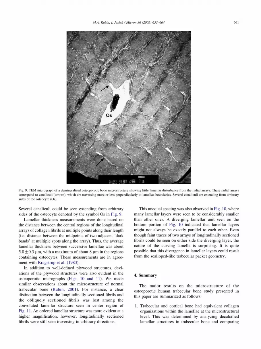
Fig. 9. TEM micrograph of a demineralized osteoporotic bone microstructure showing little lamellar disturbance from the radial arrays. These radial arrays
correspond to canaliculi (arrows), which are traversing more or less perpendicularly to lamellae boundaries. Several canaliculi are extending from arbitrary
sides of the osteocyte (Os).
M.A. Rubin, I. Jasiuk / Micron 36 (2005) 653–664 661
Several canaliculi could be seen extending from arbitrary
sides of the osteocyte denoted by the symbol Os in Fig. 9.
Lamellar thickness measurements were done based on
the distance between the central regions of the longitudinal
arrays of collagen fibrils at multiple points along their length
(i.e. distance between the midpoints of two adjacent ‘dark
bands’ at multiple spots along the array). Thus, the average
lamellar thickness between successive lamellae was about
5.8G0.3 mm, with a maximum of about 8 mm in the regions
containing osteocytes. These measurements are in agree-
ment with Kragstrup et al. (1983).
In addition to well-defined plywood structures, devi-
ations of the plywood structures were also evident in the
osteoporotic micrographs (Figs. 10 and 11). We made
similar observations about the microstructure of normal
trabecular bone (Rubin, 2001). For instance, a clear
distinction between the longitudinally sectioned fibrils and
the obliquely sectioned fibrils was lost among the
convoluted lamellar structure seen in center region of
Fig. 11. An ordered lamellar structure was more evident at a
higher magnification, however, longitudinally sectioned
fibrils were still seen traversing in arbitrary directions.
This unequal spacing was also observed in Fig. 10, where
many lamellar layers were seen to be considerably smaller
than other ones. A diverging lamellar unit seen on the
bottom portion of Fig. 10 indicated that lamellar layers
might not always be exactly parallel to each other. Even
though faint traces of two arrays of longitudinally sectioned
fibrils could be seen on either side the diverging layer, the
nature of the curving lamella is surprising. It is quite
possible that this divergence in lamellar layers could result
from the scalloped-like trabecular packet geometry.
4. Summary
The major results on the microstructure of the
osteoporotic human trabecular bone study presented in
this paper are summarized as follows:
1. Trabecular and cortical bone had equivalent collagen
organizations within the lamellae at the microstructural
level. This was determined by analyzing decalcified
lamellar structures in trabecular bone and comparing

Fig. 10. TEM micrograph of a disrupted lamellar structure resulting from the extensive networking of the radial fibril arrays. The lamellar spacing between the
bands of longitudinally sectioned fibrils varies considerably, with no two being the same. A diverging lamellar unit (arrow) is seen on the bottom-right portion
of the image.
M.A. Rubin, I. Jasiuk / Micron 36 (2005) 653–664662
them with published results on the cortical bone
structure.
2. Atypical lamellar structures were evident in osteoporotic
trabecular bone. These atypical structures, which were
mainly seen in the unmineralized regions, suggested that
the lamellar pattern in bone was much more complex
than envisioned.
3. There was evidence of lamellar structure alteration by
the osteocyte network.
The above conclusions summarize the results presented
in this paper for human osteoporotic trabecular bone.
However, they are more general as they hold for both
normal and osteoporotic trabecular bone. The results of the
normal trabecular bone study were reported elsewhere
(Rubin, 2001). In the next paragraphs we present the
summary pertaining to both normal and osteoporotic bone,
for completeness. The information on normal bone we draw
from (Rubin, 2001).
In summary, the TEM analysis of the microstructure of
human trabecular bone confirmed many of the results found
in cortical bone and MTLT, as well as surfaced a few new
questions. This investigation showed that the plywood-like
structures routinely observed in cortical bone, twisted,
rotated or orthogonal, existed in both normal and
osteoporotic human trabecular bone. However, there were
some structural differences in the lamellar motifs between
normal and osteoporotic bone. One of these differences
involved a more pristine lamellar organization in normal
trabecular bone. Observations of atypical lamellar structures
found in the mineralized and unmineralized regions of both
normal and osteoporotic trabecular bone suggested that
bone exhibited much more complicated structures than
previously reported.
The osteocyte network was shown to have some two-
dimensional structural significance on the lamellar organ-
ization in both normal and osteoporotic bone. However, it
appeared that the lamellar structure of osteoporotic bone
was more affected by this vast network, as the canaliculi
tended to be more pronounced and abundant.
Finally, the overall objectives of this research were to
investigate the ultrastructural characteristics of osteoporotic

Fig. 11. TEM micrograph from osteoporotic demineralized bone showing deviations from the plywood structures. A clear distinction between the
longitudinally sectioned fibrils and the obliquely sectioned fibrils is lost among the convoluted lamellar structure.
M.A. Rubin, I. Jasiuk / Micron 36 (2005) 653–664 663
and normal human trabecular bone by means of TEM and to
provide the characterization of the normal and osteoporotic
human trabecular bone hierarchical structure from the
mesoscale down to the nanoscale. They are summarized in
Rubin (2001). The results presented in this paper are a
subset of this research. The comparison of normal and
osteoporotic bone at sub-microstructure and nanostructure
levels was presented by us in Rubin et al. (2003).
Acknowledgements
We would like to thank Dr Robert Apkarian and
Jeannette Taylor from Integrated Microscopy and Micro-
analytical Facility at Emory University for their assistance
with TEM specimen preparation and viewing of images and
to Dr Timothy Ganey from Atlanta Medical Center for
supplying us with the bone tissue.
References
Amling, M., Posl, M., Ritzel, H., Hahn, M., Vogel, M., Wening, V.J.,
Delling, G., 1996. “Architecture and distribution of cancellous bone
yield vertebral fracture clues: A histomorphometric analysis of the
complete spinal column from 40 autopsy specimens. Arch. Orthop
Trauma Surg 115, 262–269.
Ascenzi, A., 1986. Benvenutti A Orientation of collagen fibers at the
boundary between two successive osteonic lamellae and its mechanical
interpretation. J. Biomech 19, 455–463.
Bloom, W., Fawcett, D., 1975. Bone, A Textbook of Histology, ten th 1975
(Chpt 11) pp 244–261.
Cane, V., Marotti, G., Volpi, G., Zaffe, D., Palazzini, S., 1982. “Size and
density of steocyte lacunae in different regions of long bones. Calcif.
Tissue Int. 34, 558–563.
Darby, A.J., Meunier, P.J., 1981. “Mean wall thickness and formation
periods of trabecular bone packets in idiopathic osteoporosis. Calcif.
Tissue Int. 33, 199–204.
Eriksen, E.F., Axelrod, D.W., Melsen, F., 1994. Bone Histomorphometry.
Raven Press, New York.
Eyden, B., Tzaphildou, M., 2001. Structural variations of collagen in
normal and pathological tissues: role of electron microscopy. Micron
32, 287–300.
Gebhardt, W., 1906. Uber funktionell wichtige Anordnungsweisen der
feineren und groberen Bauelemente des Wirbeltierknochens.II
Spezieller Teil. I. Arch. Entw. Mech. Org. 20, 187–322.
Giraud-Guille, M.M., 1988. Twisted plywood architecture of collagen
fibrils in human compact bone osteons. Calcif. Tissue Int. 42,
167–180.
Hoffler, C.E., McCreadie, B., Smith, E., Goldstein, S., 2000. A hierarchical
approach to exploring bone mechanical properties. In: An, Draughn
(Eds.), Mechanical Testing of Bone and the Bone-Implant Interface,
pp. 133–149 (Chpt. 8).

M.A. Rubin, I. Jasiuk / Micron 36 (2005) 653–664664
Hollister S (2001), From: www.engin.umich.edu/class/bme456/
bonestructure.
Jayasinghe, J., Jones, S.J., Boyde, A., 1993. Scanning electron microscopy
of human lumbar vertebral trabecular bone surfaces. Virchows Archiv
A Pathol. Anat. 422, 25–34.
Kessel, R., Kardon, R., 1979. Tissue and Organs: A Text Atlas of Scanning
Electron Microscopy. W.H. Freeman and Company.
Kragstrup, J., Melsen, F., 1983. Three dimensional morphology of
trabecular bone osteons reconstructed from serial sections. Metab.
Bone Dis. Rel. Res. 5, 127–130.
Kragstrup, J., Melsen, F., Mosekilde, L., 1983. Thickness of lamellae
in normal human iliac trabecular bone. Metab. Bone Dis. Rel. Res. 4,
291–295.
Lakes, R., 1993. Materials with structural hierarchy. Nature 361, 511–
515.
Landis, Song, 1993. Mineral and organic matrix interaction in normally
calcifying tendon visualized in three dimensions by high voltage
electron microscopic tomography and graphic image reconstruction.
J. Struct. Biology 110, 39–54.
Lips, P., Courpron, P., Meunier, P.J., 1978. Mean wall thickness of
trabecular bone packets in the human iliac crest: changes with age.
Calcif. Tissue Int. 26, 13–17.
Lozupone, E., Favia, A., 1990. The structure of the trabeculae of cancellous
bone. 2. Long Bones and Mastiod. Calcif. Tissue Int. 46, 367–372.
Marks, S., Cielinski, M., Sundquist, K., 1996. Bone surface
morphology reflects local skeletal metabolism. Micro. Res. And
Tech. 33, 121–127.
Marotti, G., 1993. A new theory of bone lamellation. Calcif. Tissue Int. 53,
S47–S56.
Martin, B., Burr, D., Sharkey, N., 1998. Skeletal Tissue Mechanics.
Springer-Verlag, New York.
Morita, M., Ebihara, A., Itoman, M., Sasada, T., 1994. Progression of
osteoporosis in cancellous bone depending on trabecular structure. Ann.
of Biomed. Eng. 22, 532–539.
Mosekilde, L., 1990. Consequences of the remodeling process for vertebral
trabecular bone-structure - A scanning electron-microscopy study
(uncoupling of unloaded structures). Bone Miner. 10, 13–35.
Myers, E.R., Wilson, S.E., 1997. Biomechanics of osteoporosis and
vertebral fracture. Spine 22 (24S), 25S–31S.
National Institutes of Health, The Osteoporosis and Related Bone Diseases
wNational Resource Center (2000), From: http//www.osteo.org/docs/
183.199143826.htm.
Parafitt, A., Chir, B., 1987. Bone remodeling and bone loss: Understanding
the pathophysiology of osteoporosis. Clin. Obstet. Gynecol. 30 (4),
789–811.
Prostak, K.S., Lees, S., 1996. Visualization of crystal matrix structure. In
Situ demineralization of mineralized turkey tendon and bone. Calcif.
Tissue Int. 59, 474–479.
Rai, D.V., Behari, J., 1986. Biophysical characterization of osteoporotic
bone. Env. Res. 40, 68–83.
Raspanti, M., Guizzardi, S., Strocchi, R., Ruggeri, A., 1996. Collagen fibril
patterns in compact bone: Preliminary ultrastructural observations. Acta
Anatomica 155, 249–256.
Rho, J.Y., Kuhn-Spearing, L., Zioupos, P., 1998. Mechanical properties and
the hierarchical structure of bone. Med Eng & Phys 20, 92–102.
Rubin, M. A (2001), Multiscale characterization of the ultrastructure of
trabecular bone in osteoporotic and normal humans and in two inbred
strains of mice, M.S. thesis, Georgia Institute of Technology.
Rubin, M.A., Jasiuk, I., Taylor, J., Rubin, J., Ganey, T., Apkarian, R.P.,
2003. TEM analysis of the nanostructure of normal and osteoporotic
human trabecular bone. Bone 33, 270–282.
Sato, K., Wakamatsu, E., et al., 1986. Histomorphometric study of
trabecular channels in normal iliac bone. Calcif. Tissue Int. 39, 2–7.
Snyder, B., Piazza, S., Edwards, W., Hayes, W., 1993. Role of trabecular
morphology in the etiology of age-related vertebral fractures. Calcif.
Tissue Int. 53 (1), S14–S22.
Takita, T., Naguro, T., Yamamoto, K., 1992. Fine structure of remodeling
sites on iliac cancellous bone in senile osteoporosis: A study by
scanning electron microscopy using an improved organic specimen
preparation method. Cells and Materials 2 (2), 135–142.
Traub, W., Arad, T., Weiner, S., 1989. Three-dimensional ordered
distribution of crystals in turkey in tendon collagen fibers. Proc. Natl.
Acad Sci. USA 86, 9822–9826.
Weiner, S., Traub, W., 1992. Bone structure: from angstroms to microns.
FASEB 6, 879–885.
Weiner, S., Arad, T., Sabanay, I., Traub, W., 1997. Rotated plywood
structure of primary lamellar bone in the rat: orientations of the collagen
fibril arrays. Bone 20 (6), 509–514.
Weiner, S., Traub, W., Wagner, H., 1999. Lamellar bone: Structure-
function relations. J. Struc. Bio. 126, 241–255.
Werner, H.J., Martin, H., Behrend, D., Schmitz, K.P., Schober, H.C., 1996.
The loss of stiffness as osteoporosis progresses. Med. Eng. Phys. 18,
601–606.
Ziv, V., Wagner, H., Weiner, S., 1996. Transitional structures in lamellar
bone. Microsc. Res. Tech. 33, 203–213.


![[Secondary prevention in osteoporotic fractures. The GIOS project]](https://img.pdfslide.net/doc/110x75/63487f43de40dd034d093bb9/secondary-prevention-in-osteoporotic-fractures-the-gios-project.jpg)


