Embed Size (px)
Citation preview

CORRESPONDENCE
Further Analysis Is Required to Identify an Early Stopping Rule for Peginterferon Therapy That Is Validfor All Hepatitis B e Antigen–Positive Patients
To the Editor:
We read with interest the article by Sonneveld et al.,1 whoreported an association between on-treatment hepatitis B surfaceantigen (HBsAg) levels and a sustained response to peginterferonalfa-2b in hepatitis B e antigen (HBeAg)–positive patients (n ¼221). No HBsAg decline by week 12 of therapy was associatedwith a low chance of a sustained response (97% probability ofnonresponse) and was proposed as an early stopping rule for pegin-terferon therapy. Because this rule needs to be validated in otherstudies, we investigated how the rule would have performed inHBeAg-positive patients treated with peginterferon alfa-2a duringtwo independent, large-scale studies.2,3
HBeAg-positive patients received peginterferon alfa-2a (180 lg/week) with or without lamivudine (100 mg/day) for 48 weeks aspart of a phase 3 study2 (n ¼ 542) or peginterferon alfa-2a (180lg/week) for 48 weeks as part of the Nephrotic Syndrome StudyNetwork (NEPTUNE) study (n ¼ 136).3 Overall, the rates ofHBeAg loss and hepatitis B virus (HBV) DNA levels < 10,000copies/mL in the phase 3 and NEPTUNE peginterferon alfa-2astudies were similar (25% and 24%, respectively), and they werehigher than those in Sonneveld et al.’s analysis (19%).1 In accord-ance with Sonneveld et al.’s data, the HBsAg decline was morepronounced in patients with a response 6 months post-treatmentversus nonresponders. Patients with no HBsAg decline from thebaseline to week 12 had 82% (80/97) and 71% (22/31) probabil-ities of nonresponse in the phase 3 and NEPTUNE studies, respec-tively; these were considerably lower than the probability of 97%in Sonneveld et al.’s study (Fig. 1). The probabilities of response inpatients with no HBsAg decline were 18% (17/97) and 29% (9/31), respectively. Applying the stopping rule would have resultedin premature treatment discontinuation in some patients (17 and9, respectively) who would have responded. HBeAg seroconversion
6 months post-treatment, rather than HBeAg loss and HBV DNAlevels <10,000 copies/mL, was the primary endpoint in the pegin-terferon alfa-2a studies. Using this more robust indicator of sus-tained immune control would have resulted in some patients inthe phase 3 and NEPTUNE studies (30 and 12, respectively) dis-continuing their treatment prematurely if the stopping rule hadbeen applied.
Differences in the study populations could explain the varyingresponse rates and the fact that the proposed stopping rule couldnot be validated by the peginterferon alfa-2a analyses. Sonneveldet al.’s analysis1 was a European study in which only 20% of thepatients were Asian, whereas the populations of the phase 3 andNEPTUNE peginterferon alfa-2a studies were predominantly Asian(>80%). This influenced the genotype distribution; Sonneveldet al.’s study had a high proportion of genotype A or D patients,whereas the peginterferon alfa-2a studies included predominantlygenotype B and C patients. In combination with the differences inthe treatment regimens (peginterferon alfa-2a versus peginterferonalfa-2b and 48 weeks of therapy versus 52 weeks) and in the num-bers of patients included in the analyses, this may account for thedifferences in the results.
Monitoring HBsAg levels during peginterferon therapy provides agood indication of the treatment response and helps in identifyingearly success. However, it is clear that further analysis is required ei-ther to identify an early stopping rule for peginterferon therapy that isvalid for all genotypes or to develop genotype-specific algorithms.
TEERHA PIRATVISUTH, M.D.1
PATRICK MARCELLIN, M.D.21NKC Institute of Gastroenterology and Hepatology
Songklanagarind HospitalPrince of Songkla UniversityHat Yai, Thailand
Fig. 1. Flowcharts for anydecline in HBsAg levels from thebaseline to week 12 with respectto the sustained response post-treatment. Response is de¢nedas HBeAg loss and HBV DNAlevels < 10,000 copies/mL. Anasterisk indicates patients withHBsAg values available for thebaseline, for weeks 12, 24, and48 of therapy, and for 6 monthspost-treatment. A dagger indi-cates patients with HBsAg val-ues available for the baselineand for week 12 of therapy.
1054

2Service d’HepatologieCentre de Recherche Biologique Bichat Beaujon (Unite 773)Hopital BeaujonUniversity of Paris, Clichy, France
References1. Sonneveld MJ, Rijckborst V, Boucher CA, Hansen BE, Janssen HL. Pre-
diction of sustained response to peginterferon alfa-2b for HBeAg-positivechronic hepatitis B using on-treatment HBsAg decline. HEPATOLOGY
2010;52:1251-1257.2. Lau GK, Piratvisuth T, Luo KX, Marcellin P, Thongsawat S, Cooksley G,
et al. Peginterferon alfa-2a, lamivudine, and the combination for HBeAg-positive chronic hepatitis B. N Engl J Med 2005;352:2682-2695.
3. Liaw Y-F, Xie Q, Han KH, Gane EJ, Piratvisuth T, McCloud PI, et al.Shorter duration and lower dose of peginterferon alfa-2a therapy resultsin inferior HBeAg seroconversion rates compared with the duration anddose of 48 weeks and 180 lg: NEPTUNE study. Paper presented at:61st Annual Meeting of the American Association for the Study of LiverDiseases; October 29-November 2, 2010; Boston, MA.
Copyright VC 2010 by the American Association for the Study of Liver Diseases.View this article at wileyonlinelibrary.com.DOI 10.1002/hep.24136
Potential conflict of interest: Dr. Piratvisuth advises, serves on the speakers’bureau of, and received grants from Roche and Novartis. Dr. Piratvisuth advisesand serves on the speakers’ bureau of GlaxoSmithKline and MSD. Dr. Piratvisuthalso serves on the speakers’ bureau of Bristol-Myers Squibb.Dr. Marcellin serves on the speakers’ bureau of, advises, and received grants fromRoche, Schering Plough, and Gilead. Dr. Marcellin advises and serves on thespeakers’ bureau of Bristol-Myers Squibb, Vertex, Novartis, Tibotec, andIntermune. Dr. Marcellin also advises Pharmasset, MSD, Boehringer, Biolex,and Zymogenetics.
Reply
We thank Piratvisuth and Marcellin for their valuable contribu-tion to the debate. First of all, it is important to note that ourfindings of a more pronounced hepatitis B surface antigen(HBsAg) decline in hepatitis B e antigen (HBeAg)-positive patientswith a response to peginterferon were confirmed in their study,reflecting the induction of an immune response in these patients.1
Their analysis shows that failure to achieve a decline in HBsAglevels through 12 weeks of therapy does not predict nonresponse aswell in their cohort as it did in our study. Possible explanations forthese discrepant findings could be the type of peginterferon or du-ration of therapy, as suggested by Piratvisuth and Marcellin. How-ever, the most probable explanation is the difference in hepatitis Bvirus (HBV) genotype distribution between the study cohorts. Pre-
liminary data from our group show that HBsAg decline amongHBeAg-positive patients treated with peginterferon is strongly relatedto HBV genotype.2 These differences across genotypes, particularlyamong patients who fail to achieve a response, may be an importantdeterminant of the performance of a threshold-based stopping rule.Consequently, the performance of any threshold is primarily depend-ent upon the distribution of HBV genotypes in the study cohort.
The importance of HBV genotype when applying stopping rulesfor peginterferon therapy in chronic hepatitis B was recently illustratedby a validation study of our stopping rule for HBeAg-negativepatients. This stopping rule, recommending discontinuation of pegin-terferon in patients who fail to achieve a decline in HBsAg and adecline in HBV DNA of >2 log at week 12, was based on a cohortof mostly genotype D patients.3 When validated in two independentstudy cohorts, performance was best in genotype D patients treatedwith either 48 or 96 weeks of peginterferon.4
In conclusion, monitoring of HBsAg levels during peginterferontherapy of chronic hepatitis B may provide valuable insight into apatient’s probability of achieving a response. However, it appearsthat differences in HBsAg decline across HBV genotypes have tobe taken into consideration. A pooled analysis of the data fromour respective studies, stratified by HBV genotype and possiblyincorporating HBV DNA levels, appears to be a crucial next step.
MILAN J. SONNEVELD
VINCENT RIJCKBORST
HARRY L.A. JANSSEN
Department of Gastroenterology and Hepatology, Erasmus MCUniversity Medical Center, Rotterdam, The Netherlands
References1. Sonneveld MJ, Rijckborst V, Boucher CA, Hansen BE, Janssen HL. Pre-
diction of sustained response to peginterferon alfa-2b for hepatitis B eantigen-positive chronic hepatitis B using on-treatment hepatitis B sur-face antigen decline. HEPATOLOGY 2010;52:1251-1257.
2. Sonneveld MJ, Rijckborst V, Senturk H, Zeuzem S, Akarca U, SimonK, et al. HBsAg decline during peginterferon alfa-2b therapy forHBeAg-positive chronic hepatitis B depends on HBV genotype: relationto sustained response [Abstract 441]. HEPATOLOGY 2010;52(Suppl.).
3. Rijckborst V, Hansen BE, Cakaloglu Y, Ferenci P, Tabak F, Akdogan M,Simon K, et al. Early on-treatment prediction of response to peginter-feron alfa-2a for HBeAg-negative chronic hepatitis B using HBsAg andHBV DNA levels. HEPATOLOGY 2010;52:454-461.
4. Rijckborst V, Hansen B, Ferenci P, Brunetto M, Tabak F, Cakaloglu Y,et al. Early on-treatment HBsAg and HBV DNA levels identify HBeAg-negative patients not responding to 48 or 96 weeks of peginterferonalfa-2a therapy [Abstract 479]. HEPATOLOGY 2010;52(Suppl.).
Copyright VC 2010 by the American Association for the Study of Liver Diseases.View this article at wileyonlinelibrary.com.
DOI 10.1002/hep.24182Potential conflict of interest: H.L.A. Janssen received grants from and is a
consultant for Bristol-Myers Squibb, Gilead Sciences, Novartis, Roche, andSchering-Plough. Vincent Rijckborst is a consultant for Roche.
Optimal Duration of Treatment for Acute Hepatitis C in Human ImmunodeficiencyVirus–Positive Individuals?
To the Editor:
In a recent article, Piroth et al.1 report on the outcomes follow-ing treatment of acute hepatitis C in 40 human immunodeficiencyvirus (HIV)-positive men who have sex with men (MSM), 38 of
This work was supported by a research grant from F. Hoffmann-La Roche
(Basel, Switzerland).
Editorial support was provided by Dr. Liesje Thomas (Elements Communica-
tions, Ltd., Westerham, United Kingdom) and was funded by F. Hoffmann-La
Roche (Basel, Switzerland).
whom received combination therapy with pegylated interferon andribavirin (the HEPAIG study). The overall sustained viral response(SVR) rate of 82% is encouraging, especially given that 81% of theircohort had genotype (GT) 1 or 4 infection, and supports guidelinesfor recommending treatment in this setting.2 However, we question
HEPATOLOGY, Vol. 53, No. 3, 2011 CORRESPONDENCE 1055

the conclusions the authors draw from their data regarding optimalduration of therapy.
The authors argue that those patients treated for longer than 28weeks had a significantly greater SVR rate than those treated for lessthan 28 weeks (92% versus 64%, respectively, P ¼ 0.03), and thatthe rate of SVR (25%) in those who did not achieve rapid virologi-cal response (RVR) but received <28 weeks of therapy merits exten-sion to 48 weeks for all patients with non-RVR. The evidence forthese specific recommendations, however, is weak and confused byhow data from the ‘‘null responder’’ group is dealt with in this non-randomized design. Five patients were reported as ‘‘never respond-ing’’ to therapy presumably defined as no RVR or early viralresponse (EVR) and ceased therapy before 28 weeks. In the analysisexamining SVR rates the authors appear to have included these sub-jects in the group receiving less than 28 weeks (SVR 9/14, 64%)versus longer duration (SVR 23/25, 92%) resulting in the ‘‘shortarm’’ appearing to be inferior. In fact the true question to examineis how common relapse was in non-RVR subjects who then achievedEVR and were subsequently treated for less than 28 weeks. A highrate of relapse in this situation would suggest an inadequate lengthof treatment course. In the HEPAIG study it appears that 13 non-RVR patients subsequently achieved EVR but only one of these wastreated for <28 weeks and this patient subsequently achieved SVR.
In the Australian Trial in Acute Hepatitis C (ATAHC), 35HIV-positive MSM were treated with 24 weeks combination ther-apy with pegylated interferon and ribavirin and RVR was achievedin 12 (34%).3 In the 23 non-RVR subjects, three had no EVR andwere discontinued and of the remaining 20 (50% GT 1), onlythree (2 GT 1 and 1 GT 3) relapsed after treatment completion,demonstrating that 24 weeks of combination therapy was adequatein 85% of subjects with no RVR but EVR. Given the additionalexpense and toxicity of extending therapy to 48 weeks (we notethe 40% use of growth factors in HEPAIG), the costs would out-weigh any potential marginal benefit. The HEPAIG study recom-mendation is even less appealing given the likelihood of new thera-pies available for retreatment within the next few years for thosewho do relapse.
In summary, we agree with the HEPAIG authors that combina-tion therapy is optimal in this setting and that treatment should bediscontinued in those with complete nonresponse at week 12.However, we believe their treatment duration recommendations arenot based on available evidence and that this question thereforeremains unanswered.
GAIL V. MATTHEWS, MBCHB (UK) PHDGREGORY J. DORE, MD PHDViral Hepatitis Clinical Research Program
National Centre in HIV Epidemiology and Clinical ResearchUniversity of New South Wales, Sydney, Australia
References1. Piroth L, Larsen C, Binquet C, et al. Treatment of acute hepatitis C in
human immunodeficiency virus-infected patients: the HEPAIG study.HEPATOLOGY 2010;52:1915-1921.
2. Rockstroh J. Acute hepatitis C in HIV-infected individuals—recommen-dations from the NEAT consensus conference. AIDS. Epub ahead ofprint, Jan 03, 2011.
3. Dore GJ, Hellard M, Matthews G, et al. Effective treatment of injectingdrug users with recently acquired hepatitis C virus infection. AustralianTrial In Acute Hepatitis C Study Group. Gastroenterology 2010;138:123-135.e2.
Copyright VC 2010 by the American Association for the Study of Liver Diseases.View this article at wileyonlinelibrary.com.DOI 10.1002/hep.24152Potential conflict of interest: Nothing to report.
Reply
We thank Matthews and Dore for agreeing with our suggestionto treat human immunodeficiency virus (HIV)-infected patientswith acute hepatitis C by using combination therapy,1 althoughthey advocate a 24-week duration because 85% of their patientswith early virological response (EVR) but not rapid virologicalresponse (RVR) achieved sustained virological response (SVR) aftera 24-week course in the recent ATAHC study.2 This is a key point,because the optimal duration would be the best compromisebetween reducing expense and toxicity on the one hand, and maxi-mal efficacy on the other hand, given the faster and more severeevolution of HCV infection in HIV-infected patients.
We agree that excluding patients who failed in the HEPAIGstudy to achieve RVR or EVR (n ¼ 4) would enhance the SVRrate of 24-week course therapy. Indeed, the SVR rate in patientsexperiencing EVR, when assessed, was 88% (8/9), which is higherthan that observed in ATAHC (74%)2 and close to the 92%obtained with a longer course in HEPAIG.1 However, the RVRrate in HEPAIG was high, particularly for the 14 patients treatedfor 24 6 4 weeks (57%), compared with that observed in ATAHC(34%), whereas the EVR rate was lower (75% versus 91%).Because HCV therapy and doses were similar in these studies, thismay be linked to differences in HCV genotype distribution (with apotential cluster effect), in the clinical presentation of acute hepati-tis C, or in the characteristics of the patients (e.g., the HCV trans-mission route and perhaps the distribution in IL28B gene poly-morphisms). Whatever the reasons, most patients with EVR inHEPAIG had RVR previously. Only two patients in HEPAIGexperienced EVR but not RVR. Although both of these patientsachieved SVR, this finding is not sufficient to draw conclusions, incontrast with the ATAHC study, where 17/20 patients with EVRbut not RVR experienced SVR.2 Whether the three patients whodid not experience SVR would have benefited from a longer coursecannot be established from the ATAHC study.
Recent cohort studies on acute hepatitis C in HIV-infectedpatients are quite supportive and complementary in helping todefine the best strategy in treating acute hepatitis C in HIV-infected patients. They highlight the pivotal role of HCV kineticsassessment on the management of HCV therapy. From both theHEPAIG1 and ATAHC reports,2,3 patients with RVR have to betreated for 24 weeks. For patients experiencing EVR but not RVR,SVR rate following a 24-week course ranges from 75% in the Eu-ropean Cohort Study4 (albeit including some patients with a 48-week course therapy) to 85% in ATAHC,2 compared with a hepa-titis C virus (HCV) eradication rate of 100% (10/10) with a lon-ger course in HEPAIG.1 Whether this mean difference of roughly15%-20% is significant, relevant, or marginal may be debated andshould only be addressed in randomized, controlled trials. Never-theless, our conclusions parallel the recent recommendations of theNEAT consensus conference stating that it would be reasonable toaim for a 24-week course, with a longer duration reserved for thosewithout RVR but with EVR.5 Finally, we also agree that newHCV therapies will probably lead to modifications in these propo-sitions, not only by improving the success rate of retreatment ofpatients failing to respond to first line pegylated interferon–riba-virin therapy, but also as a first-line treatment of acute hepatitis C.
LIONEL PIROTH1
CHRISTINE LARSEN2
ELISABETH DELAROCQUE-ASTAGNEAU2,3
STANISLAS POL4
On behalf of the steering committee of the HEPAIG study1Infectious Diseases Department, University Hospital, and Universite de
Bourgogne, Dijon, France2Institut de Veille Sanitaire, Saint Maurice, France
1056 CORRESPONDENCE HEPATOLOGY, March 2011
3Unite d’epidemiologie des maladies emergentes, Institut Pasteur, Paris,France
4Universite Paris Descartes, Institut Cochin Inserm U1016 and serviced’Hepatologie, APHP-Hopital Cochin, Paris, France
References1. Piroth L, Larsen C, Binquet C, Alric L, Auperin I, Chaix ML, et al.
Treatment of acute hepatitis C in human immunodeficiency virus-infected patients: the HEPAIG study. HEPATOLOGY 2010;52:1915-1921.
2. Dore GJ, Hellard M, Matthews GV, Grebely J, Haber PS, PetoumenosK, et al. Effective treatment of injecting drug users with recentlyacquired hepatitis C virus infection. Gastroenterology 2010;138:123-135.
3. Matthews GV, Hellard M, Haber P, Yeung B, Marks P, Baker D, et al.Characteristics and treatment outcomes among HIV-infected individuals
in the Australian Trial in Acute Hepatitis C. Clin Infect Dis 2009;48:650-658.
4. Vogel M, Dominguez S, Bhagani S, Azwa A, Page E, Guiguet M,et al. Treatment of acute HCV infection in HIV-positive patients: ex-perience from a multicentre European cohort. Antivir Ther 2010;15:267-279.
5. The European AIDS Treatment Network (NEAT) Acute Hepatitis CInfection Consensus Panel.Acute hepatitis C in HIV-infected individuals:recommendations from the European AIDS Treatment Network(NEAT) Consensus Conference. AIDS 2010; doi:10.1097/QAD.0b013e328343443b.
Copyright VC 2010 by the American Association for the Study of Liver Diseases.View this article at wileyonlinelibrary.com.DOI 10.1002/hep.24181Potential conflict of interest: Nothing to report.
The Wide Spectrum of Hepatic Iron Overload
To the Editor:We read with great interest the article by Nelson et al.1 The
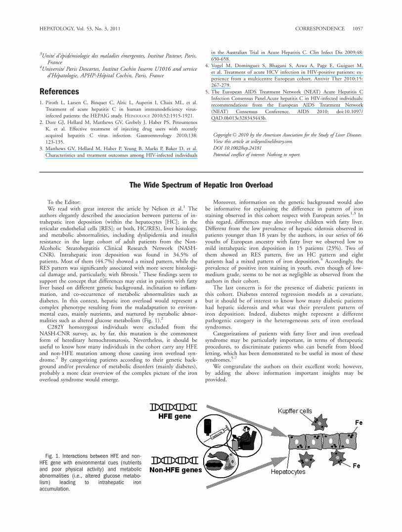
authors elegantly described the association between patterns of in-trahepatic iron deposition (within the hepatocytes [HC]; in thereticular endothelial cells [RES]; or both, HC/RES), liver histology,and metabolic abnormalities, including dyslipidemia and insulinresistance in the large cohort of adult patients from the Non-Alcoholic Steatohepatitis Clinical Research Network (NASH-CNR). Intrahepatic iron deposition was found in 34.5% ofpatients. Most of them (44.7%) showed a mixed pattern, while theRES pattern was significantly associated with more severe histologi-cal damage and, particularly, with fibrosis.1 These findings seem tosupport the concept that differences may exist in patients with fattyliver based on different genetic background, inclination to inflam-mation, and co-occurrence of metabolic abnormalities such asdiabetes. In this context, hepatic iron overload would represent acomplex phenotype resulting from the maladaptation to environ-mental cues, mainly nutrients, and nurtured by metabolic abnor-malities such as altered glucose metabolism (Fig. 1).2
C282Y homozygous individuals were excluded from theNASH-CNR survey, as, by far, this mutation is the commonestform of hereditary hemochromatosis. Nevertheless, it should beuseful to know how many individuals in the cohort carry any HFEand non-HFE mutation among those causing iron overload syn-drome.2 By categorizing patients according to their genetic back-ground and/or prevalence of metabolic disorders (mainly diabetes),probably a more clear overview of the complex picture of the ironoverload syndrome would emerge.
Moreover, information on the genetic background would alsobe informative for explaining the difference in pattern of ironstaining observed in this cohort respect with European series.1,3 Inthis regard, differences may also involve children with fatty liver.Different from the low prevalence of hepatic siderosis observed inpatients younger than 18 years by the authors, in our series of 66youths of European ancestry with fatty liver we observed low tomild intrahepatic iron deposition in 15 patients (23%). Two ofthem showed an RES pattern, five an HC pattern and eightpatients had a mixed pattern of iron deposition.4 Accordingly, theprevalence of positive iron staining in youth, even though of low-medium grade, seems to be not as negligible as observed from theauthors in their cohort.
The last concern is for the presence of diabetic patients inthis cohort. Diabetes entered regression models as a covariate,but it should be of interest to know how many diabetic patientshad hepatic siderosis and what was their prevalent pattern ofiron deposition. Indeed, diabetes might represent a differentpathogenic category in the heterogeneous sets of iron overloadsyndromes.
Categorizations of patients with fatty liver and iron overloadsyndrome may be particularly important, in terms of therapeuticprocedures, to discriminate patients who can benefit from bloodletting, which has been demonstrated to be useful in most of thesesyndromes.5-7
We congratulate the authors on their excellent work; however,by adding the above information important insights may beprovided.
Fig. 1. Interactions between HFE and non-HFE gene with environmental cues (nutrientsand poor physical activity) and metabolicabnormalities (i.e., altered glucose metabo-lism) leading to intrahepatic ironaccumulation.
HEPATOLOGY, Vol. 53, No. 3, 2011 CORRESPONDENCE 1057

MELANIA MANCO, M.D., PH.D., F.A.C.N.ANNA ALISI, PH.D.ANTONELLA MOSCA, M.D.VALERIO NOBILI, M.D.Laboratorio di Malattie Epatiche Auto-Immuni e Metaboliche Ospedale
Pediatrico Bambino Gesu IRCCS, Centro di Nutrizione e DieteticaDipartimento di Pediatria Universita La Sapienza Roma, Italia
References1. Nelson JE, Wilson L, Brunt EM, Yeh MM, Kleiner DE, Unalp-Arida A,
et al. Relationship between the pattern of hepatic iron deposition andhistological severity in nonalcoholic fatty liver disease. HEPATOLOGY 2010Oct 11 [Epub ahead of print].
2. Pietrangelo A.Hereditary hemochromatosis: pathogenesis, diagnosis, andtreatment. Gastroenterology 2010;139:393-408.
3. Valenti L, Fracanzani AL, Bugianesi E, Dongiovanni P, Galmozzi E,Vanni E, et al. HFE genotype, parenchimal iron accumulation, and liverfibrosis in patients with non-alcoholic fatty liver disease. Gastroenterol-ogy 2010;138:905-912.
4. Manco M, Alisi A, Fernandez Real JM, Equitani F, DeVito R, ValentiL, et al. Early interplay of intra-hepatic iron and insulin resistance inchildren with non-alcoholic fatty liver disease. J Hepatol 2010 [Epubahead of print].
5. Equitani F, Fernandez-Real JM, Menichella G, Koch M, Calvani M,Nobili V, et al. Bloodletting ameliorates insulin sensitivity and secretionin parallel to reducing liver iron in carriers of HFE gene mutations.Diabetes Care 2008;31:3-8.
6. Fernandez-Real JM, Penarroja G, Castro A, Garcıa-Bragado F, Hernandez-Aguado I, Ricart W.Blood letting in high-ferritin type 2 diabetes: effects oninsulin sensitivity and beta-cell function. Diabetes 2002;51:1000-1004.
7. Valenti L, Fracanzani AL, Dongiovanni P, Bugianesi E, Marchesini G, ManziniP, et al. Iron depletion by phlebotomy improves insulin resistance in patientswith non-alcoholic fatty liver disease and hyperferritinemia: evidence from acase-control study. Am J Gastroenterol 2007;102:1251-1258.
Copyright VC 2010 by the American Association for the Study of Liver Diseases.View this article at wileyonlinelibrary.com.DOI 10.1002/hep.24142Potential conflict of interest: Nothing to report.
Reply
We would like to thank Manco and colleagues for their interestin our article.1 We agree that stratification of our study cohortaccording the presence of diabetes and HFE (hereditary hemochro-matosis) genotype status could be of interest. We have conductedadditional analyses with regard to diabetes status in our cohort.HFE genotyping was not routinely performed as part of the Nonal-coholic Steatohepatitis Clinical Research Network (NASH CRN)Database study; this is currently in progress in an ancillary study.Among the 849 adult subjects in our study, 221 were enrolled inthe PIVENS (Pioglitazone or Vitamin E for NASH Study) clinicaltrial,2 which excluded diabetic patients; therefore, to prevent anypotential bias, we report here the relationship of diabetes and irondeposition in the remaining 628 subjects.
As shown in Table 1, diabetic NASH CRN subjects were lesslikely to have hepatic iron deposition. In particular, diabeticpatients had significantly less hepatocellular but not reticuloendo-thelial system iron deposition. There are several possible explana-tions for these findings. The iron regulatory hormone hepcidin isexpressed in adipose tissue and has been shown to correlate tobody mass index.3 It is possible that increased visceral fat in our di-abetic subjects could lead to greater circulating hepcidin levels,
reduced iron absorption, and less hepatocellular iron deposition.Recently, alcohol has been shown to down-regulate hepcidin geneexpression.4 Because diabetic subjects in our study consumed lessalcohol than subjects without diabetes, this could also contributeto higher hepcidin levels in these patients. Diabetic subjects werealso more likely to be women, and physiologic blood loss throughmenstruation and childbirth may have resulted in decreased bodyiron stores. Additional studies are warranted to define the mecha-nism for decreased hepatocellular iron content in diabetic patientswith NAFLD.
In contrast to Manco et al.,5 who have reported hepatic irondeposition in 29% of their Italian pediatric subjects, which was notassociated with measurements of insulin resistance, the prevalenceof iron deposition in the NASH CRN pediatric subjects was only4%. It is likely that, as in adults, differences in the Italian andU.S. cohorts, including body mass index (25.2 in Manco et al. ver-sus 33.0 in the NASH CRN pediatric subjects) account for theobserved differences.6
JAMES NELSON, PH.D.KRIS KOWDLEY, M.D.Benaroya Research Institute
Virginia Mason Medical CenterSeattle, WA
References1. Nelson JE, Wilson L, Brunt EM, Yeh MM, Kleiner DE, Unalp-Arida A,
et al. Relationship between the pattern of hepatic iron deposition andhistological severity in nonalcoholic fatty liver disease. HEPATOLOGY
2010; doi:10.1002/hep.24038.2. Sanyal AJ, Chalasani N, Kowdley KV, McCullough A, Diehl AM, Bass
NM, et al.; NASH CRN.Pioglitazone, vitamin E, or placebo for nonal-coholic steatohepatitis. N Engl J Med 2010;362:1675-1685.
3. Bekri S, Gual P, Anty R, Luciani N, Dahman M, Ramesh B, et al.
Increased adipose tissue expression of hepcidin in severe obesity is
Table 1. Iron Stain Status of 628 NASH CRNDatabase Study Subjects
No diabetes
(N 5 394)
Diabetes
(N 5 234) P Value*
Iron stain status
Any iron present 146 (37) 68 (29) 0.041
HC iron present 109 (28) 42 (18) 0.006
RES iron present 110 (28) 56 (24) 0.273
HC iron only 36 (9) 12 (5) 0.068
RES iron only 37 (9) 26 (11) 0.488
Mixed HC/RES iron present 73 (18) 30 (13) 0.062
Potential factors influencing iron staining
Body mass index (kg/m2) 33.6 6 6.2 35.6 6 6.0 <0.0001
Female sex 244 (62) 167 (71) 0.016
Any alcohol consumption 205 (52) 75 (32) <0.001
Alcohol consumption
(>2 drinks/week)
26 (7) 2 (1) <0.001
Alcohol consumption
(drinks/drinking day)
0.18 6 0.47 0.08 6 0.36 0.045
History of any
gastrointestinal bleeding
31 (8) 27 (12) 0.125
Dietary iron consumption
(mg/day)
13.9 6 7.3 12.9 6 6.6 0.105
Dietary vitamin C
consumption (mg/day)
109 6 75 96 6 66 0.007
Values are N (%) or mean 6 standard deviation.
*P values from chi-square, Fisher’s exact test orWilcoxon rank sum test.Abbreviations: HC, hepatocellular; RES, reticuloendothelial system.
1058 CORRESPONDENCE HEPATOLOGY, March 2011

independent from diabetes and NASH. Gastroenterology 2006;131:
788-796.
4. Harrison-Findik DD, Schafer D, Klein E, Timchenko NA, Kulaksiz
H, Clemens D, et al. Alcohol metabolism-mediated oxidative stress
down-regulates hepcidin transcription and leads to increased duode-
nal iron transporter expression. J Biol Chem 2006;281:22974-
22982.5. Manco M, Alisi A, Fernandez Real JM, Equitani F, DeVito R, Valenti
L, et al. Early interplay of intra-hepatic iron and insulin resistance in
children with non-alcoholic fatty liver disease. J Hepatol 2010; doi:
10.1016/j.jhep.2010.12.007.
6. Kowdley KV. The role of iron in nonalcoholic fatty liver disease: thestory continues. Gastroenterology 2010;138:817-819.
Copyright VC 2010 by the American Association for the Study of Liver Diseases.View this article at wileyonlinelibrary.com.DOI 10.1002/hep.24184Potential conflict of interest: Nothing to report.
Treatment of Hepatic Encephalopathy with Rifaximin: More to Think About
To the Editor:
The recent publication of ‘‘Drug Therapy: Rifaximin’’ by Bajajand Riggio1 offers interesting observations by colleagues. Theyvoice concern that continuous rifaximin administration ‘‘couldhave the potential to increase resistance to rifaximin,’’ but they citeno objective clinical data in support of their hypothesis. They alsocite the two cases of Clostridium difficile in the rifaximin groupreported in the registration study of rifaximin for the treatment ofhepatic encephalopathy by Bass and colleagues,2 and they advisevigilance against C. difficile in patients with cirrhosis treated withrifaximin. At the US Food and Drug Administration meeting inMarch 2010, this matter was extensively studied and discussed.Both patients who developed C. difficile had concurrently receivedother antimicrobials known to cause C. difficile infection. Bajaj andRiggio’s suggestion of pulse therapy with rifaximin (to reduce costs)is without precedent or merit in the realm of antimicrobial therapy.Their statement regarding rifaximin that ‘‘the current role appearsto be a second-line [therapy]’’ is again without scientific merit.Lactulose is an effective therapy for hepatic encephalopathy; how-ever, its use and patient compliance are severely limited and re-stricted by its well-recognized adverse event profile of nausea, vom-iting, bloating, diarrhea, and incontinence. Rifaximin is very welltolerated, and it not only improves the duration of remission ofhepatic encephalopathy but also lessens the need for repeated hos-pitalization.2 Both factors require consideration when one is calcu-lating the overall cost of the two agents, their beneficial effects,and patient preference, compliance, and quality of life.
NORMAN GITLIN, M.D.Atlanta Gastroenterology Associates
Atlanta, GA
References1. Bajaj JS, Riggio O. Drug therapy: rifaximin. HEPATOLOGY 2010;52:
1484-1488.2. Bass NM, Mullen KD, Sanyal A, Poordad F, Neff G, Leevy CB, et al.
Rifaximin treatment in hepatic encephalopathy. N Engl J Med 2010;362:1071-1081.
Copyright VC 2010 by the American Association for the Study of Liver Diseases.View this article at wileyonlinelibrary.com.DOI 10.1002/hep.24112Potential conflict of interest: Dr. Gitlin is on the speakers’ bureau of Salix.
Reply
We read with interest the letter by Gitlin, which raises severalimportant points pertaining to the use of rifaximin for hepatic
encephalopathy (HE). Long-term use of any antibiotic does carrythe risk of resistance; however, the low likelihood of this appearingwith rifaximin due to the nonplasmid nature of its resistance wasalso cited in the article.1 The U.S. Food and Drug Administration(FDA) approval for rifaximin, granted as a result of the sameMarch 2010 meeting that is mentioned in the letter, comes alongwith the warning, as with all antibiotics, for the continuous moni-toring of patients on this therapy for Clostridium difficile.2
The article referred to the differences in the use of rifaximinacross countries, cyclical therapy in Italy, and continuous therapythat is being currently used in the United States and presents possi-ble advantages and disadvantages of both approaches. The prece-dence and scientific merit of this therapy is evident in the publica-tions and the clinical practice in Italy regarding the use ofrifaximin, which was referenced in the article.3,4 Although thepoints about the adverse events of lactulose as pointed out in theletter are noted and it is also known from clinical experience thatrifaximin is well tolerated in patients, prospective evidence using alarge-scale, head-to-head comparison of rifaximin compared to lac-tulose is still awaited.5,6 Bass et al. indeed demonstrated thatadministration of rifaximin with lactulose prevented hospitaliza-tions and HE recurrences compared to administration of lactulosealone in patients who had two prior HE episodes, but this needsfurther evaluation in patients who have had their first HE episode.7
There is also a huge cost difference between rifaximin and lactuloseand the evidence that rifaximin reduces overall costs and hospital-izations outside the selected population in the trial by Bass et al. isfrom retrospective, single-center studies.1,8,9 Therefore, to answerthese important questions, the FDA has mandated postmarketingtrials comparing lactulose and rifaximin for HE and to study rifaxi-min in patients who have a Model for End-Stage Liver Diseasescore �19.10 Until these trials are completed, from an evidence-based medicine approach, we stand by the recommendations in thearticle regarding the role of rifaximin
JASMOHAN S. BAJAJ, M.D.1
OLIVIERO RIGGIO, M.D.21Division of Gastroenterology, Hepatology and Nutrition, Virginia
Commonwealth University and McGuire Veterans AdministrationMedical Center, Richmond, VA
2Department of Clinical Medicine, Center for the Diagnosis andTreatment of Portal Hypertension, Sapienza University of RomeRome, Italy
References1. Bajaj JS, Riggio O. Drug therapy: rifaximin. HEPATOLOGY 2010;52:
1484-1488.2. Xifaxan prescribing information. http://www.accessdata.fda.gov/
drugsatfda_docs/label/2010/022554lbl.pdf. Accessed January 2011.
HEPATOLOGY, Vol. 53, No. 3, 2011 CORRESPONDENCE 1059

3. Alcorn J. Review: rifaximin is equally or more effective than other antibiot-ics and lactulose for hepatic encephalopathy. ACP J Club 2008;149:11.
4. Loguercio C, Federico A, De Girolamo V, Ferrieri A, Del VecchioBlanco C. Cyclic treatment of chronic hepatic encephalopathy withrifaximin. Results of a double-blind clinical study. Minerva GastroenterolDietol 2003;49:53-62.
5. Bajaj JS, Sanyal AJ, Bell D, Gilles H, Heuman DM. Predictors of therecurrence of hepatic encephalopathy in lactulose-treated patients. Ali-ment Pharmacol Ther 2010;31:1012-1017.
6. Kalaitzakis E, Bjornsson E. Lactulose treatment for hepatic encephalop-athy, gastrointestinal symptoms, and health-related quality of life. HEPA-
TOLOGY 2007;46:949-950.7. Bass NM, Mullen KD, Sanyal A, Poordad F, Neff G, Leevy CB, et al.
Rifaximin treatment in hepatic encephalopathy. N Engl J Med;362:1071-1081.
8. Neff GW, Kemmer N, Zacharias VC, Kaiser T, Duncan C, McHenry R,et al. Analysis of hospitalizations comparing rifaximin versus lactulose in themanagement of hepatic encephalopathy. Transplant Proc 2006;38:3552-3555.
9. Leevy CB, Phillips JA. Hospitalizations during the use of rifaximin ver-sus lactulose for the treatment of hepatic encephalopathy. Dig Dis Sci2007;52:737-741.
10. New Drug Approval Letter for Xifaxan. http://www.accessdata.fda.gov/drugsatfda_docs/appletter/2010/022554s000ltr.pdf. Accessed January 2011.
Copyright VC 2010 by the American Association for the Study of Liver Diseases.View this article at wileyonlinelibrary.com.DOI 10.1002/hep.24187Potential conflict of interest: Dr. Bajaj advises, consults for, and received grants
from Salix.
Alpha-Fetoprotein Should Be Included in the Hepatocellular Carcinoma Surveillance Guidelines ofthe American Association for the Study of Liver Diseases
To the Editor:
We read with interest the updated hepatocellular carcinoma(HCC) guidelines by the American Association for the Study ofLiver Diseases.1 We were surprised by the omission of alpha-feto-protein (AFP) testing in the recommendations for HCC surveil-lance. We disagree with these recommendations.
In making recommendations, the writers of practice guidelinesshould consider the quality of the evidence. The HCC guidelinesignore a significant amount of data about the use of AFP in thesurveillance of patients at risk for HCC. The only available level 1evidence for HCC surveillance comes from one randomized con-trolled trial of ultrasonography (US) combined with AFP testingevery 6 months in a hepatitis B carrier population.2 The next bestavailable evidence comes from a population-based cohort surveil-lance program involving hepatitis B carriers in Alaska that showedimproved outcomes.3 The remainder of the literature includes pop-ulation-based and non–population-based cohorts and case-controlstudies open to multiple sources of bias.4,5 Although it may be rea-sonable to generalize the findings of the available randomized trialand population-based study to other patient groups with cirrhosisor hepatitis C, we feel that it is inappropriate to drop one of theinterventions (i.e., AFP) found to work.
The guidelines cite the Hepatitis C Antiviral Long-Term Treat-ment Against Cirrhosis (HALT-C) study as the main source for thelack of efficacy of AFP in patients with cirrhosis.6 There are signifi-cant limitations to this study. First, only 40% of the patients hadcirrhosis. Second, HCC surveillance was not the primary purposeof HALT-C. Third, AFP had a sensitivity and specificity at thetime of HCC diagnosis of 61% and 81%, respectively, whereas UShad a sensitivity of only 58%, which is inadequate according tothe criteria stated in the guidelines. Interestingly, 40% of thepatients with early-stage HCC were diagnosed by an increasingAFP level alone or in combination with US. Therefore, AFPappears to complement US for the surveillance of HCC.
In addition to ignoring the highest level of evidence for the effi-cacy of US combined with AFP in research studies, the HCC guide-lines also neglect the effectiveness of the tests in clinical practice. Testreproducibility, a major determinant of translating the results ofresearch studies into practice, has never been evaluated for US as anHCC surveillance test. Another issue is underutilization of surveillancetests. In the only population-based study evaluating surveillance forHCC, only 17% of patients with HCC underwent regular surveil-lance before their diagnosis.7 Dropping AFP from the guidelines maypotentially lower the percentage of patients undergoing surveillance.
Surveillance for HCC has a whole host of confounding factorsthat make it impossible to detect benefit through personal experiencesand clinical observations alone.8 Therefore, randomized controlledstudies are the only reliable way of evaluating surveillance and chang-ing clinical practice. In the absence of randomized studies in patientswith cirrhosis, the current evidence points to US combined with se-rum AFP as the most effective surveillance strategy for patients at riskfor HCC. The guidelines should be revised to recommend US withAFP as the best available surveillance strategy.
JORGE A. MARRERO, M.D., M.S.1
HASHEM B. EL-SERAG, M.D., M.P.H.21Division of Gastroenterology, University of Michigan
Ann Arbor, MI2Michael E DeBakey VA Medical Center
Baylor College of Medicine, Houston, TX
References1. Bruix J, Sherman M. Management of hepatocellular carcinoma. HEPATOLOGY
2010;52.2. Zhang BH, Yang BH, Tang ZY. Randomized controlled trial of screening
for hepatocellular carcinoma. J Cancer Res Clin Oncol 2004;130:417-422.3. McMahon BJ, Bulkow L, Harpster A, Snowball M, Lanier A, Sacco F, et al.
Screening for hepatocellular carcinoma in Alaska natives infected with chronichepatitis B: a 16-year population-based study. HEPATOLOGY 2000;32(pt 1):842-846.
4. Thompson Coon J, Rogers G, Hewson P, Wright D, Anderson R, CrampM, et al. Surveillance of cirrhosis for hepatocellular carcinoma: a systematicreview and economic analysis. Health Technol Assess 2007;11:1-206.
5. Singal A, Volk ML, Waljee A, Salgia R, Higgins P, Rogers MA, et al. Meta-analysis: surveillance with ultrasound for early-stage hepatocellular carcinomain patients with cirrhosis. Aliment Pharmacol Ther 2009;30:37-47.
6. Lok AS, Sterling RK, Everhart JE, Wright EC, Hoefs JC, Di Bisceglie AM,et al. Des-gamma-carboxy prothrombin and alpha-fetoprotein for the earlydetection of hepatocellular carcinoma. Gastroenterology 2010;138:493-502.
7. Davila JA, Morgan RO, Richardson PA, Du XL, McGlynn KA, El-SeragHB. Use of surveillance for hepatocellular carcinoma among patientswith cirrhosis in the United States. HEPATOLOGY 2010;52:132-141.
8. Brawley OW, Kramer BS. Cancer screening in theory and in practice.J Clin Oncol 2005;23:293-300.
Copyright VC 2010 by the American Association for the Study of Liver Diseases.View this article at wileyonlinelibrary.com.DOI 10.1002/hep.24033Potential conflict of interest: Nothing to report.
1060 CORRESPONDENCE HEPATOLOGY, March 2011

Reply:
We thank Drs. Marrero and El-Serag1 for their comments on therecently published guidelines on the management of hepatocellularcarcinoma (HCC).2 The use of alpha-fetoprotein (AFP) as a screen-ing test for HCC has long been controversial, although it should notbe so. The evidence is clear that AFP is a poor screening test.
In response to Drs. Marrero and El-Serag,1 we offer the follow-ing comments:
We agree that a randomized controlled trial is the best evidenceof the efficacy of screening, and the only such trial is that pub-lished from China in 2004.3 The study compared screening withAFP and ultrasound (US) to no screening and demonstrated a37% decrease in mortality in the screened group. This outcomecould have been due to AFP alone, to ultrasonography alone, or toboth. Drs. Marrero and El Serag have assumed that both AFP andUS are necessary. However, a separate analysis of data from thesame study indicated that adding AFP to US did increase thedetection rate slightly, but also increased the false-positive rate andmore than doubled the cost per tumor found compared to USalone.4 Other support comes from cost efficacy analyses. Of theseveral that have been published so far, the one that most closelymimics our current approach to HCC is that of Andersson et al.,5
which shows that screening with US is cost-effective, but when AFPis added there is little benefit and considerable increased cost. A sepa-rate analysis6 suggested that the most cost-effective method of screen-ing was with AFP alone, but adding US increased efficacy although italso increased cost. Some of the assumptions about the sensitivity andspecificity of AFP in this analysis was suspect, so the AFP analysismust be taken with a grain of salt. Thus, these studies would leadone to conclude that adding AFP to US does not add much.
Furthermore, there are many studies that have examined the ef-ficacy of AFP as a diagnostic test,7-10 but few that have evaluatedAFP as a screening test.11,12 Results from these studies are com-pletely discordant, with one showing that AFP as the sole screeningtest is effective in identifying treatable cancers12 and the other sug-gesting the opposite.11 In general, as expected, those studies inwhich AFP is used as a diagnostic test show a better performance.For example, Trevisani et al.7 showed that as a diagnostic test, thepositive predictive value of AFP in a population where 50% of thecases had HCC, and at a cutoff of 20 ng/mL, was 85%. However,if the prevalence of HCC was reduced to 5% (still much too highfor a screening population), the PPV was only 25%. Even if thecutoff was raised to 400 ng/mL, the PPV was only 60% at HCCprevalence of 5%. Dr. Marrero undertook a similar study recently,8
and demonstrated that the sensitivity of AFP at a cutoff of about11 ng/mL overall was only 66%. In early stage disease (definedessentially as the Milan transplant criteria, or BCLC stage A), thesensitivity was only 53% at an AFP cutoff of 20 ng/mL. It is aquestion of whether the glass is half full or half empty. To Dr.Marrero and his colleagues, this is adequately sensitive as a screen-ing test, but to us, it is not. We do not believe that identificationof lesions of 5 cm or multiple lesions should be the target size fora screen-detected lesion. The cure rate (apart from transplantation)is less than optimal for this size of lesion. A 5-cm tumor fallswithin criteria for transplantation, which has a high cure rate, butthe proportion of all patients with HCC who get a transplant isvery small,13 and is unlikely to have any effect on overall mortality.(Reduction in mortality is ultimately the goal of screening, andtests that do not reduce mortality in the screened population areuseless). AFP is not sensitive enough to identify the majority ofthese small HCCs. US, in contrast, does have adequate sensitivity,at least in Europe, Japan, and Canada, where the majority ofHCCs identified in patients undergoing screening are smaller than3 cm. Even in the United States, a meta-analysis in which Dr.Marrero participated showed that US was more sensitive than AFP,the HALT-C (Hepatitis C Antiviral Long-Term Treatment AgainstCirrhosis) study results notwithstanding.14
There seem to be two reasons why AFP testing remains popu-lar. One is an inadequate appreciation of the appropriate targetlesion size as discussed above, and the second, we think, is thatAFP is a marker of increased HCC risk. Therefore, it is not sur-prising that HCC is found more often in patients with an elevatedAFP than in those with normal AFP. However, this does not makeAFP a good screening test. The false-positive rate of AFP testing ishigh, but there is little data on the costs and harms of investigatingthese false-positives. Furthermore, there is ample evidence that AFPis a marker of poor prognosis. Screening should identify goodprognosis lesions. Finding poor prognosis lesions is unlikely toimprove overall survival.
Drs. Marrero and El Serag imply that effectiveness (as opposedto efficacy) of screening may be reduced because of the need forregular US that might reduce the frequency of screening. This is atheoretical concern, but the alternative would be not to performUS and only to use AFP. There is no data to support this practice.No guideline suggests using only AFP. Given all the negative data,to recommend that AFP be used as the single screening test is, inour view, unsupportable.
Thus, although the randomized controlled study of HCCscreening used both AFP and US, we believe that contribution ofAFP to the outcome was minimal, and maintain our position thatit should not be used. US is a better test, and we should not berecommending inferior tests in guidelines such as this.
MORRIS SHERMAN, M.D.1
JORDI BRUIX, M.D.21Toronto General Hospital, Toronto, Canada2Liver Unit, BCLC Hospital Clınic, Barcelona, Spain
References1. Marrero J, El-Serag H. Alpha-fetoprotein should be included in the
hepatocellular carcinoma surveillance guidelines of the AmericanAssociation for the Study of Liver Diseases. HEPATOLOGY 2010; doi:10.1002/hep.24033.
2. Bruix J, Sherman M. Management of hepatocellular carcinoma: anupdate. Published July 2010. http://www.aasld.org/practiceguidelines/.Accessed February 2011.
3. Zhang BH, Yang BH, Tang ZY. Randomized controlled trial of screeningfor hepatocellular carcinoma. J Cancer Res Clin Oncol 2004;130:417-422.
4. Zhang B, Yang B. Combined alpha fetoprotein testing and ultrasonographyas a screening test for primary liver cancer. J Med Screen 1999;6:108-110.
5. Andersson KL, Salomon JA, Goldie SJ, Chung RT. Cost effectiveness ofalternative surveillance strategies for hepatocellular carcinoma in patientswith cirrhosis. Clin Gastroenterol Hepatol 2008;6:1418-1424.
6. Thompson Coon J, Rogers G, Hewson P, Wright D, Anderson R,Jackson S, et al. Surveillance of cirrhosis for hepatocellular carcinoma: acost-utility analysis. Br J Cancer 2008;98:1166-1175.
7. Trevisani F, D’Intino PE, Morselli-Labate AM, Mazzella G, Accogli E,Caraceni P, et al. Serum alpha-fetoprotein for diagnosis of hepatocellularcarcinoma in patients with chronic liver disease: influence of HBsAg andanti-HCV status. J Hepatol 2001;34:570-575.
8. Marrero JA, Feng Z, Wang Y, Nguyen MH, Befeler AS, Roberts LR,et al. Alpha-fetoprotein, des-gamma carboxyprothrombin, and lectin-bound alpha-fetoprotein in early hepatocellular carcinoma. Gastroenter-ology 2009;137:110-118.
9. Durazo FA, Blatt LM, Corey WG, Lin JH, Han S, Saab S, et al. Des-gamma-carboxyprothrombin, alpha-fetoprotein and AFP-L3 in patientswith chronic hepatitis, cirrhosis and hepatocellular carcinoma. J Gastro-enterol Hepatol 2008;23:1541-1548.
10. Lok AS, Sterling RK, Everhart JE, Wright EC, Hoefs JC, Di BisceglieAM, et al.; HALT-C Trial Group. Des-gamma-carboxy prothrombinand alpha-fetoprotein as biomarkers for the early detection of hepato-cellular carcinoma. Gastroenterology 2010;138:493-502.
HEPATOLOGY, Vol. 53, No. 3, 2011 CORRESPONDENCE 1061

11. Sherman M, Peltekian KM, Lee C. Screening for hepatocellular carci-noma in chronic carriers of hepatitis B virus: incidence and prevalenceof hepatocellular carcinoma in a North American urban population.HEPATOLOGY 1995;22:432-438.
12. McMahon BJ, Bulkow L, Harpster A, Snowball M, Lanier A, Sacco F,et al. Screening for hepatocellular carcinoma in Alaska natives infectedwith chronic hepatitis B: a 16-year population-based study. HEPATOLOGY
2000;32(4 Pt 1):842-846
13. Stravitz RT, Heuman DM, Chand N, Sterling RK, Shiffman ML,Luketic VA, et al. Surveillance for hepatocellular carcinoma in patientswith cirrhosis improves outcome. Am J Med 2008;121:119-126.
14. Singal A, Volk ML, Waljee A, Salgia R, Higgins P, Rogers MA, et al. Meta-analysis: surveillance with ultrasound for early-stage hepatocellular carcinomain patients with cirrhosis. Aliment Pharmacol Ther 2009;30:37-47.
Copyright VC 2010 by the American Association for the Study of Liver Diseases.View this article at wileyonlinelibrary.com.DOI 10.1002/hep.24237
Potential conflict of interest: Dr. Bruix consults for Sunimoto, Pharmexa,Elsai, Biocompatibles, Angiodynamics, Kowa, and Imclong; advises for Bayer,Lilly, Novartis, Arqule, Schering Plough.
Meta-Analysis Using Individual Participant Data Is the Gold Standard for Diagnostic Studies
To the Editor:
We read the article by Lin et al. with great interest.1 Usingaggregate data (AD), the authors performed a meta-analysis toassess the accuracy of the aminotransferase-to-platelet ratio indexin predicting fibrosis stage in hepatitis C virus (HCV)-monoin-fected individuals and individuals coinfected with HCV andhuman immunodeficiency virus. However, we would like tocomment on the concerns raised over their data collectionapproach.
As we know, AD usually refers to averaged or estimated datataken directly from reported literature; it is less accurate and caneasily misinform readers. Therefore, individual participant data(IPD) is urgently needed.2 IPD meta-analysis (IPDMA) is widelyconsidered to be more reliable than AD meta-analysis, and thesetwo approaches may lead to wholly opposite conclusions.3,4
Currently, the number of published articles using IPDMA has risendramatically from a few articles per year in the early 1990s to anaverage of 50 per year since 2005 (Fig. 1).
In contrast to AD meta-analysis on diagnostic studies, IPDMAhas the potential to establish the value of test combinations.2,5,6
First, IPD can be considered as original continuous data ratherthan dichotomous classification data and can be analyzed frombeginning to end. In addition, this approach is essential to deter-mining a relation between test result and disease, because the testaccuracy could be estimated at different cutoff values. Second, the
Fig. 1. Number of distinct, applied meta-analyses of individual par-ticipant data published from January 1991 to December 25, 2010, asidentified by a systematic review of PubMed.
association across patient-level characteristic or between patientlevel and study level characteristic (study design, setting) can beassessed, without the ecological fallacy problem.
In summary, IPDMA needs to be applied in the diagnosticstudy.
MING-HUA ZHENG, M.D.1
KE-QING SHI, M.D.1
YU-CHEN FAN, M.D.2
YONG-PING CHEN, M.D.11Department of Infection and Liver Diseases, Liver Research Center
The First Affiliated Hospital of Wenzhou Medical CollegeWenzhou, China
2Department of Hepatology, Qilu Hospital of Shandong UniversityJinan, China
References1. Lin ZH, Xin YN, Dong QJ, Wang Q, Jiang XJ, Zhan SH, et al. Perform-
ance of the aspartate aminotransferase-to-platelet ratio index for the stag-ing of hepatitis C-related fibrosis: An updated meta-analysis. HEPATOLOGY
2010; doi:10.1002/hep.24105.2. Riley RD, Lambert PC, Abo-Zaid G. Meta-analysis of individual partici-
pant data: rationale, conduct, and reporting. BMJ 2010;340:c221.3. Jeng GT, Scott JR, Burmeister LF. A comparison of meta-analytic results
using literature vs individual patient data. Paternal cell immunization forrecurrent miscarriage. JAMA 1995;274:830-836.
4. Oxman AD, Clarke MJ, Stewart LA. From science to practice. Meta-anal-yses using individual patient data are needed. JAMA 1995;274:845-846.
5. Cooper H, Patall EA. The relative benefits of meta-analysis conductedwith individual participant data versus aggregated data. Psychol Methods2009;14:165-176.
6. Jones AP, Riley RD, Williamson PR, Whitehead A. Meta-analysis ofindividual patient data versus aggregate data from longitudinal clinicaltrials. Clin Trials 2009;6:16-27.
Copyright VC 2010 by the American Association for the Study of Liver Diseases.View this article at wileyonlinelibrary.com.DOI 10.1002/hep.24188Potential conflict of interest: Nothing to report.
Reply:
We highly appreciate the comments by Zheng et al. on the useof individual participant data (IPD) in diagnostic studies. We agreethat meta-analysis using IPD from multiple clinical studies enables
1062 CORRESPONDENCE HEPATOLOGY, March 2011

detailed investigation of diagnostic studies. We also found thatincreasing numbers of IPD meta-analyses (IPDMA) from obser-vational data are being conducted to enhance the statisticalpower and detail of epidemiological studies,1 and this approachis being increasingly applied in many research studies.2 How-ever, there is some skepticism as to whether IPDMA is really thegold standard for diagnostic studies, considering the fact that itinvolves a number of challenges. On this point, we have ourown view.
First, in order to get a better comparison with the previousstudy conducted by Shaheen and Myers in 2007,3 we used aggre-gate data (AD) instead of IPD to perform this updated meta-analysis.
Second, extra cost in effort, time, and complexity is requiredto obtain and manage raw data in IPDMA. Commendableexamples of IPDMA are those conducted by the Emerging RiskFactors Collaboration (ERFC),4 who have remarkably collectedIPD from 116 prospective studies and more than 1.2 millionparticipants. Although meta-analysis methods using AD are wellestablished and fairly routine, methods for IPDMA are morecomplex but less well known. Currently, although the numberof published articles related to ‘‘meta-analysis’’ has risen dramat-ically from hundreds of articles per year in the early 1990s tothousands of articles every year since 2003, the number ofarticles of IPDMA only accounts for a negligible proportion ofless than 1.71% (Fig. 1).
Finally, should one embark on an IPDMA when few studiesprovide their IPD, making it difficult to estimate random effects?Likewise, is an IPDMA reliable when only a proportion of existingstudies provide IPD? Unfortunately this is the current situationthat regrettably leads to what Riley5 referred to as availabilitybias—a human cognitive bias that tends to overestimate probabil-ities of events associated with memorable or vivid occurrences,where studies that provide IPD are a kind of biased subset of allexisting studies.
In conclusion, we think that methods for IPDMA are prone tobe affected by bias with inadequate generalizability despite theirwidely recognized strength. The appropriate strategy at thismoment is probably to use both approaches in a complementaryfashion, in which an AD meta-analysis is conducted in the first
step rather than an IPDMA. As Riley pointed out, IPD is not thebe-all and end-all for meta-analysis just yet.5
YONG-NING XIN, M.D.1,2
ZHONG-HUA LIN, M.D.2,3
AN-JIN CHEN, M.D.1,2
SHI-YING XUAN, M.D.1,21College of Medicine and Pharmaceutics
Ocean University of ChinaQingdao, Shandong Province, China
2Qingdao Municipal Hospital, QingdaoShandong Province, China
3School of Medicine, Qingdao University, QingdaoShandong Province, China
References1. Thompson S, Kaptoge S, White I, Wood A, Perry P, Danesh J, et al.
Statistical methods for the time-to-event analysis of individual partici-pant data from multiple epidemiological studies. Int J Epidemiol 2010;39:1345-1359.
2. Riley RD, Lambert PC, Abo-Zaid G. Meta-analysis of individualparticipant data: rationale, conduct, and reporting. BMJ 2010;340:c221.
3. Shaheen AA, Myers RP. Diagnostic accuracy of the aspartate aminotrans-ferase-to-platelet ratio index for the prediction of hepatitis C-related fi-brosis: a systematic review. HEPATOLOGY 2007;46:912-921.
4. Emerging Risk Factors Collaboration,Danesh J, Erqou S, Walker M,Thompson SG, Tipping R, et al. The Emerging Risk Factors Collabora-tion: analysis of individual data on lipid, inflammatory and othermarkers in over 1.1 million participants in 104 prospective studies ofcardiovascular diseases. Eur J Epidemiol 2007;22:839-869.
5. Riley RD. Commentary: like it and lump it? Meta-analysis using indi-vidual participant data. Int J Epidemiol 2010;39:1359-1361.
Copyright VC 2010 by the American Association for the Study of Liver Diseases.View this article at wileyonlinelibrary.com.DOI 10.1002/hep.24215Potential conflict of interest: Nothing to report.
Fig. 1. Number of articlesrelated ‘‘meta-analyses’’ publishedin PubMed from January 1991 toDecember 25, 2010 comparedwith number of distinct, appliedIPDMA published from January1991 to December 25, 2010, asidentified by a systematic reviewof PubMed.
HEPATOLOGY, Vol. 53, No. 3, 2011 CORRESPONDENCE 1063

Noncirrhotic Presinusoidal Portal Hypertension Is Common in CysticFibrosis–Associated Liver Disease
To the Editor:
We read with great interest the article by Lewindon et al.,1 whodemonstrated the importance of liver biopsies in the detection ofcystic fibrosis–associated liver disease (CFLD). They demonstratedthat in CF complications of portal hypertension can occur before theonset of cirrhosis as determined by liver biopsy. They could partiallyattribute this to sampling error, although a great effort was made toreduce sampling error by performing dual pass biopsies. However, weare not convinced that sampling error is the main issue. We believe,from Lewindon et al.1 and our own findings, that CFLD oftenpresents as noncirrhotic portal hypertension (NCPH) in which thedevelopment of portal hypertension precedes that of cirrhosis.
In a retrospective analysis of CF patients of two centers (Uni-versity Hospitals Leuven, Leuven, Belgium, and Cliniques St Luc,Universite Catholique de Louvain, Brussels, Belgium), we gatheredbiopsies of 12 patients with portal hypertension (10 with esopha-geal varices, 11 with splenomegaly and thrombocytopenia, and onewith previous surgical shunting procedure). Of these 12, therewere only five with possible cirrhosis on biopsy. The other seven
had varying degrees of fibrosis (F3 in four patients, F2 in onepatient, F1 in one patient, and F0 in one patient) and thereforerepresent NCPH. Four of these seven were scored on liver explantsand therefore sampling error certainly did not play a role.
In two of these patients, we were able to perform hemodynamicmeasurements that revealed a hepatic venous pressure gradient of 5and 9 mm Hg, respectively, despite the presence of esophageal vari-ces. This is clear proof of an important presinusoidal componentin the portal hypertension consistent with NCPH.2 Using thistechnique, there is also no sampling error.
Portal hypertension out of proportion with the fibrosis suggestsNCPH and therefore an important vascular component. Analysis ofour biopsies revealed portal branch venopathy in all the patients withNCPH (most notably, absence of portal veins in more than 40% ofportal tracts3) (Fig. 1). These findings were clearly more prevalent inour patients with NCPH than in a reanalyzed control group4 of 20patients with CFLD without portal hypertension (P ¼ 0.008) addingto the evidence of a presinusoidal vascular component.
The development of this portal branch venopathy remainsobscure. It could be due to spillover of the inflammatory infiltrate
Fig. 1. Portal branch venopathy: absence of portal veins. Hematoxylin-eosin stain of four portal tracts from different liver biopsies of patientswith CF (original magnification: �200). None of these portal tracts have demonstrable portal vein branches. Bile inspissations are present in (A;asterix) and (D; brown pigment). Note that (B) has no inflammatory infiltrate.
1064 CORRESPONDENCE HEPATOLOGY, March 2011

of the bile ducts (as suggested in other biliary diseases with presi-nusoidal portal hypertension5), due to microthrombosis (plateletsare hyperactive in CF6), or due to primary endothelialitis (CF isassociated with a rise in markers of vasculitis7).
Although the findings of Lewindon et al. and our findings dem-onstrate the importance of liver biopsies in CF, extreme care mustbe taken not to underestimate the degree of portal hypertensionbased on these biopsies. In view of the good hepatic synthetic func-tion, management of patients with CF who have NCPH shouldprobably seek the alleviation of this portal hypertension by shunt-ing procedures (that is, transjugular intrahepatic portosystemicshunt) rather than referring these patients for liver transplantation.Also in that respect, performing liver biopsies and hemodynamicmeasurements seems indicated.
PETER WITTERS, M.D., PH.D.1,6
LOUIS LIBBRECHT, M.D., PH.D.6,7
TANIA ROSKAMS, M.D., PH.D.2
KRIS DE BOECK, M.D., PH.D.1,8
LIEVEN DUPONT, M.D., PH.D.9
MARIJKE PROESMANS, M.D., PH.D.1,8
FRANCOIS VERMEULEN, M.D.1,8
BIRGITTA STRANDVIK, M.D., PH.D.10
ANDERS LINDBLAD, M.D., PH.D.11
XAVIER STEPHENNE, M.D., PH.D.12
ETIENNE SOKAL, M.D., PH.D.12
SERGE GOSSEYE, M.D., PH.D.13
SAM HEYE, M.D.3
GEERT MALEUX, , M.D., PH.D.3
RAYMOND AERTS, PH.D.4
DIETHARD MONBALIU, M.D., PH.D.4
JACQUES PIRENNE, M.D., PH.D.4
ILSE HOFFMAN, M.D., PH.D.1
FREDERIK NEVENS, M.D., PH.D.5,6
DAVID CASSIMAN, M.D., PH.D.5,61Department of Pediatrics, 2Department of Pathology, 3Department of
Interventional Radiology, 4Abdominal Transplant Surgery, and5Department of Hepatology, University Hospitals Leuven, Leuven,Belgium
6Liver Research Facility, Katholieke Universiteit Leuven, Leuven,Belgium
7Department of Pathology, Ghent University Hospital, Ghent, Belgium8Department of Pediatrics and 9Department of Pulmonology, Cystic
Fibrosis Center, University Hospitals Leuven, Leuven, Belgium10Department of Biosciences and Nutrition, NOVUM, Karolinska
Institutet, Stockholm, Sweden11Cystic Fibrosis Center, Department of Pediatrics, Sahlgrenska
University Hospital, Goteborg, Sweden12Department of Pediatrics and 13Department of Pathology, Cliniques St
Luc, Universite Catholique de Louvain, Brussels, Belgium
References1. Lewindon PJ, Shepherd RW, Walsh MJ, Greer RM, Williamson R,
Pereira TN, et al. Importance of hepatic fibrosis in cystic fibrosis andthe predictive value of liver biopsy. HEPATOLOGY 2010; doi:10.1002/hep.24014.
2. Bosch J, Abraldes JG, Berzigotti A, Garcıa-Pagan JC. The clinical use ofHVPG measurements in chronic liver disease. Nat Rev GastroenterolHepatol 2009;6:573-582.
3. Crawford AR, Lin XZ, Crawford JM. The normal adult human liverbiopsy: a quantitative reference standard. HEPATOLOGY 1998;28:323-331.
4. Lindblad A, Glaumann H, Strandvik B. Natural history of liver diseasein cystic fibrosis. HEPATOLOGY 1999;30:1151-1158.
5. Wanless IR. Understanding noncirrhotic portal hypertension: menage afois. HEPATOLOGY 1988;8:192-193.
6. O’Sullivan BP, Linden MD, Frelinger AL 3rd, Barnard MR, Spencer-Manzon M, Morris JE, et al. Platelet activation in cystic fibrosis. Blood2005;105:4635-4641.
7. Sediva A, Bartunkova J, Bartosova J, Jennette C, Falk RJ, Jethwa HS.Antineutrophil cytoplasmic antibodies directed against bactericidal/per-meability-increasing protein detected in children with cystic fibrosis in-hibit neutrophil-mediated killing of Pseudomonas aeruginosa. MicrobesInfect 2003;5:27-30.
Copyright VC 2010 by the American Association for the Study of Liver Diseases.View this article at wileyonlinelibrary.com.DOI 10.1002/hep.24183Potential conflict of interest: Nothing to report.
Cystic Fibrosis—Cirrhosis, Portal Hypertension, and Liver Biopsy: Reply
We welcome the letter by Witters et al.1 highlighting the im-portant issue of noncirrhotic portal hypertension (NCPH) in cysticfibrosis (CF). This group’s data are complementary to ours,2 focus-ing on liver biopsy findings in 12 patients (ages not supplied), allwith established portal hypertension yet only five with establishedcirrhosis. Of seven cases without cirrhosis, four were fromexplanted livers permitting direct inspection and avoidance of sam-pling error. The presinusoidal origin of the portal hypertension wasconfirmed in two patients who had hepatic venous pressure gra-dients measured at only 5 and 9 mm Hg. Witters et al. also reportportal venopathy in liver biopsies from all patients classified asNCPH (whose fibrosis on biopsy did not reach criteria for estab-lished cirrhosis), a finding more prevalent than in biopsies from anuncharacterized cohort of 20 children with CF-associated liver dis-ease (CFLD) without portal hypertension.
In our study cohort of 40 patients, a group earlier in the natu-ral history of CFLD with progression of some to advanced portalhypertension (nine at diagnosis and a further eight after 12 yearsof follow-up),2 there was no venopathy reported on biopsy. Two
patients in our cohort who subsequently underwent transplantationhad established cirrhosis in the explanted liver, and neither hadportal venopathy. Portal venopathy is characterized by progressivehistological changes, and we suspect that our failure to discern sig-nificant venopathy in patients with NCPH is because biopsy speci-mens were from patients earlier in the natural history of CFLD.We previously identified markedly increased numbers of activatedhepatic stellate cells and myofibroblasts expressing a-smooth mus-cle actin (a-SMA) with contractile potential, within portal tractsand around hepatic sinusoids in children with CFLD without fi-brosis.3 In our more recent study, we reported greatly increased a-SMA expression in the biopsies of children with CFLD, which wassignificantly associated with increasing stage of hepatic fibrosis.2
We suspect these contractile cells to have a major role in the devel-opment of early NCPH in CFLD.
The findings of Witters et al. give support to the experienceof CF centers where development of portal hypertension precedesliver failure by many years or is not followed by failure at all.One child in our study with CF and moderate fibrosis had
HEPATOLOGY, Vol. 53, No. 3, 2011 CORRESPONDENCE 1065

varices, proceeded to splenectomy for severe hypersplenism, and12 years later continues to have normal liver function(untransplanted).
We agree that care must be taken not to underestimate thedegree of portal hypertension based on liver biopsy. Portalhypertension is a dynamic process, where liver biopsy is a snap-shot of histology, and severity of portal hypertension and of cir-rhosis, although closely related, do not always match up. Wit-ters et al. further highlight the context in which we wish ourstudy to be interpreted. We demonstrated the value of liver bi-opsy to predict later morbidity and mortality in children sus-pected as having liver disease,2 whereas Witters et al. confirmwhat we only alluded to: the important role of NCPH inpatients with advanced portal hypertension, particularly thoseconsidered for transplantation.1 Although sampling error isgreatest in patients who are found to have established cirrhosis,the findings of Witters et al. give further support to the role ofliver biopsy in CF and may guide clinicians to consider non-transplant alternatives in patients with problematic portalhypertension, particularly if biopsy has revealed the absence ofadvanced liver damage.
We commend Witters et al. for their important contribution toelucidating the enigma that is CF liver disease and providing fur-ther understanding of the role of liver biopsy in this setting.
PETER J. LEWINDON, F.R.A.C.P.1,2,3
GRANT A. RAMM, PH.D.11The Hepatic Fibrosis Group, The Queensland Institute of Medical
Research, 2Department of Gastroenterology, Royal Children’sHospital, and 3Pediatrics and Child Health, The University ofQueensland, Brisbane, Australia
References1. Witters P, Libbrecht L, Roskams T, De Boeck K, Dupont L, Proesmans
M, et al. Noncirrhotic presinusoidal portal hypertension is common incystic fibrosis associated liver disease. HEPATOLOGY 2011; doi:10.1002/hep.24183.
2. Lewindon PJ, Shepherd RW, Walsh MJ, Greer RM, Williamson R, Pe-reira TN, et al. Importance of hepatic fibrosis in cystic fibrosis and thepredictive value of liver biopsy. HEPATOLOGY 2011;53:193-201.
3. Lewindon PJ, Pereira TN, Hoskins AC, Bridle KR, Williamson RM,Shepherd RW, et al. The role of hepatic stellate cells and transforminggrowth factor-beta (1) in cystic fibrosis liver disease. Am J Pathol 2002;160:1705-1715.
Copyright VC 2010 by the American Association for the Study of Liver Diseases.View this article at wileyonlinelibrary.com.DOI 10.1002/hep.24212Potential conflict of interest: Nothing to report.
Genotype 4 Hepatitis C Virus: Beware of False-Negative RNA Detection
To the Editor:
We have followed with interest the debate regarding the abilityof the COBAS AmpliPrep/COBAS TaqMan (CAP/CTM) hepatitisC virus (HCV) test (Roche, Meylan, France) to accurately detectand quantify genotype 4 HCV.1-4
We recently identified seven genotype 4 samples [4h (4); 4k(2); 4l (1)] from HCV antibody–positive patients; we repeatedlyfound them HCV RNA undetectable with CAP/CTM, but we dis-covered viral loads greater than 5 log10 IU/mL with the AbbottRealTime HCV assay (Abbott, Rungis, France). When the 50-non-coding gene of these undetected samples was compared to sequen-ces from 29 genotype 4 samples [4 (5); 4a (6); 4c (1); 4d (9); 4f(1); 4g (2); 4h (2); 4k (2); 4r (1)], significant sequence differencesbetween underquantified samples (difference between the twoassays > 1 log10 IU/mL), undetected strains, and samples withcomparable viral loads were identified at positions 145 (P <0.0001), 165 (P < 0.0001), 203 (P < 0.0001), and 204 (P ¼0.0002) with the chi-square test. Positions 203 and 204 represent anucleotide insertion in a few subtypes (f, g, h, k, o, p, and q) andare unlikely to play a role in CAP/CTM underquantification.However, any mutation at position 145 (rarely described) coulddramatically impair the performance of the Roche assay, whereas amutated nucleotide at position 165 (constantly found in subtypesg, h, k, l, m, o, and q) leads to decreased quantification of manygenotype 4 subtypes.2,5,6
In contrast to the statement by Halfon et al.4 on the possibleuse of CAP/CTM, we would like to stress the risk incurredwhen this assay is used.4 Because highly sensitive real-time poly-merase chain reaction–based assays for viral load monitoring arealso used as first-line tools to document active HCV replication,our strict recommendation is to not use CAP/CTM to initiallyidentify an active HCV infection or in the case of acute hepati-tis because the risk of missing a genuine HCV infection is not
negligible.2 Even though the prevalence of particular mutantscarrying both 145 and 165 nucleotide substitutions is probablylow, it is our duty not to deliver a false reassuring diagnosis ofcleared HCV infection.
SEPIDEH AKHAVAN, M.D.1,2
CHRISTOPHE RONSIN, M.D.3
SYRIA LAPERCHE, M.D.4
VINCENT THIBAULT, M.D.1,21Virology Laboratory
Hopital Pitie-SalpetriereAssistance Publique–Hopitaux de Paris, Paris, France
2Pierre et Marie Curie UniversityParis, France
3Laboratoire BiomnisIvry sur Seine, France
4Institut National de la Transfusion SanguineParis, France
References1. Chevaliez S, Bouvier-Alias M, Castera L, Pawlotsky JM. The Cobas
AmpliPrep-Cobas TaqMan real-time polymerase chain reaction assay failsto detect hepatitis C virus RNA in highly viremic genotype 4 clinicalsamples. HEPATOLOGY 2009;49:1397-1398.
2. Chevaliez S, Fix J, Soulier A, Pawlotsky JM. Underestimation of hepati-tis C virus genotype 4 RNA levels by the Cobas AmpliPrep/Cobas Taq-Man assay. HEPATOLOGY 2009;50:1681-1681.
3. Germer JJ, Bommersbach CE, Schmidt DM, Bendel JL, Yao JDC.Quantification of genotype 4 hepatitis C virus RNA by the COBASAmpliPrep/COBAS TaqMan hepatitis C virus test. HEPATOLOGY 2009;50:1679-1680.
4. Halfon P, Martinot-Peignoux M, Khiri H, Marcellin P. Quantification ofgenotype 4 serum samples: impact of hepatitis C virus genetic variability.Hepatology 2010;52:401.
1066 CORRESPONDENCE HEPATOLOGY, March 2011
5. Chevaliez S, Bouvier-Alias M, Brillet R, Pawlotsky JM. Overestimationand underestimation of hepatitis C virus RNA levels in a widely usedreal-time polymerase chain reaction-based method. HEPATOLOGY 2007;46:22-31.
6. Sarrazin C, Gartner BC, Sizmann D, Babiel R, Mihm U, Hofmann WP,et al. Comparison of conventional PCR with real-time PCR andbranched DNA-based assays for hepatitis C virus RNA quantification
and clinical significance for genotypes 1 to 5. J Clin Microbiol 2006;44:729-737.
Copyright VC 2010 by the American Association for the Study of Liver Diseases.View this article at wileyonlinelibrary.com.DOI 10.1002/hep.23975Potential conflict of interest: Nothing to report.
On the Mechanism of Action of Vitamin E for Nonalcoholic Steatohepatitis
To the Editor:
Nonalcoholic steatohepatitis (NASH) is a serious form of non-alcoholic fatty liver disease and can progress to cirrhosis. A recentclinical study reported that the most important lipophilic antioxi-dant, vitamin E, was superior to a placebo for the treatmentof NASH in adults without diabetes.1 Dufour2 has providedcomprehensive comments on the findings and particularly on themechanism of action of vitamin E for NASH.
Natural vitamin E exists in eight natural analogues: four toco-pherols (a-tocopherol, b-tocopherol, c-tocopherol, and d-tocoph-erol) and four tocotrienols (a-tocotrienol, b-tocotrienol, c-tocotrie-nol, and d-tocotrienol). Because of the significant role of oxidativestress in NASH pathogenesis, the prevailing view is that antioxi-dant activity should be the main mechanism of action of vitaminE. However, the antioxidant mechanism of vitamin E is doubtedby Dufour2 because the eight analogues possess equal antioxidantpotency and yet individually lead to type-specific cellular outcomes.However, we think that the type-specific cellular outcomes do notdisprove the antioxidant mechanism of vitamin E in the treatmentof NASH. The different cellular outcomes for vitamin E analoguesmay arise from their different antioxidant characters and especiallyfrom the variance in the free-radical species that they canscavenge.3-5 a-Tocopherol possesses a strong reactive oxygen spe-cies–scavenging ability. In comparison, it has been proved that c-tocopherol is more nucleophilic and thus is more efficient than a-tocopherol in scavenging reactive nitrogen species.3-5 For instance,Christen et al.4 investigated the efficacy of a-tocopherol and c-to-copherol in inhibiting peroxynitrite-induced lipid peroxidation andfound that the two tocopherols showed fundamentally differentabilities and that c-tocopherol was more effective than a-tocoph-erol. Cooney et al.5 reported that nitrogen dioxide–mediated nitro-sation of morpholine could be inhibited effectively only by c-to-copherol and not by a-tocopherol. Thus, the different antioxidantcharacters of the vitamin E analogues may account, at least in part,for the type-specific cellular outcomes. The antioxidant mechanismof vitamin E certainly does not conflict with other pathways, andwe also agree with the regulation of signaling enzymes by a vitaminE–associated mechanism as proposed by Dufour.
Even though the mechanism of action of vitamin E for NASHmay be complicated, operating under the premise of an antioxidantmechanism, we should devote more effort to enhancing its antioxi-dant capacity (e.g., through its combination with other antioxi-dants) with the aim of further increasing the rate of NASHimprovement with vitamin E therapy.
HONG-FANG JI, PH.DLIANG SHEN, PH.DShandong Provincial Research Center for
Bioinformatic Engineering and TechniqueShandong University of TechnologyZibo, People’s Republic of China
References1. Sanyal AJ, Chalasani N, Kowdley KV, McCullough A, Diehl AM, Bass
NM, et al. Pioglitazone, vitamin E, or placebo for nonalcoholic steatohe-patitis. N Engl J Med 2010;362:1675-1685.
2. Dufour JF. Vitamin E for nonalcoholic steatohepatitis: ready for primetime? HEPATOLOGY 2010;52:789-792.
3. Cooney RV, Harwood PJ, Franke AA, Narala K, Sundstrom A-K, Bergg-ren P-O, et al. Products of c-tocopherol reaction with NO2 and theirformation in rat insulinoma (RINm5F) cells. Free Radic Biol Med1995;19:259-269.
4. Christen S, Woodall AA, Shigenaga MK, Southwell-Keely PT, DuncanMW, Ames BN. c-Tocopherol traps mutagenic electrophiles such asNOx and complements a-tocopherol: physiological implications. ProcNatl Acad Sci U S A 1997;94:3217-3222.
5. Cooney RV, Franke AA, Harwood PJ, Hatch-Pigott V, Custer LJ, Mor-dan LJ. c-Tocopherol detoxification of nitrogen dioxide: superiority toa-tocopherol. Proc Natl Acad Sci U S A 1993;90:1771-1775.
Copyright VC 2010 by the American Association for the Study of Liver Diseases.View this article at wileyonlinelibrary.com.DOI 10.1002/hep.23950Potential conflict of interest: Nothing to report.
Early Determination of Hepatitis C Virus RNA May Help to Decide the Duration of Therapy forChronic Hepatitis C Virus Genotype 2/3 Infection
To the Editor:
Diago et al.1 reported results for genotype 2/3–infected patientstreated for at least 80% of the planned duration in the a random-ized, open-label study of the effect of PEGASYS and ribavirin
combination therapy on sustained virologic response in interferon-naıve patients with chronic hepatitis C genotype 2 or 3 infection(ACCELERATE) trial. Among patients achieving a rapid virologi-cal response [RVR; i.e., undetectable hepatitis C virus (HCV)RNA after 4 weeks of therapy according to an assay with a limit
HEPATOLOGY, Vol. 53, No. 3, 2011 CORRESPONDENCE 1067

of detection of <50 IU/mL], sustained virological response (SVR)rates of 82% and 91% were observed for patients treated for 16 and24 weeks, respectively. When this analysis was restricted to patientswith a baseline viral load less than or equal to 400,000 IU/mL (25%of all patients), the SVR rates were 91% and 95%, respectively.
We recently presented results from a trial of HCV genotype 2/3–infected patients (the NORDynamIC study; n ¼ 382) who receivedpeginterferon alfa-2a weekly plus 800 mg of ribavirin daily for 12 or24 weeks. Serial HCV RNA samples were analyzed with reverse-tran-scription polymerase chain reaction with a limit of detection of <15IU/mL.2 Three hundred three patients (79%) received at least 80%of the target doses of peginterferon alfa-2a and ribavirin for at least80% of the target treatment duration, and they were thus included ina per protocol analysis. In comparison with 12 weeks of treatment,24 weeks of treatment resulted in higher SVR rates in patients withundetectable HCV RNA (P < 0.0001, chi-square test and Fisher’sexact test), in patients with HCV RNA levels of 15-50 IU/mL (P ¼0.02, Fisher’s exact test), and in patients with HCV RNA levels < 50IU/mL on day 29 (P ¼ 0.009, Fisher’s exact test). When this analysiswas restricted to patients with a baseline viral load less than or equalto 400,000 IU/mL (n ¼ 73), as suggested by Diago et al.,1 35 of 37patients (95%) in the 12-week study arm achieved SVR, whereas 36of 36 patients (100%) in the 24-week arm achieved SVR. Theseresults support the idea that short-term therapy is suitable for patientswith low baseline viral loads who achieve RVR and do not requiremajor dose reductions.
However, the algorithm proposed by Diago et al.1 requires twoHCV RNA analyses (at the baseline and in week 4). The results ofthe NORDynamIC trial imply that a single HCV RNA analysiswith a cutoff level of 1000 IU/mL on day 7 predicts SVR as accu-rately as RVR and identifies at least as many candidates suitable forshort-term therapy (28% versus 25% for the algorithm proposedby Diago et al.). Also, the suggested cutoff level of 1000 IU/mL isstably quantifiable by most currently available assays and is notprone to redefinition as the limits of detection of HCV RNA anal-yses further improve over time.
MARTIN LAGGING, M.D.1
ASA ALSIO, M.D.1
NINA LANGELAND, M.D.2
COURT PEDERSEN, M.D.3
MARTTI FARKKILA, M.D.4
MADS RAUNING BUHL, M.D.5
KRISTINE MØRCH, M.D.2
KRISTOFFER HELLSTRAND, M.D.1
GUNNAR NORKRANS, M.D.11Department of Infectious Diseases
Gothenburg UniversityGothenburg, Sweden
2Department of Infectious DiseasesHaukeland University Hospital and Institute of MedicineUniversity of Bergen, Bergen, Norway
3Department of Infectious DiseasesUniversity of Southern DenmarkOdense, Denmark
4Department of GastroenterologyHelsinki UniversityHelsinki, Finland
5Department of Infectious DiseasesAarhus UniversityAarhus, Denmark
References1. Diago M, Shiffman ML, Bronowicki JP, Zeuzem S, Rodriguez-Torres
M, Pappas SC, et al. Identifying hepatitis C virus genotype 2/3patients who can receive a 16-week abbreviated course of peginter-feron alfa-2a (40KD) plus ribavirin. HEPATOLOGY 2010;51:1897-1903.
2. Lagging M, Langeland N, Pedersen C, Farkkila M, Buhl MR, Morch K,et al. Randomized comparison of 12 or 24 weeks of peginterferonalpha-2a and ribavirin in chronic hepatitis C virus genotype 2/3 infec-tion. HEPATOLOGY 2008;47:1837-1845.
Copyright VC 2010 by the American Association for the Study of Liver Diseases.View this article at wileyonlinelibrary.com.DOI 10.1002/hep.23962
Potential conflict of interest: Dr. Farkkila consults for and advises Roche. He isconsults for, advises, and received grants from MSD.
Effects of Beta-Blockers on Survival for Patients With Cirrhosis and Refractory Ascites
To the Editor:I read with interest the article by Serste et al.,1 who showed
that beta-blockers are deleterious to the survival of patients withcirrhosis and refractory ascites. Because the study was performedwith two groups of patients with cirrhosis with striking differencesin their characteristics, it should be interpreted with caution. Asindicated in the editorial by Wong and Salerno,2 the patients onbeta-blockers were indeed sicker: they had higher bilirubin levels,lower albumin levels, lower arterial pressures, and lower serum so-dium levels, and more patients had hepatic encephalopathy, hepa-tocellular carcinoma, and Child-Pugh class C cirrhosis. In addition,the patients receiving beta-blockers were taking them for the pre-vention of variceal hemorrhaging, but only 4% of the patients notreceiving beta-blockers had esophageal varices. I do not knowwhether the patients received beta-blockers mainly for primary orsecondary prevention of variceal bleeding. If patients had episodesof variceal bleeding with concomitant refractory ascites and hepato-cellular carcinoma, the outcome was naturally very dismal. More-
The Swedish Society of Medicine, the Swedish Medical Research Council, the
Torsten and Ragnar Soderberg Foundation, Cancerfonden, the Swedish
Society of Microbiology, Avtal om lakarutbildning och forskning (ALF)
funds, and Roche affiliates in the Nordic region supported this study.
This letter was written for the NORDynamIC study group.
over, Serste et al. did not disclose the proportions of (1) hepatitisB patients receiving antiviral treatment, (2) alcoholic patients whowere abstinent, and (3) patients whose refractory ascites was man-aged with transjugular intrahepatic portosystemic stent shunts inthe two groups. All these factors may partly influence the outcomesof patients with advanced cirrhosis.3,4 The causes of death in thetwo groups were not revealed in detail. As many as 25 patients(17%) died at home of unspecified causes. It is generally believedthat the use of beta-blockers is contraindicated in patients with hy-potension or bradycardia. However, some patients with hypoten-sion or bradycardia were still enrolled in the beta-blocker group.This appears to be against the principle of using propranolol toprevent gastrointestinal hemorrhaging.5 My group has previouslyshown that patients receiving medical therapy have improved sur-vival in comparison with those receiving endoscopic ligation toprevent variceal rebleeding.6 For patients with cirrhosis, refractoryascites, and a high risk of variceal bleeding, controlled trials are stillrequired to compare the efficacy and safety of beta-blockers andendoscopic ligation; otherwise, liver transplantation will be needed.
1068 CORRESPONDENCE HEPATOLOGY, March 2011

GIN-HO LO, M.D.Department of Medical Nutrition
I-Shou UniversityKaohsiung, Taiwan
References1. Serste T, Melot C, Durand F, Rautou PE, Valla D, Moreau R, et al.
Deleterious effects of beta-blockers on survival in patients with cirrhosisand refractory ascites. HEPATOLOGY 2010;52:1017-1022.
2. Wong F, Salerno F. Beta-blockers in cirrhosis: friend and foe? HEPATO-
LOGY 2010;52:811-813.3. Runyon BA. Management of adult patients with ascites due to cirrhosis:
an update. HEPATOLOGY 2009;49:2087-2107.
4. Lo GH. The management of cirrhotic ascites. HEPATOLOGY 2009;50:1320.
5. Lebrec D, Poynard T, Hillon P, Benhamou J-P. Propranolol for preven-tion of recurrent gastrointestinal bleeding in patients with cirrhosis: acontrolled study. N Engl Med 1981;305:1371-1374.
6. Lo GH, Chen WC, Lin CK, Tsai WL, Chan HH, Chen TA. Improvedsurvival in patients receiving medical therapy as compared with bandingligation for the prevention of esophageal variceal rebleeding. HEPATOLOGY
2008;48:580-587.
Copyright VC 2010 by the American Association for the Study of Liver Diseases.View this article at wileyonlinelibrary.com.DOI 10.1002/hep.23986Potential conflict of interest: Nothing to report.
Activation of the Endotoxin/Toll-Like Receptor 4 Pathway: The Way to Go From NonalcoholicSteatohepatitis up to Hepatocellular Carcinoma
To the Editor:
We read with great interest the article by Yu et al.1 on the roleof gut-derived endotoxin in hepatocarcinogenesis. In particular, theauthors demonstrated that increased levels of endotoxemia observedin an experimental animal model of chemically induced hepatocar-cinogenesis, were protective against liver cell apoptosis and seemedto promote the development of hepatocellular carcinoma (HCC).In fact, Yu et al. found that treatment with antibiotics partiallyprotected rats from diethylnitrosamine-induced hepatocarcinogen-esis, which reduced the number of tumors and their maximumsize. In addition, the animals treated with antibiotics showeddecreased plasma levels of lipopolysaccharide (LPS) and hepaticlevels of tumor necrosis factor a and interleukin-6 messengerRNA. Interestingly, diethylnitrosamine-induced HCC was reducedin toll-like receptor 4 (TLR4) mutant mice, and they displayed alower incidence of tumors and a smaller maximum tumor sizeassociated with reduced infiltration of macrophages with respect tothe control animals.
The authors hypothesized that LPS could promote survival sig-naling by TLR4; this would allow tumor cells to escape apoptosis,induce compensatory proliferation in hepatocytes, and lead toHCC. It is well known that the development and progression ofHCC can depend on several etiological factors, including viralinfections, alcohol abuse, and nonalcoholic steatohepatitis(NASH).2,3 Often, all these factors coexist; this leads to more rapidprogression of the liver disease and increases the risk for HCC.Interestingly, recent studies have shown that the gut-liver axis andparticularly gut-derived endotoxin seem to play crucial roles in thepathogenesis of chronic inflammatory liver diseases such asNASH.4 Although the real molecular mechanisms are stillunknown, undoubtedly the activation of the endotoxin/TLR4 sig-naling pathway is pivotal to the pathogenic effect of the proinflam-matory immune response in NASH.5
All these findings reinforce the idea that the crucial balanceexisting between gut microbial flora, intestinal permeability, theinnate immune response, hepatocyte function, and Kupffer cellactivation is decisive in the maintenance of liver cell homeostasis.However, Yu et al.1 add to this puzzle a novel piece of relevant
information about potential mechanisms leading to HCC. In fact,by enhancing proinflammatory signals, LPS-dependent TLR4 activ-ity possibly may not only favor progression from NASH to fibrosisbut also trigger HCC next. We believe that this last point espe-cially should make us reflect on the relevance of the innateimmune pathogenesis of several chronic liver diseases and HCC.
ANNA ALISI, PH.D1
NADIA PANERA1
CLARA BALSANO, M.D2
VALERIO NOBILI, M.D1
1Unit of Metabolic and Autoimmune Liver DiseasesBambino Gesu Children’s Hospital andResearch Institute, Rome, Italy
2Department of Internal MedicineUniversity of L’AquilaL’Aquila, Italy
References1. Yu LX, Yan HX, Liu Q, Yang W, Wu HP, Dong W, et al. Endotoxin
accumulation prevents carcinogen-induced apoptosis and promotes livertumorigenesis in rodents. HEPATOLOGY 2010;52:1322-1333.
2. Morgan TR, Mandayam S, Jamal MM. Alcohol and hepatocellular carci-noma. Gastroenterology 2004;127(Suppl 1):87-96.
3. Starley BQ, Calcagno CJ, Harrison SA. Nonalcoholic fatty liver diseaseand hepatocellular carcinoma: a weighty connection. HEPATOLOGY 2010;51:1820-1832.
4. Valenti L, Fracanzani AL, Fargion S. The immunopathogenesis of alco-holic and nonalcoholic steatohepatitis: two triggers for one disease?Semin Immunopathol 2009;31:359-369.
5. Alisi A, Panera N, Nobili V. Toll-like receptor 4: a starting point forproinflammatory signals in fatty liver disease. HEPATOLOGY 2010;51:714-715.
Copyright VC 2010 by the American Association for the Study of Liver Diseases.View this article at wileyonlinelibrary.com.DOI 10.1002/hep.24003Potential conflict of interest: Nothing to report.
HEPATOLOGY, Vol. 53, No. 3, 2011 CORRESPONDENCE 1069

Necrosis Versus Apoptosis in Acetaminophen-Induced Hepatotoxicity
To the Editor:
We read with interest the article by Hu and Colletti investigat-ing the mechanisms of acetaminophen (N-acetyl-para-aminophenol[APAP])-induced liver injury.1 The authors suggest that APAP hep-atotoxicity is caused by the mitochondrial apoptosis pathway andfacilitated by chemokine (C-X-C motif ) receptor 2 (CXCR2) re-ceptor signaling. We would like to bring to the readers’ attentionthat, due to the increasing knowledge of nonapoptotic cell deathand the development of novel biomarkers, recent evidence indicatesthat acute liver failure (ALF) following an APAP overdose is mainlymediated by necrosis rather than by apoptosis.2,3 Moreover, wehave reported that not only in experimental models, but even incritically ill patients with ALF, necrosis is the predominant cause ofAPAP hepatotoxicity.4 A distinction between both cell death mech-anisms is important, because there are now increasing possibilitiesfor therapeutic interventions with these distinct cell death forms.Results from our and other groups further suggest that determina-tion of the mode of cell death might be of predictive value for thedisease outcome of patients with ALF.4-6
The mechanism of APAP-induced liver injury involves the genera-tion of the toxic metabolite N-acetyl-p-benzoquinoneimine by thecytochrome P450 system, which causes glutathione depletion, oxida-tive stress, alterations of calcium homeostasis, and finally results in mi-tochondrial damage and adenosine triphosphate (ATP) depletion.However, even though mild forms of APAP intoxication might causesigns of apoptosis, there is now a general agreement that apoptosis isstrictly ATP-dependent and therefore inhibited under conditions ofATP depletion.7 Instead, necrosis as a result of mitochondrial dysfunc-tion is consistent with high lactate levels that are especially observedin patients with ALF with a poor outcome.8
Unfortunately, the methods used by Hu and Colletti preclude theevaluation of the relevant cell death pathways, but might have led tothe misinterpretation of apoptosis as the principal mechanism ofAPAP hepatotoxicity. First, the authors show that a caspase inhibitor,which was solubilized in dimethylsulfoxide (DMSO), prevented termi-nal deoxynucleotidyl transferase-mediated deoxyuridine triphosphatenick-end labeling (TUNEL) staining in hepatocytes. TUNEL is anunspecific marker that cannot distinguish between apoptotic and ne-crotic DNA fragmentation. Moreover, a DMSO control was not pre-sented but is mandatory, because DMSO is a radical scavenger andby itself exerts cytoprotective effects.9 Second, although the authorsshow that APAP-induced DNA fragmentation was prevented, itremains unclear whether caspase inhibition indeed improves the sur-vival of mice. There are many cases known in which caspase inhibi-tors prevent apoptotic alterations but do not affect cell survival.Finally, on the basis of the assessment of proteolytic caspase fragments,the authors suggest that caspase-9 is activated by APAP. However,they do not present data on the enzymatic caspase activity. Indeed,caspase-9 does not require cleavage to be activated. Moreover, calpainsthat are activated by APAP can induce proteolytic cleavage of caspase-9.10 These cleavages generate fragments of similar size but occur atsites that render the caspase-9 proteolytically inactive. Hence, the
mere cleavage of caspase-9 cannot be taken as sufficient evidence forits activation.
Altogether, we have serious concerns regarding the interpretationof the results by Hu and Colletti. Apoptosis is certainly of major im-portance in many chronic liver diseases. APAP-induced ALF is, how-ever, one of the few examples where necrosis but not apoptosis pre-dominates. An understanding of the cell death processes will beessential for effective interventions in ALF and other liver diseases.
KLAUS SCHULZE-OSTHOFF, M.D1
HEIKE BANTEL, M.D2
1Interfaculty Institute for Biochemistry, University of Tubingen,Tubingen, Germany
2Department of Gastroenterology, Hepatology, and Endocrinology,Hannover Medical School, Hannover, Germany
References1. Hu B, Colletti LM. CXC receptor-2 knockout genotype increases X-
linked inhibitor of apoptosis protein and protects mice from acetamino-phen hepatotoxicity. HEPATOLOGY 2010;52:691-702.
2. Jaeschke H, Bajt ML. Intracellular signaling mechanisms of acetamino-phen-induced liver cell death. Toxicol Sci 2006;89:31-41.
3. Malhi H, Gores GJ. Cellular and molecular mechanisms of liver injury.Gastroenterology 2008;134:1641-1654.
4. Volkmann X, Anstaett M, Hadem J, Stiefel P, Bahr MJ, Lehner F, et al.Caspase activation is associated with spontaneous recovery from acuteliver failure. HEPATOLOGY 2008;47:1624-1633.
5. Hofer S, Brenner T, Bopp C, Steppan J, Lichtenstern C, Weitz J, et al.Cell death serum biomarkers are early predictors for survival in severeseptic patients with hepatic dysfunction. Crit Care 2009;13:R93.
6. Bechmann LP, Marquitan G, Jochum C, Saner F, Gerken G, Canbay A.Apoptosis versus necrosis rate as a predictor in acute liver failure follow-ing acetaminophen intoxication compared with acute-on-chronic liverfailure. Liver Int 2008;28:713-716.
7. Leist M, Single B, Castoldi AF, Kuhnle S, Nicotera P. Intracellular aden-osine triphosphate (ATP) concentration: a switch in the decision betweenapoptosis and necrosis. J Exp Med 1997;185:1481-1486.
8. Bernal W, Donaldson N, Wyncoll D, Wendon J. Blood lactate as anearly predictor of outcome in paracetamol-induced acute liver failure: acohort study. Lancet 2002;359:558-563.
9. Yu ZW, Quinn PJ. Dimethyl sulphoxide: a review of its applications incell biology. Biosci Rep 1994;14:259-281.
10. Wolf BB, Goldstein JC, Stennicke HR, Beere H, Amarante-MendesGP, Salvesen GS, et al. Calpain functions in a caspase-independentmanner to promote apoptosis-like events during platelet activation.Blood 1999;94:1683-1692.
Copyright VC 2010 by the American Association for the Study of Liver Diseases.View this article at wileyonlinelibrary.com.DOI 10.1002/hep.24027Potential conflict of interest: Nothing to report.
Is Achievement of Sustained Virologic Response an Independent Predictor or a Cofactor for theDevelopment of Esophageal Varices?
To the Editor:
We read with great interest the study by Bruno et al., whichconcluded that achievement of sustained virologic responseprevents the development of esophageal varices in patients with
compensated hepatitis C virus (HCV)-induced cirrhosis. Theirmultivariable analysis also showed that HCV genotype 1b is anindependent predictor for the development of esophageal vari-ces.1 The interpretation of the results, however, raises aconcern.
1070 CORRESPONDENCE HEPATOLOGY, March 2011

It is well known that the clinical outcome of antiviral ther-apy is significantly influenced by viral genotype.2,3 That is tosay, patients with different HCV genotypes may have differentresponses to the antiviral therapy. This is confirmed by the ob-servation that patients infected with genotype 1b have lower rateof sustained virologic response.1 Therefore, HCV genotype 1bmay be more likely to be an independent predictor for the de-velopment of esophageal varices, and achievement of sustainedvirologic response may be merely a cofactor associated with thehost response to antiviral therapy. Further study is needed toclarify this concern.
LI-PING YE, M.D.XIN-LI MAO, M.D.Department of Gastroenterology
Taizhou Hospital of Zhejiang ProvinceLinhai, China
References1. Bruno S, Crosignani A, Facciotto C, Rossi S, Roffi L, Redaelli A, et al. Sus-
tained virologic response prevents the development of esophageal varices incompensated, Child-Pugh class A hepatitis C virus-induced cirrhosis. A 12-year prospective follow-up study. HEPATOLOGY 2010;51:2069-2076.
2. Taliani G, Badolato MC, Pasquazzi C. Hepatitis C virus genotype, hepatitis Cvirus RNA titers, and response to interferon. Ann Intern Med 1997;127:89.
3. Zein NN, Rakela J, Krawitt EL, Reddy KR, Tominaga T, Persing DH.Hepatitis C virus genotypes in the United States: epidemiology, pathoge-nicity, and response to interferon therapy. Collaborative Study Group.Ann Intern Med 1996;125:634-639.
Copyright VC 2010 by the American Association for the Study of Liver Diseases.View this article at wileyonlinelibrary.com.DOI 10.1002/hep.24026Potential conflict of interest: Nothing to report.
HEPATOLOGY, Vol. 53, No. 3, 2011 CORRESPONDENCE 1071


