Embed Size (px)
Citation preview
UNIVERSITÉ DE STRASBOURG
ÉCOLE DOCTORALE DES SCIENCES DE LA VIE ET DE LA SANTE
IGBMC – IGFL – ENS de Lyon
THÈSE présentée par
Eric SAMARUT
soutenue le 16 décembre 2013
pour obtenir le grade de
Docteur de l’université de Strasbourg
Sciences du Vivant – Aspects Moléculaires et Cellulaires de La Biologie
ETUDE FONCTIONNELLE ET EVOLUTIVE DE LA VOIE DE L’ACIDE RETINOIQUE ET DE LA PHOSPHORYLATION DES RECEPTEURS
CHEZ LE POISSON ZEBRE
Soutenance publique devant le jury composé de
DIRECTEURS DE THESE
Dr Cécile Rochette-Egly (Université de Strasbourg)
Pr Vincent Laudet (ENS de Lyon)
RAPPORTEURS Denis Duboule
(Ecole Polytechnique Fédérale de Lausanne) Frédéric Michon
(University of Helsinki)
EXAMINATEUR Julien Vermot
(Université de Strasbourg)

ii

iii
A mes poissons morts pour la Science, A Maurice Star,
iv
Remerciements
Bien évidemment, je tiens en tout premier lieu à remercier mes deux directeurs de
thèse avec qui cela a toujours été un plaisir de discuter science, de confronter des idées, et
d’imaginer la suite. Cette codirection a été une très bonne
balance de « liberté cadrée » entre accompagnement et
autonomie, suivi et confiance, choucroute et gratin dauphinois ! Merci au dynamisme et à
la réactivité de Cécile, merci à la souplesse et à la confiance de Vincent.
Cette codirection a aussi été l’occasion pour
moi de découvrir Strasbourg et l’IGBMC. Je ne serai jamais assez reconnaissant de l’accueil
qui m’a été réservé à mon arrivée au laboratoire. Un merci particulier à la dinde Christine et
au grand steak Sébastien pour leurs conseils techniques, leurs recommandations
culinaires et leur bonne-humeur. J’ai passé une première année très agréable
au sein de l’équipe aux côtés de Régis, Vanessa, Nathalie, Aleksandr et Gabi. Un
grand merci à Lady Samia et Ziad avec qui j’ai passé une grande partie de ma seconde
année strasbourgeoise. Les pauses cafés (p’tit caf ?) sur des
fonds de discussions plus ou moins scientifiques, les soirées
arrosées au Barco Latino et au Seven me resteront en souvenir ! Je remercie également les
Vermot et particulièrement Emilie et Stéphane pour m’avoir fourni
en poissons et m’avoir fait une petite place à l’animalerie ! Merci
aussi à tous les membres des services communs de l’IGBMC dont
Mustapha et ses anticorps, Betty et ses cellules, Pascal et ses peptides et plus
particulièrement La Blonde et ses vacances (Dear everyone…).
Après deux ans passés au grand Nord, c’est le retour sur Lyon. Merci au bureau de
m’avoir fait une place et de m’avoir accueilli au sein de leurs délires de chef ligoté et autres
élans déchaînés ! J’ai pris un plaisir tout particulier à parler le vieux avec Florent ou encore
réapprendre l’espagnol avec Juli avec quelques verres dans
le nez ! Un immense merci à Laure le pétoncle, la sirène de
l’animalerie, la poissonnière de l’IGFL pour sa gentillesse, sa
compréhension, sa naïveté et les passe-droits qu’elle a pu m’accorder ! Un merci tout
particulier à Cyril le crazy dude qui m’a été d’une RAR inspiration ! Grâce à lui, je sais que
v
mon Mac peut parler, et ce fût un plaisir de passer une grande partie de mon temps au sein
de l’équipe GOSAM©…je reste optimiste pour qu’elle se monte
un jour dans un labo junior inoccupé! Le déménagement dans
le nouvel IGFL a été l’occasion de nouveaux rapprochements,
en particulier avec les Volfettes, ex-réfugiées du froid sous-sol. Merci à Domi et sa douceur à
toute épreuve ainsi qu’à Dédé, son rire ravageur et sa
gentillesse authentique! J’ai beaucoup apprécié les moments
en votre compagnie au sein de la DreamTeam avec Roro le puriste et les membres de
l’équipe Aïe-Troudp(o)ut’ ! Sandrine, merci d’avoir
toujours modéré nos propos masculins, Benj et Romain,
merci de toujours les enrichir de nouvelles données (quoi ? un hareng géant ?). Merci
également pour cet engouement sportif dans lequel
vous avez réussi à m’enliser !
Je souhaite remercier la sympathie et l’aide de nombreuses autres personnes au sein
de l’équipe mais aussi des équipes environnantes. En particulier, merci à Marie qui a aidé
SamSam dans l’analyse de ses données. Je remercie les Viriot (Manu et al..), les Samarut
(Damien, Karine et al.), les Mérabet (Marilyne, Manon et al.), les Flamant (Fabrice et al.), les
Vanacker (Violaine et al.), les Volff (Fred, Emilien, Magalie et al.) et les Ruggiero (Elise et al.).
Un immense merci à nos chefs financières qui sont Sonia et Fabienne : merci d’avoir
accepté avec gentillesse et bonne humeur mes demandes d’achat improbables entre
grillage anti-moustique pour fabriquer des pondoirs, à des lots de poissons exotiques dont
même le CNRS doutait de leur utilisation à des fins scientifiques !
Enfin, je souhaite remercier mes amis et proches pour m’avoir soutenu et pour être
encore aux côtés d’un fou qui travaille sur… …des poissons-zèbres ! Merci à mes parents de
m’avoir initié à la science dès mon plus jeune âge en observant le développement d’œufs
de grenouille pêchés dans une marre autour de Saint-Christophe, à Sylvain pour m’avoir
donné le goût de l’aquariophilie et à Camille qui s’intéresse à mon travail en ressassant des
vieux mots de TP de S.V.T !

vi
Merci aux belles-sœurs et beaux-frères de tous
horizons (Marion, Marlène, Chloé, Marine, Arthur,
Tiébo) ainsi qu’à ma nouvellement belle-famille
Dufour qui se sont toujours interrogés sur le
pourquoi du comment je travaille sur le poisson-
zèbre. Un merci tout particulier à ma femme Liselotte qui m’a supporté dans les durs
moments de désespoir de la thèse et pour sa relecture attentive de
ce manuscrit ! Nous aurons finalement réussi à combiner avec brio
un mariage, une thèse et un départ prochain dans les grands froids
canadiens ! Merci pour ton enthousiasme et pour m’avoir toujours soutenu et suivi dans ces
projets d’éloignements géographiques ! Merci également à mes amis, les futurs Enderlin,
Alcaras, Galopin, Sanchiz et Brunon pour leur présence et pour s’être toujours intéressés de
près ou de loin à ce que je pouvais bien « rechercher »…
Un grand merci à JazzRadio reprise, à ChromaRadio, à Illy, à Coca-Cola Zéro et à
Dinausorus, qui m’ont soutenu au sens propre du terme tout au long de cette rédaction !
Pour finir, je remercie par avance tous ceux que j’aurais pu oublier, dans la
précipitation du dernier jour de rédaction, de m’excuser de cette étourderie !
vii
Publications Samarut E, Fraher D, Laudet V, Gibert Y. (2014) zebRA : an overview of retinoic acid effects during zebrafish development. Soumis, Biochim Biophys Acta……………………….......page 66 Samarut E*, Gaudin C*, Hughes S, Gillet B, De Bernard S, Jouve P.E, Buffat L, Alliot A, Lecompte O, Rochette-Egly C, Laudet V. (2013) RAR-subtype specific transcriptotypes in early zebrafish embryos. Soumis, Mol Endo. ……………………………………….................page 93 Samarut E*, Gibert Y*, Pasco-Viel E, Bernard L, Schulte-Merker S, Labbe C, Viriot L, Laudet V. (2013) Retinoic acid controls tooth morphology and number in Cypriniformes. Soumis, Cell reports.....................................................................................................................................page 177 Tohmé M, Prud’homme S.M, Boulahtouf A, Samarut E, Brunet F, Bernard L, Gibert Y, Balaguer, P and Laudet V. (2013) ERRγ is an in vivo receptor of bisphenol A. En révision, FASEB J. Seritrakul P, Samarut E, Lama TT, Gibert Y, Laudet V, Jackman WR. (2012) Retinoic acid expands the evolutionarily reduced dentition of zebrafish. FASEB J. 26(12):5014-24………………………………………………………………………………………………………page 166 Samarut E, Rochette-Egly C. (2012) Nuclear retinoic acid receptors: Conductors of the retinoic acid symphony during development. Mol Cell Endocrinol. 348(2):348-60…….page 52 Ferry C, Gaouar S, Fischer B, Boeglin M, Paul N, Samarut E, Piskunov A, Pankotai-Bodo G, Brino L, and Rochette-Egly C. (2011) Cullin 3 mediates SRC-3 ubiquitination and degradation to control the retinoic acid response. PNAS 108(51):20603-8. ........................................page 221 Lalevée S, Anno Y. N, Chatagnon A, Samarut E, Poch O, Laudet V, Benoit G, Lecompte O and Rochette-Egly C. (2011) Genome-wide in silico identification of conserved and functional DR5 retinoic acid receptors response elements. J Biol Chem. 286(38):33322-34. ...........................................................................................................................................page 207 Samarut E, Amal I, Markov GV, Stote R, Dejaegere A, Laudet V, Rochette-Egly C. (2011) Evolution of nuclear retinoic acid receptor alpha phosphorylation sites. Serine gain provides fine-tuned regulation. Mol Biol Evol. 28(7):2125-37. .........................................................page 142 Grijota-Martínez C, Samarut E, Scanlan TS, Morte B, Bernal J. (2011) In vivo activity of the thyroid hormone receptor beta- and _-selective agonists GC-24 and CO23 on rat liver, heart, and brain. Endocrinology 152(3):1136-42. Lalevée S, Bour G, Quinternet M, Samarut E, Kessler P, Vitorino M, Bruck N, Delsuc MA, Vonesch JL, Kieffer B, Rochette-Egly C. (2010) Vinexinß, an atypical "sensor" of retinoic acid receptor gamma signaling:union and sequestration, separation, and phosphorylation. FASEB J. 24(11):4523-34. ..................................................................................................................page 195

viii
Introduction – Etat de l’Art ............................. 1
A. ASPECTS MOLECULAIRES DE LA VOIE DE L’ACIDE RETINOIQUE ..... 2
I. METABOLISME DE L’ACIDE RETINOIQUE ....................................................................... 2 II. RAR et RXR: UNE STRUCTURE MODULAIRE ................................................................... 3
1. LE DOMAINE DE LIAISON A L’ADN (DBD) ............................................................................................... 4 2. LE DOMAINE DE LIAISON DU LIGAND (LBD) ........................................................................................... 5 3. LE DOMAINE N-TERMINAL (NTD) ............................................................................................................. 6 4. LA REGION CHARNIERE D ........................................................................................................................ 7 5. LA REGION F C-TERMINALE ...................................................................................................................... 8
III. VOIE CLASSIQUE DE TRANSDUCTION DU SIGNAL DE L’AR : LA VOIE GENOMIQUE 8 1. LE MODELE BINAIRE .................................................................................................................................. 8
1.1. OFF : Répression active en absence de ligand. .......................................................................... 9 1.2. ON : Activation de la transcription en présence du ligand. ..................................................... 10 1.3. Au delà du modèle classique. ..................................................................................................... 12
2. L’ADN COMME LIGAND ALLOSTERIQUE DES RAR ............................................................................... 12 2.1. Les RARE canoniques ..................................................................................................................... 12 2.2. Un éventail plus large d’éléments de réponse .......................................................................... 14 2.3. Distribution distale des éléments de réponse ............................................................................. 15 2.4. Dialogue avec l’épigénome ........................................................................................................ 16
IV. VOIE NON GENOMIQUE DE L’AR : NOUVEAUX CONCEPTS ................................... 17 1. ACTIVATION DE VOIES KINASIQUES ET RAR MEMBRANAIRES ............................................................. 17 2. INTEGRATION NUCLEAIRE DES ACTIVATIONS KINASIQUES ................................................................. 18
2.1. Phosphorylation des RAR ............................................................................................................... 18 i. Les RAR sont des phosphoprotéines ............................................................................................................... 18 ii. Cascade de phosphorylations des RAR en réponse à l’AR ....................................................................... 19
2.2. Phosphorylation des partenaires .................................................................................................. 21 i. RXR ...................................................................................................................................................................... 21 ii. Corégulateurs ................................................................................................................................................... 22 iii. Histones ............................................................................................................................................................. 22
3. NOUVEAUX PARTENAIRES DES RAR ....................................................................................................... 23 3.1. Interaction avec des ARNm ......................................................................................................... 23 3.2. Interaction avec des protéines liant l’actine (ABP) ................................................................... 23
B. EFFETS DEVELOPPEMENTAUX DE L’AR ET IMPLICATIONS EVOLUTIVES ............................................................................................ 26
I. ORIGINE ET EVOLUTION DE LA VOIE DE L’AR ............................................................. 26 1. ORIGINE EVOLUTIVE DE LA VOIE DE L’AR ............................................................................................ 26 2. EVOLUTION DES RAR CHEZ LES VERTEBRES ........................................................................................... 27
II. EFFETS PLEIOTROPES DE L’AR AU COURS DU DEVELOPPEMENT DES CHORDES ....... 29 1. NEUROECTODERME, SYSTEME NERVEUX CENTRAL ET DEVELOPPEMENT NEURAL ............................ 31
1.1. Développement du cerveau postérieur ..................................................................................... 31 1.2. Spécification neuronale et neurogénèse ................................................................................... 32
2. MESODERME, SOMITOGENESE ET ASSYMETRIE GAUCHE/DROITE ...................................................... 34 2.1. Somitogénèse ................................................................................................................................. 34 2.2. Symétrie Gauche/Droite (G/D) .................................................................................................... 35 2.3. Autres dérivés mésodermiques .................................................................................................... 36
3. ENDODERME, PHARYNX ET ORGANES INTERNES ................................................................................. 37
ix
3.1. Pharynx ............................................................................................................................................ 37 3.2. Organes internes ............................................................................................................................ 38
III. EVO-DEVO : COMMENT L’AR PEUT-IL ENGENDRER DES INNOVATIONS PHENOTYPIQUES ? ............................................................................................................. 39
1. L’EVO-DEVO : L’EVOLUTION PAR LE DEVELOPPEMENT ...................................................................... 39 1.1. Une tentative de définition ........................................................................................................... 39 1.2. Où et comment agit l’évolution ? ............................................................................................... 40
i. Mutations ........................................................................................................................................................... 40 ii. Duplication de gènes ...................................................................................................................................... 41
1.3. Evo-Devo de la voie de l’AR ......................................................................................................... 42 2. IMPLICATION DE LA VOIE DE L’AR DANS L’EVOLUTION DES STRUCTURES CRANIO-FACIALES ........ 43
2.1. Effets tératogènes craniaux-faciaux induits par l’AR ................................................................. 43 2.2. Le rôle de l’AR dans l’évolution des structures cranio-faciales. ............................................... 44
3. VARIATION INTERSPECIFIQUE DE LA MORPHOLOGIE DE L’INTESTIN DE XENOPE INDUIT PAR L’AR . 45 4. LA PERTE DES DENTS ORALES CHEZ LES CYPRINIFORMES .................................................................... 47
4.1. Les dents de poisson comme modèle d’étude en évo-dévo. ................................................ 47 4.2. Scénario impliquant la voie de l’AR pour la perte des dents orales chez les cypriniformes. 49
C. LES RAR COORDONNENT LE SIGNAL DE L’AR AU COURS DU DEVELOPPEMENT ................................................................................... 51
D. ZEBRAR : LE ROLE DE L’AR AU COURS DU DEVELOPPEMENT DU POISSON-ZEBRE ..................................................................................... 65
Objectifs ........................................................ 87
Chapitre#1 .................................................... 89
I. Contexte Scientifique ................................................................................................. 90 II. Principaux résultats .................................................................................................... 91 III. Résultats - Manuscrit en préparation ...................................................................... 92 IV. Travaux complémentaires - Discussion – Perspectives ..................................... 133
1. Comment est régulée la spécificité d’action des sous-types de RAR ? ....................................... 133 1.1. Régulation par la séquence d’ADN ? ....................................................................................... 133 1.2. Régulation au niveau du récepteur ? ....................................................................................... 134
i. RAR ? ................................................................................................................................................................ 134 ii. RXR ? ................................................................................................................................................................ 135
1.3. Régulation par l’environnement chromatinien ? ..................................................................... 135 2. Génération de nouveaux outils ......................................................................................................... 136
2.1. Lignées transgéniques KO ........................................................................................................... 136 2.2. Anticorps ....................................................................................................................................... 136
Chapitre#2 .................................................. 139
I. Contexte Scientifique ............................................................................................... 140

x
II. Principaux résultats .................................................................................................. 140 III. Publication ............................................................................................................... 141 IV. Discussion - Perspectives ...................................................................................... 155
1. L’acquisition de modifications post-traductionnelles (PTM) par mutations dans les séquences codantes des gènes. ................................................................................................................................... 155 2. Régulation allostérique et changements dynamiques induits par la phosphorylation. ............. 156 3. Vers l’étude du rôle de la phosphorylation des RAR in vivo ........................................................... 157
3.1. La différenciation de cellules souches en neurones induite par l’AR requiert la phosphorylation de RARγ. ..................................................................................................................... 158 3.2. Etudier le rôle de la phosphorylation des RAR au cours du développement du poisson zèbre. 159
i. Sauvetage phénotypique ............................................................................................................................. 159 ii. Génération de lignées transgéniques ......................................................................................................... 160
Chapitre#3 .................................................. 163
I. Contexte Scientifique ............................................................................................... 164 II. Principaux résultats .................................................................................................. 164 III. Publications ............................................................................................................. 165
1. Retinoic acid expands the evolutionary reduced dentition of zebrafish ..................................... 165 2. Altered retinoic acid signalling underpins dentition evolution ....................................................... 177
IV. Discussion - Perspectives ...................................................................................... 189 1. La voie de l’AR au cœur de l’évolution. ........................................................................................... 189
1.1. Le potentiel d’une voie de signalisation pléiotrope ................................................................ 189 1.2. La force des gènes paralogues ................................................................................................. 190
2. Comment des modulations de la voie de l’AR se traduisent-elles au niveau des gènes ? ....... 191 i. Différences d’expression ................................................................................................................................ 191 ii. Différences d’activité .................................................................................................................................... 191
3. Une diversité de denture, mais pourquoi ? ....................................................................................... 192
Conclusion générale ................................. 193
Annexes ...................................................... 195
Bibliographie ............................................... 233

xi

xii
Liste des abréviations A/P: Antéro-Postérieur ABP: Actin Binding Protein ADH: Alcohol Dehydrogenase AF-1: Activation Fonction 1 AF-2: Activation Fonction 2 Akt: Protéine kinase B AR: Acide Rétinoique ARNm: Acide RiboNucléique messager CAK: CDK-Activating Kinase Cdk7: Cyclin-dependant kinase 7 Cdk8: Cyclin-dependant kinase 8 Cellule ES: Cellules Embryonnaires Souches ChIP-seq: Chromatin ImmunoPrecipitation sequencing ChIP: Chromatin ImmunoPrecipitation CoR-NR: CoRepressor Nuclear Receptor CRABPII: Cytoplasmic Retinoic Acid Binding Protein II CRBPI, II, III: Cellular Retinol Binding Protein I, II, III CYP1A1, B1: Cytochrome P450 1A1, B1 CYP26: Cytochrome P450 26 D/V: Dorso-Ventral DAD: Deacetylase Activating Domain DBD: DNA Binding Domain DBX: Developing Brain, homeoboX DEAB: 4-DiEthylAminoBenzaldehyde DLX: Distal-Less homeoboX DR5: Direct Repeat 5 EDN: EnDotheliN EDNRA: EnDotheliN Receptor type A ERK: Extracellular signal Regulated Kinase EVX: Even-skipped homeoboX FGF: Fibroblast Growth Factor Fli1: Flightless 1 FSGD: Fish-Specific Genome Duplication Gli3: GLI-Kruppel family member 3 GluR1,2: Glutamate Receptor 1, 2 HAT: Histone Acetyl-Transferase HDAC: Histone DeACetylase HOX: HomeOboX Hpf: heures post-fertilisation IUD: Intrinsically Unstructured Domain JNK: c-Jun N-terminal Kinases KO: Knock-Out LBD: Ligand Binding Domain LBP: Ligand Binding Pocket LCoR: Ligand-dependent CoRepressor MAPK: Mitogen Activated Protein Kinase MAT1: Ménage A Trois 1 MEIS: Myeloid Ecotropic viral Integration Site MSK1: Mitogen and Stress Activated Kinase 1
xiii
NCC: Neural Crest Cells NCoR: Nuclear hormone receptor Corepressor NID: Nuclear receptor Interaction Domain NLS: Nuclear Localization Signal NTD: N-Terminal Domain NVP: Nodal Vesicular Parcels Oca2: Oculocutaneous albinism II OTX: OrThodenticle homeoboX P160: Protéine coactivatrice de 160kDa PAX: PAired boX gene PBX: Pre-B-cell leukemia homeoboX PI3K: PhosphatidylInositol 3 Kinase PIC: Pre-Initiation Complex PITX: Paired-like homeodomain transcription factor PKA: Protein Kinase A PKC: Protein Kinase C PPAR: Peroxisome Proliferator Activated Receptor PRAME: Preferentially expressed Antigen in Melanoma PRM: Proline-Rich Motif PSM: PreSomitic Mesoderm PTM: Post-Translational Modification RAE: Retinoic Acid Embryopathy RALDH: RetinALdehyde DeHydrogenases RAR: Récepteur de l'Acide Rétinoique RARE: Retinoic Acid Response Element RIP140: Receptor Integrating Protein of 140 kDa RN: Récepteur Nucléaire RNA pol II: RNA polymerase II RXR: Récepteur X des Rétinoides SDR: Short-chain Dehydrogenases/Reductases SH3: Src Homology 3 SHH: Sonic HedgeHog SMRT: Silencing Mediator of Retinoid and Thyroid hormone receptors SNC: Système nerveux Central SRC: Steroid Receptor Coactivator SUG1: Suppressor for Gal1 TACC1: Transforming Acidic Coiled-Coil 1 TBLR1: Transducin Beta-Like Related Protein 1 TFIIH: Transcription Factor II H TR: Thyroid Hormone Receptor TSS: Transcription Start Site UICPA: Union internationale de chimie pure et appliquée VAD: Vitamin A Deficiency VDR: Vitamin D Receptor WGD: Whole Genome Duplication Wnt: Wingless-related integration site WW: Tryptophane/Tryptophane XPD: Xeroderma Pigmentosum group D-complementing protein ZFN: Zinc Finger Nuclease Zic2: Zic family member 2
xiv
Liste des figures Figure 1 : Métabolisme de l’AR
Figure 2 : Organisation modulaire des RAR.
Figure 3 : Organisation et structure 3D du DBD de RAR sur l’ADN.
Figure 4 : Structure 3D du LBD et changements structuraux après fixation du ligand.
Figure 5 : Structure 3D de l’hétérodimère RXR-VDR.
Figure 6 : Modèle d’action binaire classique des RAR.
Figure 7 : Corépresseurs et complexes associés.
Figure 8 : Coactivateurs et complexes associés.
Figure 9 : Les différents types de RARE.
Figure 10 : Illustration du rôle synergique des RARE proximaux et distaux.
Figure 11 : Un pool de RARα membranaire permet l’activation de voies kinasiques en
réponse à l’AR.
Figure 12 : Principales phosphorylations de RAR.
Figure 13 : Conservation des sites de phosphorylation entre les sous-types de RAR de
mammifères.
Figure 14 : Cascade de phosphorylation coordonnée de RARα.
Figure 15 : Domaines et site de phosphorylation des RXR et kinases associées.
Figure 16 : Régulation de la traduction des ARNm GluR1 par RARα.
Figure 17 : Coopération des effets génomiques et non génomiques de l’AR.
Figure 18 : Gènes de la voie de l’AR chez les métazoaires.
Figure 19 : Phylogénie des Deutérostomes.
Figure 20 : Néofonctionnalisation des RAR chez les vertébrés.
Figure 21 : Illustration des différents feuillets embryonnaires.
Figure 22 : Identité moléculaire des rhombomères lors du développement du cerveau
postérieur.
Figure 23 : Spécification dorso-ventrale des neurones au sein du cerveau postérieur.
Figure 24 : Réseau de signalisation impliqué dans la dynamique de somitogénèse.
Figure 25 : Etablissement du flux nodal et asymétrie gauche/droite.
Figure 26 : Evo-Devo: des gènes du développement à l’évolution.
Figure 27 : Différents scénarios après une duplication de gènes.
Figure 28 : Effets tératogènes de l’AR sur les structures craniofaciales.
xv
Figure 29 : Variation interspécifique de la morphologie de l’intestin de larves d’anoures
induite par l’AR.
Figure 30 : Localisation des dents orales et pharyngiennes chez les poissons
Figure 31 : Scénario évolutif de la perte des dents chez les cypriniformes.
Figure 32 : Génération de lignées transgéniques mutantes pour RARα-A et RARα-B.
Figure 33 : Génération d’anticorps reconnaissant spécifiquement les RARα de poisson-zèbre.
Figure 34 : Mutations en cis- ou trans-?
Figure 35 : Points chauds de divergence au sein du LBD des RAR.
Figure 36 : Changements de la dynamique au sein du LBD après phosphorylation de S(LBD).
Figure 37 : Phénotype de perte des nageoires pectorales par invalidation de RARα-B.
Figure 38 : Cinétique d’expression de la protéine RARα-B après injection d’ARNm.
Figure 39 : Mutagénèse par TALEN pour la génération de lignées KO et phosphomutante
ΔS(NTD).
Figure 40 : Les conséquences d’une modulation de la voie de l’AR sont différentes dans le
temps.

xvi
xvii
Liste des annexes Lalevée et al. (2010) Vinexinß, an atypical "sensor" of retinoic acid receptor gamma signaling: union and sequestration, separation, and phosphorylation. FASEB J. 24(11): 4523-34.............................................................................................................................................page 195 Lalevée et al. (2011) Genome-wide in silico identification of conserved and functional DR5 retinoic acid receptors response elements. J Biol Chem. 286(38):33322-34. ...........................................................................................................................................page 207 Ferry et al. (2011) Cullin 3 mediates SRC-3 ubiquitination and degradation to control the retinoic acid response. PNAS 108(51):20603-8. .................................................................page 221

xviii
xix
Avant-Propos on travail de thèse s’inscrit dans ma volonté de connecter plusieurs aspects de
la biologie, en reliant des observations moléculaires au développement et à la
physiologie et de conférer à la génomique fonctionnelle une dimension
évolutive. Relier la structure à la fonction d’une protéine et l’étudier dans un contexte
développemental et évolutif est ainsi à la base de mes travaux de recherche. Durant mon
master, j’ai étudié la voie de l’acide rétinoïque (AR) chez le poisson-zèbre ce qui m’a
naturellement conduit à vouloir développer ce sujet en accord avec mes envies de
« biologie multidimensionnelle ». Le projet initial de mon doctorat était d’apporter une
dimension in vivo au rôle de la phosphorylation des récepteurs de l’acide rétinoïque (RAR)
en utilisant le poisson-zèbre comme modèle de biologie du développement. Je liais ainsi
l’expertise en biologie moléculaire du laboratoire de Cécile Rochette-Egly à la biologie du
développement au sein de l’équipe de Vincent Laudet. Au cours des années de doctorat,
mes travaux ont intégrés une dimension évolutive à laquelle j’ai pris goût, tout en
conservant mon désir premier de génomique fonctionnelle. Ainsi, mon travail durant ces
quatre années de thèse se décompose en trois parties majeures :
• Une étude fonctionnelle de l’activité transcriptionnelle des différents sous-types de
RAR dans l’embryon précoce de poisson-zèbre : La biologie moléculaire et la
génomique fonctionnelle s’y entremêlent.
• Une étude d’Evo-Fun qui corrèle l’Evolution, la structure et la Fonction des RAR en
s’intéressant au rôle des phosphorylations dans l’évolution de la régulation de leur
activité.
• Une étude d’Evo-Devo qui met en avant le rôle de la voie de l’AR dans le
Développement des dents et l’Evolution de la denture chez les poissons.
Après un état de l’art qui se veut complet mais concis sur les aspects moléculaires de
la voie de l’AR et de son rôle dans le développement, j’ai divisé ce manuscrit en trois
chapitres. Chacun est composé d’une brève introduction retraçant le contexte scientifique
dans lequel ont été effectué mes recherches, des résultats publiés ou en cours de
soumission, et d’une discussion ouvrant sur les perspectives à venir.
Très bonne lecture.
M

xx

1
Introduction – Etat de l’Art
De la molécule d’acide rétinoïque à son rôle dans le développement et
l’évolution
2
A. ASPECTS MOLECULAIRES DE LA VOIE DE L’ACIDE RETINOIQUE
I. METABOLISME DE L’ACIDE RETINOIQUE
L’acide rétinoïque (AR) est le dérivé actif majeur de la vitamine A, avec le rétinal 11-
cis, et existe sous forme de différents isomères dont les principaux sont l’AR 9-cis et all-trans.
La forme majoritaire dans l’organisme et celle dont la majorité des effets est décrite est la
forme all-trans alors que la détection de l’isomère 9-cis n’a que rarement été décrite in vivo
jusqu'à maintenant (Kane et al., 2010). L’acide rétinoïque présent dans l’organisme provient
exclusivement de l’alimentation (vitamine A ou pro-vitamine A) et ne peut être synthétisé
de novo. De ce fait, la vitamine A doit être convertie via plusieurs réactions enzymatiques
(Gutierrez-Mazariegos et al., 2011; Maden, 2007; Theodosiou et al., 2010). Brièvement, et
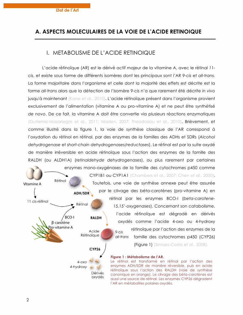
comme illustré dans la figure 1, la voie de synthèse classique de l’AR correspond à
l’oxydation du rétinol en rétinal, par des enzymes de la familles des ADHs et SDRs (Alcohol
dehydrogenase et short-chain dehydrogenases/reductases). Le rétinal est par la suite oxydé
de manière irréversible en acide rétinoïque sous l’action des enzymes de la famille des
RALDH (ou ALDH1A) (retinaldehyde dehydrogenases), ou plus rarement par certaines
enzymes mono-oxygénases de la famille des cytochromes p450 comme
CYP1B1 ou CYP1A1 (Chambers et al., 2007; Chen et al., 2000).
Toutefois, une voie de synthèse annexe peut être assurée
par le clivage des béta-carotènes (pro-vitamine A) en
rétinal par les enzymes BCO-I (beta-carotene-
15,15’-oxygenases). Concernant son catabolisme,
l’acide rétinoïque est dégradé en dérivés
oxydés comme l’acide 4-oxo ou 4-hydroxy
rétinoïque par l’action des enzymes de la
famille des cytochromes p450 (CYP26)
(Figure 1) (Simoes-Costa et al., 2008).
Figure 1 : Métabolisme de l’AR. Le rétinol est transformé en rétinal par l’action des enzymes ADH/SDR de manière réversible, puis en acide rétinoïque sous l’action des RALDH (voie de synthèse canonique en orange). Le clivage des béta-carotènes est aussi une source de rétinal. Les enzymes CYP26 dégradent l’AR en métabolites polaires oxydés.
3
Ainsi, le terme « rétinoïdes » regroupe à la fois le rétinol, l’AR, les dérivés métaboliques
et les composés synthétiques actifs (Sporn et al., 1994).
Les rétinoïdes sont des substances de petite taille, hydrophobes et liposolubles, ce qui
leur permet de traverser facilement la bicouche lipidique membranaire. Une fois dans le
cytoplasme, ils sont pris en charge par les protéines CRBP (Cellular retinol binding protein) et
CRABP (Cellular retinoic acid binding protein) qui régulent leur métabolisme soit en les
stockant, soit en facilitant leur présentation aux différentes enzymes pour des réactions
d’estérification ou d’hydrolyse (Theodosiou et al., 2010).
II. RAR et RXR: UNE STRUCTURE MODULAIRE
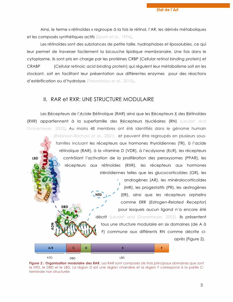
Les Récepteurs de l’Acide Rétinoïque (RAR) ainsi que les Récepteurs X des Rétinoïdes
(RXR) appartiennent à la superfamille des Récepteurs Nucléaires (RN) (Laudet and
Gronemeyer, 2002). Au moins 48 membres ont été identifiés dans le génome humain
(Robinson-Rechavi et al., 2001) et peuvent être regroupés en plusieurs sous-
familles incluant les récepteurs aux hormones thyroïdiennes (TR), à l’acide
rétinoïque (RAR), à la vitamine D (VDR), à l’ecdysone (EcR), les récepteurs
contrôlant l’activation de la prolifération des peroxysomes (PPAR), les
récepteurs aux rétinoïdes (RXR), les récepteurs aux hormones
stéroïdiennes telles que les glucocorticoïdes (GR), les
androgènes (AR), les minéralocorticoïdes
(MR), les progestatifs (PR), les œstrogènes
(ER), ainsi que les récepteurs orphelins
comme ERR (Estrogen-Related Receptor)
pour lesquels aucun ligand n’a encore été
décrit (Laudet and Gronemeyer, 2002). Ils présentent
tous une structure modulaire en six domaines (de A à
F) commune aux différents RN comme décrite ci-
après (Figure 2).
Figure 2 : Organisation modulaire des RAR. Les RAR sont composés de trois principaux domaines que sont le NTD, le DBD et le LBD. La région D est une région charnière et la région F correspond à la partie C-terminale non structurée
4
Les RAR sont des facteurs de transcription dépendants du ligand qui agissent sous
forme d’hétérodimères avec les RXR pour transduire le signal de l’AR. Trois sous-types de RAR
codés par des gènes distincts sont présents chez les mammifères : α (NR1B1), β (NR1B2) et γ
(NR1B3) (Chambon, 1996; Germain et al., 2006a) ainsi que pour RXR : α (NR2B1), β (NR2B2), γ
(NR2B3)(Germain et al., 2006b). Pour chaque sous-type, différentes isoformes différant
seulement dans leur région N-terminale peuvent être transcrits par l’utilisation de différents
promoteurs et par épissage alternatif.
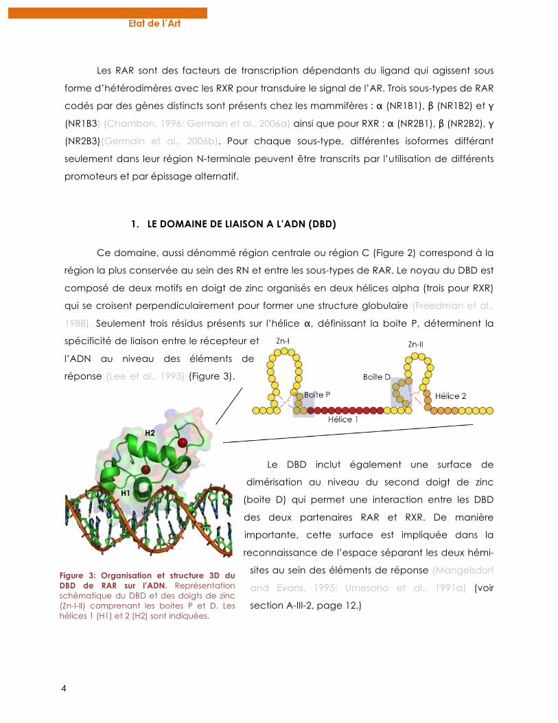
1. LE DOMAINE DE LIAISON A L’ADN (DBD)
Ce domaine, aussi dénommé région centrale ou région C (Figure 2) correspond à la
région la plus conservée au sein des RN et entre les sous-types de RAR. Le noyau du DBD est
composé de deux motifs en doigt de zinc organisés en deux hélices alpha (trois pour RXR)
qui se croisent perpendiculairement pour former une structure globulaire (Freedman et al.,
1988). Seulement trois résidus présents sur l’hélice α, définissant la boite P, déterminent la
spécificité de liaison entre le récepteur et
l’ADN au niveau des éléments de
réponse (Lee et al., 1993) (Figure 3).
Le DBD inclut également une surface de
dimérisation au niveau du second doigt de zinc
(boite D) qui permet une interaction entre les DBD
des deux partenaires RAR et RXR. De manière
importante, cette surface est impliquée dans la
reconnaissance de l’espace séparant les deux hémi-
sites au sein des éléments de réponse (Mangelsdorf
and Evans, 1995; Umesono et al., 1991a) (voir
section A-III-2, page 12.)
Figure 3: Organisation et structure 3D du DBD de RAR sur l’ADN. Représentation schématique du DBD et des doigts de zinc (Zn-I-II) comprenant les boites P et D. Les hélices 1 (H1) et 2 (H2) sont indiquées.
5
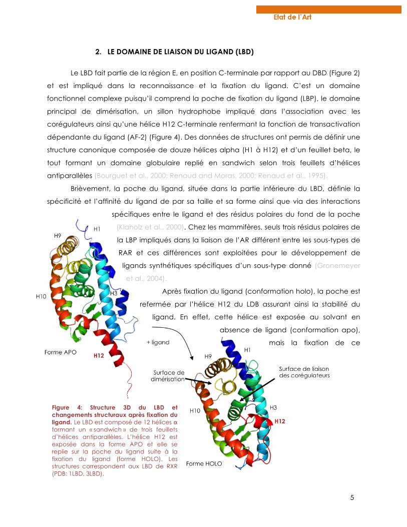
2. LE DOMAINE DE LIAISON DU LIGAND (LBD)
Le LBD fait partie de la région E, en position C-terminale par rapport au DBD (Figure 2)
et est impliqué dans la reconnaissance et la fixation du ligand. C’est un domaine
fonctionnel complexe puisqu’il comprend la poche de fixation du ligand (LBP), le domaine
principal de dimérisation, un sillon hydrophobe impliqué dans l’association avec les
corégulateurs ainsi qu’une hélice H12 C-terminale renfermant la fonction de transactivation
dépendante du ligand (AF-2) (Figure 4). Des données de structures ont permis de définir une
structure canonique composée de douze hélices alpha (H1 à H12) et d’un feuillet beta, le
tout formant un domaine globulaire replié en sandwich selon trois feuillets d’hélices
antiparallèles (Bourguet et al., 2000; Renaud and Moras, 2000; Renaud et al., 1995).
Brièvement, la poche du ligand, située dans la partie inférieure du LBD, définie la
spécificité et l’affinité du ligand de par sa taille et sa forme ainsi que via des interactions
spécifiques entre le ligand et des résidus polaires du fond de la poche
(Klaholz et al., 2000). Chez les mammifères, seuls trois résidus polaires de
la LBP impliqués dans la liaison de l’AR différent entre les sous-types de
RAR et ces différences sont exploitées pour le développement de
ligands synthétiques spécifiques d’un sous-type donné (Gronemeyer
et al., 2004).
Après fixation du ligand (conformation holo), la poche est
refermée par l’hélice H12 du LDB assurant ainsi la stabilité du
ligand. En effet, cette hélice est exposée au solvant en
absence de ligand (conformation apo),
mais la fixation de ce
Figure 4: Structure 3D du LBD et changements structuraux après fixation du ligand. Le LBD est composé de 12 hélices α formant un « sandwich » de trois feuillets d’hélices antiparallèles. L’hélice H12 est exposée dans la forme APO et elle se replie sur la poche du ligand suite à la fixation du ligand (forme HOLO). Les structures correspondent aux LBD de RXR (PDB: 1LBD, 3LBD).
6
dernier mène à des repositionnement d’hélices au sein du LBD (transconformation (Moras
and Gronemeyer, 1998)), en particulier l’alignement des hélices H10 et H11, qui résulte in
fine au basculement de l’hélice H12 sur la LBP, la refermant comme un piège à souris (Figure
4)(Moras and Gronemeyer, 1998; Renaud et al., 1995). Dans cette conformation, des ponts
salins au sein du LBD stabilisent et rigidifient la structure (Bourguet et al., 1995) et de
nouveaux résidus sont exposés à la surface du LBD formant un sillon hydrophobe (domaine
AF-2) propice à l’interaction avec des coactivateurs. Enfin, il a été montré que la fixation du
ligand induit également des changements allostériques entre la LBP et la surface de
dimérisation, conférant une stabilité de l’hétérodimère RAR-RXR (Brelivet et al., 2004;
Pogenberg et al., 2005).
En conclusion, le LBD est le domaine majeur du récepteur lui conférant sa spécificité
de ligand. C’est également ce qui le distingue d’autres facteurs de transcription en
conférant à son activité, une dépendance au ligand. C’est un domaine complexe au sein
duquel de nombreuses régions sont énergétiquement liées favorisant ainsi des
changements allostériques subtils et importants permettant une adaptation du récepteur en
fonction de son environnement moléculaire. Ces communications intramoléculaires sont
particulièrement discutées dans le chapitre 2, page 156.
3. LE DOMAINE N-TERMINAL (NTD)
Le NTD correspond aux deux régions A et B et contient une fonction de
transactivation indépendante du ligand AF-1 (Nagpal et al., 1993; Nagpal et al., 1992). Il est
le domaine qui diffère le plus en séquence et en longueur au sein des RN mais aussi entre les
sous-types de RAR et RXR. De plus pour chaque sous-type, il existe plusieurs isoformes qui
diffèrent au niveau de la région A suite à des épissages alternatifs ou à l’utilisation de
promoteurs différents (Chambon, 1996). A l’inverse, la région B est très conservée entre les
différents sous-types de RAR. A lui seul, le NTD est capable d’activer la transcription de
gènes cibles en absence de ligand du fait de sa fonction AF-1. Bien que cette fonction
redevienne dépendante du ligand dans le contexte du récepteur entier, cela suggère de
fortes relations synergiques entre le NTD et le LBD des RAR (Taneja et al., 1997).
Du fait de sa structure naturellement désordonnée (IUD : Intrinsically Unstructured
Domain) (McEwan et al., 2007), caractérisée par l’absence d’une structure tertiaire stable,
le NTD est très sensible à la protéolyse et aucune donnée structurale n’a jamais été obtenue
7
concernant ce domaine des RAR. Cependant, ces IUD peuvent adopter des états
conformationnels pseudo-structurés transitoires suite à l’interaction avec d’autres protéines
ou après liaison à l’ADN (Dyson and Wright, 2005; Lavery and McEwan, 2005; Wright and
Dyson, 2009). De plus, le NTD des RAR possède un motif riche en proline contenant un motif
consensus de phosphorylation au niveau d’une serine (voir section A-IV-2.1, page 18.) qui
est susceptible de former des pseudo-structures hélicoïdales transitoires (Bielska and Zondlo,
2006; Rochette-Egly et al., 1997). Enfin ces domaines riches en prolines (PRM : Proline rich
motif) ont la capacité d’interagir avec des protéines à domaines SH3 ou WW (Reimand et
al., 2012).
Récemment, plusieurs études ont révélé des interactions entre le NTD et certaines
protéines comme HACE1, Acinus-S’ ou la vinexine beta réprimant l’activité transcriptionnelle
des RAR (Bour et al., 2005b; Vucetic et al., 2008; Zhao et al., 2009). De manière intéressante,
de par la proximité du NTD avec le DBD, des interactions protéiques au niveau des régions A
et B seraient susceptibles de réguler la liaison du récepteur à l’ADN (Lalevee et al., 2010).
Cette hypothèse est renforcée par la localisation du domaine A/B proche de l’ADN révélée
par des données récentes de structure du complexe RXR/VDR lié à l’ADN (Orlov et al., 2012).
En conclusion, bien que le NTD soit encore très peu étudié de par sa nature
désordonnée à l’inverse du LBD et du DBD, son implication fonctionnelle dans l’activité
transcriptionnelle des RAR est de plus en plus reconnue. De plus, le fait que ce domaine
diffère grandement entre les différents sous-types de RAR permet de spéculer sur son
implication dans la spécificité d’action de chaque sous-type de RAR (discuté en chapitre 1,
page 134).
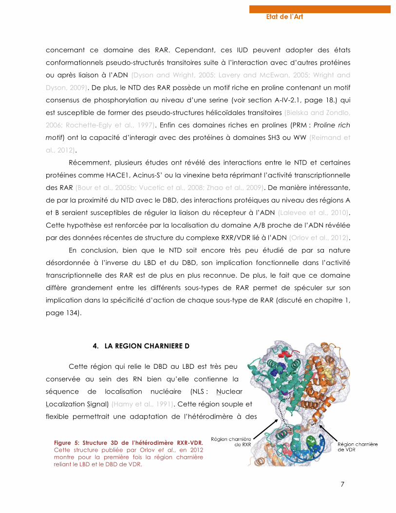
4. LA REGION CHARNIERE D
Cette région qui relie le DBD au LBD est très peu
conservée au sein des RN bien qu’elle contienne la
séquence de localisation nucléaire (NLS : Nuclear
Localization Signal) (Hamy et al., 1991). Cette région souple et
flexible permettrait une adaptation de l’hétérodimère à des
Figure 5: Structure 3D de l’hétérodimère RXR-VDR. Cette structure publiée par Orlov et al., en 2012 montre pour la première fois la région charnière reliant le LBD et le DBD de VDR.
8
éléments de réponses différents au niveau de l’ADN (voir section A-III-2, page 12.). Bien
qu’elle ne soit pas toujours considérée comme une entité à part entière, la région D a été
mise en évidence par des données structurales récentes au sein du complexe RXR/PPAR ou
RXR/VDR (Chandra et al., 2008; Orlov et al., 2012). Ces études décrivent un rôle clé de cette
région charnière dans la conformation et la stabilisation de l’hétérodimère sur l’ADN (Figure
5).
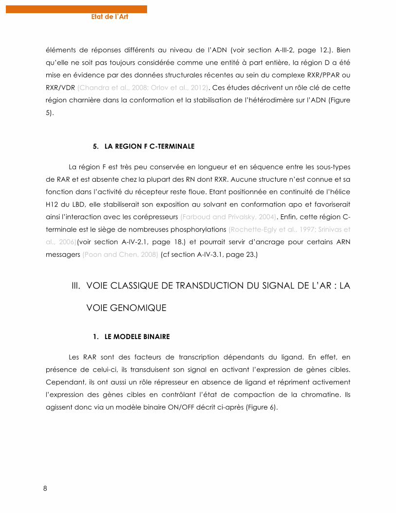
5. LA REGION F C-TERMINALE
La région F est très peu conservée en longueur et en séquence entre les sous-types
de RAR et est absente chez la plupart des RN dont RXR. Aucune structure n’est connue et sa
fonction dans l’activité du récepteur reste floue. Etant positionnée en continuité de l’hélice
H12 du LBD, elle stabiliserait son exposition au solvant en conformation apo et favoriserait
ainsi l’interaction avec les corépresseurs (Farboud and Privalsky, 2004). Enfin, cette région C-
terminale est le siège de nombreuses phosphorylations (Rochette-Egly et al., 1997; Srinivas et
al., 2006)(voir section A-IV-2.1, page 18.) et pourrait servir d’ancrage pour certains ARN
messagers (Poon and Chen, 2008) (cf section A-IV-3.1, page 23.)
III. VOIE CLASSIQUE DE TRANSDUCTION DU SIGNAL DE L’AR : LA
VOIE GENOMIQUE
1. LE MODELE BINAIRE
Les RAR sont des facteurs de transcription dépendants du ligand. En effet, en
présence de celui-ci, ils transduisent son signal en activant l’expression de gènes cibles.
Cependant, ils ont aussi un rôle répresseur en absence de ligand et répriment activement
l’expression des gènes cibles en contrôlant l’état de compaction de la chromatine. Ils
agissent donc via un modèle binaire ON/OFF décrit ci-après (Figure 6).
9
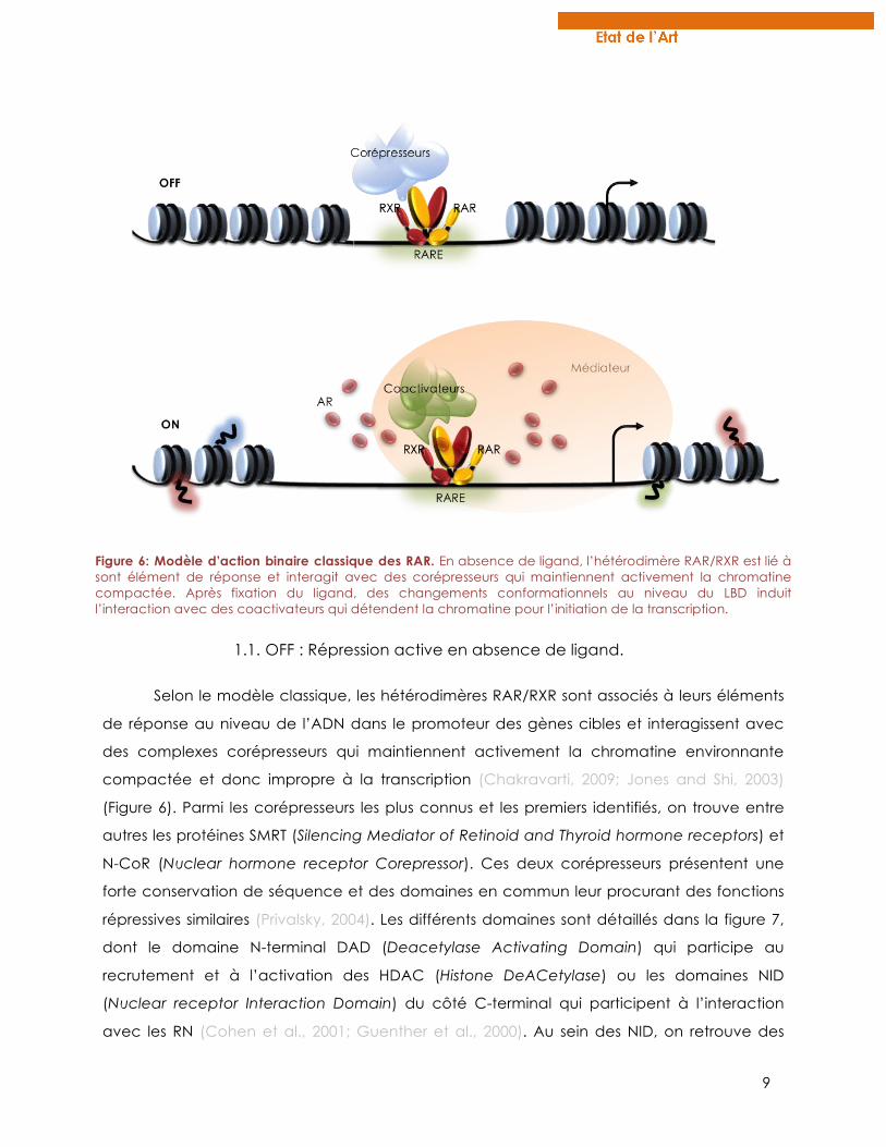
1.1. OFF : Répression active en absence de ligand.
Selon le modèle classique, les hétérodimères RAR/RXR sont associés à leurs éléments
de réponse au niveau de l’ADN dans le promoteur des gènes cibles et interagissent avec
des complexes corépresseurs qui maintiennent activement la chromatine environnante
compactée et donc impropre à la transcription (Chakravarti, 2009; Jones and Shi, 2003)
(Figure 6). Parmi les corépresseurs les plus connus et les premiers identifiés, on trouve entre
autres les protéines SMRT (Silencing Mediator of Retinoid and Thyroid hormone receptors) et
N-CoR (Nuclear hormone receptor Corepressor). Ces deux corépresseurs présentent une
forte conservation de séquence et des domaines en commun leur procurant des fonctions
répressives similaires (Privalsky, 2004). Les différents domaines sont détaillés dans la figure 7,
dont le domaine N-terminal DAD (Deacetylase Activating Domain) qui participe au
recrutement et à l’activation des HDAC (Histone DeACetylase) ou les domaines NID
(Nuclear receptor Interaction Domain) du côté C-terminal qui participent à l’interaction
avec les RN (Cohen et al., 2001; Guenther et al., 2000). Au sein des NID, on retrouve des
Figure 6: Modèle d’action binaire classique des RAR. En absence de ligand, l’hétérodimère RAR/RXR est lié à sont élément de réponse et interagit avec des corépresseurs qui maintiennent activement la chromatine compactée. Après fixation du ligand, des changements conformationnels au niveau du LBD induit l’interaction avec des coactivateurs qui détendent la chromatine pour l’initiation de la transcription.
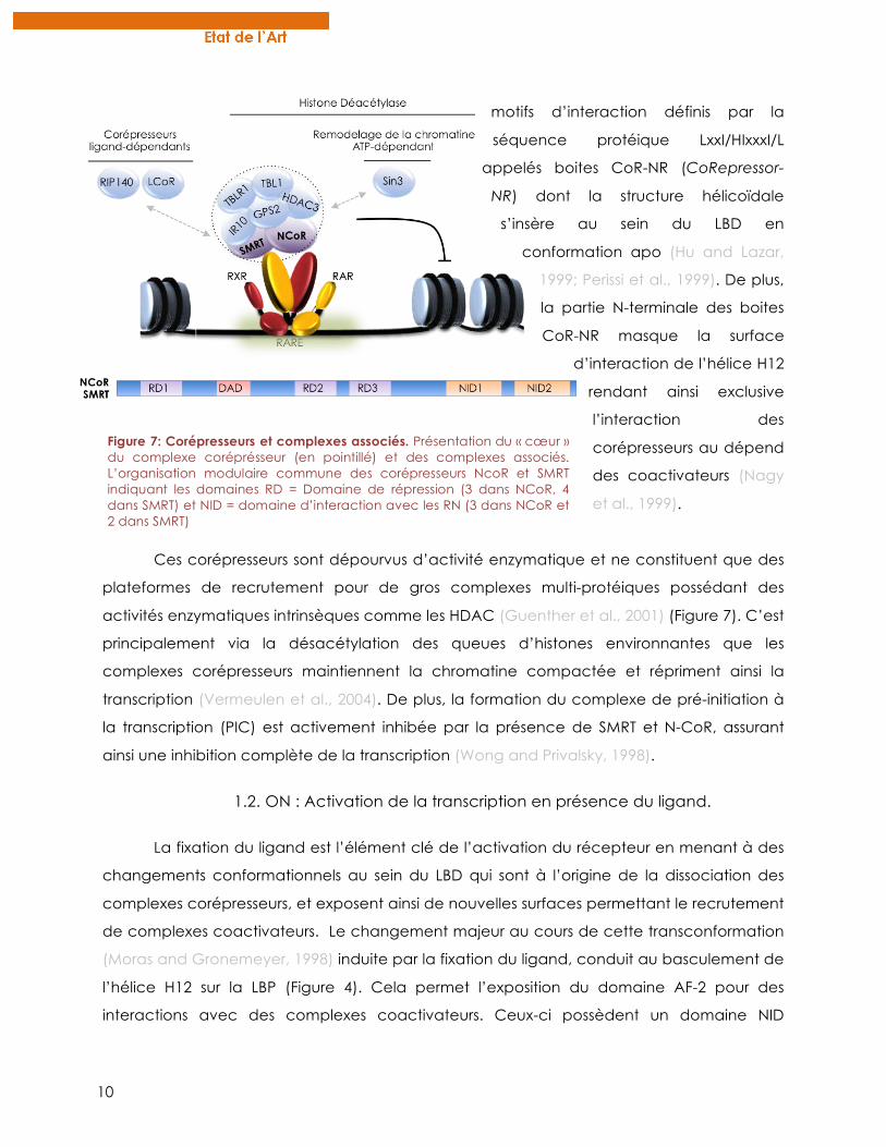
10
motifs d’interaction définis par la
séquence protéique LxxI/HIxxxI/L
appelés boites CoR-NR (CoRepressor-
NR) dont la structure hélicoïdale
s’insère au sein du LBD en
conformation apo (Hu and Lazar,
1999; Perissi et al., 1999). De plus,
la partie N-terminale des boites
CoR-NR masque la surface
d’interaction de l’hélice H12
rendant ainsi exclusive
l’interaction des
corépresseurs au dépend
des coactivateurs (Nagy
et al., 1999).
Ces corépresseurs sont dépourvus d’activité enzymatique et ne constituent que des
plateformes de recrutement pour de gros complexes multi-protéiques possédant des
activités enzymatiques intrinsèques comme les HDAC (Guenther et al., 2001) (Figure 7). C’est
principalement via la désacétylation des queues d’histones environnantes que les
complexes corépresseurs maintiennent la chromatine compactée et répriment ainsi la
transcription (Vermeulen et al., 2004). De plus, la formation du complexe de pré-initiation à
la transcription (PIC) est activement inhibée par la présence de SMRT et N-CoR, assurant
ainsi une inhibition complète de la transcription (Wong and Privalsky, 1998).
1.2. ON : Activation de la transcription en présence du ligand.
La fixation du ligand est l’élément clé de l’activation du récepteur en menant à des
changements conformationnels au sein du LBD qui sont à l’origine de la dissociation des
complexes corépresseurs, et exposent ainsi de nouvelles surfaces permettant le recrutement
de complexes coactivateurs. Le changement majeur au cours de cette transconformation
(Moras and Gronemeyer, 1998) induite par la fixation du ligand, conduit au basculement de
l’hélice H12 sur la LBP (Figure 4). Cela permet l’exposition du domaine AF-2 pour des
interactions avec des complexes coactivateurs. Ceux-ci possèdent un domaine NID
Figure 7: Corépresseurs et complexes associés. Présentation du « cœur » du complexe coréprésseur (en pointillé) et des complexes associés. L’organisation modulaire commune des corépresseurs NcoR et SMRT indiquant les domaines RD = Domaine de répression (3 dans NCoR, 4 dans SMRT) et NID = domaine d’interaction avec les RN (3 dans NCoR et 2 dans SMRT)
11
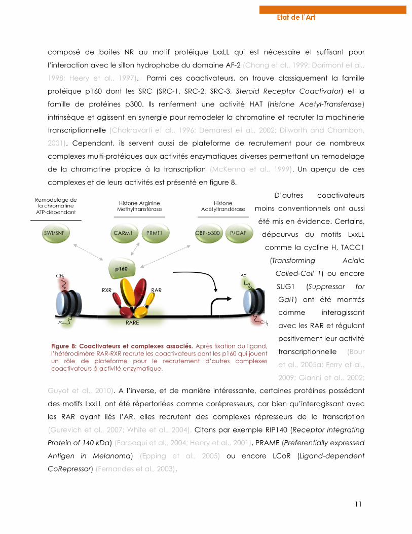
composé de boites NR au motif protéique LxxLL qui est nécessaire et suffisant pour
l’interaction avec le sillon hydrophobe du domaine AF-2 (Chang et al., 1999; Darimont et al.,
1998; Heery et al., 1997). Parmi ces coactivateurs, on trouve classiquement la famille
protéique p160 dont les SRC (SRC-1, SRC-2, SRC-3, Steroid Receptor Coactivator) et la
famille de protéines p300. Ils renferment une activité HAT (Histone Acetyl-Transferase)
intrinsèque et agissent en synergie pour remodeler la chromatine et recruter la machinerie
transcriptionnelle (Chakravarti et al., 1996; Demarest et al., 2002; Dilworth and Chambon,
2001). Cependant, ils servent aussi de plateforme de recrutement pour de nombreux
complexes multi-protéiques aux activités enzymatiques diverses permettant un remodelage
de la chromatine propice à la transcription (McKenna et al., 1999). Un aperçu de ces
complexes et de leurs activités est présenté en figure 8.
D’autres coactivateurs
moins conventionnels ont aussi
été mis en évidence. Certains,
dépourvus du motifs LxxLL
comme la cycline H, TACC1
(Transforming Acidic
Coiled-Coil 1) ou encore
SUG1 (Suppressor for
Gal1) ont été montrés
comme interagissant
avec les RAR et régulant
positivement leur activité
transcriptionnelle (Bour
et al., 2005a; Ferry et al.,
2009; Gianni et al., 2002;
Guyot et al., 2010). A l’inverse, et de manière intéressante, certaines protéines possédant
des motifs LxxLL ont été répertoriées comme corépresseurs, car bien qu’interagissant avec
les RAR ayant liés l’AR, elles recrutent des complexes répresseurs de la transcription
(Gurevich et al., 2007; White et al., 2004). Citons par exemple RIP140 (Receptor Integrating
Protein of 140 kDa) (Farooqui et al., 2004; Heery et al., 2001), PRAME (Preferentially expressed
Antigen in Melanoma) (Epping et al., 2005) ou encore LCoR (Ligand-dependent
CoRepressor) (Fernandes et al., 2003).
Figure 8: Coactivateurs et complexes associés. Après fixation du ligand, l’hétérodimère RAR-RXR recrute les coactivateurs dont les p160 qui jouent un rôle de plateforme pour le recrutement d’autres complexes coactivateurs à activité enzymatique.
12
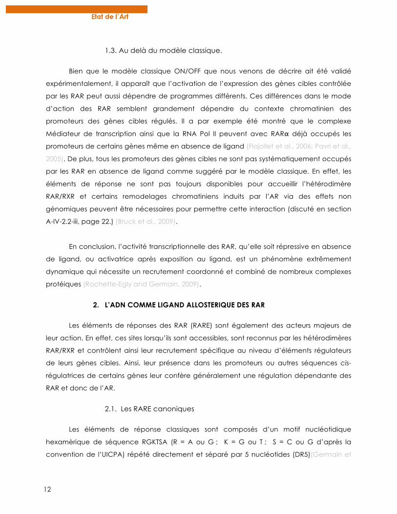
1.3. Au delà du modèle classique.
Bien que le modèle classique ON/OFF que nous venons de décrire ait été validé
expérimentalement, il apparaît que l’activation de l’expression des gènes cibles contrôlée
par les RAR peut aussi dépendre de programmes différents. Ces différences dans le mode
d’action des RAR semblent grandement dépendre du contexte chromatinien des
promoteurs des gènes cibles régulés. Il a par exemple été montré que le complexe
Médiateur de transcription ainsi que la RNA Pol II peuvent avec RARα déjà occupés les
promoteurs de certains gènes même en absence de ligand (Flajollet et al., 2006; Pavri et al.,
2005). De plus, tous les promoteurs des gènes cibles ne sont pas systématiquement occupés
par les RAR en absence de ligand comme suggéré par le modèle classique. En effet, les
éléments de réponse ne sont pas toujours disponibles pour accueillir l’hétérodimère
RAR/RXR et certains remodelages chromatiniens induits par l’AR via des effets non
génomiques peuvent être nécessaires pour permettre cette interaction (discuté en section
A-IV-2.2-iii, page 22.) (Bruck et al., 2009).
En conclusion, l’activité transcriptionnelle des RAR, qu’elle soit répressive en absence
de ligand, ou activatrice après exposition au ligand, est un phénomène extrêmement
dynamique qui nécessite un recrutement coordonné et combiné de nombreux complexes
protéiques (Rochette-Egly and Germain, 2009).
2. L’ADN COMME LIGAND ALLOSTERIQUE DES RAR
Les éléments de réponses des RAR (RARE) sont également des acteurs majeurs de
leur action. En effet, ces sites lorsqu’ils sont accessibles, sont reconnus par les hétérodimères
RAR/RXR et contrôlent ainsi leur recrutement spécifique au niveau d’éléments régulateurs
de leurs gènes cibles. Ainsi, leur présence dans les promoteurs ou autres séquences cis-
régulatrices de certains gènes leur confère généralement une régulation dépendante des
RAR et donc de l’AR.
2.1. Les RARE canoniques
Les éléments de réponse classiques sont composés d’un motif nucléotidique
hexamèrique de séquence RGKTSA (R = A ou G ; K = G ou T ; S = C ou G d’après la
convention de l’UICPA) répété directement et séparé par 5 nucléotides (DR5)(Germain et
13
al., 2003; Leid et al., 1992; Umesono et al., 1991b). Plusieurs RARE de type DR5 ont été
caractérisés dans les promoteurs de gènes cibles canoniques comme RARb2 (de The et al.,
1990), cyp26a1 (Loudig et al., 2000) ou certains gènes hox (Dupe et al., 1997). La recherche
in silico à l’échelle du génome entier de motifs DR5 conservés entre espèces de vertébrés a
permis d’élargir la liste de RARE de type DR5 fonctionnels dans les promoteurs de gènes
cibles pour lesquels aucun élément de réponse n’était encore connu ((Lalevee et al., 2011)
publication en annexe, page 207).
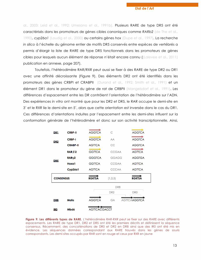
Toutefois, l’hétérodimère RAR/RXR peut aussi se fixer à des RARE de type DR2 ou DR1
avec une affinité décroissante (Figure 9). Des éléments DR2 ont été identifiés dans les
promoteurs des gènes CRBPI et CRABPII (Durand et al., 1992; Smith et al., 1991) et un
élément DR1 dans le promoteur du gène de rat de CRBPII (Mangelsdorf et al., 1991). Les
différences d’espacement entre les DR contrôlent l’orientation de l’hétérodimère sur l’ADN.
Des expériences in vitro ont montré que pour les DR2 et DR5, le RAR occupe le demi-site en
3’ et le RXR lie le demi-site en 5’, alors que cette orientation est inversée dans le cas du DR1.
Ces différences d’orientations induites par l’espacement entre les demi-sites influent sur la
conformation générale de l’hétérodimère et donc sur son activité transcriptionnelle. Ainsi,
Figure 9: Les différents types de RARE. L’hétérodimère RAR-RXR peut se fixer sur des RARE avec différents espacements. Les RARE de type DR1, DR2 et DR5 ont été les premiers décrits et définissent la séquence consensus. Récemment, des concaténations de DR0 et DR2 en DR8 ainsi que des IR0 ont été mis en évidence. Les séquences données correspondant aux RARE trouvés dans les gènes de souris correspondants. Les demi-sites occupés par RAR sont en rouge et ceux par RXR en jaune
14
alors que la fixation de RAR/RXR sur un DR5 a un rôle activateur, l’hétérodimère devient
répresseur sur un élément de type DR1 du fait d’une interaction constitutive avec des
corépresseurs (Zamir et al., 1997). En plus de l’espacement, il a été montré dans le cas du
récepteur aux glucocorticoïdes (GR), que la séquence nucléotidique de l’élément de
réponse pouvait également jouer un rôle sur la conformation du DBD (Meijsing et al., 2009).
Ainsi, des éléments de réponse de même conformation mais de séquences variables
peuvent réguler différemment l’activité du récepteur.
L’ADN peut donc être considéré comme un ligand allostérique des RAR car sa
liaison aux récepteurs régule la dynamique d’association-dissociation avec des
corégulateurs et influence ainsi leur activité transcriptionnelle au même titre que leur ligand
moléculaire qu’est l’AR (Kurokawa et al., 1995; Meijsing et al., 2009).
2.2. Un éventail plus large d’éléments de réponse
Des études récentes de séquençage haut débit après immunoprécipitation de la
chromatine (ChiP-seq) dans des cellules indifférenciées de souris (cellules F9 ou corps
embryonnaire de cellules souches) ont permis de mettre en évidence la gamme d’éléments
de réponse reconnus effectivement par RAR. En effet dans ces modèles cellulaires, les RAR
occupent un large répertoire de sites DR0, DR8 et IR0 (répétition inversée, figure 9) et
l’affinité du récepteur pour ces RARE est aussi forte que celle pour les éléments classiques
de type DR5 ou DR2 (données in vitro) (Moutier et al., 2012). Le nombre de sites occupés
arborant un DR0 est même grandement supérieur à ceux comprenant un DR5. Cependant,
contrairement aux DR8 et IR0, le motif DR0 ne semble pas pouvoir à lui seul réguler
l’expression du gène qui lui est associé. Toutefois, et de manière intéressante, il est souvent
observé une concaténation d’éléments DR0 et DR2 en DR8 (figure 9). Ce composite DR8 est
fonctionnel et est retrouvé fréquemment dans les nouvelles données de ChIP-seq comme
liant RAR/RXR, il est alors proposé de considérer les composites DR8 comme de nouveaux
éléments de réponse pour les RAR. Concernant les sites liant RAR/RXR mais ne permettant
pas de régulation transcriptionnelle, comme les DR0, ils pourraient être impliqués dans des
occupations compétitives d’éléments normalement lié par d’autres facteurs de
transcription (Gu et al., 2005).
En plus de l’espacement, la séquence consensus des demi-sites ne semble pas
toujours répondre strictement au motif RGKTSA décrit jusqu’alors. En effet, de nombreux
RARE dégénérés, c’est à dire différant par un ou deux nucléotides dans au moins un de leurs
15
demi-sites, ont été identifiés (Delacroix et al., 2010). Alors que les sites les plus fortement
occupés par RAR/RXR dans des cellules souches de souris sont composés de RARE
canoniques, les sites dégénérés correspondent à des sites moins fréquemment occupés.
Cela suggère qu’il existe un continuum de différents RARE (en séquence et en espacement)
qui lient l’hétérodimère avec des affinités différentes et dans différents contextes. Ainsi, en
fonction du contexte cellulaire (quiescence, prolifération, différentiation) ou
développemental, les sites occupés par les RAR pourraient être variables du fait des
différences d’organisation de la chromatine, et ainsi participer à la régulation fine de
l’activité des RAR (Delacroix et al., 2010; Lalevee et al., 2011).
Ces résultats issus de nouvelles techniques révèlent une grande diversité de
topologies et d’espacement des RARE. Cette diversité semble être spécifique des RAR
puisque d’autres RN comme VDR ou PPAR ont un répertoire de site de liaison beaucoup
plus restrictif (Nielsen et al., 2008; Ramagopalan et al., 2010). La dégénérescence des RARE
pourrait participer à réguler finement la liaison du récepteur à l’ADN en engendrant des
niveaux d’affinité différents. Enfin, le répertoire de RARE occupés par les RAR diffèrent d’un
type cellulaire à l’autre suggérant que la fonctionnalité de ces sites dépend du contexte
cellulaire.
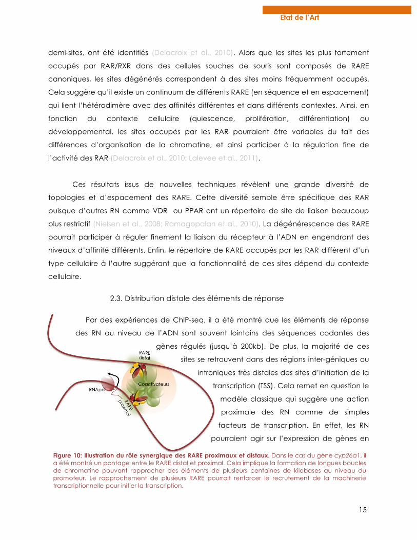
2.3. Distribution distale des éléments de réponse
Par des expériences de ChIP-seq, il a été montré que les éléments de réponse
des RN au niveau de l’ADN sont souvent lointains des séquences codantes des
gènes régulés (jusqu’à 200kb). De plus, la majorité de ces
sites se retrouvent dans des régions inter-géniques ou
introniques très distales des sites d’initiation de la
transcription (TSS). Cela remet en question le
modèle classique qui suggère une action
proximale des RN comme de simples
facteurs de transcription. En effet, les RN
pourraient agir sur l’expression de gènes en
Figure 10: Illustration du rôle synergique des RARE proximaux et distaux. Dans le cas du gène cyp26a1, il a été montré un pontage entre le RARE distal et proximal. Cela implique la formation de longues boucles de chromatine pouvant rapprocher des éléments de plusieurs centaines de kilobases au niveau du promoteur. Le rapprochement de plusieurs RARE pourrait renforcer le recrutement de la machinerie transcriptionnelle pour initier la transcription.
16
tant qu’enhancers à longue portée (long-range enhancers) de par leur position distale par
rapport aux promoteurs des gènes cibles (Carroll et al., 2006; Moutier et al., 2012; Nielsen et
al., 2008; Welboren et al., 2009). De ce fait, la régulation de ces gènes ferait intervenir de
longues boucles chromosomiques regroupant ainsi des éléments de réponse lointains au
niveau des promoteurs des gènes régulés (Figure 10) (Biddie et al., 2010; Carroll et al., 2005).
En particulier, des études récentes du laboratoire de Cécile Rochette-Egly ont montré que
les deux éléments de réponse du gène cyp26a1 de mammifère (DR5 distal et proximal) sont
rapidement pontés après fixation des récepteurs à l’ADN et coopèrent ainsi pour l’initiation
de la transcription induite par l’AR (Bruck et al., 2009).
2.4. Dialogue avec l’épigénome
L’épigénome qui constitue l’ensemble des modifications épigénétiques d’une
cellule, joue également un rôle important dans la liaison des récepteurs à l’ADN. En effet, en
plus de la séquence brute des éléments de réponse, les modifications épigénétiques de la
chromatine vont, entre autres, déterminer leur accessibilité (Biddie et al., 2010). Comme vu
précédemment, les RN recrutent des complexes protéiques à activité enzymatique qui
modifient la chromatine environnante pour la garder compacte ou pour la rendre
accessible (voir section A-III-1, page 8). Ainsi, de nombreuses modifications post-
traductionnelles de queues d’histone favorisant l’accessibilité de la chromatine ont été
corrélées à la liaison des RN au niveau des éléments de réponse (Kininis et al., 2007; Lupien
et al., 2009). Cependant, il a été montré que la liaison même du récepteur est sensible à
l’environnement épigénétique et que ces modifications jouent un rôle primordial dans le
recrutement du récepteur au niveau de l’élément de réponse (Eeckhoute et al., 2006; John
et al., 2008; Martens et al., 2011). En d’autres termes, certains prérequis épigénétiques sont
nécessaires pour permettre l’accessibilité des éléments de réponse et donc la liaison du
récepteur à l’ADN. Ces observations remettent en question le rôle des RN comme facteurs
premiers permettant le remodelage chromatinien mais suggèrent plutôt un dialogue étroit
avec d’autres voies de signalisation cellulaire qui régulent l’épigénome en amont. Dans le
cas des RAR, il a été montré que certaines kinases activées par l’AR participent à
l’accessibilité de la chromatine au niveau des RARE (Bruck et al., 2009) (voir section A-IV-2.2-
iii, page 22.).
17
IV. VOIE NON GENOMIQUE DE L’AR : NOUVEAUX CONCEPTS
De nombreuses études, certaines issues du laboratoire de Cécile Rochette-Egly, ont
montré avec évidence qu’en plus des effets génomiques classiques de l’AR via l’activité
transcriptionnelle des RAR, le signal de l’AR était également transduit par des voies non
génomiques moins conventionnelles, mais faisant également intervenir les RAR (Al Tanoury
et al., 2013).
1. ACTIVATION DE VOIES KINASIQUES ET RAR MEMBRANAIRES
Dans différentes lignées cellulaires, il a été montré que l’AR active rapidement la
protéine kinase PKC (Kambhampati et al., 2003) ainsi que la voie de signalisation PI3K/Akt
(Bastien et al., 2006; Masia et al., 2007). L’AR active aussi la voie des MAPK/ERK dans
différentes lignées de cellules neuronales lors de la différentiation des cellules souches
embryonnaires en neurones (Miloso et al., 2004; Stavridis et al., 2010). Enfin, l’AR active la
voie des p38MAPK et de la kinase MSK1 en aval dans plusieurs lignées cellulaires de
mammifères (Alsayed et al., 2001; Gianni et al., 2002; Ren et al., 2007). De par l’activation
extrêmement rapide de cette voie après traitement à l’AR (quelques minutes) (Bruck et al.,
2009), ces effets ne peuvent être qu’indépendants de tous phénomènes transcriptionnels.
Toutefois, il a été montré que l’activation de cette voie kinasique nécessite RARα puisque
l’activation des p38MAPK n’est plus observée dans des cellules mutées (knock-out) pour ce
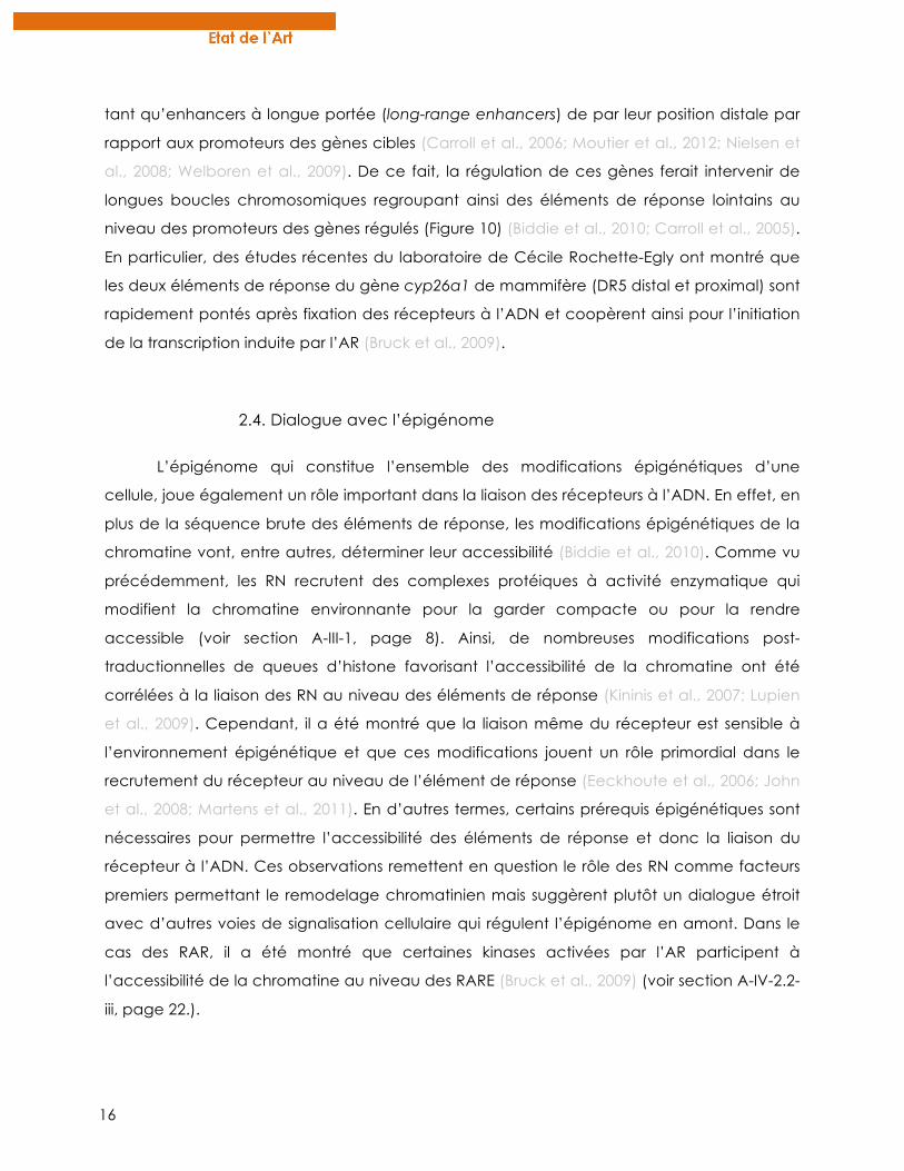
gène (Bruck et al., 2009). Alors que les
mécanismes moléculaires d’une telle
activation via des processus non génomiques
restaient peu compris, des études récentes du
laboratoire de Cécile Rochette-Egly ont montré
qu’une fraction du pool cellulaire de RARα est localisée
Figure 11: Un pool de RARα membranaire permet l’activation de voies kinasiques en réponse à l’AR. Dans des cellules humaines, RARα interagit rapidement avec la protéine G Gαq en réponse à l’AR au sein de radeaux lipidiques (en rouge). Cette interaction est nécessaire pour l’activation de la voie des p38MAPK et l’activation subséquente de MSK1 dans le noyau. Les mécanismes d’ancrage de RARα à la membrane sont inconnus. Dans des cellules de cancer du sein qui ne répondent plus à l’effet antiprolifératif de l’AR, l’interaction entre RARα et Gαq n’est pas observée et la voie p38MAPK n’est pas activée (Piskunov and Rochette-Egly, 2012).
18
dans la membrane cellulaire au niveau de radeaux lipidiques. C’est cette fraction
membranaire de récepteur qui, via l’interaction avec la protéine G Gαq, permet
l’activation de la voie des p38MAPK (Piskunov and Rochette-Egly, 2011b) (Figure 11).
Contrairement aux autres récepteurs des stéroïdes, les RAR n’ont pas de site de
palmitoylation qui faciliterait leur ancrage à la membrane (Pedram et al., 2007). Cependant,
la partie N-terminale (région A/B) est requise pour la localisation de RARα au niveau des
radeaux lipidiques membranaires suggérant que ce domaine non conservé entre les
différents sous-types de RAR joue un rôle pour la localisation extranucléaire (Piskunov and
Rochette-Egly, 2011b). A noter que d’autres RN, comme ER ou GR sont aussi capables
d’activer la voie des MAPK via un pool de récepteurs membranaires (Le Romancer et al.,
2011; Marquez et al., 2006; Matthews et al., 2008; Piskunov and Rochette-Egly, 2011a).
2. INTEGRATION NUCLEAIRE DES ACTIVATIONS KINASIQUES
Dans plusieurs types cellulaires, il a été montré qu’une fois activées par l’AR dans le
cytosol, les kinases sont rapidement transloquées dans le noyau où elles activent d’autres
kinases en aval. Dans le cas de l’activation des p38MAPK par l’AR dans des cellules de
mammifères, c’est la kinase MSK1 qui est finalement activée dans le noyau (Bruck et al.,
2009). Là, elle va transduire le signal de l’AR en phosphorylant de nombreuses protéines de
manière coordonnée.
2.1. Phosphorylation des RAR
i. Les RAR sont des phosphoprotéines
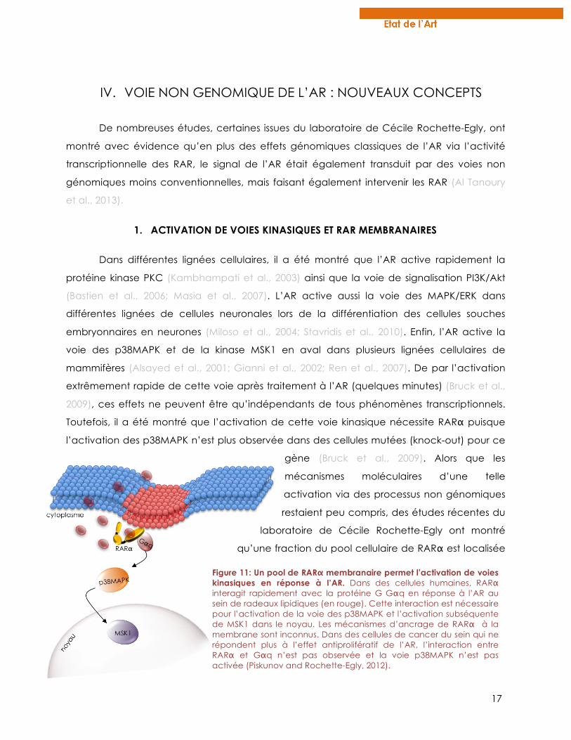
Différents sites de phosphorylations ont été décrits au sein des domaines des RAR et
ont été associés à des fonctions spécifiques (récapitulés dans la figure 12.) De nombreux
signaux, en activant des kinases cytosoliques comme Akt, PKC ou JNK qui transloquent
ensuite dans le noyau, peuvent induire la phosphorylation des RAR à diverses positions et
ainsi réguler certaines de leurs activités et leur localisation (Figure 12) (Rochette-Egly and
Germain, 2009). Nous nous intéresserons par la suite, plus particulièrement aux résidus qui
sont phosphorylés en réponse à l’AR et qui modulent l’activité transcriptionnelle des RAR.
19
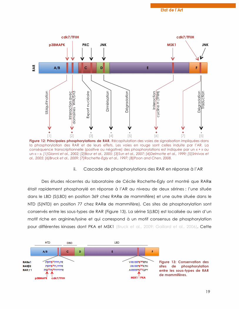
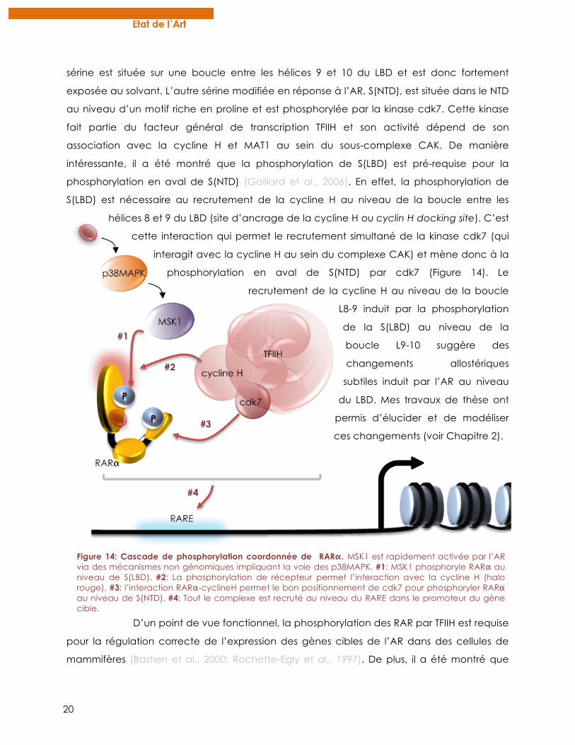
ii. Cascade de phosphorylations des RAR en réponse à l’AR
Des études récentes du laboratoire de Cécile Rochette-Egly ont montré que RARα
était rapidement phosphorylé en réponse à l’AR au niveau de deux sérines : l’une située
dans le LBD (S(LBD) en position 369 chez RARα de mammifère) et une autre située dans le
NTD (S(NTD) en position 77 chez RARα de mammifère). Ces sites de phosphorylation sont
conservés entre les sous-types de RAR (Figure 13). La sérine S(LBD) est localisée au sein d’un
motif riche en arginine/lysine et qui correspond à un motif consensus de phosphorylation
pour différentes kinases dont PKA et MSK1 (Bruck et al., 2009; Gaillard et al., 2006). Cette
Figure 12: Principales phosphorylations de RAR. Récapitulation des voies de signalisation impliquées dans la phosphorylation des RAR et de leurs effets. Les voies en rouge sont celles induite par l’AR. La conséquence transcriptionnelle (positive ou négative) des phosphorylations est indiquée par un « + » ou un « – ». [1]Gianni et al., 2002; [2]Bour et al., 2005; [3]Sun et al., 2007; [4]Delmotte et al., 1999; [5]Srinivas et al., 2005; [6]Bruck et al., 2009; [7]Rochette-Egly et al., 1997; [8]Poon and Chen, 2008.
Figure 13: Conservation des sites de phosphorylation entre les sous-types de RAR de mammifères.
20
sérine est située sur une boucle entre les hélices 9 et 10 du LBD et est donc fortement
exposée au solvant. L’autre sérine modifiée en réponse à l’AR, S(NTD), est située dans le NTD
au niveau d’un motif riche en proline et est phosphorylée par la kinase cdk7. Cette kinase
fait partie du facteur général de transcription TFIIH et son activité dépend de son
association avec la cycline H et MAT1 au sein du sous-complexe CAK. De manière
intéressante, il a été montré que la phosphorylation de S(LBD) est pré-requise pour la
phosphorylation en aval de S(NTD) (Gaillard et al., 2006). En effet, la phosphorylation de
S(LBD) est nécessaire au recrutement de la cycline H au niveau de la boucle entre les
hélices 8 et 9 du LBD (site d’ancrage de la cycline H ou cyclin H docking site). C’est
cette interaction qui permet le recrutement simultané de la kinase cdk7 (qui
interagit avec la cycline H au sein du complexe CAK) et mène donc à la
phosphorylation en aval de S(NTD) par cdk7 (Figure 14). Le
recrutement de la cycline H au niveau de la boucle
L8-9 induit par la phosphorylation
de la S(LBD) au niveau de la
boucle L9-10 suggère des
changements allostériques
subtiles induit par l’AR au niveau
du LBD. Mes travaux de thèse ont
permis d’élucider et de modéliser
ces changements (voir Chapitre 2).
D’un point de vue fonctionnel, la phosphorylation des RAR par TFIIH est requise
pour la régulation correcte de l’expression des gènes cibles de l’AR dans des cellules de
mammifères (Bastien et al., 2000; Rochette-Egly et al., 1997). De plus, il a été montré que
Figure 14: Cascade de phosphorylation coordonnée de RARα. MSK1 est rapidement activée par l’AR via des mécanismes non génomiques impliquant la voie des p38MAPK. #1: MSK1 phosphoryle RARα au niveau de S(LBD). #2: La phosphorylation de récepteur permet l’interaction avec la cycline H (halo rouge). #3: l’interaction RARα-cyclineH permet le bon positionnement de cdk7 pour phosphoryler RARα au niveau de S(NTD). #4: Tout le complexe est recruté au niveau du RARE dans le promoteur du gène cible.
21
dans des cellules de patients atteint de la maladie Xeroderma pigmentosum où le gène
d’une sous-unité du facteur TFIIH est muté (sous-unité XPD) , RARα n’est plus phosphorylé
correctement en réponse à l’AR et la réponse transcriptionnelle de l’AR est anormale (Keriel
et al., 2002). Plus récemment, il a été montré par immunoprécipitation de chromatine (ChIP)
que la phosphorylation de S(NTD) de RARα par cdk7 est nécessaire pour son recrutement à
l’ADN au niveau des RARE du gène cyp26a1 (Bruck et al., 2009). Ainsi, des modifications
post-traductionnelles au sein du NTD semblent réguler la dynamique d’interaction du
récepteur avec l’ADN. Des études récentes de structures sur des récepteurs nucléaires
stéroïdiens ont montré que le NTD est très proche du DBD et de l’ADN (Orlov et al., 2012),
ainsi des protéines interagissant avec le NTD pourraient gêner physiquement la liaison du
DBD à l’ADN. Aussi, la phosphorylation des RAR au niveau de S(NTD) régule
l’association/dissociation de certains corégulateurs non conventionnels comme la vinexine
beta (Lalevee et al., 2010), publication en annexe page 195. Ainsi, il est envisageable que la
régulation de ces interactions par la phosphorylation de S(NTD) soit à l’origine du contrôle
du recrutement du récepteur à l’ADN.
2.2. Phosphorylation des partenaires
i. RXR
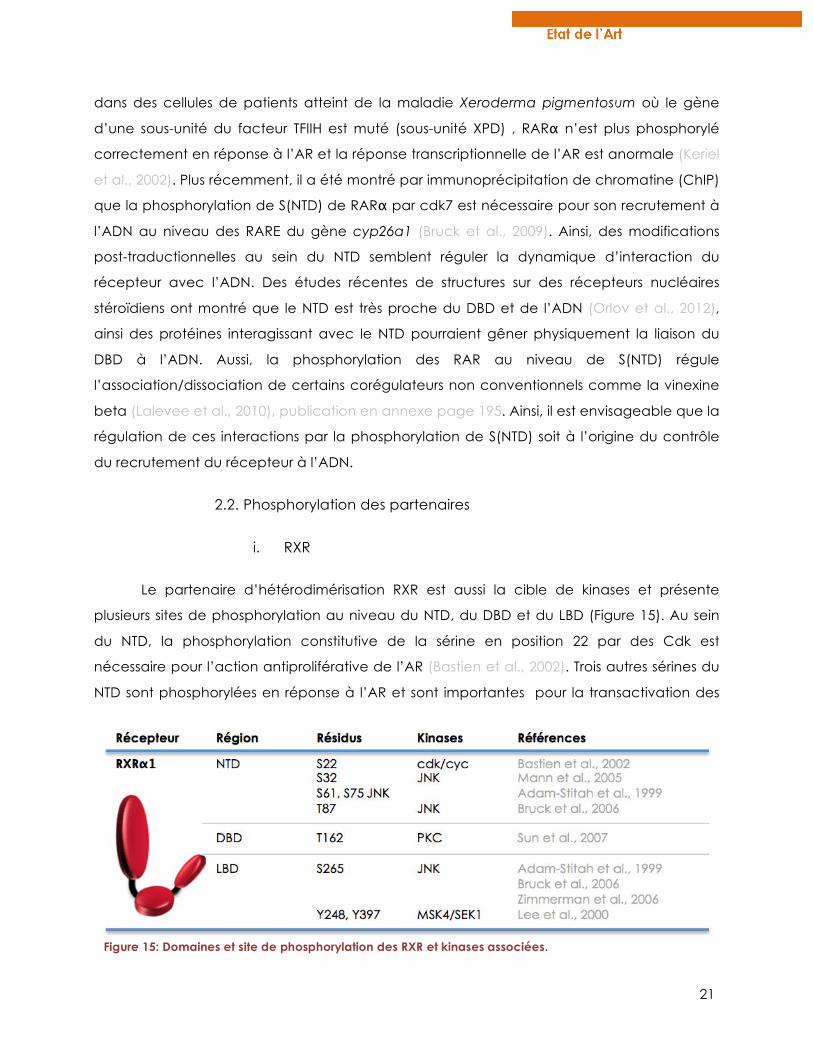
Le partenaire d’hétérodimérisation RXR est aussi la cible de kinases et présente
plusieurs sites de phosphorylation au niveau du NTD, du DBD et du LBD (Figure 15). Au sein
du NTD, la phosphorylation constitutive de la sérine en position 22 par des Cdk est
nécessaire pour l’action antiproliférative de l’AR (Bastien et al., 2002). Trois autres sérines du
NTD sont phosphorylées en réponse à l’AR et sont importantes pour la transactivation des
Figure 15: Domaines et site de phosphorylation des RXR et kinases associées.
22
gènes cibles de l’AR (Adam-Stitah et al., 1999; Gianni et al., 2003). Au niveau du DBD, la
thréonine en position 162 est phosphorylée par PKC et semble impliquée dans la localisation
cytoplasmique de RXR à la fin de son processus transcriptionnel (Sun et al., 2007). Enfin, trois
résidus sont phosphorylés dans le LBD (S265, Y248, Y397) et semblent être associés à l’export
nucléaire et à l’inhibition de l’activité de RXR après un stress cellulaire (Lee et al., 2000;
Zimmerman et al., 2006).
ii. Corégulateurs
Les corégulateurs, répresseurs ou activateurs sont aussi sujets à des phosphorylations
qui participeraient à leur interaction dynamique avec les RAR. Par exemple, la protéine
TBLR1 associée au complexe corépresseur est phosphorylée en réponse à l’AR et cette
modification permet le recrutement du protéasome pour la dégradation des protéines du
complexe comme SMRT ou NCoR (Perissi et al., 2008). Cela suggère une coopération entre
la dissociation des corépresseurs suite à la fixation du ligand au niveau du LBD et leur
dégradation induite par ces phosphorylations en réponse à l’AR.
Le coactivateur SRC-3 est également phosphorylé en réponse à l’AR par la kinase
p38MAPK. Cette modification favorise aussi sa dissociation des RAR puis sa dégradation par
le protéasome pour permettre le recrutement d’autre corégulateurs (Ferry et al., 2011),
publication en annexe page 221. Ainsi, tout au long du processus transcriptionnel induit par
l’AR, les phosphorylations réguleraient la dynamique et la coordination du recrutement des
différents partenaires des RAR
iii. Histones
Dans des expériences de traitement de cellules de mammifères avec des agonistes
des rétinoïdes, il a été montré que les queues des histones H3 étaient phosphorylées,
spécifiquement au niveau des promoteurs de gènes cibles de l’acide rétinoïque comme
RARb2 (Lefebvre et al., 2002). Selon le code histone, la phosphorylation au niveau de la
sérine 10 des histones H3 est associée a une décompaction de la chromatine et une
activation de la transcription (Vicent et al., 2006). De plus, la kinase MSK1 activée par l’AR
est recrutée au niveau des RARE du gène cyp26a1 et est associée à la phosphorylation de
H3S10 (Bruck et al., 2009). Ainsi, il est suggéré que MSK1, en plus de cibler les RAR, prépare la
chromatine pour l’initiation de la transcription des gènes cibles de l’AR (Figure 17, page 25).
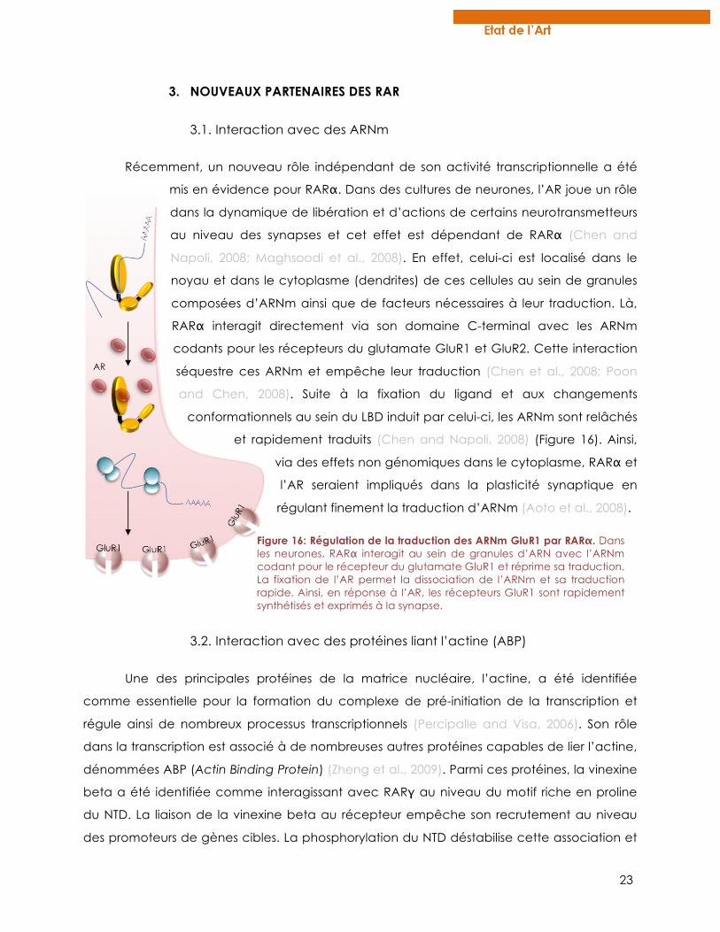
23
3. NOUVEAUX PARTENAIRES DES RAR
3.1. Interaction avec des ARNm
Récemment, un nouveau rôle indépendant de son activité transcriptionnelle a été
mis en évidence pour RARα. Dans des cultures de neurones, l’AR joue un rôle
dans la dynamique de libération et d’actions de certains neurotransmetteurs
au niveau des synapses et cet effet est dépendant de RARα (Chen and
Napoli, 2008; Maghsoodi et al., 2008). En effet, celui-ci est localisé dans le
noyau et dans le cytoplasme (dendrites) de ces cellules au sein de granules
composées d’ARNm ainsi que de facteurs nécessaires à leur traduction. Là,
RARα interagit directement via son domaine C-terminal avec les ARNm
codants pour les récepteurs du glutamate GluR1 et GluR2. Cette interaction
séquestre ces ARNm et empêche leur traduction (Chen et al., 2008; Poon
and Chen, 2008). Suite à la fixation du ligand et aux changements
conformationnels au sein du LBD induit par celui-ci, les ARNm sont relâchés
et rapidement traduits (Chen and Napoli, 2008) (Figure 16). Ainsi,
via des effets non génomiques dans le cytoplasme, RARα et
l’AR seraient impliqués dans la plasticité synaptique en
régulant finement la traduction d’ARNm (Aoto et al., 2008).
3.2. Interaction avec des protéines liant l’actine (ABP)
Une des principales protéines de la matrice nucléaire, l’actine, a été identifiée
comme essentielle pour la formation du complexe de pré-initiation de la transcription et
régule ainsi de nombreux processus transcriptionnels (Percipalle and Visa, 2006). Son rôle
dans la transcription est associé à de nombreuses autres protéines capables de lier l’actine,
dénommées ABP (Actin Binding Protein) (Zheng et al., 2009). Parmi ces protéines, la vinexine
beta a été identifiée comme interagissant avec RARγ au niveau du motif riche en proline
du NTD. La liaison de la vinexine beta au récepteur empêche son recrutement au niveau
des promoteurs de gènes cibles. La phosphorylation du NTD déstabilise cette association et
Figure 16: Régulation de la traduction des ARNm GluR1 par RARα. Dans les neurones, RARα interagit au sein de granules d’ARN avec l’ARNm codant pour le récepteur du glutamate GluR1 et réprime sa traduction. La fixation de l’AR permet la dissociation de l’ARNm et sa traduction rapide. Ainsi, en réponse à l’AR, les récepteurs GluR1 sont rapidement synthétisés et exprimés à la synapse.
24
permet à RARγ de lier les RARE des gènes régulés. Ainsi la vinexine beta est une ABP
considérée comme corépresseur des RAR (Lalevee et al., 2010).
De nombreuses autres ABP interagissent avec d’autres récepteurs nucléaires
telles que la gelsoline, Fli1, Actinine 2 ou encore la cofiline (Gettemans et al., 2005; Huang et
al., 2004; Lee et al., 2004; Ting and Chang, 2008). Ces résultats confirment l’implication de
l’actine et de ses protéines associées dans la régulation de la transcription. De plus, ils
élargissent la liste de protéines régulatrices des RN et suggèrent un rôle important de la
matrice nucléaire dans leur activité transcriptionnelle.
En conclusion, l’ensemble de ces résultats suggère une coopération des effets
génomiques et non génomiques de l’AR pour initier la transcription de gènes cibles (Figure
17). De plus, il apparaît que ce processus transcriptionnel est extrêmement dynamique
avec des recrutements et des échanges rapides et coordonnés de nombreux corégulateurs
et autres partenaires. Les phosphorylations apparaissent comme des modifications majeures
qui, de par leur caractère rapide et réversible, agissent comme de véritables horloges
moléculaires régulant les interactions protéiques au cours du processus transcriptionnel
(Rochette-Egly and Germain, 2009; Samarut and Rochette-Egly, 2012). De manière
intéressante, mes travaux de thèse ont permis de mettre en évidence que les sites de
phosphorylation des RAR en réponse à l’AR ne sont pas tous conservés au sein des vertébrés
et que l’acquisition de résidus phosphorylables au cours de l’évolution aurait permis l’ajout
d’un mécanisme de régulation fine de l’activité des RAR (Chapitre 2, page 139).
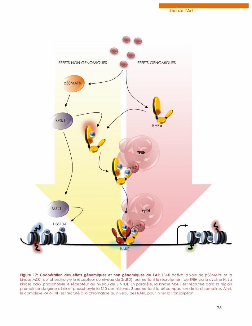
25
Figure 17: Coopération des effets génomiques et non génomiques de l’AR. L’AR active la voie de p38MAPK et la kinase MSK1 qui phosphoryle le récepteur au niveau de S(LBD), permettant le recrutement de TFIIH via la cycline H. La kinase cdk7 phosphoryle le récepteur au niveau de S(NTD). En parallèle, la kinase MSK1 est recrutée dans la région promotrice du gène cible et phosphoryle la S10 des histones 3 permettant la décompaction de la chromatine. Ainsi, le complexe RAR-TFIIH est recruté à la chromatine au niveau des RARE pour initier la transcription.
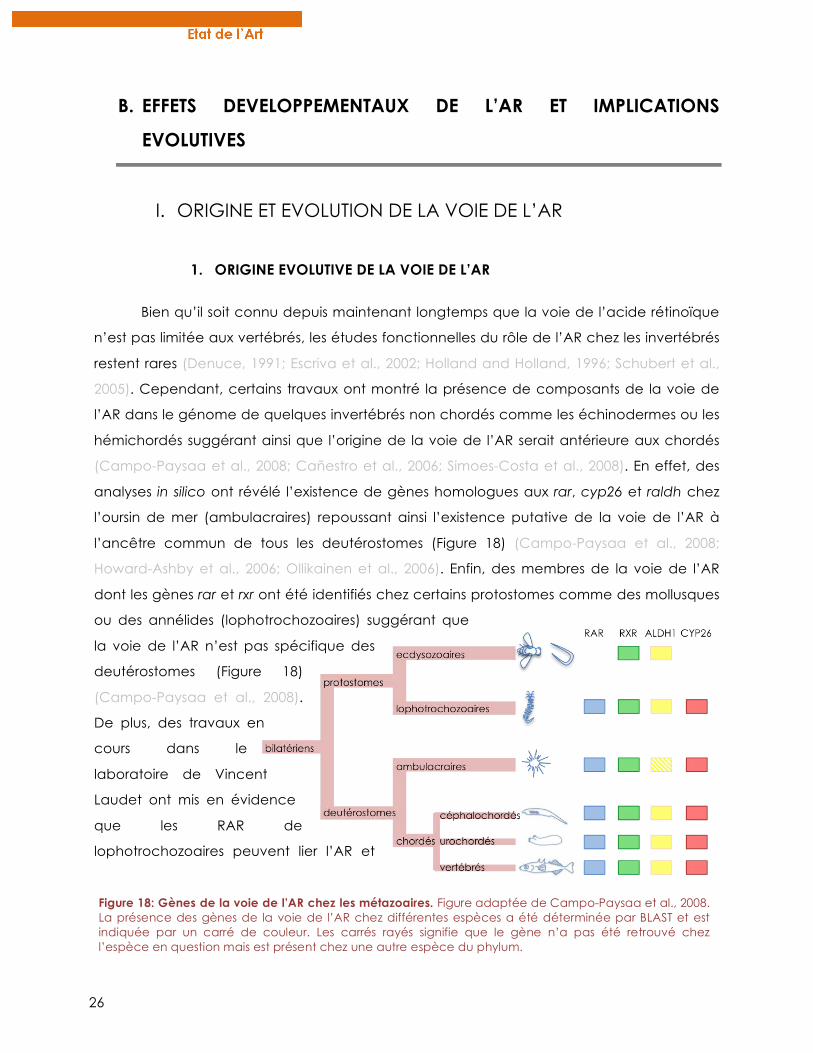
26
B. EFFETS DEVELOPPEMENTAUX DE L’AR ET IMPLICATIONS
EVOLUTIVES
I. ORIGINE ET EVOLUTION DE LA VOIE DE L’AR
1. ORIGINE EVOLUTIVE DE LA VOIE DE L’AR
Bien qu’il soit connu depuis maintenant longtemps que la voie de l’acide rétinoïque
n’est pas limitée aux vertébrés, les études fonctionnelles du rôle de l’AR chez les invertébrés
restent rares (Denuce, 1991; Escriva et al., 2002; Holland and Holland, 1996; Schubert et al.,
2005). Cependant, certains travaux ont montré la présence de composants de la voie de
l’AR dans le génome de quelques invertébrés non chordés comme les échinodermes ou les
hémichordés suggérant ainsi que l’origine de la voie de l’AR serait antérieure aux chordés
(Campo-Paysaa et al., 2008; Cañestro et al., 2006; Simoes-Costa et al., 2008). En effet, des
analyses in silico ont révélé l’existence de gènes homologues aux rar, cyp26 et raldh chez
l’oursin de mer (ambulacraires) repoussant ainsi l’existence putative de la voie de l’AR à
l’ancêtre commun de tous les deutérostomes (Figure 18) (Campo-Paysaa et al., 2008;
Howard-Ashby et al., 2006; Ollikainen et al., 2006). Enfin, des membres de la voie de l’AR
dont les gènes rar et rxr ont été identifiés chez certains protostomes comme des mollusques
ou des annélides (lophotrochozoaires) suggérant que
la voie de l’AR n’est pas spécifique des
deutérostomes (Figure 18)
(Campo-Paysaa et al., 2008).
De plus, des travaux en
cours dans le
laboratoire de Vincent
Laudet ont mis en évidence
que les RAR de
lophotrochozoaires peuvent lier l’AR et
Figure 18: Gènes de la voie de l’AR chez les métazoaires. Figure adaptée de Campo-Paysaa et al., 2008. La présence des gènes de la voie de l’AR chez différentes espèces a été déterminée par BLAST et est indiquée par un carré de couleur. Les carrés rayés signifie que le gène n’a pas été retrouvé chez l’espèce en question mais est présent chez une autre espèce du phylum.
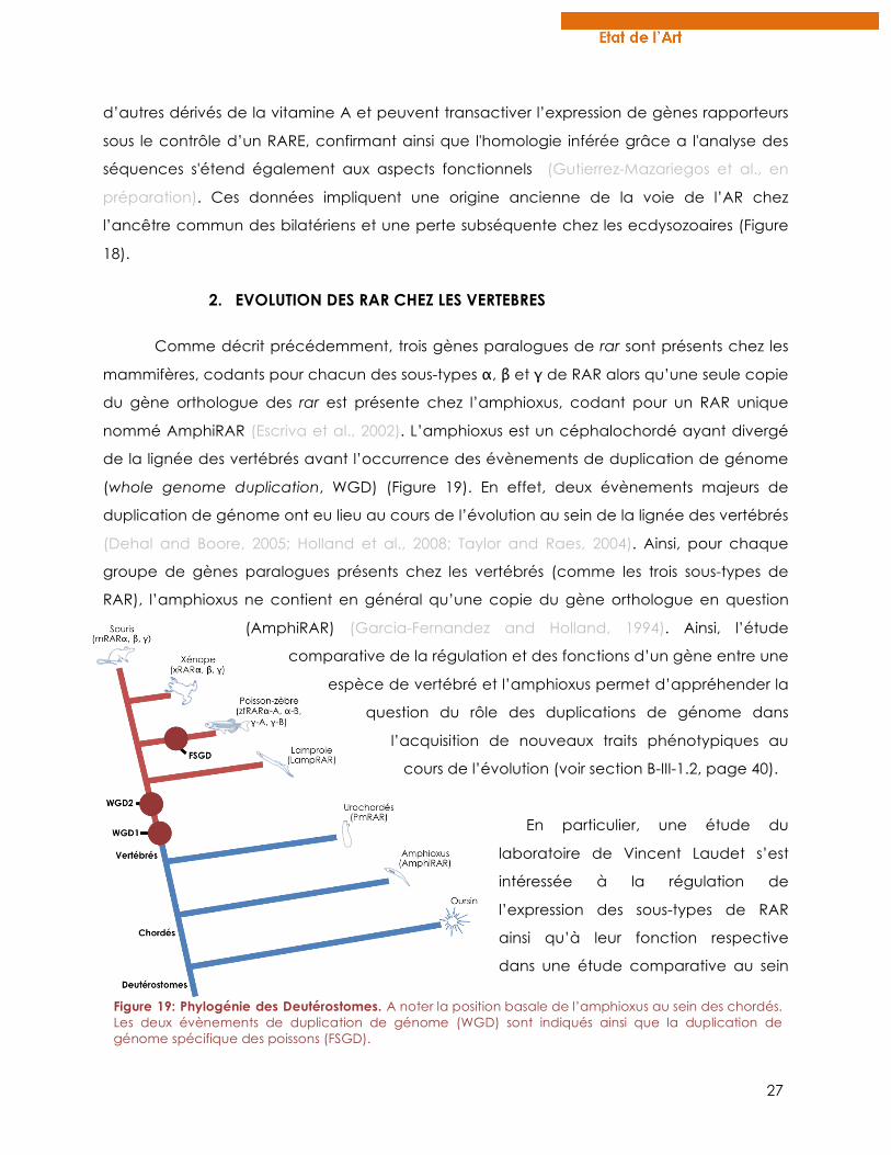
27
d’autres dérivés de la vitamine A et peuvent transactiver l’expression de gènes rapporteurs
sous le contrôle d’un RARE, confirmant ainsi que l'homologie inférée grâce a l'analyse des
séquences s'étend également aux aspects fonctionnels (Gutierrez-Mazariegos et al., en
préparation). Ces données impliquent une origine ancienne de la voie de l’AR chez
l’ancêtre commun des bilatériens et une perte subséquente chez les ecdysozoaires (Figure
18).
2. EVOLUTION DES RAR CHEZ LES VERTEBRES
Comme décrit précédemment, trois gènes paralogues de rar sont présents chez les
mammifères, codants pour chacun des sous-types α, β et γ de RAR alors qu’une seule copie
du gène orthologue des rar est présente chez l’amphioxus, codant pour un RAR unique
nommé AmphiRAR (Escriva et al., 2002). L’amphioxus est un céphalochordé ayant divergé
de la lignée des vertébrés avant l’occurrence des évènements de duplication de génome
(whole genome duplication, WGD) (Figure 19). En effet, deux évènements majeurs de
duplication de génome ont eu lieu au cours de l’évolution au sein de la lignée des vertébrés
(Dehal and Boore, 2005; Holland et al., 2008; Taylor and Raes, 2004). Ainsi, pour chaque
groupe de gènes paralogues présents chez les vertébrés (comme les trois sous-types de
RAR), l’amphioxus ne contient en général qu’une copie du gène orthologue en question
(AmphiRAR) (Garcia-Fernandez and Holland, 1994). Ainsi, l’étude
comparative de la régulation et des fonctions d’un gène entre une
espèce de vertébré et l’amphioxus permet d’appréhender la
question du rôle des duplications de génome dans
l’acquisition de nouveaux traits phénotypiques au
cours de l’évolution (voir section B-III-1.2, page 40).
En particulier, une étude du
laboratoire de Vincent Laudet s’est
intéressée à la régulation de
l’expression des sous-types de RAR
ainsi qu’à leur fonction respective
dans une étude comparative au sein
Figure 19: Phylogénie des Deutérostomes. A noter la position basale de l’amphioxus au sein des chordés. Les deux évènements de duplication de génome (WGD) sont indiqués ainsi que la duplication de génome spécifique des poissons (FSGD).
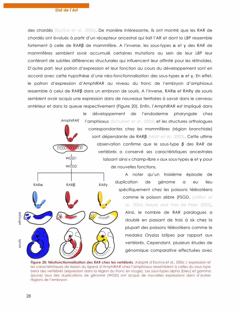
28
des chordés (Escriva et al., 2006). De manière intéressante, ils ont montré que les RAR de
chordés ont évolués à partir d’un récepteur ancestral qui liait l’AR et dont la LBP ressemble
fortement à celle de RARβ de mammifère. A l’inverse, les sous-types α et γ des RAR de
mammifères semblent avoir accumulé certaines mutations au sein de leur LBP leur
conférant de subtiles différences structurales qui influencent leur affinité pour les rétinoïdes.
D’autre part, leur patron d’expression et leur fonction au cours du développement sont en
accord avec cette hypothèse d’une néo-fonctionnalisation des sous-types α et γ. En effet,
le patron d’expression d’AmphiRAR au niveau du tronc de l’embryon d’amphioxus
ressemble à celui de RARβ dans un embryon de souris. A l’inverse, RARα et RARγ de souris
semblent avoir acquis une expression dans de nouveaux territoires à savoir dans le cerveau
antérieur et dans la queue respectivement (Figure 20). Enfin, l’AmphiRAR est impliqué dans
le développement de l’endoderme pharyngale chez
l’amphioxus (Schubert et al., 2005) et les structures orthologues
correspondantes chez les mammifères (région branchiale)
sont dépendante de RARβ (Matt et al., 2003). Cette ultime
observation confirme que le sous-type β des RAR de
vertébrés a conservé ses caractéristiques ancestrales
laissant ainsi « champ-libre » aux sous-types α et γ pour
de nouvelles fonctions.
A noter qu’un troisième épisode de
duplication de génome a eu lieu
spécifiquement chez les poissons téléostéens
comme le poisson zèbre (FSGD, (Jaillon et
al., 2004; Meyer and Van de Peer, 2005).
Ainsi, le nombre de RAR paralogues a
doublé en passant de trois à six chez la
plupart des poissons téléostéens comme le
medaka Oryzias latipes par rapport aux
vertébrés. Cependant, plusieurs études de
génomique comparative effectuées avec
Figure 20: Néofonctionnalisation des RAR chez les vertébrés. Adapté d’Escriva et al., 2006: L’expression et les caractéristiques de liaison du ligand d’AmphiRAR chez l’amphioxus ressemblent à celles du sous-type beta des vertébrés (expression dans la région du tronc en rouge). Les sous-types alpha (bleu) et gamma (jaune) issus des duplications de génome (WGD) ont acquis de nouvelles expressions dans d’autres régions de l’embryon.
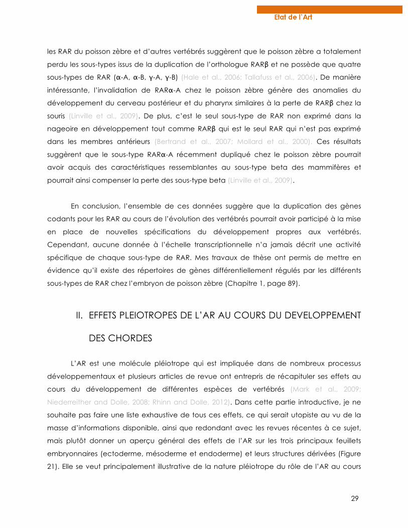
29
les RAR du poisson zèbre et d’autres vertébrés suggèrent que le poisson zèbre a totalement
perdu les sous-types issus de la duplication de l’orthologue RARβ et ne possède que quatre
sous-types de RAR (α-A, α-B, γ-A, γ-B) (Hale et al., 2006; Tallafuss et al., 2006). De manière
intéressante, l’invalidation de RARα-A chez le poisson zèbre génère des anomalies du
développement du cerveau postérieur et du pharynx similaires à la perte de RARβ chez la
souris (Linville et al., 2009). De plus, c’est le seul sous-type de RAR non exprimé dans la
nageoire en développement tout comme RARβ qui est le seul RAR qui n’est pas exprimé
dans les membres antérieurs (Bertrand et al., 2007; Mollard et al., 2000). Ces résultats
suggèrent que le sous-type RARα-A récemment dupliqué chez le poisson zèbre pourrait
avoir acquis des caractéristiques ressemblantes au sous-type beta des mammifères et
pourrait ainsi compenser la perte des sous-type beta (Linville et al., 2009).
En conclusion, l’ensemble de ces données suggère que la duplication des gènes
codants pour les RAR au cours de l’évolution des vertébrés pourrait avoir participé à la mise
en place de nouvelles spécifications du développement propres aux vertébrés.
Cependant, aucune donnée à l’échelle transcriptionnelle n’a jamais décrit une activité
spécifique de chaque sous-type de RAR. Mes travaux de thèse ont permis de mettre en
évidence qu’il existe des répertoires de gènes différentiellement régulés par les différents
sous-types de RAR chez l’embryon de poisson zèbre (Chapitre 1, page 89).
II. EFFETS PLEIOTROPES DE L’AR AU COURS DU DEVELOPPEMENT
DES CHORDES
L’AR est une molécule pléiotrope qui est impliquée dans de nombreux processus
développementaux et plusieurs articles de revue ont entrepris de récapituler ses effets au
cours du développement de différentes espèces de vertébrés (Mark et al., 2009;
Niederreither and Dolle, 2008; Rhinn and Dolle, 2012). Dans cette partie introductive, je ne
souhaite pas faire une liste exhaustive de tous ces effets, ce qui serait utopiste au vu de la
masse d’informations disponible, ainsi que redondant avec les revues récentes à ce sujet,
mais plutôt donner un aperçu général des effets de l’AR sur les trois principaux feuillets
embryonnaires (ectoderme, mésoderme et endoderme) et leurs structures dérivées (Figure
21). Elle se veut principalement illustrative de la nature pléiotrope du rôle de l’AR au cours
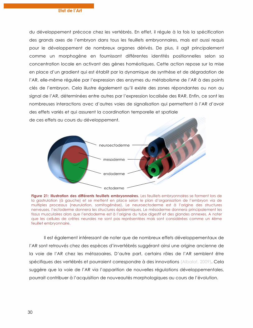
30
du développement précoce chez les vertébrés. En effet, il régule à la fois la spécification
des grands axes de l’embryon dans tous les feuillets embryonnaires, mais est aussi requis
pour le développement de nombreux organes dérivés. De plus, il agit principalement
comme un morphogène en fournissant différentes identités positionnelles selon sa
concentration locale en activant des gènes homéotiques. Cette action repose sur la mise
en place d’un gradient qui est établit par la dynamique de synthèse et de dégradation de
l’AR, elle-même régulée par l’expression des enzymes du métabolisme de l’AR à des points
clés de l’embryon. Cela illustre également qu’il existe des zones répondantes ou non au
signal de l’AR, déterminées entre autres par l’expression localisée des RAR. Enfin, ce sont les
nombreuses interactions avec d’autres voies de signalisation qui permettent à l’AR d’avoir
des effets variés et qui assurent la coordination temporelle et spatiale
de ces effets au cours du développement.
Il est également intéressant de noter que de nombreux effets développementaux de
l’AR sont retrouvés chez des espèces d’invertébrés suggérant ainsi une origine ancienne de
la voie de l’AR chez les métazoaires. D’autre part, certains rôles de l’AR semblent être
spécifiques des vertébrés et pourraient correspondre à des innovations (Albalat, 2009). Cela
suggère que la voie de l’AR via l’apparition de nouvelles régulations développementales,
pourrait contribuer à l’acquisition de nouveautés morphologiques au cours de l’évolution.
Figure 21: Illustration des différents feuillets embryonnaires. Les feuillets embryonnaires se forment lors de la gastrulation (à gauche) et se mettent en place selon le plan d’organisation de l’embryon via de multiples processus (neurulation, somitogénèse). Le neuroectoderme est à l’origine des structures nerveuses, l’ectoderme donnera les structures épidermiques, Le mésoderme donnera principalement les tissus musculaires alors que l’endoderme est à l’origine du tube digestif et des glandes annexes. A noter que les cellules de crêtes neurales ne sont pas représentées mais sont considérées comme un 4ème feuillet embryonnaire.
31
1. NEUROECTODERME, SYSTEME NERVEUX CENTRAL ET DEVELOPPEMENT
NEURAL
1.1. Développement du cerveau postérieur
Le rôle de l’AR au cours de la mise en place du système nerveux central (SNC) des
vertébrés a été principalement étudié dans le contexte de la mise en place de l’axe
antéro-postérieur (A/P). En effet, l’AR a des actions spécifiques sur différentes zones du SNC
lors de sa mise en place, tout particulièrement dans le cerveau postérieur. Là, il agit comme
un morphogène en spécifiant différentes régions du cerveau postérieur, les rhombomères,
selon sa concentration suivant un gradient antéro-postérieur (Godsave et al., 1998;
Niederreither et al., 2000). Il semblerait que l’action de l’AR soit double au cours du
développement du cerveau postérieur en induisant en premier lieu la segmentation
physique des rhombomères puis, plus tardivement, en spécifiant l’identité de chacun d’eux
(Marshall et al., 1992; Wood et al., 1994). L’identité de chacun des rhombomères est définie
par l’expression spécifique de différents facteurs de transcription dont les gènes pbx,
teashirt, meis ou encore hox (Figure 22) (Erickson et al., 2011; Lumsden and Krumlauf, 1996;
Moens and Prince, 2002). L’expression des gènes hox est colinéaire le
long de l’axe A/P et cette colinéarité est fortement régulée par
l’AR (Glover et al., 2006; Murakami et al., 2004; Wada et
al., 1999). En effet, l’altération de la voie de l’AR au
cours de la mise en place du cerveau postérieur mène
à des transformations homéotiques au niveau des
rhombomères (Dupe and Lumsden, 2001; Marshall et
al., 1992). En particulier, un excès d’AR provoque
une extension antérieur du cerveau postérieur et
inhibe la formation de structures cérébrales
antérieures (Durston et al., 1989). Au niveau
moléculaire, l’AR active l’expression de gènes
exprimés dans le cerveau postérieur comme hoxb3
Figure 22: Identité moléculaire des rhombomères lors du développement du cerveau postérieur. Adaptée de Niederreither et al., 2000. L’expression des marqueurs dans le cerveau postérieur murin de 8.5 dpc est indiqué par des carrés colorés, les nuances d’intensité reflètent le niveau d’expression. La morphologie des rhombomères chez un embryon sauvage est à gauche et les altérations d’identités chez un mutant raldh2-/- sont illustrées à droite (r1-r8: rhombomères; me: moelle épinière; ot: vésicule otique; *: identité non clairement définie).
32
ou hoxb4 et réprime des marqueurs de structures plus antérieures comme otx2 ou engrailed-
2 (Godsave et al., 1998). Ainsi, une concentration forte d’AR spécifie une identité
postérieure alors qu’un niveau plus faible d’AR détermine les rhombomères antérieurs. Ces
différences d’activité dépendantes de la concentration en AR nécessitent l’expression
dynamique des enzymes de synthèse et de dégradation de l’AR dans l’embryon. En
particulier, il apparaît que l’expression des enzymes CYP26 est finement régulée pour
permettre le contrôle robuste de la concentration en AR tout au long de l’axe A/P (Abu-
Abed et al., 2001; Emoto et al., 2005; Hernandez et al., 2007; White et al., 2007). La mise en
place du gradient A/P de l’AR par l’expression des enzymes du métabolisme de l’AR est
détaillée dans la section D de l’introduction, page 65. L’action de l’AR dans la mise en
place du cerveau postérieur dépend également d’autres voies de signalisation, comme la
voie des FGF (Fibroblast Growth Factor) qui permet, entre autres de définir clairement la
limite entre le cerveau postérieur et cerveau moyen (hindbrain/midbrain boundary) en
antagonisant les effets postériorisants de l’AR (Schilling, 2008). La voie des Wnt semble aussi
impliquée dans la régulation de l’expression de cyp26a1 et donc dans le contrôle du
gradient d’AR (Kudoh et al., 2002).
Des études effectuées sur le céphalochordé amphioxus et chez certaines
espèces d’urochordés ont montré que l’expression colinéaire des gènes hox dans le
cerveau postérieur est également régulée par l’AR (Katsuyama and Saiga, 1998; Nagatomo
and Fujiwara, 2003; Schubert et al., 2006). De plus, les mêmes effets postériorisants de l’AR y
ont été décrits ce qui laisse penser que la régulation de l’expression des marqueurs du
cerveau postérieur par l’AR est conservée au sein des chordés (Wada et al., 2006).
1.2. Spécification neuronale et neurogénèse
Un autre rôle de l’AR a été mis en évidence dans la spécification neuronale le long
de l’axe dorso-ventral (D/V) dans la moelle épinière antérieure (Maden, 2002). En effet, l’AR
est capable de spécifier in vitro la différentiation de la plaque neurale en un certain type
d’inter-neurones, exprimant des marqueurs qui leur sont propres comme certains gènes dbx
et evx (Pierani et al., 1999) et cette population d’inter-neurones est absente chez des
embryons de cailles déficients en AR (Wilson et al., 2004). De plus, il a été noté chez ces
mutants une expansion des populations ventrales de neurones aux dépends des
populations dorsales et des observations similaires ont été effectuées chez l’amphioxus
(Schubert et al., 2006). Ainsi l’AR aurait un rôle conservé au sein des chordés dans
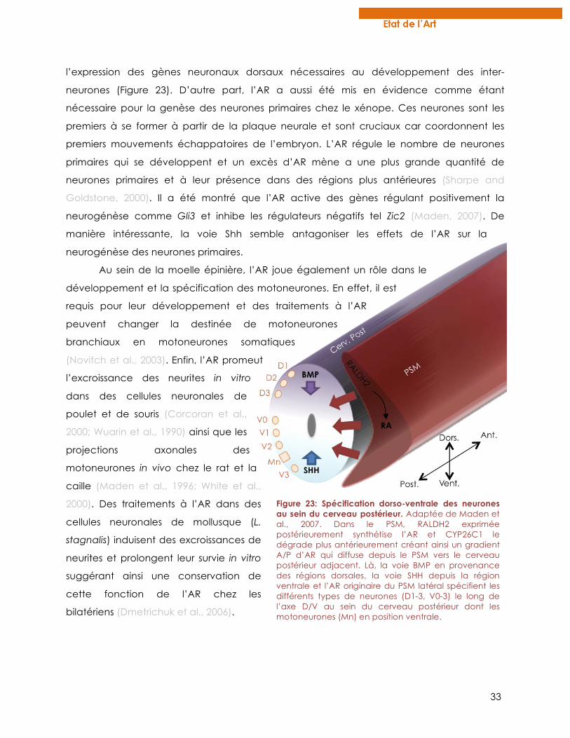
33
l’expression des gènes neuronaux dorsaux nécessaires au développement des inter-
neurones (Figure 23). D’autre part, l’AR a aussi été mis en évidence comme étant
nécessaire pour la genèse des neurones primaires chez le xénope. Ces neurones sont les
premiers à se former à partir de la plaque neurale et sont cruciaux car coordonnent les
premiers mouvements échappatoires de l’embryon. L’AR régule le nombre de neurones
primaires qui se développent et un excès d’AR mène a une plus grande quantité de
neurones primaires et à leur présence dans des régions plus antérieures (Sharpe and
Goldstone, 2000). Il a été montré que l’AR active des gènes régulant positivement la
neurogénèse comme Gli3 et inhibe les régulateurs négatifs tel Zic2 (Maden, 2007). De
manière intéressante, la voie Shh semble antagoniser les effets de l’AR sur la
neurogénèse des neurones primaires.
Au sein de la moelle épinière, l’AR joue également un rôle dans le
développement et la spécification des motoneurones. En effet, il est
requis pour leur développement et des traitements à l’AR
peuvent changer la destinée de motoneurones
branchiaux en motoneurones somatiques
(Novitch et al., 2003). Enfin, l’AR promeut
l’excroissance des neurites in vitro
dans des cellules neuronales de
poulet et de souris (Corcoran et al.,
2000; Wuarin et al., 1990) ainsi que les
projections axonales des
motoneurones in vivo chez le rat et la
caille (Maden et al., 1996; White et al.,
2000). Des traitements à l’AR dans des
cellules neuronales de mollusque (L.
stagnalis) induisent des excroissances de
neurites et prolongent leur survie in vitro
suggérant ainsi une conservation de
cette fonction de l’AR chez les
bilatériens (Dmetrichuk et al., 2006).
Figure 23: Spécification dorso-ventrale des neurones au sein du cerveau postérieur. Adaptée de Maden et al., 2007. Dans le PSM, RALDH2 exprimée postérieurement synthétise l’AR et CYP26C1 le dégrade plus antérieurement créant ainsi un gradient A/P d’AR qui diffuse depuis le PSM vers le cerveau postérieur adjacent. Là, la voie BMP en provenance des régions dorsales, la voie SHH depuis la région ventrale et l’AR originaire du PSM latéral spécifient les différents types de neurones (D1-3, V0-3) le long de l’axe D/V au sein du cerveau postérieur dont les motoneurones (Mn) en position ventrale.
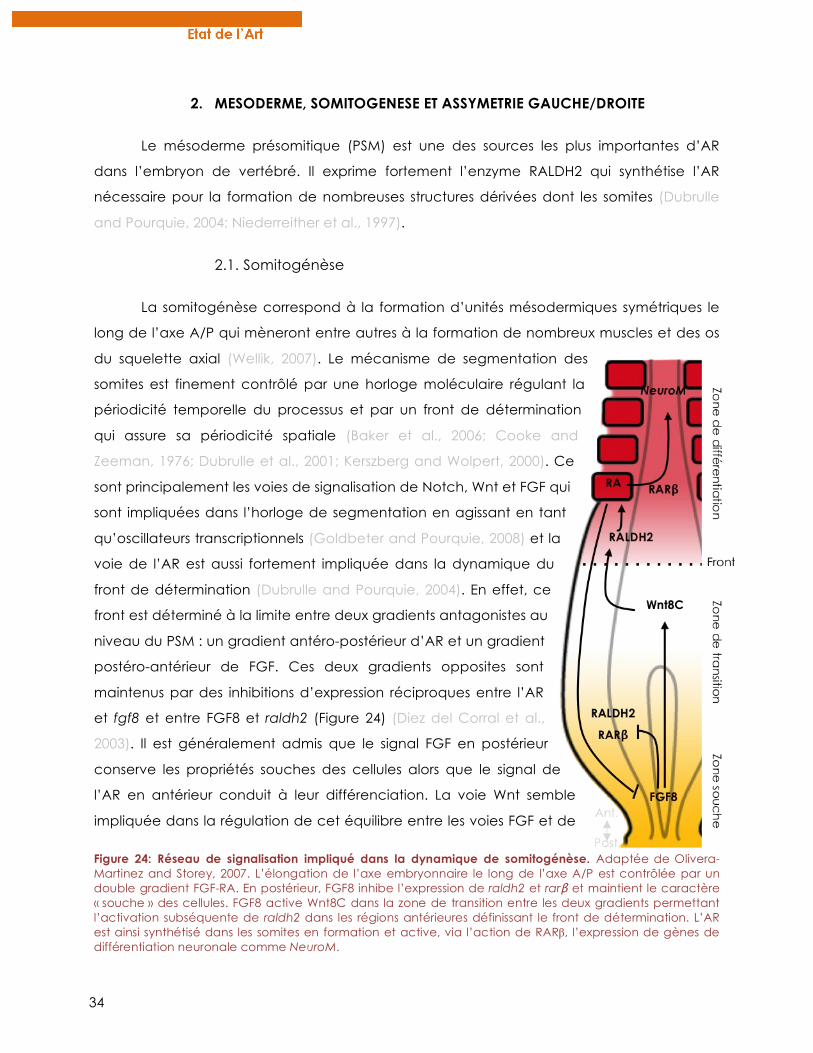
34
2. MESODERME, SOMITOGENESE ET ASSYMETRIE GAUCHE/DROITE
Le mésoderme présomitique (PSM) est une des sources les plus importantes d’AR
dans l’embryon de vertébré. Il exprime fortement l’enzyme RALDH2 qui synthétise l’AR
nécessaire pour la formation de nombreuses structures dérivées dont les somites (Dubrulle
and Pourquie, 2004; Niederreither et al., 1997).
2.1. Somitogénèse
La somitogénèse correspond à la formation d’unités mésodermiques symétriques le
long de l’axe A/P qui mèneront entre autres à la formation de nombreux muscles et des os
du squelette axial (Wellik, 2007). Le mécanisme de segmentation des
somites est finement contrôlé par une horloge moléculaire régulant la
périodicité temporelle du processus et par un front de détermination
qui assure sa périodicité spatiale (Baker et al., 2006; Cooke and
Zeeman, 1976; Dubrulle et al., 2001; Kerszberg and Wolpert, 2000). Ce
sont principalement les voies de signalisation de Notch, Wnt et FGF qui
sont impliquées dans l’horloge de segmentation en agissant en tant
qu’oscillateurs transcriptionnels (Goldbeter and Pourquie, 2008) et la
voie de l’AR est aussi fortement impliquée dans la dynamique du
front de détermination (Dubrulle and Pourquie, 2004). En effet, ce
front est déterminé à la limite entre deux gradients antagonistes au
niveau du PSM : un gradient antéro-postérieur d’AR et un gradient
postéro-antérieur de FGF. Ces deux gradients opposites sont
maintenus par des inhibitions d’expression réciproques entre l’AR
et fgf8 et entre FGF8 et raldh2 (Figure 24) (Diez del Corral et al.,
2003). Il est généralement admis que le signal FGF en postérieur
conserve les propriétés souches des cellules alors que le signal de
l’AR en antérieur conduit à leur différenciation. La voie Wnt semble
impliquée dans la régulation de cet équilibre entre les voies FGF et de
Figure 24: Réseau de signalisation impliqué dans la dynamique de somitogénèse. Adaptée de Olivera-Martinez and Storey, 2007. L’élongation de l’axe embryonnaire le long de l’axe A/P est contrôlée par un double gradient FGF-RA. En postérieur, FGF8 inhibe l’expression de raldh2 et rarβ et maintient le caractère « souche » des cellules. FGF8 active Wnt8C dans la zone de transition entre les deux gradients permettant l’activation subséquente de raldh2 dans les régions antérieures définissant le front de détermination. L’AR est ainsi synthétisé dans les somites en formation et active, via l’action de RARβ, l’expression de gènes de différentiation neuronale comme NeuroM.
35
l’AR en jouant un rôle de médiateur moléculaire entre ces deux signaux au niveau de la
zone de transition du PSM (Figure 24) (Olivera-Martinez and Storey, 2007).
En plus de réguler la dynamique du front de détermination, les voies antagonistes
AR/FGF contrôlent également l’expression différentielle dès gènes hox au sein du PSM le
long de l’axe A/P. Cela va permettre la spécification de l’identité des structures issues du
PSM ce qui est essentiel pour la future identité des vertèbres par exemple. Alors que FGF8
active préférentiellement les gènes hox postérieurs situés en 5’ du cluster génique des hox,
l’AR permet l’expression de gènes hox antérieurs situés en 3’ du cluster. Ainsi, ces voies de
signalisation assurent la coordination de la segmentation des somites ainsi que la
spécification de leur identité le long de l’axe A/P (Dubrulle et al., 2001; Zákány et al., 2001).
Les mécanismes de la somitogénèse semblent très bien conservés chez différentes espèces
de vertébrés, cependant l’implication de l’AR dans ce processus en dehors des vertébrés
reste incertaine. Il n’existe pas de PSM chez l’amphioxus et les somites se forment par
bourgeonnement à partir du bourgeon caudal. Ce processus ne semble pas requérir l’AR,
cependant il est important pour le positionnement des somites le long de l’axe A/P
(Schubert et al., 2006). Ainsi, le rôle de l’AR dans la segmentation des somites semble être
apparu chez les vertébrés parallèlement à l’acquisition du PSM.
2.2. Symétrie Gauche/Droite (G/D)
Au cours de la somitogénèse, l’AR régule également la formation synchrone et
bilatérale des somites de chaque côté de la notocorde le long de l’axe G/D. En effet,
l’altération de la synthèse endogène d’AR chez des mutants de souris, de poulet ou encore
de poisson-zèbre résulte en la production de somites asymétriques et de manière
désynchronisée (Kawakami et al., 2005; Vermot et al., 2005). Il est admis que l’AR produit
antérieurement dans le PSM tamponne des signaux d’asymétrie G/D émanant des zones
postérieures du PSM et qui sont requis pour le développement asymétrique de certains
organes internes comme les viscères (Vermot and Pourquie, 2005).
Par ailleurs, la mise en place de cette asymétrie globale au sein de l’embryon est
également dépendante de la voie de l’AR. Ce processus repose sur l’établissement d’un
courant au niveau du nœud embryonnaire, appelé flux nodal, qui est généré par un
mécanisme hydrodynamique. En effet, les cellules ventrales du nœud sont ciliées et la
rotation synchronisée des cils permet la formation du flux de la droite vers la gauche chez la
souris (Figure 25) (Hirokawa et al., 2009). Le flux nodal permet entre autres le transport de
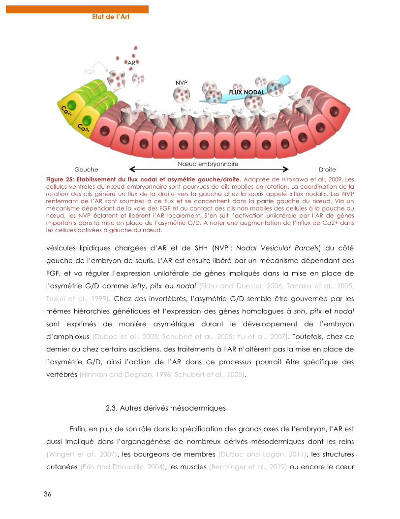
36
vésicules lipidiques chargées d’AR et de SHH (NVP : Nodal Vesicular Parcels) du côté
gauche de l’embryon de souris. L’AR est ensuite libéré par un mécanisme dépendant des
FGF, et va réguler l’expression unilatérale de gènes impliqués dans la mise en place de
l’asymétrie G/D comme lefty, pitx ou nodal (Sirbu and Duester, 2006; Tanaka et al., 2005;
Tsukui et al., 1999). Chez des invertébrés, l’asymétrie G/D semble être gouvernée par les
mêmes hiérarchies génétiques et l’expression des gènes homologues à shh, pitx et nodal
sont exprimés de manière asymétrique durant le développement de l’embryon
d’amphioxus (Duboc et al., 2005; Schubert et al., 2005; Yu et al., 2007). Toutefois, chez ce
dernier ou chez certains ascidiens, des traitements à l’AR n’altèrent pas la mise en place de
l’asymétrie G/D, ainsi l’action de l’AR dans ce processus pourrait être spécifique des
vertébrés (Hinman and Degnan, 1998; Schubert et al., 2005).
2.3. Autres dérivés mésodermiques
Enfin, en plus de son rôle dans la spécification des grands axes de l’embryon, l’AR est
aussi impliqué dans l’organogénèse de nombreux dérivés mésodermiques dont les reins
(Wingert et al., 2007), les bourgeons de membres (Duboc and Logan, 2011), les structures
cutanées (Prin and Dhouailly, 2004), les muscles (Bentzinger et al., 2012) ou encore le cœur
Figure 25: Etablissement du flux nodal et asymétrie gauche/droite. Adaptée de Hirokawa et al., 2009. Les cellules ventrales du nœud embryonnaire sont pourvues de cils mobiles en rotation. La coordination de la rotation des cils génère un flux de la droite vers la gauche chez la souris appelé « flux nodal ». Les NVP renfermant de l’AR sont soumises à ce flux et se concentrent dans la partie gauche du nœud. Via un mécanisme dépendant de la voie des FGF et au contact des cils non mobiles des cellules à la gauche du nœud, les NVP éclatent et libèrent l’AR localement. S’en suit l’activation unilatérale par l’AR de gènes importants dans la mise en place de l’asymétrie G/D. A noter une augmentation de l’influx de Ca2+ dans les cellules activées à gauche du nœud.
37
(Collop et al., 2006). De nombreuses études sur les vertébrés dont le poisson-zèbre ont
permis de décrypter la mécanistique précise des effets de l’AR ainsi que les nombreuses
interactions avec d’autres voies de signalisation pour le développement de ces organes.
Quelques exemples sont présentés dans la revue de la section D de l’introduction, page 65.
A l’inverse, rares sont les données du rôle de l’AR dans l’organogénèse des invertébrés, ce
qui ne permet donc aucune spéculation sur l’origine évolutive du rôle de l’AR sur le
développement de ces structures.
3. ENDODERME, PHARYNX ET ORGANES INTERNES
3.1. Pharynx
L’AR est impliqué dans la spécification des structures endodermiques le long de l’axe
A/P au sein de l’endoderme pharyngien. En effet, chez la caille et le poisson-zèbre, des
mutants de la voie de synthèse de l’AR présentent une expansion de l’endoderme
pharyngien au dépend des structures de l’intestin antérieur (Grandel et al., 2002; Quinlan et
al., 2002). A l’inverse, un excès d’AR provoque une postériorisation de ces structures en
transformant par exemple les structures antérieures de l’intestin en foie ou en pancréas
(Stafford and Prince, 2002). Au niveau des arcs branchiaux, une concentration élevée d’AR
est nécessaire pour le développement des arcs postérieurs 3 à 6 chez l’embryon de souris,
alors que la différentiation du second arc requiert une dose plus faible d’AR (Mark et al.,
2004). En effet, chez des embryons de souris ou de poisson-zèbre où la voie de l’AR est
altérée, les arcs 3 à 6 sont manquants et l’expression de certains gènes hox et pax est
modifiée. Ces gènes étant importants dans la différentiation des arcs branchiaux postérieurs
(Wendling et al., 2000), cela suggère que l’AR agit au niveau des cellules de l’endoderme
pharyngien en leur pourvoyant une identité positionnelle via l’expression de certains gènes
de détermination. A noter que le sous-type beta des RAR est spécifiquement requis pour
l’effet tératogénique de l’AR au niveau des arcs branchiaux (Matt et al., 2003). En
conclusion, la régulation fine de la structuration des différents arcs branchiaux semble être
corrélée à des zones d’activité de l’AR très localisées au sein de l’endoderme pharyngien.
Chez l’amphioxus également, la structuration de l’endoderme pharyngien est dépendante
de l’AR. (Escriva et al., 2002). En effet, des traitements à l’AR mènent à la perte de certaines
structures pharyngiennes et postériorisent l’expression de marqueurs comme les gènes pax
ou otx (Schubert et al., 2005). Les mêmes effets ont aussi été observés chez certaines
38
ascidies ce qui suggère que l’AR était déjà impliqué dans la spécification de l’endoderme
pharyngien chez l’ancêtre commun de tous les chordés.
A noter que l’action de l’AR sur l’endoderme pharyngien est indépendante de
l’action des cellules de crêtes neurales (NCC) (Veitch et al., 1999) bien que certaines NCC
craniales migrent dans les arcs branchiaux et participent activement à leur morphogénèse
(Santagati and Rijli, 2003). En effet, chez l’amphioxus, l’AR joue un rôle dans la spécification
du pharynx bien que ce chordé soit dépourvu de NCC (Escriva et al., 2002). Toutefois, l’AR a
un rôle important dans l’induction et la migration des NCC ainsi que dans la morphogénèse
des arcs branchiaux. La revue de la section D de l’introduction, page 65, fournie plus de
détails sur les effets de l’AR sur les NCC.
3.2. Organes internes
L’AR synthétisé dans le mésoderme via l’activité de RALDH2 est impliqué dans
l’organogénèse de certaines structures dérivées de l’endoderme adjacent (Molotkov et al.,
2005). Par exemple, la différentiation du foie et du pancréas nécessite des signaux de l’AR
depuis le mésoderme des plaques latérales (Kumar et al., 2003; Kumar and Melton, 2003).
De manière générale, il apparaît que comme dans le mésoderme et dans le CNS, l’AR agit
comme agent postériorisant dans l’endoderme. En effet, des traitements antagonistes de la
voie de l’AR mènent à une absence des marqueurs normalement exprimés dans le foie et le
pancréas. A l’inverse, un excès d’AR mène à une expansion de ces marqueurs au niveau
de l’endoderme antérieur (Stafford and Prince, 2002). De plus, l’AR promeut la
différenciation de cellules progénitrices en cellules béta dans le pancréas en activant des
gènes de différenciation (Ostrom et al., 2008). Cependant, les organes dérivés de
l’endoderme postérieur comme l’intestin ne semblent pas requérir l’AR pour leur
développement ce qui suggère qu’au sein de l’endoderme, il existe des zones répondant à
l’AR et d’autres qui ne le sont pas. Ces zones de réponse à l’AR le long de l’axe A/P de
l’endoderme sont définies par l’expression dynamique spatio-temporelle des enzymes de
synthèse (RALDH) et de dégradation (CYP26) de l’AR (Blentic et al., 2003; Reijntjes et al.,
2005). De plus, l’expression localisée des différents sous-types de RAR dans l’endoderme
parachève l’établissement des zones d’activités de l’AR le long de l’axe A/P (Mollard et al.,
2000). Aussi, l’expression différentielle des RAR le long de l’axe D/V de l’endoderme pourrait
jouer un rôle dans la régionalisation de l’action de l’AR au sein de l’endoderme (Pan et al.,
2007).
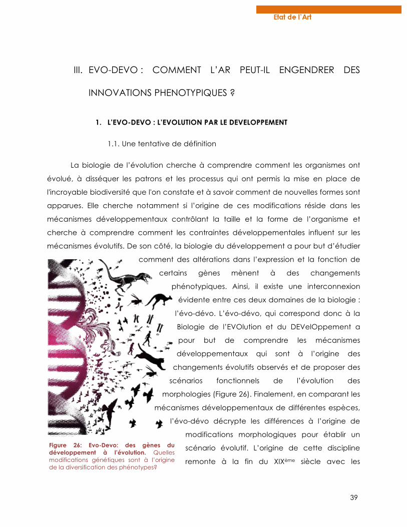
39
III. EVO-DEVO : COMMENT L’AR PEUT-IL ENGENDRER DES
INNOVATIONS PHENOTYPIQUES ?
1. L’EVO-DEVO : L’EVOLUTION PAR LE DEVELOPPEMENT
1.1. Une tentative de définition
La biologie de l’évolution cherche à comprendre comment les organismes ont
évolué, à disséquer les patrons et les processus qui ont permis la mise en place de
l'incroyable biodiversité que l'on constate et à savoir comment de nouvelles formes sont
apparues. Elle cherche notamment si l’origine de ces modifications réside dans les
mécanismes développementaux contrôlant la taille et la forme de l’organisme et
cherche à comprendre comment les contraintes développementales influent sur les
mécanismes évolutifs. De son côté, la biologie du développement a pour but d’étudier
comment des altérations dans l’expression et la fonction de
certains gènes mènent à des changements
phénotypiques. Ainsi, il existe une interconnexion
évidente entre ces deux domaines de la biologie :
l’évo-dévo. L’évo-dévo, qui correspond donc à la
Biologie de l’EVOlution et du DEVelOppement a
pour but de comprendre les mécanismes
développementaux qui sont à l’origine des
changements évolutifs observés et de proposer des
scénarios fonctionnels de l’évolution des
morphologies (Figure 26). Finalement, en comparant les
mécanismes développementaux de différentes espèces,
l’évo-dévo décrypte les différences à l’origine de
modifications morphologiques pour établir un
scénario évolutif. L’origine de cette discipline
remonte à la fin du XIXème siècle avec les
Figure 26: Evo-Devo: des gènes du développement à l’évolution. Quelles modifications génétiques sont à l’origine de la diversification des phénotypes?
40
premières études d’embryologie. En fait elle a été récemment remis en lumière après la
découverte des gènes homéotiques et de leur unicité fonctionnelle chez les
métazoaires (Duboule and Dolle, 1989; Graham et al., 1989) et plus récemment par la
disponibilité de plus en plus de génomes d’espèces non modèles (Carroll, 2005).
1.2. Où et comment agit l’évolution ?
i. Mutations
Les mutations génétiques sont indéniablement essentielles pour la modification des
gènes du développement et donc du plan d’organisation embryonnaire. La fonction
propre de chaque gène dépend à la fois de la fonction de la protéine codée, mais
également des conditions sous lesquelles la protéine est exprimée. Ainsi, les mutations
peuvent intervenir dans des régions régulatrices (régions cis) pour modifier l’expression de
gènes du développement, ou bien peuvent cibler les séquences codantes des protéines et
ainsi changer leur fonction.
Les mutations dans les séquences codantes des gènes sont supposées être moins
fréquentes et plus facilement délétères car la fonction propre d’une protéine soumet sa
séquence codante à une forte pression de sélection. Cependant, quelques exemples de
mutations dans la partie codante de gènes du développement ont été mis en évidence
comme étant à l’origine d’innovations morphologiques adaptatives. C’est par exemple le
cas dans l’acquisition de l’albinisme chez différentes populations de poissons cavernicoles
après mutation du gène Oca2 (Protas et al., 2006). Des mutations au sein de ce gène sont
également responsables de différences de pigmentation de la peau observées chez
l’homme (Lamason et al., 2005). Il est proposé que des mutations dans les séquences
codantes ne sont possibles que si le gène ne présente qu’une faible pléiotropie. En effet,
des mutations dans des gènes pleiotropes vont avoir de multiples conséquences et seront
ainsi fréquemment délétères (Carroll et al., 2004). Cependant, certaines mutations dans des
facteurs pléiotropes comme le récepteur de l’ectodysplasine (EDAR) ont été décrites et
semblent avoir été sélectionnées dans certaines populations d’Asie de l’est (Sadier et al.,
2013).
A l’inverse, King et Wilson déclaraient en 1975 que « le faible niveau de divergence
des séquences codantes des gènes entre l’homme et le chimpanzé, ne pouvait à lui seul,
41
expliquer les différences phénotypiques importantes entres ces deux espèces » (King and
Wilson, 1975). Depuis, il a été généralement admis que les modifications au niveau des
séquences cis-régulatrices jouent un rôle principal dans la genèse de nouveaux traits
phénotypiques (Wray, 2007). En effet, de nombreuses validations expérimentales ont montré
que des mutations dans les séquences cis-régulatrices des gènes peuvent induire des
modifications développementales et des changements morphologiques adaptatifs. Cela a
par exemple été montré pour des gènes impliqués dans la pigmentation des ailes de
drosophile (Gompel et al., 2005) ou encore pour le gène pitx1 dont les changements
d’expression chez différentes espèces d’épinoches ont conduit à la perte de leur armure
osseuse (Chan et al., 2010).
Mes travaux de thèse ont mis en évidence que des substitutions dans les séquences
codantes des gènes peuvent permettre une régulation plus fine de l’activité des protéines
via l’acquisition de résidus ciblés par des modifications post-traductionnelles comme les
phosphorylations. Mes résultats suggèrent ainsi que des mutations dans des gènes exprimés
dans de nombreux organes et ayant un rôle pléiotrope peuvent ne pas avoir d’effets
nécessairement délétères. (Chapitre 3, page 139)
ii. Duplication de gènes
Il a été suggéré que l’apparition de nouvelles morphologies et de nouveaux
phénotypes pouvait être corrélée à l’augmentation de la complexité génomique induite
par des évènements de duplication de génome (WGD)(Ohno, 1970). En particulier, deux
évènements de duplication de génome au cours de l’évolution ont joué un rôle majeur
dans la diversification du plan d’organisation des différentes espèces de vertébrés (Dehal
and Boore, 2005). Cette théorie a été également été validée expérimentalement chez les
poissons téléostéens dont le génome a subit une duplication supplémentaire (FSGD) par
rapport aux autres vertébrés (Jaillon et al., 2004) (Figure 19, page 27).
Selon l’hypothèse de Ohno, l’augmentation du nombre de gènes suite aux
évènements de WGD permet l’acquisition de nouvelles fonctions pour les gènes dupliqués
(gènes paralogues) et est ainsi à l’origine de la diversification des phénotypes. Selon le
modèle dit de Duplication, Dégénération, Complémentation (DDC) (Force et al., 1999), il est
généralement admis que la duplication d’un gène ancestral en plusieurs gènes paralogues
peut mener à trois scénarios différents (Figure 27):
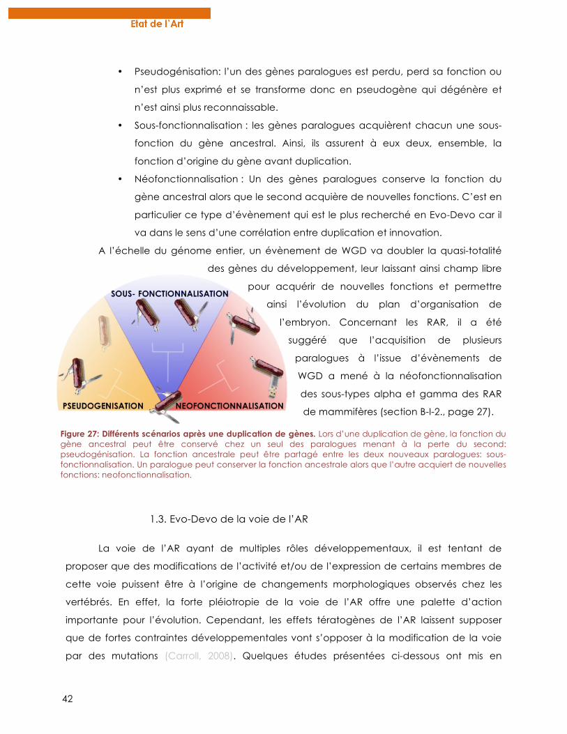
42
• Pseudogénisation: l’un des gènes paralogues est perdu, perd sa fonction ou
n’est plus exprimé et se transforme donc en pseudogène qui dégénère et
n’est ainsi plus reconnaissable.
• Sous-fonctionnalisation : les gènes paralogues acquièrent chacun une sous-
fonction du gène ancestral. Ainsi, ils assurent à eux deux, ensemble, la
fonction d’origine du gène avant duplication.
• Néofonctionnalisation : Un des gènes paralogues conserve la fonction du
gène ancestral alors que le second acquière de nouvelles fonctions. C’est en
particulier ce type d’évènement qui est le plus recherché en Evo-Devo car il
va dans le sens d’une corrélation entre duplication et innovation.
A l’échelle du génome entier, un évènement de WGD va doubler la quasi-totalité
des gènes du développement, leur laissant ainsi champ libre
pour acquérir de nouvelles fonctions et permettre
ainsi l’évolution du plan d’organisation de
l’embryon. Concernant les RAR, il a été
suggéré que l’acquisition de plusieurs
paralogues à l’issue d’évènements de
WGD a mené à la néofonctionnalisation
des sous-types alpha et gamma des RAR
de mammifères (section B-I-2., page 27).
1.3. Evo-Devo de la voie de l’AR
La voie de l’AR ayant de multiples rôles développementaux, il est tentant de
proposer que des modifications de l’activité et/ou de l’expression de certains membres de
cette voie puissent être à l’origine de changements morphologiques observés chez les
vertébrés. En effet, la forte pléiotropie de la voie de l’AR offre une palette d’action
importante pour l’évolution. Cependant, les effets tératogènes de l’AR laissent supposer
que de fortes contraintes développementales vont s’opposer à la modification de la voie
par des mutations (Carroll, 2008). Quelques études présentées ci-dessous ont mis en
Figure 27: Différents scénarios après une duplication de gènes. Lors d’une duplication de gène, la fonction du gène ancestral peut être conservé chez un seul des paralogues menant à la perte du second: pseudogénisation. La fonction ancestrale peut être partagé entre les deux nouveaux paralogues: sous-fonctionnalisation. Un paralogue peut conserver la fonction ancestrale alors que l’autre acquiert de nouvelles fonctions: neofonctionnalisation.
43
évidence un rôle de la voie de l’AR dans la genèse de nouvelles morphologies. Mes travaux
de thèse ont également participé à montrer que des modifications subtiles du métabolisme
local de l’AR in vivo pouvaient être à l’origine de la diversité de microstructures comme les
dents pharyngiennes de poissons (chapitre 3, page 163)
2. IMPLICATION DE LA VOIE DE L’AR DANS L’EVOLUTION DES STRUCTURES
CRANIO-FACIALES
2.1. Effets tératogènes craniaux-faciaux induits par l’AR
L’AR est un puissant tératogène lorsqu’il est administré durant le développement
embryonnaire des vertébrés et de certains invertébrés (Escriva et al., 2002; Soprano and
Soprano, 1995). Il existe un continuum d’anomalies développementales induites par l’AR
selon la concentration, la durée et le stade développemental de l’embryon exposé et
l’ensemble des ces malformations est regroupé sous le terme d’embryopathie dépendante
de l’AR (RAE). Une des malformations caractéristiques induites par l’AR, est un défaut dans
la morphogénèse des structures craniofaciales dont la mâchoire et l’oreille interne (Figure
28) (Lammer et al., 1985; Mallo, 1997). Chez l’embryon de souris, les défauts craniofaciaux
sont caractérisés par une hypoplasie et une fusion des deux premiers arcs pharyngiens qui
sont à l’origine de nombreuses structures craniofaciales affectées par l’effet tératogène de
l’AR. Les arcs pharyngiens sont rapidement colonisés par les NCC craniales qui vont
participer à leur morphogénèse ainsi qu’au développement des structures dérivées des arcs
pharyngiens (Santagati and Rijli, 2003). Il a été montré que les effets tératogènes de l’AR
n’impliquent pas un effet de l’AR sur la survie ou la migration des NCC chez la souris. Plutôt,
l’AR perturbe les voies de signalisation en provenance de l’épithélium et de l’endoderme
pharyngien et qui déterminent la destinée des NCC via l’expression des gènes dlx (Figure
28). Les gènes dlx codent pour des facteurs de transcriptions à homéodomaines qui, à
l’image des gènes hox, sont essentiels dans la spécification de l’identité des structures
craniofaciales (Acampora et al., 1999). Ainsi, il est admis que les gènes dlx ont un rôle clé
dans le développement et l’évolution de la mâchoire chez les vertébrés (Depew et al.,
2005; Takechi et al., 2013). De manière intéressante, les mêmes anomalies que celles induites
par les effets tératogènes de l’AR sur la mâchoire sont retrouvées chez des mutants de souris
pour les gènes dlx5 et dlx6 (Beverdam et al., 2002). En cohérence avec ces observations, il a
été montré que l’AR perturbe l’activation des gènes dlx dans les NCC craniales
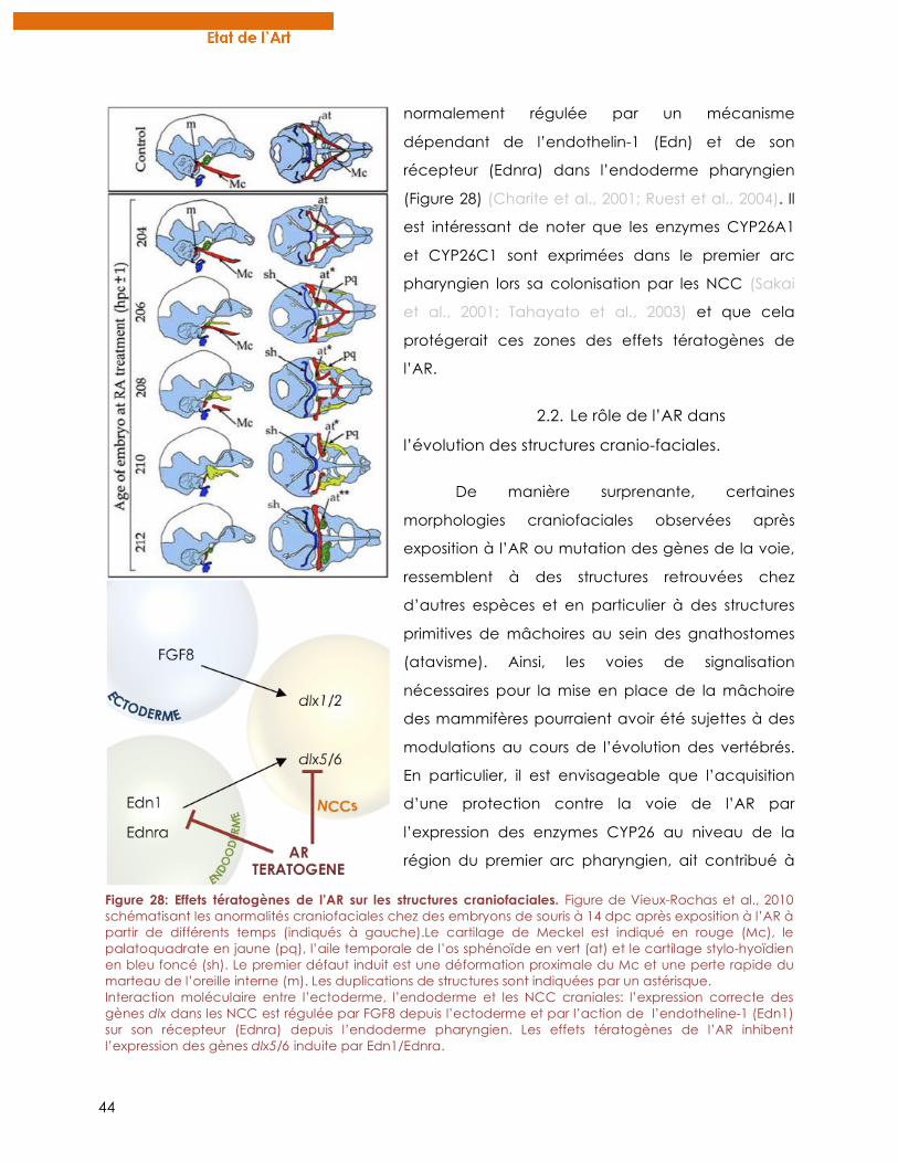
44
normalement régulée par un mécanisme
dépendant de l’endothelin-1 (Edn) et de son
récepteur (Ednra) dans l’endoderme pharyngien
(Figure 28) (Charite et al., 2001; Ruest et al., 2004). Il
est intéressant de noter que les enzymes CYP26A1
et CYP26C1 sont exprimées dans le premier arc
pharyngien lors sa colonisation par les NCC (Sakai
et al., 2001; Tahayato et al., 2003) et que cela
protégerait ces zones des effets tératogènes de
l’AR.
2.2. Le rôle de l’AR dans
l’évolution des structures cranio-faciales.
De manière surprenante, certaines
morphologies craniofaciales observées après
exposition à l’AR ou mutation des gènes de la voie,
ressemblent à des structures retrouvées chez
d’autres espèces et en particulier à des structures
primitives de mâchoires au sein des gnathostomes
(atavisme). Ainsi, les voies de signalisation
nécessaires pour la mise en place de la mâchoire
des mammifères pourraient avoir été sujettes à des
modulations au cours de l’évolution des vertébrés.
En particulier, il est envisageable que l’acquisition
d’une protection contre la voie de l’AR par
l’expression des enzymes CYP26 au niveau de la
région du premier arc pharyngien, ait contribué à
Figure 28: Effets tératogènes de l’AR sur les structures craniofaciales. Figure de Vieux-Rochas et al., 2010 schématisant les anormalités craniofaciales chez des embryons de souris à 14 dpc après exposition à l’AR à partir de différents temps (indiqués à gauche).Le cartilage de Meckel est indiqué en rouge (Mc), le palatoquadrate en jaune (pq), l’aile temporale de l’os sphénoïde en vert (at) et le cartilage stylo-hyoïdien en bleu foncé (sh). Le premier défaut induit est une déformation proximale du Mc et une perte rapide du marteau de l’oreille interne (m). Les duplications de structures sont indiquées par un astérisque. Interaction moléculaire entre l’ectoderme, l’endoderme et les NCC craniales: l’expression correcte des gènes dlx dans les NCC est régulée par FGF8 depuis l’ectoderme et par l’action de l’endotheline-1 (Edn1) sur son récepteur (Ednra) depuis l’endoderme pharyngien. Les effets tératogènes de l’AR inhibent l’expression des gènes dlx5/6 induite par Edn1/Ednra.
45
l’élaboration de la mâchoire des gnathostomes modernes (Vieux-Rochas et al., 2007). Une
étude récente a montré que chez le xénope, il existe une période très courte de 6 heures
pendant laquelle une exposition à l’AR induit des malformations craniofaciales sans affecter
le reste de l’embryon (Vieux-Rochas et al., 2010). Ces fenêtres d’action sont des périodes
clés durant lesquelles une modulation de la voie de l’AR pourrait permettre l’acquisition de
nouvelles caractéristiques au niveau de la mâchoire, tout en contournant les fortes
contraintes développementales liées à l’action de l’AR sur le développement. De plus, une
exposition de seulement une minute à l’AR suffit à induire des malformations craniofaciales
irréversibles, suggérant que des modifications très fines dans les voies de régulation sont
suffisantes pour l’apparition de structures modifiées (Vieux-Rochas et al., 2010). Ces effets
très subtils pourraient être à l’origine de légères différences morphologiques craniofaciales
intra-spécifiques, mais aussi plus largement à la base de la diversité interspécifique acquise
au cours de l’évolution.
Enfin cette discussion du rôle éventuel de l’AR dans l’évolution des caractéristiques
de la mâchoire est renforcée par des données plus anciennes issues de mutants de souris
dans la voie de l’AR. En effet, la double invalidation (KO) de RARγ et RARα chez la souris
induit la formation de structures surnuméraires au niveau du crâne des embryons (Lohnes et
al., 1994). De manière intéressante, ces éléments correspondent à des structures
atavistiques retrouvées chez des fossiles de reptiles mammaliens (thérapsides). Ainsi, bien
que l’interprétation de ces mutations atavistes soit très délicate, cela suggère qu’au cours
de l’évolution, et plus précisément lors de la transition reptile-mammifère (divergence
synapside/diapside), des mécanismes dépendants de la voie de l’AR pourraient être à
l’origine de certaines modifications des structures crâniennes de reptiles ayant généré des
caractéristiques propres aux crânes de mammifères,
3. VARIATION INTERSPECIFIQUE DE LA MORPHOLOGIE DE L’INTESTIN DE
XENOPE INDUIT PAR L’AR
La morphologie et la physiologie de l’appareil digestif des animaux sont souvent
corrélées à la niche écologique occupée par l’animal et la diversification de ces organes
au cours de l’évolution va de pair avec l’alimentation disponible (Grant and Grant, 2011).
En particulier, le régime alimentaire est influencé par la configuration et la longueur du tube
digestif (Stevens and Hume, 2004). Chez les larves d’anoures (amphibiens sans queue), on
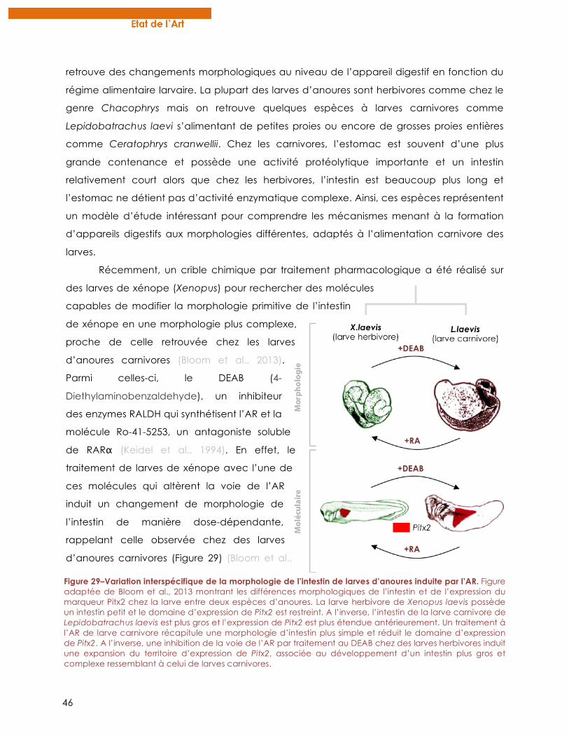
46
retrouve des changements morphologiques au niveau de l’appareil digestif en fonction du
régime alimentaire larvaire. La plupart des larves d’anoures sont herbivores comme chez le
genre Chacophrys mais on retrouve quelques espèces à larves carnivores comme
Lepidobatrachus laevi s’alimentant de petites proies ou encore de grosses proies entières
comme Ceratophrys cranwellii. Chez les carnivores, l’estomac est souvent d’une plus
grande contenance et possède une activité protéolytique importante et un intestin
relativement court alors que chez les herbivores, l’intestin est beaucoup plus long et
l’estomac ne détient pas d’activité enzymatique complexe. Ainsi, ces espèces représentent
un modèle d’étude intéressant pour comprendre les mécanismes menant à la formation
d’appareils digestifs aux morphologies différentes, adaptés à l’alimentation carnivore des
larves.
Récemment, un crible chimique par traitement pharmacologique a été réalisé sur
des larves de xénope (Xenopus) pour rechercher des molécules
capables de modifier la morphologie primitive de l’intestin
de xénope en une morphologie plus complexe,
proche de celle retrouvée chez les larves
d’anoures carnivores (Bloom et al., 2013).
Parmi celles-ci, le DEAB (4-
Diethylaminobenzaldehyde), un inhibiteur
des enzymes RALDH qui synthétisent l’AR et la
molécule Ro-41-5253, un antagoniste soluble
de RARα (Keidel et al., 1994). En effet, le
traitement de larves de xénope avec l’une de
ces molécules qui altèrent la voie de l’AR
induit un changement de morphologie de
l’intestin de manière dose-dépendante,
rappelant celle observée chez des larves
d’anoures carnivores (Figure 29) (Bloom et al.,
Figure 29–Variation interspécifique de la morphologie de l’intestin de larves d’anoures induite par l’AR. Figure adaptée de Bloom et al., 2013 montrant les différences morphologiques de l’intestin et de l’expression du marqueur Pitx2 chez la larve entre deux espèces d’anoures. La larve herbivore de Xenopus laevis possède un intestin petit et le domaine d’expression de Pitx2 est restreint. A l’inverse, l’intestin de la larve carnivore de Lepidobatrachus laevis est plus gros et l’expression de Pitx2 est plus étendue antérieurement. Un traitement à l’AR de larve carnivore récapitule une morphologie d’intestin plus simple et réduit le domaine d’expression de Pitx2. A l’inverse, une inhibition de la voie de l’AR par traitement au DEAB chez des larves herbivores induit une expansion du territoire d’expression de Pitx2, associée au développement d’un intestin plus gros et complexe ressemblant à celui de larves carnivores.
47
2013). Des changements similaires sont observés après traitement de larves de Chacophrys,
une espèce herbivore à la morphologie intestinale simple. A l’inverse, une exposition à l’AR
de larves d’anoures carnivores (Lepidobatrachus) semble restaurer une morphologie
intestinale plus simple ressemblant aux structures primitives des espèces herbivores. Au
niveau moléculaire, l’expression du marqueur pitx2, un facteur de transcription impliqué
dans la mise en place de l’asymétrie G/D des viscères, est différente entre les espèces à
larves herbivores et carnivores. En effet, l’expression de pitx2 est plus étendue chez les larves
carnivores ce qui pourrait expliquer la formation de structures digestives de plus grande
taille en déterminant un plus grand territoire présomptif. De manière intéressante, une
altération de la voie de l’AR chez des larves herbivores étend le territoire d’expression de
pitx2 comme observé chez les larves carnivores et vice-versa (Bloom et al., 2013) (Figure 29).
Ainsi, les auteurs concluent qu’une modification de l’expression de gènes impliqués dans
l’asymétrie G/D et régulés par l’AR (comme pitx2) pourrait avoir participé à l’émergence de
nouvelles morphologies intestinales interspécifiques chez les anoures. Celles-ci auraient été
sélectionnées positivement au cours de l’évolution car étant plus adaptées à un certain
régime alimentaire. Reste à déterminer comment une telle modulation de la voie de l’AR a
pu être mise en place sans altérer le développement global d’autres structures
embryonnaires dépendantes de l’AR.
4. LA PERTE DES DENTS ORALES CHEZ LES CYPRINIFORMES
4.1. Les dents de poisson comme modèle d’étude en évo-dévo.
Les dents correspondent à des structures spécifiques des vertébrés et leur mode de
développement semble conservé au sein du groupe (Jernvall and Salazar-Ciudad, 2007;
Smith et al., 2009). De part leur grande diversité de nombre, de forme et de localisation,
l’étude des mécanismes menant au développement d’un certain nombre de dents à
différents endroits dans la cavité buccale et/ou le pharynx est d’un intérêt tout particulier
pour des études évo-dévo. Plus de la moitié des espèces des vertébrés sont des
acténoptérygiens c’est à dire des poissons à nageoires rayonnées, le groupe de vertébré le
plus diversifié avec plus de 27 000 espèces recensées (Nelson, 2006). Ils présentent une
incroyable diversité morphologique de forme et de couleur et leur denture est également
très diversifiée. En effet, selon les espèces on peut retrouver des dents sur les mâchoires
inférieures et/ou supérieures, sur le palais, dans différentes régions de la bouche et tout le
long du pharynx (Pasco-Viel et al., 2010; Stock, 2007) (Figure 30). A titre d’exemple, entre les
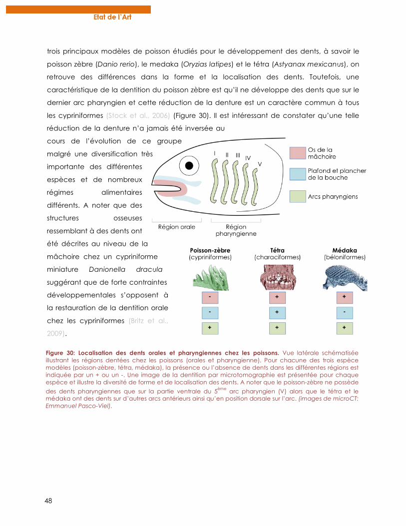
48
trois principaux modèles de poisson étudiés pour le développement des dents, à savoir le
poisson zèbre (Danio rerio), le medaka (Oryzias latipes) et le tétra (Astyanax mexicanus), on
retrouve des différences dans la forme et la localisation des dents. Toutefois, une
caractéristique de la dentition du poisson zèbre est qu’il ne développe des dents que sur le
dernier arc pharyngien et cette réduction de la denture est un caractère commun à tous
les cypriniformes (Stock et al., 2006) (Figure 30). Il est intéressant de constater qu’une telle
réduction de la denture n’a jamais été inversée au
cours de l’évolution de ce groupe
malgré une diversification très
importante des différentes
espèces et de nombreux
régimes alimentaires
différents. A noter que des
structures osseuses
ressemblant à des dents ont
été décrites au niveau de la
mâchoire chez un cypriniforme
miniature Danionella dracula
suggérant que de forte contraintes
développementales s’opposent à
la restauration de la dentition orale
chez les cypriniformes (Britz et al.,
2009).
Figure 30: Localisation des dents orales et pharyngiennes chez les poissons. Vue latérale schématisée illustrant les régions dentées chez les poissons (orales et pharyngienne). Pour chacune des trois espèce modèles (poisson-zèbre, tétra, médaka), la présence ou l’absence de dents dans les différentes régions est indiquée par un + ou un -. Une image de la dentition par microtomographie est présentée pour chaque espèce et illustre la diversité de forme et de localisation des dents. A noter que le poisson-zèbre ne possède
des dents pharyngiennes que sur la partie ventrale du 5ème arc pharyngien (V) alors que le tétra et le médaka ont des dents sur d’autres arcs antérieurs ainsi qu’en position dorsale sur l’arc. (images de microCT: Emmanuel Pasco-Viel).
49
4.2. Scénario impliquant la voie de l’AR pour la perte des dents orales
chez les cypriniformes.
Quels sont les mécanismes développementaux à l’origine de la réduction de la
denture chez les cypriniformes ? Comment une telle réduction n’a-t-elle jamais été
inversée ? Une étude comparative du développement des dents entre le poisson-zèbre et
d’autres espèces possédant des dents dans la cavité buccale (médaka, tétra) permettrait
ainsi de comprendre le scénario évolutif de la réduction de la denture des cypriniformes.
Une étude du laboratoire de Vincent Laudet a montré que la voie de l’AR était nécessaire
pour le développement des dents pharyngiennes chez le poisson-zèbre (Gibert et al., 2010).
En effet, l’inhibition de la synthèse endogène d’AR ou le traitement des embryons à des
antagonistes des RAR empêche l’expression des marqueurs dentaires comme dlx2b ou
pitx2a et inhibe ainsi le développement de la première paire de dents. Au niveau du
pharynx, l’enzyme RALDH2 est exprimée dans le pharynx postérieur ventral entourant le
cinquième arc branchial. Ainsi, il est proposé que RALDH2 synthétise localement l’AR qui va
servir de signal pour l’induction des bourgeons dentaires chez le poisson-zèbre via
l’expression de marqueurs spécifiques comme pitx2a ou dlx2a. Cependant, de manière
surprenante, l’induction des dents orales et pharyngiennes chez le médaka et le tétra n’est
pas dépendante de la voie de l’AR (Gibert et al., 2010). Ces données sont soutenues par
l’observation que chez le tétra, l’enzyme RALDH2 n’est pas exprimée dans la région du
pharynx où sont induites les dents (Figure 31).
Ainsi, l’ensemble de ces résultats suggère que chez les cypriniformes, le
développement des dents a acquis une dépendance à la voie de l’AR, qui peut être
corrélée au décalage de l’expression de RALDH2 au niveau du pharynx postérieur chez ces
espèces. Etant donné que le développement des dents pharyngiennes et orales repose sur
des mécanismes moléculaires similaires, il est vraisemblable que la dépendance à l’AR ait
également touché le développement des dents orales. Or, les enzymes RADLH2 et RALDH3
n’étant pas exprimées dans la région orale chez le poisson zèbre (White et al., 2007) l’AR n’y
est donc pas synthétisé empêchant ainsi le développement de dents orales (Figure 31).
Il est peu probable que la perte des dents orales chez les cypriniformes soit due à des
mutations touchant la fonction et la régulation des gènes impliqués dans le développement
des dents comme les gènes dlx. En effet, de telles mutations auraient certainement des
effets importants sur le développement d’autres structures, comme les arcs pharyngiens, et
mèneraient à des défauts délétères. Ainsi, ce scénario basé sur l’acquisition d’une
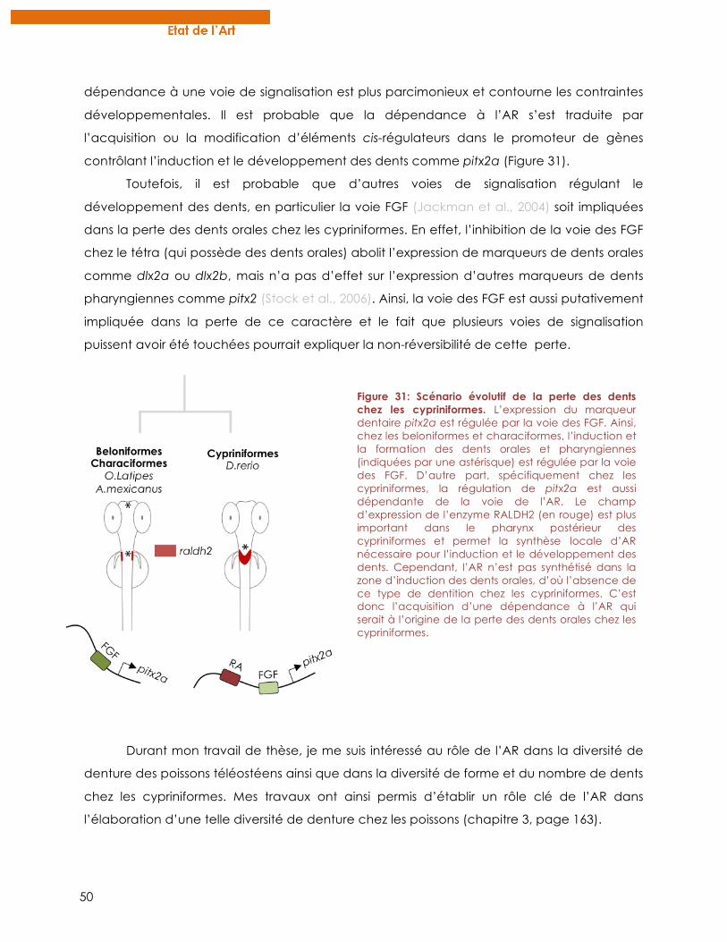
50
dépendance à une voie de signalisation est plus parcimonieux et contourne les contraintes
développementales. Il est probable que la dépendance à l’AR s’est traduite par
l’acquisition ou la modification d’éléments cis-régulateurs dans le promoteur de gènes
contrôlant l’induction et le développement des dents comme pitx2a (Figure 31).
Toutefois, il est probable que d’autres voies de signalisation régulant le
développement des dents, en particulier la voie FGF (Jackman et al., 2004) soit impliquées
dans la perte des dents orales chez les cypriniformes. En effet, l’inhibition de la voie des FGF
chez le tétra (qui possède des dents orales) abolit l’expression de marqueurs de dents orales
comme dlx2a ou dlx2b, mais n’a pas d’effet sur l’expression d’autres marqueurs de dents
pharyngiennes comme pitx2 (Stock et al., 2006). Ainsi, la voie des FGF est aussi putativement
impliquée dans la perte de ce caractère et le fait que plusieurs voies de signalisation
puissent avoir été touchées pourrait expliquer la non-réversibilité de cette perte.
Durant mon travail de thèse, je me suis intéressé au rôle de l’AR dans la diversité de
denture des poissons téléostéens ainsi que dans la diversité de forme et du nombre de dents
chez les cypriniformes. Mes travaux ont ainsi permis d’établir un rôle clé de l’AR dans
l’élaboration d’une telle diversité de denture chez les poissons (chapitre 3, page 163).
Figure 31: Scénario évolutif de la perte des dents chez les cypriniformes. L’expression du marqueur dentaire pitx2a est régulée par la voie des FGF. Ainsi, chez les beloniformes et characiformes, l’induction et la formation des dents orales et pharyngiennes (indiquées par une astérisque) est régulée par la voie des FGF. D’autre part, spécifiquement chez les cypriniformes, la régulation de pitx2a est aussi dépendante de la voie de l’AR. Le champ d’expression de l’enzyme RALDH2 (en rouge) est plus important dans le pharynx postérieur des cypriniformes et permet la synthèse locale d’AR nécessaire pour l’induction et le développement des dents. Cependant, l’AR n’est pas synthétisé dans la zone d’induction des dents orales, d’où l’absence de ce type de dentition chez les cypriniformes. C’est donc l’acquisition d’une dépendance à l’AR qui serait à l’origine de la perte des dents orales chez les cypriniformes.
51
C. LES RAR COORDONNENT LE SIGNAL DE L’AR AU COURS DU
DEVELOPPEMENT
Cette partie introductive correspond à une revue publiée en 2011 dans un numéro
spécial du journal Molecular and Cellular Endocrinology intitulé « Nuclear Receptor
Structure : Dynamics and Function ». Elle a pour but d’exposer le rôle des RAR dans la
transduction du signal de l’AR au cours du développement dans un contexte in vivo. Plutôt
que de cataloguer les nombreux effets de l’AR décrits jusqu’alors, principalement chez la
souris, nous avons voulu corréler les aspects moléculaires de la structure des RAR à leur
fonction in vivo au cours du développement. Cette revue illustre le rôle central des RAR
comme coordinateurs de la réponse de l’AR, mais nécessitant l’intégrité de nombreux
partenaires et d’autres voies de signalisation pour transduire harmonieusement le signal de
l’AR au cours du développement.
52
Review
Nuclear retinoic acid receptors: Conductors of the retinoic acid symphony duringdevelopment
Eric Samarut, Cécile Rochette-Egly ⇑
IGBMC (Institut de Génétique et de Biologie Moléculaire et Cellulaire), INSERM, U596; CNRS, UMR7104; Université de Strasbourg, 1 rue Laurent Fries, BP 10142, 67404 Illkirch Cedex,France
a r t i c l e i n f o
Article history:Received 7 January 2011Received in revised form 21 March 2011Accepted 31 March 2011Available online 8 April 2011
Keywords:RARARPhosphorylationTranscriptionDevelopmentDifferentiation
a b s t r a c t
The vitamin A derivative, retinoic acid (RA), is essential for embryonic development through the activa-tion of cognate nuclear receptors, RARs, which work as ligand dependent regulators of transcription. Invitro studies revealed how RARs control gene expression at the molecular level and now it appears thatit is fine-tuned by a phosphorylation code. In addition, several genetic approaches provided valuableinsights on the functions of RARs during development and on the influence of other actors such as theenzymes involved in RA synthesis and degradation and other signaling pathways. It appears that RARsare the conductors of the RA signaling symphony through controlling the dynamics and the coordinationof the different players and development steps.
! 2011 Elsevier Ireland Ltd. All rights reserved.
Contents
1. Introduction . . . . . . . . . . . . . . . . . . . . . . . . . . . . . . . . . . . . . . . . . . . . . . . . . . . . . . . . . . . . . . . . . . . . . . . . . . . . . . . . . . . . . . . . . . . . . . . . . . . . . . . . . 3482. Nuclear retinoic acid receptors are the functional effectors of RA signaling . . . . . . . . . . . . . . . . . . . . . . . . . . . . . . . . . . . . . . . . . . . . . . . . . . . . . . 349
2.1. RARs structure and canonical RAR-mediated regulation of transcription . . . . . . . . . . . . . . . . . . . . . . . . . . . . . . . . . . . . . . . . . . . . . . . . . . . 3492.2. RA synthesis and degradation . . . . . . . . . . . . . . . . . . . . . . . . . . . . . . . . . . . . . . . . . . . . . . . . . . . . . . . . . . . . . . . . . . . . . . . . . . . . . . . . . . . . . 3502.3. New pictures of RAR signaling: kinase cascade activation . . . . . . . . . . . . . . . . . . . . . . . . . . . . . . . . . . . . . . . . . . . . . . . . . . . . . . . . . . . . . . . 3512.4. Multi-modulation of RAR-target genes through phosphorylation . . . . . . . . . . . . . . . . . . . . . . . . . . . . . . . . . . . . . . . . . . . . . . . . . . . . . . . . . 351
3. Dynamic expression of the RA signaling players provides a robust and coordinated RA response . . . . . . . . . . . . . . . . . . . . . . . . . . . . . . . . . . . . 3513.1. RARs are the conductors transducing RA signaling . . . . . . . . . . . . . . . . . . . . . . . . . . . . . . . . . . . . . . . . . . . . . . . . . . . . . . . . . . . . . . . . . . . . . 3513.2. RAR expression is subtly orchestrated during development . . . . . . . . . . . . . . . . . . . . . . . . . . . . . . . . . . . . . . . . . . . . . . . . . . . . . . . . . . . . . 3523.3. Other players in the RA symphony: RAR coregulators . . . . . . . . . . . . . . . . . . . . . . . . . . . . . . . . . . . . . . . . . . . . . . . . . . . . . . . . . . . . . . . . . . 3543.4. Importance of the RA ‘‘sources and sinks’’. . . . . . . . . . . . . . . . . . . . . . . . . . . . . . . . . . . . . . . . . . . . . . . . . . . . . . . . . . . . . . . . . . . . . . . . . . . . 354
4. RARs conduct RA signaling in an integrative and coordinated manner . . . . . . . . . . . . . . . . . . . . . . . . . . . . . . . . . . . . . . . . . . . . . . . . . . . . . . . . . . 3564.1. Crosstalks with other signaling pathways fine-shape RA boundaries . . . . . . . . . . . . . . . . . . . . . . . . . . . . . . . . . . . . . . . . . . . . . . . . . . . . . . 3564.2. RARs molecular targets: from direct target genes to coordinated gene cascades . . . . . . . . . . . . . . . . . . . . . . . . . . . . . . . . . . . . . . . . . . . . . 3564.3. Epigenetic and chromatin modifications: a dynamic ‘‘language’’ to modulate RAR-target gene expression. . . . . . . . . . . . . . . . . . . . . . . . 357
5. Conclusions and perspectives . . . . . . . . . . . . . . . . . . . . . . . . . . . . . . . . . . . . . . . . . . . . . . . . . . . . . . . . . . . . . . . . . . . . . . . . . . . . . . . . . . . . . . . . . . . 357Acknowledgements . . . . . . . . . . . . . . . . . . . . . . . . . . . . . . . . . . . . . . . . . . . . . . . . . . . . . . . . . . . . . . . . . . . . . . . . . . . . . . . . . . . . . . . . . . . . . . . . . . . 357References . . . . . . . . . . . . . . . . . . . . . . . . . . . . . . . . . . . . . . . . . . . . . . . . . . . . . . . . . . . . . . . . . . . . . . . . . . . . . . . . . . . . . . . . . . . . . . . . . . . . . . . . . . 357
1. Introduction
Vitamin A (retinol) is essential for embryonic development,organogenesis and homeostasis of most body tissues as well asfor vision, immune functions and reproduction. Such an important
0303-7207/$ - see front matter ! 2011 Elsevier Ireland Ltd. All rights reserved.doi:10.1016/j.mce.2011.03.025
⇑ Corresponding author. Tel.: +33 3 88 65 34 59; fax: +33 3 88 65 32 01.E-mail addresses: [email protected] (E. Samarut), [email protected] (C. Rochette-Egly).
Molecular and Cellular Endocrinology 348 (2012) 348–360
Contents lists available at SciVerse ScienceDirect
Molecular and Cellular Endocrinology
journal homepage: www.elsevier .com/locate /mce
53
role has been revealed by both experimental approaches andclinical observations. Indeed deficiency of vitamin A leads to neo-natal growth retardation and a large array of congenital malforma-tions affecting the ocular, cardiac, respiratory and urogenitalsystems collectively referred to as the fetal vitamin A deficiency(VAD) syndrome (Clagett-Dame and DeLuca, 2002; Zile, 2001).
The only source of vitamin A in the organism is diet-derived andretinol has to be converted to active compounds such as retinoicacid (RA). Then in cells and tissues, RA levels must be controlledthrough a balance between synthesis and catabolism (Fig. 1A).Now it is admitted that RA supports the effects of vitamin A. Thisis based on the ability of RA to replace vitamin A during embryo-genesis and to reverse some developmental defects in VADanimals.
Newly synthesized RA binds to cellular RA binding proteins(CRABP-I and CRABP-II) and enters the nucleus (Budhu and Noy,2002; Delva et al., 1999) where it controls gene expression throughthe activation of specific nuclear receptors, RARs, which consist ofthree subtypes, a (NR1B1), b (NR1B2) and c (NR1B3) encoded byseparate genes (for review see Chambon, 1996; Germain et al.,2006a,c). In mammals, for each subtype, there are several isoforms,which are generated by differential promoter usage and alternativesplicing and differ only in their N-terminal regions. RARs functionas ligand-dependent transcriptional regulators, heterodimerizedwith retinoid X receptors (RXRs), which also consist of three types,a (NR2B1), b (NR2B2) and c (NR2B3) (Germain et al., 2006b). Assuch, they regulate the expression of subsets of target genes in-volved in cellular differentiation, proliferation and apoptosis, andmediate the pleiotropic effects of RA.
In this review we will recapitulate how RARs conduct RA sig-naling during development, focusing on molecular aspects andon three main questions: (i) when and where are RARs neededduring development (ii) when and where is RA available (iii) doRARs integrate other developmental networks and then whenand where.
2. Nuclear retinoic acid receptors are the functional effectors ofRA signaling
2.1. RARs structure and canonical RAR-mediated regulation oftranscription
The basics of RARs’ structure and function have been recapitu-lated in several recent reviews (Bastien and Rochette-Egly, 2004;Chambon, 1996; Laudet and Gronemeyer, 2001; Lefebvre et al.,2005; Rochette-Egly and Germain, 2009). Briefly, RARs exhibit awell-defined domain organization and structure consisting mainlyof a central DNA-binding domain (DBD) and a C-terminal ligand-binding domain (LBD) (Fig. 1B). Crystallographic and nuclear mag-netic resonance analysis of the structures of these domains and thecharacterization of the associated multiprotein complexes pro-vided a wealth of information on how RARs regulate transcription.However, recent studies highlighted the importance of the N-ter-minal domain (NTD), which, in contrast to the DBD and the LBD,is naturally disordered. Nevertheless, it contains a conserved pro-line-rich motif, which forms helices that can bind proteins withSH3 or WW domains (Lalevee et al., 2010). It has been suggestedthat binding of such coregulatory proteins might have profoundimpacts on the structural properties of the NTD and/or on thedynamics of the adjacent domains.
The LBD is formed by 12 conserved alpha helices and a beta-turn, which are folded into a three-layered, and parallel helicalsandwich (Fig. 1B) (Renaud and Moras, 2000; Renaud et al.,1995). It is functionally complex as it contains the ligand-bindingpocket (LBP), the main dimerization domain and a hydrophobic
cleft involved in coregulators binding. Analysis of the crystal struc-tures of the unliganded and ligand-bound LBDs highlighted that RAbinding induces conformational changes, the most striking onebeing the repositionment of helix 12 (le Maire et al., 2010; Renaudet al., 1995).
The core of the DBD, which confers sequence specific DNA rec-ognition, is composed of two zinc-nucleated modules and two a-helices, which cross at right angles and fold into a single globulardomain (Zechel et al., 1994a,b). The DBD also includes a dimeriza-tion surface that allows RARs bind to DNA as asymmetric, orientedheterodimers with RXRs. Note that the DBD of RXR depicts a thirda-helix that facilitates DNA binding and homodimerization (Leeet al., 1993).
RXR/RAR heterodimers bind to specific DNA sequences or RAresponse elements (RAREs), composed typically of two direct re-peats of a core hexameric motif, (A/G)G [G/T] TCA and located inthe regulatory sequences of target genes (Leid et al., 1992; Man-gelsdorf and Evans, 1995). The classical RARE is a 5 bp-spaced di-rect repeat (referred to as DR5). However, the heterodimers alsobind to direct repeats separated by 1 bp (DR1) or 2 bp (DR2).RAREs have been identified in the promoters of a large numberof RA-target genes implicated in a wide variety of functions includ-ing RA metabolism and cellular differentiation (Balmer and Blom-hoff, 2005; Bour et al., 2006). For instance, the classical DR5elements are found in the promoters of the rarb2 gene itself, thecyp26A1 gene, the foxA1 gene and several Homeobox (hox) genes.Recently the development of high throughput technologies suchas chromatin immunoprecipitation (ChIP) coupled with arrayhybridization or flow cell sequencing expanded the repertoire ofpotential high affinity response elements (Delacroix et al., 2010;Hua et al., 2009). However it also revealed degenerative andnon-identifiable DRs, as well as DRs overlapping with binding sitesfor other transcription factors, increasing the complexity of RAR-mediated gene transcription.
The transcriptional regulation of RA-target genes relies on RARsbinding to DNA and on ligand-induced conformational changes inthe LBD that direct the association/dissociation of several coregula-tor complexes (Fig. 1C). According to the canonical model (Dil-worth and Chambon, 2001), in the absence of ligand and in acontext of chromatin where the nucleosomes do not impede bind-ing to RAREs, RAREs are occupied by RAR/RXR heterodimers, asso-ciated with large protein complexes endowed with enzymaticactivities, which maintain chromatin in a condensed and repressedstate. According to recent studies, repression in the absence of RAmay also involve Topoisomerase II (McNamara et al., 2008) andpolycomb group proteins (Gillespie and Gudas, 2007a,b). Upon li-gand binding, RARs undergo conformational changes that provokecorepressors release and initiate an ordered and coordinatedrecruitment of coactivators within large complexes with differentenzymatic activities (histone acetyl and methyl transferases andDNA-dependent ATPases) (Perissi et al., 2010; Perissi and Rosen-feld, 2005), which will alter the chromatin structure surroundingthe promoter of the target genes and pave the way for the recruit-ment of the transcription machinery including the multisubunitmediator complex, RNA polymerase II and the general transcrip-tion factors.
However, recent studies highlighted that, depending on thetarget gene promoter, RARs can employ different programs forgene activation. Indeed, the mediator and RNA PolII can alreadyoccupy with RARa the promoters of some endogenous genes evenin the absence of RA (Flajollet et al., 2006; Pavri et al., 2005). Inthat context, gene-specific factors like PARP-1 are determinantfor switching the Mediator to its active form. According to otherstudies, only a small fraction of RAREs are occupied by RXR/RARheterodimers in the absence of RA and RARs are recruited toRAREs upon ligand binding (Biddie et al., 2010; Bruck et al.,
E. Samarut, C. Rochette-Egly / Molecular and Cellular Endocrinology 348 (2012) 348–360 349
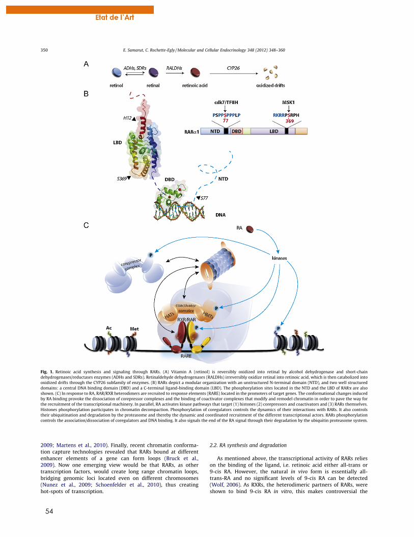
54
2009; Martens et al., 2010). Finally, recent chromatin conforma-tion capture technologies revealed that RARs bound at differentenhancer elements of a gene can form loops (Bruck et al.,2009). Now one emerging view would be that RARs, as othertranscription factors, would create long range chromatin loops,bridging genomic loci located even on different chromosomes(Nunez et al., 2009; Schoenfelder et al., 2010), thus creatinghot-spots of transcription.
2.2. RA synthesis and degradation
As mentioned above, the transcriptional activity of RARs relieson the binding of the ligand, i.e. retinoic acid either all-trans or9-cis RA. However, the natural in vivo form is essentially all-trans-RA and no significant levels of 9-cis RA can be detected(Wolf, 2006). As RXRs, the heterodimeric partners of RARs, wereshown to bind 9-cis RA in vitro, this makes controversial the
Fig. 1. Retinoic acid synthesis and signaling through RARs. (A) Vitamin A (retinol) is reversibly oxidized into retinal by alcohol dehydrogenase and short-chaindehydrogenases/reductases enzymes (ADHs and SDRs). Retinaldehyde dehydrogenases (RALDHs) irreversibly oxidize retinal into retinoic acid, which is then catabolized intooxidized drifts through the CYP26 subfamily of enzymes. (B) RARs depict a modular organization with an unstructured N-terminal domain (NTD), and two well structureddomains: a central DNA binding domain (DBD) and a C-terminal ligand-binding domain (LBD). The phosphorylation sites located in the NTD and the LBD of RARa are alsoshown. (C) In response to RA, RAR/RXR heterodimers are recruited to response elements (RARE) located in the promoters of target genes. The conformational changes inducedby RA binding provoke the dissociation of corepressor complexes and the binding of coactivator complexes that modify and remodel chromatin in order to pave the way forthe recruitment of the transcriptional machinery. In parallel, RA activates kinase pathways that target (1) histones (2) corepressors and coactivators and (3) RARs themselves.Histones phosphorylation participates in chromatin decompaction. Phosphorylation of coregulators controls the dynamics of their interactions with RARs. It also controlstheir ubiquitination and degradation by the proteasome and thereby the dynamic and coordinated recruitment of the different transcriptional actors. RARs phosphorylationcontrols the association/dissociation of coregulators and DNA binding. It also signals the end of the RA signal through their degradation by the ubiquitin proteasome system.
350 E. Samarut, C. Rochette-Egly / Molecular and Cellular Endocrinology 348 (2012) 348–360
55
in vivo relevance of retinoids binding to RXRs (Calleja et al., 2006;Mic et al., 2003).
Retinoic acid is a derivative of vitamin A that cannot be synthe-sized de novo. Therefore RA supply in the organism is exclusivelydiet-derived and requires several conversions by key enzymes thatrepresent an important part of the large RA signaling family com-ponents (Duester, 2008; Maden, 2007; Theodosiou et al., 2010).Briefly, RA is synthesized through retinol oxidation to retinal byalcohol dehydrogenase and short-chain dehydrogenases/reduc-tases enzymes (ADHs and SDRs) (Fig. 1A). Retinal is then irrevers-ibly oxidized into RA through the action of retinaldehydedehydrogenases (RALDHs). Three RALDHs (RALDH1, RALDH2 andRALDH3) have been identified and exhibit distinct tissue expres-sion patterns during mouse development, which collectively corre-late with the dynamics of RA production. Note that according tosome reports, RA synthesis during embryonic patterning may alsooccur independently of RALDHs and involve CYP1B1 or CYP1A1,members of the cytochrome p450 family of mono-oxygenases(Chambers et al., 2007; Chen et al., 2000).
On the other hand, RA is catabolized into more polar oxidizedmetabolites such as 4-hydroxy or 4-oxo RA through enzymes ofthe cytochrome P450 family (Fig. 1A). The mammalian genomescontain three cyp26 genes, cyp26A1, cyp26B1 and cyp26C1 andorthologs have been cloned in other vertebrates like zebrafish(White and Schilling, 2008). They depict specific expression pat-terns, suggesting individual and combinatorial roles for each onein the control of RA levels.
Interestingly, both RA metabolic cascade and RAR-mediated RAsignaling are conserved during evolution (Campo-Paysaa et al.,2008; Escriva et al., 2006; Marletaz et al., 2006; Samarut et al.,2011; Theodosiou et al., 2010), emphasizing the robustness andthe significance of this pathway in many biological processesincluding embryonic development.
2.3. New pictures of RAR signaling: kinase cascade activation
Today it is becoming increasingly evident that, in addition tothe classical genomic effects, liganded RARs also induce non-genomic responses such as the rapid and transient activation ofseveral kinase cascades (Fig. 1C). These non-genomic effects aremediated by a subpopulation of RARs anchored at the cytoplas-mic side of the cell membrane (Masia et al., 2007; Piskunovand Rochette-Egly, in press). Indeed, in several cell types, RA-bound RARs activate p38MAPK, which in turn, translocates intothe nucleus and activates a downstream mitogen and stress-acti-vated kinase, MSK1 (Bruck et al., 2009). However, this processappears to be cell-type specific as RARs rather activate Erks inneuronal and Sertoli cells (Chen and Napoli, 2008; Gupta et al.,2008; Masia et al., 2007; Pan et al., 2005). Nevertheless this sug-gests a new paradigm by which RARs integrate membrane/cyto-plasm events orchestrating several coordinated cascades ofphosphorylations of nuclear proteins, in order to fine-tune tran-scription (Fig. 1C).
In line with this, it emerged that in the nucleus, RA-activatedMSK1 phosphorylates rapidly RARa at a serine residue (S369) lo-cated in loop L9-10 within the LBD (Bruck et al., 2009) (Fig. 1B).Phosphorylation of this serine increases the dynamics/flexibilityof the nearby loop L8-9 (Samarut et al., 2011), which correspondsto the docking site of cyclin H, that forms with CDK7 and MAT1 theCAK subcomplex of the general transcription factor TFIIH. Conse-quently, RARa becomes able to bind cyclin H, allowing the phos-phorylation of S77 located in the proline-rich domain of the NTDby the CDK7 kinase (Bour et al., 2005; Gaillard et al., 2006)(Fig. 1B). Finally, phosphorylation of the NTD drives the recruit-ment of RARa to promoters. Note that in the case of the RARc sub-type, phosphorylation of the NTD also induces the dissociation of
vinexinb, an actin-binding protein with SH3 domains, which re-presses RARc activity (Lalevee et al., 2010). Thus RA-induced phos-phorylations appear to play an important role in the dynamics ofRAR-mediated transcription.
2.4. Multi-modulation of RAR-target genes through phosphorylation
Now it emerges that RARs act through the combination of geno-mic and non-genomic effects, introducing the notion of transcrip-tional dynamics so that the right proteins are present with theright activity at the right place and at the right time.
Indeed, at the very beginning of the RA switch, RA-activatedMSK1 is required for making RAREs accessible to RAR/RXR hetero-dimers through binding to chromatin and phosphorylation of his-tones H3 (H3S10), an event that promotes other histonesmodifications and chromatin remodeling, according to the histonecode (Bruck et al., 2009; Vermeulen et al., 2009). MSK1 is also re-quired for DNA recruitment of RARa (Bruck et al., 2009), throughRARa phosphorylation (see Section 2.1).
Whether MSK1 also controls the association/dissociation ofcoregulator complexes is still unclear but it has been shown thatthe upstream kinase p38MAPK phosphorylates corepressors(Perissi et al., 2008; Rosenfeld et al., 2006), as well as the coac-tivator SRC-3 (Gianni et al., 2006). Then phosphorylation pro-motes their ubiquitination according to a code very similar tothe histone code and finally their degradation by the 26S protea-some (Gianni et al., 2006) (Fig. 1C). It has been proposed thatphosphorylation, ubiquitination and degradation cooperate tocontrol the corepressors/coactivators exchanges in line with theidea that transcription is a highly dynamic process. Lastly, atthe end of the RA response, RARs are also ubiquitinated and de-graded (Gianni et al., 2002; Kopf et al., 2000), a radical processfor turning off the transcriptional signal (Bour et al., 2007; Roch-ette-Egly and Germain, 2009).
3. Dynamic expression of the RA signaling players provides arobust and coordinated RA response
3.1. RARs are the conductors transducing RA signaling
Numerous studies described the importance of RA signalingduring early development and organogenesis (Clagett-Dame andDeLuca, 2002; Maden, 2007; Mark et al., 2006; Zile, 2001) withmouse being one of the most studied vertebrate model. These stud-ies demonstrated that RA is crucial for the correct patterning ofearly structures in the embryo such as neural tube developmentand closure, spinal chord formation, rhombomeres patterningand specification, somitogenesis, limb bud morphogenesis andheart development.
How RA could exert such pleiotropic effects was a long-stand-ing question until the discovery of RARs. Subsequently, geneticapproaches in the animal corroborated that RARs are indeedthe major conductors of RA signaling during embryonic develop-ment and decoded how they function at the molecular level(Mark et al., 2006, 2009). Mutant mice lacking RARa, RARb orRARc were first generated. Unexpectedly these single KO micewere viable and displayed a limited number of defects that didnot mirror all those seen in the fetal and postnatal VAD syn-dromes (Table 1). In fact, complete VAD phenotypes were reca-pitulated only in double KO mutant mice (Table 1), suggestingthat RARs are largely functionally redundant. The exhaustiveanalysis of the different double KO combinations allowed draw-ing a list assigning specific phenotypes and thus specific func-tions for RARs during development. Note that RAR doublemutants also exhibited congenital abnormalities that were not
E. Samarut, C. Rochette-Egly / Molecular and Cellular Endocrinology 348 (2012) 348–360 351
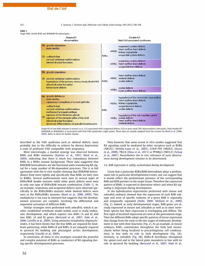
56
described in the VAD syndrome such as skeletal defects, mostprobably due to the difficulty to achieve by dietary deprivationa state of profound VAD compatible with pregnancy.
Most interestingly, a marked synergy was observed betweenRARs and RXRs mutations (Kastner et al., 1997; Mark et al.,2009), indicating that there is much less redundancy betweenRARs in a RXRa mutant background. These data suggested thatRXR/RAR heterodimers are the functional units transducing RA sig-nal for a large number of RA-dependent processes. This is in fullagreement with the in vitro studies showing that RXR/RAR hetero-dimers bind more tightly and specifically than RARs on their ownto RAREs. Several malformations were seen in several types ofRXRa/RAR double mutants while other given defects were seenin only one type of RXRa/RAR mutant combination (Table 1). Asan example, respiratory and urogenital defects were observed spe-cifically in the RXRa/RARa mutants while skeletal defects wereseen in the RXRa/RARc mutants (Mark et al., 2006, 2009). Thismultiplicity is not easy to interpret but suggests that the develop-mental processes are complex, involving the differential andsequential activation of different RARs.
Similar strategies were performed with zebrafish, which is an-other established vertebrate model widely used to study embry-onic development, and which express two RARa (A and B) andtwo RARc (A and B) genes (Bertrand et al., 2007; Hale et al.,2006; Linville et al., 2009). Using antisense morpholino oligonucle-otides, it has been shown that the four RARs are required for hind-brain patterning, while RARa-B and RARc-A are uniquely requiredin pectoral fin budding and pharyngeal arches development,respectively (Linville et al., 2009).
In conclusion, all these in vivo strategies revealed the powerfuland complex potential of RARs as conductors of RA signaling dur-ing specific developmental processes.
Note however that some recent in vitro studies suggested thatRA signaling could be mediated by other receptors such as RORb(NR1F2) (Stehlin-Gaon et al., 2003), COUP-TFII (NR2F2) (Kruseet al., 2008), TR2/4 (Zhou et al., 2011) or PPARb/d (NR1C2) (Schuget al., 2007). Nevertheless the in vivo relevance of such observa-tions during development remains to be determined.
3.2. RAR expression is subtly orchestrated during development
Given that a particular RXRa/RAR heterodimer plays a predom-inant role in particular developmental events, one can suggest thatit would reflect the predominant presence of the correspondingRAR and RXR partners in the target tissue. Therefore the expressionpattern of RARs, is expected to determine where and when RA sig-naling is important during development.
In situ hybridization experiments performed with mouse andzebrafish embryos showed that the expression of each RAR sub-type and even of specific isoforms of a given RAR is regionallyand temporally regulated (Dolle, 2009; Mollard et al., 2000)(Fig. 2). Indeed, at early developmental stages, RAR genes are al-ready expressed in mouse and zebrafish as well as in most verte-brate species but their expression is overlapping and diffuse. Thefirst signs of localized expression are seen at the gastrulation stage.Then the different RARs adopt specific patterns of tissue expressionthat change from the early to the late stages of embryonic develop-ment in line with their functions (Fig. 2). As an example, in mouseembryos, RARc concentrates throughout the limb bud mesen-chyme before being localized to precartilaginous cell condensa-tions in line with its role in limb development (Fig. 2A).Similarly, in zebrafish (Fig. 2B), RARa-B distributes throughoutthe spinal cord and in the lateral plate mesoderm in line with itsrole in pectoral fin budding (Bertrand et al., 2007; Hale et al.,
Table 1Single RAR, double RAR and RXR/RAR KO phenotypes.
Single KO of one RAR subtype in mouse (a, b, c) is associated with congenital defects (CD) or post-natal (PN) abnormalities (left panel). Only double KO(RAR/RAR or RAR/RXRa) is associated with fetal VAD syndromes (right panel). These data are mainly adapted from the reviews by Mark et al. (2006,2009). Refer to them for further details
352 E. Samarut, C. Rochette-Egly / Molecular and Cellular Endocrinology 348 (2012) 348–360
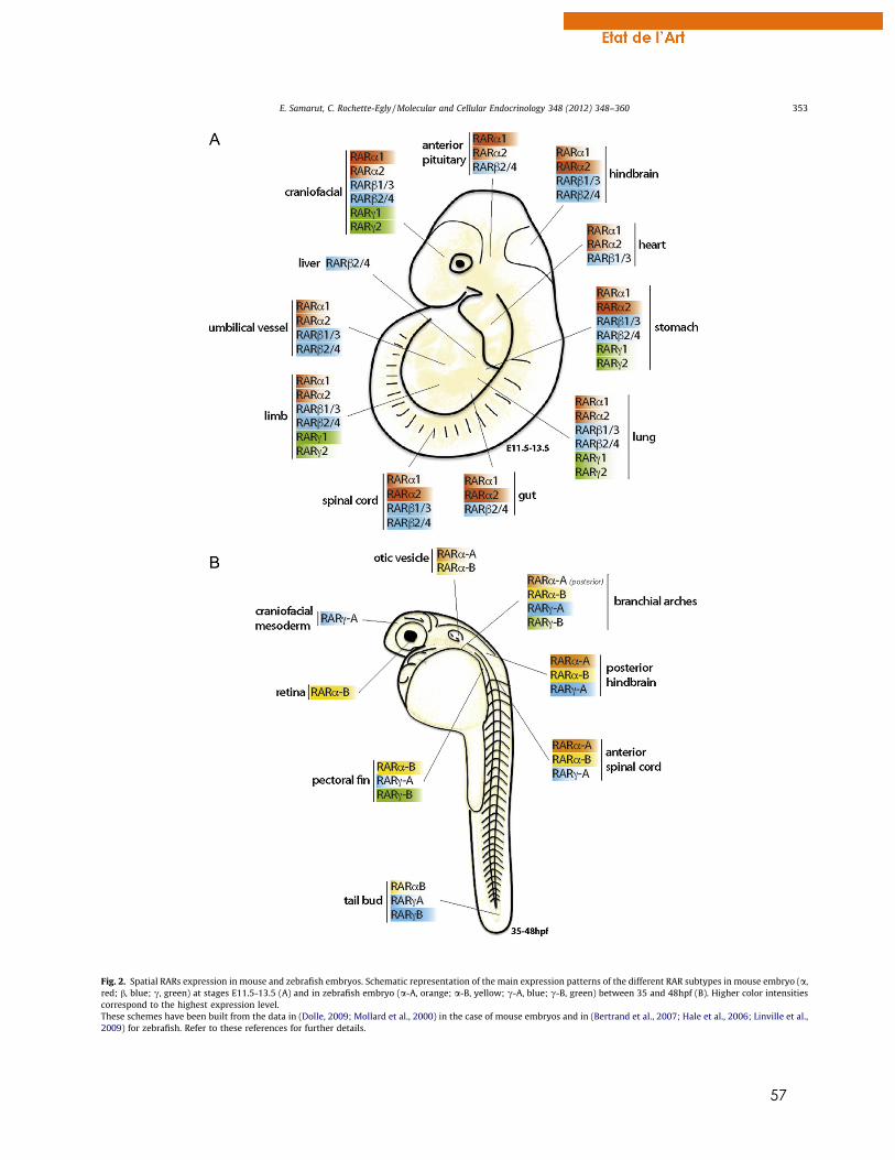
57
Fig. 2. Spatial RARs expression in mouse and zebrafish embryos. Schematic representation of the main expression patterns of the different RAR subtypes in mouse embryo (a,red; b, blue; c, green) at stages E11.5-13.5 (A) and in zebrafish embryo (a-A, orange; a-B, yellow; c-A, blue; c-B, green) between 35 and 48hpf (B). Higher color intensitiescorrespond to the highest expression level.These schemes have been built from the data in (Dolle, 2009; Mollard et al., 2000) in the case of mouse embryos and in (Bertrand et al., 2007; Hale et al., 2006; Linville et al.,2009) for zebrafish. Refer to these references for further details.
E. Samarut, C. Rochette-Egly / Molecular and Cellular Endocrinology 348 (2012) 348–360 353
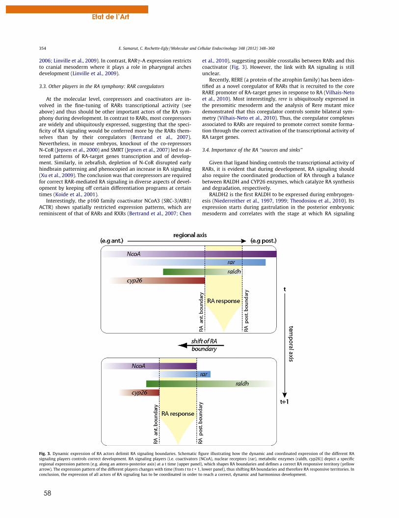
58
2006; Linville et al., 2009). In contrast, RARc-A expression restrictsto cranial mesoderm where it plays a role in pharyngeal archesdevelopment (Linville et al., 2009).
3.3. Other players in the RA symphony: RAR coregulators
At the molecular level, corepressors and coactivators are in-volved in the fine-tuning of RARs transcriptional activity (seeabove) and thus should be other important actors of the RA sym-phony during development. In contrast to RARs, most corepressorsare widely and ubiquitously expressed, suggesting that the speci-ficity of RA signaling would be conferred more by the RARs them-selves than by their coregulators (Bertrand et al., 2007).Nevertheless, in mouse embryos, knockout of the co-repressorsN-CoR (Jepsen et al., 2000) and SMRT (Jepsen et al., 2007) led to al-tered patterns of RA-target genes transcription and of develop-ment. Similarly, in zebrafish, depletion of N-CoR disrupted earlyhindbrain patterning and phenocopied an increase in RA signaling(Xu et al., 2009). The conclusion was that corepressors are requiredfor correct RAR-mediated RA signaling in diverse aspects of devel-opment by keeping off certain differentiation programs at certaintimes (Koide et al., 2001).
Interestingly, the p160 family coactivator NCoA3 (SRC-3/AIB1/ACTR) shows spatially restricted expression patterns, which arereminiscent of that of RARs and RXRs (Bertrand et al., 2007; Chen
et al., 2010), suggesting possible crosstalks between RARs and thiscoactivator (Fig. 3). However, the link with RA signaling is stillunclear.
Recently, RERE (a protein of the atrophin family) has been iden-tified as a novel coregulator of RARs that is recruited to the coreRARE promoter of RA-target genes in response to RA (Vilhais-Netoet al., 2010). Most interestingly, rere is ubiquitously expressed inthe presomitic mesoderm and the analysis of Rere mutant micedemonstrated that this coregulator controls somite bilateral sym-metry (Vilhais-Neto et al., 2010). Thus, the coregulator complexesassociated to RARs are required to promote correct somite forma-tion through the correct activation of the transcriptional activity ofRA target genes.
3.4. Importance of the RA ‘‘sources and sinks’’
Given that ligand binding controls the transcriptional activity ofRARs, it is evident that during development, RA signaling shouldalso require the coordinated production of RA through a balancebetween RALDH and CYP26 enzymes, which catalyze RA synthesisand degradation, respectively.
RALDH2 is the first RALDH to be expressed during embryogen-esis (Niederreither et al., 1997, 1999; Theodosiou et al., 2010). Itsexpression starts during gastrulation in the posterior embryonicmesoderm and correlates with the stage at which RA signaling
Fig. 3. Dynamic expression of RA actors delimit RA signaling boundaries. Schematic figure illustrating how the dynamic and coordinated expression of the different RAsignaling players controls correct development. RA signaling players (i.e. coactivators (NCoA), nuclear receptors (rar), metabolic enzymes (raldh, cyp26)) depict a specificregional expression pattern (e.g. along an antero-posterior axis) at a t time (upper panel), which shapes RA boundaries and defines a correct RA responsive territory (yellowarrow). The expression pattern of the different players changes with time (from t to t + 1, lower panel), thus shifting RA boundaries and therefore RA responsive territories. Inconclusion, the expression of all actors of RA signaling has to be coordinated in order to reach a correct, dynamic and harmonious development.
354 E. Samarut, C. Rochette-Egly / Molecular and Cellular Endocrinology 348 (2012) 348–360
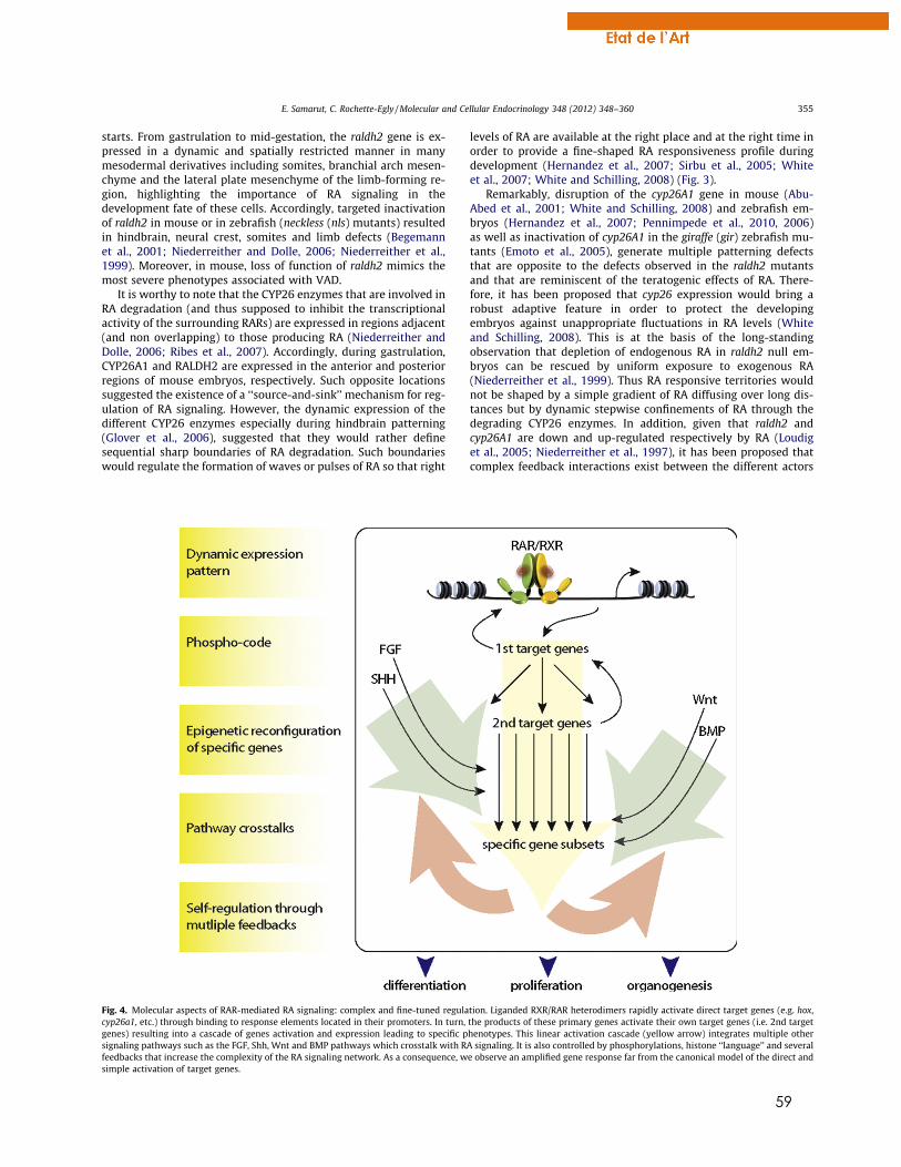
59
starts. From gastrulation to mid-gestation, the raldh2 gene is ex-pressed in a dynamic and spatially restricted manner in manymesodermal derivatives including somites, branchial arch mesen-chyme and the lateral plate mesenchyme of the limb-forming re-gion, highlighting the importance of RA signaling in thedevelopment fate of these cells. Accordingly, targeted inactivationof raldh2 in mouse or in zebrafish (neckless (nls) mutants) resultedin hindbrain, neural crest, somites and limb defects (Begemannet al., 2001; Niederreither and Dolle, 2006; Niederreither et al.,1999). Moreover, in mouse, loss of function of raldh2 mimics themost severe phenotypes associated with VAD.
It is worthy to note that the CYP26 enzymes that are involved inRA degradation (and thus supposed to inhibit the transcriptionalactivity of the surrounding RARs) are expressed in regions adjacent(and non overlapping) to those producing RA (Niederreither andDolle, 2006; Ribes et al., 2007). Accordingly, during gastrulation,CYP26A1 and RALDH2 are expressed in the anterior and posteriorregions of mouse embryos, respectively. Such opposite locationssuggested the existence of a ‘‘source-and-sink’’ mechanism for reg-ulation of RA signaling. However, the dynamic expression of thedifferent CYP26 enzymes especially during hindbrain patterning(Glover et al., 2006), suggested that they would rather definesequential sharp boundaries of RA degradation. Such boundarieswould regulate the formation of waves or pulses of RA so that right
levels of RA are available at the right place and at the right time inorder to provide a fine-shaped RA responsiveness profile duringdevelopment (Hernandez et al., 2007; Sirbu et al., 2005; Whiteet al., 2007; White and Schilling, 2008) (Fig. 3).
Remarkably, disruption of the cyp26A1 gene in mouse (Abu-Abed et al., 2001; White and Schilling, 2008) and zebrafish em-bryos (Hernandez et al., 2007; Pennimpede et al., 2010, 2006)as well as inactivation of cyp26A1 in the giraffe (gir) zebrafish mu-tants (Emoto et al., 2005), generate multiple patterning defectsthat are opposite to the defects observed in the raldh2 mutantsand that are reminiscent of the teratogenic effects of RA. There-fore, it has been proposed that cyp26 expression would bring arobust adaptive feature in order to protect the developingembryos against unappropriate fluctuations in RA levels (Whiteand Schilling, 2008). This is at the basis of the long-standingobservation that depletion of endogenous RA in raldh2 null em-bryos can be rescued by uniform exposure to exogenous RA(Niederreither et al., 1999). Thus RA responsive territories wouldnot be shaped by a simple gradient of RA diffusing over long dis-tances but by dynamic stepwise confinements of RA through thedegrading CYP26 enzymes. In addition, given that raldh2 andcyp26A1 are down and up-regulated respectively by RA (Loudiget al., 2005; Niederreither et al., 1997), it has been proposed thatcomplex feedback interactions exist between the different actors
Fig. 4. Molecular aspects of RAR-mediated RA signaling: complex and fine-tuned regulation. Liganded RXR/RAR heterodimers rapidly activate direct target genes (e.g. hox,cyp26a1, etc.) through binding to response elements located in their promoters. In turn, the products of these primary genes activate their own target genes (i.e. 2nd targetgenes) resulting into a cascade of genes activation and expression leading to specific phenotypes. This linear activation cascade (yellow arrow) integrates multiple othersignaling pathways such as the FGF, Shh, Wnt and BMP pathways which crosstalk with RA signaling. It is also controlled by phosphorylations, histone ‘‘language’’ and severalfeedbacks that increase the complexity of the RA signaling network. As a consequence, we observe an amplified gene response far from the canonical model of the direct andsimple activation of target genes.
E. Samarut, C. Rochette-Egly / Molecular and Cellular Endocrinology 348 (2012) 348–360 355
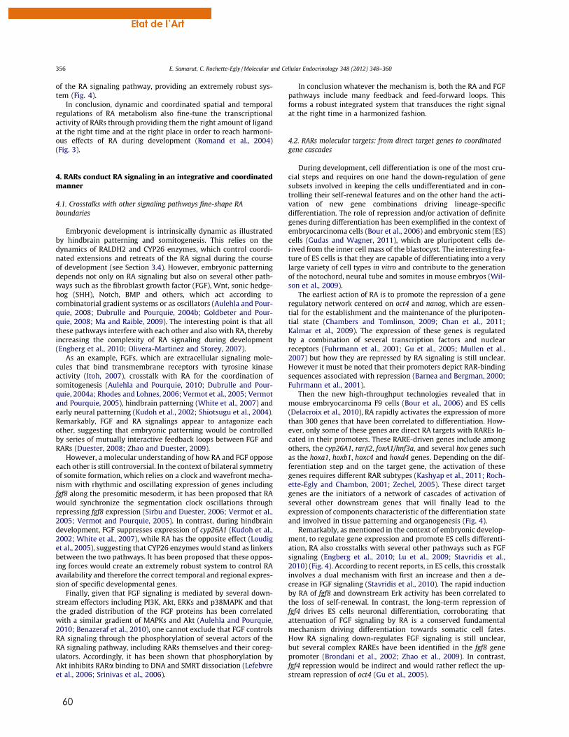
60
of the RA signaling pathway, providing an extremely robust sys-tem (Fig. 4).
In conclusion, dynamic and coordinated spatial and temporalregulations of RA metabolism also fine-tune the transcriptionalactivity of RARs through providing them the right amount of ligandat the right time and at the right place in order to reach harmoni-ous effects of RA during development (Romand et al., 2004)(Fig. 3).
4. RARs conduct RA signaling in an integrative and coordinatedmanner
4.1. Crosstalks with other signaling pathways fine-shape RAboundaries
Embryonic development is intrinsically dynamic as illustratedby hindbrain patterning and somitogenesis. This relies on thedynamics of RALDH2 and CYP26 enzymes, which control coordi-nated extensions and retreats of the RA signal during the courseof development (see Section 3.4). However, embryonic patterningdepends not only on RA signaling but also on several other path-ways such as the fibroblast growth factor (FGF), Wnt, sonic hedge-hog (SHH), Notch, BMP and others, which act according tocombinatorial gradient systems or as oscillators (Aulehla and Pour-quie, 2008; Dubrulle and Pourquie, 2004b; Goldbeter and Pour-quie, 2008; Ma and Raible, 2009). The interesting point is that allthese pathways interfere with each other and also with RA, therebyincreasing the complexity of RA signaling during development(Engberg et al., 2010; Olivera-Martinez and Storey, 2007).
As an example, FGFs, which are extracellular signaling mole-cules that bind transmembrane receptors with tyrosine kinaseactivity (Itoh, 2007), crosstalk with RA for the coordination ofsomitogenesis (Aulehla and Pourquie, 2010; Dubrulle and Pour-quie, 2004a; Rhodes and Lohnes, 2006; Vermot et al., 2005; Vermotand Pourquie, 2005), hindbrain patterning (White et al., 2007) andearly neural patterning (Kudoh et al., 2002; Shiotsugu et al., 2004).Remarkably, FGF and RA signalings appear to antagonize eachother, suggesting that embryonic patterning would be controlledby series of mutually interactive feedback loops between FGF andRARs (Duester, 2008; Zhao and Duester, 2009).
However, a molecular understanding of how RA and FGF opposeeach other is still controversial. In the context of bilateral symmetryof somite formation, which relies on a clock and wavefront mecha-nism with rhythmic and oscillating expression of genes includingfgf8 along the presomitic mesoderm, it has been proposed that RAwould synchronize the segmentation clock oscillations throughrepressing fgf8 expression (Sirbu and Duester, 2006; Vermot et al.,2005; Vermot and Pourquie, 2005). In contrast, during hindbraindevelopment, FGF suppresses expression of cyp26A1 (Kudoh et al.,2002; White et al., 2007), while RA has the opposite effect (Loudiget al., 2005), suggesting that CYP26 enzymes would stand as linkersbetween the two pathways. It has been proposed that these oppos-ing forces would create an extremely robust system to control RAavailability and therefore the correct temporal and regional expres-sion of specific developmental genes.
Finally, given that FGF signaling is mediated by several down-stream effectors including PI3K, Akt, ERKs and p38MAPK and thatthe graded distribution of the FGF proteins has been correlatedwith a similar gradient of MAPKs and Akt (Aulehla and Pourquie,2010; Benazeraf et al., 2010), one cannot exclude that FGF controlsRA signaling through the phosphorylation of several actors of theRA signaling pathway, including RARs themselves and their coreg-ulators. Accordingly, it has been shown that phosphorylation byAkt inhibits RARa binding to DNA and SMRT dissociation (Lefebvreet al., 2006; Srinivas et al., 2006).
In conclusion whatever the mechanism is, both the RA and FGFpathways include many feedback and feed-forward loops. Thisforms a robust integrated system that transduces the right signalat the right time in a harmonized fashion.
4.2. RARs molecular targets: from direct target genes to coordinatedgene cascades
During development, cell differentiation is one of the most cru-cial steps and requires on one hand the down-regulation of genesubsets involved in keeping the cells undifferentiated and in con-trolling their self-renewal features and on the other hand the acti-vation of new gene combinations driving lineage-specificdifferentiation. The role of repression and/or activation of definitegenes during differentiation has been exemplified in the context ofembryocarcinoma cells (Bour et al., 2006) and embryonic stem (ES)cells (Gudas and Wagner, 2011), which are pluripotent cells de-rived from the inner cell mass of the blastocyst. The interesting fea-ture of ES cells is that they are capable of differentiating into a verylarge variety of cell types in vitro and contribute to the generationof the notochord, neural tube and somites in mouse embryos (Wil-son et al., 2009).
The earliest action of RA is to promote the repression of a generegulatory network centered on oct4 and nanog, which are essen-tial for the establishment and the maintenance of the pluripoten-tial state (Chambers and Tomlinson, 2009; Chan et al., 2011;Kalmar et al., 2009). The expression of these genes is regulatedby a combination of several transcription factors and nuclearreceptors (Fuhrmann et al., 2001; Gu et al., 2005; Mullen et al.,2007) but how they are repressed by RA signaling is still unclear.However it must be noted that their promoters depict RAR-bindingsequences associated with repression (Barnea and Bergman, 2000;Fuhrmann et al., 2001).
Then the new high-throughput technologies revealed that inmouse embryocarcinoma F9 cells (Bour et al., 2006) and ES cells(Delacroix et al., 2010), RA rapidly activates the expression of morethan 300 genes that have been correlated to differentiation. How-ever, only some of these genes are direct RA targets with RAREs lo-cated in their promoters. These RARE-driven genes include amongothers, the cyp26A1, rarb2, foxA1/hnf3a, and several hox genes suchas the hoxa1, hoxb1, hoxc4 and hoxd4 genes. Depending on the dif-ferentiation step and on the target gene, the activation of thesegenes requires different RAR subtypes (Kashyap et al., 2011; Roch-ette-Egly and Chambon, 2001; Zechel, 2005). These direct targetgenes are the initiators of a network of cascades of activation ofseveral other downstream genes that will finally lead to theexpression of components characteristic of the differentiation stateand involved in tissue patterning and organogenesis (Fig. 4).
Remarkably, as mentioned in the context of embryonic develop-ment, to regulate gene expression and promote ES cells differenti-ation, RA also crosstalks with several other pathways such as FGFsignaling (Engberg et al., 2010; Lu et al., 2009; Stavridis et al.,2010) (Fig. 4). According to recent reports, in ES cells, this crosstalkinvolves a dual mechanism with first an increase and then a de-crease in FGF signaling (Stavridis et al., 2010). The rapid inductionby RA of fgf8 and downstream Erk activity has been correlated tothe loss of self-renewal. In contrast, the long-term repression offgf4 drives ES cells neuronal differentiation, corroborating thatattenuation of FGF signaling by RA is a conserved fundamentalmechanism driving differentiation towards somatic cell fates.How RA signaling down-regulates FGF signaling is still unclear,but several complex RAREs have been identified in the fgf8 genepromoter (Brondani et al., 2002; Zhao et al., 2009). In contrast,fgf4 repression would be indirect and would rather reflect the up-stream repression of oct4 (Gu et al., 2005).
356 E. Samarut, C. Rochette-Egly / Molecular and Cellular Endocrinology 348 (2012) 348–360
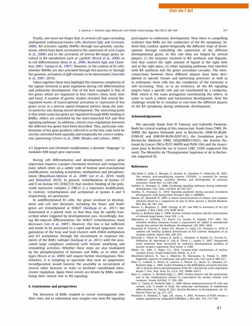
61
Finally, one must not forget that, in several cell types includingmultipotent embryocarcinoma cells (Rochette-Egly and Germain,2009), RA activates rapidly MAPKs through non-genomic mecha-nisms, which have been correlated to the repression of oct4 (Guptaet al., 2008) and to the activation of several RA-target genes in-volved in RA metabolism such as cyp26A1 (Bruck et al., 2009) orin cell differentiation (Bour et al., 2006; Rochette-Egly and Cham-bon, 2001; Taneja et al., 1997). However, in the context of ES cells,whether MAPKs are also activated through this process or throughthe genomic activation of fgf8 remains to be determined (Stavridiset al., 2007, 2010).
Taken together these data highlight the immense complexity ofthe signals involved in gene regulation during cell differentiationand embryonic development. One of the best examples is that ofhox genes which are organized in four clusters (hoxa, hoxb, hoxcand hoxd). A number of genetic studies revealed that several RA-regulated waves of transcriptional activation or repression of hoxgenes occur in a precise spatio-temporal pattern along the ante-ro-posterior axis during neural development. The interesting pointis that while some hox genes are regulated through RARs binding toRAREs, others are controlled by the interconnected FGF and Wntsignaling pathways. In addition, critical cross-regulations betweenthe different hox genes have been described. Thus, particular com-binations of hox gene products referred to as the Hox code must bestrictly controlled both spatially and temporally for correct embry-onic patterning (Glover et al., 2006; Rhodes and Lohnes, 2006).
4.3. Epigenetic and chromatin modifications: a dynamic ‘‘language’’ tomodulate RAR-target gene expression
During cell differentiation and development, correct geneexpression requires a proper chromatin structure and compactionstate, which relies on a subtle code of histone post-translationalmodifications, including acetylation, methylation and phosphory-lation (Bloushtain-Qimron et al., 2009; Lee et al., 2010; Smithand Shilatifard, 2010). In general, trimethylation of lysines 27and 9 on histone H3 (H3K27,9) that involves binding of the poly-comb repressive complex 2 (PRC2) is a repressive modification.In contrast, trimethylation and acetylation at lysines 4 and 9respectively, are permissive.
In undifferentiated ES cells, the genes involved in develop-ment and cell fate decisions, including the hoxa1 and hoxb1genes are trimethylated at H3K27 (Kashyap et al., 2011) butmaintained in a poised state so that they may be rapidly tran-scribed when triggered by developmental cues. Accordingly, dur-ing RA-induced differentiation, the H3K27 trimethylation markdecreases (Lee et al., 2007). However, this loss is not sufficientand needs to be associated to a rapid and broad epigenetic reor-ganization of the hoxa and hoxb clusters with H3K4 methylationand H3 acetylation, through the recruitment to response ele-ments of the RARc subtype (Kashyap et al., 2011) with the asso-ciated large complexes endowed with histone modifying andremodeling activities. Whether these steps are also modulatedby the phosphorylation of histones and RARs, as in other celltypes (Bruck et al., 2009) will require further investigations. Nev-ertheless, it is tempting to speculate that such an epigenomicreconfiguration would favorize the assembly or recruitment ofseveral other factories in order to facilitate coordinated intra-cluster regulation. Again these events are driven by RARs, under-lining their central role in RA-signaling.
5. Conclusions and perspectives
The discovery of RARs coupled to recent investigations intotheir roles, led to substantial new insights into how RA signaling
participates to embryonic development. Now there is compellingevidence that RARs are the conductors of the RA symphony. In-deed they conduct spatio-temporally the different steps of devel-opment through controlling the expression of the differentdevelopmental genes. In this role they are helped by severalplayers, (i) the enzymes involved in RA synthesis and degrada-tion that control the right amount of ligand at the right timeand at the right place, (ii) other signaling pathways that interferewith RA synthesis and the genes activation cascades. The inter-connections between these different players have been deci-phered in specific tissues and patterning processes as well asin embryonic stem cells but the complexity of the networks isstill increasing. Thus, as in an orchestra, all the RA signalingplayers have a specific role and are coordinated by a conductor,RAR, which is the main protagonist coordinating the others, inorder to reach a robust and harmonious development. Now thechallenge would be to visualize in real-time the different playersof the RA symphony during embryonic development.
Acknowledgements
We specially thank Ziad Al Tanoury and Gabriella Pankotai-Bodó for critical reading of this manuscript. Funds from CNRS, IN-SERM, the Agence Nationale pour la Recherche (ANR-05-BLAN-0390-02 and ANR-09-BLAN-0297-01), the Foundation pour laRecherche Medicale (FRM, DEQ20090515423), the Institut Na-tional du Cancer (INCa-PLO7-96099 and PL09-194) and the Associ-ation pour la Recherche sur le Cancer (ARC 3169) supported thiswork. The Ministère de l’Enseignement Supérieur et de la Recher-che supported ES.
References
Abu-Abed, S., Dolle, P., Metzger, D., Beckett, B., Chambon, P., Petkovich, M., 2001.The retinoic acid-metabolizing enzyme, CYP26A1, is essential for normalhindbrain patterning, vertebral identity, and development of posteriorstructures. Genes Dev. 15, 226–240.
Aulehla, A., Pourquie, O., 2008. Oscillating signaling pathways during embryonicdevelopment. Curr. Opin. Cell Biol. 20, 632–637.
Aulehla, A., Pourquie, O., 2010. Signaling gradients during paraxial mesodermdevelopment. Cold Spring Harb. Perspect. Biol. 2, a000869.
Balmer, J.E., Blomhoff, R., 2005. A robust characterization of retinoic acid responseelements based on a comparison of sites in three species. J. Steroid Biochem.Mol. Biol. 96, 347–354.
Barnea, E., Bergman, Y., 2000. Synergy of SF1 and RAR in activation of Oct-3/4promoter. J. Biol. Chem. 275, 6608–6619.
Bastien, J., Rochette-Egly, C., 2004. Nuclear retinoid receptors and the transcriptionof retinoid-target genes. Gene 328, 1–16.
Begemann, G., Schilling, T.F., Rauch, G.J., Geisler, R., Ingham, P.W., 2001. Thezebrafish neckless mutation reveals a requirement for raldh2 in mesodermalsignals that pattern the hindbrain. Development 128, 3081–3094.
Benazeraf, B., Francois, P., Baker, R.E., Denans, N., Little, C.D., Pourquie, O., 2010. Arandom cell motility gradient downstream of FGF controls elongation of anamniote embryo. Nature 466, 248–252.
Bertrand, S., Thisse, B., Tavares, R., Sachs, L., Chaumot, A., Bardet, P.L., Escriva, H.,Duffraisse, M., Marchand, O., Safi, R., Thisse, C., Laudet, V., 2007. Unexpectednovel relational links uncovered by extensive developmental profiling ofnuclear receptor expression. PLoS Genet. 3, e188.
Biddie, S.C., John, S., Hager, G.L., 2010. Genome-wide mechanisms of nuclearreceptor action. Trends Endocrinol. Metab. 21, 3–9.
Bloushtain-Qimron, N., Yao, J., Shipitsin, M., Maruyama, R., Polyak, K., 2009.Epigenetic patterns of embryonic and adult stem cells. Cell Cycle 8, 809–817.
Bour, G., Gaillard, E., Bruck, N., Lalevee, S., Plassat, J.L., Busso, D., Samama, J.P.,Rochette-Egly, C., 2005. Cyclin H binding to the RAR{alpha} activation function(AF)-2 domain directs phosphorylation of the AF-1 domain by cyclin-dependentkinase 7. Proc. Natl. Acad. Sci. U.S.A. 102, 16608–16613.
Bour, G., Lalevee, S., Rochette-Egly, C., 2007. Protein kinases and the proteasomejoin in the combinatorial control of transcription by nuclear retinoic acidreceptors. Trends Cell Biol. 17, 302–309.
Bour, G., Taneja, R., Rochette-Egly, C., 2006. Mouse embryocarcinoma F9 cells andretinoic acid. A model to study the molecular mechanisms of endodermaldifferentiation. In: Taneja, R. (Ed.), Nuclear Receptors in Development, vol. 16.Elsevier Press Inc, pp. 211–253.
Brondani, V., Klimkait, T., Egly, J.M., Hamy, F., 2002. Promoter of FGF8 reveals aunique regulation by unliganded RARalpha. J. Mol. Biol. 319, 715–728.
E. Samarut, C. Rochette-Egly / Molecular and Cellular Endocrinology 348 (2012) 348–360 357
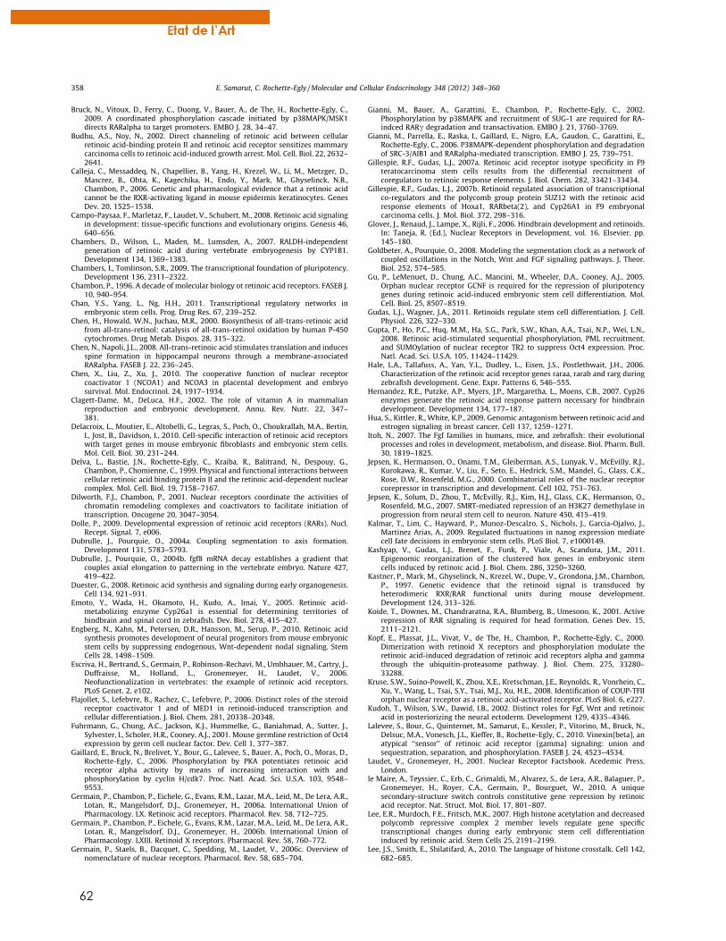
62
Bruck, N., Vitoux, D., Ferry, C., Duong, V., Bauer, A., de The, H., Rochette-Egly, C.,2009. A coordinated phosphorylation cascade initiated by p38MAPK/MSK1directs RARalpha to target promoters. EMBO J. 28, 34–47.
Budhu, A.S., Noy, N., 2002. Direct channeling of retinoic acid between cellularretinoic acid-binding protein II and retinoic acid receptor sensitizes mammarycarcinoma cells to retinoic acid-induced growth arrest. Mol. Cell. Biol. 22, 2632–2641.
Calleja, C., Messaddeq, N., Chapellier, B., Yang, H., Krezel, W., Li, M., Metzger, D.,Mascrez, B., Ohta, K., Kagechika, H., Endo, Y., Mark, M., Ghyselinck, N.B.,Chambon, P., 2006. Genetic and pharmacological evidence that a retinoic acidcannot be the RXR-activating ligand in mouse epidermis keratinocytes. GenesDev. 20, 1525–1538.
Campo-Paysaa, F., Marletaz, F., Laudet, V., Schubert, M., 2008. Retinoic acid signalingin development: tissue-specific functions and evolutionary origins. Genesis 46,640–656.
Chambers, D., Wilson, L., Maden, M., Lumsden, A., 2007. RALDH-independentgeneration of retinoic acid during vertebrate embryogenesis by CYP1B1.Development 134, 1369–1383.
Chambers, I., Tomlinson, S.R., 2009. The transcriptional foundation of pluripotency.Development 136, 2311–2322.
Chambon, P., 1996. A decade of molecular biology ot retinoic acid receptors. FASEB J.10, 940–954.
Chan, Y.S., Yang, L., Ng, H.H., 2011. Transcriptional regulatory networks inembryonic stem cells. Prog. Drug Res. 67, 239–252.
Chen, H., Howald, W.N., Juchau, M.R., 2000. Biosynthesis of all-trans-retinoic acidfrom all-trans-retinol: catalysis of all-trans-retinol oxidation by human P-450cytochromes. Drug Metab. Dispos. 28, 315–322.
Chen, N., Napoli, J.L., 2008. All-trans-retinoic acid stimulates translation and inducesspine formation in hippocampal neurons through a membrane-associatedRARalpha. FASEB J. 22, 236–245.
Chen, X., Liu, Z., Xu, J., 2010. The cooperative function of nuclear receptorcoactivator 1 (NCOA1) and NCOA3 in placental development and embryosurvival. Mol. Endocrinol. 24, 1917–1934.
Clagett-Dame, M., DeLuca, H.F., 2002. The role of vitamin A in mammalianreproduction and embryonic development. Annu. Rev. Nutr. 22, 347–381.
Delacroix, L., Moutier, E., Altobelli, G., Legras, S., Poch, O., Choukrallah, M.A., Bertin,I., Jost, B., Davidson, I., 2010. Cell-specific interaction of retinoic acid receptorswith target genes in mouse embryonic fibroblasts and embryonic stem cells.Mol. Cell. Biol. 30, 231–244.
Delva, L., Bastie, J.N., Rochette-Egly, C., Kraiba, R., Balitrand, N., Despouy, G.,Chambon, P., Chomienne, C., 1999. Physical and functional interactions betweencellular retinoic acid binding protein II and the retinoic acid-dependent nuclearcomplex. Mol. Cell. Biol. 19, 7158–7167.
Dilworth, F.J., Chambon, P., 2001. Nuclear receptors coordinate the activities ofchromatin remodeling complexes and coactivators to facilitate initiation oftranscription. Oncogene 20, 3047–3054.
Dolle, P., 2009. Developmental expression of retinoic acid receptors (RARs). Nucl.Recept. Signal. 7, e006.
Dubrulle, J., Pourquie, O., 2004a. Coupling segmentation to axis formation.Development 131, 5783–5793.
Dubrulle, J., Pourquie, O., 2004b. fgf8 mRNA decay establishes a gradient thatcouples axial elongation to patterning in the vertebrate embryo. Nature 427,419–422.
Duester, G., 2008. Retinoic acid synthesis and signaling during early organogenesis.Cell 134, 921–931.
Emoto, Y., Wada, H., Okamoto, H., Kudo, A., Imai, Y., 2005. Retinoic acid-metabolizing enzyme Cyp26a1 is essential for determining territories ofhindbrain and spinal cord in zebrafish. Dev. Biol. 278, 415–427.
Engberg, N., Kahn, M., Petersen, D.R., Hansson, M., Serup, P., 2010. Retinoic acidsynthesis promotes development of neural progenitors from mouse embryonicstem cells by suppressing endogenous, Wnt-dependent nodal signaling. StemCells 28, 1498–1509.
Escriva, H., Bertrand, S., Germain, P., Robinson-Rechavi, M., Umbhauer, M., Cartry, J.,Duffraisse, M., Holland, L., Gronemeyer, H., Laudet, V., 2006.Neofunctionalization in vertebrates: the example of retinoic acid receptors.PLoS Genet. 2, e102.
Flajollet, S., Lefebvre, B., Rachez, C., Lefebvre, P., 2006. Distinct roles of the steroidreceptor coactivator 1 and of MED1 in retinoid-induced transcription andcellular differentiation. J. Biol. Chem. 281, 20338–20348.
Fuhrmann, G., Chung, A.C., Jackson, K.J., Hummelke, G., Baniahmad, A., Sutter, J.,Sylvester, I., Scholer, H.R., Cooney, A.J., 2001. Mouse germline restriction of Oct4expression by germ cell nuclear factor. Dev. Cell 1, 377–387.
Gaillard, E., Bruck, N., Brelivet, Y., Bour, G., Lalevee, S., Bauer, A., Poch, O., Moras, D.,Rochette-Egly, C., 2006. Phosphorylation by PKA potentiates retinoic acidreceptor alpha activity by means of increasing interaction with andphosphorylation by cyclin H/cdk7. Proc. Natl. Acad. Sci. U.S.A. 103, 9548–9553.
Germain, P., Chambon, P., Eichele, G., Evans, R.M., Lazar, M.A., Leid, M., De Lera, A.R.,Lotan, R., Mangelsdorf, D.J., Gronemeyer, H., 2006a. International Union ofPharmacology. LX. Retinoic acid receptors. Pharmacol. Rev. 58, 712–725.
Germain, P., Chambon, P., Eichele, G., Evans, R.M., Lazar, M.A., Leid, M., De Lera, A.R.,Lotan, R., Mangelsdorf, D.J., Gronemeyer, H., 2006b. International Union ofPharmacology. LXIII. Retinoid X receptors. Pharmacol. Rev. 58, 760–772.
Germain, P., Staels, B., Dacquet, C., Spedding, M., Laudet, V., 2006c. Overview ofnomenclature of nuclear receptors. Pharmacol. Rev. 58, 685–704.
Gianni, M., Bauer, A., Garattini, E., Chambon, P., Rochette-Egly, C., 2002.Phosphorylation by p38MAPK and recruitment of SUG-1 are required for RA-indced RARc degradation and transactivation. EMBO J. 21, 3760–3769.
Gianni, M., Parrella, E., Raska, I., Gaillard, E., Nigro, E.A., Gaudon, C., Garattini, E.,Rochette-Egly, C., 2006. P38MAPK-dependent phosphorylation and degradationof SRC-3/AIB1 and RARalpha-mediated transcription. EMBO J. 25, 739–751.
Gillespie, R.F., Gudas, L.J., 2007a. Retinoic acid receptor isotype specificity in F9teratocarcinoma stem cells results from the differential recruitment ofcoregulators to retinoic response elements. J. Biol. Chem. 282, 33421–33434.
Gillespie, R.F., Gudas, L.J., 2007b. Retinoid regulated association of transcriptionalco-regulators and the polycomb group protein SUZ12 with the retinoic acidresponse elements of Hoxa1, RARbeta(2), and Cyp26A1 in F9 embryonalcarcinoma cells. J. Mol. Biol. 372, 298–316.
Glover, J., Renaud, J., Lampe, X., Rijli, F., 2006. Hindbrain development and retinoids.In: Taneja, R. (Ed.), Nuclear Receptors in Development, vol. 16. Elsevier, pp.145–180.
Goldbeter, A., Pourquie, O., 2008. Modeling the segmentation clock as a network ofcoupled oscillations in the Notch, Wnt and FGF signaling pathways. J. Theor.Biol. 252, 574–585.
Gu, P., LeMenuet, D., Chung, A.C., Mancini, M., Wheeler, D.A., Cooney, A.J., 2005.Orphan nuclear receptor GCNF is required for the repression of pluripotencygenes during retinoic acid-induced embryonic stem cell differentiation. Mol.Cell. Biol. 25, 8507–8519.
Gudas, L.J., Wagner, J.A., 2011. Retinoids regulate stem cell differentiation. J. Cell.Physiol. 226, 322–330.
Gupta, P., Ho, P.C., Huq, M.M., Ha, S.G., Park, S.W., Khan, A.A., Tsai, N.P., Wei, L.N.,2008. Retinoic acid-stimulated sequential phosphorylation, PML recruitment,and SUMOylation of nuclear receptor TR2 to suppress Oct4 expression. Proc.Natl. Acad. Sci. U.S.A. 105, 11424–11429.
Hale, L.A., Tallafuss, A., Yan, Y.L., Dudley, L., Eisen, J.S., Postlethwait, J.H., 2006.Characterization of the retinoic acid receptor genes raraa, rarab and rarg duringzebrafish development. Gene. Expr. Patterns 6, 546–555.
Hernandez, R.E., Putzke, A.P., Myers, J.P., Margaretha, L., Moens, C.B., 2007. Cyp26enzymes generate the retinoic acid response pattern necessary for hindbraindevelopment. Development 134, 177–187.
Hua, S., Kittler, R., White, K.P., 2009. Genomic antagonism between retinoic acid andestrogen signaling in breast cancer. Cell 137, 1259–1271.
Itoh, N., 2007. The Fgf families in humans, mice, and zebrafish: their evolutionalprocesses and roles in development, metabolism, and disease. Biol. Pharm. Bull.30, 1819–1825.
Jepsen, K., Hermanson, O., Onami, T.M., Gleiberman, A.S., Lunyak, V., McEvilly, R.J.,Kurokawa, R., Kumar, V., Liu, F., Seto, E., Hedrick, S.M., Mandel, G., Glass, C.K.,Rose, D.W., Rosenfeld, M.G., 2000. Combinatorial roles of the nuclear receptorcorepressor in transcription and development. Cell 102, 753–763.
Jepsen, K., Solum, D., Zhou, T., McEvilly, R.J., Kim, H.J., Glass, C.K., Hermanson, O.,Rosenfeld, M.G., 2007. SMRT-mediated repression of an H3K27 demethylase inprogression from neural stem cell to neuron. Nature 450, 415–419.
Kalmar, T., Lim, C., Hayward, P., Munoz-Descalzo, S., Nichols, J., Garcia-Ojalvo, J.,Martinez Arias, A., 2009. Regulated fluctuations in nanog expression mediatecell fate decisions in embryonic stem cells. PLoS Biol. 7, e1000149.
Kashyap, V., Gudas, L.J., Brenet, F., Funk, P., Viale, A., Scandura, J.M., 2011.Epigenomic reorganization of the clustered hox genes in embryonic stemcells induced by retinoic acid. J. Biol. Chem. 286, 3250–3260.
Kastner, P., Mark, M., Ghyselinck, N., Krezel, W., Dupe, V., Grondona, J.M., Chambon,P., 1997. Genetic evidence that the retinoid signal is transduced byheterodimeric RXR/RAR functional units during mouse development.Development 124, 313–326.
Koide, T., Downes, M., Chandraratna, R.A., Blumberg, B., Umesono, K., 2001. Activerepression of RAR signaling is required for head formation. Genes Dev. 15,2111–2121.
Kopf, E., Plassat, J.L., Vivat, V., de The, H., Chambon, P., Rochette-Egly, C., 2000.Dimerization with retinoid X receptors and phosphorylation modulate theretinoic acid-induced degradation of retinoic acid receptors alpha and gammathrough the ubiquitin-proteasome pathway. J. Biol. Chem. 275, 33280–33288.
Kruse, S.W., Suino-Powell, K., Zhou, X.E., Kretschman, J.E., Reynolds, R., Vonrhein, C.,Xu, Y., Wang, L., Tsai, S.Y., Tsai, M.J., Xu, H.E., 2008. Identification of COUP-TFIIorphan nuclear receptor as a retinoic acid-activated receptor. PLoS Biol. 6, e227.
Kudoh, T., Wilson, S.W., Dawid, I.B., 2002. Distinct roles for Fgf, Wnt and retinoicacid in posteriorizing the neural ectoderm. Development 129, 4335–4346.
Lalevee, S., Bour, G., Quinternet, M., Samarut, E., Kessler, P., Vitorino, M., Bruck, N.,Delsuc, M.A., Vonesch, J.L., Kieffer, B., Rochette-Egly, C., 2010. Vinexin{beta}, anatypical ‘‘sensor’’ of retinoic acid receptor {gamma} signaling: union andsequestration, separation, and phosphorylation. FASEB J. 24, 4523–4534.
Laudet, V., Gronemeyer, H., 2001. Nuclear Receptor Factsbook. Acedemic Press,London.
le Maire, A., Teyssier, C., Erb, C., Grimaldi, M., Alvarez, S., de Lera, A.R., Balaguer, P.,Gronemeyer, H., Royer, C.A., Germain, P., Bourguet, W., 2010. A uniquesecondary-structure switch controls constitutive gene repression by retinoicacid receptor. Nat. Struct. Mol. Biol. 17, 801–807.
Lee, E.R., Murdoch, F.E., Fritsch, M.K., 2007. High histone acetylation and decreasedpolycomb repressive complex 2 member levels regulate gene specifictranscriptional changes during early embryonic stem cell differentiationinduced by retinoic acid. Stem Cells 25, 2191–2199.
Lee, J.S., Smith, E., Shilatifard, A., 2010. The language of histone crosstalk. Cell 142,682–685.
358 E. Samarut, C. Rochette-Egly / Molecular and Cellular Endocrinology 348 (2012) 348–360
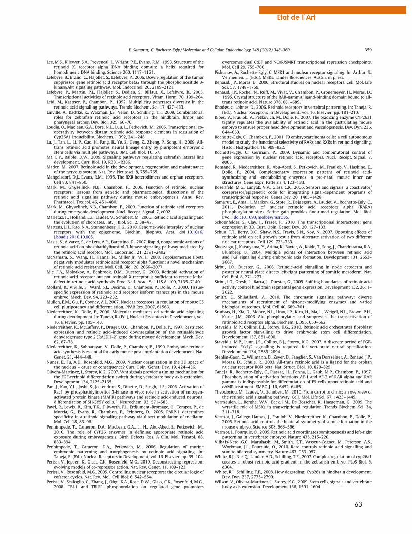
63
Lee, M.S., Kliewer, S.A., Provencal, J., Wright, P.E., Evans, R.M., 1993. Structure of theretinoid X receptor alpha DNA binding domain: a helix required forhomodimeric DNA binding. Science 260, 1117–1121.
Lefebvre, B., Brand, C., Flajollet, S., Lefebvre, P., 2006. Down-regulation of the tumorsuppressor gene retinoic acid receptor beta2 through the phosphoinositide 3-kinase/Akt signaling pathway. Mol. Endocrinol. 20, 2109–2121.
Lefebvre, P., Martin, P.J., Flajollet, S., Dedieu, S., Billaut, X., Lefebvre, B., 2005.Transcriptional activities of retinoic acid receptors. Vitam. Horm. 70, 199–264.
Leid, M., Kastner, P., Chambon, P., 1992. Multiplicity generates diversity in theretinoic acid signalling pathways. Trends Biochem. Sci. 17, 427–433.
Linville, A., Radtke, K., Waxman, J.S., Yelon, D., Schilling, T.F., 2009. Combinatorialroles for zebrafish retinoic acid receptors in the hindbrain, limbs andpharyngeal arches. Dev. Biol. 325, 60–70.
Loudig, O., Maclean, G.A., Dore, N.L., Luu, L., Petkovich, M., 2005. Transcriptional co-operativity between distant retinoic acid response elements in regulation ofCyp26A1 inducibility. Biochem. J. 392, 241–248.
Lu, J., Tan, L., Li, P., Gao, H., Fang, B., Ye, S., Geng, Z., Zheng, P., Song, H., 2009. All-trans retinoic acid promotes neural lineage entry by pluripotent embryonicstem cells via multiple pathways. BMC Cell Biol. 10, 57.
Ma, E.Y., Raible, D.W., 2009. Signaling pathways regulating zebrafish lateral linedevelopment. Curr. Biol. 19, R381–R386.
Maden, M., 2007. Retinoic acid in the development, regeneration and maintenanceof the nervous system. Nat. Rev. Neurosci. 8, 755–765.
Mangelsdorf, D.J., Evans, R.M., 1995. The RXR heterodimers and orphan receptors.Cell 83, 841–850.
Mark, M., Ghyselinck, N.B., Chambon, P., 2006. Function of retinoid nuclearreceptors: lessons from genetic and pharmacological dissections of theretinoic acid signaling pathway during mouse embryogenesis. Annu. Rev.Pharmacol. Toxicol. 46, 451–480.
Mark, M., Ghyselinck, N.B., Chambon, P., 2009. Function of retinoic acid receptorsduring embryonic development. Nucl. Recept. Signal. 7, e002.
Marletaz, F., Holland, L.Z., Laudet, V., Schubert, M., 2006. Retinoic acid signaling andthe evolution of chordates. Int. J. Biol. Sci. 2, 38–47.
Martens, J.H., Rao, N.A., Stunnenberg, H.G., 2010. Genome-wide interplay of nuclearreceptors with the epigenome. Biochim. Biophys. Acta. doi:10.1016/j.bbadis.2010.10.005.
Masia, S., Alvarez, S., de Lera, A.R., Barettino, D., 2007. Rapid, nongenomic actions ofretinoic acid on phosphatidylinositol-3-kinase signaling pathway mediated bythe retinoic acid receptor. Mol. Endocrinol. 21, 2391–2402.
McNamara, S., Wang, H., Hanna, N., Miller Jr., W.H., 2008. Topoisomerase IIbetanegatively modulates retinoic acid receptor alpha function: a novel mechanismof retinoic acid resistance. Mol. Cell. Biol. 28, 2066–2077.
Mic, F.A., Molotkov, A., Benbrook, D.M., Duester, G., 2003. Retinoid activation ofretinoic acid receptor but not retinoid X receptor is sufficient to rescue lethaldefect in retinoic acid synthesis. Proc. Natl. Acad. Sci. U.S.A. 100, 7135–7140.
Mollard, R., Viville, S., Ward, S.J., Decimo, D., Chambon, P., Dolle, P., 2000. Tissue-specific expression of retinoic acid receptor isoform transcripts in the mouseembryo. Mech. Dev. 94, 223–232.
Mullen, E.M., Gu, P., Cooney, A.J., 2007. Nuclear receptors in regulation of mouse EScell pluripotency and differentiation. PPAR Res. 2007, 61563.
Niederreither, K., Dolle, P., 2006. Molecular mediators od retinoic acid signalingduring development. In: Taneja, R. (Ed.), Nuclear Receptors in Development, vol.16. Elsevier, pp. 105–143.
Niederreither, K., McCaffery, P., Drager, U.C., Chambon, P., Dolle, P., 1997. Restrictedexpression and retinoic acid-induced downregulation of the retinaldehydedehydrogenase type 2 (RALDH-2) gene during mouse development. Mech. Dev.62, 67–78.
Niederreither, K., Subbarayan, V., Dolle, P., Chambon, P., 1999. Embryonic retinoicacid synthesis is essential for early mouse post-implantation development. Nat.Genet. 21, 444–448.
Nunez, E., Fu, X.D., Rosenfeld, M.G., 2009. Nuclear organization in the 3D space ofthe nucleus – cause or consequence? Curr. Opin. Genet. Dev. 19, 424–436.
Olivera-Martinez, I., Storey, K.G., 2007. Wnt signals provide a timing mechanism forthe FGF-retinoid differentiation switch during vertebrate body axis extension.Development 134, 2125–2135.
Pan, J., Kao, Y.L., Joshi, S., Jeetendran, S., Dipette, D., Singh, U.S., 2005. Activation ofRac1 by phosphatidylinositol 3-kinase in vivo: role in activation of mitogen-activated protein kinase (MAPK) pathways and retinoic acid-induced neuronaldifferentiation of SH-SY5Y cells. J. Neurochem. 93, 571–583.
Pavri, R., Lewis, B., Kim, T.K., Dilworth, F.J., Erdjument-Bromage, H., Tempst, P., deMurcia, G., Evans, R., Chambon, P., Reinberg, D., 2005. PARP-1 determinesspecificity in a retinoid signaling pathway via direct modulation of mediator.Mol. Cell 18, 83–96.
Pennimpede, T., Cameron, D.A., MacLean, G.A., Li, H., Abu-Abed, S., Petkovich, M.,2010. The role of CYP26 enzymes in defining appropriate retinoic acidexposure during embryogenesis. Birth Defects Res. A Clin. Mol. Teratol. 88,883–894.
Pennimpede, T., Cameron, D.A., Petkovich, M., 2006. Regulation of murineembryonic patterning and morphogenesis by retinoic acid signaling. In:Taneja, R. (Ed.), Nuclear Receptors in Development, vol. 16. Elsevier, pp. 65–104.
Perissi, V., Jepsen, K., Glass, C.K., Rosenfeld, M.G., 2010. Deconstructing repression:evolving models of co-repressor action. Nat. Rev. Genet. 11, 109–123.
Perissi, V., Rosenfeld, M.G., 2005. Controlling nuclear receptors: the circular logic ofcofactor cycles. Nat. Rev. Mol. Cell Biol. 6, 542–554.
Perissi, V., Scafoglio, C., Zhang, J., Ohgi, K.A., Rose, D.W., Glass, C.K., Rosenfeld, M.G.,2008. TBL1 and TBLR1 phosphorylation on regulated gene promoters
overcomes dual CtBP and NCoR/SMRT transcriptional repression checkpoints.Mol. Cell 29, 755–766.
Piskunov, A., Rochette-Egly, C. MSK1 and nuclear receptor signaling. In: Arthur, S.,Vermeulen, L. (Eds.), MSKs. Landes Biosciences, Austin, in press.
Renaud, J.P., Moras, D., 2000. Structural studies on nuclear receptors. Cell. Mol. LifeSci. 57, 1748–1769.
Renaud, J.P., Rochel, N., Ruff, M., Vivat, V., Chambon, P., Gronemeyer, H., Moras, D.,1995. Crystal structure of the RAR-gamma ligand-binding domain bound to all-trans retinoic acid. Nature 378, 681–689.
Rhodes, c., Lohnes, D., 2006. Retinoid receptors in vertebral patterning. In: Taneja, R.(Ed.), Nuclear Receptors in Development, vol. 16. Elsevier, pp. 181–210.
Ribes, V., Fraulob, V., Petkovich, M., Dolle, P., 2007. The oxidizing enzyme CYP26a1tightly regulates the availability of retinoic acid in the gastrulating mouseembryo to ensure proper head development and vasculogenesis. Dev. Dyn. 236,644–653.
Rochette-Egly, C., Chambon, P., 2001. F9 embryocarcinoma cells: a cell autonomousmodel to study the functional selectivity of RARs and RXRs in retinoid signaling.Histol. Histopathol. 16, 909–922.
Rochette-Egly, C., Germain, P., 2009. Dynamic and combinatorial control ofgene expression by nuclear retinoic acid receptors. Nucl. Recept. Signal. 7,e005.
Romand, R., Niederreither, K., Abu-Abed, S., Petkovich, M., Fraulob, V., Hashino, E.,Dolle, P., 2004. Complementary expression patterns of retinoid acid-synthesizing and -metabolizing enzymes in pre-natal mouse inner earstructures. Gene Expr. Patterns 4, 123–133.
Rosenfeld, M.G., Lunyak, V.V., Glass, C.K., 2006. Sensors and signals: a coactivator/corepressor/epigenetic code for integrating signal-dependent programs oftranscriptional response. Genes Dev. 20, 1405–1428.
Samarut, E., Amal, I., Markov, G., Stote, R., Dejaegere, A., Laudet, V., Rochette-Egly, C.,2011. Evolution of nuclear retinoic acid receptors alpha (RARa)phosphorylation sites. Serine gain provides fine-tuned regulation. Mol. Biol.Evol., doi:10.1093/molbev/msrO35.
Schoenfelder, S., Clay, I., Fraser, P., 2010. The transcriptional interactome: geneexpression in 3D. Curr. Opin. Genet. Dev. 20, 127–133.
Schug, T.T., Berry, D.C., Shaw, N.S., Travis, S.N., Noy, N., 2007. Opposing effects ofretinoic acid on cell growth result from alternate activation of two differentnuclear receptors. Cell 129, 723–733.
Shiotsugu, J., Katsuyama, Y., Arima, K., Baxter, A., Koide, T., Song, J., Chandraratna, R.A.,Blumberg, B., 2004. Multiple points of interaction between retinoic acidand FGF signaling during embryonic axis formation. Development 131, 2653–2667.
Sirbu, I.O., Duester, G., 2006. Retinoic-acid signalling in node ectoderm andposterior neural plate directs left-right patterning of somitic mesoderm. Nat.Cell Biol. 8, 271–277.
Sirbu, I.O., Gresh, L., Barra, J., Duester, G., 2005. Shifting boundaries of retinoic acidactivity control hindbrain segmental gene expression. Development 132, 2611–2622.
Smith, E., Shilatifard, A., 2010. The chromatin signaling pathway: diversemechanisms of recruitment of histone-modifying enzymes and variedbiological outcomes. Mol. Cell 40, 689–701.
Srinivas, H., Xia, D., Moore, N.L., Uray, I.P., Kim, H., Ma, L., Weigel, N.L., Brown, P.H.,Kurie, J.M., 2006. Akt phosphorylates and suppresses the transactivation ofretinoic acid receptor alpha. Biochem. J. 395, 653–662.
Stavridis, M.P., Collins, B.J., Storey, K.G., 2010. Retinoic acid orchestrates fibroblastgrowth factor signalling to drive embryonic stem cell differentiation.Development 137, 881–890.
Stavridis, M.P., Lunn, J.S., Collins, B.J., Storey, K.G., 2007. A discrete period of FGF-induced Erk1/2 signalling is required for vertebrate neural specification.Development 134, 2889–2894.
Stehlin-Gaon, C., Willmann, D., Zeyer, D., Sanglier, S., Van Dorsselaer, A., Renaud, J.P.,Moras, D., Schule, R., 2003. All-trans retinoic acid is a ligand for the orphannuclear receptor ROR beta. Nat. Struct. Biol. 10, 820–825.
Taneja, R., Rochette-Egly, C., Plassat, J.L., Penna, L., Gaub, M.P., Chambon, P., 1997.Phosphorylation of activation functions AF-1 and AF-2 of RAR alpha and RARgamma is indispensable for differentiation of F9 cells upon retinoic acid andcAMP treatment. EMBO J. 16, 6452–6465.
Theodosiou, M., Laudet, V., Schubert, M., 2010. From carrot to clinic: an overview ofthe retinoic acid signaling pathway. Cell. Mol. Life Sci. 67, 1423–1445.
Vermeulen, L., Berghe, W.V., Beck, I.M., De Bosscher, K., Haegeman, G., 2009. Theversatile role of MSKs in transcriptional regulation. Trends Biochem. Sci. 34,311–318.
Vermot, J., Gallego Llamas, J., Fraulob, V., Niederreither, K., Chambon, P., Dolle, P.,2005. Retinoic acid controls the bilateral symmetry of somite formation in themouse embryo. Science 308, 563–566.
Vermot, J., Pourquie, O., 2005. Retinoic acid coordinates somitogenesis and left-rightpatterning in vertebrate embryos. Nature 435, 215–220.
Vilhais-Neto, G.C., Maruhashi, M., Smith, K.T., Vasseur-Cognet, M., Peterson, A.S.,Workman, J.L., Pourquie, O., 2010. Rere controls retinoic acid signalling andsomite bilateral symmetry. Nature 463, 953–957.
White, R.J., Nie, Q., Lander, A.D., Schilling, T.F., 2007. Complex regulation of cyp26a1creates a robust retinoic acid gradient in the zebrafish embryo. PLoS Biol. 5,e304.
White, R.J., Schilling, T.F., 2008. How degrading: Cyp26s in hindbrain development.Dev. Dyn. 237, 2775–2790.
Wilson, V., Olivera-Martinez, I., Storey, K.G., 2009. Stem cells, signals and vertebratebody axis extension. Development 136, 1591–1604.
E. Samarut, C. Rochette-Egly / Molecular and Cellular Endocrinology 348 (2012) 348–360 359
64
Wolf, G., 2006. Is 9-cis-retinoic acid the endogenous ligand for the retinoic acid-Xreceptor? Nutr. Rev. 64, 532–538.
Xu, F., Li, K., Tian, M., Hu, P., Song, W., Chen, J., Gao, X., Zhao, Q., 2009. N-CoR isrequired for patterning the anterior-posterior axis of zebrafish hindbrain byactively repressing retinoid signaling. Mech. Dev. 126, 771–780.
Zechel, C., 2005. Requirement of retinoic acid receptor isotypes alpha, beta, andgamma during the initial steps of neural differentiation of PCC7 cells. Mol.Endocrinol. 19, 1629–1645.
Zechel, C., Shen, X.Q., Chambon, P., Gronemeyer, H., 1994a. Dimerization interfacesformed between the DNA binding domains determine the cooperative bindingof RXR/RAR and RXR/TR heterodimers to DR5 and DR4 elements. EMBO J. 13,1414–1424.
Zechel, C., Shen, X.Q., Chen, J.Y., Chen, Z.P., Chambon, P., Gronemeyer, H., 1994b. Thedimerization interfaces formed between the DNA binding domains of RXR, RAR
and TR determine the binding specificity and polarity of the full-lengthreceptors to direct repeats. EMBO J. 13, 1425–1433.
Zhao, X., Duester, G., 2009. Effect of retinoic acid signaling on Wnt/beta-catenin andFGF signaling during body axis extension. Gene Expr. Patterns 9, 430–435.
Zhao, X., Sirbu, I.O., Mic, F.A., Molotkova, N., Molotkov, A., Kumar, S., Duester, G.,2009. Retinoic acid promotes limb induction through effects on body axisextension but is unnecessary for limb patterning. Curr. Biol. 19, 1050–1057.
Zhou, X.E., Suino-Powell, K.M., Xu, Y., Chan, C.W., Tanabe, O., Kruse, S.W., Reynolds,R., Engel, J.D., Xu, H.E., 2011. The orphan nuclear receptor TR4 is a vitamin A-activated nuclear receptor. J. Biol. Chem. 286, 2877–2885.
Zile, M.H., 2001. Function of vitamin A in vertebrate embryonic development. J.Nutr. 131, 705–708.
360 E. Samarut, C. Rochette-Egly / Molecular and Cellular Endocrinology 348 (2012) 348–360

65
D. ZEBRAR : LE ROLE DE L’AR AU COURS DU DEVELOPPEMENT DU
POISSON-ZEBRE
Cette revue a été soumise pour publication au journal Biochimica et Biophysica Acta
dans le cadre d’un numéro spécial intitulé Nuclear receptors in animal development qui
sera publié en 2014. L’objectif de cette revue est de mettre en avant les avantages du
poisson-zèbre en tant que modèle de biologie du développement pour l'étude in vivo du
rôle de RA et de ses récepteurs. De plus, elle illustre la pléiotropie de la voie de l’AR dans le
développement en se basant sur des études utilisant le poisson-zèbre. De ce fait, c’est la
première revue qui récapitule les principaux rôles de l’AR au cours de l’embryogénèse du
poisson-zèbre et devrait être ainsi d’un fort intérêt pour la communauté.

66
ZebRA: an overview of retinoic acid effects during zebrafish development
Eric Samarut1, Daniel Fraher2, Vincent Laudet1 and Yann Gibert2*
1: Institut de Génomique Fonctionnelle de Lyon; Ecole Normale Supérieure de Lyon; CNRS UMR 5242 ;
Université Lyon 1; Université de Lyon; 46 allée d'Italie, 69364 Lyon Cedex 07, France;
2: Metabolic Genetic Diseases Laboratory, Metabolic Research Unit, Deakin School of Medicine, 75 Pigdons
Road, Waurn Ponds VIC 3217, Australia
*: Corresponding author: E-mail: [email protected]
Keywords: Retinoic acid, RAR, zebrafish, organogenesis, patterning
67
ABSTRACT
Retinoic Acid (RA), the main active vitamin A derivative is undeniably crucial for embryo
development regulating cellular processes, embryo patterning and organogenesis. Many studies
performed in mammalian or avian models undertook successfully the investigation of the role
played by RA during embryogenesis. However, since the early 1980’s, zebrafish (Danio rerio) has
emerged as powerful developmental model. Unlike other vertebrate models, zebrafish
embryogenesis is external allowing the observation of the see-through embryo from the earliest
steps, but also providing an easily accessible system for pharmacological treatment or genetic
approaches. Therefore, zebrafish research largely participates in deciphering the role of RA
during development and this review aims at illustrating different concepts of RA signaling based
on the research performed on zebrafish. Indeed, RA action relies on many cross-talks with
different other signaling pathways and requires a coordinated, dynamic and fine-regulation of its
level and activity in both temporal and spatial dimensions. This review also highlights major
advances that have been discovered using zebrafish such as the observation for the first time of
the RA gradient in vivo. Finally, it underlines that zebrafish is a convenient and powerful model for
functional genomics.
INTRODUCTION
Retinoic acid (RA) is the active derivate of retinol, the form of vitamin A ingested from
animal food. Other forms of vitamin A can be found in carotenes (alpha, beta, gamma-
carotene and the xanthophyll beta-cryptoxanthin) which act as provitamin A and can be
cleaved by 15-15'-dioxygenase (BCO) and therefore transformed into retinol (Figure 1). Animals,
including the zebrafish, are dependant of their nutrition as source of vitamin A as it cannot be
synthesized de novo. Importance of Vitamin A during embryonic development comes from work
in avian and rodent where it has been determine that Vitamin A deficiency (VAD) syndrome
lead to a wide range of abnormalities to the developing foetus. In human, VAD is still common in
developing countries and manifest as xerophthalmia, keratomalacia or even complete blindness
(WHO). Due to its impact on human health, numerous studies have tried to elucidate the
molecular basis of Vitamin A. It was only in 1987 that two independent research group identified
a receptor protein that was able to bind all-trans-retinoic acid [1, 2]. This receptor protein was
then name retinoic acid receptor (RAR) and found to belong to a gene family with three
members in human: RARα (NR1B1), RARβ (NR1B2) and RARγ (NR1B3).
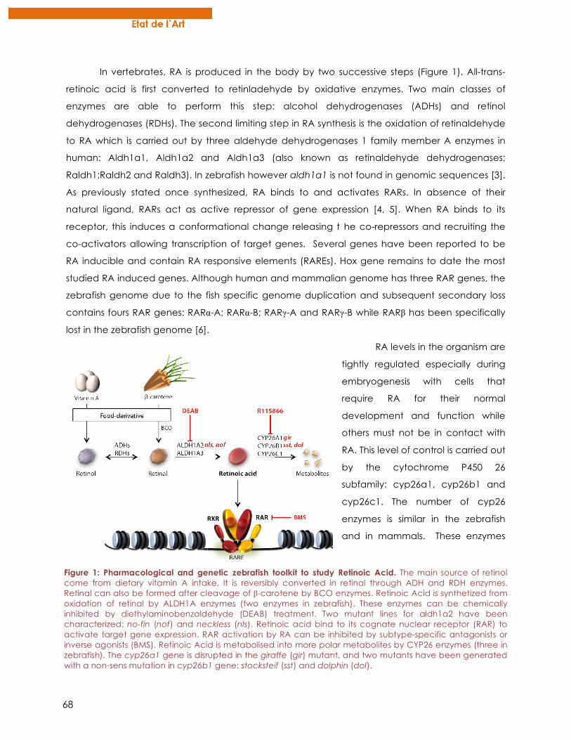
68
In vertebrates, RA is produced in the body by two successive steps (Figure 1). All-trans-
retinoic acid is first converted to retinladehyde by oxidative enzymes. Two main classes of
enzymes are able to perform this step: alcohol dehydrogenases (ADHs) and retinol
dehydrogenases (RDHs). The second limiting step in RA synthesis is the oxidation of retinaldehyde
to RA which is carried out by three aldehyde dehydrogenases 1 family member A enzymes in
human: Aldh1a1, Aldh1a2 and Aldh1a3 (also known as retinaldehyde dehydrogenases:
Raldh1;Raldh2 and Raldh3). In zebrafish however aldh1a1 is not found in genomic sequences [3].
As previously stated once synthesized, RA binds to and activates RARs. In absence of their
natural ligand, RARs act as active repressor of gene expression [4, 5]. When RA binds to its
receptor, this induces a conformational change releasing t he co-repressors and recruiting the
co-activators allowing transcription of target genes. Several genes have been reported to be
RA inducible and contain RA responsive elements (RAREs). Hox gene remains to date the most
studied RA induced genes. Although human and mammalian genome has three RAR genes, the
zebrafish genome due to the fish specific genome duplication and subsequent secondary loss
contains fours RAR genes: RARα-A; RARα-B; RARγ-A and RARγ-B while RARβ has been specifically
lost in the zebrafish genome [6].
RA levels in the organism are
tightly regulated especially during
embryogenesis with cells that
require RA for their normal
development and function while
others must not be in contact with
RA. This level of control is carried out
by the cytochrome P450 26
subfamily: cyp26a1, cyp26b1 and
cyp26c1. The number of cyp26
enzymes is similar in the zebrafish
and in mammals. These enzymes
Figure 1: Pharmacological and genetic zebrafish toolkit to study Retinoic Acid. The main source of retinol come from dietary vitamin A intake. It is reversibly converted in retinal through ADH and RDH enzymes. Retinal can also be formed after cleavage of β-carotene by BCO enzymes. Retinoic Acid is synthetized from oxidation of retinal by ALDH1A enzymes (two enzymes in zebrafish). These enzymes can be chemically inhibited by diethylaminobenzaldehyde (DEAB) treatment. Two mutant lines for aldh1a2 have been characterized: no-fin (nof) and neckless (nls). Retinoic acid bind to its cognate nuclear receptor (RAR) to activate target gene expression. RAR activation by RA can be inhibited by subtype-specific antagonists or inverse agonists (BMS). Retinoic Acid is metabolised into more polar metabolites by CYP26 enzymes (three in zebrafish). The cyp26a1 gene is disrupted in the giraffe (gir) mutant, and two mutants have been generated with a non-sens mutation in cyp26b1 gene: stocksteif (sst) and dolphin (dol).
69
catalyse reactions that convert RA into 4-hydroxy-RA that is in turn oxidized to 4-oxo-RA (Figure 1).
Therefore cyp26 enzymes function is to prevent activation of RARs by RA in specific cells. The
complementary expression pattern of RA synthesizing enzymes (aldh1a) and RA degrading
enzymes (cyp26) allow normal distribution of this morphogen and therefore normal development
of embryos and foetuses.
Thanks to its external development, zebrafish is a convenient model to assay
pharmacological treatment. By simply treating the medium in which embryos are developing
with chemicals altering RA metabolism, researchers have a unique occasion to investigate the
role of RA pathway during embryogenesis. Different compounds are commercially available to
specifically inhibit (i) ALDH1A-mediated endogenous RA synthesis with
diethylaminobenzaldehyde (DEAB), (ii) endogenous CYP26-mediated RA metabolism with
R115866 inhibitor, or even (iii) RAR activation by RA thanks to specific RAR-antagonist or inverse
agonist (e.g BMS493) (Figure 1). Moreover, zebrafish is a very suitable model for genetic
approach and different mutant lines have already been generated with mutation in aldh1a2
gene (neckless, no-fin), in cyp26a1 (giraffe) or in cyp26b1 (stocksteif, dolphin) [7-11]. As a result,
both genetic and chemical tools are available for studying RA effects on the see-through and
fast-developing zebrafish embryo but also in adult fish.
EARLY EMBRYOGENESIS
Brain patterning
Numerous studies have focused on the implication of RA signaling during hindbrain
patterning. Hindbrain patterning is characterized by the formation of equal segments called
rhombomeres (r). The number of rhombomeres varies depending on the authors between 7 and
8. Each rhombomere acquires a specific identity depending on its position along the antero-
posteiror axis. Identification of RA as a major signal in rhombomere development came from
analysis of VAD syndrome in avian where a lack of RA leads to the absence of rhombomere 4-8
into an enlarged rhombomere 3 [12]. In zebrafish the first data on hindbrain patterning and RA
signaling came from the analysis of the neckless (nls) mutant deficient in alhh1a2 enzyme [7],
later confirmed by the identification of a second aldh1a2 mutant named no fin (nof) [8].
However in these zebrafish mutants the severity of posterior rhombemere loss was less that in the
VAD quail syndrome. Therefore it has been postulated that other source(s) of RA synthesis were
present in the zebrafish or that maternal supply of RA might help early hindbrain patterning. Use
of pharmacological compounds that prevent RA synthesis such as 4-Diethylaminobenzaldehyde
(DEAB) or the pan-RAR inverse agonist such as BMS 493 after the maternal to zygotic transition
70
generated embryos with a much reduced hindbrain with the posterior limit of the hindbrain
being found at the r4/r5 boundary [13]. Therefore during zebrafish embryonic development, at
least one other source of RA independent of aldh1a2 is implicated in hindbrain patterning. This
other source of RA production is yet to be identified. The most obvious secondary source of RA
production will lies in aldh1a3, however morpholino knock-down of aldh1a3 does not seem to
have any hindbrain patterning abnormality [14]. Therefore the zebrafish may possess another
enzyme with retinaldehyde dehydrogenase activity or has another way to produce RA in an
aldh1a independent manner.
Although the anterior part of the brain is qualified as being a RA free zone in zebrafish,
Aldh1a2 homozygous mutant in mouse display forebrain deficiencies such as defective growth
and morphogenesis of the optic tectum [15, 16] while analysis of the Aldh1a2/Aldh1a3 double
homozygous mutant challenges the view of a role played by RA in forebrain formation [17]. A
definitive role of RA signalling during forebrain patterning came from recent studies in zebrafish
where Gongal and colleagues demonstrated that aldh1a2 expression is controlled and
maintained by the H6-homeobox (HMX) 4 [18]. Surprisingly, exogenous RA can rescue the
forebrain phenotype in Hmx4 morphants while DEAB treatments do not affect forebrain
patterning. Therefore the authors proposed a model in which Hmx4 regulates RA production
which in turns control sonic hedgehog (shh) signalling for a normal forebrain patterning in
zebrafish.
RA Gradient
RA is a morphogen molecule that diffuses from the posterior to the anterior of the embryo
through a concentration gradient and specifies positional information within paraxial mesoderm
and for rhombomere patterning in the hindbrain [13, 19, 20]. In the past few years, studies using
zebrafish as a model provided new insights in how this RA gradient is dynamically regulated
during early embryogenesis, and is RA accurately acting through a canonical morphogen
mechanism. Many studies performed in mammals, birds and fish pointed out the posteriorizing
effect of RA. Indeed, when applied exogenously, RA promotes the formation of posterior
structures at the expanse of anterior features (e.g eyes, forebrain) [8] and conversely, depleting
RA synthesis prevent the formation of posterior rhombomeres in the hindbrain [19]. This
posteriorizing effect of RA is consistent with the expression of ALDH1A2 enzyme in the posterior
mesoderm where RA is synthetized, therefore providing the “source” of the gradient. However,
the fact that RA deficiency can be rescued by a homogeneous exposure to exogenous RA [21]
means that RA gradient is not simply generated through diffusion from a synthetizing zone. In
fact, many studies on zebrafish embryos revealed that RA availability is prevented in the anterior
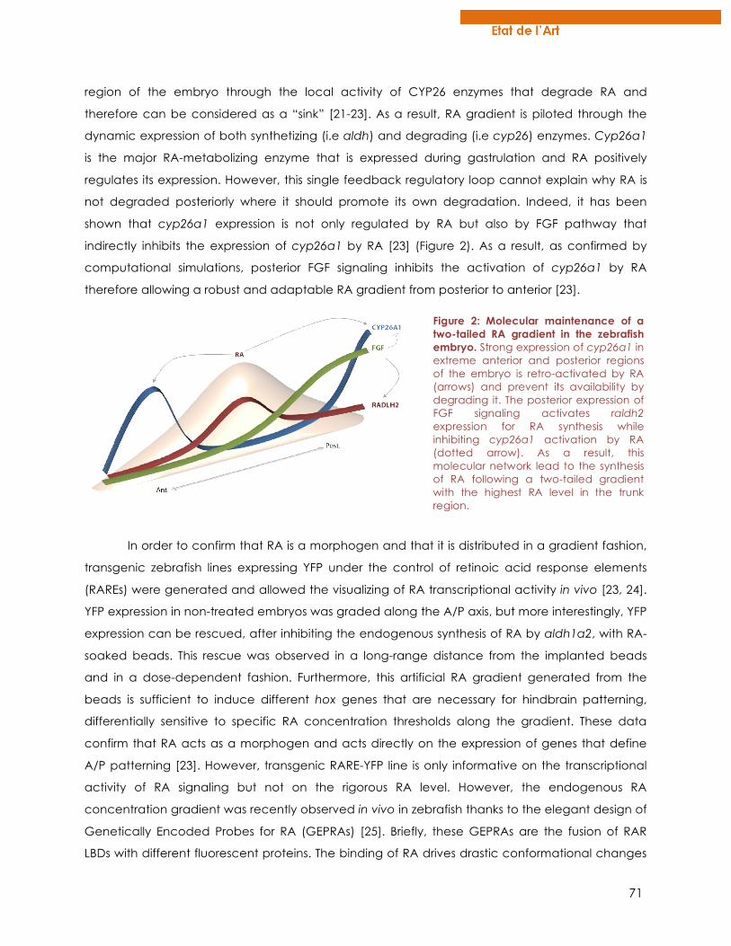
71
region of the embryo through the local activity of CYP26 enzymes that degrade RA and
therefore can be considered as a “sink” [21-23]. As a result, RA gradient is piloted through the
dynamic expression of both synthetizing (i.e aldh) and degrading (i.e cyp26) enzymes. Cyp26a1
is the major RA-metabolizing enzyme that is expressed during gastrulation and RA positively
regulates its expression. However, this single feedback regulatory loop cannot explain why RA is
not degraded posteriorly where it should promote its own degradation. Indeed, it has been
shown that cyp26a1 expression is not only regulated by RA but also by FGF pathway that
indirectly inhibits the expression of cyp26a1 by RA [23] (Figure 2). As a result, as confirmed by
computational simulations, posterior FGF signaling inhibits the activation of cyp26a1 by RA
therefore allowing a robust and adaptable RA gradient from posterior to anterior [23].
In order to confirm that RA is a morphogen and that it is distributed in a gradient fashion,
transgenic zebrafish lines expressing YFP under the control of retinoic acid response elements
(RAREs) were generated and allowed the visualizing of RA transcriptional activity in vivo [23, 24].
YFP expression in non-treated embryos was graded along the A/P axis, but more interestingly, YFP
expression can be rescued, after inhibiting the endogenous synthesis of RA by aldh1a2, with RA-
soaked beads. This rescue was observed in a long-range distance from the implanted beads
and in a dose-dependent fashion. Furthermore, this artificial RA gradient generated from the
beads is sufficient to induce different hox genes that are necessary for hindbrain patterning,
differentially sensitive to specific RA concentration thresholds along the gradient. These data
confirm that RA acts as a morphogen and acts directly on the expression of genes that define
A/P patterning [23]. However, transgenic RARE-YFP line is only informative on the transcriptional
activity of RA signaling but not on the rigorous RA level. However, the endogenous RA
concentration gradient was recently observed in vivo in zebrafish thanks to the elegant design of
Genetically Encoded Probes for RA (GEPRAs) [25]. Briefly, these GEPRAs are the fusion of RAR
LBDs with different fluorescent proteins. The binding of RA drives drastic conformational changes
Figure 2: Molecular maintenance of a two-tailed RA gradient in the zebrafish embryo. Strong expression of cyp26a1 in extreme anterior and posterior regions of the embryo is retro-activated by RA (arrows) and prevent its availability by degrading it. The posterior expression of FGF signaling activates raldh2 expression for RA synthesis while inhibiting cyp26a1 activation by RA (dotted arrow). As a result, this molecular network lead to the synthesis of RA following a two-tailed gradient with the highest RA level in the trunk region.
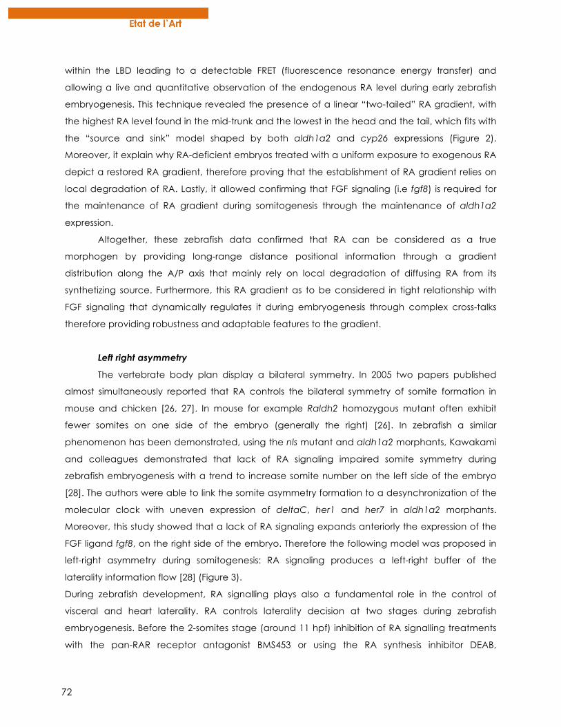
72
within the LBD leading to a detectable FRET (fluorescence resonance energy transfer) and
allowing a live and quantitative observation of the endogenous RA level during early zebrafish
embryogenesis. This technique revealed the presence of a linear “two-tailed” RA gradient, with
the highest RA level found in the mid-trunk and the lowest in the head and the tail, which fits with
the “source and sink” model shaped by both aldh1a2 and cyp26 expressions (Figure 2).
Moreover, it explain why RA-deficient embryos treated with a uniform exposure to exogenous RA
depict a restored RA gradient, therefore proving that the establishment of RA gradient relies on
local degradation of RA. Lastly, it allowed confirming that FGF signaling (i.e fgf8) is required for
the maintenance of RA gradient during somitogenesis through the maintenance of aldh1a2
expression.
Altogether, these zebrafish data confirmed that RA can be considered as a true
morphogen by providing long-range distance positional information through a gradient
distribution along the A/P axis that mainly rely on local degradation of diffusing RA from its
synthetizing source. Furthermore, this RA gradient as to be considered in tight relationship with
FGF signaling that dynamically regulates it during embryogenesis through complex cross-talks
therefore providing robustness and adaptable features to the gradient.
Left right asymmetry
The vertebrate body plan display a bilateral symmetry. In 2005 two papers published
almost simultaneously reported that RA controls the bilateral symmetry of somite formation in
mouse and chicken [26, 27]. In mouse for example Raldh2 homozygous mutant often exhibit
fewer somites on one side of the embryo (generally the right) [26]. In zebrafish a similar
phenomenon has been demonstrated, using the nls mutant and aldh1a2 morphants, Kawakami
and colleagues demonstrated that lack of RA signaling impaired somite symmetry during
zebrafish embryogenesis with a trend to increase somite number on the left side of the embryo
[28]. The authors were able to link the somite asymmetry formation to a desynchronization of the
molecular clock with uneven expression of deltaC, her1 and her7 in aldh1a2 morphants.
Moreover, this study showed that a lack of RA signaling expands anteriorly the expression of the
FGF ligand fgf8, on the right side of the embryo. Therefore the following model was proposed in
left-right asymmetry during somitogenesis: RA signaling produces a left-right buffer of the
laterality information flow [28] (Figure 3).
During zebrafish development, RA signalling plays also a fundamental role in the control of
visceral and heart laterality. RA controls laterality decision at two stages during zebrafish
embryogenesis. Before the 2-somites stage (around 11 hpf) inhibition of RA signalling treatments
with the pan-RAR receptor antagonist BMS453 or using the RA synthesis inhibitor DEAB,
73
randomized the left-right positioning of visceral organs [29]. This lack of correct left right
asymmetry of visceral organ was a consequence of bilateral expression of the asymmetric genes
southpaw and lefty2 in the lateral plate mesoderm [29]. Analysis of the RAR revealed that
knockdown of RARγ-B results in bilateral livers without impacting liver size [30]. This study therefore
revealed that the primitive endoderm is able to form liver on both the left and right side of the
embryo. This study also reveals that the bilaterality of liver observed in RARγ-B morphants can be
rescued by overexpressing bmp2b indicating that RA signalling acts upstream of BMP signalling
during liver positioning [30].
Depletion or RA signalling after the 2-somite stage has no effect of the laterality of
visceral organ (southpaw and lefty2 are expressed normally) but perturbs the expression of bmp4
in the cardiac field resulting in a randomized
cardiac looping of the heart. Analysis of
Tg(cmlc2:GFP) embryos, which express GFP in
both the atrium and the ventricle, depleted of
RA signalling lead to a majority of embryos with
heart displaying a L-Looped heart or a non-
looped heart at the midline [29].
The question remains to understand how
RA signalling can influence left-right asymmetry
of visceral organs during zebrafish development.
The answer lies within the study of the Kupffer’s
vesicle (KV), the assumed equivalent of the
murine embryonic node implicated in left-right
asymmetry. Depletion of RA signalling during
early zebrafish development does not affect KV
formation but increases cilia length as marked
by alpha-Acetylated tubulin antibody staining
[29]. As a result, cilia in the Kupffer’s vesicle lose
their well-organized counter-clockwise
directional flow to a non-directional flow in RA
Figure 3: Multiple roles of RA in controlling asymmetric flow within the Kuppfer’s vesicle (KV) and buffering assymetric signal for bilateral somite development. Within the KV, ENC1L expression is expressed and ensure correct cilia length and number in KV cells. Its expression is regulated by both RA and FGF signalling
thus RA is required for correct flow within the KV. This flow is responsible of an increase in Ca2+ influx in left cells of the KV (green cells) and therefore promote the unilateral activation of genes (e.g spaw) involved in L/R asymmetry. Furthermore, anterior RA signaling from the forming somites acts as a buffer against assymetric signals (e.g Notch, FGF) from the posterior, and allow synchronized bilateral somite formation.
74
compromised embryos. Using an ectoderm-Neural Cortex1-like (enc1l) morpholino, Qian and
collaborators showed that this gene was a direct downstream effector of RA signalling pathway
in modulating KV ciliogenesis in zebrafish [31] (Figure 3). The length and number of cilia in cells
within the KV is important to maintain a directional flow, equivalent to the nodal flow described
in mammals [32, 33], and leading to the unilateral activation of genes required for asymmetrical
patterning [34].
ORGANOGENESIS
Heart development
Thanks to its external development, see-through zebrafish embryos allow easy and non-
invasive heart and blood cell observation during embryogenesis. Moreover, embryogenesis is not
dependent of blood circulation since the embryo can receive enough oxygen from the external
medium by passive diffusion. As a result and unlike mammalian models, many zebrafish mutants
with strong cardiovascular defects can still survive until the end of organogenesis, therefore
providing unprecedented investigations on cardiovascular developmental.
The heart is the first organ to develop and to function and its development requires
coordinated and combinatorial mechanisms that rely on many developmental pathways. In
zebrafish, the embryonic heart originates from cardiogenic precursors that are already
determined in the late blastula and which involute in the early gastrula to then migrate to the
lateral plate mesoderm (LPM) near the midbrain-hindbrain boundary [35]. RA signaling is firstly
involved in the mesoderm commitment to cardiac lineage and early RA exposure of zebrafish
gastrula embryos lead to early loss of all cardiac precursors [36, 37]. The bilateral populations of
cardiogenic precursors from LPM are already composed of myocardial precursors (that will form
the outer myocardial layer of the heart) and endocardial precursors (that will form the inner
endocardial layer), both expressing specific markers. At around 19 hpf, they all migrate towards
the embryo midline where they fuse to form a linear heart tube. While migrating, myocardial
precursors terminate their differentiation by expressing specific genes conferring them contractile
activity. The linear heart tube then undergo many morphogenetic changes that will shape it as a
multi-chambered organ, the main observables structures being the ventricule and the atrium
along the A/P axis [38]. The formation of cardiac chambers and of its A/P patterning requires RA
signaling. Indeed, exogenous RA treatment of zebrafish embryo leads to a posteriorized heart by
inhibiting the developmental of ventricular cells [37]. This is consistent with the expression of
aldh1a2 in the preatrial region of the murine heart tube [39] that suggests that endogenous RA
signaling favors atrial development at the expense of ventricular development, during heart
chamber formation. However, RA signaling also plays a major role before chamber formation in
75
the commitment of cardiogenic precursors into atrial versus ventricular differentiation. In the
neckless zebrafish mutant that lacks endogenous synthesis of RA through ALDH1A2 activity, an
excess of cardiomyocytes is observed [40]. Consistently, by treating zebrafish embryos with a
pan-RAR antagonist (BMS 189453) at the gastrula stage, both ventricular and atrial precursor
populations are more numerous. When applied at early somite-stage, BMS 189453 treatment only
increases the number of atrial cells thus suggesting a role of RA signaling in restricting atrial cell
number at this stage. Interestingly, this RA-dependent restriction of atrial cell fate is mediated
through a non-autonomous action of hoxb5b. In fact, this hox gene is expressed in the nearby
pectoral fin (forelimb) field at the early-somite stage and its expression in induced by RA.
Morpholino knockdown of hoxb5b phenocopies the lack of RA signaling induced by BMS 189453
treatment and this phenotype can be rescued by ectopic sources of hoxb5b delivered by
transplanted cells expressing an hyperactive vp16-hoxb5b [41]. More recently, FGF signaling
(fgf8) has been confirmed as acting downstream to RA signaling in this forelimb and heart
coordinated development [42] which is consistent with some studies in mice. In fact, increasing
FGF signaling lead to an increased number of cardiac cells and conversely, down-regulation of
fgf8a (that is expressed in cardiac progenitors) by morpholino injection, prevent the phenotype
induced by RA deficiency. Fgf8 is also involved in the development of cardiac precursors and is
required for the proper expression of the heart precursor marker within the LPM [43].
Later in development, RA exposure rapidly induces cardio-toxicity in the zebrafish larvae
such as edema or heart elongation [37]. In fact, RA-treated embryos at 72 hpf develop cardiac
malformations less than 24 hours post-treatment. The precise mechanisms induced by RA that
lead to such toxicity is not well understood but a micro-array assay pointed out NR2F5 (a
member of the COUP-TF family) as being rapidly activated by RA in the heart. Interestingly, NR2F5
overexpression phenocopies RA-induced cardiac phenotype and conversely, morpholino
injection against NR2F5 prevents RA-induced cardio-toxicity, therefore suggesting a role for
NR2F5 in mediating RA-teratogenic effect on the heart [44].
Hematopoiesis
Studies mainly performed in mice, pointed out that RA signaling regulate developmental
hematopoiesis, and that it tightly balances the determination of precursor cells to a specific
hematopoietic lineage, but generalized conclusions about RA effects were not easily drawn.
Complementary studies using zebrafish as a model helped to have a better view of RA effects on
the early phases of hematopoiesis [45]. Briefly, like in all vertebrates, hematopoiesis is established
in two main waves in zebrafish during embryogenesis [46]. The first wave called “primitive”
hematopoiesis mainly generates red blood cell population and few myeloid cells from cells
76
located in the anterior and posterior LPM respectively, where they express specific markers such
as scl or genes from the gata family [47]. The second wave called “definitive” hematopoiesis
starts at around 36 hpf and leads to the production of all the different blood cell lineages for
lifetime, from a pool of hematopoietic stem cells located in the kidney marrow. Interestingly, RA
signaling has been reported as playing a major role in the early primitive hematopoiesis. In fact,
zebrafish embryos treated with exogenous RA show a strong dose-dependent reduction of
gata1-expressing blood cells at the 5-somites stage [45]. Moreover, this reduced expression is
rescued by the injection of scl mRNA, therefore proving that the RA effects observed are specific
to the hematopoietic cells and also suggesting that RA is acting upstream to scl for correct
primitive erythropoiesis. Conversely, inhibition of RA signaling by DEAB treatment leads to an
increased expression of gata1. However, these effects of RA are only observed after early
treatment of zebrafish embryos at the gastrula stage suggesting that RA signaling is regulating
primitive hematopoiesis during the early step of mesodermal patterning. Recently, RA signaling
has been reported as also regulating restrictively zebrafish primitive myelopoiesis [48].
Neural crest, pharyngeal arches and tooth
Neural crest cells (NCCs) are transient migratory cells that emerge from the dorsal part of
the neural tube (neural folds) during its closure (Figure 4). Of note is that in teleosts, the formation
of the neural tube is processed through a mechanism called secondary neurulation that
corresponds to a thickening of the ectoderm followed by a lumen cavitation [49]. Basically, at
the molecular level, the induction model commonly admitted is that ectoderm can give rise to
both epidermis and neural tissue following a BMP gradient. While BMP signaling promotes
induction of epidermis, BMP antagonists (e.g noggin, chordin, follistatin) from the organizer
region, potentiate the determination of neural tissue. Following this model, an intermediate level
of BMP give rise to an intermediate fate therefore forming neural tube borders, roughly
corresponding to the neural folds [50, 51] (Figure 4). Secondary signaling emerging from the
epidermis and/or the lateral mesoderm are required to posteriorize the anterior neural plate for
the induction of neural crests. The main signaling pathways involved are FGF, Wnt and BMP [50,
52, 53] but RA signaling has also been reported in Xenopus as being required for the induction of
neural crests that express specific markers such as Slug [54]. Although paraxial mesoderm has
been shown to be required for NC induction in Xenopus [55], its requirement in zebrafish is less
obvious since absence of involuted mesoderm in zebrafish embryos in which Nodal signaling is
impaired, does not impair NC induction [56]. Once induced, NCCs delaminate from the dorsal
neural tube to migrate within the entire embryo through a complex journey driven by many
repulsive signals [52, 57]. Trunk NCCs mainly generate pigment cells or ventral derivatives such as
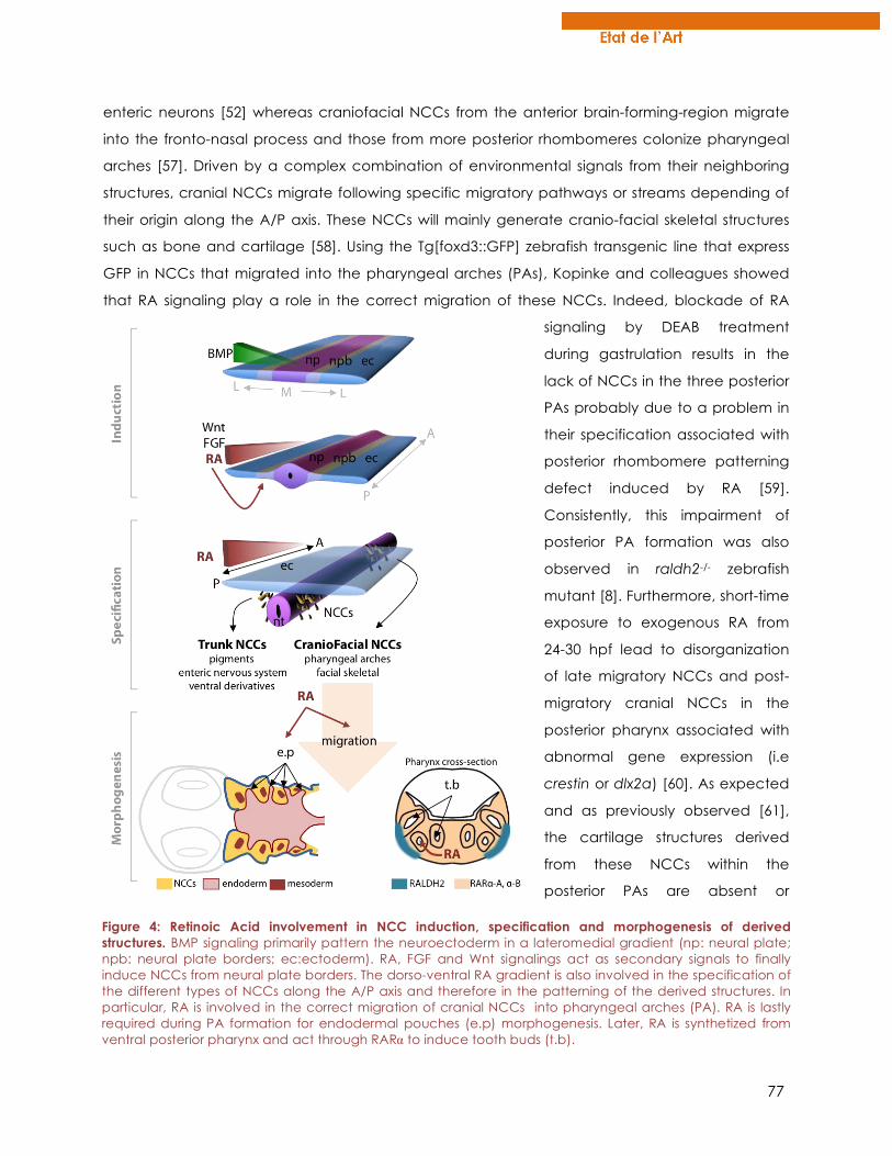
77
enteric neurons [52] whereas craniofacial NCCs from the anterior brain-forming-region migrate
into the fronto-nasal process and those from more posterior rhombomeres colonize pharyngeal
arches [57]. Driven by a complex combination of environmental signals from their neighboring
structures, cranial NCCs migrate following specific migratory pathways or streams depending of
their origin along the A/P axis. These NCCs will mainly generate cranio-facial skeletal structures
such as bone and cartilage [58]. Using the Tg[foxd3::GFP] zebrafish transgenic line that express
GFP in NCCs that migrated into the pharyngeal arches (PAs), Kopinke and colleagues showed
that RA signaling play a role in the correct migration of these NCCs. Indeed, blockade of RA
signaling by DEAB treatment
during gastrulation results in the
lack of NCCs in the three posterior
PAs probably due to a problem in
their specification associated with
posterior rhombomere patterning
defect induced by RA [59].
Consistently, this impairment of
posterior PA formation was also
observed in raldh2-/- zebrafish
mutant [8]. Furthermore, short-time
exposure to exogenous RA from
24-30 hpf lead to disorganization
of late migratory NCCs and post-
migratory cranial NCCs in the
posterior pharynx associated with
abnormal gene expression (i.e
crestin or dlx2a) [60]. As expected
and as previously observed [61],
the cartilage structures derived
from these NCCs within the
posterior PAs are absent or
Figure 4: Retinoic Acid involvement in NCC induction, specification and morphogenesis of derived structures. BMP signaling primarily pattern the neuroectoderm in a lateromedial gradient (np: neural plate; npb: neural plate borders; ec:ectoderm). RA, FGF and Wnt signalings act as secondary signals to finally induce NCCs from neural plate borders. The dorso-ventral RA gradient is also involved in the specification of the different types of NCCs along the A/P axis and therefore in the patterning of the derived structures. In particular, RA is involved in the correct migration of cranial NCCs into pharyngeal arches (PA). RA is lastly required during PA formation for endodermal pouches (e.p) morphogenesis. Later, RA is synthetized from ventral posterior pharynx and act through RARα to induce tooth buds (t.b).
78
malformed whereas cartilage structures derives from the anterior PAs are developing almost
normally [59]. Linville and colleagues confirmed this phenotype (i.e the lack of dlx2 expression in
the posterior PAs) and they showed that this RA effect is mediated through RARγ-A subtype since
specific morpholino knockdown of this receptor subtype is sufficient to prevent the formation of
posterior PAs [62]. Interestingly, if RA synthesis inhibition is achieved after gastrulation, NCCs do
migrate within the five PAs but the correct separation between the most posterior PAs is not
accomplished. This is explained by the fact that RA also plays a role in the formation and
morphogenesis of the endodermal pouches that normally separate PAs [59, 63] (Figure 4). All
these data confirmed that RA, by acting within different tight time-windows, is required all along
NCCs journey, from their specification to their migration and for the correct morphogenesis and
segmentation of their derived structures such as PAs.
Cranial NCCs are also giving rise to the dental mesenchyme required for tooth
development [64] and recent studies using zebrafish showed the involvement of RA signaling in
this process. Zebrafish, like all cypriniformes, lack oral teeth and only depict pharyngeal dentition.
Impairing RA synthesis by DEAB treatment during the timeframe of induction of the first tooth-pair
(named 4V1) result in the lack of tooth induction [65]. By using RAR-subtype specific antagonists,
we were able to show that RA mediates its actions on tooth induction through the alpha
subtypes of RARs (i.e RARα-A and RARα-B). Furthermore, aldh1a2 expression pattern observed by
in situ hybridization revealed a local and transient expression in the posterior ventral pharynx, that
provide local source of RA that can diffuse toward the tooth bud mesenchyme [65]. Recently,
we showed that cyp26b1, a RA-degrading enzyme, is expressed from 48 hpf in the tooth-forming
region and play the role of a sink to deplete RA from this region (unpublished). As a result, the
temporal expression of both synthetizing (i.e aldh1a2) and degrading (i.e cyp26b1) enzymes
drive a precise temporal and regional window of action for RA signaling to regulate tooth
development. Interestingly, we showed that small changes in the temporal expression of
cyp26b1 are correlated with different dental phenotypes within wild cypriniformes species
(unpublished). Besides, exposure of zebrafish embryos to exogenous RA leads to different dental
phenotypes that resemble dentition of other teleosts, such as anterior expansion of dentition [60]
or change in tooth shape and number depending of the time and amount of exogenous RA
dispensed (unpublished). Altogether, these results suggest that RA signaling is a good molecular
candidate that is thought to have generated a huge diversity of dentition within cypriniformes
and more widely in vertebrates.
79
Bone
As with many other organ systems, bone tissue and its development are highly conserved
between zebrafish and mammals, positioning the zebrafish as a reliable model organism to study
bone. As is seen in mammals, zebrafish create bone by both endochondral ossification, in which
bone progenitor cells condense and differentiate into chondrocytes before differentiating into
bone cells, and intramembranous ossification, where the progenitors condense and differentiate
directly into bone cells [66-68]. Consistent with mammals, zebrafish also have osteoblasts (bone-
building cells) and osteoclasts (bone-resorbing cells).[69, 70] In zebrafish, osteoblasts begin
differentiating at 36 hpf, initially in the craniofacial region where the first bones will develop,
including the opercle and the cleithrum.[71]. As these osteoblasts mature, they express many of
the same genes that are seen in mammals including runx2 and osterix during bone
development, and osteocalcin and osteonectin as fully mature cells.[71]. As in mammals,
zebrafish osteoclasts are hematopoietically derived, firstly present in zebrafish around 10-12 dpf,
and are detectable by tartrate-resistant acid phosphatase staining (TRAP) [70]. Similar to other
vertebrates, zebrafish osteoclasts are responsible for degrading bone matrix [70]. Taken together,
these similarities show that the zebrafish is an adequate model for studying bone development.
Along with the critical roles of RA in early patterning and organogenesis of the vertebrate
embryo, it is also involved in bone development. Early research identified that RA treatment
caused changes in the size and shape of bones in embryonic mice and could induce osteoblast
differentiation in murine cell lines [72, 73]. However, subsequent research has provided conflicting
results of the effects that RA has on bone development. Some publications have claimed that
RA caused an increase in osteoblast differentiation and bone development while others have
stated that it caused a decrease in differentiation [74-77]. Much of this research was performed
in cell lines or in mammalian models, hindering observations at stages of early bone
development in an in vivo context. The zebrafish stocksteif and dolphin mutants both have a
non-sense mutation in cyp26b1, causing increased endogenous RA levels. These mutants present
phenotypes of hyperossification that are visible with alizarin red staining by 8 dpf [9, 11]. Fittingly,
wild-type fish pharmacologically treated with RA phenocopy both of these mutations.
Interestingly, it was shown that the increase in bone mineralization of the stocksteif mutant was
not caused by an increase in osteoblast number or by mis-localization of osteoblasts, but was
rather proposed to be caused by increased osteoblast activity [9]. Also, in situ hybridization
showed that expression of osterix and col10a1, two markers of mature osteoblasts in zebrafish
[71] was unaffected in the stocksteif mutant compared to siblings at 4 dpf. Further research using
exogenous RA and DEAB treatment has looked at acute effects that RA signalling has on bone
development [78]. It was shown that, in contrast to the stoscksteif mutant, RA treatment at 48-52
80
hpf decreased the expression of osterix along with runx2b, an earlier marker of osteoblasts, while
DEAB treatment at this time interval increased the expression of these genes. RA treatment from
48-54 hpf was also shown to decrease bone mineralization at 144 hpf. While this data may seem
to conflict with other results, it was shown that RA treatment of zebrafish at 84-96 hpf, when
mature osteoblasts are present, increased bone mineralization. However, it remains unclear why
the stocksteif mutant shows normal development of osteoblasts while acute RA treatment
hinders osteoblast maturation. Interestingly, these experiments reveal a possible dual role for RA
in bone development. RA may play a role inhibiting osteoblast differentiation, as shown by the
exogenous RA treatments early in development. Later in development, after osteoblasts have
matured, RA may have an opposite role in promoting osteoblast activity to enhance bone
mineralization. This proposed dual role of RA in bone development may also explain why there
have been conflicting results from in vitro experiments.
Lipid
It has been known for over thirty years that RA signalling has an effect on fat
development. It was initially shown that RA inhibited the differentiation of mammalian cells into
adipocytes [79, 80]. However, subsequent in vitro studies have produced conflicting results, some
showing a pro-adipogenic effect of RA treatment [81-83] while others claiming an inhibitory role
for RA in adipocyte differentiation [84, 85]. The contradictory effects seen in these in vitro studies
may have arisen from differences in RA concentration or from the differences in the relative
timing that the treatment was given during differentiation. Mammalian studies have primarily
shown that RA acts to reduce adiposity and overall body weight and can increase energy
expenditure in mature animals [86-88]. It has been proposed that the reduced adiposity seen in
RA treated rodents is caused by the combination of two aspects of RA signalling. Firstly, RA has
been shown to increase expression of pref-1, SOX9, and KLF2, which are expressed in
preadipocytes and may inhibit the cells from further differentiating into mature adipocytes [87].
Secondly, in already mature adipocytes, RA may signal through RARs and PPARβ and PPARδ to
increase energy expenditure [87]. While these studies reveal a role for RA in reducing adiposity in
rodent models, the effects of RA on adiposity in humans is less clear. There is an association
between low dietary vitamin A intake and high adiposity [89]. However, humans receiving RA as
a treatment for cancer have experienced increased weight gain [90]. In fact, the effect of RA on
adiposity in mammals is further complicated by some rodent studies that have shown that RA
and increased vitamin A intake can cause higher adiposity [91, 92].
Over the past few years, the use of zebrafish for lipid research has grown in popularity.
Chemicals such as Oil-Red-O (ORO) and Nile Red have been used to stain for neutral lipids and
81
to fluorescently label adipocytes respectively [93]. Fluorescent lipid analogs have also been
developed to study the metabolic functions of zebrafish once feeding has begun [94]. These
fluorescent analogs can be used to trace lipid intake, processing, and incorporation into tissue.
These visualization techniques have helped to establish the homology between zebrafish and
mammalian adipocyte development and lipid metabolism [93, 95, 96]. Because of these recent
developments, the zebrafish has recently become a model used to analyze the effects of RA on
adipose development. In the neckless (aldh1a2) mutant, which has a mutation in the RA-
synthezing enzyme retinaldehyde dehydrogenase type 2, causing a reduction of endogenous
RA, there was decreased lipid deposition labelled with ORO [78]. Consistent with the mutant
phenotype, DEAB treatment, inhibiting RA synthesis, also showed a reduction in ORO labelling.
Not only did both the mutant and DEAB treated embryos show a decrease in ORO, but they also
showed decreased expression of C/ebpα, an adipocyte lineage marker. Fittingly, it was also
shown that exogenous RA treatment increased the expression of C/ebpα. These results show that
RA has a pro-adipogenic role during early development in zebrafish, which is contradictory to
the effects primarily seen in the adult mammal studies discussed previously. These differing
effects could be due to differences in lipid metabolism during development and adulthood.
Further investigation is required to elucidate a specific role for RA in lipid metabolism early and
late in zebrafish life.
CONCLUSION
In this review, we aimed at illustrating the power of zebrafish as a developmental model.
It is not only used for standard forward and reverse genetics, but is also easily accessible for
pharmacological treatments. Moreover it is also a suitable model for the generation of
transgenic lines integrating the latest genetic engineering advances. Its use for RA research is
now largely widespread and it allowed shedding light on new knowledge of the role of RA
mainly in early patterning and also in organogenesis. Its fast external development and its
transparency are major advantages to investigate in details developing structures.
REFERENCES
[1] M. Petkovich, N.J. Brand, A. Krust, P. Chambon, A human retinoic acid receptor which belongs to the family of nuclear receptors, Nature, 330 (1987) 444-450. [2] V. Giguere, E.S. Ong, P. Segui, R.M. Evans, Identification of a receptor for the morphogen retinoic acid, Nature, 330 (1987) 624-629. [3] S. Pittlik, S. Domingues, A. Meyer, G. Begemann, Expression of zebrafish aldh1a3 (raldh3) and absence of aldh1a1 in teleosts, Gene expression patterns : GEP, 8 (2008) 141-147. [4] T. Koide, M. Downes, R.A. Chandraratna, B. Blumberg, K. Umesono, Active repression of RAR signaling is required for head formation, Genes & development, 15 (2001) 2111-2121. [5] A.D. Weston, B. Blumberg, T.M. Underhill, Active repression by unliganded retinoid receptors in development: less is sometimes more, The Journal of cell biology, 161 (2003) 223-228.
82
[6] S. Bertrand, B. Thisse, R. Tavares, L. Sachs, A. Chaumot, P.L. Bardet, H. Escriva, M. Duffraisse, O. Marchand, R. Safi, C. Thisse, V. Laudet, Unexpected novel relational links uncovered by extensive developmental profiling of nuclear receptor expression, PLoS genetics, 3 (2007) e188. [7] G. Begemann, T.F. Schilling, G.J. Rauch, R. Geisler, P.W. Ingham, The zebrafish neckless mutation reveals a requirement for raldh2 in mesodermal signals that pattern the hindbrain, Development, 128 (2001) 3081-3094. [8] H. Grandel, K. Lun, G.J. Rauch, M. Rhinn, T. Piotrowski, C. Houart, P. Sordino, A.M. Kuchler, S. Schulte-Merker, R. Geisler, N. Holder, S.W. Wilson, M. Brand, Retinoic acid signalling in the zebrafish embryo is necessary during pre-segmentation stages to pattern the anterior-posterior axis of the CNS and to induce a pectoral fin bud, Development, 129 (2002) 2851-2865. [9] K.M. Spoorendonk, J. Peterson-Maduro, J. Renn, T. Trowe, S. Kranenbarg, C. Winkler, S. Schulte-Merker, Retinoic acid and Cyp26b1 are critical regulators of osteogenesis in the axial skeleton, Development, 135 (2008) 3765-3774. [10] Y. Emoto, H. Wada, H. Okamoto, A. Kudo, Y. Imai, Retinoic acid-metabolizing enzyme Cyp26a1 is essential for determining territories of hindbrain and spinal cord in zebrafish, Developmental biology, 278 (2005) 415-427. [11] K. Laue, M. Jänicke, N. Plaster, C. Sonntag, M. Hammerschmidt, Restriction of retinoic acid activity by Cyp26b1 is required for proper timing and patterning of osteogenesis during zebrafish development, Development, 135 (2008) 3775-3787. [12] E. Gale, M. Zile, M. Maden, Hindbrain respecification in the retinoid-deficient quail, Mechanisms of development, 89 (1999) 43-54. [13] G. Begemann, M. Marx, K. Mebus, A. Meyer, M. Bastmeyer, Beyond the neckless phenotype: influence of reduced retinoic acid signaling on motor neuron development in the zebrafish hindbrain, Developmental biology, 271 (2004) 119-129. [14] M. Yahyavi, H. Abouzeid, G. Gawdat, A.S. de Preux, T. Xiao, T. Bardakjian, A. Schneider, A. Choi, E. Jorgenson, H. Baier, M. El Sada, D.F. Schorderet, A.M. Slavotinek, ALDH1A3 loss of function causes bilateral anophthalmia/microphthalmia and hypoplasia of the optic nerve and optic chiasm, Human molecular genetics, 22 (2013) 3250-3258. [15] V. Ribes, Z. Wang, P. Dolle, K. Niederreither, Retinaldehyde dehydrogenase 2 (RALDH2)-mediated retinoic acid synthesis regulates early mouse embryonic forebrain development by controlling FGF and sonic hedgehog signaling, Development, 133 (2006) 351-361. [16] F.A. Mic, A. Molotkov, N. Molotkova, G. Duester, Raldh2 expression in optic vesicle generates a retinoic acid signal needed for invagination of retina during optic cup formation, Dev Dyn, 231 (2004) 270-277. [17] N. Molotkova, A. Molotkov, G. Duester, Role of retinoic acid during forebrain development begins late when Raldh3 generates retinoic acid in the ventral subventricular zone, Developmental biology, 303 (2007) 601-610. [18] P.A. Gongal, L.D. March, V.L. Holly, L.M. Pillay, K.M. Berry-Wynne, H. Kagechika, A.J. Waskiewicz, Hmx4 regulates Sonic hedgehog signaling through control of retinoic acid synthesis during forebrain patterning, Developmental biology, 355 (2011) 55-64. [19] L. Maves, C.B. Kimmel, Dynamic and sequential patterning of the zebrafish posterior hindbrain by retinoic acid, Developmental biology, 285 (2005) 593-605. [20] A. Aulehla, O. Pourquie, Signaling gradients during paraxial mesoderm development, Cold Spring Harb Perspect Biol, 2 (2010) a000869. [21] R.E. Hernandez, A.P. Putzke, J.P. Myers, L. Margaretha, C.B. Moens, Cyp26 enzymes generate the retinoic acid response pattern necessary for hindbrain development, Development, 134 (2007) 177-187. [22] R.J. White, T.F. Schilling, How degrading: Cyp26s in hindbrain development, Dev Dyn, 237 (2008) 2775-2790. [23] R.J. White, Q. Nie, A.D. Lander, T.F. Schilling, Complex regulation of cyp26a1 creates a robust retinoic acid gradient in the zebrafish embryo, PLoS biology, 5 (2007) e304. [24] A. Perz-Edwards, N.L. Hardison, E. Linney, Retinoic acid-mediated gene expression in transgenic reporter zebrafish, Developmental biology, 229 (2001) 89-101. [25] S. Shimozono, T. Iimura, T. Kitaguchi, S. Higashijima, A. Miyawaki, Visualization of an endogenous retinoic acid gradient across embryonic development, Nature, 496 (2013) 363-366. [26] J. Vermot, J. Gallego Llamas, V. Fraulob, K. Niederreither, P. Chambon, P. Dolle, Retinoic acid controls the bilateral symmetry of somite formation in the mouse embryo, Science, 308 (2005) 563-566. [27] J. Vermot, O. Pourquie, Retinoic acid coordinates somitogenesis and left-right patterning in vertebrate embryos, Nature, 435 (2005) 215-220.
83
[28] Y. Kawakami, A. Raya, R.M. Raya, C. Rodriguez-Esteban, J.C. Izpisua Belmonte, Retinoic acid signalling links left-right asymmetric patterning and bilaterally symmetric somitogenesis in the zebrafish embryo, Nature, 435 (2005) 165-171. [29] S. Huang, J. Ma, X. Liu, Y. Zhang, L. Luo, Retinoic acid signaling sequentially controls visceral and heart laterality in zebrafish, The Journal of biological chemistry, 286 (2011) 28533-28543. [30] M.K. Garnaas, C.C. Cutting, A. Meyers, P.B. Kelsey, Jr., J.M. Harris, T.E. North, W. Goessling, Rargb regulates organ laterality in a zebrafish model of right atrial isomerism, Developmental biology, 372 (2012) 178-189. [31] M. Qian, S. Yao, L. Jing, J. He, C. Xiao, T. Zhang, W. Meng, H. Zhu, H. Xu, X. Mo, ENC1-like integrates the retinoic acid/FGF signaling pathways to modulate ciliogenesis of Kupffer's Vesicle during zebrafish embryonic development, Developmental biology, 374 (2013) 85-95. [32] N. Hirokawa, Y. Tanaka, Y. Okada, Left-right determination: involvement of molecular motor KIF3, cilia, and nodal flow, Cold Spring Harb Perspect Biol, 1 (2009) a000802. [33] J.J. Essner, K.J. Vogan, M.K. Wagner, C.J. Tabin, H.J. Yost, M. Brueckner, Conserved function for embryonic nodal cilia, Nature, 418 (2002) 37-38. [34] T. Matsui, Y. Bessho, Left-right asymmetry in zebrafish, Cell Mol Life Sci, 69 (2012) 3069-3077. [35] D.Y. Stainier, Zebrafish genetics and vertebrate heart formation, Nature reviews. Genetics, 2 (2001) 39-48. [36] K.E. Yutzey, D. Bader, Diversification of cardiomyogenic cell lineages during early heart development, Circulation research, 77 (1995) 216-219. [37] D.Y. Stainier, M.C. Fishman, Patterning the zebrafish heart tube: acquisition of anteroposterior polarity, Developmental biology, 153 (1992) 91-101. [38] D. Yelon, D.Y. Stainier, Patterning during organogenesis: genetic analysis of cardiac chamber formation, Seminars in cell & developmental biology, 10 (1999) 93-98. [39] J.B. Moss, J. Xavier-Neto, M.D. Shapiro, S.M. Nayeem, P. McCaffery, U.C. Drager, N. Rosenthal, Dynamic patterns of retinoic acid synthesis and response in the developing mammalian heart, Developmental biology, 199 (1998) 55-71. [40] B.R. Keegan, J.L. Feldman, G. Begemann, P.W. Ingham, D. Yelon, Retinoic acid signaling restricts the cardiac progenitor pool, Science, 307 (2005) 247-249. [41] J.S. Waxman, B.R. Keegan, R.W. Roberts, K.D. Poss, D. Yelon, Hoxb5b acts downstream of retinoic acid signaling in the forelimb field to restrict heart field potential in zebrafish, Developmental cell, 15 (2008) 923-934. [42] M.R.J. Sorrell, J.S. Waxman, Restraint of Fgf8 signaling by retinoic acid signaling is required for proper heart and forelimb formation, Developmental biology, 358 (2011) 44-55. [43] F. Reifers, E.C. Walsh, S. Leger, D.Y. Stainier, M. Brand, Induction and differentiation of the zebrafish heart requires fibroblast growth factor 8 (fgf8/acerebellar), Development, 127 (2000) 225-235. [44] N. Chen, J.L. Napoli, All-trans-retinoic acid stimulates translation and induces spine formation in hippocampal neurons through a membrane-associated RARalpha, Faseb J, 22 (2008) 236-245. [45] J.L. de Jong, A.J. Davidson, Y. Wang, J. Palis, P. Opara, E. Pugach, G.Q. Daley, L.I. Zon, Interaction of retinoic acid and scl controls primitive blood development, Blood, 116 (2010) 201-209. [46] A.J. Davidson, L.I. Zon, The 'definitive' (and 'primitive') guide to zebrafish hematopoiesis., Oncogene, 23 (2004) 7233-7246. [47] N. Hsia, L.I. Zon, Transcriptional regulation of hematopoietic stem cell development in zebrafish, Experimental hematology, 33 (2005) 1007-1014. [48] D. Liang, W. Jia, J. Li, K. Li, Q. Zhao, Retinoic acid signaling plays a restrictive role in zebrafish primitive myelopoiesis, PloS one, 7 (2012) e30865. [49] C. Papan, J.A. Campos-Ortega, On the formation of the neural keel and neural tube in the zebrafish Danio (Brachydanio) rerio, Roux's archives of developmental biology, 203 (1993) 178-186. [50] A.K. Knecht, M. Bronner-Fraser, Induction of the neural crest: a multigene process, Nature reviews. Genetics, 3 (2002) 453-461. [51] V.H. Nguyen, B. Schmid, J. Trout, S.A. Connors, M. Ekker, M.C. Mullins, Ventral and lateral regions of the zebrafish gastrula, including the neural crest progenitors, are established by a bmp2/swirl pathway of genes, Developmental Biology (1998) 103-110. [52] M.W. Klymkowsky, C.C. Rossi, K.B. Artinger, Mechanisms driving neural crest induction and migration in the zebrafish and Xenopus laevis, Cell adhesion & migration, 4 (2010) 595-608. [53] M.J. Aybar, R. Mayor, Early induction of neural crest cells: lessons learned from frog, fish and chick, Current opinion in genetics & development, 12 (2002) 452-458.
84
[54] S. Villanueva, A. Glavic, P. Ruiz, R. Mayor, Posteriorization by FGF, Wnt, and Retinoic Acid Is Required for Neural Crest Induction, Developmental biology, 241 (2002) 289-301. [55] L. Bonstein, S. Elias, D. Frank, Paraxial-fated mesoderm is required for neural crest induction in Xenopus embryos, Developmental biology, 193 (1998) 156-168. [56] J.W. Ragland, D.W. Raible, Signals derived from the underlying mesoderm are dispensable for zebrafish neural crest induction, Developmental biology, 276 (2004) 16-30. [57] M. Minoux, F.M. Rijli, Molecular mechanisms of cranial neural crest cell migration and patterning in craniofacial development, Development, 137 (2010) 2605-2621. [58] F. Santagati, F.M. Rijli, Cranial neural crest and the building of the vertebrate head, Nature reviews. Neuroscience, 4 (2003) 806-818. [59] D. Kopinke, J. Sasine, J. Swift, W.Z. Stephens, T. Piotrowski, Retinoic Acid Is Required for Endodermal Pouch Morphogenesis and Not for Pharyngeal Endoderm Specification, Developmental Dynamics, 235 (2006) 2695-2709. [60] P. Seritrakul, E. Samarut, T.T. Lama, Y. Gibert, V. Laudet, W.R. Jackman, Retinoic acid expands the evolutionarily reduced dentition of zebrafish, FASEB J, 26 (2012) 5014-5024. [61] D.L. Ellies, R.M. Langille, C.C. Martin, M.A. Akimenko, M. Ekker, Specific craniofacial cartilage dysmorphogenesis coincides with a loss of dlx gene expression in retinoic acid-treated zebrafish embryos, Mechanisms of development, 61 (1997) 23-36. [62] A. Linville, K. Radtke, J.S. Waxman, D. Yelon, T.F. Schilling, Combinatorial roles for zebrafish retinoic acid receptors in the hindbrain, limbs and pharyngeal arches, Developmental biology, 325 (2009) 60-70. [63] A. Graham, A. Smith, Patterning the pharyngeal arches, BioEssays : news and reviews in molecular, cellular and developmental biology, 23 (2001) 54-61. [64] I. Miletich, P.T. Sharpe, Neural crest contribution to mammalian tooth formation, Birth defects research. Part C, Embryo today : reviews, 72 (2004) 200-212. [65] Y. Gibert, L. Bernard, M. Debiais-Thibaud, F. Bourrat, J.S. Joly, K. Pottin, A. Meyer, S. Retaux, D.W. Stock, W.R. Jackman, P. Seritrakul, G. Begemann, V. Laudet, Formation of oral and pharyngeal dentition in teleosts depends on differential recruitment of retinoic acid signaling, FASEB J, 24 (2010) 3298-3309. [66] C.C. Cubbage, P.M. Mabee, Development of the cranium and paired fins in the zebrafish Danio rerio (Ostariophysi, Cyprinidae), Journal of Morphology, 229 (1996) 121-160. [67] H.M. Kronenberg, Developmental regulation of the growth plate, Nature, 423 (2003) 332-336. [68] N.C. Bird, P.M. Mabee, Developmental morphology of the axial skeleton of the zebrafish, Danio rerio (Ostariophysi: Cyprinidae), Developmental Dynamics, 228 (2003) 337-357. [69] C.L. Hammond, S. Schulte-Merker, Two populations of endochondral osteoblasts with differential sensitivity to Hedgehog signalling, Development, 136 (2009) 3991-4000. [70] P.E. Witten, A. Hansen, B.K. Hall, Features of mono‐and multinucleated bone resorbing cells of the zebrafish Danio rerio and their contribution to skeletal development, remodeling, and growth, Journal of Morphology, 250 (2001) 197-207. [71] N. Li, K. Felber, P. Elks, P. Croucher, H.H. Roehl, Tracking gene expression during zebrafish osteoblast differentiation, Developmental Dynamics, 238 (2009) 459-466. [72] D. Kochhar, Limb development in mouse embryos. I. Analysis of teratogenic effects of retinoic acid, Teratology, 7 (1973) 289-298. [73] D. Gazit, R. Ebner, A. Kahn, R. Derynck, Modulation of expression and cell surface binding of members of the transforming growth factor-beta superfamily during retinoic acid-induced osteoblastic differentiation of multipotential mesenchymal cells, Molecular Endocrinology, 7 (1993) 189-198. [74] J. Skillington, L. Choy, R. Derynck, Bone morphogenetic protein and retinoic acid signaling cooperate to induce osteoblast differentiation of preadipocytes, The Journal of cell biology, 159 (2002) 135-146. [75] H.J.M. Song, R.P. Nacamuli, W. Xia, A.S. Bari, Y.Y. Shi, T.D. Fang, M.T. Longaker, High‐dose retinoic acid modulates rat calvarial osteoblast biology, Journal of cellular physiology, 202 (2005) 255-262. [76] K. Iba, H. Chiba, T. Yamashita, S. Ishii, N. Sawada, Phase-independent inhibition by retinoic acid of mineralization correlated with loss of tetranectin expression in a human osteoblastic cell line, Cell structure and function, 26 (2001) 227-233. [77] A. Cohen-Tanugi, N. Forest, Retinoic acid suppresses the osteogenic differentiation capacity of murine osteoblast-like 3/A/1D-1M cell cultures, Differentiation, 63 (1998) 115-123. [78] N. Li, R.N. Kelsh, P. Croucher, H.H. Roehl, Regulation of neural crest cell fate by the retinoic acid and Pparg signalling pathways, Development, 137 (2010) 389-394. [79] W. Kuri-Harcuch, Differentiation of 3T3-F442A cells into adipocytes is inhibited by retinoic acid, Differentiation, 23 (1982) 164-169.
85
[80] T. Murray, T.R. Russell, Inhibition of adipose conversion in 3T3‐L2 cells by retinoic acid, Journal of supramolecular structure, 14 (1980) 255-266. [81] I. Safonova, C. Darimont, E.Z. Amri, P. Grimaldi, G. Ailhaud, U. Reichert, B. Shroot, Retinoids are positive effectors of adipose cell differentiation, Molecular and cellular endocrinology, 104 (1994) 201-211. [82] K. Morikawa, H. Hanada, K. Hirota, M. Nonaka, C. Ikeda, All‐trans retinoic acid displays multiple effects on the growth, lipogenesis and adipokine gene expression of AML‐I preadipocyte cell line, Cell biology international, 37 (2013) 36-46. [83] B.W. Phillips, C. Vernochet, C. Dani, Differentiation of embryonic stem cells for pharmacological studies on adipose cells, Pharmacological research, 47 (2003) 263-268. [84] E.J. Schwarz, M.J. Reginato, D. Shao, S.L. Krakow, M.A. Lazar, Retinoic acid blocks adipogenesis by inhibiting C/EBPbeta-mediated transcription, Molecular and cellular biology, 17 (1997) 1552-1561. [85] J.C. Xue, E.J. Schwarz, A. Chawla, M.A. Lazar, Distinct stages in adipogenesis revealed by retinoid inhibition of differentiation after induction of PPARgamma, Molecular and cellular biology, 16 (1996) 1567-1575. [86] F. Felipe, M.L. Bonet, J. Ribot, A. Palou, Modulation of resistin expression by retinoic acid and vitamin A status, Diabetes, 53 (2004) 882-889. [87] D.C. Berry, D. DeSantis, H. Soltanian, C.M. Croniger, N. Noy, Retinoic Acid Upregulates Preadipocyte Genes to Block Adipogenesis and Suppress Diet-Induced Obesity, Diabetes, 61 (2012) 1112-1121. [88] J. Mercader, J. Ribot, I. Murano, F. Felipe, S. Cinti, M.L. Bonet, A. Palou, Remodeling of white adipose tissue after retinoic acid administration in mice, Endocrinology, 147 (2006) 5325-5332. [89] M.Á. ZULET, B. Puchau, H.H. Hermsdorff, C. Navarro, J.A. Martinez, Vitamin A intake is inversely related with adiposity in healthy young adults, Journal of nutritional science and vitaminology, 54 (2008) 347-352. [90] E. Patatanian, D. Thompson, Retinoic acid syndrome: a review, Journal of clinical pharmacy and therapeutics, 33 (2008) 331-338. [91] L. Sedova, O. Seda, D. Krenova, V. Kren, L. Kazdova, Isotretinoin and fenofibrate induce adiposity with distinct effect on metabolic profile in a rat model of the insulin resistance syndrome, International journal of obesity, 28 (2004) 719-725. [92] A. Redonnet, C. Ferrand, C. Bairras, P. Higueret, C. Noël-Suberville, P. Cassand, C. Atgié, Synergic effect of vitamin A and high-fat diet in adipose tissue development and nuclear receptor expression in young rats, British Journal of Nutrition, 100 (2008) 722-730. [93] E.J. Flynn, C.M. Trent, J.F. Rawls, Ontogeny and nutritional control of adipogenesis in zebrafish (Danio rerio), Journal of lipid research, 50 (2009) 1641-1652. [94] J.D. Carten, M.K. Bradford, S.A. Farber, Visualizing digestive organ morphology and function using differential fatty acid metabolism in live zebrafish, Developmental biology, 360 (2011) 276-285. [95] S.A. Farber, M. Pack, S.-Y. Ho, I.D. Johnson, D.S. Wagner, R. Dosch, M.C. Mullins, H.S. Hendrickson, E.K. Hendrickson, M.E. Halpern, Genetic analysis of digestive physiology using fluorescent phospholipid reporters, Science, 292 (2001) 1385-1388. [96] A. Schlegel, D.Y. Stainier, Microsomal triglyceride transfer protein is required for yolk lipid utilization and absorption of dietary lipids in zebrafish larvae, Biochemistry, 45 (2006) 15179-15187.

86
87
Objectifs
Au vu de l’état actuel des connaissances sur les aspects moléculaires et
développementaux de la voie de l’AR détaillés précédemment, les
objectifs de ma thèse étaient les suivants :
RAR et PROCESSUS TRANSCRIPTIONNEL
Mieux comprendre la dynamique moléculaire autour de l’activité transcriptionnelle
des RAR. Comment est régulé, au niveau moléculaire, le recrutement des RAR au niveau de
l’ADN? Comment l’activité des RAR est-elle contrôlée au cours du processus
transcriptionnel ? Etudier le comportement transcriptionnel des RAR endogènes in vivo.
PHOSPHORYLATION DES RAR
Quels sont les changements allostériques induits par la phosphorylation des RAR?
Comment ont évolué les sites de phosphorylations des RAR et quelles sont les conséquences
sur la régulation de leur activité ? Quel est le rôle des phosphorylations des RAR in vivo ?
EVO-DEVO de la VOIE de L’AR
La voie de l’AR est-elle impliquée dans la genèse de nouveaux phénotypes ?
Comment la voie de l’AR peut-elle être finement modulée sous la pression des contraintes
développementales ?
COMMENT ?
Utiliser tout le potentiel de la simplicité de cultures de cellules et en complément, tirer
profit du modèle poisson-zèbre pour étudier l’activité des RAR in vivo en travaillant sur les
protéines endogènes. Utiliser les techniques de séquençage de dernière génération et de
nouveaux outils bioinformatiques puissants. Collaborer et échanger. Etudier la voie de l’AR
chez d’autres espèces non modèles. Comparer et inférer.

88
89
Chapitre#1
Activité transcriptionnelle spécifique des sous-types de RAR chez le poisson-
zèbre
90
I. Contexte Scientifique
Alors qu’il existe trois sous-types de RAR chez les mammifères (α, β, γ), on retrouve
quatre sous-types différents chez le poisson zèbre (α-A, α-B, γ-A, γ-B). En effet, le nombre de
gènes paralogues codants pour les RAR a doublé chez les téléostéens à la suite d’une
duplication du génome spécifique des poissons (FSGD) (Bertrand et al., 2007; Jaillon et al.,
2004). Le poisson-zèbre semble avoir perdu les deux sous-types beta alors que d’autres
poissons téléostéens comme le médaka ont conservé les six gènes paralogues. De
nombreuses études chez la souris et chez le poisson-zèbre se sont intéressées au rôle des
différents sous-types de RAR dans le développement. En effet, en invalidant un seul sous-
type à la fois, par knock-out chez la souris ou par injection de morpholino (knock-down)
chez le poisson-zèbre, des phénotypes développementaux spécifiques de chaque sous-
type de RAR ont été mis en évidence.
Par exemple, il a été montré chez le poisson-zèbre que l’invalidation de RARα-B était
suffisante pour inhiber la formation des nageoires pectorales ou encore que RARα-A est
spécifiquement requis pour la détermination correcte du pharynx et du cerveau postérieur
(Linville et al., 2009). De même, chez la souris, certaines anormalités comportementales ne
sont observées que chez les mutants RARβ (Mark et al., 2009). Ces résultats suggèrent donc
des rôles spécifiques pour chaque sous-type de RAR au cours du développement et il est
concevable que cette spécificité d’action soit due à l’expression exclusive d’un sous-type
donné dans un tissu en particulier. A l’inverse, certains phénotypes ne sont observés
qu’après invalidation de plusieurs sous-types de RAR simultanément. C’est par exemple le
cas des phénotypes typiques du syndrome lié à la déficience en vitamine A (VAD) qui ne
sont retrouvés que chez des doubles mutants (KO) de RAR chez la souris (Mark et al., 2009).
Ces résultats suggèrent qu’il peut exister une redondance fonctionnelle entre les différents
RAR dans les tissus où plusieurs sous-types sont exprimés. Cependant, la spécificité d’action
et la redondance fonctionnelle des différents sous-types de RAR n’ont jamais été étudiées
au niveau transcriptionnel. Une étude récente sur le récepteur aux hormones thyroïdiennes
(TR) a montré que le répertoire de gènes activés par les deux sous-types TRα et TRβ ainsi que
leurs cistromes respectifs sont différents dans des cellules neurales (Chatonnet et al., 2013).
Ainsi, cela suggère qu’il existe une spécificité d’action des différents TR au niveau
transcriptionnel mais ces travaux reposent sur l’analyse de protéines surexprimées dans un
91
contexte cellulaire et ne reflètent pas nécessairement le comportement des protéines
endogènes.
Dans notre étude, nous avons décidé d’étudier l’activité transcriptionnelle des
différents sous-types de RAR endogènes in vivo chez l’embryon précoce de poisson-zèbre.
Pour ce faire, nous avons combiné l’utilisation de morpholinos pour invalider
différentiellement les sous-types de RAR dans l’embryon et l’analyse du transcriptome des
différents morphants par séquençage haut-débit.
II. Principaux résultats
o Description de différents isoformes pour chaque sous-type de RAR de
poisson-zèbre. Toutes les études sur les RAR de poisson-zèbre réalisées jusqu’à présent
se concentrent sur un unique variant bien que deux isoformes soient prédites dans la
base de données génomiques Ensembl. Nous avons validé l’existence de deux
isoformes pour chacun des sous-types de RAR de poisson-zèbre et avons généré de
nouveaux morpholinos pour leur invalidation.
o Etablissement d’une liste de gènes répondants à l’AR dans l’embryon
précoce de poisson-zèbre. Nous avons établi, pour la première fois chez le poisson-
zèbre, une liste de gènes répondant à l’AR chez l’embryon précoce (gastrula). Nos
données constituent la première analyse transcriptomique par séquençage haut débit
après traitement à l’AR chez le poisson-zèbre. Parmi ces gènes, certains gènes cibles
canoniques comme raldh2, cyp26a1 ou certains gènes hox sont retrouvés et valident
notre approche.
o Activité transcriptionnelle spécifique des sous-types de RAR. Nous avons
généré des morphants « alpha » dans lesquels les sous-types alpha de RAR ont été
invalidés et des morphants « gamma » où les sous-types gamma sont invalidés. Nous
avons également produit des morphants totaux où tous les RAR sont invalidés. Par
comparaison de l’expression des gènes répondant à l’AR chez les différents
morphants, nous avons mis en évidence qu’il existe des répertoires de gènes
spécifiquement régulés par les sous-types alpha et d’autres par les sous-types gamma.
De plus, certains gènes nécessitent les quatre sous-types pour être régulés par l’AR et
d’autres semblent être indifféremment régulés par n’importe quels sous-types. En
92
conclusion, nos résultats suggèrent qu’il existe une activité transcriptionnelle spécifique
des différents RAR de poisson-zèbre in vivo dans l’embryon précoce.
o Différents niveaux de robustesse des gènes répondant à l’AR. De manière
inattendue, deux gènes de la voie de l’AR (cyp26a1 et raraa) ne semblent pas
affectés par le knock-down des RAR et leur régulation par l’AR résiste même chez les
morphants « totaux ». Cela suggère qu’il existe un haut niveau de robustesse de la
régulation de ces gènes par l’AR ce qui pourrait participer au maintien d’une voie de
signalisation intègre et résistante.
III. Résultats - Manuscrit en préparation
Ce manuscrit présente l’ensemble de nos résultats à ce sujet et a été soumis pour
publication dans le journal Molecular Endocrinology. Pour en faciliter la lecture, les figures
principales et supplémentaires sont implémentées dans le texte et les tableaux
supplémentaires sont présentés à la fin.
93
FULL TITLE: Retinoic Acid Receptor subtype-specific transcriptotypes in the early zebrafish embryo
ABBREVIATED TITLE: RAR-subtype specific transcriptotypes
Eric Samarut1,2*, Cyril Gaudin1*, Sandrine Hughes1, Benjamin Gillet1, Simon de Bernard3, Pierre-Emmanuel
Jouve3, Laurent Buffat3, Alexis Allot2, Odile Lecompte2, Liubov Berekelya1, Cécile Rochette-Egly2, Vincent
Laudet1
1 Institut de Génomique Fonctionnelle de Lyon, Université de Lyon, Université Lyon 1, Centre National de la
Recherche Scientifique (CNRS), Ecole Normale Supérieure de Lyon, 46 allée d'Italie, 69364 Lyon CEDEX 07,
France.
2 IGBMC (Institut de Génétique et de Biologie Moléculaire et Cellulaire), Institut National de la Sante et de la
Recherche Medicale (INSERM), U596, Centre National de la Recherche Scientifique (CNRS), UMR7104,
Université de Strasbourg, 1 rue Laurent Fries, BP 10142, 67404 Illkirch Cedex, France.
3 AltraBio SAS, Lyon, France.
* authors contributed equally to this work.
CONTACT: Vincent Laudet, IGFL-ENS de Lyon, 32-34 avenue Tony Garnier, 69007 LYON; +334 26 73 13 76;
KEY WORDS: Retinoic acid, Retinoic Acid Receptor, Transcriptome, Zebrafish
DISCLOSURE STATEMENT: The authors have nothing to disclose
94
ABSTRACT
Retinoic acid (RA) controls many aspect of embryonic development by binding to specific
receptors (RARs) that regulate complex transcriptional network. Three different subtypes of RARs
are present in vertebrates and play both common and specific roles in transducing RA signaling.
Specific activity of each receptor subtype can be correlated with its exclusive expression
pattern, and shared activities between different subtypes are generally assimilated to functional
redundancy. However, the question remains if some subtype-specific activity still exists in regions
or organs co-expressing multiple RAR subtypes. We tackled this issue at the transcriptional level
using early zebrafish embryo as a model. Using morpholino knockdown, we specifically
invalidated the zebrafish endogenous RAR subtypes in an in vivo context. After building up a list
of RA-responsive genes in the zebrafish gastrula through a whole-transcriptome analysis, we
compared this panel of genes with those that still respond to RA in embryos lacking one or
another RAR subtype. Our work reveal that RAR subtypes do not have fully redundant functions
at the transcriptional level but can transduce RA signal in a subtype-specific fashion. As a result,
we define RAR subtype-specific transcriptotypes that correspond to different repertoires of genes
activated by different RAR subtypes. Finally, we found genes of the RA pathway (cyp26a1,
raraa) whose regulation by RA is highly robust and can even resist to the knockdown of all RARs.
This suggests that RA-responsive genes are differentially sensitive to alterations in the RA pathway
and that in particular cyp26a1 and raraa are under a high pressure to maintain signaling
integrity.
INTRODUCTION
Retinoic acid (RA) is the main active metabolite of vitamin A and has high pleiotropic
effects being involved in major cellular processes such as proliferation and differentiation (1-3). It
acts during development as a crucial morphogen for axes patterning and organogenesis and
regulates adult homeostasis (4-6). Its wide action relies on the fact that it controls a complex
gene regulatory network and connect to several other important signaling pathways (e.g. FGF,
Hox etc.) controlling physiology and embryonic development. At the molecular level, RA acts
through binding to nuclear retinoic acid receptors (RARs) that harbor a modular structure
encompassing two main structured domains, a DNA Binding Domain (DBD) and a Ligand Binding
Domain (LBD), and act as ligand-dependent transcription factors (7-10). Basically, RARs work as
heterodimers with retinoid X receptors (RXRs) another nuclear receptor, following a classical
ON/OFF binary model of action (11). In the absence of ligand (apo state), RAR/RXR heterodimer
is thought to be bound to specific sequences located in the regulatory sequences of target
genes, named RAREs for retinoic acid response elements, where it interacts with large
95
corepressor protein complexes that keep the chromatin in a silent state, thus preventing
transcription (OFF) (12). Upon binding of RA (holo state), drastic changes in the conformation of
the RAR LBD allow the dissociation of corepressors and favor the interaction with large
coactivator complexes endowing enzymatic activities that are able to modify the surrounding
chromatin to an active state (ON) (13,14). As a result, RA initiates the sequential and coordinated
recruitment of the transcriptional machinery to activate transcription of the associated genes.
There are three RAR subtypes in mammals, α (NR1B1), β (NR1B2) and γ (NR1B3) that are
encoded by distinct genes (15). The specific functions of each of them have been deeply
investigated in mutant mice lacking RARα, RARβ and/or RARγ (16,17). Although RAR single KO
mutants displayed congenital and post-natal abnormalities, they were viable and did not
recapitulate the retinoic acid-deprived phenotypes defining the Vitamin A Deficiency (VAD)
syndromes (18,19). Recapitulation of VAD phenotypes was only observed in double KO mutants
in which at least two retinoid receptors were invalidated leading to the conclusion that RARs are,
at least partially, functionally redundant.
As a result of a third-round of whole genome duplication (3R-WGD) in the teleost lineage,
teleost fishes doubled their rar genes. However, in the zebrafish genome only four rar genes are
present, raraa, rarab, rarga, rargb, because of the secondary loss of the beta subtypes,
specifically in zebrafish (21,22,28). Knockdown of RARs using morpholino-oligonucleotides in
zebrafish also revealed some shared and specific functions for each RAR subtype. Indeed, RARα-
B is necessary for pectoral fin development whereas RARγ-A and RARγ-B are required for correct
pharyngeal arches patterning (20). During zebrafish embryogenesis, RARs depict both exclusive
and overlapping expression patterns (20-22). As an example, between 35 and 48 hours post-
fertilization (hpf), the retina only express RARα-B subtype, the craniofacial mesoderm specifically
express RARγ-A subtype while all four RARs are expressed in branchial arches. The specific
function of one RAR subtype can therefore be correlated to its particular expression pattern.
However, in regions in which different RAR subtypes are coexpressed, it has never been
addressed if RAR function is fully redundant or if each of them have subtype-specific intrinsic
properties that drive specific responses. For example, some embryonic regions such as the
pectoral fin bud express several RAR subtypes (i.e RARα-B, RARγ-A, RARγ-B), but only one is
required for the development of the organ suggesting that, even expressed in the same region,
each RAR subtype could have specific functions.
Shedding light on such functional subtype-specificity would be of crucial interest for the
understanding of the mechanisms controlling RAR activity in vivo. In this paper, we tackled this
issue in vivo at the transcriptomic level using knockdown strategy in the early zebrafish embryo.
Zebrafish gastrula presents many advantages as being an easily accessible in vivo model, but
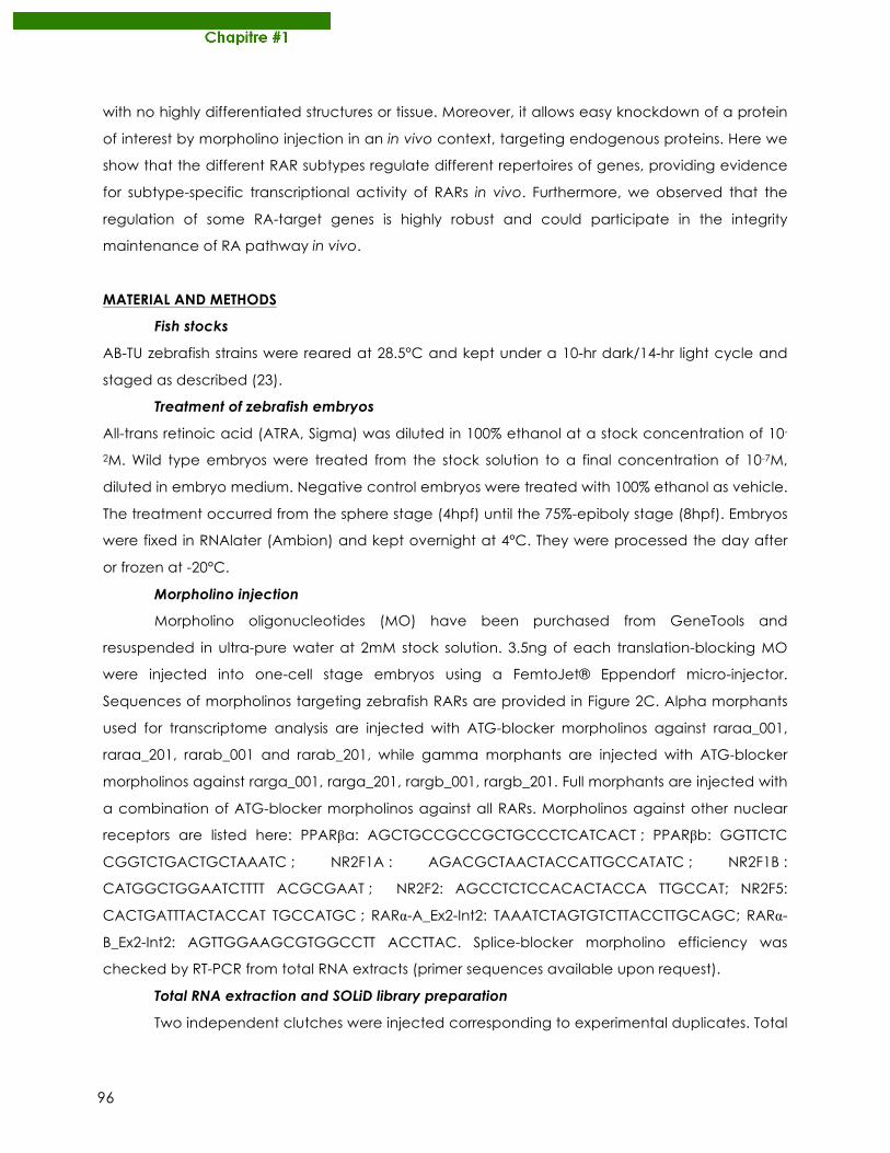
96
with no highly differentiated structures or tissue. Moreover, it allows easy knockdown of a protein
of interest by morpholino injection in an in vivo context, targeting endogenous proteins. Here we
show that the different RAR subtypes regulate different repertoires of genes, providing evidence
for subtype-specific transcriptional activity of RARs in vivo. Furthermore, we observed that the
regulation of some RA-target genes is highly robust and could participate in the integrity
maintenance of RA pathway in vivo.
MATERIAL AND METHODS
Fish stocks
AB-TU zebrafish strains were reared at 28.5°C and kept under a 10-hr dark/14-hr light cycle and
staged as described (23).
Treatment of zebrafish embryos
All-trans retinoic acid (ATRA, Sigma) was diluted in 100% ethanol at a stock concentration of 10-
2M. Wild type embryos were treated from the stock solution to a final concentration of 10-7M,
diluted in embryo medium. Negative control embryos were treated with 100% ethanol as vehicle.
The treatment occurred from the sphere stage (4hpf) until the 75%-epiboly stage (8hpf). Embryos
were fixed in RNAlater (Ambion) and kept overnight at 4°C. They were processed the day after
or frozen at -20°C.
Morpholino injection
Morpholino oligonucleotides (MO) have been purchased from GeneTools and
resuspended in ultra-pure water at 2mM stock solution. 3.5ng of each translation-blocking MO
were injected into one-cell stage embryos using a FemtoJet® Eppendorf micro-injector.
Sequences of morpholinos targeting zebrafish RARs are provided in Figure 2C. Alpha morphants
used for transcriptome analysis are injected with ATG-blocker morpholinos against raraa_001,
raraa_201, rarab_001 and rarab_201, while gamma morphants are injected with ATG-blocker
morpholinos against rarga_001, rarga_201, rargb_001, rargb_201. Full morphants are injected with
a combination of ATG-blocker morpholinos against all RARs. Morpholinos against other nuclear
receptors are listed here: PPARβa: AGCTGCCGCCGCTGCCCTCATCACT ; PPARβb: GGTTCTC
CGGTCTGACTGCTAAATC ; NR2F1A : AGACGCTAACTACCATTGCCATATC ; NR2F1B :
CATGGCTGGAATCTTTT ACGCGAAT ; NR2F2: AGCCTCTCCACACTACCA TTGCCAT; NR2F5:
CACTGATTTACTACCAT TGCCATGC ; RARα-A_Ex2-Int2: TAAATCTAGTGTCTTACCTTGCAGC; RARα-
B_Ex2-Int2: AGTTGGAAGCGTGGCCTT ACCTTAC. Splice-blocker morpholino efficiency was
checked by RT-PCR from total RNA extracts (primer sequences available upon request).
Total RNA extraction and SOLiD library preparation
Two independent clutches were injected corresponding to experimental duplicates. Total
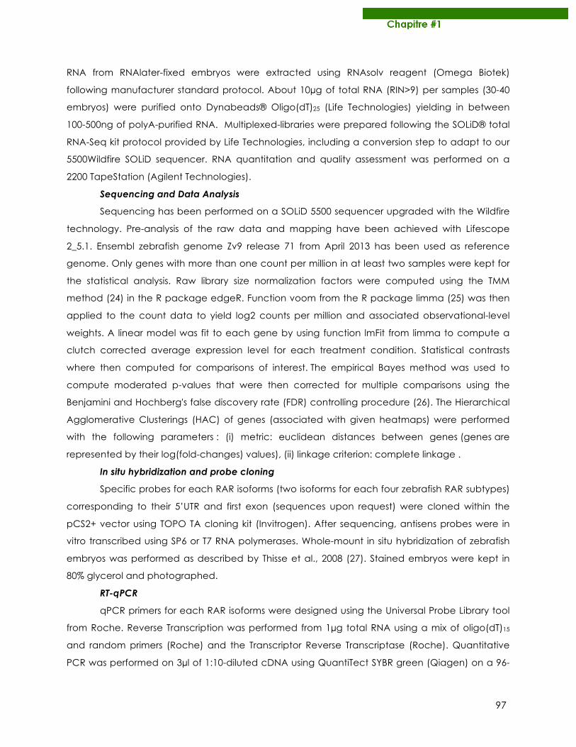
97
RNA from RNAlater-fixed embryos were extracted using RNAsolv reagent (Omega Biotek)
following manufacturer standard protocol. About 10µg of total RNA (RIN>9) per samples (30-40
embryos) were purified onto Dynabeads® Oligo(dT)25 (Life Technologies) yielding in between
100-500ng of polyA-purified RNA. Multiplexed-libraries were prepared following the SOLiD® total
RNA-Seq kit protocol provided by Life Technologies, including a conversion step to adapt to our
5500Wildfire SOLiD sequencer. RNA quantitation and quality assessment was performed on a
2200 TapeStation (Agilent Technologies).
Sequencing and Data Analysis
Sequencing has been performed on a SOLiD 5500 sequencer upgraded with the Wildfire
technology. Pre-analysis of the raw data and mapping have been achieved with Lifescope
2_5.1. Ensembl zebrafish genome Zv9 release 71 from April 2013 has been used as reference
genome. Only genes with more than one count per million in at least two samples were kept for
the statistical analysis. Raw library size normalization factors were computed using the TMM
method (24) in the R package edgeR. Function voom from the R package limma (25) was then
applied to the count data to yield log2 counts per million and associated observational-level
weights. A linear model was fit to each gene by using function lmFit from limma to compute a
clutch corrected average expression level for each treatment condition. Statistical contrasts
where then computed for comparisons of interest. The empirical Bayes method was used to
compute moderated p-values that were then corrected for multiple comparisons using the
Benjamini and Hochberg's false discovery rate (FDR) controlling procedure (26). The Hierarchical
Agglomerative Clusterings (HAC) of genes (associated with given heatmaps) were performed
with the following parameters : (i) metric: euclidean distances between genes (genes are
represented by their log(fold-changes) values), (ii) linkage criterion: complete linkage .
In situ hybridization and probe cloning
Specific probes for each RAR isoforms (two isoforms for each four zebrafish RAR subtypes)
corresponding to their 5’UTR and first exon (sequences upon request) were cloned within the
pCS2+ vector using TOPO TA cloning kit (Invitrogen). After sequencing, antisens probes were in
vitro transcribed using SP6 or T7 RNA polymerases. Whole-mount in situ hybridization of zebrafish
embryos was performed as described by Thisse et al., 2008 (27). Stained embryos were kept in
80% glycerol and photographed.
RT-qPCR
qPCR primers for each RAR isoforms were designed using the Universal Probe Library tool
from Roche. Reverse Transcription was performed from 1µg total RNA using a mix of oligo(dT)15
and random primers (Roche) and the Transcriptor Reverse Transcriptase (Roche). Quantitative
PCR was performed on 3µl of 1:10-diluted cDNA using QuantiTect SYBR green (Qiagen) on a 96-
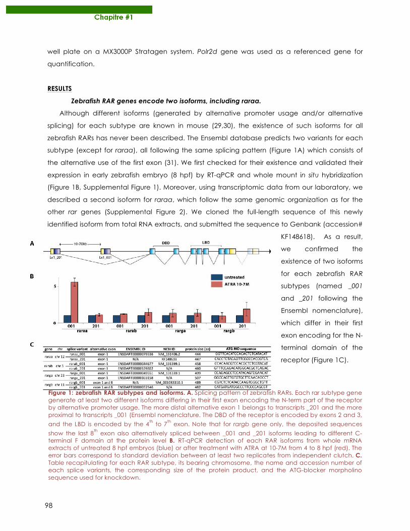
98
well plate on a MX3000P Stratagen system. Polr2d gene was used as a referenced gene for
quantification.
RESULTS
Zebrafish RAR genes encode two isoforms, including raraa.
Although different isoforms (generated by alternative promoter usage and/or alternative
splicing) for each subtype are known in mouse (29,30), the existence of such isoforms for all
zebrafish RARs has never been described. The Ensembl database predicts two variants for each
subtype (except for raraa), all following the same splicing pattern (Figure 1A) which consists of
the alternative use of the first exon (31). We first checked for their existence and validated their
expression in early zebrafish embryo (8 hpf) by RT-qPCR and whole mount in situ hybridization
(Figure 1B, Supplemental Figure 1). Moreover, using transcriptomic data from our laboratory, we
described a second isoform for raraa, which follow the same genomic organization as for the
other rar genes (Supplemental Figure 2). We cloned the full-length sequence of this newly
identified isoform from total RNA extracts, and submitted the sequence to Genbank (accession#
KF148618). As a result,
we confirmed the
existence of two isoforms
for each zebrafish RAR
subtypes (named _001
and _201 following the
Ensembl nomenclature),
which differ in their first
exon encoding for the N-
terminal domain of the receptor (Figure 1C).
Figure 1: zebrafish RAR subtypes and isoforms. A. Splicing pattern of zebrafish RARs. Each rar subtype gene generate at least two different isoforms differing in their first exon encoding the N-term part of the receptor by alternative promoter usage. The more distal alternative exon 1 belongs to transcripts _201 and the more proximal to transcripts _001 (Ensembl nomenclature. The DBD of the receptor is encoded by exons 2 and 3,
and the LBD is encoded by the 4th to 7th exon. Note that for rargb gene only, the deposited sequences
show the last 8th exon also alternatively spliced between _001 and _201 isoforms leading to different C-terminal F domain at the protein level B. RT-qPCR detection of each RAR isoforms from whole mRNA extracts of untreated 8 hpf embryos (blue) or after treatment with ATRA at 10-7M from 4 to 8 hpf (red). The error bars correspond to standard deviation between at least two replicates from independent clutch. C. Table recapitulating for each RAR subtype, its bearing chromosome, the name and accession number of each splice variants, the corresponding size of the protein product, and the ATG-blocker morpholino sequence used for knockdown.
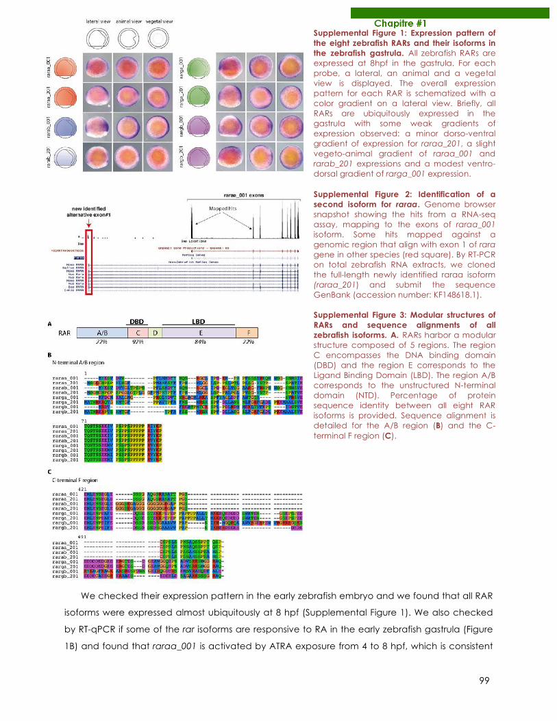
99
We checked their expression pattern in the early zebrafish embryo and we found that all RAR
isoforms were expressed almost ubiquitously at 8 hpf (Supplemental Figure 1). We also checked
by RT-qPCR if some of the rar isoforms are responsive to RA in the early zebrafish gastrula (Figure
1B) and found that raraa_001 is activated by ATRA exposure from 4 to 8 hpf, which is consistent
Supplemental Figure 1: Expression pattern of the eight zebrafish RARs and their isoforms in the zebrafish gastrula. All zebrafish RARs are expressed at 8hpf in the gastrula. For each probe, a lateral, an animal and a vegetal view is displayed. The overall expression pattern for each RAR is schematized with a color gradient on a lateral view. Briefly, all RARs are ubiquitously expressed in the gastrula with some weak gradients of expression observed: a minor dorso-ventral gradient of expression for raraa_201, a slight vegeto-animal gradient of raraa_001 and rarab_201 expressions and a modest ventro-dorsal gradient of rarga_001 expression. Supplemental Figure 2: Identification of a second isoform for raraa. Genome browser snapshot showing the hits from a RNA-seq assay, mapping to the exons of raraa_001 isoform. Some hits mapped against a genomic region that align with exon 1 of rara gene in other species (red square). By RT-PCR on total zebrafish RNA extracts, we cloned the full-length newly identified raraa isoform (raraa_201) and submit the sequence GenBank (accession number: KF148618.1). Supplemental Figure 3: Modular structures of RARs and sequence alignments of all zebrafish isoforms. A. RARs harbor a modular structure composed of 5 regions. The region C encompasses the DNA binding domain (DBD) and the region E corresponds to the Ligand Binding Domain (LBD). The region A/B corresponds to the unstructured N-terminal domain (NTD). Percentage of protein sequence identity between all eight RAR isoforms is provided. Sequence alignment is detailed for the A/B region (B) and the C-terminal F region (C).
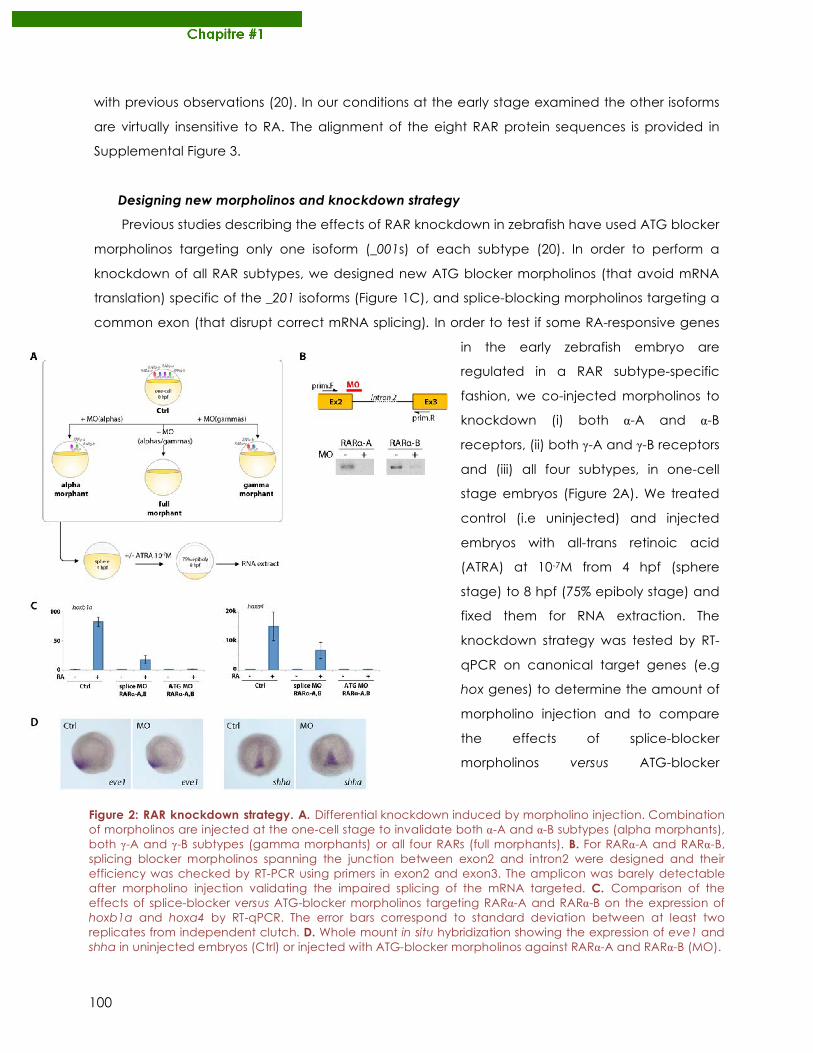
100
with previous observations (20). In our conditions at the early stage examined the other isoforms
are virtually insensitive to RA. The alignment of the eight RAR protein sequences is provided in
Supplemental Figure 3.
Designing new morpholinos and knockdown strategy
Previous studies describing the effects of RAR knockdown in zebrafish have used ATG blocker
morpholinos targeting only one isoform (_001s) of each subtype (20). In order to perform a
knockdown of all RAR subtypes, we designed new ATG blocker morpholinos (that avoid mRNA
translation) specific of the _201 isoforms (Figure 1C), and splice-blocking morpholinos targeting a
common exon (that disrupt correct mRNA splicing). In order to test if some RA-responsive genes
in the early zebrafish embryo are
regulated in a RAR subtype-specific
fashion, we co-injected morpholinos to
knockdown (i) both α-A and α-B
receptors, (ii) both γ-A and γ-B receptors
and (iii) all four subtypes, in one-cell
stage embryos (Figure 2A). We treated
control (i.e uninjected) and injected
embryos with all-trans retinoic acid
(ATRA) at 10-7M from 4 hpf (sphere
stage) to 8 hpf (75% epiboly stage) and
fixed them for RNA extraction. The
knockdown strategy was tested by RT-
qPCR on canonical target genes (e.g
hox genes) to determine the amount of
morpholino injection and to compare
the effects of splice-blocker
morpholinos versus ATG-blocker
Figure 2: RAR knockdown strategy. A. Differential knockdown induced by morpholino injection. Combination of morpholinos are injected at the one-cell stage to invalidate both α-A and α-B subtypes (alpha morphants), both γ-A and γ-B subtypes (gamma morphants) or all four RARs (full morphants). B. For RARα-A and RARα-B, splicing blocker morpholinos spanning the junction between exon2 and intron2 were designed and their efficiency was checked by RT-PCR using primers in exon2 and exon3. The amplicon was barely detectable after morpholino injection validating the impaired splicing of the mRNA targeted. C. Comparison of the effects of splice-blocker versus ATG-blocker morpholinos targeting RARα-A and RARα-B on the expression of hoxb1a and hoxa4 by RT-qPCR. The error bars correspond to standard deviation between at least two replicates from independent clutch. D. Whole mount in situ hybridization showing the expression of eve1 and shha in uninjected embryos (Ctrl) or injected with ATG-blocker morpholinos against RARα-A and RARα-B (MO).
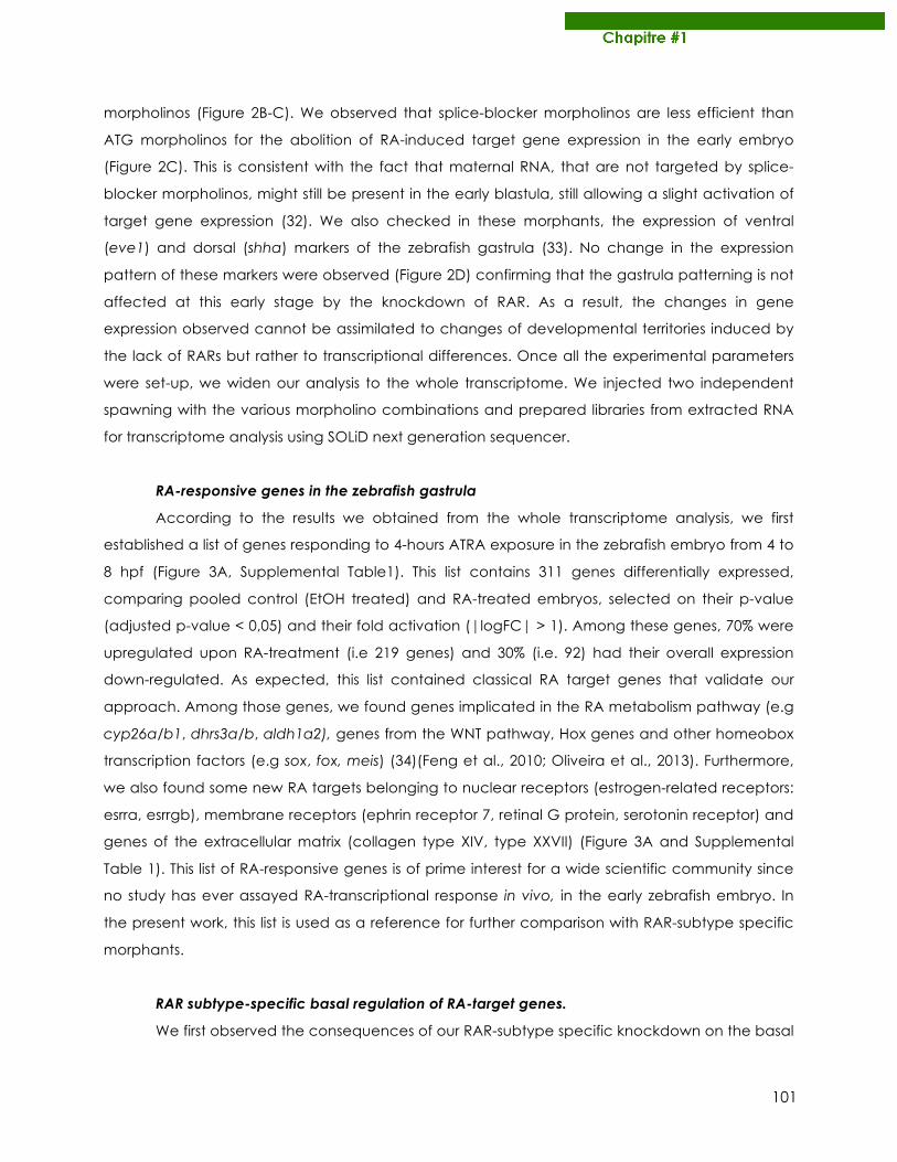
101
morpholinos (Figure 2B-C). We observed that splice-blocker morpholinos are less efficient than
ATG morpholinos for the abolition of RA-induced target gene expression in the early embryo
(Figure 2C). This is consistent with the fact that maternal RNA, that are not targeted by splice-
blocker morpholinos, might still be present in the early blastula, still allowing a slight activation of
target gene expression (32). We also checked in these morphants, the expression of ventral
(eve1) and dorsal (shha) markers of the zebrafish gastrula (33). No change in the expression
pattern of these markers were observed (Figure 2D) confirming that the gastrula patterning is not
affected at this early stage by the knockdown of RAR. As a result, the changes in gene
expression observed cannot be assimilated to changes of developmental territories induced by
the lack of RARs but rather to transcriptional differences. Once all the experimental parameters
were set-up, we widen our analysis to the whole transcriptome. We injected two independent
spawning with the various morpholino combinations and prepared libraries from extracted RNA
for transcriptome analysis using SOLiD next generation sequencer.
RA-responsive genes in the zebrafish gastrula
According to the results we obtained from the whole transcriptome analysis, we first
established a list of genes responding to 4-hours ATRA exposure in the zebrafish embryo from 4 to
8 hpf (Figure 3A, Supplemental Table1). This list contains 311 genes differentially expressed,
comparing pooled control (EtOH treated) and RA-treated embryos, selected on their p-value
(adjusted p-value < 0,05) and their fold activation (|logFC| > 1). Among these genes, 70% were
upregulated upon RA-treatment (i.e 219 genes) and 30% (i.e. 92) had their overall expression
down-regulated. As expected, this list contained classical RA target genes that validate our
approach. Among those genes, we found genes implicated in the RA metabolism pathway (e.g
cyp26a/b1, dhrs3a/b, aldh1a2), genes from the WNT pathway, Hox genes and other homeobox
transcription factors (e.g sox, fox, meis) (34)(Feng et al., 2010; Oliveira et al., 2013). Furthermore,
we also found some new RA targets belonging to nuclear receptors (estrogen-related receptors:
esrra, esrrgb), membrane receptors (ephrin receptor 7, retinal G protein, serotonin receptor) and
genes of the extracellular matrix (collagen type XIV, type XXVII) (Figure 3A and Supplemental
Table 1). This list of RA-responsive genes is of prime interest for a wide scientific community since
no study has ever assayed RA-transcriptional response in vivo, in the early zebrafish embryo. In
the present work, this list is used as a reference for further comparison with RAR-subtype specific
morphants.
RAR subtype-specific basal regulation of RA-target genes.
We first observed the consequences of our RAR-subtype specific knockdown on the basal
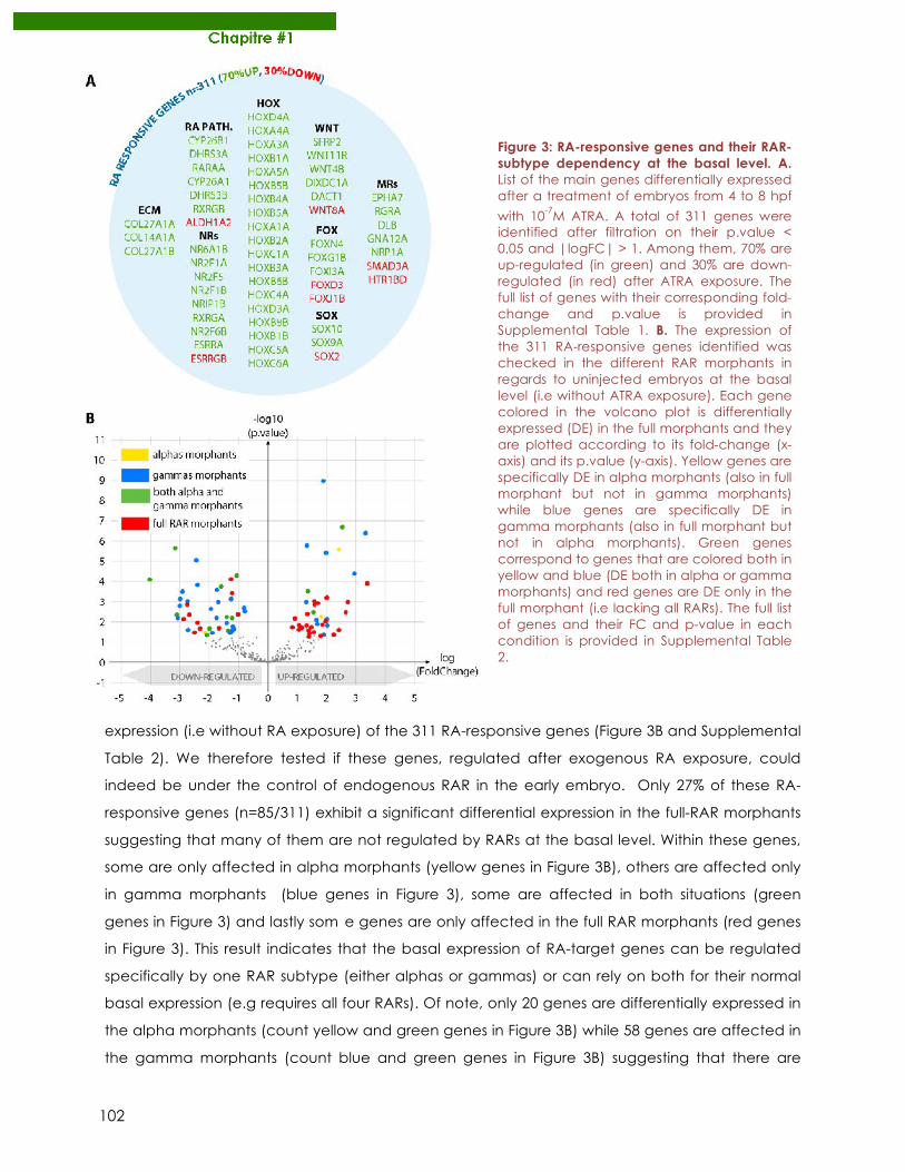
102
expression (i.e without RA exposure) of the 311 RA-responsive genes (Figure 3B and Supplemental
Table 2). We therefore tested if these genes, regulated after exogenous RA exposure, could
indeed be under the control of endogenous RAR in the early embryo. Only 27% of these RA-
responsive genes (n=85/311) exhibit a significant differential expression in the full-RAR morphants
suggesting that many of them are not regulated by RARs at the basal level. Within these genes,
some are only affected in alpha morphants (yellow genes in Figure 3B), others are affected only
in gamma morphants (blue genes in Figure 3), some are affected in both situations (green
genes in Figure 3) and lastly som e genes are only affected in the full RAR morphants (red genes
in Figure 3). This result indicates that the basal expression of RA-target genes can be regulated
specifically by one RAR subtype (either alphas or gammas) or can rely on both for their normal
basal expression (e.g requires all four RARs). Of note, only 20 genes are differentially expressed in
the alpha morphants (count yellow and green genes in Figure 3B) while 58 genes are affected in
the gamma morphants (count blue and green genes in Figure 3B) suggesting that there are
Figure 3: RA-responsive genes and their RAR-subtype dependency at the basal level. A. List of the main genes differentially expressed after a treatment of embryos from 4 to 8 hpf
with 10-7M ATRA. A total of 311 genes were identified after filtration on their p.value < 0,05 and |logFC| > 1. Among them, 70% are up-regulated (in green) and 30% are down-regulated (in red) after ATRA exposure. The full list of genes with their corresponding fold-change and p.value is provided in Supplemental Table 1. B. The expression of the 311 RA-responsive genes identified was checked in the different RAR morphants in regards to uninjected embryos at the basal level (i.e without ATRA exposure). Each gene colored in the volcano plot is differentially expressed (DE) in the full morphants and they are plotted according to its fold-change (x-axis) and its p.value (y-axis). Yellow genes are specifically DE in alpha morphants (also in full morphant but not in gamma morphants) while blue genes are specifically DE in gamma morphants (also in full morphant but not in alpha morphants). Green genes correspond to genes that are colored both in yellow and blue (DE both in alpha or gamma morphants) and red genes are DE only in the full morphant (i.e lacking all RARs). The full list of genes and their FC and p-value in each condition is provided in Supplemental Table 2.
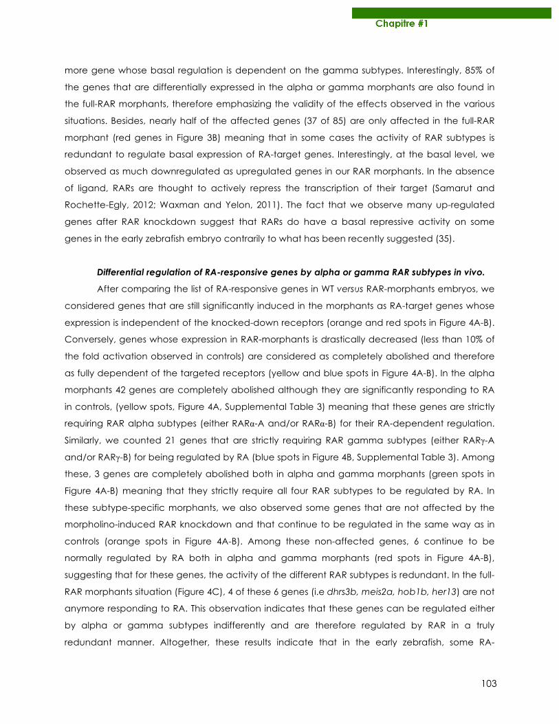
103
more gene whose basal regulation is dependent on the gamma subtypes. Interestingly, 85% of
the genes that are differentially expressed in the alpha or gamma morphants are also found in
the full-RAR morphants, therefore emphasizing the validity of the effects observed in the various
situations. Besides, nearly half of the affected genes (37 of 85) are only affected in the full-RAR
morphant (red genes in Figure 3B) meaning that in some cases the activity of RAR subtypes is
redundant to regulate basal expression of RA-target genes. Interestingly, at the basal level, we
observed as much downregulated as upregulated genes in our RAR morphants. In the absence
of ligand, RARs are thought to actively repress the transcription of their target (Samarut and
Rochette-Egly, 2012; Waxman and Yelon, 2011). The fact that we observe many up-regulated
genes after RAR knockdown suggest that RARs do have a basal repressive activity on some
genes in the early zebrafish embryo contrarily to what has been recently suggested (35).
Differential regulation of RA-responsive genes by alpha or gamma RAR subtypes in vivo.
After comparing the list of RA-responsive genes in WT versus RAR-morphants embryos, we
considered genes that are still significantly induced in the morphants as RA-target genes whose
expression is independent of the knocked-down receptors (orange and red spots in Figure 4A-B).
Conversely, genes whose expression in RAR-morphants is drastically decreased (less than 10% of
the fold activation observed in controls) are considered as completely abolished and therefore
as fully dependent of the targeted receptors (yellow and blue spots in Figure 4A-B). In the alpha
morphants 42 genes are completely abolished although they are significantly responding to RA
in controls, (yellow spots, Figure 4A, Supplemental Table 3) meaning that these genes are strictly
requiring RAR alpha subtypes (either RARα-A and/or RARα-B) for their RA-dependent regulation.
Similarly, we counted 21 genes that are strictly requiring RAR gamma subtypes (either RARγ-A
and/or RARγ-B) for being regulated by RA (blue spots in Figure 4B, Supplemental Table 3). Among
these, 3 genes are completely abolished both in alpha and gamma morphants (green spots in
Figure 4A-B) meaning that they strictly require all four RAR subtypes to be regulated by RA. In
these subtype-specific morphants, we also observed some genes that are not affected by the
morpholino-induced RAR knockdown and that continue to be regulated in the same way as in
controls (orange spots in Figure 4A-B). Among these non-affected genes, 6 continue to be
normally regulated by RA both in alpha and gamma morphants (red spots in Figure 4A-B),
suggesting that for these genes, the activity of the different RAR subtypes is redundant. In the full-
RAR morphants situation (Figure 4C), 4 of these 6 genes (i.e dhrs3b, meis2a, hob1b, her13) are not
anymore responding to RA. This observation indicates that these genes can be regulated either
by alpha or gamma subtypes indifferently and are therefore regulated by RAR in a truly
redundant manner. Altogether, these results indicate that in the early zebrafish, some RA-
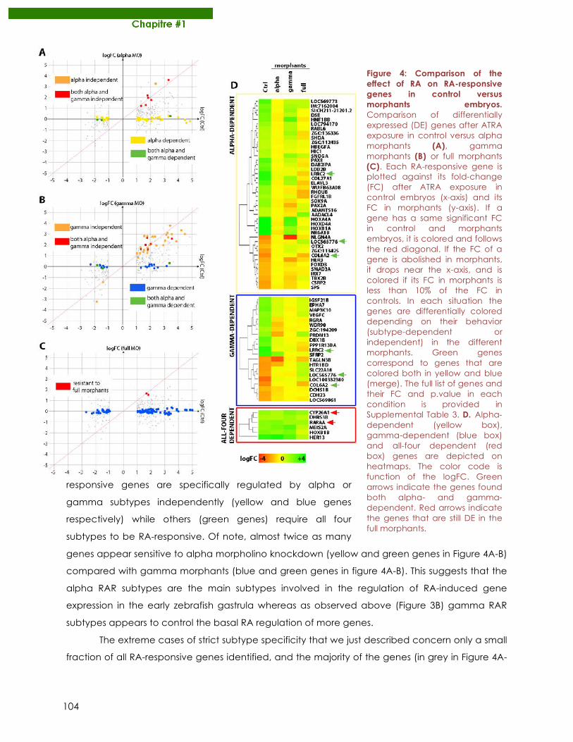
104
responsive genes are specifically regulated by alpha or
gamma subtypes independently (yellow and blue genes
respectively) while others (green genes) require all four
subtypes to be RA-responsive. Of note, almost twice as many
genes appear sensitive to alpha morpholino knockdown (yellow and green genes in Figure 4A-B)
compared with gamma morphants (blue and green genes in figure 4A-B). This suggests that the
alpha RAR subtypes are the main subtypes involved in the regulation of RA-induced gene
expression in the early zebrafish gastrula whereas as observed above (Figure 3B) gamma RAR
subtypes appears to control the basal RA regulation of more genes.
The extreme cases of strict subtype specificity that we just described concern only a small
fraction of all RA-responsive genes identified, and the majority of the genes (in grey in Figure 4A-
Figure 4: Comparison of the effect of RA on RA-responsive genes in control versus morphants embryos. Comparison of differentially expressed (DE) genes after ATRA exposure in control versus alpha morphants (A), gamma morphants (B) or full morphants (C). Each RA-responsive gene is plotted against its fold-change (FC) after ATRA exposure in control embryos (x-axis) and its FC in morphants (y-axis). If a gene has a same significant FC in control and morphants embryos, it is colored and follows the red diagonal. If the FC of a gene is abolished in morphants, it drops near the x-axis, and is colored if its FC in morphants is less than 10% of the FC in controls. In each situation the genes are differentially colored depending on their behavior (subtype-dependent or independent) in the different morphants. Green genes correspond to genes that are colored both in yellow and blue (merge). The full list of genes and their FC and p.value in each condition is provided in Supplemental Table 3. D. Alpha-dependent (yellow box), gamma-dependent (blue box) and all-four dependent (red box) genes are depicted on heatmaps. The color code is function of the logFC. Green arrows indicate the genes found both alpha- and gamma-dependent. Red arrows indicate the genes that are still DE in the full morphants.
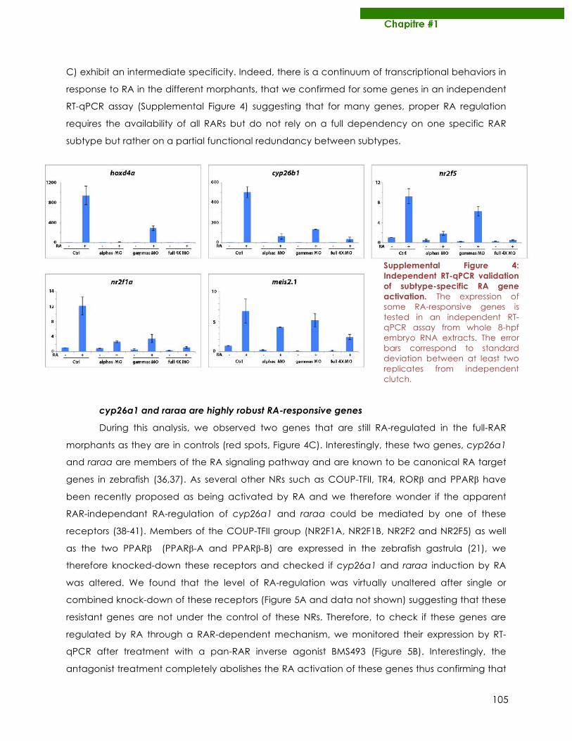
105
C) exhibit an intermediate specificity. Indeed, there is a continuum of transcriptional behaviors in
response to RA in the different morphants, that we confirmed for some genes in an independent
RT-qPCR assay (Supplemental Figure 4) suggesting that for many genes, proper RA regulation
requires the availability of all RARs but do not rely on a full dependency on one specific RAR
subtype but rather on a partial functional redundancy between subtypes.
cyp26a1 and raraa are highly robust RA-responsive genes During this analysis, we observed two genes that are still RA-regulated in the full-RAR
morphants as they are in controls (red spots, Figure 4C). Interestingly, these two genes, cyp26a1
and raraa are members of the RA signaling pathway and are known to be canonical RA target
genes in zebrafish (36,37). As several other NRs such as COUP-TFII, TR4, RORβ and PPARβ have
been recently proposed as being activated by RA and we therefore wonder if the apparent
RAR-independant RA-regulation of cyp26a1 and raraa could be mediated by one of these
receptors (38-41). Members of the COUP-TFII group (NR2F1A, NR2F1B, NR2F2 and NR2F5) as well
as the two PPARβ (PPARβ-A and PPARβ-B) are expressed in the zebrafish gastrula (21), we
therefore knocked-down these receptors and checked if cyp26a1 and raraa induction by RA
was altered. We found that the level of RA-regulation was virtually unaltered after single or
combined knock-down of these receptors (Figure 5A and data not shown) suggesting that these
resistant genes are not under the control of these NRs. Therefore, to check if these genes are
regulated by RA through a RAR-dependent mechanism, we monitored their expression by RT-
qPCR after treatment with a pan-RAR inverse agonist BMS493 (Figure 5B). Interestingly, the
antagonist treatment completely abolishes the RA activation of these genes thus confirming that
Supplemental Figure 4: Independent RT-qPCR validation of subtype-specific RA gene activation. The expression of some RA-responsive genes is tested in an independent RT-qPCR assay from whole 8-hpf embryo RNA extracts. The error bars correspond to standard deviation between at least two replicates from independent clutch.
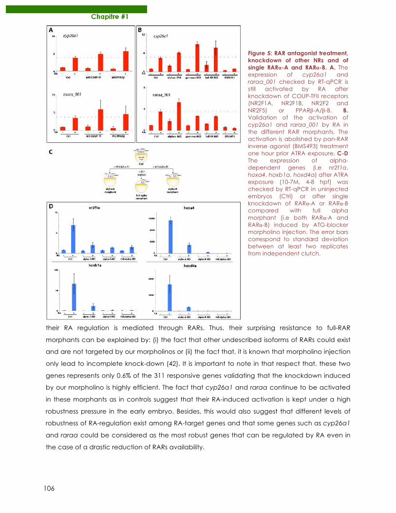
106
their RA regulation is mediated through RARs. Thus, their surprising resistance to full-RAR
morphants can be explained by: (i) the fact that other undescribed isoforms of RARs could exist
and are not targeted by our morpholinos or (ii) the fact that, it is known that morpholino injection
only lead to incomplete knock-down (42). It is important to note in that respect that, these two
genes represents only 0.6% of the 311 responsive genes validating that the knockdown induced
by our morpholino is highly efficient. The fact that cyp26a1 and raraa continue to be activated
in these morphants as in controls suggest that their RA-induced activation is kept under a high
robustness pressure in the early embryo. Besides, this would also suggest that different levels of
robustness of RA-regulation exist among RA-target genes and that some genes such as cyp26a1
and raraa could be considered as the most robust genes that can be regulated by RA even in
the case of a drastic reduction of RARs availability.
Figure 5: RAR antagonist treatment, knockdown of other NRs and of single RARα-A and RARα-B. A. The expression of cyp26a1 and raraa_001 checked by RT-qPCR is still activated by RA after knockdown of COUP-TFII receptors (NR2F1A, NR2F1B, NR2F2 and NR2F5) or PPARβ-A/β-B. B. Validation of the activation of cyp26a1 and raraa_001 by RA in the different RAR morphants. The activation is abolished by pan-RAR inverse agonist (BMS493) treatment one hour prior ATRA exposure. C-D The expression of alpha-dependent genes (i.e nr2f1a, hoxa4, hoxb1a, hoxd4a) after ATRA exposure (10-7M, 4-8 hpf) was checked by RT-qPCR in uninjected embryos (Ctrl) or after single knockdown of RARα-A or RARα-B compared with full alpha morphant (i.e both RARα-A and RARα-B) induced by ATG-blocker morpholino injection. The error bars correspond to standard deviation between at least two replicates from independent clutch.
107
The transcriptional activity of RARα-A and RARα-B is not redundant.
In order to decipher in more details the RAR-subtype-specific transcriptional activity, we
decided to knockdown the RAR alpha subtypes (i.e RARα-A and RARα-B) separately. Therefore,
we specifically knocked-down RARα-A or RARα-B by morpholino injection (Figure 5C) and we
checked by RT-qPCR the expression of genes that have been categorized as alpha-dependent
in our whole-transcriptome assay (Figure 4A). For the four genes tested, each single knockdown
of RARα-A or RARα-B strongly reduces their RA-induced expression. This suggests that both RARα-A
and RARα-B are required for proper RA regulation of these genes. Although the expression level in
the single alpha morphants is not as reduced as in the full-alpha morphants these results suggest
that, contrarily to what is commonly thought, there is not a full functional redundancy at the
transcriptional level between each RAR alpha subtypes, at least at this stage. Of note is that the
relative abundance of raraa and rarab transcripts in the early zebrafish embryo is the same
(Oliveira et al., 2013), therefore the changes observed are not due to differences of expression
level between both subtypes.
DISCUSSION
Many studies successfully deciphered the specific function of each RAR subtypes in mice
and zebrafish using standard forward genetics approaches looking at developmental
phenotypes that can be associated with the lack of one specific RAR subtype (19,20). However,
it is still not understood why one developmental process could require only one specific RAR
subtype although others are expressed in the same region of interest. This is expected to rely on
differences in the recruitment and/or transcriptional activity of the different RAR subtypes. In this
work, we provide evidences that the different RAR subtypes, even co-expressed in vivo, can
drive RA transcriptional response in a subtype-specific fashion. Indeed, we were able to
discriminate genes that (i) are specifically regulated by one or another RAR subtype (ii) strictly
require all subtypes for their correct RA-regulation and (iii) can be regulated either by one or
another subtype indifferently. Drawing the parallel with RAR-subtype specific phenotypes that
have been characterized in zebrafish (20,43), our results revealed what we call RAR-subtype
specific “transcriptotypes” that correspond to specific repertoire of genes activated in a
subtype-specific fashion in response to RA. Moreover, we observed that the gamma subtypes of
RARs appear to be more involved in the basal regulation of RA-responsive gene expression,
whereas alpha subtypes are most implicated in the transcriptional RA-response in the early
zebrafish embryo. These differences observed are unlikely to be due to different expression levels
since in a 12hpf-embryo, the relative mRNA level of alpha (i.e α-A and α-B) and gamma (i.e γ-A
ang γ-B) subtypes are equivalent (44).
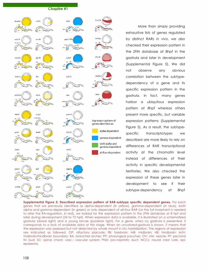
108
More than simply providing
exhaustive lists of genes regulated
by distinct RARs in vivo, we also
checked their expression pattern in
the ZFIN database at 8hpf in the
gastrula and later in development
(Supplemental Figure 5). We did
not observe any obvious
correlation between the subtype-
dependency of a gene and its
specific expression pattern in the
gastrula. In fact, many genes
harbor a ubiquitous expression
pattern at 8hpf whereas others
present more specific, but variable
expression patterns (Supplemental
Figure 5). As a result, the subtype-
specific transcriptotypes we
described are more likely to rely on
differences of RAR transcriptional
activity at the chromatin level
instead of differences of their
activity in specific developmental
territories. We also checked the
expression of these genes later in
development to see if their
subtype-dependency at 8hpf
Supplemental Figure 5: Described expression pattern of RAR-subtype specific dependent genes. For each genes that we previously identified as alpha-dependent (in yellow), gamma-dependent (in blue), both alpha and gamma-dependent (in green) or only dependent of all-four RAR (i.e the full morphant is needed to alter the RA-regulation, in red), we looked for the expression pattern in the ZFIN database at 8 hpf and later during development (24 to 72 hpf). When expression data is available, it is illustrated on a schematized gastrula (dorsal right) and a young larvae (posterior right). For a gene, when no gastrula is presented, it corresponds to a lack of available data at this stage. When an uncolored-gastrula is shown, it means that the expression was assessed but not detected by whole mount in situ hybridization. The regions of expression are indicated as followed. OP: olfactory placode; FB: forebrain; MB: midbrain; HB: hindbrain; M/H: midbrain/hindbrain boundary; BA: branchial arches; PP: pharyngeal pouches; OV: otic vesicle; PF; pectoral fin bud; SC: spinal chord; vasc.: vascular system; PND: pro-nephritic duct; NCCs: neural crest cells; epi: epidermis.
109
correlates with their expression pattern later in development. Interestingly, the alpha-dependent
genes at 8hpf are mainly expressed in the central nervous system (CNS) later in development,
whereas our gamma-dependent genes are predominantly expressed in the eye (retina or lens),
heart and somites of the zebrafish larvae (Supplemental Figure 5). Although all four RARs are
expressed in the CNS, only RARα-A and RARα-B are expressed in the eye (21). Altogether, these
observations suggest that the specific subtype-dependency of a gene is not necessarily the
same at 8hpf and later in development. We therefore propose this dependency does not rely on
fixed features but rather on dynamic mechanisms.
How such specificity can be driven at the transcriptional level? Since the DBD of the
different RAR subtypes are highly conserved (92%), especially the P box (100% identity) that
determines the specific interaction between RAR and DNA (Lee et al., 1993), we do not expect
differences at the receptor level that could explain why one specific RAR subtype is specifically
involved in the regulation of specific gene as we observed here. However, one can speculate
that the specificity relies on specific features of the RAREs that would promote the recruitment of
one specific RAR subtype. We exhaustively looked for canonical and non-classical RAREs within
zebrafish genome using the PARSEC (PAtteRn Search and Contextualization) platform that have
been recently developed to identify a specific sequence pattern within the whole genome (45).
We look for direct-repeats (DR) of the consensus RARE nucleotide sequence RGKTSA (46)
separated by 1 to 10 nucleotides (DR1-DR10) in the vicinity of the coding region of our RA-
responsive genes (+/- 10kb). Unfortunately, neither specific consensus sequence nor special DR
space enrichment was found to explain the specific recruitment of one specific RAR subtype.
However, recent studies have revealed that the repertoire of RAR binding sites in vivo is highly
diverse and that the classical RARE consensus does not recapitulate all true binding sites (47). As
a result, an in vivo analysis of RAR-subtype specific binding sites by chomatin immunoprecipation
(ChIP-seq) using antibodies recognizing specifically each subtypes would be necessary to have
a better view in what could drive this RAR-subtype specificity at the DNA level, such as the
presence of binding sites for other transcription factors in the vicinity of RAREs. Lastly, the
unstructured N-terminal domain (NTD) of RARs is a good candidate for being involved in this
specificity. In fact, thanks to its proximity with the DBD, proteins interacting with the NTD could
regulate RAR recruitment onto DNA as recently hypothesized and/or the recruitment of specific
coactivator complexes (48). Since NTDs are the most divergent parts of RARs between subtypes
(Supplemental Figure 3B), one can speculate that differences in the NTDs can participate in
driving RAR-subtype specific recruitment onto chromatin through specific protein interactions.
This would require further work aiming at identifying RAR-subtype specific NTD interacting proteins
in vivo.
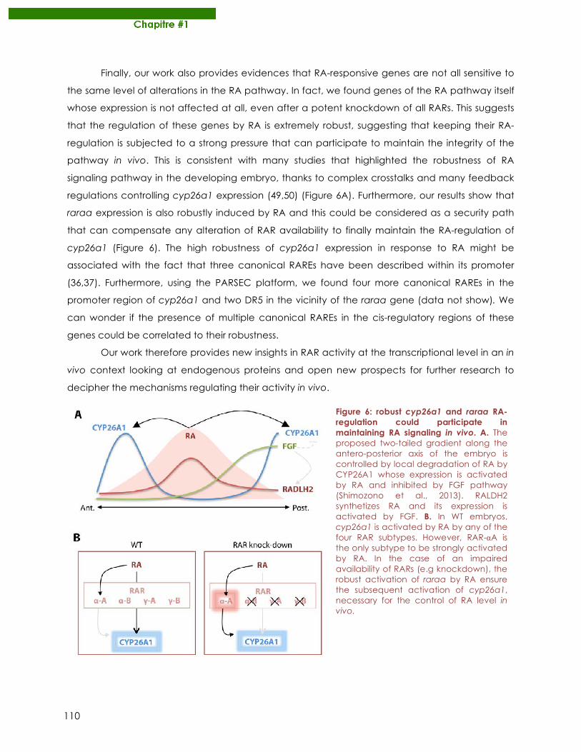
110
Finally, our work also provides evidences that RA-responsive genes are not all sensitive to
the same level of alterations in the RA pathway. In fact, we found genes of the RA pathway itself
whose expression is not affected at all, even after a potent knockdown of all RARs. This suggests
that the regulation of these genes by RA is extremely robust, suggesting that keeping their RA-
regulation is subjected to a strong pressure that can participate to maintain the integrity of the
pathway in vivo. This is consistent with many studies that highlighted the robustness of RA
signaling pathway in the developing embryo, thanks to complex crosstalks and many feedback
regulations controlling cyp26a1 expression (49,50) (Figure 6A). Furthermore, our results show that
raraa expression is also robustly induced by RA and this could be considered as a security path
that can compensate any alteration of RAR availability to finally maintain the RA-regulation of
cyp26a1 (Figure 6). The high robustness of cyp26a1 expression in response to RA might be
associated with the fact that three canonical RAREs have been described within its promoter
(36,37). Furthermore, using the PARSEC platform, we found four more canonical RAREs in the
promoter region of cyp26a1 and two DR5 in the vicinity of the raraa gene (data not show). We
can wonder if the presence of multiple canonical RAREs in the cis-regulatory regions of these
genes could be correlated to their robustness.
Our work therefore provides new insights in RAR activity at the transcriptional level in an in
vivo context looking at endogenous proteins and open new prospects for further research to
decipher the mechanisms regulating their activity in vivo.
Figure 6: robust cyp26a1 and raraa RA-regulation could participate in maintaining RA signaling in vivo. A. The proposed two-tailed gradient along the antero-posterior axis of the embryo is controlled by local degradation of RA by CYP26A1 whose expression is activated by RA and inhibited by FGF pathway (Shimozono et al., 2013). RALDH2 synthetizes RA and its expression is activated by FGF. B. In WT embryos, cyp26a1 is activated by RA by any of the four RAR subtypes. However, RAR-αA is the only subtype to be strongly activated by RA. In the case of an impaired availability of RARs (e.g knockdown), the robust activation of raraa by RA ensure the subsequent activation of cyp26a1, necessary for the control of RA level in vivo.
111
ACKNOWLEDGMENTS
We warmly thank Laure Bernard for technical help and comprehensive support. We
benefited of the zebrafish in-breeding facilities developed by the PRECI technical platform at the
SFR Biosciences Gerland. This work was supported by La ligue contre le cancer and the French
ministry of research, the CNRS and the Ecole Normale Supérieure de Lyon. Of note it was not
funded by ANR who rejected the project.
REFERENCES
1. Samarut E, Rochette-Egly C. Nuclear retinoic acid receptors: conductors of the retinoic acid symphony during development. Mol Cell Endocrinol. 2012;348(2):348-360.
2. Clagett-Dame M, DeLuca HF. The role of vitamin A in mammalian reproduction and embryonic development. Annu Rev Nutr. 2002;22:347-381.
3. Zile MH. Function of vitamin A in vertebrate embryonic development. J Nutr. 2001;131(3):705-708. 4. Begemann G, Marx M, Mebus K, Meyer A, Bastmeyer M. Beyond the neckless phenotype: influence
of reduced retinoic acid signaling on motor neuron development in the zebrafish hindbrain. Dev Biol. 2004;271(1):119-129.
5. Maves L, Kimmel CB. Dynamic and sequential patterning of the zebrafish posterior hindbrain by retinoic acid. Dev Biol. 2005;285(2):593-605.
6. Aulehla A, Pourquie O. Signaling gradients during paraxial mesoderm development. Cold Spring Harb Perspect Biol. 2010;2(2):a000869.
7. Chambon P. A decade of molecular biology ot retinoic acid receptors. FASEBJ. 1996;10:940-954. 8. Germain P, Altucci L, Bourguet W, Rochette-Egly C, Gronemeyer H. Nuclear receptor superfamily:
Principles of signaling. In: Miyamoto J, Burger J, eds. Pure Appl. Chem.,. Vol 752003:1619-1664. 9. Germain P, Staels B, Dacquet C, Spedding M, Laudet V. Overview of nomenclature of nuclear
receptors. Pharmacol Rev. 2006;58(4):685-704. 10. Gronemeyer H, Gustafsson JA, Laudet V. Principles for modulation of the nuclear receptor
superfamily. Nat Rev Drug Discov. 2004;3(11):950-964. 11. Rochette-Egly C, Germain P. Dynamic and combinatorial control of gene expression by nuclear
retinoic acid receptors (RARs). Nucl Recept Signal. 2009;7:e005. 12. Chakravarti D. Volume 87: Regulatory Mechanisms in Transcriptional Signaling, 1st Edition. Elsevier
ed2009. 13. Moras D, Gronemeyer H. The nuclear receptor ligand-binding domain: structure and function. Curr
Opin Cell Biol. 1998;10(3):384-391. 14. Dilworth FJ, Chambon P. Nuclear receptors coordinate the activities of chromatin remodeling
complexes and coactivators to facilitate initiation of transcription. Oncogene. 2001;20(24):3047-3054.
15. Germain P, Chambon P, Eichele G, Evans RM, Lazar MA, Leid M, De Lera AR, Lotan R, Mangelsdorf DJ, Gronemeyer H. International Union of Pharmacology. LX. Retinoic acid receptors. Pharmacol Rev. 2006;58(4):712-725.
16. Dolle P. Developmental expression of retinoic acid receptors (RARs). Nucl Recept Signal. 2009;7:e006.
17. Niederreither K, Dolle P. Retinoic acid in development: towards an integrated view. Nat Rev Genet. 2008;9(7):541-553.
18. Mark M, Ghyselinck NB, Chambon P. Function of retinoid nuclear receptors: lessons from genetic and pharmacological dissections of the retinoic acid signaling pathway during mouse embryogenesis. Annu Rev Pharmacol Toxicol. 2006;46:451-480.
19. Mark M, Ghyselinck NB, Chambon P. Function of retinoic acid receptors during embryonic development. Nucl Recept Signal. 2009;7:e002.
20. Linville A, Radtke K, Waxman JS, Yelon D, Schilling TF. Combinatorial roles for zebrafish retinoic acid receptors in the hindbrain, limbs and pharyngeal arches. Dev Biol. 2009;325(1):60-70.
21. Bertrand S, Thisse B, Tavares R, Sachs L, Chaumot A, Bardet PL, Escriva H, Duffraisse M, Marchand O, Safi R, Thisse C, Laudet V. Unexpected novel relational links uncovered by extensive developmental profiling of nuclear receptor expression. PLoS Genet. 2007;3(11):e188.
112
22. Hale LA, Tallafuss A, Yan YL, Dudley L, Eisen JS, Postlethwait JH. Characterization of the retinoic acid receptor genes raraa, rarab and rarg during zebrafish development. Gene Expr Patterns. 2006;6(5):546-555.
23. Kimmel CB, Ballard WW, Kimmel SR, Ullmann B, Schilling TF. Stages of embryonic development of the zebrafish. Dev Dyn. 1995;203(3):253-310.
24. Robinson MD, Oshlack A. A scaling normalization method for differential expression analysis of RNA-seq data. Genome Biol. 2010;11(3):R25.
25. Smyth GK. Linear models and empirical bayes methods for assessing differential expression in microarray experiments. Stat Appl Genet Mol Biol. 2004;3:Article3.
26. Benjamini Y, Hochberg Y. Controlling the False Discovery Rate: A Practical and Powerful Approach to Multiple Testing. Journal of the Royal Statistical Society Series B (Methodological). 1995;57(1):289-300.
27. Thisse C, Thisse B. High-resolution in situ hybridization to whole-mount zebrafish embryos. Nature Protocols. 2008;3(1):59-69.
28. Waxman JS, Yelon D. Comparison of the expression patterns of newly identified zebrafish retinoic acid and retinoid X receptors. Dev Dyn. 2007;236(2):587-595.
29. Zelent A, Mendelsohn C, Kastner P, Krust A, Garnier JM, Ruffenach F, Leroy P, Chambon P. Differentially expressed isoforms of the mouse retinoic acid receptor beta generated by usage of two promoters and alternative splicing. EMBO J. 1991;10(1):71-81.
30. Mollard R, Viville S, Ward SJ, Decimo D, Chambon P, Dolle P. Tissue-specific expression of retinoic acid receptor isoform transcripts in the mouse embryo. Mech Dev. 2000;94(1-2):223-232.
31. Birney E, Andrews TD, Bevan P, Caccamo M, Chen Y, Clarke L, Coates G, Cuff J, Curwen V, Cutts T, Down T, Eyras E, Fernandez-Suarez XM, Gane P, Gibbins B, Gilbert J, Hammond M, Hotz HR, Iyer V, Jekosch K, Kahari A, Kasprzyk A, Keefe D, Keenan S, Lehvaslaiho H, McVicker G, Melsopp C, Meidl P, Mongin E, Pettett R, Potter S, Proctor G, Rae M, Searle S, Slater G, Smedley D, Smith J, Spooner W, Stabenau A, Stalker J, Storey R, Ureta-Vidal A, Woodwark KC, Cameron G, Durbin R, Cox A, Hubbard T, Clamp M. An overview of Ensembl. Genome Res. 2004;14(5):925-928.
32. Tadros W, Lipshitz HD. The maternal-to-zygotic transition: a play in two acts. Development. 2009;136(18):3033-3042.
33. Hammerschmidt M, Pelegri F, Mullins MC, Kane DA, van Eeden FJ, Granato M, Brand M, Furutani-Seiki M, Haffter P, Heisenberg CP, Jiang YJ, Kelsh RN, Odenthal J, Warga RM, Nusslein-Volhard C. dino and mercedes, two genes regulating dorsal development in the zebrafish embryo. Development. 1996;123:95-102.
34. Balmer JE, Blomhoff R. Gene expression regulation by retinoic acid. J Lipid Res. 2002;43(11):1773-1808.
35. Waxman JS, Yelon D. Zebrafish retinoic acid receptors function as context-dependent transcriptional activators. Developmental Biology. 2011;352(1):128-140.
36. Hu P, Tian M, Bao J, Xing G, Gu X, Gao X, Linney E, Zhao Q. Retinoid regulation of the zebrafish cyp26a1 promoter. Developmental Dynamics. 2008;237(12):3798-3808.
37. Li J, Hu P, Li K, Zhao Q. Identification and characterization of a novel retinoic acid response element in zebrafish cyp26a1 promoter. Anat Rec (Hoboken). 2012;295(2):268-277.
38. Zhou XE, Suino-Powell KM, Xu Y, Chan CW, Tanabe O, Kruse SW, Reynolds R, Engel JD, Xu HE. The Orphan Nuclear Receptor TR4 Is a Vitamin A-activated Nuclear Receptor. Journal of Biological Chemistry. 2011;286(4):2877-2885.
39. Stehlin-Gaon C, Willmann D, Zeyer D, Sanglier S, Van Dorsselaer A, Renaud JP, Moras D, Schule R. All-trans retinoic acid is a ligand for the orphan nuclear receptor ROR beta. Nature Structural Biology. 2003;10(10):820-825.
40. Kruse SW, Suino-Powell K, Zhou XE, Kretschman JE, Reynolds R, Vonrhein C, Xu Y, Wang L, Tsai SY, Tsai MJ, Xu HE. Identification of COUP-TFII orphan nuclear receptor as a retinoic acid-activated receptor. PLoS Biol. 2008;6(9):e227.
41. Shaw N, Elholm M, Noy N. Retinoic acid is a high affinity selective ligand for the peroxisome proliferator-activated receptor beta/delta. Journal of Biological Chemistry. 2003;278(43):41589-41592.
42. Bill BR, Petzold AM, Clark KJ, Schimmenti LA, Ekker SC. A Primer for Morpholino Use in Zebrafish. Zebrafish. 2009;6(1).
43. D'Aniello E, Rydeen AB, Anderson JL, Mandal A, Waxman JS. Depletion of Retinoic Acid Receptors Initiates a Novel Positive Feedback Mechanism that Promotes Teratogenic Increases in Retinoic Acid. Plos Genetics. 2013;9(8).
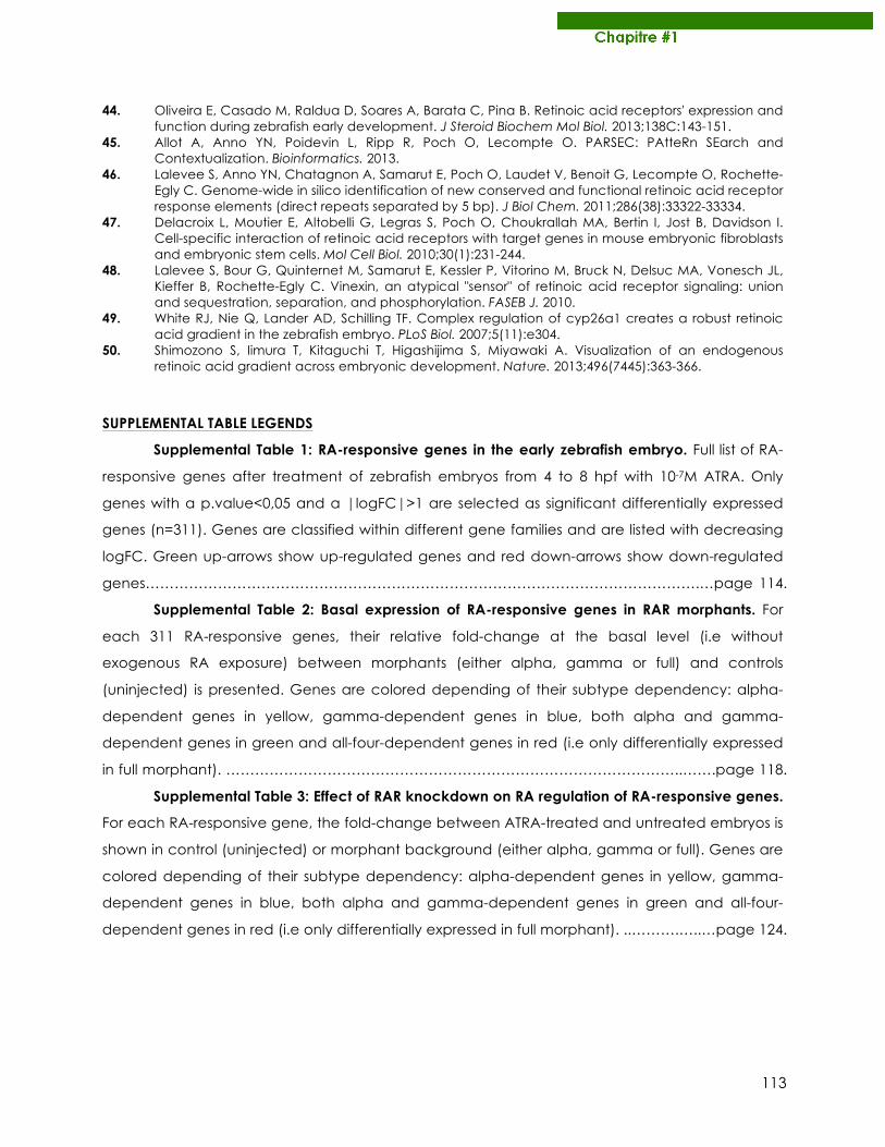
113
44. Oliveira E, Casado M, Raldua D, Soares A, Barata C, Pina B. Retinoic acid receptors' expression and function during zebrafish early development. J Steroid Biochem Mol Biol. 2013;138C:143-151.
45. Allot A, Anno YN, Poidevin L, Ripp R, Poch O, Lecompte O. PARSEC: PAtteRn SEarch and Contextualization. Bioinformatics. 2013.
46. Lalevee S, Anno YN, Chatagnon A, Samarut E, Poch O, Laudet V, Benoit G, Lecompte O, Rochette-Egly C. Genome-wide in silico identification of new conserved and functional retinoic acid receptor response elements (direct repeats separated by 5 bp). J Biol Chem. 2011;286(38):33322-33334.
47. Delacroix L, Moutier E, Altobelli G, Legras S, Poch O, Choukrallah MA, Bertin I, Jost B, Davidson I. Cell-specific interaction of retinoic acid receptors with target genes in mouse embryonic fibroblasts and embryonic stem cells. Mol Cell Biol. 2010;30(1):231-244.
48. Lalevee S, Bour G, Quinternet M, Samarut E, Kessler P, Vitorino M, Bruck N, Delsuc MA, Vonesch JL, Kieffer B, Rochette-Egly C. Vinexin, an atypical "sensor" of retinoic acid receptor signaling: union and sequestration, separation, and phosphorylation. FASEB J. 2010.
49. White RJ, Nie Q, Lander AD, Schilling TF. Complex regulation of cyp26a1 creates a robust retinoic acid gradient in the zebrafish embryo. PLoS Biol. 2007;5(11):e304.
50. Shimozono S, Iimura T, Kitaguchi T, Higashijima S, Miyawaki A. Visualization of an endogenous retinoic acid gradient across embryonic development. Nature. 2013;496(7445):363-366.
SUPPLEMENTAL TABLE LEGENDS
Supplemental Table 1: RA-responsive genes in the early zebrafish embryo. Full list of RA-
responsive genes after treatment of zebrafish embryos from 4 to 8 hpf with 10-7M ATRA. Only
genes with a p.value<0,05 and a |logFC|>1 are selected as significant differentially expressed
genes (n=311). Genes are classified within different gene families and are listed with decreasing
logFC. Green up-arrows show up-regulated genes and red down-arrows show down-regulated
genes…………………………………………………………………………………………………….…page 114.
Supplemental Table 2: Basal expression of RA-responsive genes in RAR morphants. For
each 311 RA-responsive genes, their relative fold-change at the basal level (i.e without
exogenous RA exposure) between morphants (either alpha, gamma or full) and controls
(uninjected) is presented. Genes are colored depending of their subtype dependency: alpha-
dependent genes in yellow, gamma-dependent genes in blue, both alpha and gamma-
dependent genes in green and all-four-dependent genes in red (i.e only differentially expressed
in full morphant). …………………………………………………………………………………..…….page 118.
Supplemental Table 3: Effect of RAR knockdown on RA regulation of RA-responsive genes.
For each RA-responsive gene, the fold-change between ATRA-treated and untreated embryos is
shown in control (uninjected) or morphant background (either alpha, gamma or full). Genes are
colored depending of their subtype dependency: alpha-dependent genes in yellow, gamma-
dependent genes in blue, both alpha and gamma-dependent genes in green and all-four-
dependent genes in red (i.e only differentially expressed in full morphant). ..……….…..…page 124.
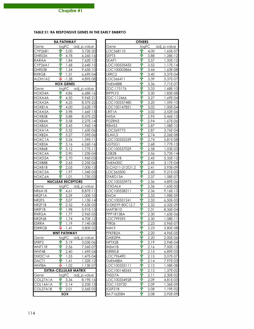
114
TABLE S1: RA RESPONSIVE GENES IN THE EARLY EMBRYO
Gene logFC adj_p.value Gene logFC adj_p.valueCYP26B1 5,00 3,72E-20 LOC568110 4,00 1,65E-07DHRS3A 4,78 6,26E-16 SEPT3 3,88 1,28E-12RARAA 1,84 7,62E-12 SKAP1 3,57 1,35E-12CYP26A1 1,68 2,64E-15 LOC100535433 3,52 1,17E-14DHRS3B 1,34 9,65E-10 LOC100003866 3,44 1,63E-08RXRGB 1,31 6,69E-04 LRRC2 3,40 3,37E-04ALDH1A2 -1,38 4,89E-08 LOC566411 3,39 5,37E-07
TMEM88B 3,36 7,71E-07Gene logFC adj_p.value ZGC:175176 3,33 1,68E-10HOXD4A 4,86 6,68E-16 RIPPLY3 3,30 1,83E-08HOXA4A 4,50 9,96E-21 ZGC:112466 3,27 1,69E-04HOXA3A 4,25 8,37E-22 LOC100537480 3,20 1,59E-10HOXB1A 4,00 3,62E-19 LOC100147851 3,02 1,00E-04HOXA5A 3,98 1,66E-15 LRIT1A 3,02 2,52E-06HOXB5B 3,88 8,37E-22 NHSA 2,95 5,46E-13HOXB4A 3,58 2,27E-14 PDZRN3 2,94 1,67E-06HOXB5A 3,45 1,96E-14 RBMS3 2,87 1,08E-10HOXA1A 3,32 1,63E-06 LOC569773 2,87 3,76E-04HOXB2A 3,27 7,59E-06 ELAVL3 2,74 2,26E-08HOXC1A 3,26 1,59E-11 LOC100332539 2,74 5,81E-08HOXB3A 3,16 6,26E-16 UGT5G1 2,68 1,77E-13HOXB6B 3,12 1,77E-11 LOC100537029 2,58 1,03E-03HOXC4A 2,90 1,23E-04 LDB2B 2,56 2,72E-14HOXD3A 2,70 1,96E-03 HAPLN1B 2,45 5,35E-12HOXB8B 2,65 2,25E-06 TMEM30C 2,45 3,17E-04HOXB1B 2,05 7,00E-13 SI:CH211-212O1.2 2,41 3,93E-09HOXC5A 1,97 1,34E-05 LOC563500 2,40 9,21E-03HOXC6A 1,01 1,75E-03 STARD13A 2,37 1,58E-07
LOC100333973 2,36 4,89E-06Gene logFC adj_p.value ST3GAL4 2,36 1,65E-03NR6A1B 4,61 8,87E-11 LOC100538211 2,36 9,16E-13NR2F1A 3,29 1,00E-10 ENO4 2,33 1,98E-09NR2F5 3,07 1,13E-14 LOC100331241 2,33 6,50E-03NR2F1B 2,52 9,63E-05 SI:DKEYP-80C12.7 2,32 6,32E-09NRIP1B 1,98 5,91E-15 MAP3K10 2,31 8,36E-04RXRGA 1,77 2,96E-05 PPP1R13BA 2,30 1,63E-06NR2F6B 1,74 4,70E-12 LOC799595 2,30 1,08E-11ESRRA 1,19 7,14E-08 FTR06 2,23 5,96E-07ESRRGB -1,41 3,80E-02 NAV3 2,23 3,80E-08
PFKFB2A 2,20 4,96E-05Gene logFC adj_p.value DAB2IPA 2,20 2,00E-06SFRP2 3,19 3,03E-06 NPTX2B 2,19 2,94E-04WNT11R 2,56 7,56E-07 INSM1B 2,16 7,00E-13WNT4B 2,40 1,69E-04 KIRRELB 2,15 6,89E-03DIXDC1A 1,55 1,47E-04 LOC796490 2,15 3,07E-07DACT1 1,41 1,32E-12 TMEM88A 2,14 7,97E-03WNT8A -1,02 1,13E-07 LOC100333111 2,12 1,58E-08
LOC100148343 2,12 1,37E-02Gene logFC adj_p.value THSD7A 2,11 2,30E-02COL27A1A 3,36 8,19E-16 LOC100334928 2,09 4,81E-03COL14A1A 2,14 2,23E-13 ZGC:153720 2,09 1,36E-09COL27A1B 2,01 2,56E-02 IGSF21B 2,08 1,19E-02
IM:7162084 2,08 2,92E-09SOX
RA PATHWAY OTHERS
HOX GENES
NUCLEAR RECEPTORS
WNT PATHWAY
EXTRA-CELLULAR MATRIX
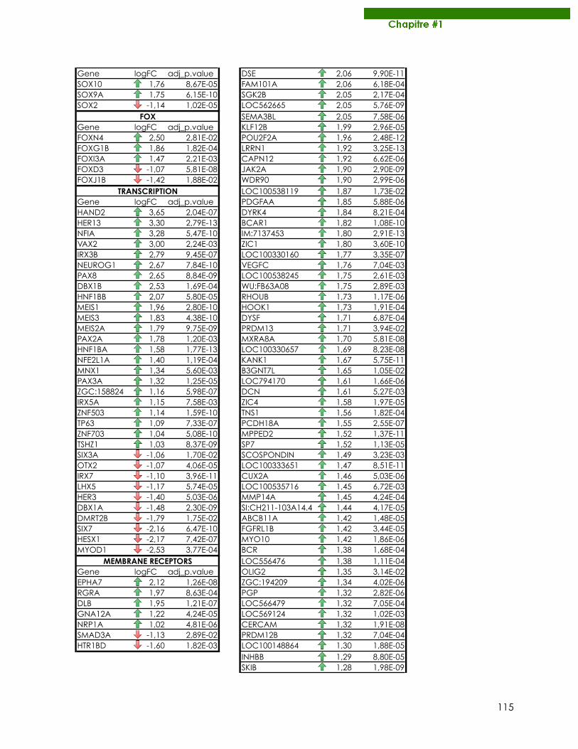
115
Gene logFC adj_p.value DSE 2,06 9,90E-11SOX10 1,76 8,67E-05 FAM101A 2,06 6,18E-04SOX9A 1,75 6,15E-10 SGK2B 2,05 2,17E-04SOX2 -1,14 1,02E-05 LOC562665 2,05 5,76E-09
SEMA3BL 2,05 7,58E-06Gene logFC adj_p.value KLF12B 1,99 2,96E-05FOXN4 2,50 2,81E-02 POU2F2A 1,96 2,48E-12FOXG1B 1,86 1,82E-04 LRRN1 1,92 3,25E-13FOXI3A 1,47 2,21E-03 CAPN12 1,92 6,62E-06FOXD3 -1,07 5,81E-08 JAK2A 1,90 2,90E-09FOXJ1B -1,42 1,88E-02 WDR90 1,90 2,99E-06
LOC100538119 1,87 1,73E-02Gene logFC adj_p.value PDGFAA 1,85 5,88E-06HAND2 3,65 2,04E-07 DYRK4 1,84 8,21E-04HER13 3,30 2,79E-13 BCAR1 1,82 1,08E-10NFIA 3,28 5,47E-10 IM:7137453 1,80 2,91E-13VAX2 3,00 2,24E-03 ZIC1 1,80 3,60E-10IRX3B 2,79 9,45E-07 LOC100330160 1,77 3,35E-07NEUROG1 2,67 7,84E-10 VEGFC 1,76 7,04E-03PAX8 2,65 8,84E-09 LOC100538245 1,75 2,61E-03DBX1B 2,53 1,69E-04 WU:FB63A08 1,75 2,89E-03HNF1BB 2,07 5,80E-05 RHOUB 1,73 1,17E-06MEIS1 1,96 2,80E-10 HOOK1 1,73 1,91E-04MEIS3 1,83 4,38E-10 DYSF 1,71 6,87E-04MEIS2A 1,79 9,75E-09 PRDM13 1,71 3,94E-02PAX2A 1,78 1,20E-03 MXRA8A 1,70 5,81E-08HNF1BA 1,58 1,77E-13 LOC100330657 1,69 8,23E-08NFE2L1A 1,40 1,19E-04 KANK1 1,67 5,75E-11MNX1 1,34 5,60E-03 B3GNT7L 1,65 1,05E-02PAX3A 1,32 1,25E-05 LOC794170 1,61 1,66E-06ZGC:158824 1,16 5,98E-07 DCN 1,61 5,27E-03IRX5A 1,15 7,58E-03 ZIC4 1,58 1,97E-05ZNF503 1,14 1,59E-10 TNS1 1,56 1,82E-04TP63 1,09 7,33E-07 PCDH18A 1,55 2,55E-07ZNF703 1,04 5,08E-10 MPPED2 1,52 1,37E-11TSHZ1 1,03 8,37E-09 SP7 1,52 1,13E-05SIX3A -1,06 1,70E-02 SCOSPONDIN 1,49 3,23E-03OTX2 -1,07 4,06E-05 LOC100333651 1,47 8,51E-11IRX7 -1,10 3,96E-11 CUX2A 1,46 5,03E-06LHX5 -1,17 5,74E-05 LOC100535716 1,45 6,72E-03HER3 -1,40 5,03E-06 MMP14A 1,45 4,24E-04DBX1A -1,48 2,30E-09 SI:CH211-103A14.4 1,44 4,17E-05DMRT2B -1,79 1,75E-02 ABCB11A 1,42 1,48E-05SIX7 -2,16 6,47E-10 FGFRL1B 1,42 3,44E-05HESX1 -2,17 7,42E-07 MYO10 1,42 1,86E-06MYOD1 -2,53 3,77E-04 BCR 1,38 1,68E-04
LOC556476 1,38 1,11E-04Gene logFC adj_p.value OLIG2 1,35 3,14E-02EPHA7 2,12 1,26E-08 ZGC:194209 1,34 4,02E-06RGRA 1,97 8,63E-04 PGP 1,32 2,82E-06DLB 1,95 1,21E-07 LOC566479 1,32 7,05E-04GNA12A 1,22 4,24E-05 LOC569124 1,32 1,02E-03NRP1A 1,02 4,81E-06 CERCAM 1,32 1,91E-08SMAD3A -1,13 2,89E-02 PRDM12B 1,32 7,04E-04HTR1BD -1,60 1,82E-03 LOC100148864 1,30 1,88E-05
INHBB 1,29 8,80E-05SKIB 1,28 1,98E-09
FOX
TRANSCRIPTION
MEMBRANE RECEPTORS
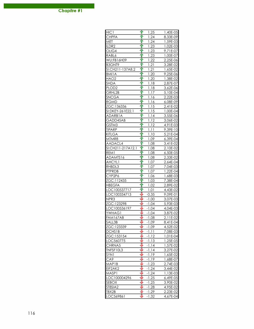
116
HIC1 1,25 1,40E-05CHPFA 1,24 8,33E-09MET 1,24 1,59E-03ILDR2 1,23 1,02E-03OLIG4 1,23 9,71E-07RABL6 1,23 1,00E-07WU:FB16H09 1,22 2,25E-06B3GNT9 1,21 3,28E-02SI:CH211-137A8.2 1,21 1,65E-02BMI1A 1,20 9,25E-06HAO2 1,20 1,38E-02SHDA 1,18 2,87E-07PLOD2 1,18 3,62E-06GRHL2B 1,17 5,10E-04SNCGA 1,16 2,22E-03RGMD 1,16 6,08E-09ZGC:136336 1,15 2,41E-02SI:DKEY-261E22.1 1,15 1,00E-04ADARB1A 1,14 3,55E-06GADD45AB 1,12 3,06E-02GSTM3 1,12 4,91E-03TIPARP 1,11 9,39E-10KITLGA 1,10 5,21E-04MTMR8 1,09 6,39E-04AADACL4 1,08 3,41E-02SI:CH211-217A12.1 1,08 2,10E-02REM1 1,08 6,50E-05ADAMTS16 1,08 2,33E-02AHCYL1 1,07 2,64E-04RHBDL3 1,07 7,04E-03PTPRDB 1,07 1,22E-04CYP2P6 1,06 1,68E-03ZGC:112435 1,03 7,38E-04HBEGFA 1,02 2,89E-02LOC100537717 1,01 4,43E-02LOC100334713 -0,35 9,09E-01NPR3 -1,00 3,07E-03ZGC:123298 -1,04 5,93E-03LOC100536197 -1,04 4,04E-03YWHAG1 -1,04 3,87E-02FAM167AB -1,08 2,11E-02SALL3B -1,09 8,41E-04ZGC:123339 -1,09 4,52E-02DCHS1B -1,11 7,08E-03ZGC:153154 -1,12 1,01E-04LOC560775 -1,13 1,25E-05CHRNA5 -1,14 1,57E-02TNFSF10L3 -1,14 3,27E-02SYN1 -1,19 1,65E-02CA9 -1,19 1,68E-07MAP1B -1,23 2,74E-03EIF2AK2 -1,24 3,44E-02MASP1 -1,24 1,13E-03LOC100004296 -1,25 6,49E-05SEBOX -1,25 3,90E-02ST8SIA2 -1,28 4,95E-02TBX2B -1,29 2,23E-02LOC569861 -1,32 4,67E-04
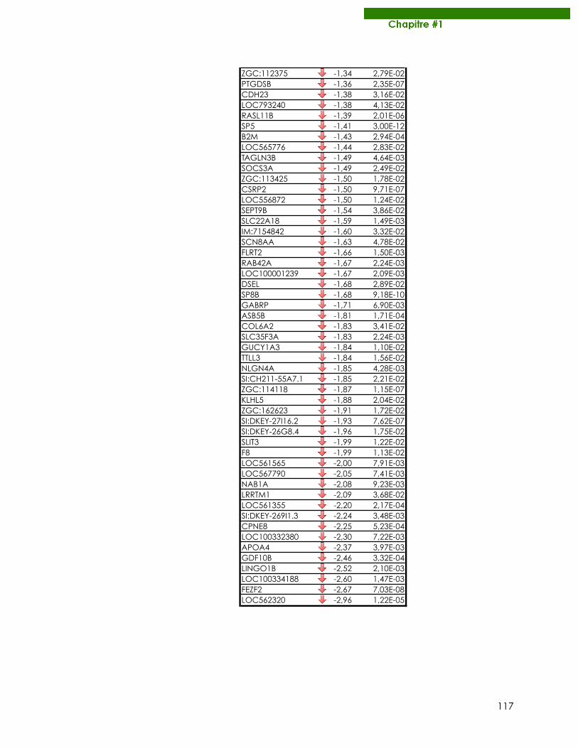
117
ZGC:112375 -1,34 2,79E-02PTGDSB -1,36 2,35E-07CDH23 -1,38 3,16E-02LOC793240 -1,38 4,13E-02RASL11B -1,39 2,01E-06SP5 -1,41 3,00E-12B2M -1,43 2,94E-04LOC565776 -1,44 2,83E-02TAGLN3B -1,49 4,64E-03SOCS3A -1,49 2,49E-02ZGC:113425 -1,50 1,78E-02CSRP2 -1,50 9,71E-07LOC556872 -1,50 1,24E-02SEPT9B -1,54 3,86E-02SLC22A18 -1,59 1,49E-03IM:7154842 -1,60 3,32E-02SCN8AA -1,63 4,78E-02FLRT2 -1,66 1,50E-03RAB42A -1,67 2,24E-03LOC100001239 -1,67 2,09E-03DSEL -1,68 2,89E-02SP8B -1,68 9,18E-10GABRP -1,71 6,90E-03ASB5B -1,81 1,71E-04COL6A2 -1,83 3,41E-02SLC35F3A -1,83 2,24E-03GUCY1A3 -1,84 1,10E-02TTLL3 -1,84 1,56E-02NLGN4A -1,85 4,28E-03SI:CH211-55A7.1 -1,85 2,21E-02ZGC:114118 -1,87 1,15E-07KLHL5 -1,88 2,04E-02ZGC:162623 -1,91 1,72E-02SI:DKEY-27I16.2 -1,93 7,62E-07SI:DKEY-26G8.4 -1,96 1,75E-02SLIT3 -1,99 1,22E-02F8 -1,99 1,13E-02LOC561565 -2,00 7,91E-03LOC567790 -2,05 7,41E-03NAB1A -2,08 9,23E-03LRRTM1 -2,09 3,68E-02LOC561355 -2,20 2,17E-04SI:DKEY-269I1.3 -2,24 3,48E-03CPNE8 -2,25 5,23E-04LOC100332380 -2,30 7,22E-03APOA4 -2,37 3,97E-03GDF10B -2,46 3,32E-04LINGO1B -2,52 2,10E-03LOC100334188 -2,60 1,47E-03FEZF2 -2,67 7,03E-08LOC562320 -2,96 1,22E-05
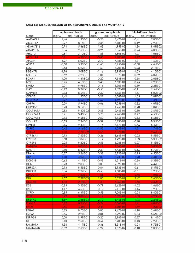
118
TABLE S2: BASAL EXPRESSION OF RA-RESPONSIVE GENES IN RAR MORPHANTS
Gene logFC adj_P.value logFC adj_P.value logFC adj_P.valueAADACL4 1,46 1,20E-01 -0,25 8,47E-01 -0,41 7,00E-01ABCB11A -0,27 8,16E-01 0,35 6,48E-01 0,19 7,97E-01ADAMTS16 0,74 5,66E-01 1,63 4,95E-02 1,36 9,61E-02ADARB1A 0,06 9,60E-01 -0,26 7,05E-01 -0,24 6,85E-01AHCYL1 -0,81 3,10E-01 -1,83 1,80E-02 -1,07 1,05E-01ALDH1A2 -0,21 6,91E-01 -0,40 2,70E-01 -1,22 3,86E-04APOA4 -1,27 5,02E-01 -2,70 7,78E-02 -1,91 1,60E-01ASB5B -0,50 5,98E-01 -1,60 3,95E-02 -0,50 4,64E-01B2M -0,63 5,22E-01 -1,50 6,90E-02 -0,93 2,18E-01B3GNT7L -1,18 5,09E-01 -1,16 3,93E-01 -1,03 4,12E-01B3GNT9 -0,52 7,28E-01 -1,04 3,37E-01 -0,52 6,02E-01BCAR1 1,00 4,37E-02 0,20 7,54E-01 0,56 2,05E-01BCR -0,95 4,18E-01 0,40 6,63E-01 -0,25 7,93E-01BMI1A -0,01 9,95E-01 0,16 8,30E-01 0,59 2,21E-01CA9 -0,12 8,37E-01 -0,55 1,02E-01 -0,11 7,54E-01CAPN12 -0,33 8,64E-01 0,32 8,15E-01 1,57 5,02E-02CDH23 1,49 1,23E-01 0,92 3,38E-01 1,05 2,09E-01CERCAM -0,97 5,67E-02 -1,62 1,59E-03 -2,40 1,46E-04CHPFA 0,29 5,94E-01 -0,06 9,25E-01 0,32 4,09E-01CHRNA5 0,23 8,19E-01 -1,10 1,25E-01 -0,90 1,84E-01COL14A1A 0,83 9,40E-02 -0,68 2,46E-01 -0,50 3,67E-01COL27A1A 0,71 3,45E-01 0,75 2,26E-01 0,47 4,55E-01COL27A1B -0,15 9,68E-01 0,50 8,16E-01 -0,33 8,61E-01COL6A2 -0,53 7,94E-01 -0,37 8,23E-01 -0,28 8,36E-01CPNE8 -0,16 9,40E-01 -1,14 3,17E-01 -0,66 5,49E-01CSRP2 -0,42 5,16E-01 -1,95 1,29E-03 -1,21 1,74E-02CUX2A 1,10 7,71E-02 0,55 3,87E-01 1,61 9,69E-04CYP26A1 0,13 7,63E-01 -0,26 3,66E-01 -0,02 9,58E-01CYP26B1 0,50 7,07E-01 1,14 1,42E-01 1,33 5,38E-02CYP2P6 -0,05 9,80E-01 -0,55 6,08E-01 0,07 9,43E-01DAB2IPA 0,34 8,06E-01 1,12 1,56E-01 1,44 3,88E-02DACT1 -0,10 8,42E-01 -0,30 3,43E-01 0,16 5,79E-01DBX1A -0,27 5,26E-01 0,32 3,19E-01 0,15 6,55E-01DBX1B 1,18 4,09E-01 2,03 3,94E-02 1,90 4,41E-02DCHS1B -0,62 4,11E-01 -0,93 1,31E-01 -0,36 5,38E-01DCN -0,23 9,08E-01 0,03 9,89E-01 0,77 4,42E-01DHRS3A -0,13 9,55E-01 0,84 3,93E-01 0,61 5,49E-01DHRS3B 0,06 9,27E-01 -0,50 1,68E-01 -0,51 1,23E-01DIXDC1A -0,37 7,80E-01 -1,06 2,49E-01 -2,40 1,06E-02DLB 1,57 7,37E-03 1,05 5,39E-02 2,42 2,60E-06DMRT2B -1,88 2,14E-01 -1,30 3,06E-01 -2,50 3,39E-02DSE -0,85 3,55E-01 -0,71 3,62E-01 -1,02 1,66E-01DSEL -1,17 4,63E-01 0,17 9,11E-01 -1,45 1,98E-01DYRK4 -0,85 6,41E-01 -0,58 7,00E-01 -0,24 8,62E-01DYSF 0,49 8,01E-01 1,12 3,09E-01 1,83 4,96E-02EIF2AK2 -3,59 2,36E-03 -2,70 6,46E-03 -1,98 2,27E-02ELAVL3 0,22 8,96E-01 -0,24 8,56E-01 1,41 4,84E-02ENO4 0,65 4,38E-01 -0,48 5,90E-01 1,69 1,29E-03EPHA7 0,23 8,75E-01 0,05 9,67E-01 -1,10 2,67E-01ESRRA -0,56 2,94E-01 -0,81 6,99E-02 -0,84 5,56E-02ESRRGB 0,00 9,99E-01 -0,20 8,96E-01 0,27 8,14E-01F8 0,13 9,61E-01 -0,54 7,40E-01 0,43 7,27E-01FAM101A -1,38 4,17E-01 -0,36 8,31E-01 0,04 9,75E-01FAM167AB -0,32 7,63E-01 -1,99 1,37E-02 -0,10 9,00E-01
alpha morphants gamma morphants full-RAR morphants
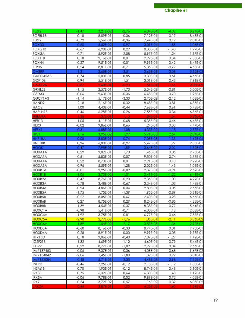
119
FEZF2 -1,47 3,21E-02 -2,76 6,04E-04 -4,02 8,24E-05FGFRL1B 0,18 8,89E-01 -0,36 7,12E-01 -0,17 8,43E-01FLRT2 -0,72 5,56E-01 -0,36 7,44E-01 0,12 9,04E-01FOXD3 0,60 5,52E-02 0,97 3,01E-04 1,88 1,06E-09FOXG1B -0,67 6,98E-01 0,29 8,38E-01 -1,43 1,99E-01FOXI3A -0,71 5,92E-01 -2,08 5,97E-02 -1,24 1,97E-01FOXJ1B 0,18 9,16E-01 0,01 9,97E-01 0,34 7,33E-01FOXN4 -0,27 9,51E-01 -0,01 9,99E-01 0,42 8,49E-01FTR06 0,24 8,81E-01 -0,71 5,35E-01 -0,79 4,53E-01GABRP -1,21 2,65E-01 -3,51 3,65E-03 -3,08 5,52E-03GADD45AB 0,74 5,00E-01 0,85 3,30E-01 0,61 4,66E-01GDF10B 0,94 5,51E-01 -1,51 3,01E-01 -0,43 7,61E-01GNA12A -0,87 2,60E-01 -0,93 1,51E-01 -1,66 2,03E-02GRHL2B -1,15 2,57E-01 -1,70 5,34E-02 -0,81 3,00E-01GSTM3 -0,06 9,63E-01 -0,36 6,48E-01 0,70 1,95E-01GUCY1A3 -1,14 5,17E-01 -3,30 2,70E-02 -2,12 1,08E-01HAND2 -2,18 2,16E-01 0,32 8,48E-01 0,81 4,85E-01HAO2 1,00 4,43E-01 -0,44 7,68E-01 0,61 5,48E-01HAPLN1B -0,46 6,28E-01 -0,26 7,55E-01 -0,34 6,36E-01HBEGFA 1,38 1,37E-01 1,13 1,84E-01 1,57 3,26E-02HER13 -1,05 4,11E-01 -0,68 5,35E-01 -0,46 6,45E-01HER3 -0,02 9,86E-01 0,66 1,24E-01 0,33 4,47E-01HESX1 -0,31 6,88E-01 -1,08 4,33E-02 -1,18 2,57E-02HIC1 1,16 3,95E-02 1,99 5,71E-05 2,54 2,04E-07HNF1BA 0,08 8,80E-01 0,74 7,05E-03 1,33 1,69E-06HNF1BB 0,96 6,00E-01 -0,97 5,47E-01 1,27 2,85E-01HOOK1 0,47 7,95E-01 1,89 3,64E-02 2,02 1,52E-02HOXA1A 0,29 9,02E-01 1,70 1,46E-01 0,05 9,75E-01HOXA3A -0,61 5,83E-01 -0,07 9,50E-01 -0,74 3,73E-01HOXA4A 0,22 8,73E-01 0,01 9,91E-01 0,10 9,22E-01HOXA5A -0,96 5,59E-01 1,28 2,02E-01 1,43 1,03E-01HOXB1A -0,01 9,95E-01 -0,09 9,37E-01 -0,91 2,59E-01HOXB1B -1,06 3,73E-02 -1,01 2,41E-02 -1,22 6,41E-03HOXB2A -0,47 8,76E-01 -0,20 9,36E-01 1,00 4,99E-01HOXB3A -0,78 3,48E-01 -0,67 3,34E-01 -1,03 1,48E-01HOXB4A -0,94 4,86E-01 0,04 9,80E-01 0,05 9,66E-01HOXB5A -1,75 1,75E-01 -1,39 1,95E-01 -0,89 3,61E-01HOXB5B -0,27 8,05E-01 0,67 2,40E-01 0,28 6,66E-01HOXB6B 0,27 8,75E-01 0,29 8,24E-01 -0,85 4,23E-01HOXB8B -1,39 4,54E-01 -0,37 8,38E-01 -0,77 5,64E-01HOXC1A -0,98 5,41E-01 -0,71 6,00E-01 1,13 2,03E-01HOXC4A -1,92 3,75E-01 -0,81 6,77E-01 -0,46 7,87E-01HOXC5A -2,90 2,77E-02 -1,76 1,05E-01 -2,11 3,86E-02HOXC6A -1,89 1,54E-02 -2,17 3,53E-03 -1,36 2,92E-02HOXD3A -0,60 8,16E-01 -0,33 8,74E-01 0,01 9,95E-01HOXD4A -0,28 8,91E-01 0,00 9,99E-01 -0,05 9,73E-01HTR1BD 0,18 9,06E-01 -0,40 7,07E-01 -1,29 1,42E-01IGSF21B -1,32 4,69E-01 -1,12 4,60E-01 -0,79 5,44E-01ILDR2 0,22 8,77E-01 -1,02 2,99E-01 0,04 9,66E-01IM:7137453 -0,06 9,37E-01 -0,36 4,08E-01 -0,68 9,67E-02IM:7154842 -2,06 1,45E-01 -1,80 1,32E-01 0,99 3,04E-01IM:7162084 -0,48 5,71E-01 -2,35 4,48E-03 -2,98 7,32E-04INHBB -0,46 7,25E-01 -0,12 9,18E-01 -1,12 1,85E-01INSM1B 0,70 1,93E-01 -0,12 8,74E-01 0,48 3,10E-01IRX3B 0,75 6,32E-01 0,64 6,30E-01 1,48 1,12E-01IRX5A -0,06 9,78E-01 0,02 9,89E-01 0,72 4,02E-01IRX7 -0,54 3,72E-02 -0,57 1,16E-02 -0,39 6,05E-02JAK2A 0,66 3,87E-01 1,05 6,23E-02 1,60 1,39E-03
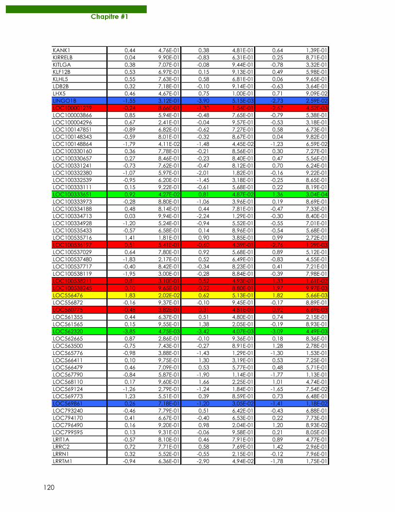
120
KANK1 0,44 4,76E-01 0,38 4,81E-01 0,64 1,39E-01KIRRELB 0,04 9,90E-01 -0,83 6,31E-01 0,25 8,71E-01KITLGA 0,38 7,07E-01 -0,08 9,44E-01 -0,78 3,32E-01KLF12B 0,53 6,97E-01 0,15 9,13E-01 0,49 5,98E-01KLHL5 0,55 7,63E-01 0,58 6,81E-01 0,06 9,65E-01LDB2B 0,32 7,18E-01 -0,10 9,14E-01 -0,63 3,64E-01LHX5 0,46 4,67E-01 0,75 1,00E-01 0,71 9,09E-02LINGO1B -1,55 3,12E-01 -3,90 5,15E-03 -2,73 2,59E-02LOC100001239 -0,24 8,66E-01 -1,30 1,54E-01 -2,67 4,52E-03LOC100003866 0,85 5,94E-01 -0,48 7,65E-01 -0,79 5,38E-01LOC100004296 0,67 2,41E-01 -0,04 9,57E-01 -0,53 3,18E-01LOC100147851 -0,89 6,82E-01 -0,62 7,27E-01 0,58 6,73E-01LOC100148343 -0,59 8,01E-01 -0,32 8,67E-01 0,04 9,82E-01LOC100148864 -1,79 4,11E-02 -1,48 4,45E-02 -1,23 6,59E-02LOC100330160 0,36 7,78E-01 -0,21 8,56E-01 0,30 7,27E-01LOC100330657 0,27 8,46E-01 -0,23 8,40E-01 0,47 5,56E-01LOC100331241 -0,73 7,62E-01 -0,47 8,12E-01 0,70 6,24E-01LOC100332380 -1,07 5,97E-01 -2,01 1,82E-01 -0,16 9,22E-01LOC100332539 -0,95 6,20E-01 -1,45 3,18E-01 -0,25 8,65E-01LOC100333111 0,15 9,22E-01 -0,61 5,68E-01 0,22 8,19E-01LOC100333651 0,92 4,27E-02 0,81 4,87E-02 1,36 3,04E-04LOC100333973 -0,28 8,80E-01 -1,06 3,96E-01 0,19 8,69E-01LOC100334188 0,48 8,14E-01 0,44 7,81E-01 -0,47 7,33E-01LOC100334713 0,03 9,94E-01 -2,24 1,29E-01 -0,30 8,40E-01LOC100334928 -1,20 5,24E-01 -0,94 5,52E-01 -0,55 7,01E-01LOC100535433 -0,57 6,58E-01 0,14 8,96E-01 -0,54 5,68E-01LOC100535716 1,41 1,81E-01 0,90 3,85E-01 0,99 2,72E-01LOC100536197 0,51 5,41E-01 -0,60 4,39E-01 -2,76 1,29E-03LOC100537029 0,64 7,80E-01 0,92 5,68E-01 0,89 5,12E-01LOC100537480 -1,83 2,17E-01 0,52 6,49E-01 -0,83 4,55E-01LOC100537717 -0,40 8,42E-01 -0,34 8,23E-01 0,41 7,21E-01LOC100538119 -1,95 3,00E-01 -0,28 8,84E-01 -0,39 7,98E-01LOC100538211 0,81 3,10E-01 0,52 4,93E-01 1,33 1,61E-02LOC100538245 0,10 9,65E-01 -0,22 8,80E-01 1,97 9,97E-03LOC556476 1,83 2,02E-02 0,62 5,13E-01 1,82 5,66E-03LOC556872 -0,16 9,37E-01 -0,10 9,45E-01 -0,17 8,89E-01LOC560775 -0,48 3,82E-01 0,31 4,81E-01 0,92 6,69E-03LOC561355 0,44 6,37E-01 0,51 4,80E-01 0,74 2,15E-01LOC561565 0,15 9,55E-01 1,38 2,05E-01 -0,19 8,93E-01LOC562320 -3,85 4,75E-03 -3,42 4,07E-03 -3,09 4,49E-03LOC562665 0,87 2,86E-01 -0,10 9,36E-01 0,18 8,36E-01LOC563500 -0,75 7,43E-01 -0,27 8,91E-01 1,28 2,78E-01LOC565776 -0,98 3,88E-01 -1,43 1,29E-01 -1,30 1,53E-01LOC566411 0,10 9,75E-01 1,30 3,19E-01 0,53 7,25E-01LOC566479 0,46 7,09E-01 0,53 5,77E-01 0,48 5,71E-01LOC567790 -0,84 5,87E-01 -1,90 1,14E-01 -1,77 1,13E-01LOC568110 0,17 9,60E-01 1,66 2,25E-01 1,01 4,74E-01LOC569124 -1,26 2,79E-01 -1,24 1,84E-01 -1,65 7,54E-02LOC569773 1,23 5,51E-01 0,39 8,59E-01 0,73 6,48E-01LOC569861 0,26 7,18E-01 -1,20 3,05E-02 -1,41 1,18E-02LOC793240 -0,46 7,79E-01 0,51 6,42E-01 -0,43 6,88E-01LOC794170 0,41 6,67E-01 -0,40 6,53E-01 0,22 7,73E-01LOC796490 0,16 9,20E-01 0,98 2,04E-01 1,20 8,93E-02LOC799595 0,13 9,31E-01 -0,06 9,58E-01 0,21 8,05E-01LRIT1A -0,57 8,10E-01 0,46 7,91E-01 0,89 4,77E-01LRRC2 0,72 7,71E-01 0,58 7,69E-01 1,42 2,96E-01LRRN1 0,32 5,52E-01 -0,55 2,15E-01 -0,12 7,96E-01LRRTM1 -0,94 6,36E-01 -2,90 4,94E-02 -1,78 1,75E-01
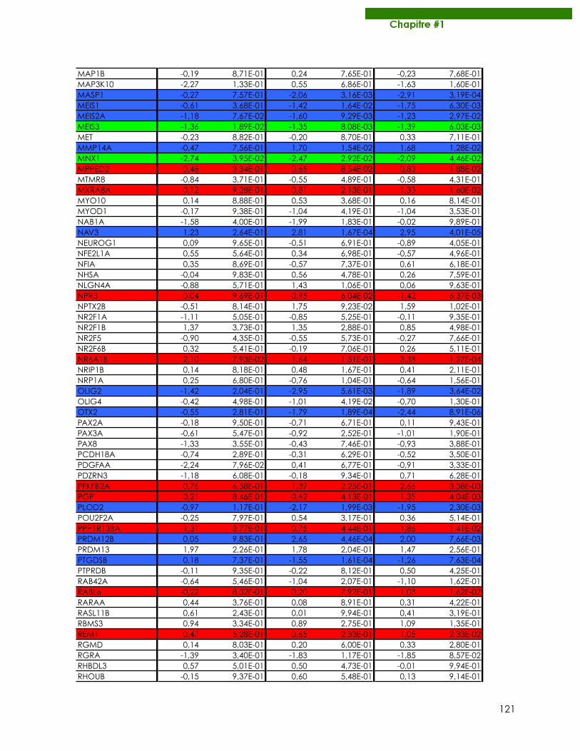
121
MAP1B -0,19 8,71E-01 0,24 7,65E-01 -0,23 7,68E-01MAP3K10 -2,27 1,33E-01 0,55 6,86E-01 -1,63 1,60E-01MASP1 -0,27 7,57E-01 -2,06 3,16E-03 -2,91 3,19E-04MEIS1 -0,61 3,68E-01 -1,42 1,64E-02 -1,75 6,30E-03MEIS2A -1,18 7,67E-02 -1,60 9,29E-03 -1,23 2,97E-02MEIS3 -1,36 1,89E-02 -1,35 8,08E-03 -1,39 6,03E-03MET -0,23 8,82E-01 -0,20 8,70E-01 0,33 7,11E-01MMP14A -0,47 7,56E-01 1,70 1,54E-02 1,68 1,28E-02MNX1 -2,74 3,95E-02 -2,47 2,92E-02 -2,09 4,46E-02MPPED2 0,48 3,34E-01 0,65 8,54E-02 0,83 1,88E-02MTMR8 -0,84 3,71E-01 -0,55 4,89E-01 -0,58 4,31E-01MXRA8A 0,12 9,28E-01 0,81 2,13E-01 1,33 1,60E-02MYO10 0,14 8,88E-01 0,53 3,68E-01 0,16 8,14E-01MYOD1 -0,17 9,38E-01 -1,04 4,19E-01 -1,04 3,53E-01NAB1A -1,58 4,00E-01 -1,99 1,83E-01 -0,02 9,89E-01NAV3 1,23 2,64E-01 2,81 1,67E-04 2,95 4,01E-05NEUROG1 0,09 9,65E-01 -0,51 6,91E-01 -0,89 4,05E-01NFE2L1A 0,55 5,64E-01 0,34 6,98E-01 -0,57 4,96E-01NFIA 0,35 8,69E-01 -0,57 7,37E-01 0,61 6,18E-01NHSA -0,04 9,83E-01 0,56 4,78E-01 0,26 7,59E-01NLGN4A -0,88 5,71E-01 1,43 1,06E-01 0,06 9,63E-01NPR3 0,04 9,69E-01 -0,95 6,04E-02 -1,42 6,37E-03NPTX2B -0,51 8,14E-01 1,75 9,23E-02 1,59 1,02E-01NR2F1A -1,11 5,05E-01 -0,85 5,25E-01 -0,11 9,35E-01NR2F1B 1,37 3,73E-01 1,35 2,88E-01 0,85 4,98E-01NR2F5 -0,90 4,35E-01 -0,55 5,73E-01 -0,27 7,66E-01NR2F6B 0,32 5,41E-01 -0,19 7,06E-01 0,26 5,11E-01NR6A1B 2,10 7,93E-02 1,64 1,51E-01 3,38 1,27E-04NRIP1B 0,14 8,18E-01 0,48 1,67E-01 0,41 2,11E-01NRP1A 0,25 6,80E-01 -0,76 1,04E-01 -0,64 1,56E-01OLIG2 -1,42 2,04E-01 -2,95 5,61E-03 -1,89 3,64E-02OLIG4 -0,42 4,98E-01 -1,01 4,19E-02 -0,70 1,30E-01OTX2 -0,55 2,81E-01 -1,79 1,89E-04 -2,44 8,91E-06PAX2A -0,18 9,50E-01 -0,71 6,71E-01 0,11 9,43E-01PAX3A -0,61 5,47E-01 -0,92 2,52E-01 -1,01 1,90E-01PAX8 -1,33 3,55E-01 -0,43 7,46E-01 -0,93 3,88E-01PCDH18A -0,74 2,89E-01 -0,31 6,29E-01 -0,52 3,50E-01PDGFAA -2,24 7,96E-02 0,41 6,77E-01 -0,91 3,33E-01PDZRN3 -1,18 6,08E-01 -0,18 9,34E-01 0,71 6,28E-01PFKFB2A 0,76 6,58E-01 1,39 2,25E-01 2,66 3,38E-03PGP -0,21 8,46E-01 -0,62 4,13E-01 1,35 4,04E-03PLOD2 -0,97 1,17E-01 -2,17 1,99E-03 -1,95 2,30E-03POU2F2A -0,25 7,97E-01 0,54 3,17E-01 0,36 5,14E-01PPP1R13BA -1,31 3,77E-01 0,75 4,44E-01 1,86 1,41E-02PRDM12B 0,05 9,83E-01 2,65 4,46E-04 2,00 7,66E-03PRDM13 1,97 2,26E-01 1,78 2,04E-01 1,47 2,56E-01PTGDSB 0,18 7,37E-01 -1,55 1,61E-04 -1,26 7,63E-04PTPRDB -0,11 9,35E-01 -0,22 8,12E-01 0,50 4,25E-01RAB42A -0,64 5,46E-01 -1,04 2,07E-01 -1,10 1,62E-01RABL6 -0,22 8,32E-01 0,20 7,92E-01 1,08 1,62E-02RARAA 0,44 3,76E-01 0,08 8,91E-01 0,31 4,22E-01RASL11B 0,61 2,43E-01 0,01 9,94E-01 0,41 3,19E-01RBMS3 0,94 3,34E-01 0,89 2,75E-01 1,09 1,35E-01REM1 0,47 5,28E-01 0,65 2,33E-01 1,05 2,33E-02RGMD 0,14 8,03E-01 0,20 6,00E-01 0,33 2,80E-01RGRA -1,39 3,40E-01 -1,83 1,17E-01 -1,85 8,57E-02RHBDL3 0,57 5,01E-01 0,50 4,73E-01 -0,01 9,94E-01RHOUB -0,15 9,37E-01 0,60 5,48E-01 0,13 9,14E-01
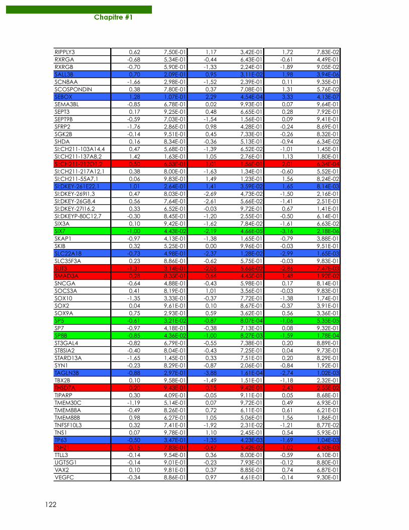
122
RIPPLY3 0,62 7,50E-01 1,17 3,42E-01 1,72 7,83E-02RXRGA -0,68 5,34E-01 -0,44 6,43E-01 -0,61 4,49E-01RXRGB -0,70 5,90E-01 -1,33 2,24E-01 -1,89 9,05E-02SALL3B 0,70 2,09E-01 0,95 3,11E-02 1,98 3,94E-06SCN8AA -1,66 2,98E-01 -1,52 2,39E-01 0,11 9,35E-01SCOSPONDIN 0,38 7,80E-01 0,37 7,08E-01 1,31 5,76E-02SEBOX 1,28 1,07E-01 2,29 4,54E-04 3,33 4,13E-07SEMA3BL -0,85 6,78E-01 0,02 9,93E-01 0,07 9,64E-01SEPT3 0,17 9,25E-01 0,48 6,65E-01 0,28 7,92E-01SEPT9B -0,59 7,03E-01 -1,54 1,56E-01 0,09 9,41E-01SFRP2 -1,76 2,86E-01 0,98 4,28E-01 -0,24 8,69E-01SGK2B -0,14 9,51E-01 0,45 7,33E-01 -0,26 8,32E-01SHDA 0,16 8,34E-01 -0,36 5,13E-01 -0,94 6,34E-02SI:CH211-103A14,4 0,47 5,68E-01 -1,39 6,52E-02 -1,01 1,45E-01SI:CH211-137A8,2 1,42 1,63E-01 1,05 2,76E-01 1,13 1,80E-01SI:CH211-212O1,2 0,50 6,53E-01 1,01 1,56E-01 2,01 6,34E-04SI:CH211-217A12,1 0,38 8,00E-01 -1,63 1,34E-01 -0,60 5,52E-01SI:CH211-55A7,1 0,06 9,83E-01 1,49 1,23E-01 1,56 8,24E-02SI:DKEY-261E22,1 1,01 2,64E-01 1,41 3,59E-02 1,65 8,14E-03SI:DKEY-269I1,3 0,47 8,03E-01 -2,69 4,73E-02 -1,50 2,16E-01SI:DKEY-26G8,4 0,56 7,64E-01 -2,61 5,66E-02 -1,41 2,51E-01SI:DKEY-27I16,2 0,33 6,52E-01 -0,03 9,72E-01 0,67 1,41E-01SI:DKEYP-80C12,7 -0,30 8,45E-01 -1,20 2,55E-01 -0,50 6,14E-01SIX3A 0,10 9,42E-01 -1,62 7,84E-02 -1,61 6,63E-02SIX7 -1,00 4,43E-02 -2,19 4,66E-05 -3,16 2,18E-06SKAP1 -0,97 4,13E-01 -1,38 1,65E-01 -0,79 3,88E-01SKIB 0,32 5,25E-01 0,00 9,96E-01 -0,03 9,51E-01SLC22A18 -0,73 4,98E-01 -2,37 1,28E-02 -2,99 1,65E-03SLC35F3A 0,23 8,86E-01 -0,62 5,75E-01 -0,03 9,83E-01SLIT3 -1,31 3,14E-01 -2,06 5,66E-02 -2,86 7,47E-03SMAD3A 0,28 8,35E-01 0,66 4,45E-01 1,48 1,92E-02SNCGA -0,64 4,88E-01 -0,43 5,98E-01 0,17 8,14E-01SOCS3A 0,41 8,19E-01 1,01 3,56E-01 -0,03 9,83E-01SOX10 -1,35 3,33E-01 -0,37 7,72E-01 -1,38 1,74E-01SOX2 0,04 9,61E-01 0,10 8,67E-01 -0,37 3,91E-01SOX9A 0,75 2,93E-01 0,59 3,62E-01 0,56 3,36E-01SP5 -0,61 3,21E-02 -0,87 8,07E-04 -1,06 5,35E-05SP7 -0,97 4,18E-01 -0,38 7,13E-01 0,08 9,32E-01SP8B -0,85 4,36E-02 -1,00 8,27E-03 -1,59 1,78E-04ST3GAL4 -0,82 6,79E-01 -0,55 7,38E-01 0,20 8,89E-01ST8SIA2 -0,40 8,04E-01 -0,43 7,25E-01 0,04 9,73E-01STARD13A -1,65 1,45E-01 0,33 7,51E-01 0,20 8,29E-01SYN1 -0,23 8,29E-01 -0,87 2,06E-01 -0,84 1,92E-01TAGLN3B -0,88 2,97E-01 -3,88 1,61E-04 -2,74 1,02E-03TBX2B 0,10 9,58E-01 -1,49 1,51E-01 -1,18 2,32E-01THSD7A 0,20 9,43E-01 0,15 9,42E-01 2,43 2,55E-02TIPARP 0,30 4,09E-01 -0,05 9,11E-01 0,05 8,68E-01TMEM30C -1,19 5,14E-01 0,07 9,72E-01 0,49 6,93E-01TMEM88A -0,49 8,26E-01 0,72 6,11E-01 0,61 6,21E-01TMEM88B 0,98 6,27E-01 1,05 5,06E-01 1,56 1,86E-01TNFSF10L3 0,32 7,41E-01 -1,92 2,31E-02 -1,21 8,77E-02TNS1 0,07 9,78E-01 1,10 2,45E-01 0,54 5,93E-01TP63 -0,50 3,47E-01 -1,35 4,23E-03 -1,69 1,04E-03TSHZ1 -0,15 7,83E-01 -0,67 5,42E-02 -1,02 4,50E-03TTLL3 -0,14 9,54E-01 0,36 8,00E-01 -0,59 6,10E-01UGT5G1 -0,14 9,01E-01 -0,23 7,93E-01 -0,12 8,80E-01VAX2 0,10 9,81E-01 0,37 8,85E-01 0,74 6,87E-01VEGFC -0,34 8,86E-01 0,97 4,61E-01 -0,14 9,30E-01
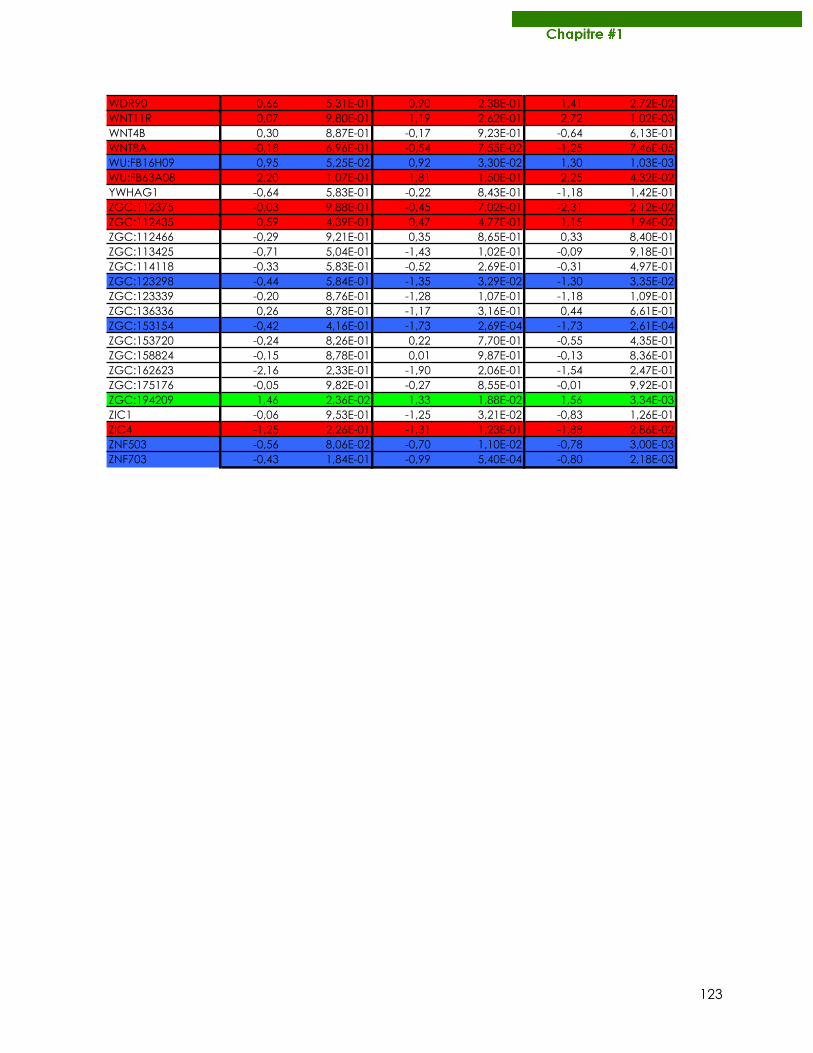
123
WDR90 0,66 5,31E-01 0,90 2,38E-01 1,41 2,72E-02WNT11R 0,07 9,80E-01 1,19 2,62E-01 2,72 1,02E-03WNT4B 0,30 8,87E-01 -0,17 9,23E-01 -0,64 6,13E-01WNT8A -0,18 6,96E-01 -0,54 7,55E-02 -1,25 7,46E-05WU:FB16H09 0,95 5,25E-02 0,92 3,30E-02 1,30 1,03E-03WU:FB63A08 2,20 1,07E-01 1,81 1,50E-01 2,25 4,32E-02YWHAG1 -0,64 5,83E-01 -0,22 8,43E-01 -1,18 1,42E-01ZGC:112375 -0,03 9,88E-01 -0,45 7,02E-01 -2,31 2,12E-02ZGC:112435 0,59 4,39E-01 0,47 4,77E-01 1,15 1,94E-02ZGC:112466 -0,29 9,21E-01 0,35 8,65E-01 0,33 8,40E-01ZGC:113425 -0,71 5,04E-01 -1,43 1,02E-01 -0,09 9,18E-01ZGC:114118 -0,33 5,83E-01 -0,52 2,69E-01 -0,31 4,97E-01ZGC:123298 -0,44 5,84E-01 -1,35 3,29E-02 -1,30 3,35E-02ZGC:123339 -0,20 8,76E-01 -1,28 1,07E-01 -1,18 1,09E-01ZGC:136336 0,26 8,78E-01 -1,17 3,16E-01 0,44 6,61E-01ZGC:153154 -0,42 4,16E-01 -1,73 2,69E-04 -1,73 2,61E-04ZGC:153720 -0,24 8,26E-01 0,22 7,70E-01 -0,55 4,35E-01ZGC:158824 -0,15 8,78E-01 0,01 9,87E-01 -0,13 8,36E-01ZGC:162623 -2,16 2,33E-01 -1,90 2,06E-01 -1,54 2,47E-01ZGC:175176 -0,05 9,82E-01 -0,27 8,55E-01 -0,01 9,92E-01ZGC:194209 1,46 2,36E-02 1,33 1,88E-02 1,56 3,34E-03ZIC1 -0,06 9,53E-01 -1,25 3,21E-02 -0,83 1,26E-01ZIC4 -1,25 2,26E-01 -1,31 1,23E-01 -1,88 2,86E-02ZNF503 -0,56 8,06E-02 -0,70 1,10E-02 -0,78 3,00E-03ZNF703 -0,43 1,84E-01 -0,99 5,40E-04 -0,80 2,18E-03
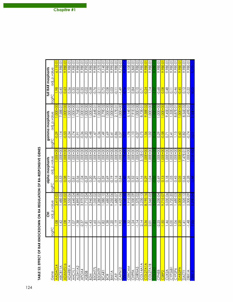
124
TABL
E S3
: EFF
ECT
OF
RAR
KN
OC
KD
OW
N O
N R
A R
EGU
LATI
ON
OF
RA-R
ESPO
NSI
VE
GEN
ES
Gene
logFC
adj_p,value
logFC
adj_p,value
logFC
adj_p,value
logFC
adj_p,value
AADACL4
1,08
3,41E-02
0,10
1,00E+00
-0,29
1,00E+00
1,98
6,11E-01
ABCB11A
1,42
1,48E-05
-0,53
1,00E+00
-1,14
7,58E-01
-0,45
9,98E-01
ADAMTS16
1,08
2,33E-02
0,08
1,00E+00
-0,53
1,00E+00
0,63
9,98E-01
ADARB1A
1,14
3,55E-06
-0,17
1,00E+00
-0,34
1,00E+00
0,56
9,98E-01
AHCYL1
1,07
2,64E-04
0,81
1,00E+00
0,74
1,00E+00
0,35
9,98E-01
ALDH1A2
-1,38
4,89E-08
-0,56
1,00E+00
-0,91
1,76E-01
-0,50
9,98E-01
APOA4
-2,37
3,97E-03
-1,04
1,00E+00
-0,36
1,00E+00
-0,55
9,98E-01
ASB5B
-1,81
1,71E-04
-0,59
1,00E+00
-0,20
1,00E+00
-0,02
9,99E-01
B2M
-1,43
2,94E-04
0,20
1,00E+00
-0,48
1,00E+00
-0,58
9,98E-01
B3GNT7L
1,65
1,05E-02
1,29
1,00E+00
1,47
9,64E-01
-0,76
9,98E-01
B3GNT9
1,21
3,28E-02
-0,21
1,00E+00
-0,36
1,00E+00
-2,11
7,57E-01
BCAR1
1,82
1,08E-10
0,59
1,00E+00
1,49
2,79E-02
0,75
9,14E-01
BCR
1,38
1,68E-04
0,69
1,00E+00
0,69
1,00E+00
-0,08
9,98E-01
BMI1A
1,20
9,25E-06
-0,19
1,00E+00
0,41
1,00E+00
0,03
9,99E-01
CA9
-1,19
1,68E-07
-0,16
1,00E+00
-0,86
2,17E-01
-0,11
9,98E-01
CAPN12
1,92
6,62E-06
0,84
1,00E+00
0,37
1,00E+00
-1,49
9,71E-01
CDH23
-1,38
3,16E-02
-0,42
1,00E+00
0,05
1,00E+00
-0,06
9,98E-01
CERCAM
1,32
1,91E-08
0,28
1,00E+00
1,78
5,14E-03
1,10
8,10E-01
CHPFA
1,24
8,33E-09
0,52
1,00E+00
1,10
7,91E-02
0,84
5,86E-01
CHRNA5
-1,14
1,57E-02
0,41
1,00E+00
-0,37
1,00E+00
0,10
9,98E-01
COL14A1A
2,14
2,23E-13
1,16
3,12E-01
0,75
8,73E-01
0,71
9,98E-01
COL27A1A
3,36
8,19E-16
0,29
1,00E+00
1,56
9,58E-02
-0,24
9,98E-01
COL27A1B
2,01
2,56E-02
1,04
1,00E+00
1,02
1,00E+00
1,14
9,98E-01
COL6A2
-1,83
3,41E-02
-0,05
1,00E+00
0,14
1,00E+00
-1,07
9,98E-01
CPNE8
-2,25
5,23E-04
-0,69
1,00E+00
0,29
1,00E+00
-0,68
9,98E-01
CSRP2
-1,50
9,71E-07
0,13
1,00E+00
-0,58
1,00E+00
0,08
9,98E-01
CUX2A
1,46
5,03E-06
0,18
1,00E+00
0,71
9,45E-01
0,48
9,98E-01
CYP26B1
5,00
3,72E-20
1,03
1,00E+00
2,41
1,32E-02
0,11
9,98E-01
CYP2P6
1,06
1,68E-03
0,45
1,00E+00
1,15
8,87E-01
-0,46
9,98E-01
DAB2IPA
2,20
2,00E-06
0,05
1,00E+00
0,60
1,00E+00
-0,45
9,98E-01
DACT1
1,41
1,32E-12
0,66
7,47E-01
0,82
8,56E-02
0,12
9,98E-01
DBX1A
-1,48
2,30E-09
-0,28
1,00E+00
-0,74
2,69E-01
-0,22
9,98E-01
DBX1B
2,53
1,69E-04
0,48
1,00E+00
-0,07
1,00E+00
0,20
9,98E-01
Ctr
la
lpha
mo
rpha
nts
ga
mm
a m
orp
hant
sfu
ll RA
R m
orp
hant
s
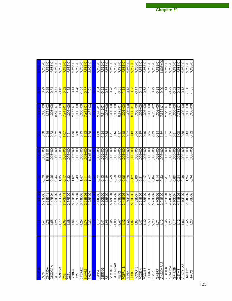
125
DCHS1B
-1,11
7,08E-03
-0,52
1,00E+00
-0,05
1,00E+00
-0,12
9,98E-01
DCN
1,61
5,27E-03
-0,70
1,00E+00
0,38
1,00E+00
0,29
9,98E-01
DHRS3A
4,78
6,26E-16
1,98
8,94E-01
2,46
4,71E-02
0,68
9,98E-01
DIXDC1A
1,55
1,47E-04
-0,65
1,00E+00
0,73
1,00E+00
0,76
9,98E-01
DLB
1,95
1,21E-07
0,46
1,00E+00
0,79
8,57E-01
0,71
9,98E-01
DMRT2B
-1,79
1,75E-02
1,35
1,00E+00
-1,28
1,00E+00
-0,13
9,98E-01
DSE
2,06
9,90E-11
-0,17
1,00E+00
1,07
7,61E-01
0,02
9,99E-01
DSEL
-1,68
2,89E-02
0,33
1,00E+00
-1,21
1,00E+00
1,58
9,98E-01
DYRK4
1,84
8,21E-04
1,09
1,00E+00
1,50
9,18E-01
-0,14
9,98E-01
DYSF
1,71
6,87E-04
-0,40
1,00E+00
0,88
1,00E+00
0,28
9,98E-01
EIF2AK2
-1,24
3,44E-02
1,67
1,00E+00
0,19
1,00E+00
-1,56
9,72E-01
ELAVL3
2,74
2,26E-08
-0,11
1,00E+00
1,43
7,61E-01
-0,79
9,98E-01
ENO4
2,33
1,98E-09
1,29
8,94E-01
2,78
1,48E-03
1,21
5,92E-01
EPHA7
2,12
1,26E-08
0,60
1,00E+00
0,15
1,00E+00
0,66
9,98E-01
ESRRA
1,19
7,14E-08
0,79
1,00E+00
1,00
2,64E-01
0,34
9,98E-01
ESRRGB
-1,41
3,80E-02
-0,43
1,00E+00
-1,26
9,94E-01
-0,32
9,98E-01
F8-1,99
1,13E-02
-2,49
1,00E+00
-0,85
1,00E+00
-0,81
9,98E-01
FAM101A
2,06
6,18E-04
0,68
1,00E+00
1,31
1,00E+00
-0,98
9,98E-01
FAM167AB
-1,08
2,11E-02
-0,58
1,00E+00
0,46
1,00E+00
-1,02
9,98E-01
FEZF2
-2,67
7,03E-08
-0,52
1,00E+00
-1,17
8,89E-01
-0,05
9,99E-01
FGFRL1B
1,42
3,44E-05
0,05
1,00E+00
0,48
1,00E+00
-1,13
9,98E-01
FLRT2
-1,66
1,50E-03
-0,28
1,00E+00
-0,22
1,00E+00
-0,15
9,98E-01
FOXD3
-1,07
5,81E-08
-0,09
1,00E+00
-0,43
8,11E-01
0,06
9,98E-01
FOXG1B
1,86
1,82E-04
0,88
1,00E+00
0,86
1,00E+00
-0,14
9,98E-01
FOXI3A
1,47
2,21E-03
-0,21
1,00E+00
0,89
1,00E+00
-0,48
9,98E-01
FOXJ1B
-1,42
1,88E-02
-0,17
1,00E+00
-0,40
1,00E+00
-0,58
9,98E-01
FOXN4
2,50
2,81E-02
0,69
1,00E+00
0,85
1,00E+00
1,07
9,98E-01
FTR06
2,23
5,96E-07
0,86
1,00E+00
2,63
6,20E-02
0,72
9,98E-01
GABRP
-1,71
6,90E-03
-1,14
1,00E+00
-0,34
1,00E+00
-0,36
9,98E-01
GADD45AB
1,12
3,06E-02
-0,53
1,00E+00
-1,39
7,99E-01
0,00
1,00E+00
GDF10B
-2,46
3,32E-04
-0,31
1,00E+00
-1,62
9,86E-01
-1,44
9,98E-01
GNA12A
1,22
4,24E-05
0,12
1,00E+00
0,36
1,00E+00
-0,92
9,98E-01
GRHL2B
1,17
5,10E-04
0,89
1,00E+00
0,81
1,00E+00
-0,02
9,99E-01
GSTM3
1,12
4,91E-03
0,84
1,00E+00
1,00
7,78E-01
0,43
9,98E-01
GUCY1A3
-1,84
1,10E-02
-0,48
1,00E+00
0,38
1,00E+00
-0,13
9,98E-01
HAND2
3,65
2,04E-07
1,96
1,00E+00
0,43
1,00E+00
-1,30
9,98E-01
HAO2
1,20
1,38E-02
-0,94
1,00E+00
-0,32
1,00E+00
-1,05
9,98E-01
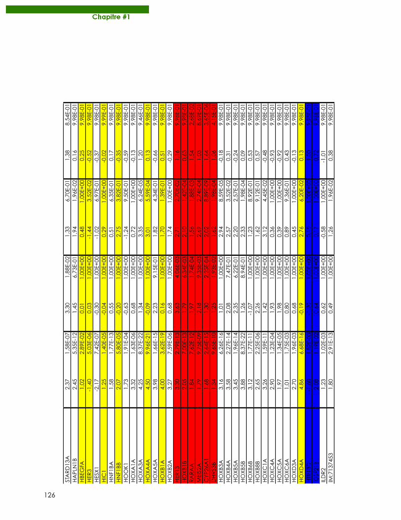
126
STARD13A
2,37
1,58E-07
3,30
1,88E-02
1,33
6,70E-01
1,38
8,54E-01
HAPLN1B
2,45
5,35E-12
1,45
6,73E-01
1,94
1,96E-02
0,16
9,98E-01
HBEGFA
1,02
2,89E-02
0,01
1,00E+00
0,48
1,00E+00
0,25
9,98E-01
HER3
-1,40
5,03E-06
0,03
1,00E+00
-1,44
3,52E-02
-0,52
9,98E-01
HESX1
-2,17
7,42E-07
-0,30
1,00E+00
-1,02
6,97E-01
-0,37
9,98E-01
HIC1
1,25
1,40E-05
-0,04
1,00E+00
0,29
1,00E+00
-0,02
9,99E-01
HNF1BA
1,58
1,77E-13
0,55
1,00E+00
0,51
6,35E-01
0,17
9,98E-01
HNF1BB
2,07
5,80E-05
-0,20
1,00E+00
2,75
3,82E-01
-0,35
9,98E-01
HOOK1
1,73
1,91E-04
-0,63
1,00E+00
-1,24
9,50E-01
-0,59
9,98E-01
HOXA1A
3,32
1,63E-06
0,68
1,00E+00
0,72
1,00E+00
-0,13
9,98E-01
HOXA3A
4,25
8,37E-22
1,34
1,00E+00
3,35
6,24E-05
1,20
9,46E-01
HOXA4A
4,50
9,96E-21
-0,09
1,00E+00
3,01
5,59E-04
0,13
9,98E-01
HOXA5A
3,98
1,66E-15
2,23
9,15E-01
1,82
4,34E-01
-0,66
9,98E-01
HOXB1A
4,00
3,62E-19
0,16
1,00E+00
1,70
1,39E-01
0,51
9,98E-01
HOXB2A
3,27
7,59E-06
0,68
1,00E+00
1,74
1,00E+00
-0,29
9,98E-01
HER13
3,30
2,79E-13
3,63
4,06E-03
2,71
2,70E-02
1,16
9,98E-01
HOXB1B
2,05
7,00E-13
1,79
6,34E-03
2,10
1,42E-04
0,63
9,98E-01
RARAA
1,84
7,62E-12
1,97
1,74E-04
1,56
1,88E-03
1,54
2,68E-02
MEIS2A
1,79
9,75E-09
2,18
9,35E-03
2,69
2,74E-04
1,03
8,69E-01
CYP26A1
1,68
2,64E-15
1,30
2,25E-04
2,02
8,89E-09
1,64
3,65E-06
DHRS3B
1,34
9,65E-10
1,25
1,93E-02
1,62
1,98E-04
1,06
4,15E-01
HOXB3A
3,16
6,26E-16
1,01
1,00E+00
2,94
8,59E-05
-0,18
9,98E-01
HOXB4A
3,58
2,27E-14
2,08
7,47E-01
2,57
3,52E-02
0,31
9,98E-01
HOXB5A
3,45
1,96E-14
2,35
6,22E-01
2,20
2,57E-01
-0,24
9,98E-01
HOXB5B
3,88
8,37E-22
1,26
8,94E-01
2,33
1,98E-04
0,09
9,98E-01
HOXB6B
3,12
1,77E-11
-1,07
1,00E+00
1,23
8,92E-01
0,53
9,98E-01
HOXB8B
2,65
2,25E-06
2,26
1,00E+00
1,62
9,12E-01
0,57
9,98E-01
HOXC1A
3,26
1,59E-11
1,42
1,00E+00
3,12
4,45E-02
-0,48
9,98E-01
HOXC4A
2,90
1,23E-04
1,93
1,00E+00
0,36
1,00E+00
-0,93
9,98E-01
HOXC5A
1,97
1,34E-05
1,98
1,00E+00
0,39
1,00E+00
-0,92
9,98E-01
HOXC6A
1,01
1,75E-03
0,80
1,00E+00
0,89
9,36E-01
0,43
9,98E-01
HOXD3A
2,70
1,96E-03
0,68
1,00E+00
0,45
1,00E+00
-0,13
9,98E-01
HOXD4A
4,86
6,68E-16
-0,19
1,00E+00
2,76
6,20E-02
0,13
9,98E-01
HTR1BD
-1,60
1,82E-03
-1,20
1,00E+00
0,15
1,00E+00
1,00
9,98E-01
IGSF21B
2,08
1,19E-02
0,64
1,00E+00
0,17
1,00E+00
0,72
9,98E-01
ILDR2
1,23
1,02E-03
0,62
1,00E+00
-0,58
1,00E+00
-1,01
9,98E-01
IM:7137453
1,80
2,91E-13
0,49
1,00E+00
1,26
1,96E-02
0,38
9,98E-01
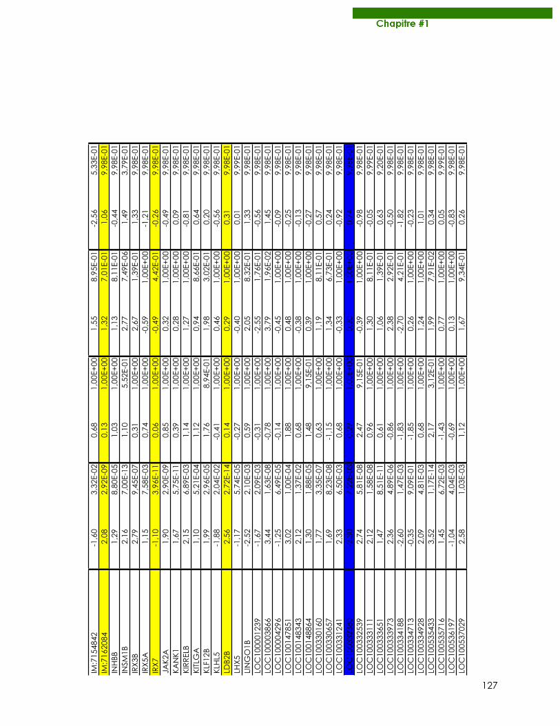
127
IM:7154842
-1,60
3,32E-02
0,68
1,00E+00
1,55
8,95E-01
-2,56
5,33E-01
IM:7162084
2,08
2,92E-09
0,13
1,00E+00
1,32
7,01E-01
1,06
9,98E-01
INHBB
1,29
8,80E-05
1,03
1,00E+00
1,13
8,11E-01
-0,44
9,98E-01
INSM1B
2,16
7,00E-13
1,10
5,52E-01
2,77
7,49E-06
1,49
3,79E-01
IRX3B
2,79
9,45E-07
0,31
1,00E+00
2,67
1,39E-01
1,33
9,98E-01
IRX5A
1,15
7,58E-03
0,74
1,00E+00
-0,59
1,00E+00
-1,21
9,98E-01
IRX7
-1,10
3,96E-11
0,06
1,00E+00
-0,49
4,42E-01
-0,26
9,98E-01
JAK2A
1,90
2,90E-09
0,85
1,00E+00
0,32
1,00E+00
-0,49
9,98E-01
KANK1
1,67
5,75E-11
0,39
1,00E+00
0,28
1,00E+00
0,09
9,98E-01
KIRRELB
2,15
6,89E-03
1,14
1,00E+00
1,27
1,00E+00
-0,81
9,98E-01
KITLGA
1,10
5,21E-04
1,12
1,00E+00
0,94
8,66E-01
0,64
9,98E-01
KLF12B
1,99
2,96E-05
1,76
8,94E-01
1,98
3,02E-01
0,20
9,98E-01
KLHL5
-1,88
2,04E-02
-0,41
1,00E+00
0,46
1,00E+00
-0,56
9,98E-01
LDB2B
2,56
2,72E-14
0,14
1,00E+00
0,29
1,00E+00
0,31
9,98E-01
LHX5
-1,17
5,74E-05
-0,27
1,00E+00
-0,40
1,00E+00
0,01
9,99E-01
LINGO1B
-2,52
2,10E-03
0,59
1,00E+00
2,05
8,32E-01
1,33
9,98E-01
LOC100001239
-1,67
2,09E-03
-0,31
1,00E+00
-2,55
1,76E-01
-0,56
9,98E-01
LOC100003866
3,44
1,63E-08
-0,78
1,00E+00
3,79
1,96E-02
1,45
9,98E-01
LOC100004296
-1,25
6,49E-05
-0,14
1,00E+00
-0,45
1,00E+00
-0,09
9,98E-01
LOC100147851
3,02
1,00E-04
1,88
1,00E+00
0,48
1,00E+00
-0,25
9,98E-01
LOC100148343
2,12
1,37E-02
0,68
1,00E+00
-0,38
1,00E+00
-0,13
9,98E-01
LOC100148864
1,30
1,88E-05
1,48
9,15E-01
0,39
1,00E+00
-0,27
9,98E-01
LOC100330160
1,77
3,35E-07
0,63
1,00E+00
1,19
8,11E-01
0,57
9,98E-01
LOC100330657
1,69
8,23E-08
-1,15
1,00E+00
1,34
6,73E-01
0,24
9,98E-01
LOC100331241
2,33
6,50E-03
0,68
1,00E+00
-0,33
1,00E+00
-0,92
9,98E-01
LOC100332380
-2,30
7,22E-03
0,29
1,00E+00
0,20
1,00E+00
0,74
9,98E-01
LOC100332539
2,74
5,81E-08
2,47
9,15E-01
-0,39
1,00E+00
-0,98
9,98E-01
LOC100333111
2,12
1,58E-08
0,96
1,00E+00
1,30
8,11E-01
-0,05
9,99E-01
LOC100333651
1,47
8,51E-11
0,61
1,00E+00
1,06
1,39E-01
0,63
9,20E-01
LOC100333973
2,36
4,89E-06
-0,86
1,00E+00
2,38
2,92E-01
-0,50
9,98E-01
LOC100334188
-2,60
1,47E-03
-1,83
1,00E+00
-2,70
4,21E-01
-1,82
9,98E-01
LOC100334713
-0,35
9,09E-01
-1,85
1,00E+00
0,26
1,00E+00
-0,23
9,98E-01
LOC100334928
2,09
4,81E-03
0,68
1,00E+00
1,24
1,00E+00
1,01
9,98E-01
LOC100535433
3,52
1,17E-14
2,17
3,12E-01
1,99
7,91E-02
0,34
9,98E-01
LOC100535716
1,45
6,72E-03
-1,43
1,00E+00
0,77
1,00E+00
0,05
9,99E-01
LOC100536197
-1,04
4,04E-03
-0,69
1,00E+00
0,13
1,00E+00
-0,83
9,98E-01
LOC100537029
2,58
1,03E-03
1,12
1,00E+00
1,67
9,34E-01
0,26
9,98E-01
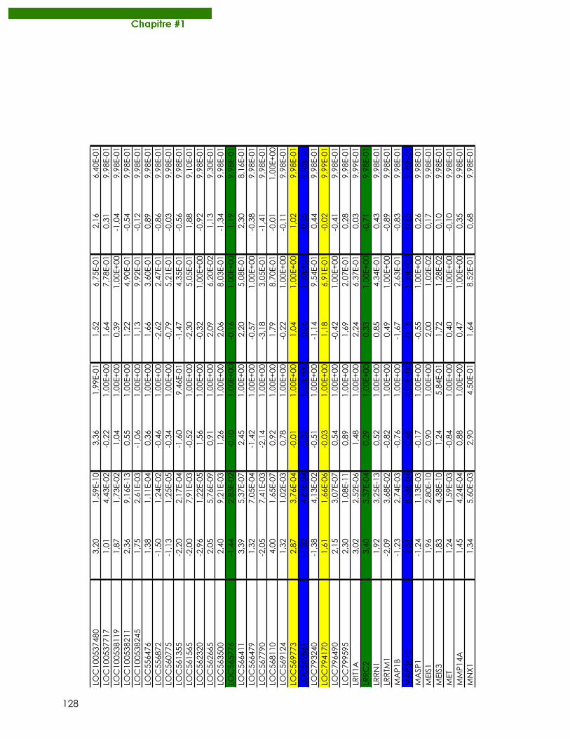
128
LOC100537480
3,20
1,59E-10
3,36
1,99E-01
1,52
6,75E-01
2,16
6,40E-01
LOC100537717
1,01
4,43E-02
-0,22
1,00E+00
1,64
7,78E-01
0,31
9,98E-01
LOC100538119
1,87
1,73E-02
1,04
1,00E+00
0,39
1,00E+00
-1,04
9,98E-01
LOC100538211
2,36
9,16E-13
0,55
1,00E+00
1,22
4,90E-01
-0,54
9,98E-01
LOC100538245
1,75
2,61E-03
-1,06
1,00E+00
1,13
9,92E-01
-0,12
9,98E-01
LOC556476
1,38
1,11E-04
0,36
1,00E+00
1,66
3,60E-01
0,89
9,98E-01
LOC556872
-1,50
1,24E-02
-0,46
1,00E+00
-2,62
2,47E-01
-0,86
9,98E-01
LOC560775
-1,13
1,25E-05
-0,34
1,00E+00
-0,79
5,21E-01
-0,03
9,98E-01
LOC561355
-2,20
2,17E-04
-1,60
9,46E-01
-1,47
4,35E-01
-0,56
9,98E-01
LOC561565
-2,00
7,91E-03
-0,52
1,00E+00
-2,30
5,05E-01
1,88
9,10E-01
LOC562320
-2,96
1,22E-05
1,56
1,00E+00
-0,32
1,00E+00
-0,92
9,98E-01
LOC562665
2,05
5,76E-09
0,91
1,00E+00
2,09
6,20E-02
1,13
9,30E-01
LOC563500
2,40
9,21E-03
1,26
1,00E+00
2,06
8,03E-01
-1,34
9,98E-01
LOC565776
-1,44
2,83E-02
-0,10
1,00E+00
-0,16
1,00E+00
1,19
9,98E-01
LOC566411
3,39
5,37E-07
2,45
1,00E+00
2,20
5,08E-01
2,30
8,16E-01
LOC566479
1,32
7,05E-04
-1,42
1,00E+00
-0,57
1,00E+00
-0,38
9,98E-01
LOC567790
-2,05
7,41E-03
-2,14
1,00E+00
-3,18
3,05E-01
-1,41
9,98E-01
LOC568110
4,00
1,65E-07
0,92
1,00E+00
1,79
8,70E-01
-0,01
1,00E+00
LOC569124
1,32
1,02E-03
0,78
1,00E+00
-0,22
1,00E+00
-0,11
9,98E-01
LOC569773
2,87
3,76E-04
-0,01
1,00E+00
1,04
1,00E+00
1,02
9,98E-01
LOC569861
-1,32
4,67E-04
-0,32
1,00E+00
-0,06
1,00E+00
-0,25
9,98E-01
LOC793240
-1,38
4,13E-02
-0,51
1,00E+00
-1,14
9,54E-01
0,44
9,98E-01
LOC794170
1,61
1,66E-06
-0,03
1,00E+00
1,18
6,91E-01
-0,02
9,99E-01
LOC796490
2,15
3,07E-07
0,54
1,00E+00
-0,42
1,00E+00
-0,41
9,98E-01
LOC799595
2,30
1,08E-11
0,89
1,00E+00
1,69
2,07E-01
0,28
9,98E-01
LRIT1A
3,02
2,52E-06
1,48
1,00E+00
2,24
6,37E-01
0,03
9,99E-01
LRRC2
3,40
3,37E-04
0,29
1,00E+00
0,33
1,00E+00
-0,71
9,98E-01
LRRN1
1,92
3,25E-13
0,52
1,00E+00
0,85
4,34E-01
0,43
9,98E-01
LRRTM1
-2,09
3,68E-02
-0,82
1,00E+00
0,49
1,00E+00
-0,89
9,98E-01
MAP1B
-1,23
2,74E-03
-0,76
1,00E+00
-1,67
2,63E-01
-0,83
9,98E-01
MAP3K10
2,31
8,36E-04
0,68
1,00E+00
-0,15
1,00E+00
0,63
9,98E-01
MASP1
-1,24
1,13E-03
-0,17
1,00E+00
-0,55
1,00E+00
0,26
9,98E-01
MEIS1
1,96
2,80E-10
0,90
1,00E+00
2,00
1,02E-02
0,17
9,98E-01
MEIS3
1,83
4,38E-10
1,24
5,84E-01
1,72
1,28E-02
0,10
9,98E-01
MET
1,24
1,59E-03
-0,84
1,00E+00
0,40
1,00E+00
-0,10
9,98E-01
MMP14A
1,45
4,24E-04
0,88
1,00E+00
0,47
1,00E+00
0,35
9,98E-01
MNX1
1,34
5,60E-03
2,90
4,50E-01
1,64
8,52E-01
0,68
9,98E-01
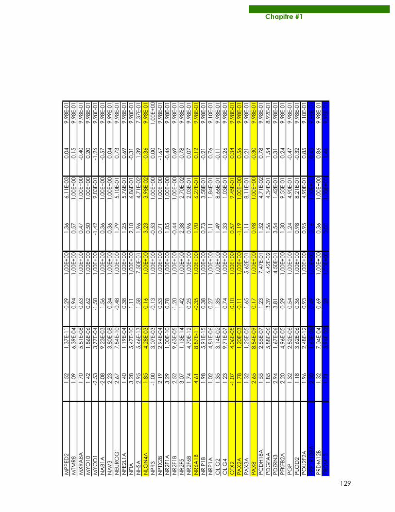
129
MPPED2
1,52
1,37E-11
-0,29
1,00E+00
1,36
6,11E-03
0,04
9,98E-01
MTMR8
1,09
6,39E-04
0,94
1,00E+00
0,57
1,00E+00
-0,15
9,98E-01
MXRA8A
1,70
5,81E-08
0,63
1,00E+00
0,47
1,00E+00
-0,40
9,98E-01
MYO10
1,42
1,86E-06
0,62
1,00E+00
0,50
1,00E+00
0,20
9,98E-01
MYOD1
-2,53
3,77E-04
-1,58
1,00E+00
-1,42
9,83E-01
-1,26
9,98E-01
NAB1A
-2,08
9,23E-03
1,56
1,00E+00
0,36
1,00E+00
-0,57
9,98E-01
NAV3
2,23
3,80E-08
0,34
1,00E+00
-0,36
1,00E+00
0,04
9,99E-01
NEUROG1
2,67
7,84E-10
-0,48
1,00E+00
1,79
5,10E-01
0,73
9,98E-01
NFE2L1A
1,40
1,19E-04
0,38
1,00E+00
1,25
5,76E-01
0,69
9,98E-01
NFIA
3,28
5,47E-10
1,11
1,00E+00
2,10
6,86E-01
-0,31
9,98E-01
NHSA
2,95
5,46E-13
1,58
7,50E-01
1,96
4,71E-02
1,39
7,37E-01
NLGN4A
-1,85
4,28E-03
0,16
1,00E+00
-3,23
3,98E-02
-0,36
9,98E-01
NPR3
-1,00
3,07E-03
-0,13
1,00E+00
-0,53
1,00E+00
0,00
1,00E+00
NPTX2B
2,19
2,94E-04
0,53
1,00E+00
0,71
1,00E+00
-1,67
9,98E-01
NR2F1A
3,29
1,00E-10
0,78
1,00E+00
1,05
1,00E+00
-0,46
9,98E-01
NR2F1B
2,52
9,63E-05
-1,20
1,00E+00
-0,44
1,00E+00
0,69
9,98E-01
NR2F5
3,07
1,13E-14
1,42
1,00E+00
2,38
2,70E-02
-0,78
9,98E-01
NR2F6B
1,74
4,70E-12
0,25
1,00E+00
0,96
2,03E-01
0,07
9,98E-01
NR6A1B
4,61
8,87E-11
-0,35
1,00E+00
1,90
6,27E-01
0,12
9,98E-01
NRIP1B
1,98
5,91E-15
0,38
1,00E+00
0,73
3,58E-01
-0,21
9,98E-01
NRP1A
1,02
4,81E-06
0,27
1,00E+00
1,11
1,84E-01
0,76
9,10E-01
OLIG2
1,35
3,14E-02
1,35
1,00E+00
1,49
8,66E-01
-0,11
9,98E-01
OLIG4
1,23
9,71E-07
0,74
1,00E+00
1,33
1,03E-01
-0,26
9,98E-01
OTX2
-1,07
4,06E-05
0,10
1,00E+00
0,57
9,45E-01
0,34
9,98E-01
PAX2A
1,78
1,20E-03
-0,11
1,00E+00
-1,19
1,00E+00
0,56
9,98E-01
PAX3A
1,32
1,25E-05
1,65
5,62E-01
1,11
8,11E-01
0,21
9,98E-01
PAX8
2,65
8,84E-09
0,17
1,00E+00
0,98
1,00E+00
-0,30
9,98E-01
PCDH18A
1,55
2,55E-07
1,23
7,47E-01
1,52
4,71E-02
0,78
9,98E-01
PDGFAA
1,85
5,88E-06
3,39
6,42E-02
1,56
4,34E-01
1,54
8,92E-01
PDZRN3
2,94
1,67E-06
3,81
4,50E-01
3,54
1,42E-01
0,31
9,98E-01
PFKFB2A
2,20
4,96E-05
-0,29
1,00E+00
1,30
9,55E-01
-0,24
9,98E-01
PGP
1,32
2,82E-06
0,54
1,00E+00
1,24
4,90E-01
-0,47
9,98E-01
PLOD2
1,18
3,62E-06
0,36
1,00E+00
0,98
8,21E-01
-0,32
9,98E-01
POU2F2A
1,96
2,48E-12
0,93
1,00E+00
0,95
4,90E-01
0,85
9,10E-01
PPP1R13BA
2,30
1,63E-06
0,49
1,00E+00
0,16
1,00E+00
-0,43
9,98E-01
PRDM12B
1,32
7,04E-04
0,69
1,00E+00
0,36
1,00E+00
0,86
9,98E-01
PRDM13
1,71
3,94E-02
-1,28
1,00E+00
0,00
1,00E+00
1,46
9,98E-01
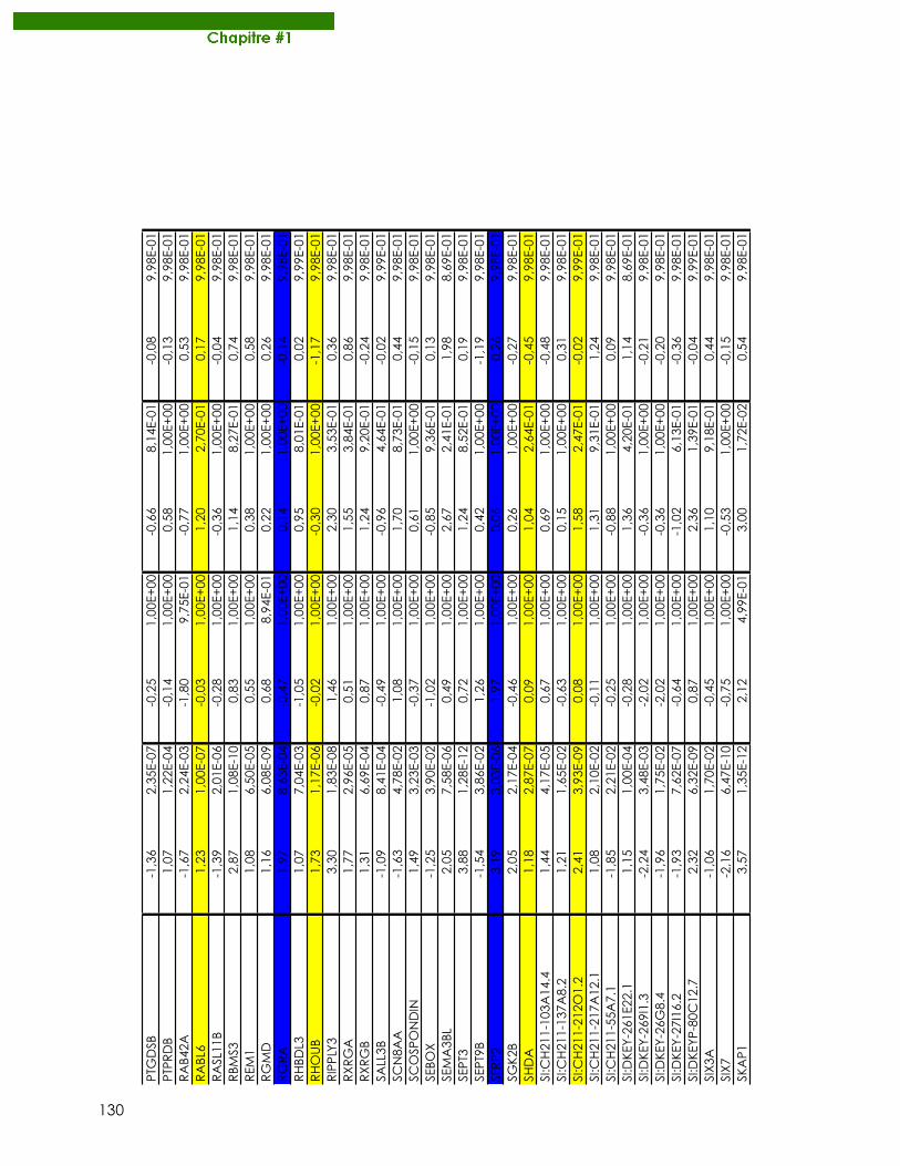
130
PTGDSB
-1,36
2,35E-07
-0,25
1,00E+00
-0,66
8,14E-01
-0,08
9,98E-01
PTPRDB
1,07
1,22E-04
-0,14
1,00E+00
0,58
1,00E+00
-0,13
9,98E-01
RAB42A
-1,67
2,24E-03
-1,80
9,75E-01
-0,77
1,00E+00
0,53
9,98E-01
RABL6
1,23
1,00E-07
-0,03
1,00E+00
1,20
2,70E-01
0,17
9,98E-01
RASL11B
-1,39
2,01E-06
-0,28
1,00E+00
-0,36
1,00E+00
-0,04
9,98E-01
RBMS3
2,87
1,08E-10
0,83
1,00E+00
1,14
8,27E-01
0,74
9,98E-01
REM1
1,08
6,50E-05
0,55
1,00E+00
0,38
1,00E+00
0,58
9,98E-01
RGMD
1,16
6,08E-09
0,68
8,94E-01
0,22
1,00E+00
0,26
9,98E-01
RGRA
1,97
8,63E-04
-0,47
1,00E+00
0,14
1,00E+00
-0,14
9,98E-01
RHBDL3
1,07
7,04E-03
-1,05
1,00E+00
0,95
8,01E-01
0,02
9,99E-01
RHOUB
1,73
1,17E-06
-0,02
1,00E+00
-0,30
1,00E+00
-1,17
9,98E-01
RIPPLY3
3,30
1,83E-08
1,46
1,00E+00
2,30
3,53E-01
0,36
9,98E-01
RXRGA
1,77
2,96E-05
0,51
1,00E+00
1,55
3,84E-01
0,86
9,98E-01
RXRGB
1,31
6,69E-04
0,87
1,00E+00
1,24
9,20E-01
-0,24
9,98E-01
SALL3B
-1,09
8,41E-04
-0,49
1,00E+00
-0,96
4,64E-01
-0,02
9,99E-01
SCN8AA
-1,63
4,78E-02
1,08
1,00E+00
1,70
8,73E-01
0,44
9,98E-01
SCOSPONDIN
1,49
3,23E-03
-0,37
1,00E+00
0,61
1,00E+00
-0,15
9,98E-01
SEBOX
-1,25
3,90E-02
-1,02
1,00E+00
-0,85
9,36E-01
0,13
9,98E-01
SEMA3BL
2,05
7,58E-06
0,49
1,00E+00
2,67
2,41E-01
1,98
8,69E-01
SEPT3
3,88
1,28E-12
0,72
1,00E+00
1,24
8,52E-01
0,19
9,98E-01
SEPT9B
-1,54
3,86E-02
1,26
1,00E+00
0,42
1,00E+00
-1,19
9,98E-01
SFRP2
3,19
3,03E-06
1,97
1,00E+00
0,06
1,00E+00
0,26
9,98E-01
SGK2B
2,05
2,17E-04
-0,46
1,00E+00
0,26
1,00E+00
-0,27
9,98E-01
SHDA
1,18
2,87E-07
0,09
1,00E+00
1,04
2,64E-01
-0,45
9,98E-01
SI:CH211-103A14.4
1,44
4,17E-05
0,67
1,00E+00
0,69
1,00E+00
-0,48
9,98E-01
SI:CH211-137A8.2
1,21
1,65E-02
-0,63
1,00E+00
0,15
1,00E+00
0,31
9,98E-01
SI:CH211-212O1.2
2,41
3,93E-09
0,08
1,00E+00
1,58
2,47E-01
-0,02
9,99E-01
SI:CH211-217A12.1
1,08
2,10E-02
-0,11
1,00E+00
1,31
9,31E-01
1,24
9,98E-01
SI:CH211-55A7.1
-1,85
2,21E-02
-0,25
1,00E+00
-0,88
1,00E+00
0,09
9,98E-01
SI:DKEY-261E22.1
1,15
1,00E-04
-0,28
1,00E+00
1,36
4,20E-01
1,14
8,69E-01
SI:DKEY-269I1.3
-2,24
3,48E-03
-2,02
1,00E+00
-0,36
1,00E+00
-0,21
9,98E-01
SI:DKEY-26G8.4
-1,96
1,75E-02
-2,02
1,00E+00
-0,36
1,00E+00
-0,20
9,98E-01
SI:DKEY-27I16.2
-1,93
7,62E-07
-0,64
1,00E+00
-1,02
6,13E-01
-0,36
9,98E-01
SI:DKEYP-80C12.7
2,32
6,32E-09
0,87
1,00E+00
2,36
1,39E-01
-0,04
9,99E-01
SIX3A
-1,06
1,70E-02
-0,45
1,00E+00
1,10
9,18E-01
0,44
9,98E-01
SIX7
-2,16
6,47E-10
-0,75
1,00E+00
-0,53
1,00E+00
-0,15
9,98E-01
SKAP1
3,57
1,35E-12
2,12
4,99E-01
3,00
1,72E-02
0,54
9,98E-01
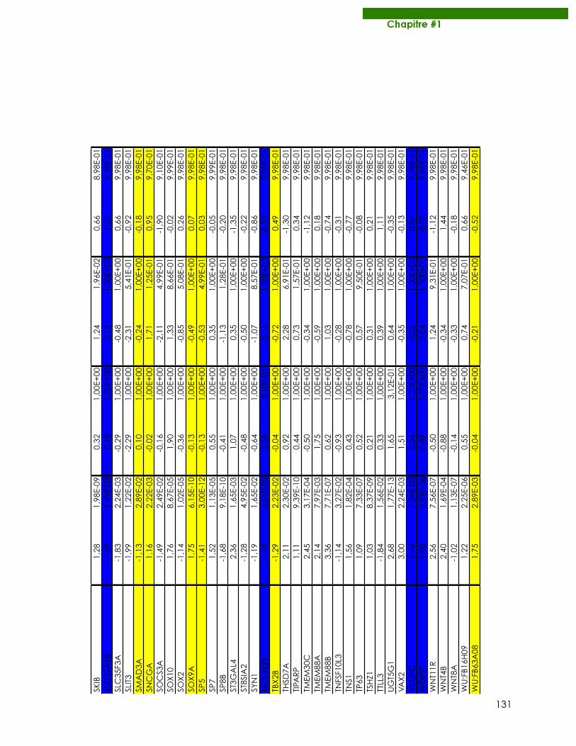
131
SKIB
1,28
1,98E-09
0,32
1,00E+00
1,24
1,96E-02
0,66
8,98E-01
SLC22A18
-1,59
1,49E-03
0,18
1,00E+00
0,13
1,00E+00
1,07
9,98E-01
SLC35F3A
-1,83
2,24E-03
-0,29
1,00E+00
-0,48
1,00E+00
0,66
9,98E-01
SLIT3
-1,99
1,22E-02
-2,29
1,00E+00
-2,31
5,41E-01
-0,92
9,98E-01
SMAD3A
-1,13
2,89E-02
0,10
1,00E+00
-0,24
1,00E+00
-0,18
9,98E-01
SNCGA
1,16
2,22E-03
-0,02
1,00E+00
1,71
1,25E-01
0,95
9,70E-01
SOCS3A
-1,49
2,49E-02
-0,16
1,00E+00
-2,11
4,99E-01
-1,90
9,10E-01
SOX10
1,76
8,67E-05
1,90
1,00E+00
1,33
8,66E-01
-0,02
9,99E-01
SOX2
-1,14
1,02E-05
-0,36
1,00E+00
-0,85
5,08E-01
0,26
9,98E-01
SOX9A
1,75
6,15E-10
-0,13
1,00E+00
-0,49
1,00E+00
0,07
9,98E-01
SP5
-1,41
3,00E-12
-0,13
1,00E+00
-0,53
4,99E-01
0,03
9,98E-01
SP7
1,52
1,13E-05
0,55
1,00E+00
0,35
1,00E+00
-0,05
9,99E-01
SP8B
-1,68
9,18E-10
-0,41
1,00E+00
-1,13
1,28E-01
-0,20
9,98E-01
ST3GAL4
2,36
1,65E-03
1,07
1,00E+00
0,35
1,00E+00
-1,35
9,98E-01
ST8SIA2
-1,28
4,95E-02
-0,48
1,00E+00
-0,50
1,00E+00
-0,22
9,98E-01
SYN1
-1,19
1,65E-02
-0,64
1,00E+00
-1,07
8,57E-01
-0,86
9,98E-01
TAGLN3B
-1,49
4,64E-03
-2,59
3,10E-01
-0,14
1,00E+00
-0,93
9,98E-01
TBX2B
-1,29
2,23E-02
-0,04
1,00E+00
-0,72
1,00E+00
0,49
9,98E-01
THSD7A
2,11
2,30E-02
0,92
1,00E+00
2,28
6,91E-01
-1,30
9,98E-01
TIPARP
1,11
9,39E-10
0,44
1,00E+00
0,73
1,57E-01
0,34
9,98E-01
TMEM30C
2,45
3,17E-04
-0,50
1,00E+00
-0,34
1,00E+00
-1,12
9,98E-01
TMEM88A
2,14
7,97E-03
1,75
1,00E+00
-0,59
1,00E+00
0,18
9,98E-01
TMEM88B
3,36
7,71E-07
0,62
1,00E+00
1,03
1,00E+00
-0,74
9,98E-01
TNFSF10L3
-1,14
3,27E-02
-0,93
1,00E+00
-0,28
1,00E+00
-0,31
9,98E-01
TNS1
1,56
1,82E-04
0,43
1,00E+00
-0,78
1,00E+00
-0,77
9,98E-01
TP63
1,09
7,33E-07
0,52
1,00E+00
0,57
9,50E-01
-0,08
9,98E-01
TSHZ1
1,03
8,37E-09
0,21
1,00E+00
0,31
1,00E+00
0,21
9,98E-01
TTLL3
-1,84
1,56E-02
0,33
1,00E+00
0,39
1,00E+00
1,11
9,98E-01
UGT5G1
2,68
1,77E-13
1,65
3,12E-01
0,64
1,00E+00
-0,35
9,98E-01
VAX2
3,00
2,24E-03
1,51
1,00E+00
-0,35
1,00E+00
-0,13
9,98E-01
VEGFC
1,76
7,04E-03
0,56
1,00E+00
-0,02
1,00E+00
0,87
9,98E-01
WDR90
1,90
2,99E-06
-0,89
1,00E+00
0,04
1,00E+00
-0,07
9,98E-01
WNT11R
2,56
7,56E-07
-0,50
1,00E+00
1,24
9,31E-01
-1,12
9,98E-01
WNT4B
2,40
1,69E-04
-0,88
1,00E+00
-0,34
1,00E+00
1,44
9,98E-01
WNT8A
-1,02
1,13E-07
-0,14
1,00E+00
-0,33
1,00E+00
-0,18
9,98E-01
WU:FB16H09
1,22
2,25E-06
0,55
1,00E+00
0,74
7,07E-01
0,66
9,46E-01
WU:FB63A08
1,75
2,89E-03
-0,04
1,00E+00
-0,21
1,00E+00
-0,52
9,98E-01
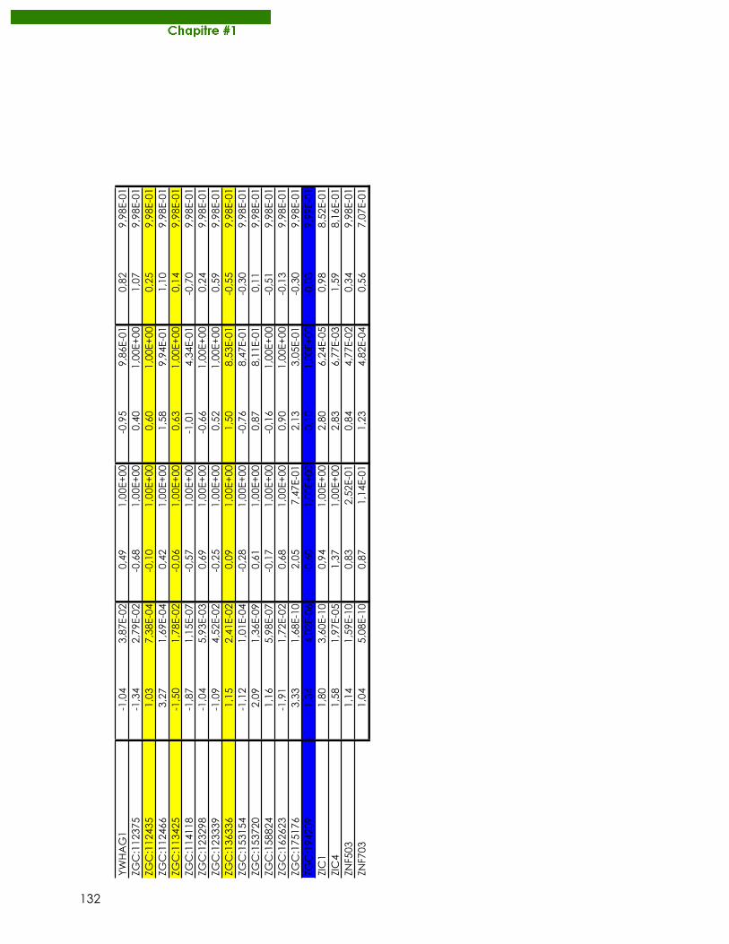
132
YWHAG1
-1,04
3,87E-02
0,49
1,00E+00
-0,95
9,86E-01
0,82
9,98E-01
ZGC:112375
-1,34
2,79E-02
-0,68
1,00E+00
0,40
1,00E+00
1,07
9,98E-01
ZGC:112435
1,03
7,38E-04
-0,10
1,00E+00
0,60
1,00E+00
0,25
9,98E-01
ZGC:112466
3,27
1,69E-04
0,42
1,00E+00
1,58
9,94E-01
1,10
9,98E-01
ZGC:113425
-1,50
1,78E-02
-0,06
1,00E+00
0,63
1,00E+00
0,14
9,98E-01
ZGC:114118
-1,87
1,15E-07
-0,57
1,00E+00
-1,01
4,34E-01
-0,70
9,98E-01
ZGC:123298
-1,04
5,93E-03
0,69
1,00E+00
-0,66
1,00E+00
0,24
9,98E-01
ZGC:123339
-1,09
4,52E-02
-0,25
1,00E+00
0,52
1,00E+00
0,59
9,98E-01
ZGC:136336
1,15
2,41E-02
0,09
1,00E+00
1,50
8,53E-01
-0,55
9,98E-01
ZGC:153154
-1,12
1,01E-04
-0,28
1,00E+00
-0,76
8,47E-01
-0,30
9,98E-01
ZGC:153720
2,09
1,36E-09
0,61
1,00E+00
0,87
8,11E-01
0,11
9,98E-01
ZGC:158824
1,16
5,98E-07
-0,17
1,00E+00
-0,16
1,00E+00
-0,51
9,98E-01
ZGC:162623
-1,91
1,72E-02
0,68
1,00E+00
0,90
1,00E+00
-0,13
9,98E-01
ZGC:175176
3,33
1,68E-10
2,05
7,47E-01
2,13
3,05E-01
-0,30
9,98E-01
ZGC:194209
1,34
4,02E-06
-0,60
1,00E+00
-0,10
1,00E+00
-0,03
9,99E-01
ZIC1
1,80
3,60E-10
0,94
1,00E+00
2,80
6,24E-05
0,98
8,52E-01
ZIC4
1,58
1,97E-05
1,37
1,00E+00
2,83
6,77E-03
1,59
8,16E-01
ZNF503
1,14
1,59E-10
0,83
2,52E-01
0,84
4,77E-02
0,34
9,98E-01
ZNF703
1,04
5,08E-10
0,87
1,14E-01
1,23
4,82E-04
0,56
7,07E-01
133
IV. Travaux complémentaires - Discussion – Perspectives
Des expériences en cours dans le laboratoire de Cécile Rochette-Egly sur des cellules
ES de souris traitées à l’AR ont également montré que les différents sous-types de RAR
activent des gènes différents. En effet, les cellules ES expriment les sous-types RARα et RARγ
et parmi l’ensemble des gènes répondant à l’AR, la moitié nécessite spécifiquement RARγ
pour leur régulation par l’AR (données de RNAseq couplées à du ChIPseq (Al Tanoury et al.,
en préparation). De manière intéressante, il semble que les gènes régulés par RARγ soient
impliqués dans la différenciation neuronale induite par l’AR, alors que RARα régule les autres
gènes cibles de l’AR. Ainsi, ces données récentes appuient nos travaux en montrant que
dans un système cellulaire exprimant plusieurs sous-types de RAR, il existe une activité
transcriptionnelle propre à chacun.
1. Comment est régulée la spécificité d’action des sous-types de RAR ?
Les transcriptotypes spécifiques des différents sous-types de RAR que nous avons
décrit dans nos résultats sont la conséquence de différences transcriptionnelles (page 101
et figure 2 page 100). Quels sont les mécanismes permettant le recrutement spécifique d’un
sous-type de RAR au niveau du promoteur d’un gène cible en particulier ? Comment les
différents sous-types de RAR, bien qu’exprimés dans les mêmes cellules, régulent des
répertoires de gènes différents ?
1.1. Régulation par la séquence d’ADN ?
C’en en premier lieu la présence d’un RARE dans le promoteur d’un gène qui est à
l’origine du recrutement des RAR pour la régulation de son expression. Bien qu’un consensus
de RARE canonique ait été établi, à savoir la répétition directe du motif RGKTSA séparé par
5 nucléotides, la description des RARE fonctionnels in vivo montre une bien plus grande
diversité de motifs et d’espacements (section A-III-2, page 12). Parmi ces différences, existe-
t-il un motif ou un espacement en particulier recrutant spécifiquement un sous-type de
récepteur ? L’analyse in silico de l’ensemble des RARE consensus trouvés dans le génome
du poisson-zèbre, aux abords des gènes répondant à l’AR, ne montre aucune corrélation
entre la spécificité d’action d’un sous-type et l’enrichissement d’un espacement en
particulier ou d’une séquence consensus spécifique. En d’autres termes, ces résultats
134
suggèrent que le recrutement spécifique des différents sous-types de RAR ne repose pas sur
des différences dans la séquence brute des éléments de réponse au niveau de l’ADN.
Toutefois, les RARE in silico pris en compte dans notre étude ont été obtenus par la
recherche de motifs RGKTSA du type DR1-10 ou IR1-10 dans l’ensemble du génome du
poisson-zèbre par la plateforme PARSEC. Ainsi, ces éléments de réponse ne correspondent
pas nécessairement aux RARE effectivement fonctionnels régulant l’expression des gènes
cibles de l’AR. Pour avoir accès à ces derniers, il serait nécessaire d’analyser par ChIP-seq le
recrutement différentiel des différents sous-types de RAR in vivo et ainsi décrire des cistromes
spécifiques pour chacun des sous-types. Ces travaux nécessitent des anticorps spécifiques
de chaque sous-type de RAR de poisson-zèbre et capables d’immunoprécipiter la protéine
in vivo. Ces travaux permettraient d’avoir un aperçu de l’ensemble des sites de recrutement
différentiels des différents sous-types de RAR (cistromes spécifiques de chaque sous-type) et
ainsi de corréler d’éventuelles spécificités de séquence et/ou d’espacement au niveau des
RARE et le recrutement spécifique d’un sous-type de RAR.
1.2. Régulation au niveau du récepteur ?
i. RAR ?
Les différences majeures dans la séquence protéique des sous-types de RAR sont
retrouvées dans le NTD et la partie C-terminale. Mais comment de telles régions non
structurées peuvent influer sur le recrutement du récepteur à l’ADN ? Des études récentes
suggèrent que de part sa proximité avec le DBD, le NTD pourrait influencer l’interaction du
récepteur avec l’ADN. En particulier, l’équipe de Cécile Rochette-Egly a montré que
certaines protéines du cytosquelette interagissent spécifiquement avec un sous-type de
RAR et peuvent réguler leur recrutement au niveau de l’ADN. C’est le cas de la vinexine
beta qui interagit spécifiquement avec le NTD de RARγ et empêche son interaction in vitro
avec un RARE, et cette interaction est abolie par la phosphorylation du NTD de RARγ,
(publication en annexe, page 195). D’autre part, des études récentes du laboratoire ont
montré que dans des cellules ES, RARγ phosphorylé était spécifiquement recruté au niveau
de RARE de type DR5 et DR7. Ainsi, il est possible que la phosphorylation du récepteur et
l’interaction de protéines au niveau du NTD agissent de manière synergique et coordonnée
pour réguler le recrutement du récepteur au niveau d’un RARE spécifique. Une telle
hypothèse suggère une régulation complexe de la spécificité d’action des différents sous-
types de RAR.
135
ii. RXR ?
Le partenaire d’hétérodimérisation RXR est aussi susceptible de jouer un rôle dans
l’activité transcriptionnelle spécifique des différents sous-types de RAR. Il existe six gènes
paralogues codants pour les RXR chez le poisson-zèbre et rxrβa est le sous-type le plus
fortement exprimé dans les premières étapes du développement du poisson-zèbre
(Bertrand et al., 2007; Oliveira et al., 2013; Tallafuss et al., 2006; Waxman and Yelon, 2007).
Ainsi, il peut se former pas moins de 24 différents hétérodimères RAR-RXR sans prendre en
compte les différentes isoformes au sein de chaque sous-type. De ce fait, il est fortement
envisageable que les différents hétérodimères puissent présenter une spécificité
d’interaction avec certains cofacteurs. De plus, il existe une relation entre la topologie
d’hétéromidérisation des DBD des deux partenaires RAR et RXR et l’espacement entre les
demi-sites des RARE. Ainsi, les différents hétérodimères peuvent présenter des différences
topologiques subtiles favorisant leur recrutement sur un type de RARE donné.
1.3. Régulation par l’environnement chromatinien ?
Les facteurs de transcription peuvent agir en synergie pour contrôler l’expression de
gènes cibles au sein de réseaux cis-régulateurs dans les promoteurs des gènes régulés. En
particulier, il a été montré que l’association de certains récepteurs nucléaires au niveau de
leurs éléments de réponse nécessite l’interaction concomitante d’autres facteurs de
transcription dans l’environnement chromatinien proche. C’est le cas des facteurs FoxA1 ou
GATA3 dont l’interaction à la chromatine est requise pour l’interaction des RAR sur les RARE
de certains gènes dans des cellules de cancer du sein (Hua et al., 2009). En effet, dans ces
cellules plus de 40% des régions occupées par les RAR sont également occupées par FoxA1
et 20% sont aussi occupées par le récepteur des œstrogènes ERα. Ainsi, il est tout à fait
envisageable que le recrutement spécifique d’un sous-type de RAR au niveau des
séquences régulatrices d’un gène cible puisse être corrélé à la liaison d’autres facteurs au
niveau de la même région de chromatine. Cette hypothèse pourrait être testée par
l’analyse des sites de liaisons de facteurs de transcription comme FoxA1 ou d’autres facteurs
aux alentours des RARE recensés par ChiP-seq.
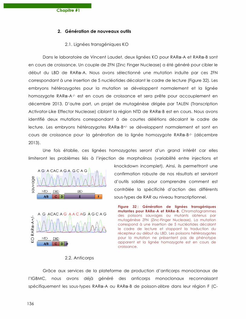
136
2. Génération de nouveaux outils
2.1. Lignées transgéniques KO
Dans le laboratoire de Vincent Laudet, deux lignées KO pour RARα-A et RARα-B sont
en cours de croissance. Un couple de ZFN (Zinc Finger Nuclease) a été généré pour cibler le
début du LBD de RARα-A. Nous avons sélectionné une mutation induite par ces ZFN
correspondant à une insertion de 5 nucléotides décalant le cadre de lecture (Figure 32). Les
embryons hétérozygotes pour la mutation se développent normalement et la lignée
homozygote RARα-A-/- est en cours de croissance et sera prête pour accouplement en
décembre 2013. D’autre part, un projet de mutagénèse dirigée par TALEN (Transcription
Activator-Like Effector Nuclease) ciblant la région NTD de RARα-B est en cours. Nous avons
identifié deux mutations correspondant à de courtes délétions décalant le cadre de
lecture. Les embryons hétérozygotes RARα-B+/- se développent normalement et sont en
cours de croissance pour- la génération de la lignée homozygote RARα-B-/- (décembre
2013).
Une fois établie, ces lignées homozygotes seront d’un grand intérêt car elles
limiteront les problèmes liés à l’injection de morpholinos (variabilité entre injections et
knockdown incomplet). Ainsi, ils permettront une
confirmation robuste de nos résultats et serviront
d’outils solides pour comprendre comment est
contrôlée la spécificité d’action des différents
sous-types de RAR au niveau transcriptionnel.
2.2. Anticorps
Grâce aux services de la plateforme de production d’anticorps monoclonaux de
l’IGBMC, nous avons déjà généré des anticorps monoclonaux reconnaissant
spécifiquement les sous-types RARα-A ou RARα-B de poisson-zèbre dans leur région F (C-
Figure 32 : Génération de lignées transgéniques mutantes pour RARα-A et RARα-B. Chromatogrammes des poissons sauvages ou mutants obtenus par mutagénèse ZFN (Zinc-Finger Nuclease). La mutation correspond à une insertion de 5 nucléotides décalant le cadre de lecture et stoppant la traduction du récepteur au début du LBD. Les poissons hétérozygotes pour la mutation ne présentent pas de phénotype apparent et la lignée homozygote est en cours de croissance.
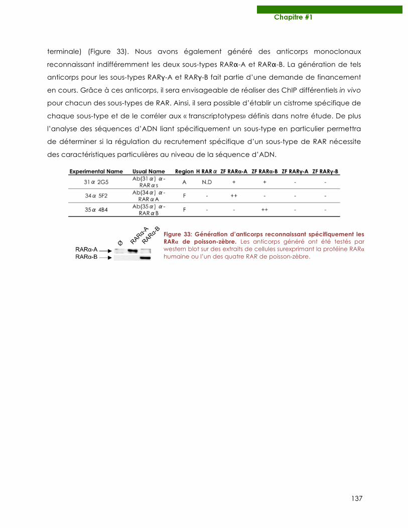
137
terminale) (Figure 33). Nous avons également généré des anticorps monoclonaux
reconnaissant indifféremment les deux sous-types RARα-A et RARα-B. La génération de tels
anticorps pour les sous-types RARγ-A et RARγ-B fait partie d’une demande de financement
en cours. Grâce à ces anticorps, il sera envisageable de réaliser des ChIP différentiels in vivo
pour chacun des sous-types de RAR. Ainsi, il sera possible d’établir un cistrome spécifique de
chaque sous-type et de le corréler aux « transcriptotypes» définis dans notre étude. De plus
l’analyse des séquences d’ADN liant spécifiquement un sous-type en particulier permettra
de déterminer si la régulation du recrutement spécifique d’un sous-type de RAR nécessite
des caractéristiques particulières au niveau de la séquence d’ADN.
Figure 33: Génération d’anticorps reconnaissant spécifiquement les RARα de poisson-zèbre. Les anticorps généré ont été testés par western blot sur des extraits de cellules surexprimant la protéine RARα humaine ou l’un des quatre RAR de poisson-zèbre.

138
139
Chapitre#2
La phosphorylation des RAR : structure – fonction – évolution
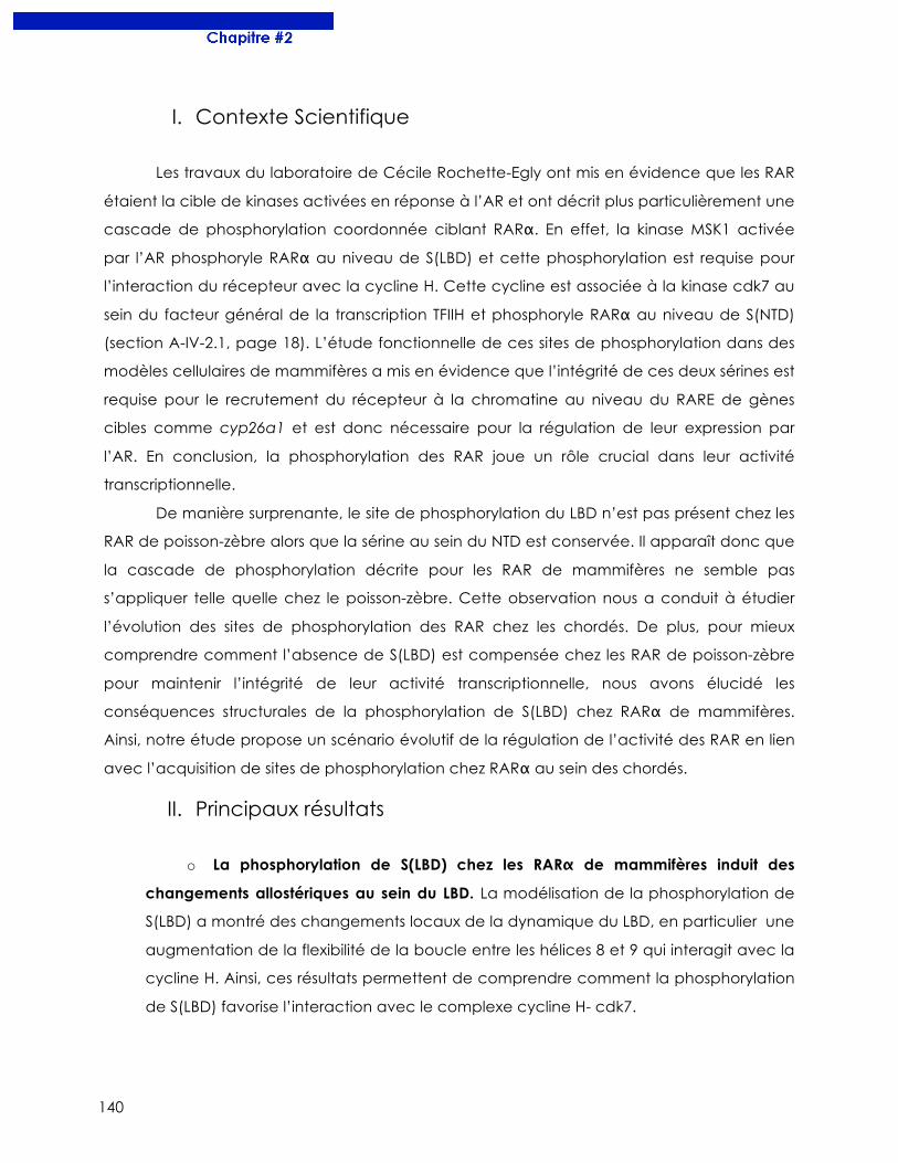
140
I. Contexte Scientifique
Les travaux du laboratoire de Cécile Rochette-Egly ont mis en évidence que les RAR
étaient la cible de kinases activées en réponse à l’AR et ont décrit plus particulièrement une
cascade de phosphorylation coordonnée ciblant RARα. En effet, la kinase MSK1 activée
par l’AR phosphoryle RARα au niveau de S(LBD) et cette phosphorylation est requise pour
l’interaction du récepteur avec la cycline H. Cette cycline est associée à la kinase cdk7 au
sein du facteur général de la transcription TFIIH et phosphoryle RARα au niveau de S(NTD)
(section A-IV-2.1, page 18). L’étude fonctionnelle de ces sites de phosphorylation dans des
modèles cellulaires de mammifères a mis en évidence que l’intégrité de ces deux sérines est
requise pour le recrutement du récepteur à la chromatine au niveau du RARE de gènes
cibles comme cyp26a1 et est donc nécessaire pour la régulation de leur expression par
l’AR. En conclusion, la phosphorylation des RAR joue un rôle crucial dans leur activité
transcriptionnelle.
De manière surprenante, le site de phosphorylation du LBD n’est pas présent chez les
RAR de poisson-zèbre alors que la sérine au sein du NTD est conservée. Il apparaît donc que
la cascade de phosphorylation décrite pour les RAR de mammifères ne semble pas
s’appliquer telle quelle chez le poisson-zèbre. Cette observation nous a conduit à étudier
l’évolution des sites de phosphorylation des RAR chez les chordés. De plus, pour mieux
comprendre comment l’absence de S(LBD) est compensée chez les RAR de poisson-zèbre
pour maintenir l’intégrité de leur activité transcriptionnelle, nous avons élucidé les
conséquences structurales de la phosphorylation de S(LBD) chez RARα de mammifères.
Ainsi, notre étude propose un scénario évolutif de la régulation de l’activité des RAR en lien
avec l’acquisition de sites de phosphorylation chez RARα au sein des chordés.
II. Principaux résultats
o La phosphorylation de S(LBD) chez les RARα de mammifères induit des
changements allostériques au sein du LBD. La modélisation de la phosphorylation de
S(LBD) a montré des changements locaux de la dynamique du LBD, en particulier une
augmentation de la flexibilité de la boucle entre les hélices 8 et 9 qui interagit avec la
cycline H. Ainsi, ces résultats permettent de comprendre comment la phosphorylation
de S(LBD) favorise l’interaction avec le complexe cycline H- cdk7.
141
o Les RARα de poisson-zèbre sont phosphorylés au niveau du NTD mais pas
dans le LBD. Les alignements de séquence entre les RARα humain et de poisson-zèbre
indiquent que S(NTD) est conservée alors que S(LBD) est substituée par un résidu non
phosphorylable chez le poisson zèbre. Nous avons confirmé par des expériences de
phosphorylations in vitro et in cellulo que S(NTD) est bien phosphorylée par cdk7 chez
le poisson-zèbre mais qu’aucune phosphorylation n’est présente dans le LBD.
o L’acquisition de S(LBD) permet une modulation plus fine de l’activité de RARα.
Nos travaux ont permis d’établir un scénario évolutif de la phosphorylation des RAR où
S(LBD) est acquise quatre fois indépendamment chez les vertébrés et permet une
régulation plus fine de l’activité du récepteur. Chez les espèces où S(LBD) est absente,
nous avons identifié certains résidus du LBD qui compensent cette absence pour
maintenir l’intégrité de l’activité de RARα.
III. Publication
Nos travaux ont été publiés dans le journal Molecular Biology and Evolution en 2011.
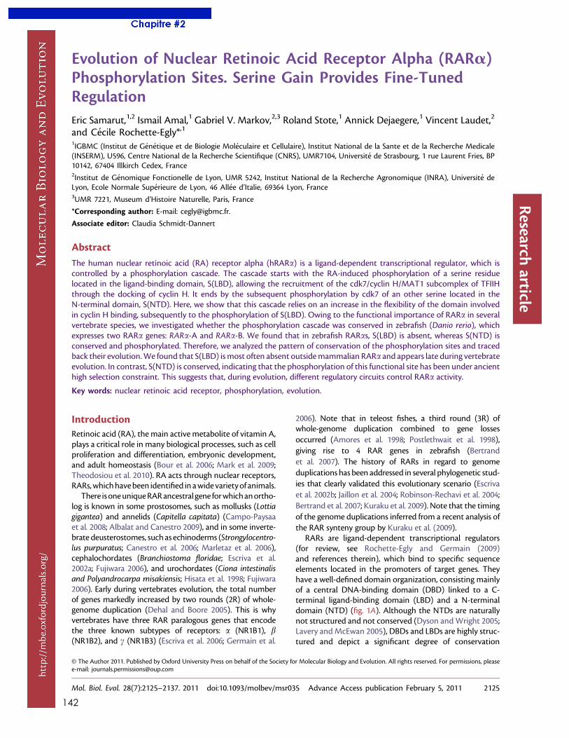
142
Evolution of Nuclear Retinoic Acid Receptor Alpha (RARa)Phosphorylation Sites. Serine Gain Provides Fine-TunedRegulationEric Samarut,1,2 Ismail Amal,1 Gabriel V. Markov,2,3 Roland Stote,1 Annick Dejaegere,1 Vincent Laudet,2
and Cecile Rochette-Egly*,1
1IGBMC (Institut de Genetique et de Biologie Moleculaire et Cellulaire), Institut National de la Sante et de la Recherche Medicale(INSERM), U596, Centre National de la Recherche Scientifique (CNRS), UMR7104, Universite de Strasbourg, 1 rue Laurent Fries, BP10142, 67404 Illkirch Cedex, France2Institut de Genomique Fonctionelle de Lyon, UMR 5242, Institut National de la Recherche Agronomique (INRA), Universite deLyon, Ecole Normale Superieure de Lyon, 46 Allee d’Italie, 69364 Lyon, France3UMR 7221, Museum d’Histoire Naturelle, Paris, France
*Corresponding author: E-mail: [email protected].
Associate editor: Claudia Schmidt-Dannert
Abstract
The human nuclear retinoic acid (RA) receptor alpha (hRARa) is a ligand-dependent transcriptional regulator, which iscontrolled by a phosphorylation cascade. The cascade starts with the RA-induced phosphorylation of a serine residuelocated in the ligand-binding domain, S(LBD), allowing the recruitment of the cdk7/cyclin H/MAT1 subcomplex of TFIIHthrough the docking of cyclin H. It ends by the subsequent phosphorylation by cdk7 of an other serine located in theN-terminal domain, S(NTD). Here, we show that this cascade relies on an increase in the flexibility of the domain involvedin cyclin H binding, subsequently to the phosphorylation of S(LBD). Owing to the functional importance of RARa in severalvertebrate species, we investigated whether the phosphorylation cascade was conserved in zebrafish (Danio rerio), whichexpresses two RARa genes: RARa-A and RARa-B. We found that in zebrafish RARas, S(LBD) is absent, whereas S(NTD) isconserved and phosphorylated. Therefore, we analyzed the pattern of conservation of the phosphorylation sites and tracedback their evolution.We found that S(LBD) ismost often absent outsidemammalian RARa and appears late during vertebrateevolution. In contrast, S(NTD) is conserved, indicating that the phosphorylation of this functional site has been under ancienthigh selection constraint. This suggests that, during evolution, different regulatory circuits control RARa activity.
Key words: nuclear retinoic acid receptor, phosphorylation, evolution.
IntroductionRetinoic acid (RA), the main active metabolite of vitamin A,plays a critical role in many biological processes, such as cellproliferation and differentiation, embryonic development,and adult homeostasis (Bour et al. 2006; Mark et al. 2009;Theodosiou et al. 2010). RA acts through nuclear receptors,RARs,whichhavebeen identified in awidevarietyof animals.
There isoneuniqueRARancestral gene forwhichanortho-log is known in some prostosomes, such as mollusks (Lottiagigantea) and annelids (Capitella capitata) (Campo-Paysaaet al. 2008; Albalat and Canestro 2009), and in some inverte-brate deusterostomes, such as echinoderms (Strongylocentro-lus purpuratus; Canestro et al. 2006; Marletaz et al. 2006),cephalochordates (Branchiostoma floridae; Escriva et al.2002a; Fujiwara 2006), and urochordates (Ciona intestinalisand Polyandrocarpa misakiensis; Hisata et al. 1998; Fujiwara2006). Early during vertebrates evolution, the total numberof genes markedly increased by two rounds (2R) of whole-genome duplication (Dehal and Boore 2005). This is whyvertebrates have three RAR paralogous genes that encodethe three known subtypes of receptors: a (NR1B1), b(NR1B2), and c (NR1B3) (Escriva et al. 2006; Germain et al.
2006). Note that in teleost fishes, a third round (3R) ofwhole-genome duplication combined to gene lossesoccurred (Amores et al. 1998; Postlethwait et al. 1998),giving rise to 4 RAR genes in zebrafish (Bertrandet al. 2007). The history of RARs in regard to genomeduplications has been addressed in several phylogenetic stud-ies that clearly validated this evolutionary scenario (Escrivaet al. 2002b; Jaillon et al. 2004; Robinson-Rechavi et al. 2004;Bertrand et al. 2007; Kuraku et al. 2009). Note that the timingof the genome duplications inferred from a recent analysis ofthe RAR synteny group by Kuraku et al. (2009).
RARs are ligand-dependent transcriptional regulators(for review, see Rochette-Egly and Germain (2009)and references therein), which bind to specific sequenceelements located in the promoters of target genes. Theyhave a well-defined domain organization, consisting mainlyof a central DNA-binding domain (DBD) linked to a C-terminal ligand-binding domain (LBD) and a N-terminaldomain (NTD) (fig. 1A). Although the NTDs are naturallynot structured and not conserved (Dyson andWright 2005;Lavery andMcEwan 2005), DBDs and LBDs are highly struc-tured and depict a significant degree of conservation
© The Author 2011. Published by Oxford University Press on behalf of the Society for Molecular Biology and Evolution. All rights reserved. For permissions, pleasee-mail: [email protected]
Mol. Biol. Evol. 28(7):2125–2137. 2011 doi:10.1093/molbev/msr035 Advance Access publication February 5, 2011 2125
Research
article
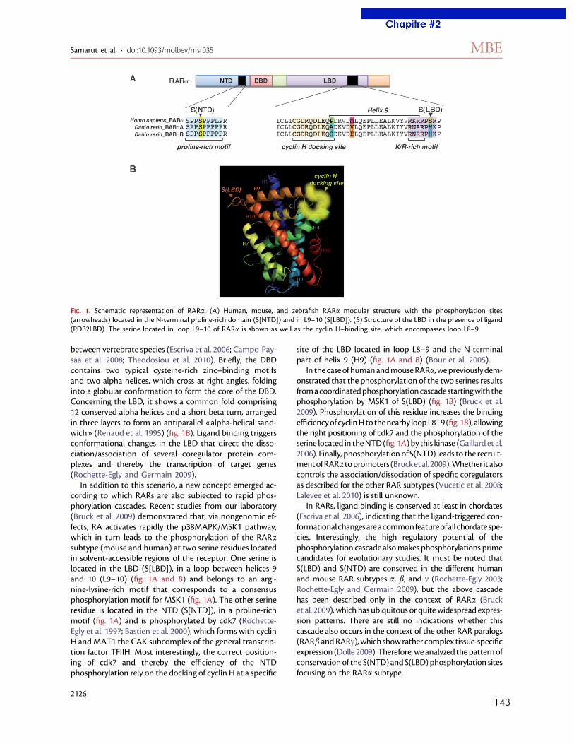
143
between vertebrate species (Escriva et al. 2006; Campo-Pay-saa et al. 2008; Theodosiou et al. 2010). Briefly, the DBDcontains two typical cysteine-rich zinc–binding motifsand two alpha helices, which cross at right angles, foldinginto a globular conformation to form the core of the DBD.Concerning the LBD, it shows a common fold comprising12 conserved alpha helices and a short beta turn, arrangedin three layers to form an antiparallel «alpha-helical sand-wich» (Renaud et al. 1995) (fig. 1B). Ligand binding triggersconformational changes in the LBD that direct the disso-ciation/association of several coregulator protein com-plexes and thereby the transcription of target genes(Rochette-Egly and Germain 2009).
In addition to this scenario, a new concept emerged ac-cording to which RARs are also subjected to rapid phos-phorylation cascades. Recent studies from our laboratory(Bruck et al. 2009) demonstrated that, via nongenomic ef-fects, RA activates rapidly the p38MAPK/MSK1 pathway,which in turn leads to the phosphorylation of the RARasubtype (mouse and human) at two serine residues locatedin solvent-accessible regions of the receptor. One serine islocated in the LBD (S[LBD]), in a loop between helices 9and 10 (L9–10) (fig. 1A and B) and belongs to an argi-nine-lysine-rich motif that corresponds to a consensusphosphorylation motif for MSK1 (fig. 1A). The other serineresidue is located in the NTD (S[NTD]), in a proline-richmotif (fig. 1A) and is phosphorylated by cdk7 (Rochette-Egly et al. 1997; Bastien et al. 2000), which forms with cyclinH andMAT1 the CAK subcomplex of the general transcrip-tion factor TFIIH. Most interestingly, the correct position-ing of cdk7 and thereby the efficiency of the NTDphosphorylation rely on the docking of cyclin H at a specific
site of the LBD located in loop L8–9 and the N-terminalpart of helix 9 (H9) (fig. 1A and B) (Bour et al. 2005).
In thecaseof humanandmouseRARa,wepreviouslydem-onstrated that the phosphorylation of the two serines resultsfromacoordinatedphosphorylationcascadestartingwiththephosphorylation by MSK1 of S(LBD) (fig. 1B) (Bruck et al.2009). Phosphorylation of this residue increases the bindingefficiencyof cyclinHtothenearby loopL8–9(fig.1B), allowingthe right positioning of cdk7 and the phosphorylation of theserine located in theNTD(fig. 1A) by this kinase (Gaillard et al.2006). Finally, phosphorylation of S(NTD) leads to the recruit-mentofRARa topromoters (Brucketal.2009).Whether italsocontrols the association/dissociation of specific coregulatorsas described for the other RAR subtypes (Vucetic et al. 2008;Lalevee et al. 2010) is still unknown.
In RARs, ligand binding is conserved at least in chordates(Escriva et al. 2006), indicating that the ligand-triggered con-formationalchangesareacommonfeatureofall chordatespe-cies. Interestingly, the high regulatory potential of thephosphorylation cascade alsomakes phosphorylations primecandidates for evolutionary studies. It must be noted thatS(LBD) and S(NTD) are conserved in the different humanand mouse RAR subtypes a, b, and c (Rochette-Egly 2003;Rochette-Egly and Germain 2009), but the above cascadehas been described only in the context of RARa (Brucket al. 2009), which has ubiquitous or quitewidespread expres-sion patterns. There are still no indications whether thiscascade also occurs in the context of the other RAR paralogs(RARb andRARc), which show rather complex tissue-specificexpression (Dolle 2009). Therefore,weanalyzed thepatternofconservationof the S(NTD) and S(LBD)phosphorylation sitesfocusing on the RARa subtype.
FIG. 1. Schematic representation of RARa. (A) Human, mouse, and zebrafish RARa modular structure with the phosphorylation sites(arrowheads) located in the N-terminal proline-rich domain (S[NTD]) and in L9–10 (S[LBD]). (B) Structure of the LBD in the presence of ligand(PDB2LBD). The serine located in loop L9–10 of RARa is shown as well as the cyclin H–binding site, which encompasses loop L8–9.
Samarut et al. · doi:10.1093/molbev/msr035 MBE
2126
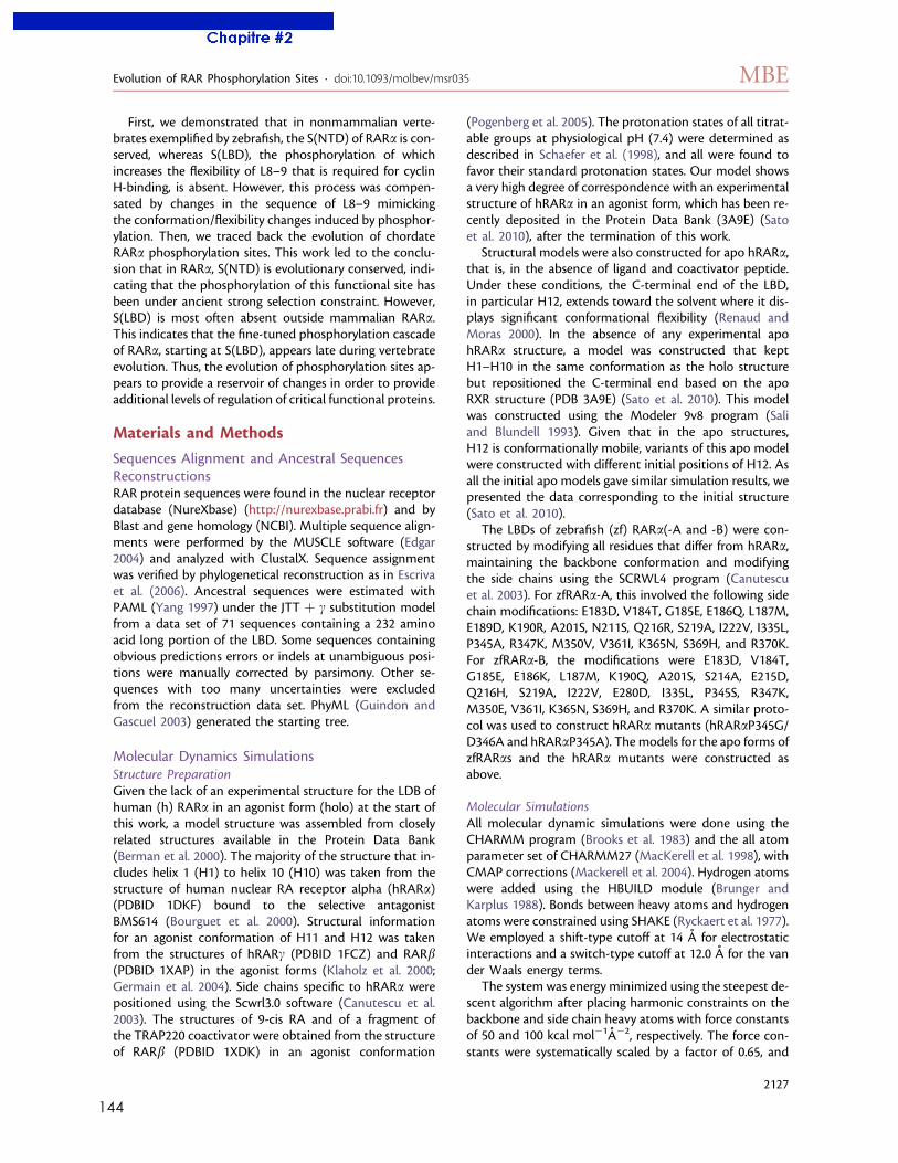
144
First, we demonstrated that in nonmammalian verte-brates exemplified by zebrafish, the S(NTD) of RARa is con-served, whereas S(LBD), the phosphorylation of whichincreases the flexibility of L8–9 that is required for cyclinH-binding, is absent. However, this process was compen-sated by changes in the sequence of L8–9 mimickingthe conformation/flexibility changes induced by phosphor-ylation. Then, we traced back the evolution of chordateRARa phosphorylation sites. This work led to the conclu-sion that in RARa, S(NTD) is evolutionary conserved, indi-cating that the phosphorylation of this functional site hasbeen under ancient strong selection constraint. However,S(LBD) is most often absent outside mammalian RARa.This indicates that the fine-tuned phosphorylation cascadeof RARa, starting at S(LBD), appears late during vertebrateevolution. Thus, the evolution of phosphorylation sites ap-pears to provide a reservoir of changes in order to provideadditional levels of regulation of critical functional proteins.
Materials and Methods
Sequences Alignment and Ancestral SequencesReconstructionsRAR protein sequences were found in the nuclear receptordatabase (NureXbase) (http://nurexbase.prabi.fr) and byBlast and gene homology (NCBI). Multiple sequence align-ments were performed by the MUSCLE software (Edgar2004) and analyzed with ClustalX. Sequence assignmentwas verified by phylogenetical reconstruction as in Escrivaet al. (2006). Ancestral sequences were estimated withPAML (Yang 1997) under the JTT þ c substitution modelfrom a data set of 71 sequences containing a 232 aminoacid long portion of the LBD. Some sequences containingobvious predictions errors or indels at unambiguous posi-tions were manually corrected by parsimony. Other se-quences with too many uncertainties were excludedfrom the reconstruction data set. PhyML (Guindon andGascuel 2003) generated the starting tree.
Molecular Dynamics SimulationsStructure PreparationGiven the lack of an experimental structure for the LDB ofhuman (h) RARa in an agonist form (holo) at the start ofthis work, a model structure was assembled from closelyrelated structures available in the Protein Data Bank(Berman et al. 2000). The majority of the structure that in-cludes helix 1 (H1) to helix 10 (H10) was taken from thestructure of human nuclear RA receptor alpha (hRARa)(PDBID 1DKF) bound to the selective antagonistBMS614 (Bourguet et al. 2000). Structural informationfor an agonist conformation of H11 and H12 was takenfrom the structures of hRARc (PDBID 1FCZ) and RARb(PDBID 1XAP) in the agonist forms (Klaholz et al. 2000;Germain et al. 2004). Side chains specific to hRARa werepositioned using the Scwrl3.0 software (Canutescu et al.2003). The structures of 9-cis RA and of a fragment ofthe TRAP220 coactivator were obtained from the structureof RARb (PDBID 1XDK) in an agonist conformation
(Pogenberg et al. 2005). The protonation states of all titrat-able groups at physiological pH (7.4) were determined asdescribed in Schaefer et al. (1998), and all were found tofavor their standard protonation states. Our model showsa very high degree of correspondence with an experimentalstructure of hRARa in an agonist form, which has been re-cently deposited in the Protein Data Bank (3A9E) (Satoet al. 2010), after the termination of this work.
Structural models were also constructed for apo hRARa,that is, in the absence of ligand and coactivator peptide.Under these conditions, the C-terminal end of the LBD,in particular H12, extends toward the solvent where it dis-plays significant conformational flexibility (Renaud andMoras 2000). In the absence of any experimental apohRARa structure, a model was constructed that keptH1–H10 in the same conformation as the holo structurebut repositioned the C-terminal end based on the apoRXR structure (PDB 3A9E) (Sato et al. 2010). This modelwas constructed using the Modeler 9v8 program (Saliand Blundell 1993). Given that in the apo structures,H12 is conformationally mobile, variants of this apo modelwere constructed with different initial positions of H12. Asall the initial apo models gave similar simulation results, wepresented the data corresponding to the initial structure(Sato et al. 2010).
The LBDs of zebrafish (zf) RARa(-A and -B) were con-structed by modifying all residues that differ from hRARa,maintaining the backbone conformation and modifyingthe side chains using the SCRWL4 program (Canutescuet al. 2003). For zfRARa-A, this involved the following sidechain modifications: E183D, V184T, G185E, E186Q, L187M,E189D, K190R, A201S, N211S, Q216R, S219A, I222V, I335L,P345A, R347K, M350V, V361I, K365N, S369H, and R370K.For zfRARa-B, the modifications were E183D, V184T,G185E, E186K, L187M, K190Q, A201S, S214A, E215D,Q216H, S219A, I222V, E280D, I335L, P345S, R347K,M350E, V361I, K365N, S369H, and R370K. A similar proto-col was used to construct hRARa mutants (hRARaP345G/D346A and hRARaP345A). The models for the apo forms ofzfRARas and the hRARa mutants were constructed asabove.
Molecular SimulationsAll molecular dynamic simulations were done using theCHARMM program (Brooks et al. 1983) and the all atomparameter set of CHARMM27 (MacKerell et al. 1998), withCMAP corrections (Mackerell et al. 2004). Hydrogen atomswere added using the HBUILD module (Brunger andKarplus 1988). Bonds between heavy atoms and hydrogenatoms were constrained using SHAKE (Ryckaert et al. 1977).We employed a shift-type cutoff at 14 A for electrostaticinteractions and a switch-type cutoff at 12.0 A for the vander Waals energy terms.
The system was energy minimized using the steepest de-scent algorithm after placing harmonic constraints on thebackbone and side chain heavy atoms with force constantsof 50 and 100 kcal mol"1A"2, respectively. The force con-stants were systematically scaled by a factor of 0.65, and
Evolution of RAR Phosphorylation Sites · doi:10.1093/molbev/msr035 MBE
2127
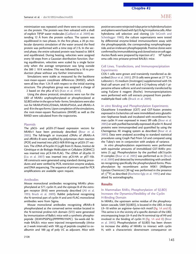
145
minimization was repeated until there were no constraintson the protein. The protein was then solvated with a shellof explicit TIP3P water molecules (Gaillard et al. 2009) ex-tending 12 A from the protein surface. The system wasequilibrated in two phases. In the first phase, a 20 ps mo-lecular dynamics simulation of the water around the fixedprotein was performed with a time step of 2 fs. In the sec-ond phase, the entire solvated protein was heated to 300 Kand equilibrated. During heating, velocities were assignedevery 50 steps from a Gaussian distribution function. Dur-ing equilibration, velocities were scaled by a single factoronly when the average temperature was lying outsidethe 300 ± 10 K window. This was followed by a 10 ns pro-duction phase without any further intervention.
Simulations were stable as measured by the backboneroot-mean-square coordinate differences (RMSD), whichwere all less than 1.24 A with respect to the initial startingstructure. The phosphate group was assigned a charge of!2 based on the pKa of 6.5 (Kast et al. 2010).
Using the above protocol, simulations were run for theLBD of hRARa unphosphorylated or phosphorylated atS(LBD)either in theapoorholo- forms. Simulationswerealsorun for hRARaP345G/D346A, hRARaP345A, and zfRARa-Aand -B in theapo forms.Uponcompletionof the simulations,the root-mean-square fluctuations (RMSfl) as well as theRMSD were calculated from the trajectories.
PlasmidsThe pSG5- and pGEX-2T-based expression vectors forhRARa1 have been previously described (Bour et al.2005). The full-length or truncated cDNAs of zfRARa-Aand zfRARa-B were amplified by polymerase chain reaction(PCR) and inserted into pSG5-hER-B10-tag or pGEX-2T vec-tors. The cDNA of hcyclin H (a gift from D. Busso, Institut deGenetique et de Biologie Moleculaire et Cellulaire [IGBMC])was inserted into pCX-HA-FLAG. The cDNA of zfcyclin H(Liu et al. 2007) was inserted into pCX-HA or pET-15b.All constructs were generated using standard cloning proce-dures and were verified by PCR, restriction enzyme analysis,and DNA sequencing. The sequence of primers used for PCRamplifications are available upon request.
AntibodiesMouse monoclonal antibodies recognizing hRARa phos-phorylated at S77, cyclin H, and the epitope B of the estro-gen receptor (B10) were previously described (Ali et al.1993; Bruck et al. 2009). Rabbit polyclonal recognizingthe N-terminal part of cyclin H and anti-FLAG monoclonalantibodies were from Sigma.
Mouse monoclonal antibodies recognizing zfRARa-Bphosphorylated at the conserved serine residue located inthe N-terminal proline-rich domain (S72) were generatedby immunization of Balb/c mice with a synthetic phospho-peptide (EEMVPSSPS(p)PPPPPRVYKPC). Six-week-old fe-male BALB/c mice were injected intraperitoneally (thriceat 2-week intervals) with 100 lg of peptide coupled to ov-albumin and 100 lg of poly I/C as adjuvant. Mice with
positiveserawerereinjected4daysprior tohybridomafusionandspleenswere fusedwithSp2/0.Ag14myelomacells.Afterhybridoma cell selection and cloning (de StGroth andScheidegger 1980), the culture supernatants were testedby differential enzyme-linked immunosorbent assay withthe phosphopeptide, the corresponding nonphosphopep-tide, and an irrelevant phosphopeptide. Positive cloneswereconfirmedby immunoblottingandclonedtwiceonsoft agar.Ascites fluids were prepared by injection of 2" 106 hybrid-oma cells into pristane-primed BALB/c mice.
Cell Lines, Transfections, and ImmunoprecipitationExperimentsCOS-1 cells were grown and transiently transfected as de-scribed (Bour et al. 2005). ZF13 cells were grown at 27 !C inLeibovitz L-15 medium (Invitrogen) supplemented with 5%fetal calf serum and 15 mM acid 4-(2-hydroxy ethyl)-1 pi-perazine ethane sulfonic acid and transiently transfected byusing FuGene 6 reagent (Roche). Immunoprecipitationswere performed with cell extracts prepared from parafor-maldehyde-fixed cells (Bruck et al. 2009).
In vitro Binding and Phosphorylation ExperimentsGlutathione S-transferase (GST) and GST-fusion proteinsexpressed in Escherichia coliwere immobilized onto glutathi-one–Sepharose beads and incubated with recombinant hu-man cyclin H over expressed in insect Sf9 cells (Bour et al.2005) or with purified bacterially expressed zfcyclin H. Boundproteins were immunoprobed and quantified by using theChemigenius XE imaging system as described (Bour et al.2005). Data were analyzed according to standard statisticalprocedures using Graph Pad Prism 5.0 and compared usingthe Tukey’s test in conjunction with analysis of variance.
In vitro phosphorylation experiments were performedwith equimolar amounts of immobilized GST-RARa pro-teins (5 lg). Phosphorylation by the purified cdk7/cyclinH complex (Bour et al. 2005) was performed as in (Brucket al. 2009) and detected by immunoblotting with antibod-ies recognizing specifically the phosphorylated forms. Phos-phorylation by recombinant active MSK1 (MilliporeUpstate Chemicon) (30 ng) was performed in the presenceof c[32P] as described (Rochette-Egly et al. 1995) and visu-alized by autoradiography.
Results
In Mammalian RARa, Phosphorylation of S(LBD)Increases the Dynamics/Flexibility of the CyclinH-Binding DomainIn hRARa, the upstream serine residue of the phosphory-lation cascade, S369 [S(LBD)], is located in the LBD, in loopL9–10 within an arginine–lysine-rich motif (fig. 1A and B).This serine is in the vicinity of a specific domain of the LBD,encompassing loops L8–9 and the N-terminal tip of H9 andinvolved in the binding of cyclin H (fig. 1A and B) (Bouret al. 2005). Phosphorylation of S(LBD) has been shownto increase the ability of hRARa to interact with cyclinH, with a characteristic downstream consequence on
Samarut et al. · doi:10.1093/molbev/msr035 MBE
2128
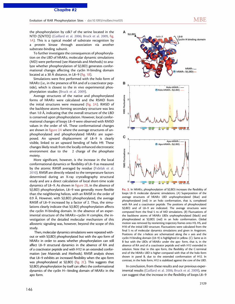
146
the phosphorylation by cdk7 of the serine located in theNTD (S[NTD]) (Gaillard et al. 2006; Bruck et al. 2009, fig.1A). This is a typical model of substrate recognition bya protein kinase through association via anothersubstrate-binding subunit.
To further investigate the consequences of phosphoryla-tion on the LBD of hRARa, molecular dynamic simulations(MD) were performed (see Materials and Methods) to ana-lyze whether phosphorylation of S(LBD) generates confor-mational changes affecting the cyclin H-binding domainlocated at a 30 A distance, in L8–9 (fig. 1B).
Simulations were first performed with the holo form ofhRARa (i.e., in the presence of RA and of a coactivator pep-tide), which is closest to the in vivo experimental phos-phorylation studies (Bruck et al. 2009).
Average structures of the native and phosphorylatedforms of hRARa were calculated and the RSMD fromthe initial structures were measured (fig. 2A). RMSD ofthe backbone atoms forming secondary structure was lessthan 1.0 A, indicating that the overall structure of the LBDis conserved upon phosphorylation. However, local confor-mational changes of loops L8–9 were observed with RMSDvalues in the order of 4A. These conformational changesare shown in figure 2A where the average structures of un-phosphorylated and phosphorylated hRARa are super-posed. An upward displacement of L8–9 is clearlyvisible, linked to an upward bending of helix H9. Thesechanges likely result from the locally enhanced electrostaticenvironment due to the !2 charge of the phosphatemoiety.
More significant, however, is the increase in the localconformational dynamics or flexibility of L8–9 as measuredby the atomic RMSfl averaged by residue (Fidelak et al.2010). RMSfl are directly related to the temperature factorsdetermined during an X-ray crystallography structuralstudy and are a direct calculation of local short-time scaledynamics of L8–9. As shown in figure 2B, in the absence ofS(LBD) phosphorylation, L8–9 was generally more flexiblethan the neighboring helices, with an RMSfl in the order of0.9 A. However, with S(LBD) phosphorylated, the averageRMSfl of L8–9 increased by a factor of 2. Thus, the simu-lations clearly indicate that S(LBD) phosphorylation affectsthe cyclin H-binding domain. In the absence of an exper-imental structure of the hRARa–cyclin H complex, the in-vestigation of the detailed molecular mechanism of thisallosteric signaling was, however, beyond the scope of thisstudy.
Then, molecular dynamics simulations were repeated with-out or with S(LBD) phosphorylated but with the apo form ofhRARa in order to assess whether phosphorylation can stillaffect L8–9 structural dynamics in the absence of RA andof a coactivator peptide and with H12 in an extended confor-mation (see Materials and Methods). RMSfl analysis showsthat L8–9 exhibits an increased flexibility when the apo formwas phosphorylated at S(LBD) (fig. 2C). This suggests thatS(LBD) phosphorylation by itself can affect the conformationaldynamics of the cyclin H–-binding domain of hRARa in theapo form.
In conclusion, from these results and our previous exper-imental results (Gaillard et al. 2006; Bruck et al. 2009), onecan suggest that the increase in the flexibility of loops L8–9
FIG. 2. In hRARa, phosphorylation of S(LBD) increases the flexibility ofloops L8–9: molecular dynamic simulations. (A) Superposition of theaverage structures of hRARa LBD unphosphorylated (blue) andphosphorylated (red) in an holo conformation, that is, complexedwith RA and a coactivator peptide. The positions of phosphorylatedS(LBD) and of L8–9 are indicated. The average structures werecomputed from the final 5 ns of MD simulations. (B) Fluctuations ofthe backbone atoms of hRARa LBDs unphosphorylated (black) andphosphorylated at S(LBD) (red) in an holo conformation. Globalmotion was removed by reorienting trajectory frames onto H3, H5, andH10 of the initial LBD structure. Fluctuations were calculated from thefinal 5 ns of molecular dynamics simulations and given in Angstrom.Positions of the a-helices are schematized along the x axis and thecyclin H-binding domain (L8–9) is highlighted in yellow. (C). Same as inB but with the LBDs of hRARa under the apo- form, that is, in theabsence of RA and of a coactivator peptide and with H12 extended insolution. Note that in this apo form, the flexibility of the C-terminalend of the hRARa LBD is higher compared with that of the holo formshown in panel B, due to the extended conformation of H12. Incontrast, in the holo form, H12 is stabilized against the core of the LBD.
Evolution of RAR Phosphorylation Sites · doi:10.1093/molbev/msr035 MBE
2129
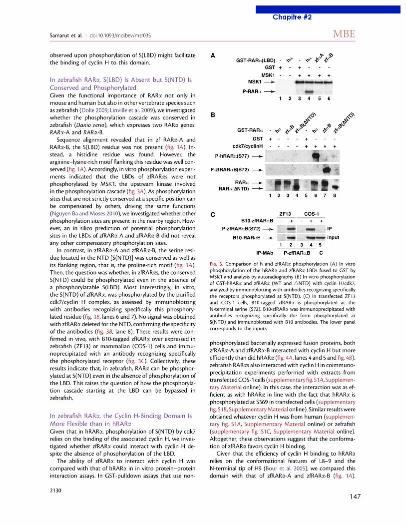
147
observed upon phosphorylation of S(LBD) might facilitatethe binding of cyclin H to this domain.
In zebrafish RARa, S(LBD) Is Absent but S(NTD) IsConserved and PhosphorylatedGiven the functional importance of RARa not only inmouse and human but also in other vertebrate species suchas zebrafish (Dolle 2009; Linville et al. 2009), we investigatedwhether the phosphorylation cascade was conserved inzebrafish (Danio rerio), which expresses two RARa genes:RARa-A and RARa-B.
Sequence alignment revealed that in zf RARa-A andRARa-B, the S(LBD) residue was not present (fig. 1A). In-stead, a histidine residue was found. However, thearginine–lysine-rich motif flanking this residue was well con-served (fig. 1A). Accordingly, in vitro phosphorylation experi-ments indicated that the LBDs of zfRARas were notphosphorylated by MSK1, the upstream kinase involvedin the phosphorylation cascade (fig. 3A). As phosphorylationsites that are not strictly conserved at a specific position canbe compensated by others, driving the same functions(Nguyen Ba andMoses 2010), we investigated whether otherphosphorylation sites are present in the nearby region. How-ever, an in silico prediction of potential phosphorylationsites in the LBDs of zfRARa-A and zfRARa-B did not revealany other compensatory phosphorylation sites.
In contrast, in zfRARa-A and zfRARa-B, the serine resi-due located in the NTD [S(NTD)] was conserved as well asits flanking region, that is, the proline-rich motif (fig. 1A).Then, the question was whether, in zfRARas, the conservedS(NTD) could be phosphorylated even in the absence ofa phosphorylatable S(LBD). Most interestingly, in vitro,the S(NTD) of zfRARa, was phosphorylated by the purifiedcdk7/cyclin H complex, as assessed by immunoblottingwith antibodies recognizing specifically this phosphory-lated residue (fig. 3B, lanes 6 and 7). No signal was obtainedwith zfRARa deleted for the NTD, confirming the specificityof the antibodies (fig. 3B, lane 8). These results were con-firmed in vivo, with B10-tagged zfRARa over expressed inzebrafish (ZF13) or mammalian (COS-1) cells and immu-noprecipitated with an antibody recognizing specificallythe phosphorylated receptor (fig. 3C). Collectively, theseresults indicate that, in zebrafish, RARa can be phosphor-ylated at S(NTD) even in the absence of phosphorylation ofthe LBD. This raises the question of how the phosphoryla-tion cascade starting at the LBD can be bypassed inzebrafish.
In zebrafish RARa, the Cyclin H-Binding Domain IsMore Flexible than in hRARaGiven that in hRARa, phosphorylation of S(NTD) by cdk7relies on the binding of the associated cyclin H, we inves-tigated whether zfRARa could interact with cyclin H de-spite the absence of phosphorylation of the LBD.
The ability of zfRARa to interact with cyclin H wascompared with that of hRARa in in vitro protein–proteininteraction assays. In GST-pulldown assays that use non-
phosphorylated bacterially expressed fusion proteins, bothzfRARa-A and zfRARa-B interacted with cyclin H but moreefficiently than did hRARa (fig. 4A, lanes 4 and 5 and fig. 4B).zebrafish RARas also interacted with cyclin H in coimmuno-precipitation experiments performed with extracts fromtransfectedCOS-1cells (supplementaryfig.S1A, Supplemen-tary Material online). In this case, the interaction was as ef-ficient as with hRARa in line with the fact that hRARa isphosphorylated at S369 in transfected cells (supplementaryfig. S1B, SupplementaryMaterial online). Similar resultswereobtained whatever cyclin H was from human (supplemen-tary fig. S1A, Supplementary Material online) or zefrafish(supplementary fig. S1C, Supplementary Material online).Altogether, these observations suggest that the conforma-tion of zfRARa favors cyclin H binding.
Given that the efficiency of cyclin H binding to hRARarelies on the conformational features of L8–9 and theN-terminal tip of H9 (Bour et al. 2005), we compared thisdomain with that of zfRARa-A and zfRARa-B (fig. 1A).
FIG. 3. Comparison of h and zfRARa phosphorylation (A) In vitrophosphorylation of the hRARa and zfRARa LBDs fused to GST byMSK1 and analysis by autoradiography (B) In vitro phosphorylationof GST-hRARa and zfRARa (WT and nNTD) with cyclin H/cdk7,analyzed by immunoblotting with antibodies recognizing specificallythe receptors phosphorylated at S(NTD). (C) In transfected ZF13and COS-1 cells, B10-tagged zfRARa is phosphorylated at theN-terminal serine (S72). B10-zfRARa was immunoprecipitated withantibodies recognizing specifically the form phosphorylated atS(NTD) and immunoblotted with B10 antibodies. The lower panelcorresponds to the inputs.
Samarut et al. · doi:10.1093/molbev/msr035 MBE
2130
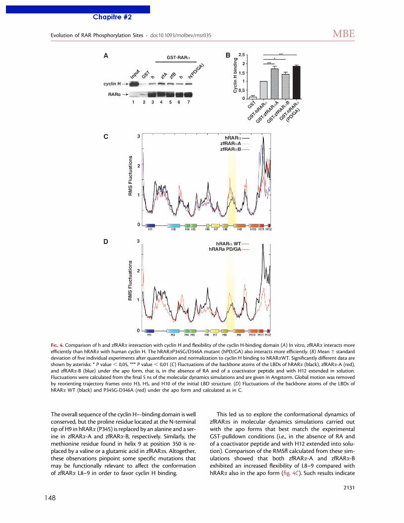
148
The overall sequence of the cyclin H–-binding domain is wellconserved, but the proline residue located at the N-terminaltip of H9 in hRARa (P345) is replaced by an alanine and a ser-ine in zfRARa-A and zfRARa-B, respectively. Similarly, themethionine residue found in helix 9 at position 350 is re-placed by a valine or a glutamic acid in zfRARas. Altogether,these observations pinpoint some specific mutations thatmay be functionally relevant to affect the conformationof zfRARa L8–9 in order to favor cyclin H binding.
This led us to explore the conformational dynamics ofzfRARas in molecular dynamics simulations carried outwith the apo forms that best match the experimentalGST-pulldown conditions (i.e., in the absence of RA andof a coactivator peptide and with H12 extended into solu-tion). Comparison of the RMSfl calculated from these sim-ulations showed that both zfRARa-A and zfRARa-Bexhibited an increased flexibility of L8–9 compared withhRARa also in the apo form (fig. 4C). Such results indicate
FIG. 4. Comparison of h and zfRARa interaction with cyclin H and flexibility of the cyclin H-binding domain (A) In vitro, zfRARa interacts moreefficiently than hRARa with human cyclin H. The hRARaP345G/D346A mutant (hPD/GA) also interacts more efficiently. (B) Mean ± standarddeviation of five individual experiments after quantification and normalization to cyclin H binding to hRARaWT. Significantly different data areshown by asterisks: * P value , 0,05, *** P value , 0,01 (C) Fluctuations of the backbone atoms of the LBDs of hRARa (black), zfRARa-A (red),and zfRARa-B (blue) under the apo form, that is, in the absence of RA and of a coactivator peptide and with H12 extended in solution.Fluctuations were calculated from the final 5 ns of the molecular dynamics simulations and are given in Angstorm. Global motion was removedby reorienting trajectory frames onto H3, H5, and H10 of the initial LBD structure. (D) Fluctuations of the backbone atoms of the LBDs ofhRARa WT (black) and P345G-D346A (red) under the apo form and calculated as in C.
Evolution of RAR Phosphorylation Sites · doi:10.1093/molbev/msr035 MBE
2131
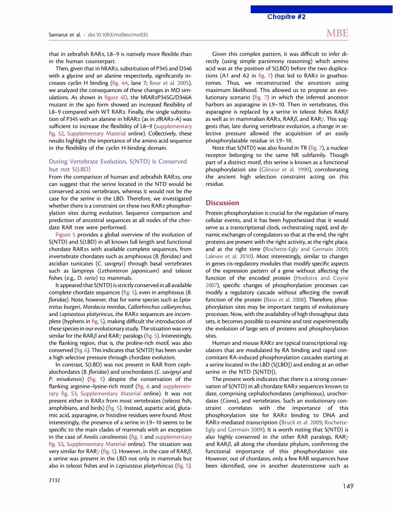
149
that in zebrafish RARa, L8–9 is natively more flexible thanin the human counterpart.
Then, given that in hRARa, substitution of P345 and D346with a glycine and an alanine respectively, significantly in-creases cyclin H binding (fig. 4A, lane 7; Bour et al. 2005),we analyzed the consequences of these changes in MD sim-ulations. As shown in figure 4D, the hRARaP345G/D346Amutant in the apo form showed an increased flexibility ofL8–9 compared with WT RARa. Finally, the single substitu-tion of P345 with an alanine in hRARa (as in zfRARa-A) wassufficient to increase the flexibility of L8–9 (supplementaryfig. S2, Supplementary Material online). Collectively, theseresults highlight the importance of the amino acid sequencein the flexibility of the cyclin H-binding domain.
During Vertebrate Evolution, S(NTD) Is Conservedbut not S(LBD)From the comparison of human and zebrafish RARas, onecan suggest that the serine located in the NTD would beconserved across vertebrates, whereas it would not be thecase for the serine in the LBD. Therefore, we investigatedwhether there is a constraint on these two RARa phosphor-ylation sites during evolution. Sequence comparison andprediction of ancestral sequences at all nodes of the chor-date RAR tree were performed.
Figure 5 provides a global overview of the evolution ofS(NTD) and S(LBD) in all known full length and functionalchordate RARas with available complete sequences, frominvertebrate chordates such as amphioxus (B. floridae) andascidian tunicates (C. savignyi) through basal vertebratessuch as lampreys (Lethenteron japonicum) and teleostfishes (e.g., D. rerio) to mammals.
It appeared that S(NTD) is strictly conserved in all availablecomplete chordate sequences (fig. 5), even in amphioxus (B.floridae). Note, however, that for some species such as Epta-tretus burgeri,Mordacia mordax, Callorhinchus callorynchus,and Lepisosteus platyrincus, the RARa sequences are incom-plete (hyphens in fig. 5), making difficult the introduction ofthese species inour evolutionary study.The situationwasverysimilar for the RARb and RARc paralogs (fig. 5). Interestingly,the flanking region, that is, the proline-rich motif, was alsoconserved (fig. 6). This indicates that S(NTD) has been undera high selective pressure through chordate evolution.
In contrast, S(LBD) was not present in RAR from ceph-alochordates (B. floridae) and urochordates (C. savignyi andP. misakensis) (fig. 5) despite the conservation of theflanking arginine–lysine-rich motif (fig. 6 and supplemen-tary fig. S3, Supplementary Material online). It was notpresent either in RARa from most vertebrates (teleost fish,amphibians, and birds) (fig. 5). Instead, aspartic acid, gluta-mic acid, asparagine, or histidine residues were found. Mostinterestingly, the presence of a serine in L9–10 seems to bespecific to the main clades of mammals with an exceptionin the case of Anolis carolinensis (fig. 5 and supplementaryfig. S3, Supplementary Material online). The situation wasvery similar for RARc (fig. 5). However, in the case of RARb,a serine was present in the LBD not only in mammals butalso in teleost fishes and in Lepisosteus platyrhincus (fig. 5).
Given this complex pattern, it was difficult to infer di-rectly (using simple parsimony reasoning) which aminoacid was at the position of S(LBD) before the two duplica-tions (A1 and A2 in fig. 7) that led to RARa in gnathos-tomes. Thus, we reconstructed the ancestors usingmaximum likelihood. This allowed us to propose an evo-lutionary scenario (fig. 7) in which the inferred ancestorharbors an asparagine in L9–10. Then in vertebrates, thisasparagine is replaced by a serine in teleost fishes RARbas well as in mammalian RARa, RARb, and RARc. This sug-gests that, late during vertebrate evolution, a change in se-lective pressure allowed the acquisition of an easilyphosphorylatable residue in L9–10.
Note that S(NTD) was also found in TR (fig. 7), a nuclearreceptor belonging to the same NR subfamily. Thoughpart of a distinct motif, this serine is known as a functionalphosphorylation site (Glineur et al. 1990), corroboratingthe ancient high selection constraint acting on thisresidue.
DiscussionProtein phosphorylation is crucial for the regulation of manycellular events, and it has been hypothesized that it wouldserve as a transcriptional clock, orchestrating rapid, and dy-namic exchanges of coregulators so that at the end, the rightproteins are present with the right activity, at the right place,and at the right time (Rochette-Egly and Germain 2009;Lalevee et al. 2010). Most interestingly, similar to changesin genes cis-regulatory modules that modify specific aspectsof the expression pattern of a gene without affecting thefunction of the encoded protein (Hoekstra and Coyne2007), specific changes of phosphorylation processes canmodify a regulatory cascade without affecting the overallfunction of the protein (Basu et al. 2008). Therefore, phos-phorylation sites may be important targets of evolutionaryprocesses. Now, with the availability of high throughput datasets, it becomes possible to examine and test experimentallythe evolution of large sets of proteins and phosphorylationsites.
Human and mouse RARa are typical transcriptional reg-ulators that are modulated by RA binding and rapid con-comitant RA-induced phosphorylation cascades starting ata serine located in the LBD (S[LBD]) and ending at an otherserine in the NTD (S[NTD]).
The present work indicates that there is a strong conser-vation of S(NTD) in all chordate RARa sequences known todate, comprising cephalochordates (amphioxus), urochor-dates (Ciona), and vertebrates. Such an evolutionary con-straint correlates with the importance of thisphosphorylation site for RARa binding to DNA andRARa-mediated transcription (Bruck et al. 2009; Rochette-Egly and Germain 2009). It is worth noting that S(NTD) isalso highly conserved in the other RAR paralogs, RARcand RARb, all along the chordate phylum, confirming thefunctional importance of this phosphorylation site.However, out of chordates, only a few RAR sequences havebeen identified, one in another deuterostome such as
Samarut et al. · doi:10.1093/molbev/msr035 MBE
2132
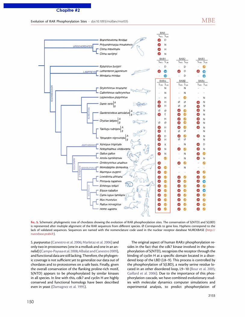
150
S. purpuratus (Canestro et al. 2006; Marletaz et al. 2006) andonly two in prostosomes (one in amollusk and one in an an-nelid)(Campo-Paysaaetal.2008;AlbalatandCanestro2009),and functional data are still lacking. Therefore, thephylogen-ic coverage is not sufficient yet to generalize our data out ofchordates and to protostomes on a safe basis. Finally, giventhe overall conservation of the flanking proline-rich motif,S(NTD) appears to be phosphorylated by similar kinasesin all species. In line with this, cdk7 and cyclin H are highlyconserved and functional homologs have been describedeven in yeast (Damagnez et al. 1995).
The original aspect of human RARa phosphorylation re-sides in the fact that the cdk7 kinase involved in the phos-phorylation of S(NTD), recognizes the receptor through thebinding of cyclin H at a specific domain located in a disor-dered loop of the LBD (L8–9). This process is controlled bythe phosphorylation of S(LBD), a nearby serine residue lo-cated in an other disordered loop, L9–10 (Bour et al. 2005;Gaillard et al. 2006). Due to the importance of this phos-phorylation cascade, we have combined evolutionary stud-ies with molecular dynamics computer simulations andexperimental analysis, to predict phosphorylation of
FIG. 5. Schematic phylogenetic tree of chordates showing the evolution of RAR phosphorylation sites. The conservation of S(NTD) and S(LBD)is represented after multiple alignment of the RAR sequences from different species. Ø Corresponds to gene loss. Hyphens correspond to thelack of validated sequences. Sequences are named with the nomenclature code used in the nuclear receptor database NUREXBASE (http://nurexbase.prabi.fr).
Evolution of RAR Phosphorylation Sites · doi:10.1093/molbev/msr035 MBE
2133
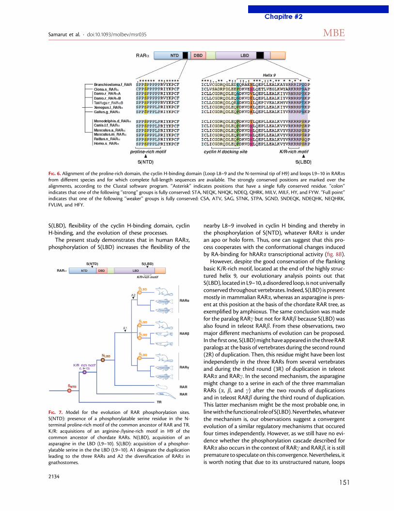
151
S(LBD), flexibility of the cyclin H-binding domain, cyclinH-binding, and the evolution of these processes.
The present study demonstrates that in human RARa,phosphorylation of S(LBD) increases the flexibility of the
nearby L8–9 involved in cyclin H binding and thereby inthe phosphorylation of S(NTD), whatever RARa is underan apo or holo form. Thus, one can suggest that this pro-cess cooperates with the conformational changes inducedby RA-binding for hRARa transcriptional activity (fig. 8B).
However, despite the good conservation of the flankingbasic K/R-rich motif, located at the end of the highly struc-tured helix 9, our evolutionary analysis points out thatS(LBD), located in L9–10, a disordered loop, is not universallyconserved throughout vertebrates. Indeed, S(LBD) is presentmostly in mammalian RARa, whereas an asparagine is pres-ent at this position at the basis of the chordate RAR tree, asexemplified by amphioxus. The same conclusion was madefor the paralog RARc but not for RARb because S(LBD) wasalso found in teleost RARb. From these observations, twomajor different mechanisms of evolution can be proposed.In thefirst one, S(LBD)mighthaveappeared in the threeRARparalogs at the basis of vertebrates during the second round(2R) of duplication. Then, this residue might have been lostindependently in the three RARs from several vertebratesand during the third round (3R) of duplication in teleostRARa and RARc. In the second mechanism, the asparaginemight change to a serine in each of the three mammalianRARs (a, b, and c) after the two rounds of duplicationsand in teleost RARb during the third round of duplication.This latter mechanism might be the most probable one, inlinewith the functional roleof S(LBD).Nevertheles,whateverthe mechanism is, our observations suggest a convergentevolution of a similar regulatory mechanisms that occuredfour times independently. However, as we still have no evi-dence whether the phosphorylation cascade described forRARa also occurs in the context of RARc and RARb, it is stillpremature to speculate on this convergence.Nevertheless, itis worth noting that due to its unstructured nature, loops
FIG. 6. Alignment of the proline-rich domain, the cyclin H-binding domain (Loop L8–9 and the N-terminal tip of H9) and loops L9–10 in RARasfrom different species and for which complete full-length sequences are available. The strongly conserved positions are marked over thealignments, according to the Clustal software program. ‘‘Asterisk’’ indicates positions that have a single fully conserved residue. ‘‘colon’’indicates that one of the following ‘‘strong’’ groups is fully conserved: STA, NEQK, NHQK, NDEQ, QHRK, MILV, MILF, HY, and FYW. ‘‘Full point’’indicates that one of the following ‘‘weaker’’ groups is fully conserved: CSA, ATV, SAG, STNK, STPA, SGND, SNDEQK, NDEQHK, NEQHRK,FVLIM, and HFY.
FIG. 7. Model for the evolution of RAR phosphorylation sites.S(NTD): presence of a phosphorylatable serine residue in the N-terminal proline-rich motif of the common ancestor of RAR and TR.K/R: acquisitions of an arginine-/lysine-rich motif in H9 of thecommon ancestor of chordate RARs. N(LBD), acquisition of anasparagine in the LBD (L9–10). S(LBD): acquisition of a phosphor-ylatable serine in the the LBD (L9–10). A1 designate the duplicationleading to the three RARs and A2 the diversification of RARa ingnathostomes.
Samarut et al. · doi:10.1093/molbev/msr035 MBE
2134
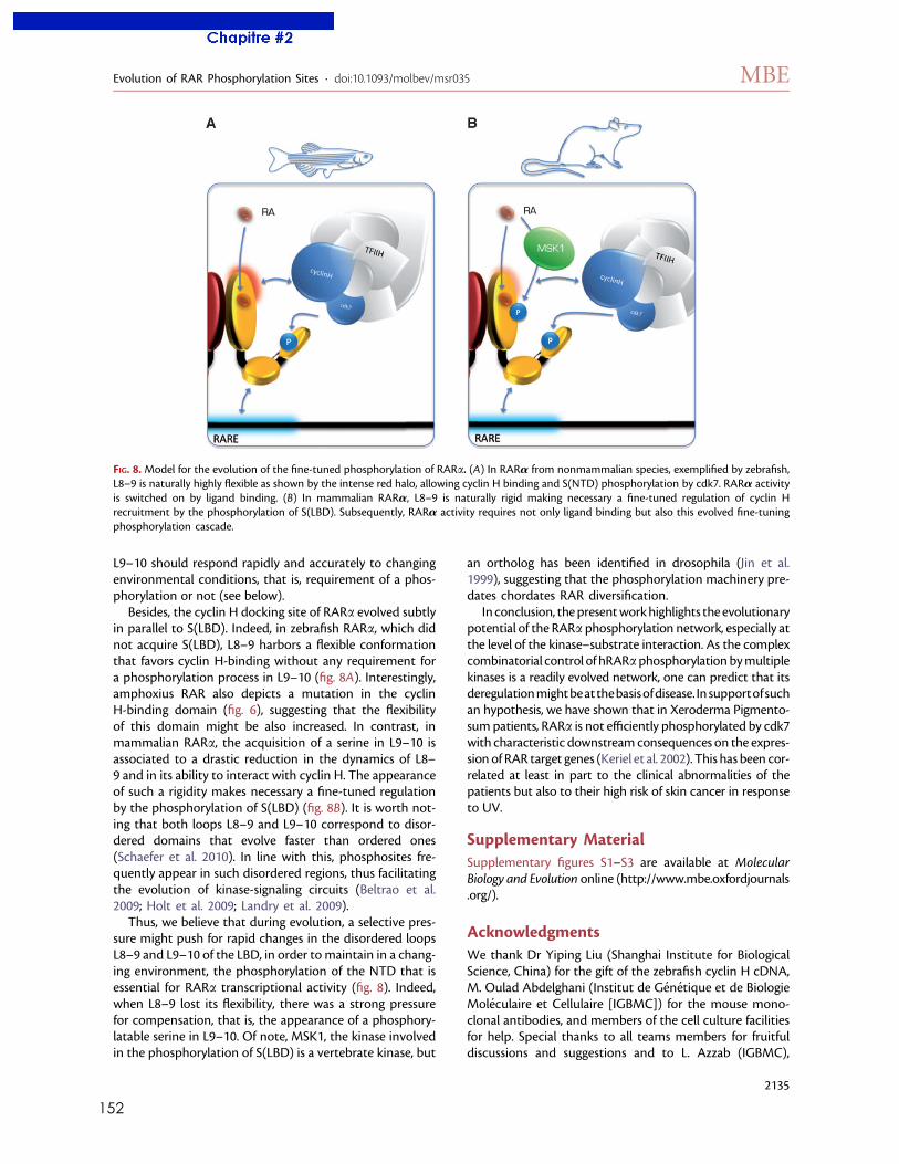
152
L9–10 should respond rapidly and accurately to changingenvironmental conditions, that is, requirement of a phos-phorylation or not (see below).
Besides, the cyclin H docking site of RARa evolved subtlyin parallel to S(LBD). Indeed, in zebrafish RARa, which didnot acquire S(LBD), L8–9 harbors a flexible conformationthat favors cyclin H-binding without any requirement fora phosphorylation process in L9–10 (fig. 8A). Interestingly,amphoxius RAR also depicts a mutation in the cyclinH-binding domain (fig. 6), suggesting that the flexibilityof this domain might be also increased. In contrast, inmammalian RARa, the acquisition of a serine in L9–10 isassociated to a drastic reduction in the dynamics of L8–9 and in its ability to interact with cyclin H. The appearanceof such a rigidity makes necessary a fine-tuned regulationby the phosphorylation of S(LBD) (fig. 8B). It is worth not-ing that both loops L8–9 and L9–10 correspond to disor-dered domains that evolve faster than ordered ones(Schaefer et al. 2010). In line with this, phosphosites fre-quently appear in such disordered regions, thus facilitatingthe evolution of kinase-signaling circuits (Beltrao et al.2009; Holt et al. 2009; Landry et al. 2009).
Thus, we believe that during evolution, a selective pres-sure might push for rapid changes in the disordered loopsL8–9 and L9–10 of the LBD, in order tomaintain in a chang-ing environment, the phosphorylation of the NTD that isessential for RARa transcriptional activity (fig. 8). Indeed,when L8–9 lost its flexibility, there was a strong pressurefor compensation, that is, the appearance of a phosphory-latable serine in L9–10. Of note, MSK1, the kinase involvedin the phosphorylation of S(LBD) is a vertebrate kinase, but
an ortholog has been identified in drosophila (Jin et al.1999), suggesting that the phosphorylation machinery pre-dates chordates RAR diversification.
In conclusion, thepresentworkhighlights the evolutionarypotential of the RARa phosphorylation network, especially atthe level of the kinase–substrate interaction. As the complexcombinatorial control of hRARaphosphorylationbymultiplekinases is a readily evolved network, one can predict that itsderegulationmightbeatthebasisofdisease. Insupportofsuchan hypothesis, we have shown that in Xeroderma Pigmento-sumpatients, RARa is not efficiently phosphorylated by cdk7with characteristic downstreamconsequences on the expres-sion of RAR target genes (Keriel et al. 2002). This has been cor-related at least in part to the clinical abnormalities of thepatients but also to their high risk of skin cancer in responseto UV.
Supplementary MaterialSupplementary figures S1–S3 are available at MolecularBiology and Evolution online (http://www.mbe.oxfordjournals.org/).
AcknowledgmentsWe thank Dr Yiping Liu (Shanghai Institute for BiologicalScience, China) for the gift of the zebrafish cyclin H cDNA,M. Oulad Abdelghani (Institut de Genetique et de BiologieMoleculaire et Cellulaire [IGBMC]) for the mouse mono-clonal antibodies, and members of the cell culture facilitiesfor help. Special thanks to all teams members for fruitfuldiscussions and suggestions and to L. Azzab (IGBMC),
FIG. 8.Model for the evolution of the fine-tuned phosphorylation of RARa. (A) In RARa from nonmammalian species, exemplified by zebrafish,L8–9 is naturally highly flexible as shown by the intense red halo, allowing cyclin H binding and S(NTD) phosphorylation by cdk7. RARa activityis switched on by ligand binding. (B) In mammalian RARa, L8–9 is naturally rigid making necessary a fine-tuned regulation of cyclin Hrecruitment by the phosphorylation of S(LBD). Subsequently, RARa activity requires not only ligand binding but also this evolved fine-tuningphosphorylation cascade.
Evolution of RAR Phosphorylation Sites · doi:10.1093/molbev/msr035 MBE
2135
153
A. Rodriguez and A. Perret (Universite de Strasbourg) forhelp in the development of simulation protocols. This workwas supported by funds from Centre National de la Re-cherche Scientifique (CNRS), INSERM, the Association pourla Recherche sur le Cancer (ARC 3169), the Agence Natio-nale pour la Recherche (ANR-05-BLAN-0390-02 and ANR-09-BLAN-0127-01), the Fondation pour la Recherche Med-icale (DEQ20090515423), and the Institut National du Can-cer (INCa-PL09-194). The Institut du Developpement et desRessources en Informatique Scientifique (IDRIS), the CentreInformatique National de l’Enseignement Superieur(CINES), and the Centre d’Etude du Calcul Parallele deStrasbourg (Universite de Strasbourg) are aknowledgedfor generous allocations of computer time. I.A. was sup-ported by the Ligue Nationale contre le cancer.
ReferencesAlbalat R, Canestro C. 2009. Identification of Aldh1a, Cyp26 and
RAR orthologs in protostomes pushes back the retinoic acidgenetic machinery in evolutionary time to the bilaterianancestor. Chem Biol Interact. 178:188–196.
Ali S, Lutz Y, Bellocq JP, Chenard-Neu MP, Rouyer N, Metzger D.1993. Production and characterization of monoclonal antibodiesrecognising defined regions of the human oestrogen receptor.Hybridoma 12:391–405.
Amores A, Force A, Yan YL, et al. (13 co-authors). 1998. Zebrafishhox clusters and vertebrate genome evolution. Science282:1711–1714.
Bastien J, Adam-Stitah S, Riedl T, Egly JM, Chambon P, Rochette-Egly C. 2000. TFIIH interacts with the retinoic acid receptorgamma and phosphorylates its AF-1-activating domain throughcdk7. J Biol Chem. 275:21896–21904.
Basu U, Wang Y, Alt FW. 2008. Evolution of phosphorylation-dependent regulation of activation-induced cytidine deaminase.Mol Cell. 32:285–291.
Beltrao P, Trinidad JC, Fiedler D, Roguev A, Lim WA, Shokat KM,Burlingame AL, Krogan NJ. 2009. Evolution of phosphoregula-tion: comparison of phosphorylation patterns across yeastspecies. PLoS Biol. 7:e1000134.
Berman HM, Westbrook J, Feng Z, Gilliland G, Bhat TN, Weissig H,Shindyalov IN, Bourne PE. 2000. The Protein Data Bank. NucleicAcids Res. 28:235–242.
Bertrand S, Thisse B, Tavares R, et al. (12 co-authors). 2007.Unexpected novel relational links uncovered by extensivedevelopmental profiling of nuclear receptor expression. PLoSGenet. 3:e188.
Bour G, Gaillard E, Bruck N, Lalevee S, Plassat JL, Busso D,Samama JP, Rochette-Egly C. 2005. Cyclin H binding to the RAR{alpha} activation function (AF)-2 domain directs phosphory-lation of the AF-1 domain by cyclin-dependent kinase 7. ProcNatl Acad Sci U S A. 102:16608–16613.
Bour G, Taneja R, Rochette-Egly C. 2006. Mouse embryocarcinomaF9 cells and retinoic acid. A model to study the molecularmechanisms of endodermal differentiation. In: Taneja R, editor.Nuclear receptors in development. New York: Elsevier Press Inc.p. 211–253.
Bourguet W, Vivat V, Wurtz JM, Chambon P, Gronemeyer H,Moras D. 2000. Crystal structure of a heterodimeric complex ofRAR and RXR ligand-binding domains. Mol Cell. 5:289–298.
Brooks BR, Bruccoleri RE, Olafson BD, States DJ, Swaminathan S.1983. CHARMM: a program for macromolecular energyminimization and dynamics calculations. J Comp Chem.4:187–217.
Bruck N, Vitoux D, Ferry C, Duong V, Bauer A, de The H, Rochette-Egly C. 2009. A coordinated phosphorylation cascade initiatedby p38MAPK/MSK1 directs RARalpha to target promoters.EMBO J. 28:34–47.
Brunger AT, Karplus M. 1988. Polar hydrogen positions in proteins:empirical energy placement and neutron diffraction compari-son. Proteins 4:148–156.
Campo-Paysaa F, Marletaz F, Laudet V, Schubert M. 2008. Retinoicacid signaling in development: tissue-specific functions andevolutionary origins. Genesis 46:640–656.
Canestro C, Postlethwait JH, Gonzalez-Duarte R, Albalat R. 2006. Isretinoic acid genetic machinery a chordate innovation? Evol Dev.8:394–406.
Canutescu AA, Shelenkov AA, Dunbrack RL Jr. 2003. A graph-theoryalgorithm for rapid protein side-chain prediction. Protein Sci.12:2001–2014.
Damagnez V, Makela TP, Cottarel G. 1995. Schizosaccharomycespombe Mop1-Mcs2 is related to mammalian CAK. EMBO J.14:6164–6172.
de StGroth SF, Scheidegger D. 1980. Production of monoclonalantibodies: strategy and tactics. J Immunol Methods. 35:1–21.
Dehal P, Boore JL. 2005. Two rounds of whole genome duplication inthe ancestral vertebrate. PLoS Biol. 3:e314.
Dolle P. 2009. Developmental expression of retinoic acid receptors(RARs). Nucl Recept Signal. 7:e006.
Dyson HJ, Wright PE. 2005. Intrinsically unstructured proteins andtheir functions. Nat Rev Mol Cell Biol. 6:197–208.
Edgar RC. 2004. MUSCLE: a multiple sequence alignment methodwith reduced time and space complexity. BMC Bioinformatics.5:113.
Escriva H, Bertrand S, Germain P, Robinson-Rechavi M, Umbhauer M,Cartry J, Duffraisse M, Holland L, Gronemeyer H, Laudet V. 2006.Neofunctionalization in vertebrates: the example of retinoic acidreceptors. PLoS Genet. 2:e102.
Escriva H, Holland ND, Gronemeyer H, Laudet V, Holland LZ. 2002a.The retinoic acid signaling pathway regulates anterior/posteriorpatterning in the nerve cord and pharynx of amphioxus,a chordate lacking neural crest. Development 129:2905–2916.
Escriva H, Manzon L, Youson J, Laudet V. 2002b. Analysis oflamprey and hagfish genes reveals a complex history of geneduplications during early vertebrate evolution. Mol Biol Evol.19:1440–1450.
Fidelak J, Ferrer S, Oberlin M, Moras D, Dejaegere A, Stote RH. 2010.Dynamic correlation networks in human peroxisomeproliferator-activated receptor-gamma nuclear receptor protein.Eur Biophys J. 39:1503–1512.
Fujiwara S. 2006. Retinoids and nonvertebrate chordate develop-ment. J Neurobiol. 66:645–652.
Gaillard E, Bruck N, Brelivet Y, Bour G, Lalevee S, Bauer A, Poch O,Moras D, Rochette-Egly C. 2006. Phosphorylation by proteinkinase A potentiates retinoic acid repeptor a activity by meansof increasing interaction with and phosphorylation by cyclin H/cdk7. Proc Natl Acad Sci U S A. 103:9548–9553.
Gaillard T, Dejaegere A, Stote RH. 2009. Dynamics of beta3 integrinI-like and hybrid domains: insight from simulations on themechanism of transition between open and closed forms.Proteins 76:977–994.
Germain P, Kammerer S, Perez E, et al. (13 co-authors). 2004.Rational design of RAR-selective ligands revealed by RARbetacrystal stucture. EMBO Rep. 5:877–882.
Germain P, Staels B, Dacquet C, Spedding M, Laudet V. 2006.Overview of nomenclature of nuclear receptors. Pharmacol Rev.58:685–704.
Glineur C, Zenke M, Beug H, Ghysdael J. 1990. Phosphorylation ofthe v-erbA protein is required for its function as an oncogene.Genes Dev. 4:1663–1676.
Samarut et al. · doi:10.1093/molbev/msr035 MBE
2136
154
Guindon S, Gascuel O. 2003. A simple, fast, and accurate algorithmto estimate large phylogenies by maximum likelihood. Syst Biol.52:696–704.
Hisata K, Fujiwara S, Tsuchida Y, Ohashi M, Kawamura K. 1998.Expression and function of a retinoic acid receptor in buddingascidians. Dev Genes Evol. 208:537–546.
Hoekstra HE, Coyne JA. 2007. The locus of evolution: evo devo andthe genetics of adaptation. Evolution 61:995–1016.
Holt LJ, Tuch BB, Villen J, Johnson AD, Gygi SP, Morgan DO. 2009.Global analysis of Cdk1 substrate phosphorylation sites providesinsights into evolution. Science 325:1682–1686.
Jaillon O, Aury JM, Brunet F, et al. (61 co-authors). 2004. Genomeduplication in the teleost fish Tetraodon nigroviridis reveals theearly vertebrate proto-karyotype. Nature 431:946–957.
Jin Y, Wang Y, Walker DL, Dong H, Conley C, Johansen J, Johansen KM.1999. JIL-1: a novel chromosomal tandem kinase implicated intranscriptional regulation in Drosophila. Mol Cell. 4:129–135.
Kast D, Espinoza-Fonseca LM, Yi C, Thomas DD. 2010. Pho-sphorylation-induced structural changes in smooth musclemyosin regulatory light chain. Proc Natl Acad Sci U S A. 107:8207–8212.
Keriel A, Stary A, Sarasin A, Rochette-Egly C, Egly JM. 2002. XPDmutations prevent TFIIH-dependent transactivation by nuclearreceptors and phosphorylation of RARalpha. Cell 109:125–135.
Klaholz BP, Mitschler A, Moras D. 2000. Structural basis for isotypeselectivity of the human retinoic acid nuclear receptor. J MolBiol. 302:155–170.
Kuraku S, Meyer A, Kuratani S. 2009. Timing of genome duplicationsrelative to the origin of the vertebrates: did cyclostomes divergebefore or after? Mol Biol Evol. 26:47–59.
Lalevee S, Bour G, Quinternet M, et al. (11 co-authors). 2010.Vinexin{beta}, an atypical ‘‘sensor’’ of retinoic acid receptor{gamma} signaling: union and sequestration, separation, andphosphorylation. FASEB J. 24:4523–4534.
Lalevee S, Ferry C, Rochette-Egly C. 2010. Phosphorylation control ofnuclear receptors. Methods Mol Biol. 647:251–266.
Landry CR, Levy ED, Michnick SW. 2009. Weak functionalconstraints on phosphoproteomes. Trends Genet. 25:193–197.
Lavery DN, McEwan IJ. 2005. Structure and function of steroidreceptor AF1 transactivation domains: induction of activeconformations. Biochem J. 391:449–464.
Linville A, Radtke K, Waxman JS, Yelon D, Schilling TF. 2009.Combinatorial roles for zebrafish retinoic acid receptors in thehindbrain, limbs and pharyngeal arches. Dev Biol. 325:60–70.
Liu QY, Wu ZL, Lv WJ, Yan YC, Li YP. 2007. Developmentalexpression of cyclin H and Cdk7 in zebrafish: the essential role ofcyclin H during early embryo development. Cell Res. 17:163–173.
MacKerell AD, Bashford D, Bellott M, et al. (29 co-authors). 1998.All-atom empirical potential for molecular modeling anddynamics studies of proteins. J Phys Chem B. 102:3586–3616.
Mackerell AD Jr, Feig M, Brooks CL. 2004. Extending the treatmentof backbone energetics in protein force fields: limitations of gas-phase quantum mechanics in reproducing protein conforma-tional distributions in molecular dynamics simulations. J ComputChem. 25:1400–1415.
Mark M, Ghyselinck NB, Chambon P. 2009. Function of retinoic acidreceptors during embryonic development. Nucl Recept Signal.7:e002.
Marletaz F, Holland LZ, Laudet V, Schubert M. 2006. Retinoic acidsignaling and the evolution of chordates. Int J Biol Sci. 2:38–47.
Nguyen Ba AN, Moses AM. Forthcoming 2010. Evolution ofcharacterized phosphorylation sites in budding yeast. Mol BiolEvol. 27:2027–2037.
Pogenberg V, Guichou JF, Vivat-Hannah V, Kammerer S, Perez E,Germain P, de Lera AR, Gronemeyer H, Royer CA, Bourguet W.2005. Characterization of the interaction between retinoic acidreceptor/retinoid X receptor (RAR/RXR) heterodimers andtranscriptional coactivators through structural and fluorescenceanisotropy studies. J Biol Chem. 280:1625–1633.
Postlethwait JH, Yan YL, Gates MA, et al. (29 co-authors). 1998.Vertebrate genome evolution and the zebrafish gene map. NatGenet. 18:345–349.
Renaud JP, Moras D. 2000. Structural studies on nuclear receptors.Cell Mol Life Sci. 57:1748–1769.
Renaud JP, Rochel N, Ruff M, Vivat V, Chambon P, Gronemeyer H,Moras D. 1995. Crystal structure of the RAR-gamma ligand-binding domain bound to all-trans retinoic acid. Nature378:681–689.
Robinson-Rechavi M, Boussau B, Laudet V. 2004. Phylogeneticdating and characterization of gene duplications in vertebrates:the cartilaginous fish reference. Mol Biol Evol. 21:580–586.
Rochette-Egly C. 2003. Nuclear receptors: integration of multiplesignalling pathways through phosphorylation. Cell Signal.15:355–366.
Rochette-Egly C, Adam S, Rossignol M, Egly JM, Chambon P. 1997.Stimulation of RAR alpha activation function AF-1 throughbinding to the general transcription factor TFIIH and phos-phorylation by CDK7. Cell 90:97–107.
Rochette-Egly C, Germain P. 2009. Dynamic and combinatorialcontrol of gene expression by nuclear retinoic acid receptors.Nucl Recep Signal. 7:e005.
Rochette-Egly C, Oulad-Abdelghani M, Staub A, Pfister V, Scheuer I,Chambon P, Gaub MP. 1995. Phosphorylation of the retinoicacid receptor-alpha by protein kinase A. Mol Endocrinol.9:860–871.
Ryckaert JP, Ciccotti G, Berendsen HJC. 1977. Numerical integrationof the cartesian equations of motion of a system withconstraints: molecular dynamics of n-alkanes. J Comp Phys.23:327–341.
Sali A, Blundell TL. 1993. Comparative protein modelling bysatisfaction of spatial restraints. J Mol Biol. 234:779–815.
Sato Y, Ramalanjaona N, Huet T, et al. (13 co-authors). 2010. The‘‘phantom effect’’ of the rexinoid LG100754: structural andfunctional insights. PloS One. 5:e15119.
Schaefer C, Schlessinger A, Rost B. 2010. Protein secondary structureappears to be robust under in silico evolution while proteindisorder appears not to be. Bioinformatics 26:625–631.
Schaefer M, van Vlijmen HW, Karplus M. 1998. Electrostaticcontributions to molecular free energies in solution. Adv ProteinChem. 51:1–57.
Theodosiou M, Laudet V, Schubert M. 2010. From carrot to clinic:an overview of the retinoic acid signaling pathway. Cell Mol LifeSci. 67:1423–1445.
Vucetic Z, Zhang Z, Zhao J, Wang F, Soprano KJ, Soprano DR. 2008.Acinus-S’ represses retinoic acid receptor (RAR)-regulated geneexpression through interaction with the B domains of RARs. MolCell Biol. 28:2549–2558.
Yang Z. 1997. PAML: a program package for phylogenetic analysis bymaximum likelihood. Comput Appl Biosci. 13:555–556.
Evolution of RAR Phosphorylation Sites · doi:10.1093/molbev/msr035 MBE
2137
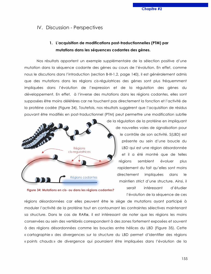
155
IV. Discussion - Perspectives
1. L’acquisition de modifications post-traductionnelles (PTM) par
mutations dans les séquences codantes des gènes.
Nos résultats apportent un exemple supplémentaire de la sélection positive d’une
mutation dans la séquence codante des gènes au cours de l’évolution. En effet, comme
nous le discutions dans l’introduction (section B-III-1.2, page 140), il est généralement admis
que des mutations dans les régions cis-régulatrices des gènes sont plus fréquemment
impliquées dans l’évolution de l’expression et de la régulation des gènes du
développement. En effet, à l’inverse des mutations dans les régions codantes, elles sont
supposées être moins délétères car ne touchent pas directement la fonction et l’activité de
la protéine codée (Figure 34), Toutefois, nos résultats suggèrent que l’acquisition de résidus
pouvant être modifiés en post-traductionnel (PTM) peut permettre une modification subtile
de la régulation de la protéine en impliquant
de nouvelles voies de signalisation pour
le contrôle de son activité. S(LBD) est
présente au sein d’une boucle du
LBD qui est une région désordonnée
et il a été montré que de telles
régions semblent évoluer plus
rapidement du fait qu’elles sont moins
directement impliquées dans le
maintien strict d’une structure. Ainsi, il
serait intéressant d’étudier
l’évolution de la séquence de ces
régions désordonnées car elles peuvent être le siège de mutations ayant participé à
moduler l’activité de la protéine tout en contournant les contraintes sélectives maintenant
sa structure. Dans le cas de RARα, il est intéressant de noter que les régions les moins
conservées au sein des vertébrés correspondent à des zones fortement exposées et souvent
à des régions désordonnées comme les boucles entre hélices du LBD (Figure 35). Cette
« cartographie » des divergences sur la structure du LBD permet d’identifier des régions
« points chauds » de divergence qui pourraient être impliquées dans l’évolution de la
Figure 34: Mutations en cis- ou dans les régions codantes?
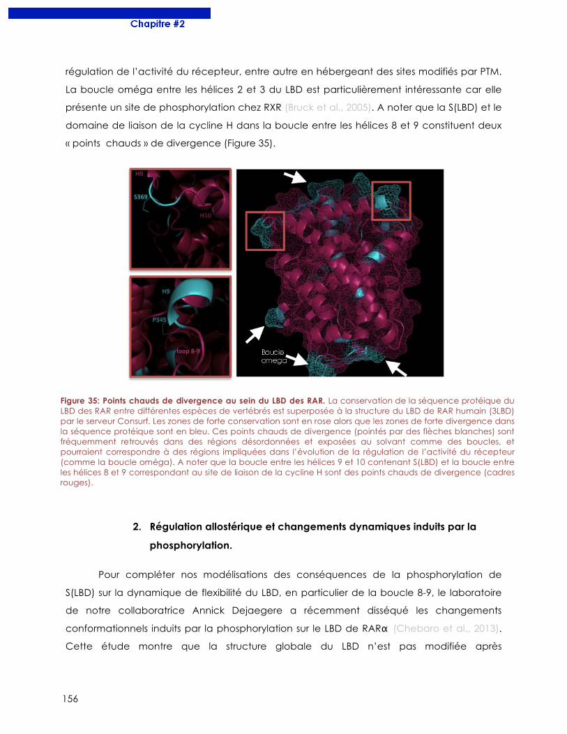
156
régulation de l’activité du récepteur, entre autre en hébergeant des sites modifiés par PTM.
La boucle oméga entre les hélices 2 et 3 du LBD est particulièrement intéressante car elle
présente un site de phosphorylation chez RXR (Bruck et al., 2005). A noter que la S(LBD) et le
domaine de liaison de la cycline H dans la boucle entre les hélices 8 et 9 constituent deux
« points chauds » de divergence (Figure 35).
2. Régulation allostérique et changements dynamiques induits par la
phosphorylation.
Pour compléter nos modélisations des conséquences de la phosphorylation de
S(LBD) sur la dynamique de flexibilité du LBD, en particulier de la boucle 8-9, le laboratoire
de notre collaboratrice Annick Dejaegere a récemment disséqué les changements
conformationnels induits par la phosphorylation sur le LBD de RARα (Chebaro et al., 2013).
Cette étude montre que la structure globale du LBD n’est pas modifiée après
Figure 35: Points chauds de divergence au sein du LBD des RAR. La conservation de la séquence protéique du LBD des RAR entre différentes espèces de vertébrés est superposée à la structure du LBD de RAR humain (3LBD) par le serveur Consurf. Les zones de forte conservation sont en rose alors que les zones de forte divergence dans la séquence protéique sont en bleu. Ces points chauds de divergence (pointés par des flèches blanches) sont fréquemment retrouvés dans des régions désordonnées et exposées au solvant comme des boucles, et pourraient correspondre à des régions impliquées dans l’évolution de la régulation de l’activité du récepteur (comme la boucle oméga). A noter que la boucle entre les hélices 9 et 10 contenant S(LBD) et la boucle entre les hélices 8 et 9 correspondant au site de liaison de la cycline H sont des points chauds de divergence (cadres rouges).
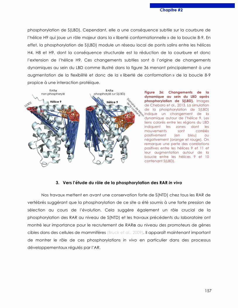
157
phosphorylation de S(LBD). Cependant, elle a une conséquence subtile sur la courbure de
l’hélice H9 qui joue un rôle majeur dans la « liberté conformationnelle » de la boucle 8-9. En
effet, la phosphorylation de S(LBD) module un réseau local de ponts salins entre les hélices
H4, H8 et H9, dont la conséquence structurale est la réduction de la courbure et donc
l’extension de l’hélice H9. Ces changements subtiles sont à l’origine de changements
dynamiques au sein du LBD comme illustré dans la figure 36 menant principalement à une
augmentation de la flexibilité et donc de la « liberté de conformation » de la boucle 8-9
propice à une interaction protéique.
3. Vers l’étude du rôle de la phosphorylation des RAR in vivo
Nos travaux mettent en avant une conservation forte de S(NTD) chez tous les RAR de
vertébrés suggérant que la phosphorylation de ce site a été soumis à une forte pression de
sélection au cours de l’évolution. Cela suggère également un rôle crucial de la
phosphorylation des RAR au niveau de S(NTD) et les travaux précédents du laboratoire ont
montré leur importance pour le recrutement de RARα au niveau des promoteurs de gènes
cibles dans des cellules de mammifères (Bruck et al., 2009). Il apparaît maintenant important
de montrer le rôle de ces phosphorylations in vivo en particulier dans des processus
développementaux régulés par l’AR.
Figure 36: Changements de la dynamique au sein du LBD après phosphorylation de S(LBD). Images de Chebaro et al., 2013. La simulation de la phosphorylation de S(LBD) indique un changement de la dynamique autour de l’hélice 9. Les liens colorés entre les régions du LBD indiquent les zones dont les mouvements sont corrélés positivement (en bleu) ou négativement (orange et rouge). On remarque une perte des corrélations positives entre les hélices 9 et 11 et leur augmentation autour de la boucle entre les hélices 9 et 10 contenant S(LBD).
158
3.1. La différenciation de cellules souches en neurones induite par l’AR
requiert la phosphorylation de RARγ.
Le traitement de cellules souches de souris (cellule ES) à l’AR conduit à leur
différentiation en progéniteurs de neurones en quelques jours (Bibel et al., 2007). Des travaux
en cours dans le laboratoire de Cécile Rochette-Egly ont montré que c’est le sous-type
gamma de RAR qui est requis lors de cette différentiation, et plus particulièrement l’isoforme
2 de RARγ (RARγ2). En effet, alors que les cellules ES invalidées pour les RAR (KO) ne peuvent
plus se différencier en neurones, la différenciation est restaurée lorsqu’on exprime à
nouveau RARγ2, mais ne l’est pas avec RARα ou RARγ1 (Al Tanoury et al. En préparation). Il
a aussi été montré que dans les cellules ES, la voie des ERK (p42/p44MAPK) était rapidement
activée par l’AR et phosphoryle RARγ au niveau de S(NTD) (en position 68 chez RARγ murin).
De manière très intéressante, la phosphorylation de RARγ est nécessaire pour permettre la
différentiation des cellules ES en neurones. En effet, alors que l’expression de RARγ2 dans
des cellules ES KO permet de restaurer la différentiation en neurones, l’expression d’un
phospho-mutant de RARγ où la S(NTD) est substituée par une alanine (RARγ S-A) ne permet
pas cette restauration. En particulier, des expériences de ChIP ont montré que le
recrutement de RARγ au niveau du promoteur de gènes impliqués dans la différentiation de
cellules ES en neurones était abolit avec un phospho-mutant RARγ S-A. De plus, il semble
que la phosphorylation de RARγ soit particulièrement nécessaire pour le recrutement du
récepteur au niveau de RARE précis dont les DR5 et DR7. A noter qu’à l’inverse, certains
gènes cibles de l’AR non impliqués dans la différentiation neuronale ne requièrent pas la
phosphorylation de RARγ pour être régulés par l’AR.
L’ensemble de ces résultats confirme que la phosphorylation des RAR joue un rôle
essentiel dans leur activité transcriptionnelle mais montre également pour la première fois
leur rôle dans un processus biologique impliqué dans le développement. De plus, ces
données révèlent qu’il existe des gènes cibles de l’AR phospho-dépendants car ne pouvant
pas être régulés par un RAR phospho-mutant, et des gènes phospho-indépendants
répondant à l’AR même en l’absence de phosphorylation des RAR. Bien que les
mécanismes précis de cette dépendance reste flous, il semble que RARγ phosphorylé soit
spécifiquement recruté au niveau de RARE de type DR5 et DR7.
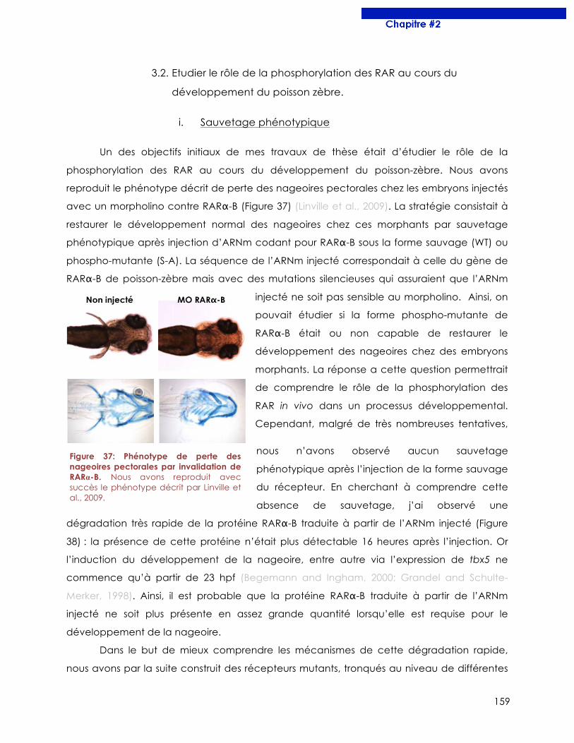
159
3.2. Etudier le rôle de la phosphorylation des RAR au cours du
développement du poisson zèbre.
i. Sauvetage phénotypique
Un des objectifs initiaux de mes travaux de thèse était d’étudier le rôle de la
phosphorylation des RAR au cours du développement du poisson-zèbre. Nous avons
reproduit le phénotype décrit de perte des nageoires pectorales chez les embryons injectés
avec un morpholino contre RARα-B (Figure 37) (Linville et al., 2009). La stratégie consistait à
restaurer le développement normal des nageoires chez ces morphants par sauvetage
phénotypique après injection d’ARNm codant pour RARα-B sous la forme sauvage (WT) ou
phospho-mutante (S-A). La séquence de l’ARNm injecté correspondait à celle du gène de
RARα-B de poisson-zèbre mais avec des mutations silencieuses qui assuraient que l’ARNm
injecté ne soit pas sensible au morpholino. Ainsi, on
pouvait étudier si la forme phospho-mutante de
RARα-B était ou non capable de restaurer le
développement des nageoires chez des embryons
morphants. La réponse a cette question permettrait
de comprendre le rôle de la phosphorylation des
RAR in vivo dans un processus développemental.
Cependant, malgré de très nombreuses tentatives,
nous n’avons observé aucun sauvetage
phénotypique après l’injection de la forme sauvage
du récepteur. En cherchant à comprendre cette
absence de sauvetage, j’ai observé une
dégradation très rapide de la protéine RARα-B traduite à partir de l’ARNm injecté (Figure
38) : la présence de cette protéine n’était plus détectable 16 heures après l’injection. Or
l’induction du développement de la nageoire, entre autre via l’expression de tbx5 ne
commence qu’à partir de 23 hpf (Begemann and Ingham, 2000; Grandel and Schulte-
Merker, 1998). Ainsi, il est probable que la protéine RARα-B traduite à partir de l’ARNm
injecté ne soit plus présente en assez grande quantité lorsqu’elle est requise pour le
développement de la nageoire.
Dans le but de mieux comprendre les mécanismes de cette dégradation rapide,
nous avons par la suite construit des récepteurs mutants, tronqués au niveau de différentes
Figure 37: Phénotype de perte des nageoires pectorales par invalidation de RARα-B. Nous avons reproduit avec succès le phénotype décrit par Linville et al., 2009.
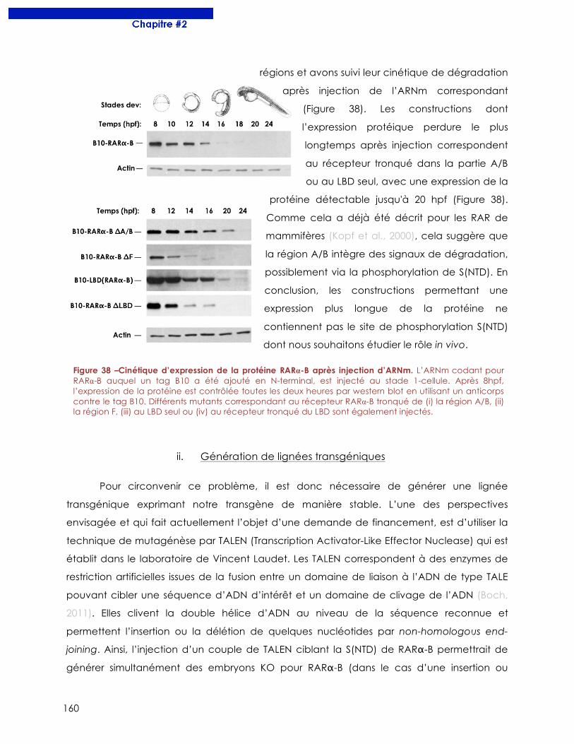
160
régions et avons suivi leur cinétique de dégradation
après injection de l’ARNm correspondant
(Figure 38). Les constructions dont
l’expression protéique perdure le plus
longtemps après injection correspondent
au récepteur tronqué dans la partie A/B
ou au LBD seul, avec une expression de la
protéine détectable jusqu'à 20 hpf (Figure 38).
Comme cela a déjà été décrit pour les RAR de
mammifères (Kopf et al., 2000), cela suggère que
la région A/B intègre des signaux de dégradation,
possiblement via la phosphorylation de S(NTD). En
conclusion, les constructions permettant une
expression plus longue de la protéine ne
contiennent pas le site de phosphorylation S(NTD)
dont nous souhaitons étudier le rôle in vivo.
ii. Génération de lignées transgéniques
Pour circonvenir ce problème, il est donc nécessaire de générer une lignée
transgénique exprimant notre transgène de manière stable. L’une des perspectives
envisagée et qui fait actuellement l’objet d’une demande de financement, est d’utiliser la
technique de mutagénèse par TALEN (Transcription Activator-Like Effector Nuclease) qui est
établit dans le laboratoire de Vincent Laudet. Les TALEN correspondent à des enzymes de
restriction artificielles issues de la fusion entre un domaine de liaison à l’ADN de type TALE
pouvant cibler une séquence d’ADN d’intérêt et un domaine de clivage de l’ADN (Boch,
2011). Elles clivent la double hélice d’ADN au niveau de la séquence reconnue et
permettent l’insertion ou la délétion de quelques nucléotides par non-homologous end-
joining. Ainsi, l’injection d’un couple de TALEN ciblant la S(NTD) de RARα-B permettrait de
générer simultanément des embryons KO pour RARα-B (dans le cas d’une insertion ou
Figure 38 –Cinétique d’expression de la protéine RARα-B après injection d’ARNm. L’ARNm codant pour RARα-B auquel un tag B10 a été ajouté en N-terminal, est injecté au stade 1-cellule. Après 8hpf, l’expression de la protéine est contrôlée toutes les deux heures par western blot en utilisant un anticorps contre le tag B10. Différents mutants correspondant au récepteur RARα-B tronqué de (i) la région A/B, (ii) la région F, (iii) au LBD seul ou (iv) au récepteur tronqué du LBD sont également injectés.
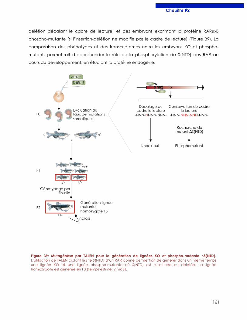
161
délétion décalant le cadre de lecture) et des embryons exprimant la protéine RARα-B
phospho-mutante (si l’insertion-délétion ne modifie pas le cadre de lecture) (Figure 39). La
comparaison des phénotypes et des transcriptomes entre les embryons KO et phospho-
mutants permettrait d’appréhender le rôle de la phosphorylation de S(NTD) des RAR au
cours du développement, en étudiant la protéine endogène.
Figure 39: Mutagénèse par TALEN pour la génération de lignées KO et phospho-mutante ΔS(NTD). L’utilisation de TALEN ciblant le site S(NTD) d’un RAR donné permettrait de générer dans un même temps une lignée KO et une lignée phospho-mutante où S(NTD) est substituée ou deletée. La lignée homozygote est générée en F3 (temps estimé: 9 mois).

162
163
Chapitre#3
Le rôle de l’AR dans la diversification de la denture chez les poissons
164
I. Contexte Scientifique
Il existe une très grande diversité de denture chez les vertébrés et particulièrement
chez les poissons. Ainsi, c’est un modèle de choix pour l’étude des mécanismes
développementaux à l’origine d’une telle diversité (section B-III-4.1, page 47). Des travaux
du laboratoire de Vincent Laudet ont mis en évidence un rôle de l’AR dans l’induction des
dents chez le poisson-zèbre. En effet, l’inhibition de la synthèse endogène d’AR inhibe
l’expression de marqueur dentaire comme dlx2a et empêche le développement des dents
(Gibert et al., 2010). Toutefois, l’effet d’un excès d’AR sur le développement des dents n’a
jamais été testé. De ce fait, nous avons entrepris d’étudier les effets d’un excès exogène ou
endogène d’AR chez le poisson-zèbre et d’autres espèces de cypriniformes.
II. Principaux résultats
o Un traitement précoce à l’AR induit une expansion antérieure de la denture
pharyngienne chez le poisson-zèbre. L’expression de marqueurs dentaires comme
pitx2 ou dlx2b est également étendue antérieurement chez des embryons traités.
L’expansion de la denture induite par l’AR ressemble à la denture d’autres poissons
téléostéens comme le médaka.
o Les enzymes raldh2 et cyp26b1 contrôlent le métabolisme de l’AR dans la
région de formation des dents chez le poisson-zèbre. La dynamique d’expression
temporelle de l’enzyme de synthèse (raldh2) et de dégradation (cyp26b1) de l’AR
définit une fenêtre d’action temporelle et locale au niveau de la région de formation
des dents dans le pharynx.
o La mutation de cyp26b1 induit la formation d’une dent supplémentaire chez
le poisson-zèbre. Alors que 5 dents sont observées sur la rangée ventrale du 5ème arc
branchial chez le poisson-zèbre adulte, les mutants hétérozygotes cyp26b1+/-
développent une 6ème dent qui est attachée normalement.
o Un traitement tardif à l’AR induit un changement de la forme des dents chez
l’embryon de poisson-zèbre. Dans de rares cas, le traitement à l’AR induit la formation
de dents bicuspides ce qui n’est jamais observé de manière naturelle chez le poisson-
zèbre. De plus, un phénotype plus pénétrant observé est un allongement et un
affinement de la forme des dents après traitement à l’AR.
165
o Les phénotypes au niveau de la denture induits par l’AR sont retrouvés chez
d’autres espèces de cypriniformes. Plusieurs espèces développent 6 dents sur la
rangée ventrale du 5ème arc branchial comme observé chez les mutants cyp26b1+ /-
de poisson zèbre. D’autre part, Tanichthys albonubes est un cypriniforme dont les
dents sont plus allongées et plus fines que le poisson-zèbre et sa denture ressemble à
celle induite par un traitement tardif à l’AR. Nous avons corrélé l’acquisition de ce
phénotype à une différence de l’expression temporelle de cyp26b1 dans le pharynx
postérieur entre les deux espèces.
III. Publications
L’ensemble de nos résultats est présenté dans les deux articles à suivre.
1. Retinoic acid expands the evolutionary reduced dentition of zebrafish
Ce travail est issu du laboratoire de William Jackman du Bowdoin College aux Etats-
Unis avec qui nous avons collaboré. Ma participation a été la description comparative de
l’expression des enzymes de synthèse de l’AR chez le poisson-zèbre et le médaka. Ces
travaux ont été publiés en 2012 dans The FASEB Journal.
166
The FASEB Journal • Research Communication
Retinoic acid expands the evolutionarily reduceddentition of zebrafish
Pawat Seritrakul,* Eric Samarut,† Tenzing T. S. Lama,* Yann Gibert,‡ Vincent Laudet,†and William R. Jackman*,1
*Department of Biology, Bowdoin College, Brunswick, Maine, USA; †Molecular Zoology Group,Institut de Génomique Fonctionnelle de Lyon, Université de Lyon, Centre National de la RechercheScientifique, Institut National de la Recherche Agronomique, Université Claude Bernard Lyon 1,Ecole Normale Supérieure de Lyon, Lyon, France; and ‡Deakin University School of Medicine,Waurn Ponds, Victoria, Australia
ABSTRACT Zebrafish lost anterior teeth during evo-lution but retain a posterior pharyngeal dentition thatrequires retinoic acid (RA) cell-cell signaling for itsdevelopment. The purposes of this study were to testthe sufficiency of RA to induce tooth development andto assess its role in evolution. We found that exposureof embryos to exogenous RA induces a dramatic ante-rior expansion of the number of pharyngeal teeth thatlater form and shifts anteriorly the expression patternsof genes normally expressed in the posterior tooth-forming region, such as pitx2 and dlx2b. After RAexposure, we also observed a correlation between car-tilage malformations and ectopic tooth induction, aswell as abnormal cranial neural crest marker geneexpression. Additionally, we observed that the RA-induced zebrafish anterior teeth resemble in patternand number the dentition of fish species that retainanterior pharyngeal teeth such as medaka but thatmedaka do not express the aldh1a2 RA-synthesizingenzyme in tooth-forming regions. We conclude that RAis sufficient to induce anterior ectopic tooth develop-ment in zebrafish where teeth were lost in evolution,potentially by altering neural crest cell development,and that changes in the location of RA synthesis corre-late with evolutionary changes in vertebrate denti-tions.—Seritrakul, P., Samarut, E., Lama, T. T. S.,Gibert, Y., Laudet, V., Jackman, W. R. Retinoic acidexpands the evolutionarily reduced dentition of ze-brafish. FASEB J. 26, 000–000 (2012). www.fasebj.org
Key Words: teeth ! neural crest cells ! retinaldehyde dehydrogenase
Primitive ray-finned fish possessed numerous teethin their pharyngeal regions, and the generally reduceddentitions of modern species are thought to be largelythe result of evolutionary tooth loss (1, 2). The ze-brafish (Danio rerio) lineage, as a member of the orderCypriniformes, lost anterodorsal pharyngeal teeth 65 or
more million years ago (3, 4), and only retain part ofthe original posteroventral pharyngeal dentition. Amore extensive dentition has not reevolved in any ofthe nearly 3000 described cypriniform species, despitediverse feeding modes, including piscivory (5). If thereis indeed selective pressure to increase the dentitionbut it is no longer possible due to developmentalconstraints, this may represent a modern example ofDollo’s law of the irreversibility of evolution (6–8).Such irreversibility may be the result of pleiotropicinteractions during development (9, 10).
Retinoic acid (RA), an oxidized form of vitamin A, isa cell-cell signaling molecule with numerous functionsin developing vertebrate embryos. Evidence of thecrucial roles RA plays in development was establishedover half a century ago, when the offspring of femalerodents fed a vitamin A-deficient diet during pregnancyshowed congenital defects in many organ systems (11).Synthesized from carotenoids, the active all-trans formof RA acts as a ligand for nuclear RA receptors, whichin turn function as transcription factors to control theexpression of numerous targets, including Hox genes(12). The amphiphilic nature of RA, with both polarand nonpolar moieties, allows this molecule to passrelatively easily through cell membranes and act as apositional cue for cells during embryogenesis (13).
In vertebrates, RA is particularly important in antero-posterior (AP) patterning and in the development ofcranial neural crest (CNC) cells. During the develop-mental specification of the AP axis, RA can directlyregulate Hox gene expression, and alterations in RAlevels result in homeotic changes in AP identity (14–16). Such RA-induced homeotic transformations in-clude the duplication of skeletal elements, such asvertebrae (17, 18). RA is also required for the properspecification of CNC cells, a class of vertebrate pluripo-tent migratory cells that give rise to cephalic structures,including cartilage, bone, and teeth (19, 20). Disrup-
1 Correspondence: Bowdoin College, 6500 College Station,Brunswick, ME 04011, USA. E-mail: [email protected]
doi: 10.1096/fj.12-209304
Abbreviations: AP, anteroposterior; CB5, 5th ceratobran-chial element; CNC, cranial neural crest; dpf, days postfertil-ization; hpf, hours postfertilization; RA, retinoic acid
10892-6638/12/0026-0001 © FASEB
The FASEB Journal article fj.12-209304. Published online August 31, 2012.
167
tions in RA signaling cause malformations of CNC-derived tissues in many vertebrates (21, 22).
Teeth exhibit conservation in their early developmentacross vertebrate species and begin their formation inboth mammals and teleost fishes as an epithelial placode(23, 24). Subsequent stages of tooth morphogenesis anddifferentiation are coordinated by reciprocal cell signal-ing between this dental epithelium and an underlyingmesenchyme, which employs members of several evolu-tionarily conserved vertebrate gene families, including thefibroblast growth factor and hedgehog signaling pathways(25–27). The mammalian dental mesenchyme is knownto be largely derived from CNC cells (28), and the dentalmesenchyme of zebrafish pharyngeal teeth also expressCNC cell markers (25).
RA signaling has been studied previously in relationto tooth development. In mice, mutants lacking RAreceptors develop deformed skulls without teeth (29).Conversely, exposing embryonic mouse mandibles totissue culture medium containing excess RA altersdental epithelial morphology, increases tooth bud sizein the diastema region, and possibly switches toothidentity between molars and incisors (30). These stud-ies provide some clues to the potential roles of RA inmammalian tooth development, although there hasbeen no gene expression data or direct functional testsat a molecular level to explain the mechanisms under-lying these phenotypes. In zebrafish, it has been shownthat tooth induction depends on an endogenoussource of RA from retinaldehyde dehydrogenase-ex-pressing cells and that chemically blocking RA synthesisresults in the complete absence of teeth (31). However,effects of RA overexpression on zebrafish tooth devel-opment have not yet been described.
Here we report that exposure of zebrafish embryos toexogenous RA induces the formation of an extensivesymmetrical pattern of supernumerary teeth. Corre-sponding with this induction, we find up-regulation ofgenes normally expressed in developing teeth and inthe nearby posterior pharyngeal region such as pitx2,dlx2b, and hoxb5a. After RA treatment, CNC cells arepresent but with disrupted arrangements and abnormalgene expression, and cartilage development is corre-spondingly disrupted. We compare the expanded RA-induced zebrafish pharyngeal dentition with the wild-type dentitions of other teleost species and findsimilarities especially with the teeth of medaka. How-ever, comparisons of retinaldehyde dehydrogenase ex-pression between these species suggest that evolution-ary changes in RA signaling were not likely responsiblefor the reduction of the zebrafish pharyngeal dentition.
MATERIALS AND METHODS
Animal husbandry
Embryos of wild-type zebrafish (Danio rerio, Hamilton 1822)were obtained from in-house stocks derived originally from acommercial supplier (LiveAquaria.com, Rhinelander, WI,
USA). Embryos were raised at 28.5°C in an embryo mediumconsisting of 30% Danieu’s medium and 0.003% phenylthio-urea to prevent pigmentation (25). Phenylthiourea use isimportant to note, as it has recently been reported toenhance certain RA-related craniofacial phenotypes in ze-brafish (32). Zebrafish staging was done as described previ-ously (33), with developmental time reported in hours post-fertilization (hpf) or days postfertilization (dpf). The fli1:GFPreporter fish were of the Tg(fli1:EGFP)y1 line (34), and thedlx2b:GFP reporters were from the Tg(dlx2b:EGFP)cs1 line(35). Astyanax mexicanus (De Filippi 1853) stages are reportedin dpf, and medaka (Oryzias latipes, Temminck and Schlegel1846) stages are reported as dpf or stage numbers (36).Animal use was in accordance with protocols approved by theInstitutional Animal Care and Use Committee at BowdoinCollege and the Université de Lyon.
Exogenous RA treatment and bead implantation
Embryos were manually dechorionated and added to embryomedium containing either 0.05% DMSO alone as a control orDMSO plus 6 ! 10"7 M all-trans RA (Biomol International,Plymouth, PA, USA). RA treatments were from 24 hpf onwardunless indicated otherwise. Embryos were kept in the dark orwith minimal lighting to avoid RA degradation (31). Toinactivate RA, embryos were washed into new medium andexposed to bright light for 5 min. AG 1-X8 beads (Bio-Rad,Hercules, CA, USA) were incubated in fresh solution of 6 !10"4 M RA and implanted as described previously (37).
Histology and in situ hybridization
Whole-mount mRNA in situ hybridization was performed asdescribed previously (25). Riboprobe sources: zebrafish dlx2b,dlx2a, and pitx2 (25); crestin (38); hoxb5a (39); sox9a (40);foxd3 (41); mitfa (42), lef1 (43); prdm1a (44); aldh1a2 (31);and medaka aldh1a2 (sequences 1367–1830 of Genbankaccession number NM_001104821). Optical confocal micros-copy sections of in situ hybridization specimens were obtainedusing 633-nm fluorescence of the BM Purple (Roche, India-napolis, IN, USA) staining product (45). Double fluorescencein situ hybridization was performed as described previously(46). Alizarin red and alcian blue skeletal single and doublestaining were performed as described previously (47).
Microscopy and statistical analysis
Embryos were visualized and photographed on a LeicaDMI3000B inverted microscope with DFC420C camera (LeicaMicrosystems, Solms, Germany), a Leica MZ16F stereomicro-scope with DFC300FX camera (Leica Microsystems), or aZeiss 510 Meta confocal laser-scanning microscope (CarlZeiss, Jena, Germany). Three-dimensional images of teethwere generated from confocal data using Volocity (PerkinElmer, San Jose, CA, USA). Photograph files were processedwith Adobe Photoshop (Adobe Systems, San Jose, CA, USA)and minimally adjusted as a whole for brightness, contrast,and/or color balance. Statistical significance of collected datawas calculated using a 2-tailed Fisher’s exact test of indepen-dence (48).
RESULTS
Exogenous RA expands zebrafish tooth developmentanteriorly and dorsally
Wild-type zebrafish larvae normally develop a singlepair of fully formed teeth on each side of the ventral,
2 Vol. 26 December 2012 SERITRAKUL ET AL.The FASEB Journal ! www.fasebj.org
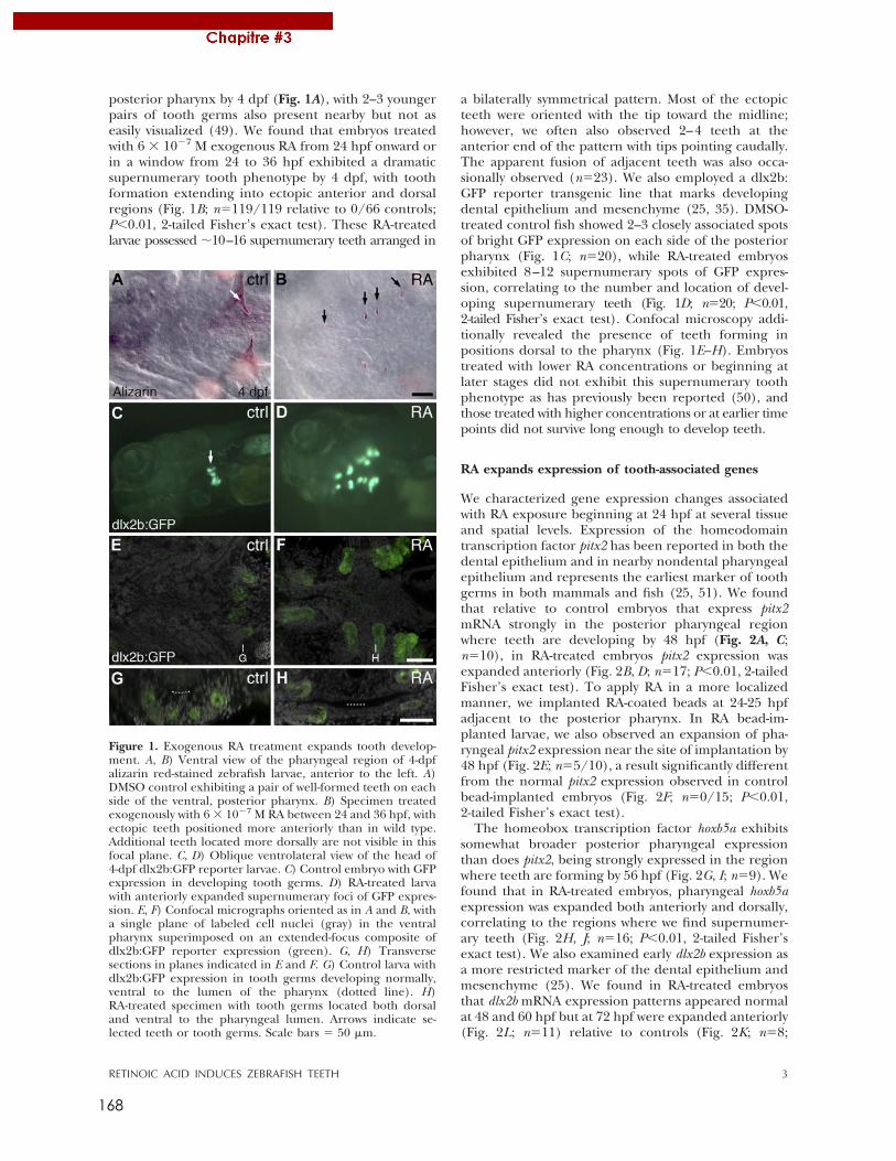
168
posterior pharynx by 4 dpf (Fig. 1A), with 2–3 youngerpairs of tooth germs also present nearby but not aseasily visualized (49). We found that embryos treatedwith 6 ! 10"7 M exogenous RA from 24 hpf onward orin a window from 24 to 36 hpf exhibited a dramaticsupernumerary tooth phenotype by 4 dpf, with toothformation extending into ectopic anterior and dorsalregions (Fig. 1B; n#119/119 relative to 0/66 controls;P$0.01, 2-tailed Fisher’s exact test). These RA-treatedlarvae possessed %10–16 supernumerary teeth arranged in
a bilaterally symmetrical pattern. Most of the ectopicteeth were oriented with the tip toward the midline;however, we often also observed 2–4 teeth at theanterior end of the pattern with tips pointing caudally.The apparent fusion of adjacent teeth was also occa-sionally observed (n#23). We also employed a dlx2b:GFP reporter transgenic line that marks developingdental epithelium and mesenchyme (25, 35). DMSO-treated control fish showed 2–3 closely associated spotsof bright GFP expression on each side of the posteriorpharynx (Fig. 1C; n#20), while RA-treated embryosexhibited 8–12 supernumerary spots of GFP expres-sion, correlating to the number and location of devel-oping supernumerary teeth (Fig. 1D; n#20; P$0.01,2-tailed Fisher’s exact test). Confocal microscopy addi-tionally revealed the presence of teeth forming inpositions dorsal to the pharynx (Fig. 1E–H). Embryostreated with lower RA concentrations or beginning atlater stages did not exhibit this supernumerary toothphenotype as has previously been reported (50), andthose treated with higher concentrations or at earlier timepoints did not survive long enough to develop teeth.
RA expands expression of tooth-associated genes
We characterized gene expression changes associatedwith RA exposure beginning at 24 hpf at several tissueand spatial levels. Expression of the homeodomaintranscription factor pitx2 has been reported in both thedental epithelium and in nearby nondental pharyngealepithelium and represents the earliest marker of toothgerms in both mammals and fish (25, 51). We foundthat relative to control embryos that express pitx2mRNA strongly in the posterior pharyngeal regionwhere teeth are developing by 48 hpf (Fig. 2A, C;n#10), in RA-treated embryos pitx2 expression wasexpanded anteriorly (Fig. 2B, D; n#17; P$0.01, 2-tailedFisher’s exact test). To apply RA in a more localizedmanner, we implanted RA-coated beads at 24-25 hpfadjacent to the posterior pharynx. In RA bead-im-planted larvae, we also observed an expansion of pha-ryngeal pitx2 expression near the site of implantation by48 hpf (Fig. 2E; n#5/10), a result significantly differentfrom the normal pitx2 expression observed in controlbead-implanted embryos (Fig. 2F; n#0/15; P$0.01,2-tailed Fisher’s exact test).
The homeobox transcription factor hoxb5a exhibitssomewhat broader posterior pharyngeal expressionthan does pitx2, being strongly expressed in the regionwhere teeth are forming by 56 hpf (Fig. 2G, I; n#9). Wefound that in RA-treated embryos, pharyngeal hoxb5aexpression was expanded both anteriorly and dorsally,correlating to the regions where we find supernumer-ary teeth (Fig. 2H, J; n#16; P$0.01, 2-tailed Fisher’sexact test). We also examined early dlx2b expression asa more restricted marker of the dental epithelium andmesenchyme (25). We found in RA-treated embryosthat dlx2b mRNA expression patterns appeared normalat 48 and 60 hpf but at 72 hpf were expanded anteriorly(Fig. 2L; n#11) relative to controls (Fig. 2K; n#8;
Figure 1. Exogenous RA treatment expands tooth develop-ment. A, B) Ventral view of the pharyngeal region of 4-dpfalizarin red-stained zebrafish larvae, anterior to the left. A)DMSO control exhibiting a pair of well-formed teeth on eachside of the ventral, posterior pharynx. B) Specimen treatedexogenously with 6 ! 10"7 M RA between 24 and 36 hpf, withectopic teeth positioned more anteriorly than in wild type.Additional teeth located more dorsally are not visible in thisfocal plane. C, D) Oblique ventrolateral view of the head of4-dpf dlx2b:GFP reporter larvae. C) Control embryo with GFPexpression in developing tooth germs. D) RA-treated larvawith anteriorly expanded supernumerary foci of GFP expres-sion. E, F) Confocal micrographs oriented as in A and B, witha single plane of labeled cell nuclei (gray) in the ventralpharynx superimposed on an extended-focus composite ofdlx2b:GFP reporter expression (green). G, H) Transversesections in planes indicated in E and F. G) Control larva withdlx2b:GFP expression in tooth germs developing normally,ventral to the lumen of the pharynx (dotted line). H)RA-treated specimen with tooth germs located both dorsaland ventral to the pharyngeal lumen. Arrows indicate se-lected teeth or tooth germs. Scale bars # 50 &m.
3RETINOIC ACID INDUCES ZEBRAFISH TEETH
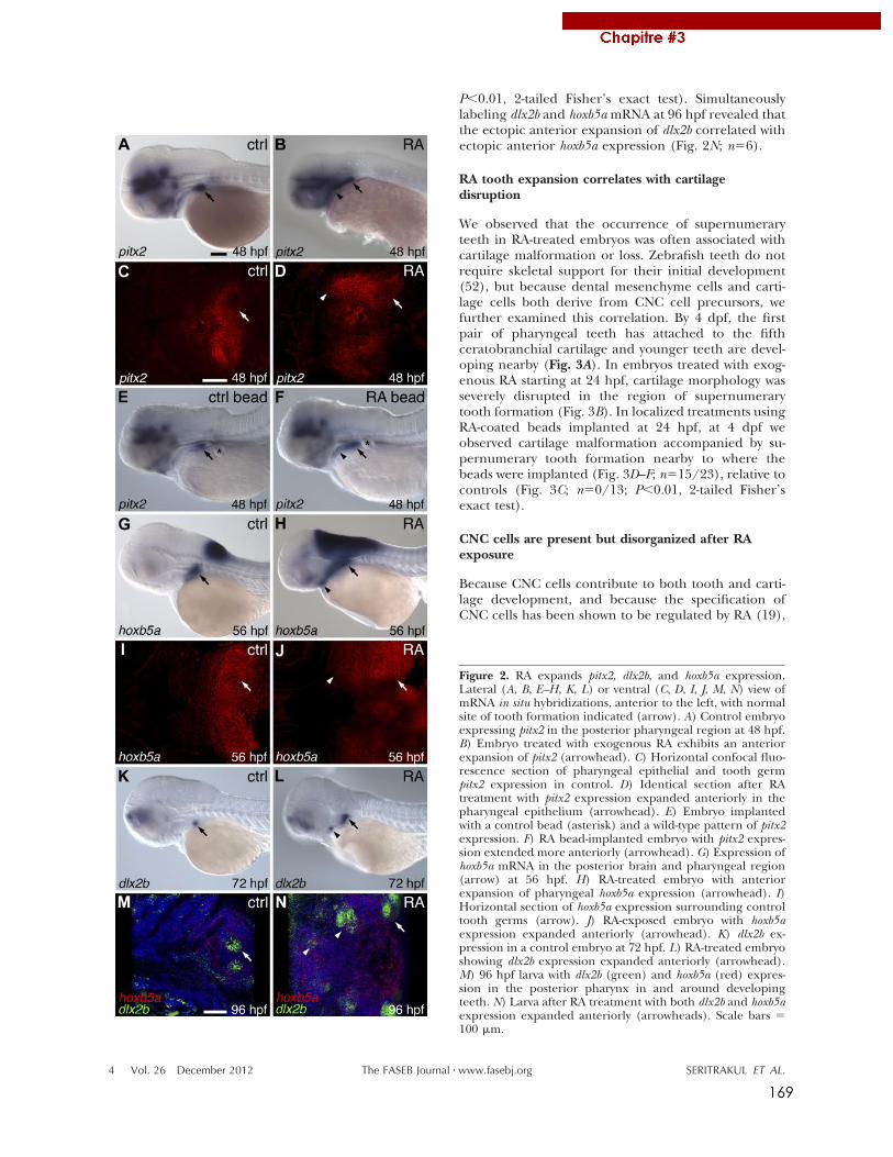
169
P!0.01, 2-tailed Fisher’s exact test). Simultaneouslylabeling dlx2b and hoxb5a mRNA at 96 hpf revealed thatthe ectopic anterior expansion of dlx2b correlated withectopic anterior hoxb5a expression (Fig. 2N; n"6).
RA tooth expansion correlates with cartilagedisruption
We observed that the occurrence of supernumeraryteeth in RA-treated embryos was often associated withcartilage malformation or loss. Zebrafish teeth do notrequire skeletal support for their initial development(52), but because dental mesenchyme cells and carti-lage cells both derive from CNC cell precursors, wefurther examined this correlation. By 4 dpf, the firstpair of pharyngeal teeth has attached to the fifthceratobranchial cartilage and younger teeth are devel-oping nearby (Fig. 3A). In embryos treated with exog-enous RA starting at 24 hpf, cartilage morphology wasseverely disrupted in the region of supernumerarytooth formation (Fig. 3B). In localized treatments usingRA-coated beads implanted at 24 hpf, at 4 dpf weobserved cartilage malformation accompanied by su-pernumerary tooth formation nearby to where thebeads were implanted (Fig. 3D–F; n"15/23), relative tocontrols (Fig. 3C; n"0/13; P!0.01, 2-tailed Fisher’sexact test).
CNC cells are present but disorganized after RAexposure
Because CNC cells contribute to both tooth and carti-lage development, and because the specification ofCNC cells has been shown to be regulated by RA (19),
Figure 2. RA expands pitx2, dlx2b, and hoxb5a expression.Lateral (A, B, E–H, K, L) or ventral (C, D, I, J, M, N) view ofmRNA in situ hybridizations, anterior to the left, with normalsite of tooth formation indicated (arrow). A) Control embryoexpressing pitx2 in the posterior pharyngeal region at 48 hpf.B) Embryo treated with exogenous RA exhibits an anteriorexpansion of pitx2 (arrowhead). C) Horizontal confocal fluo-rescence section of pharyngeal epithelial and tooth germpitx2 expression in control. D) Identical section after RAtreatment with pitx2 expression expanded anteriorly in thepharyngeal epithelium (arrowhead). E) Embryo implantedwith a control bead (asterisk) and a wild-type pattern of pitx2expression. F) RA bead-implanted embryo with pitx2 expres-sion extended more anteriorly (arrowhead). G) Expression ofhoxb5a mRNA in the posterior brain and pharyngeal region(arrow) at 56 hpf. H) RA-treated embryo with anteriorexpansion of pharyngeal hoxb5a expression (arrowhead). I)Horizontal section of hoxb5a expression surrounding controltooth germs (arrow). J) RA-exposed embryo with hoxb5aexpression expanded anteriorly (arrowhead). K) dlx2b ex-pression in a control embryo at 72 hpf. L) RA-treated embryoshowing dlx2b expression expanded anteriorly (arrowhead).M) 96 hpf larva with dlx2b (green) and hoxb5a (red) expres-sion in the posterior pharynx in and around developingteeth. N) Larva after RA treatment with both dlx2b and hoxb5aexpression expanded anteriorly (arrowheads). Scale bars "100 #m.
4 Vol. 26 December 2012 SERITRAKUL ET AL.The FASEB Journal ! www.fasebj.org
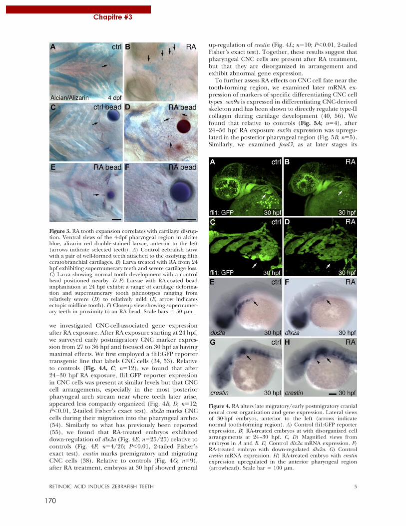
170
we investigated CNC-cell-associated gene expressionafter RA exposure. After RA exposure starting at 24 hpf,we surveyed early postmigratory CNC marker expres-sion from 27 to 36 hpf and focused on 30 hpf as havingmaximal effects. We first employed a fli1:GFP reportertransgenic line that labels CNC cells (34, 53). Relativeto controls (Fig. 4A, C; n!12), we found that after24–30 hpf RA exposure, fli1:GFP reporter expressionin CNC cells was present at similar levels but that CNCcell arrangements, especially in the most posteriorpharyngeal arch stream near where teeth later arise,appeared less compactly organized (Fig. 4B, D; n!12;P"0.01, 2-tailed Fisher’s exact test). dlx2a marks CNCcells during their migration into the pharyngeal arches(54). Similarly to what has previously been reported(55), we found that RA-treated embryos exhibiteddown-regulation of dlx2a (Fig. 4E; n!25/25) relative tocontrols (Fig. 4F; n!4/26; P"0.01, 2-tailed Fisher’sexact test). crestin marks premigratory and migratingCNC cells (38). Relative to controls (Fig. 4G; n!9),after RA treatment, embryos at 30 hpf showed general
up-regulation of crestin (Fig. 4L; n!10; P"0.01, 2-tailedFisher’s exact test). Together, these results suggest thatpharyngeal CNC cells are present after RA treatment,but that they are disorganized in arrangement andexhibit abnormal gene expression.
To further assess RA effects on CNC cell fate near thetooth-forming region, we examined later mRNA ex-pression of markers of specific differentiating CNC celltypes. sox9a is expressed in differentiating CNC-derivedskeleton and has been shown to directly regulate type-IIcollagen during cartilage development (40, 56). Wefound that relative to controls (Fig. 5A; n!4), after24–56 hpf RA exposure sox9a expression was upregu-lated in the posterior pharyngeal region (Fig. 5B; n!5).Similarly, we examined foxd3, as at later stages its
Figure 4. RA alters late migratory/early postmigratory cranialneural crest organization and gene expression. Lateral viewsof 30-hpf embryos, anterior to the left (arrows indicatenormal tooth-forming region). A) Control fli1:GFP reporterexpression. B) RA-treated embryos at with disorganized cellarrangements at 24–30 hpf. C, D) Magnified views fromembryos in A and B. E) Control dlx2a mRNA expression. F)RA-treated embryo with down-regulated dlx2a. G) Controlcrestin mRNA expression. H) RA-treated embryo with crestinexpression upregulated in the anterior pharyngeal region(arrowhead). Scale bar ! 100 #m.
Figure 3. RA tooth expansion correlates with cartilage disrup-tion. Ventral views of the 4-dpf pharyngeal region in alcianblue, alizarin red double-stained larvae, anterior to the left(arrows indicate selected teeth). A) Control zebrafish larvawith a pair of well-formed teeth attached to the ossifying fifthceratobranchial cartilages. B) Larva treated with RA from 24hpf exhibiting supernumerary teeth and severe cartilage loss.C) Larva showing normal tooth development with a controlbead positioned nearby. D–F) Larvae with RA-coated beadimplantation at 24 hpf exhibit a range of cartilage deforma-tion and supernumerary tooth phenotypes ranging fromrelatively severe (D) to relatively mild (E, arrow indicatesectopic midline tooth). F) Closeup view showing supernumer-ary teeth in proximity to an RA bead. Scale bars ! 50 #m.
5RETINOIC ACID INDUCES ZEBRAFISH TEETH
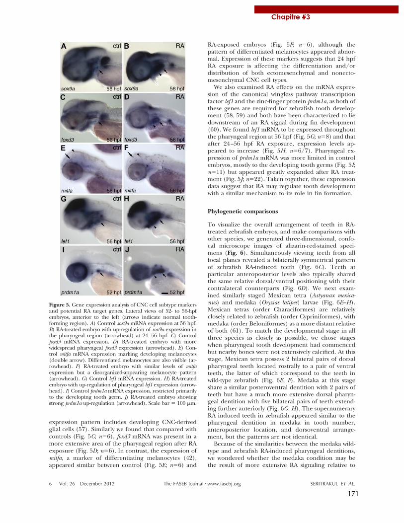
171
expression pattern includes developing CNC-derivedglial cells (57). Similarly we found that compared withcontrols (Fig. 5C; n!6), foxd3 mRNA was present in amore extensive area of the pharyngeal region after RAexposure (Fig. 5D; n!6). In contrast, the expression ofmitfa, a marker of differentiating melanocytes (42),appeared similar between control (Fig. 5E; n!6) and
RA-exposed embryos (Fig. 5F; n!6), although thepattern of differentiated melanocytes appeared abnor-mal. Expression of these markers suggests that 24 hpfRA exposure is affecting the differentiation and/ordistribution of both ectomesenchymal and nonecto-mesenchymal CNC cell types.
We also examined RA effects on the mRNA expres-sion of the canonical wingless pathway transcriptionfactor lef1 and the zinc-finger protein prdm1a, as both ofthese genes are required for zebrafish tooth develop-ment (58, 59) and both have been characterized to liedownstream of an RA signal during fin development(60). We found lef1 mRNA to be expressed throughoutthe pharyngeal region at 56 hpf (Fig. 5G; n!8) and thatafter 24–56 hpf RA exposure, expression levels ap-peared to increase (Fig. 5H; n!6/7). Pharyngeal ex-pression of prdm1a mRNA was more limited in controlembryos, mostly to the developing tooth germs (Fig. 5I;n!11) but appeared greatly expanded after RA treat-ment (Fig. 5J; n!22). Taken together, these expressiondata suggest that RA may regulate tooth developmentwith a similar mechanism to its role in fin formation.
Phylogenetic comparisons
To visualize the overall arrangement of teeth in RA-treated zebrafish embryos, and make comparisons withother species, we generated three-dimensional, confo-cal microscope images of alizarin-red-stained speci-mens (Fig. 6). Simultaneously viewing teeth from allfocal planes revealed a bilaterally symmetrical patternof zebrafish RA-induced teeth (Fig. 6C). Teeth atparticular anteroposterior levels also typically sharedthe same relative dorsal/ventral positioning with theircontralateral counterparts (Fig. 6D). We next exam-ined similarly staged Mexican tetra (Astyanax mexica-nus) and medaka (Oryzias latipes) larvae (Fig. 6E–H).Mexican tetras (order Characiformes) are relativelyclosely related to zebrafish (order Cypriniformes), withmedaka (order Beloniformes) as a more distant relativeof both (61). To match the developmental stage in allthree species as closely as possible, we chose stageswhen pharyngeal tooth development had commencedbut nearby bones were not extensively calcified. At thisstage, Mexican tetra possess 2 bilateral pairs of dorsalpharyngeal teeth located rostrally to a pair of ventralteeth, the latter of which correspond to the teeth inwild-type zebrafish (Fig. 6E, F). Medaka at this stageshare a similar posteroventral dentition with 2 pairs ofteeth but have a much more extensive dorsal pharyn-geal dentition with five bilateral pairs of teeth extend-ing further anteriorly (Fig. 6G, H). The supernumeraryRA induced teeth in zebrafish appeared similar to thepharyngeal dentition in medaka in tooth number,anteroposterior location, and dorsoventral arrange-ment, but the patterns are not identical.
Because of the similarities between the medaka wild-type and zebrafish RA-induced pharyngeal dentitions,we wondered whether the medaka condition may bethe result of more extensive RA signaling relative to
Figure 5. Gene expression analysis of CNC cell subtype markersand potential RA target genes. Lateral views of 52- to 56-hpfembryos, anterior to the left (arrows indicate normal tooth-forming region). A) Control sox9a mRNA expression at 56 hpf.B) RA-treated embryo with up-regulation of sox9a expression inthe pharyngeal region (arrowhead) at 24–56 hpf. C) Controlfoxd3 mRNA expression. D) RA-treated embryo with morewidespread pharyngeal foxd3 expression (arrowhead). E) Con-trol mitfa mRNA expression marking developing melanocytes(double arrow). Differentiated melanocytes are also visible (ar-rowhead). F) RA-treated embryo with similar levels of mitfaexpression but a disorganized-appearing melanocyte pattern(arrowhead). G) Control lef1 mRNA expression. H) RA-treatedembryo with up-regulation of pharyngeal lef1 expression (arrow-head). I) Control prdm1a mRNA expression, restricted primarilyto the developing tooth germ. J) RA-treated embryo showingstrong prdm1a up-regulation (arrowhead). Scale bar ! 100 "m.
6 Vol. 26 December 2012 SERITRAKUL ET AL.The FASEB Journal ! www.fasebj.org
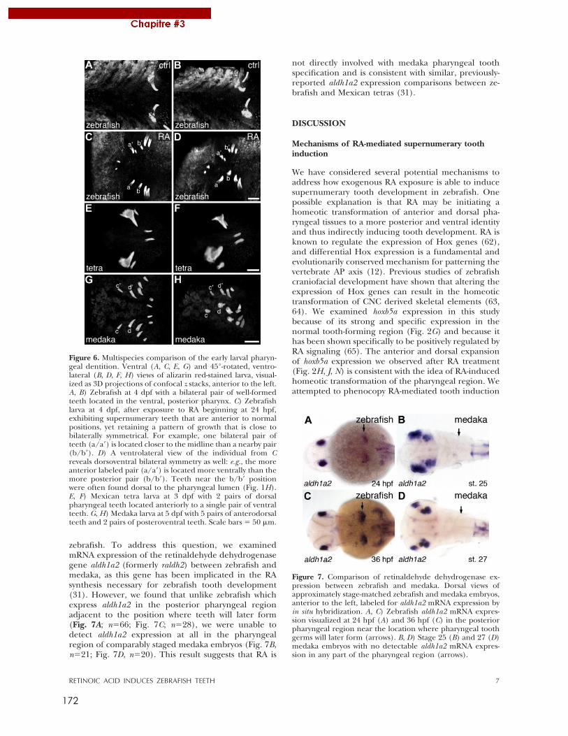
172
zebrafish. To address this question, we examinedmRNA expression of the retinaldehyde dehydrogenasegene aldh1a2 (formerly raldh2) between zebrafish andmedaka, as this gene has been implicated in the RAsynthesis necessary for zebrafish tooth development(31). However, we found that unlike zebrafish whichexpress aldh1a2 in the posterior pharyngeal regionadjacent to the position where teeth will later form(Fig. 7A; n!66; Fig. 7C; n!28), we were unable todetect aldh1a2 expression at all in the pharyngealregion of comparably staged medaka embryos (Fig. 7B,n!21; Fig. 7D, n!20). This result suggests that RA is
not directly involved with medaka pharyngeal toothspecification and is consistent with similar, previously-reported aldh1a2 expression comparisons between ze-brafish and Mexican tetras (31).
DISCUSSION
Mechanisms of RA-mediated supernumerary toothinduction
We have considered several potential mechanisms toaddress how exogenous RA exposure is able to inducesupernumerary tooth development in zebrafish. Onepossible explanation is that RA may be initiating ahomeotic transformation of anterior and dorsal pha-ryngeal tissues to a more posterior and ventral identityand thus indirectly inducing tooth development. RA isknown to regulate the expression of Hox genes (62),and differential Hox expression is a fundamental andevolutionarily conserved mechanism for patterning thevertebrate AP axis (12). Previous studies of zebrafishcraniofacial development have shown that altering theexpression of Hox genes can result in the homeotictransformation of CNC derived skeletal elements (63,64). We examined hoxb5a expression in this studybecause of its strong and specific expression in thenormal tooth-forming region (Fig. 2G) and because ithas been shown specifically to be positively regulated byRA signaling (65). The anterior and dorsal expansionof hoxb5a expression we observed after RA treatment(Fig. 2H, J, N) is consistent with the idea of RA-inducedhomeotic transformation of the pharyngeal region. Weattempted to phenocopy RA-mediated tooth induction
Figure 7. Comparison of retinaldehyde dehydrogenase ex-pression between zebrafish and medaka. Dorsal views ofapproximately stage-matched zebrafish and medaka embryos,anterior to the left, labeled for aldh1a2 mRNA expression byin situ hybridization. A, C) Zebrafish aldh1a2 mRNA expres-sion visualized at 24 hpf (A) and 36 hpf (C) in the posteriorpharyngeal region near the location where pharyngeal toothgerms will later form (arrows). B, D) Stage 25 (B) and 27 (D)medaka embryos with no detectable aldh1a2 mRNA expres-sion in any part of the pharyngeal region (arrows).
Figure 6. Multispecies comparison of the early larval pharyn-geal dentition. Ventral (A, C, E, G) and 45°-rotated, ventro-lateral (B, D, F, H) views of alizarin red-stained larva, visual-ized as 3D projections of confocal z stacks, anterior to the left.A, B) Zebrafish at 4 dpf with a bilateral pair of well-formedteeth located in the ventral, posterior pharynx. C) Zebrafishlarva at 4 dpf, after exposure to RA beginning at 24 hpf,exhibiting supernumerary teeth that are anterior to normalpositions, yet retaining a pattern of growth that is close tobilaterally symmetrical. For example, one bilateral pair ofteeth (a/a=) is located closer to the midline than a nearby pair(b/b=). D) A ventrolateral view of the individual from Creveals dorsoventral bilateral symmetry as well: e.g., the moreanterior labeled pair (a/a=) is located more ventrally than themore posterior pair (b/b=). Teeth near the b/b= positionwere often found dorsal to the pharyngeal lumen (Fig. 1H).E, F) Mexican tetra larva at 3 dpf with 2 pairs of dorsalpharyngeal teeth located anteriorly to a single pair of ventralteeth. G, H) Medaka larva at 5 dpf with 5 pairs of anterodorsalteeth and 2 pairs of posteroventral teeth. Scale bars ! 50 "m.
7RETINOIC ACID INDUCES ZEBRAFISH TEETH
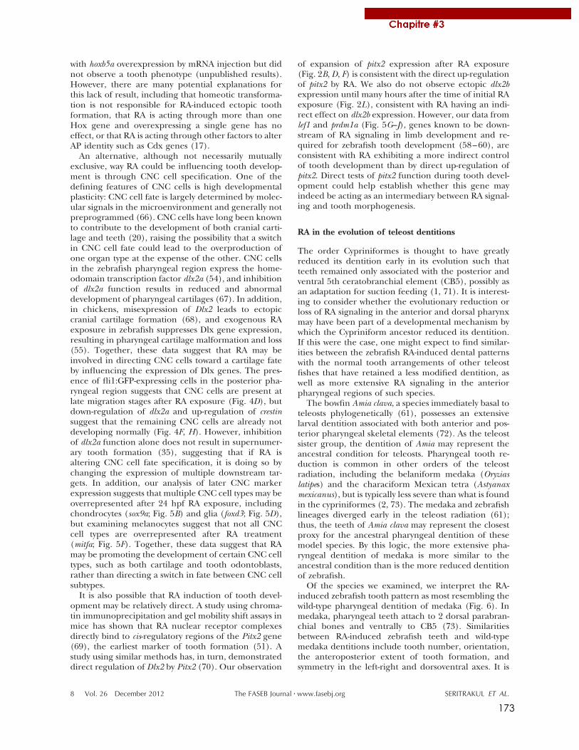
173
with hoxb5a overexpression by mRNA injection but didnot observe a tooth phenotype (unpublished results).However, there are many potential explanations forthis lack of result, including that homeotic transforma-tion is not responsible for RA-induced ectopic toothformation, that RA is acting through more than oneHox gene and overexpressing a single gene has noeffect, or that RA is acting through other factors to alterAP identity such as Cdx genes (17).
An alternative, although not necessarily mutuallyexclusive, way RA could be influencing tooth develop-ment is through CNC cell specification. One of thedefining features of CNC cells is high developmentalplasticity: CNC cell fate is largely determined by molec-ular signals in the microenvironment and generally notpreprogrammed (66). CNC cells have long been knownto contribute to the development of both cranial carti-lage and teeth (20), raising the possibility that a switchin CNC cell fate could lead to the overproduction ofone organ type at the expense of the other. CNC cellsin the zebrafish pharyngeal region express the home-odomain transcription factor dlx2a (54), and inhibitionof dlx2a function results in reduced and abnormaldevelopment of pharyngeal cartilages (67). In addition,in chickens, misexpression of Dlx2 leads to ectopiccranial cartilage formation (68), and exogenous RAexposure in zebrafish suppresses Dlx gene expression,resulting in pharyngeal cartilage malformation and loss(55). Together, these data suggest that RA may beinvolved in directing CNC cells toward a cartilage fateby influencing the expression of Dlx genes. The pres-ence of fli1:GFP-expressing cells in the posterior pha-ryngeal region suggests that CNC cells are present atlate migration stages after RA exposure (Fig. 4D), butdown-regulation of dlx2a and up-regulation of crestinsuggest that the remaining CNC cells are already notdeveloping normally (Fig. 4F, H). However, inhibitionof dlx2a function alone does not result in supernumer-ary tooth formation (35), suggesting that if RA isaltering CNC cell fate specification, it is doing so bychanging the expression of multiple downstream tar-gets. In addition, our analysis of later CNC markerexpression suggests that multiple CNC cell types may beoverrepresented after 24 hpf RA exposure, includingchondrocytes (sox9a; Fig. 5B) and glia (foxd3; Fig. 5D),but examining melanocytes suggest that not all CNCcell types are overrepresented after RA treatment(mitfa; Fig. 5F). Together, these data suggest that RAmay be promoting the development of certain CNC celltypes, such as both cartilage and tooth odontoblasts,rather than directing a switch in fate between CNC cellsubtypes.
It is also possible that RA induction of tooth devel-opment may be relatively direct. A study using chroma-tin immunoprecipitation and gel mobility shift assays inmice has shown that RA nuclear receptor complexesdirectly bind to cis-regulatory regions of the Pitx2 gene(69), the earliest marker of tooth formation (51). Astudy using similar methods has, in turn, demonstrateddirect regulation of Dlx2 by Pitx2 (70). Our observation
of expansion of pitx2 expression after RA exposure(Fig. 2B, D, F) is consistent with the direct up-regulationof pitx2 by RA. We also do not observe ectopic dlx2bexpression until many hours after the time of initial RAexposure (Fig. 2L), consistent with RA having an indi-rect effect on dlx2b expression. However, our data fromlef1 and prdm1a (Fig. 5G–J), genes known to be down-stream of RA signaling in limb development and re-quired for zebrafish tooth development (58–60), areconsistent with RA exhibiting a more indirect controlof tooth development than by direct up-regulation ofpitx2. Direct tests of pitx2 function during tooth devel-opment could help establish whether this gene mayindeed be acting as an intermediary between RA signal-ing and tooth morphogenesis.
RA in the evolution of teleost dentitions
The order Cypriniformes is thought to have greatlyreduced its dentition early in its evolution such thatteeth remained only associated with the posterior andventral 5th ceratobranchial element (CB5), possibly asan adaptation for suction feeding (1, 71). It is interest-ing to consider whether the evolutionary reduction orloss of RA signaling in the anterior and dorsal pharynxmay have been part of a developmental mechanism bywhich the Cypriniform ancestor reduced its dentition.If this were the case, one might expect to find similar-ities between the zebrafish RA-induced dental patternswith the normal tooth arrangements of other teleostfishes that have retained a less modified dentition, aswell as more extensive RA signaling in the anteriorpharyngeal regions of such species.
The bowfin Amia clava, a species immediately basal toteleosts phylogenetically (61), possesses an extensivelarval dentition associated with both anterior and pos-terior pharyngeal skeletal elements (72). As the teleostsister group, the dentition of Amia may represent theancestral condition for teleosts. Pharyngeal tooth re-duction is common in other orders of the teleostradiation, including the belaniform medaka (Oryziaslatipes) and the characiform Mexican tetra (Astyanaxmexicanus), but is typically less severe than what is foundin the cypriniformes (2, 73). The medaka and zebrafishlineages diverged early in the teleost radiation (61);thus, the teeth of Amia clava may represent the closestproxy for the ancestral pharyngeal dentition of thesemodel species. By this logic, the more extensive pha-ryngeal dentition of medaka is more similar to theancestral condition than is the more reduced dentitionof zebrafish.
Of the species we examined, we interpret the RA-induced zebrafish tooth pattern as most resembling thewild-type pharyngeal dentition of medaka (Fig. 6). Inmedaka, pharyngeal teeth attach to 2 dorsal parabran-chial bones and ventrally to CB5 (73). Similaritiesbetween RA-induced zebrafish teeth and wild-typemedaka dentitions include tooth number, orientation,the anteroposterior extent of tooth formation, andsymmetry in the left-right and dorsoventral axes. It is
8 Vol. 26 December 2012 SERITRAKUL ET AL.The FASEB Journal ! www.fasebj.org
174
also notable that RA-exposed zebrafish often developteeth dorsal to the pharyngeal lumen, a position whereteeth never form in zebrafish or any other cypriniformspecies (5). These similarities prompted us to wonderwhether RA signaling may have been ancestrally moreprevalent in the pharyngeal region, and whether theevolutionary reduction of the cypriniform dentitionmay have been the consequence of reduction in ante-rior pharyngeal RA signaling. This scenario wouldpredict that a species with a more extensive pharyngealdentition, such as medaka, would have more RA signal-ing associated with the development of the anteriorpharynx. To test this idea, we compared the zebrafishand medaka mRNA expression of aldh1a2, as this genehas been implicated as a key regulator of RA levelsrequired for zebrafish tooth and craniofacial develop-ment (21, 31). However, in contrast to the abovescenario, our data suggest instead that medaka have noretinaldehyde dehydrogenase expression associatedwith pharyngeal tooth development (Fig. 7). This resultis consistent with previous work where RA inhibitionhad a much lesser effect on tooth development inmedaka than it had in zebrafish (31). We thereforespeculate that the RA-induced ectopic dentition inzebrafish may not be recapitulating an ancestral condi-tion; however, phylogenetic comparisons of RA expres-sion and function during the tooth development ofother species will shed further light on this question.
However, even if raising RA signaling levels in ze-brafish does not actually restore a more ancestral-likedentition, the number of teeth and area in which theyreside is clearly increased by increasing RA, so why hasthis not occurred during cypriniform evolution to bringback teeth in species that would benefit from a moreextensive dentition? Examples of cypriniforms whereadaptive pressure may be present to produce moreteeth include piscivorous pikeminnows (74) and Dani-onella dracula, which has evolved fang-like jawboneprojections used in male displays (75). The answer tothis question may lie with the corresponding cartilagedisruptions we have observed to accompany RA-in-duced tooth expansion. It is possible to induce a moreextensive dentition by gross RA overexpression, but toincrease tooth number without adversely affectingnearby skeletal elements may require more sophisti-cated control. Such fine control over RA levels, distri-bution pattern, or the simultaneous coexpression ofmodulatory cofactors may be difficult to evolve; thusconstraining how cypriniforms could possibly reacquirea more extensive dentition. Further comparative studyof the mechanisms of RA action on tooth and skeletaldevelopment in zebrafish and medaka and other non-cypriniform species, as well as similar comparativeanalyses of other developmental signaling pathways,will help to explain this apparent case of evolutionarydevelopmental constraint.
The authors are very grateful to Kristin Artinger (Univer-sity of Colorado, Aurora, CO, USA) for sharing ideas, pre-publication data, and reagents; Vicki Prince (University of
Chicago, Chicago, IL, USA) and Paul Henion (Ohio StateUniversity, Columbus, OH, USA) for reagents; and MikePalopoli, Andrea Jowdry, and David Rivers for helpful sugges-tions. This work was supported by U.S. National Institutes ofHealth grants 5P20-RR-016463-12 and 8P20-GM-103423-12(to W.R.J.) and Agence Nationale de la Recherche grantANR-09-BLAN-0127-01 (to V.L.).
REFERENCES
1. Gosline, W. (1973) Considerations regarding the phylogeny ofcypriniform fishes, with special reference to structures associ-ated with feeding. Copeia 1973, 761–776
2. Stock, D. W. (2001) The genetic basis of modularity in thedevelopment and evolution of the vertebrate dentition. Philos.Trans. R Soc. Lond. B Biol. Sci. 356, 1633–1653
3. Patterson, C. (1993) Osteichthyes: teleostei. In The Fossil Record2 (Benton, M., ed.) pp. 621–656, Chapman & Hall, London
4. Sibbing, F. (1991) Food capture and oral processing. In CyprinidFishes: Systematics, Biology, and Exploitation (Winfield, I., andNelson, J., eds.) pp. 377–412, Chapman & Hall, New York
5. Stock, D. W. (2007) Zebrafish dentition in comparative context.J. Exp. Zoolog. B Mol. Dev. Evol. 308B, 523–549
6. Dollo, L. (1893) Les lois de l’évolution. Bull. Soc. Bel. Géol. Pal.Hydr. 7, 164–166
7. Goldberg, E. E., and Igic, B. (2008) On phylogenetic tests ofirreversible evolution. Evolution 62, 2727–2741
8. Marshall, C. R., Raff, E. C., and Raff, R. A. (1994) Dollo’s lawand the death and resurrection of genes. Proc. Natl. Acad. Sci.U. S. A. 91, 12283–12287
9. Galis, F., Arntzen, J. W., and Lande, R. (2010) Dollo’s law andthe irreversibility of digit loss in Bachia. Evolution 64, 2466–2485
10. Lande, R. (1978) Evolutionary mechanisms of limb loss intetrapods. Evolution 32, 73–92
11. Wilson, J., Roth, C., and Warkany, J. (1953) An analysis of thesyndrome of malformations induced by maternal vitamin Adeficiency. Effects of restoration of vitamin A at various timesduring gestation. Am. J. Anat. 92, 189–217
12. Mallo, M., Wellik, D. M., and Deschamps, J. (2010) Hox genesand regional patterning of the vertebrate body plan. Dev. Biol.344, 7–15
13. Niederreither, K., and Dollé, P. (2008) Retinoic acid in devel-opment: towards an integrated view. Nat. Rev. Genet. 9, 541–553
14. Allan, D., Houle, M., Bouchard, N., Meyer, B. I., Gruss, P., andLohnes, D. (2001) RAR! and Cdx1 interactions in vertebralpatterning. Dev. Biol. 240, 46–60
15. Manzanares, M., Wada, H., Itasaki, N., Trainor, P. A., Krumlauf,R., and Holland, P. W. (2000) Conservation and elaboration ofHox gene regulation during evolution of the vertebrate head.Nature 408, 854–857
16. Kessel, M., and Gruss, P. (1991) Homeotic transformations ofmurine vertebrae and concomitant alteration of Hox codesinduced by retinoic acid. Cell 67, 89–104
17. Houle, M., Sylvestre, J.-R., and Lohnes, D. (2003) Retinoic acidregulates a subset of Cdx1 function in vivo. Development 130,6555–6567
18. Yamaguchi, M., Nakamoto, M., Honda, H., Nakagawa, T., Fujita,H., Nakamura, T., Hirai, H., Narumiya, S., and Kakizuka, A.(1998) Retardation of skeletal development and cervical abnor-malities in transgenic mice expressing a dominant-negativeretinoic acid receptor in chondrogenic cells. Proc. Natl. Acad. Sci.U. S. A. 95, 7491–7496
19. Minoux, M., and Rijli, F. M. (2010) Molecular mechanisms ofcranial neural crest cell migration and patterning in craniofacialdevelopment. Development 137, 2605–2621
20. De Beer, G. (1947) The differentiation of neural crest cells intovisceral cartilages and odontoblasts in Amblystoma, and a re-examination of the germ-layer theory. Proc. R Soc. Lond. B Biol.Sci. 134, 377–398
21. Begemann, G., Schilling, T. F., Rauch, G. J., Geisler, R., andIngham, P. W. (2001) The zebrafish neckless mutation reveals arequirement for raldh2 in mesodermal signals that pattern thehindbrain. Development 128, 3081–3094
9RETINOIC ACID INDUCES ZEBRAFISH TEETH
175
22. Lohnes, D., Mark, M., Mendelsohn, C., Dollé, P., Dierich, A.,Gorry, P., Gansmuller, A., and Chambon, P. (1994) Function ofthe retinoic acid receptors (RARs) during development (I).Craniofacial and skeletal abnormalities in RAR double mutants.Development 120, 2723–2748
23. Huysseune, A., Van der heyden, C., and Sire, J.-Y. (1998) Earlydevelopment of the zebrafish (Danio rerio) pharyngeal dentition(Teleostei, Cyprinidae). Anat. Embryol. 198, 289–305
24. Peters, H., and Balling, R. (1999) Teeth. Where and how tomake them. Trends Genet. 15, 59–65
25. Jackman, W. R., Draper, B. W., and Stock, D. W. (2004) Fgfsignaling is required for zebrafish tooth development. Dev. Biol.274, 139–157
26. Jackman, W. R., Yoo, J. J., and Stock, D. W. (2010) Hedgehogsignaling is required at multiple stages of zebrafish toothdevelopment. BMC Dev. Biol. 10, 119
27. Klein, O. D., Lyons, D. B., Balooch, G., Marshall, G. W., Basson,M. A., Peterka, M., Boran, T., Peterkova, R., and Martin, G. R.(2008) An FGF signaling loop sustains the generation of differ-entiated progeny from stem cells in mouse incisors. Development135, 377–385
28. Chai, Y., Jiang, X., Ito, Y., Bringas, P., Han, J., Rowitch, D. H.,Soriano, P., McMahon, A. P., and Sucov, H. M. (2000) Fate ofthe mammalian cranial neural crest during tooth and mandib-ular morphogenesis. Development 127, 1671–1679
29. Mark, M., Lohnes, D., Mendelsohn, C., Dupé, V., Vonesch, J. L.,Kastner, P., Rijli, F., Bloch-Zupan, A., and Chambon, P. (1995)Roles of retinoic acid receptors and of Hox genes in thepatterning of the teeth and of the jaw skeleton. Int. J. Dev. Biol.39, 111–121
30. Kronmiller, J. E., Nguyen, T., and Berndt, W. (1995) Instructionby retinoic acid of incisor morphology in the mouse embryonicmandible. Arch. Oral Biol. 40, 589–595
31. Gibert, Y., Bernard, L., Debiais-Thibaud, M., Bourrat, F., Joly,J.-S., Pottin, K., Meyer, A., Rétaux, S., Stock, D. W., Jackman,W. R., Seritrakul, P., Begemann, G., and Laudet, V. (2010)Formation of oral and pharyngeal dentition in teleosts dependson differential recruitment of retinoic acid signaling. FASEB J.24, 3298–3309
32. Bohnsack, B. L., Gallina, D., and Kahana, A. (2011) Phenothio-urea sensitizes zebrafish cranial neural crest and extraocularmuscle development to changes in retinoic acid and IGFsignaling. PLoS ONE 6, e22991
33. Kimmel, C. B., Ballard, W. W., Kimmel, S. R., Ullmann, B., andSchilling, T. F. (1995) Stages of embryonic development of thezebrafish. Dev. Dyn. 203, 253–310
34. Lawson, N. D., and Weinstein, B. M. (2002) In vivo imaging ofembryonic vascular development using transgenic zebrafish.Dev. Biol. 248, 307–318
35. Jackman, W. R., and Stock, D. W. (2006) Transgenic analysis ofDlx regulation in fish tooth development reveals evolutionaryretention of enhancer function despite organ loss. Proc. Natl.Acad. Sci. U. S. A. 103, 19390–19395
36. Iwamatsu, T. (2004) Stages of normal development in themedaka Oryzias latipes. Mech. Dev. 121, 605–618
37. White, R. J., Nie, Q., Lander, A. D., and Schilling, T. F. (2007)Complex regulation of cyp26a1 creates a robust retinoic acidgradient in the zebrafish embryo. PLoS Biol. 5, e304
38. Luo, R., An, M., Arduini, B. L., and Henion, P. D. (2001)Specific pan-neural crest expression of zebrafish Crestinthroughout embryonic development. Dev. Dyn. 220, 169–174
39. Prince, V. E., Joly, L., Ekker, M., and Ho, R. K. (1998) Zebrafishhox genes: genomic organization and modified colinear expres-sion patterns in the trunk. Development 125, 407–420
40. Chiang, E. F., Pai, C. I., Wyatt, M., Yan, Y. L., Postlethwait, J., andChung, B. (2001) Two sox9 genes on duplicated zebrafishchromosomes: expression of similar transcription activators indistinct sites. Dev. Biol. 231, 149–163
41. Odenthal, J., and Nüsslein-Volhard, C. (1998) Fork head do-main genes in zebrafish. Dev. Genes Evol. 208, 245–258
42. Lister, J. A., Robertson, C. P., Lepage, T., Johnson, S. L., andRaible, D. W. (1999) nacre encodes a zebrafish microphthalmia-related protein that regulates neural-crest-derived pigment cellfate. Development 126, 3757–3767
43. Dorsky, R. I., Snyder, A., Cretekos, C. J., Grunwald, D. J., Geisler,R., Haffter, P., Moon, R. T., and Raible, D. W. (1999) Maternal
and embryonic expression of zebrafish lef1. Mech. Dev. 86,147–150
44. Hernandez-Lagunas, L., Choi, I. F., Kaji, T., Simpson, P.,Hershey, C., Zhou, Y., Zon, L., Mercola, M., and Artinger, K. B.(2005) Zebrafish narrowminded disrupts the transcription factorprdm1 and is required for neural crest and sensory neuronspecification. Dev. Biol. 278, 347–357
45. Trinh, L. A., McCutchen, M. D., Bonner-Fraser, M., Fraser, S. E.,Bumm, L. A., and McCauley, D. W. (2007) Fluorescent in situhybridization employing the conventional NBT/BCIP chromo-genic stain. Biotechniques 42, 756–759
46. Talbot, J. C., Johnson, S. L., and Kimmel, C. B. (2010) Hand andDlx genes specify dorsal, intermediate and ventral domainswithin zebrafish pharyngeal arches. Development 137, 2507–2517
47. Walker, M. B., and Kimmel, C. B. (2007) A two-color acid-freecartilage and bone stain for zebrafish larvae. Biotech. Histochem.82, 23–28
48. McDonald, J. (2009) Fisher’s exact test of independence. InHandbook of Biological Statistics, pp. 70–75, Sparky House, Balti-more
49. Van der Heyden, C., and Huysseune, A. (2000) Dynamics oftooth formation and replacement in the zebrafish (Danio rerio)(Teleostei, Cyprinidae). Dev. Dyn. 219, 486–496
50. Laue, K., Jänicke, M., Plaster, N., Sonntag, C., and Hammer-schmidt, M. (2008) Restriction of retinoic acid activity byCyp26b1 is required for proper timing and patterning ofosteogenesis during zebrafish development. Development 135,3775–3787
51. Mucchielli, M. L., Mitsiadis, T. A., Raffo, S., Brunet, J. F., Proust,J. P., and Goridis, C. (1997) Mouse Otlx2/RIEG expression in theodontogenic epithelium precedes tooth initiation and requiresmesenchyme-derived signals for its maintenance. Dev. Biol. 189,275–284
52. Schilling, T. F., Piotrowski, T., Grandel, H., Brand, M., Heisen-berg, C. P., Jiang, Y. J., Beuchle, D., Hammerschmidt, M., Kane,D. A., Mullins, M. C., van Eeden, F. J., Kelsh, R. N., Furutani-Seiki, M., Granato, M., Haffter, P., Odenthal, J. Warga, R. M.,Trowe, T., and Nusslein-Volhard, C. (1996) Jaw and branchialarch mutants in zebrafish I: branchial arches. Development 123,329–344
53. Crump, J. G., Maves, L., Lawson, N. D., Weinstein, B. M., andKimmel, C. B. (2004) An essential role for Fgfs in endodermalpouch formation influences later craniofacial skeletal pattern-ing. Development 131, 5703–5716
54. Akimenko, M. A., Ekker, M., Wegner, J., Lin, W., and Wester-field, M. (1994) Combinatorial expression of three zebrafishgenes related to distal-less: part of a homeobox gene code forthe head. J. Neurosci. 14, 3475–3486
55. Ellies, D. L., Langille, R. M., Martin, C. C., Akimenko, M. A., andEkker, M. (1997) Specific craniofacial cartilage dysmorphogen-esis coincides with a loss of dlx gene expression in retinoicacid-treated zebrafish embryos. Mech. Dev. 61, 23–36
56. Bell, D. M., Leung, K. K., Wheatley, S. C., Ng, L. J., Zhou, S.,Ling, K. W., Sham, M. H., Koopman, P., Tam, P. P., and Cheah,K. S. (1997) SOX9 directly regulates the type-II collagen gene.Nat. Genet. 16, 174–178
57. Ignatius, M.S., Moose, H. E., El-Hodiri, H. M., and Henion, P. D.(2008) Colgate/hdac1 repression of foxd3 expression is requiredto permit mitfa-dependent melanogenesis. Dev. Biol. 313, 568–583
58. Birkholz, D. A., Olesnicky Killian, E. C., George, K. M., andArtinger, K. B. (2009) Prdm1a is necessary for posterior pha-ryngeal arch development in zebrafish. Dev. Dyn. 238, 2575–2587
59. McGraw, H. F., Drerup, C. M., Culbertson, M. D., Linbo, T.,Raible, D. W., and Nechiporuk, A. V. (2011) Lef1 is required forprogenitor cell identity in the zebrafish lateral line primordium.Development 138, 3921–3930
60. Mercader, N., Fischer, S., and Neumann, C. J. (2006) Prdm1acts downstream of a sequential RA, Wnt and Fgf signalingcascade during zebrafish forelimb induction. Development 133,2805–2815
61. Nelson, J. (2006). Fishes of the World, 4th Ed., John Wiley & Sons,Hoboken, NJ, USA
62. Marshall, H., Morrison, A., Studer, M., Pöpperl, H., and Krum-lauf, R. (1996) Retinoids and Hox genes. FASEB J. 10, 969–978
10 Vol. 26 December 2012 SERITRAKUL ET AL.The FASEB Journal ! www.fasebj.org
176
63. Hunter, M. P., and Prince, V. E. (2002) Zebrafish hox paraloguegroup 2 genes function redundantly as selector genes to patternthe second pharyngeal arch. Dev. Biol. 247, 367–389
64. Alexandre, D., Clarke, J. D., Oxtoby, E., Yan, Y. L., Jowett, T.,and Holder, N. (1996) Ectopic expression of Hoxa-1 in thezebrafish alters the fate of the mandibular arch neural crest andphenocopies a retinoic acid-induced phenotype. Development122, 735–746
65. Feng, L., Hernandez, R. E., Waxman, J. S., Yelon, D., andMoens, C. B. (2010) Dhrs3a regulates retinoic acid biosynthesisthrough a feedback inhibition mechanism. Dev. Biol. 338, 1–14
66. Baker, C. V., and Bronner-Fraser, M. (1997) The origins of theneural crest. Part II: an evolutionary perspective. Mech. Dev. 69,13–29
67. Sperber, S. M., Saxena, V., Hatch, G., and Ekker, M. (2008)Zebrafish dlx2a contributes to hindbrain neural crest survival, isnecessary for differentiation of sensory ganglia and functionswith dlx1a in maturation of the arch cartilage elements. Dev.Biol. 314, 59–70
68. Gordon, C. T., Brinas, I. M. L., Rodda, F. A., Bendall, A. J., andFarlie, P. G. (2010) Role of Dlx genes in craniofacial morpho-genesis: Dlx2 influences skeletal patterning by inducing ecto-mesenchymal aggregation in ovo. Evol. Dev. 12, 459–473
69. Kumar, S., and Duester, G. (2010) Retinoic acid signaling inperioptic mesenchyme represses Wnt signaling via induction ofPitx2 and Dkk2. Dev. Biol. 340, 67–74
70. Venugopalan, S. R., Li, X., Amen, M. A., Florez, S., Gutierrez,D., Cao, H., Wang, J., and Amendt, B. A. (2011) Hierarchicalinteractions of homeodomain and forkhead transcription fac-tors in regulating odontogenic gene expression. J. Biol. Chem.286, 21372–21383
71. Nichols, J. T. (1930) Speculation on the history of the ostario-physi. Copeia 1930, 148–151
72. Grande, L., and Bemis, W. E. (1998) A comprehensive phylo-genetic study of amiid fishes (Amiidae) based on comparativeskeletal anatomy. An empirical search for interconnected pat-terns of natural history. J. Vertebr. Paleontol. Mem. 4, 1–690
73. Debiais-Thibaud, M., Borday-Birraux, V., Germon, I., Bourrat,F., Metcalfe, C. J., Casane, D., and Laurenti, P. (2007) Develop-ment of oral and pharyngeal teeth in the medaka (Oryziaslatipes): comparison of morphology and expression of eve1 gene.J. Exp. Zool. 308, 693–708
74. Portz, D., and Tyus, H. (2004) Fish humps in two ColoradoRiver fishes: a morphological response to cyprinid predation?Env. Biol. Fishes 71, 233–245
75. Britz, R., Conway, K. W., and Rüber, L. (2009) Spectacularmorphological novelty in a miniature cyprinid fish, Danionelladracula n. sp. Proc. R. Soc. Lond. B Biol. Sci. 276, 2179–2186
Received for publication April 3, 2012.Accepted for publication August 20, 2012.
11RETINOIC ACID INDUCES ZEBRAFISH TEETH

177
2. Altered retinoic acid signalling underpins dentition evolution
Ce travail est en cours de soumission pour publication dans le journal Proceedings of
the Royal Society B.
Altered Retinoic Acid signalling underpins dentition evolution
Eric Samarut1*, Yann Gibert2*, Emmanuel Pasco-Viel1, Laure Bernard1, Alexa Sadier1 Véronique Borday-
Birraux3, Stefan Schulte-Merker4, Catherine Labbé5, Laurent Viriot1, Vincent Laudet1
1 Institut de Génomique Fonctionnelle de Lyon; UMR 5242 du CNRS; INRA; Université de Lyon; Université
Claude Bernard Lyon 1, Ecole Normale Supérieure de Lyon, 46 Allée d’Italie, 69364 Lyon Cedex 07, France
2 Metabolic Genetic Diseases Laboratory. Metabolic Research Unit, Deakin School of Medicine. 75 Pigdons
Road, Waurn Ponds 3217 VIC, Australia.
3 Laboratoire Evolution, Génomes et Spéciation, CNRS and Université Paris Diderot Sorbonne Paris Cité, 5 rue
Marie-Andrée Lagroua Weill-Hallé, 75205 Paris Cedex 13, France.
4 Hubrecht Laboratory, the Netherlands Institute for Developmental Biology, Uppsalalaan 8 Utrecht, 3584 CT
Netherlands
5 INRA UR 1037 SCRIBE, IFR 140, 35042 Rennes CEDEX, France
* These authors contributed equally to this work
Contact: Vincent Laudet, IGFL-ENS de Lyon, 32-34 avenue Tony Garnier, 69007 LYON; +334 26 73 13 76;
Authors declare no competiting interest

178
Small variations in signaling pathways have been linked to phenotypic diversity and
speciation. In vertebrates, teeth represent a reservoir of adaptive morphological structures that
are prone to evolutionary change. Cyprinid fish, display an impressive diversity in tooth number
and shape, but the signals that generate such diversity are unknown. Here, we show that retinoic
acid (RA) availability influences tooth number size and shape in Cypriniformes. Heterozygous
adult zebrafish heterozygous for cyp26b1 mutant which encodes an enzyme able to degrade RA
possess an extra tooth in the ventral row. In contrast, increased RA levels generate longer teeth.
Another Cypriniforme, Tanichthys albonubes with similarly long teeth was found to have delayed
onset of cyp26b1 expression in the tooth-forming region. Our analysis suggests that changes in
RA signaling play an important role in the diversification of teeth in Cypriniformes. Our work
illustrates that, by being prone to heterochronic variations through subtle changes in the
expression of rate-limiting enzymes, the RA pathway is an active player of tooth evolution in fish.
Introduction:
Small variations in signaling molecules linked to species variation was first demonstrated in
Darwin’s finches with bmp4 demonstrated to be a major actor in beak shape variation
(Abzhanov et al., 2004). To identify additional signaling molecules that play a role during
evolution, a number of criteria are likely to be important: (i) that a particular tissue or organ
displays sufficient variation in closely related species, (ii) that the developmental biology of the
organ or tissue development is sufficiently known and can be manipulated. Teeth represent an
ideal candidate tissue for such studies as they display an impressive diversity in terms of size,
shape, number and localization (Jernvall and Salazar-Ciudad, 2007). With more than 3000
species, Cypriniformes, is the most diverse order of freshwater fishes with an impressive diversity in
pharyngeal dentition (Stock, 2007). Retinoic acid (RA) is a morphogen molecule with pleiotropic
roles during embryonic development in vertebrates (Rhinn and Dolle, 2012) and has long been
proposed to play an important role during evolution (Fujiwara and Kawamura, 2003, Marletaz et
al., 2006). RA levels are tightly controlled in the developing embryo mainly by the expression of
specific synthetizing (aldh1a) and degrading (cyp26) enzymes. These provide the ability to
precisely govern the local levels of RA during development, alteration in which could impact
developmental processes. We have previously established that retinoic acid (RA) is required for
tooth induction in the Cypriniforme zebrafish (Gibert et al., 2010). Here we investigate how local
levels of RA signaling in the tooth forming region influence tooth number, shape and size
providing a mechanism to underpin dentition evolution in Cypriniformes.

179
Results:
Cypriniformes’s dentition is highly diverse
We previously characterized the diversity of tooth row organization and tooth shape in
Cypriniformes (Pasco-Viel et al., 2010). Whereas in most species the main tooth row contains 5
teeth, we found at least 4 independent cases in which 6 teeth are present on the fifth
ceratobranchial bone (red circles, Figure 1): in Cultrinae in which at least 3 genera form a
monophyletic group (Xiao et al., 2001), in Leuciscinae in which Rutilus and Chondrostoma
represents two independent lineages (Perea et al., 2010) harboring 6 teeth and in Raiamas, a
Danioninae distantly related to the zebrafish. The case of Rutilus is particularly interesting as it
displays an asymmetric organization with the left ceratobranchial harboring 6 teeth whereas the
right one contains only 5 (see also Figure 1-figure supplement 1 for more details). This suggests
that there were convergent gains of one tooth during evolution in these various clades.
Figure 1: Cyprinidae dentition diversity. Phylogeny of several species of Cyprinidae based on cytb, coi and rag1 markers. Phylogenetic analyses were performed using RaxML, with a GTR+G+I partitioned model, with 1,000 bootstraps (values indicated for each branch). For each species, the fifth ceratobranchial bearing pharyngeal teeth is pictured and the number of teeth on the ventral row is indicated. Species with 6 teeth on the ventral row are colored in red. Red dots indicate the events of tooth gain on the ventral row, based on parsimonious hypotheses.

180
RA signaling in the tooth-forming region.
To test if RA signaling plays a role in the diversification of the tooth row in Cypriniformes,
we first studied the expression of RA signaling pathway genes during the development of the first
three teeth (4V1 which appeared first and then 3V1 and 5V1) (Borday-Birraux et al., 2006,
Huysseune et al., 1998). Aldh1a2, the rate-limiting enzyme of RA formation required for tooth
induction, is expressed in the posterior ventral pharynx from 24 to 72 hpf (Gibert et al., 2010,
Grandel et al., 2002). We found that cyp26b1, a RA degrading enzyme (Zhao et al., 2005,
Spoorendonk et al., 2008), is expressed at 48 hpf where the first tooth 4V1 will be formed but also
in the entire proximal distal length of the ventral pharynx, where other teeth will subsequently
emerge from 48 to 56 hpf (Figure 2A, B). We also checked the expression pattern of the other
cyp26 enzymes (e.g cyp26a1, cyp26d1) and showed that they are not expressed in the tooth-
Figure 1-supplement 1: Diversity of dentition in Cyprinidae. High resolution computed microtomography scans of different Cyprinidae dental phenotypes. The species depicting 5 teeth on the ventral row are highlighted in blue and the one with 6 teeth are highlighted in red.

181
forming region at these stages (Figure 3). In zebrafish,
the RA receptor as (RARa-A and a-B) are implicated
in 4V1 induction (Gibert et al., 2010, Bertrand et al.,
2007). We monitored their expression at the time of
4V1 (43 hpf) and 3/5V1 (48 hpf) (Huysseune and
Witten, 2006) induction and found them expressed in
the entire ventral most part of the pharynx, not
restricted to the region of 4V1 or 3/5V1 induction
(Figure 2A, B). These expression patterns suggest that
RA is produced in the entire dental row but that its
availability is tightly regulated by cyp26b1 expression
that preve nts RA being present outside of its domain
of action in both a spatial and temporal manner
(Figure 2B).
RA signaling controls tooth number.
Based on this tight regulation of RA
availability in the ventral posterior pharynx, we then
studied the effect of exogenous RA on the
Figure 2: Retinoic Acid signaling in the tooth-forming region. (A) Transverse sections of the posterior ventral pharynx of post in situ hybridization embryos of 48hpf stained with cyp26b1, aldh1a2, raraa and rarab. The position of the 4V1 is indicated. (B) Summary of the temporal and regional expression pattern of RA signaling actors: at 36hpf, aldh1a2 is expressed in the ventral pharynx until 48hpf. cyp26b1 expression starts at 50hpf in the tooth bud mesenchyme. raraa and rarab are expressed through the entire ventral pharynx. The RA availability within the tooth-forming region is temporally sharpened by the expressions of both synthetizing (aldh1a2) and degrading (cyp26b1) enzymes.
Figure 3: cyp26b1 is expressed in the tooth-forming region. Whole mount expression of cyp26 enzymes revealed by in situ hybridization between 36hpf and 80hpf in zebrafish embryos. (A) cyp26a1 is mainly expressed in the pharyngeal arches as shown by brackets. (B) cyp26b1 is expressed in hindbrain and in the tooth-forming region (black arrows). (C) cyp26d1 is mainly expressed in the otic vesicule (black arrowheads).

182
development of the tooth row. Under RA exposure (at 10-8 M from 56 hpf to 12 days post
fertilization (dpf)) embryos often (55% of observed cases, n=18) display 6 rather than 5 teeth in
each pharyngeal jaw at 14 dpf detected either by alcian blue or by alizarin red staining (Figure
4A). The presence of this extra tooth is difficult to interpret as it could corresponds to an extra
tooth in the row or, alternatively, to an earlier appearance of the first replacing tooth 4V2
(Huysseune, 2006). As it is very difficult to discriminate between these possibilities we studied this
effect in adult fishes in which the amoun t of RA varies within a more physiological range. For this
we used the stocksteif (sst) mutant
in which the cyp26b1 gene is
mutated therefore compromising
RA degradation and leading to
more endogenous RA
(Spoorendonk et al., 2008),
especially in the ventral posterior
pharynx where cyp26b1 is highly
expressed (Figure 2B). Using
computed microtomography (CT)
we observed a high penetrance
(51%, n=45) of tooth rows
harboring 6 teeth in the adults sst
heterozygous mutants while 5
were observed in wild-type fishes
(Figure 4C, D). This sixth tooth is
present in an extra position in the
normal row as it is present in wild
species harboring six teeth in the
first row (e.g. Rutilus or
Pseudobrama (see Figure 1 and
Figure 1-supplement figure 1). In
many cases we observed, as in
Figure 4: Retinoic Acid controls tooth number. (A) Alizarin red staining of 12dpf untreated and treated
zebrafish (Danio) embryos with 10-8M of RA from 56hpf onwards. (B) Alizarin red staining of 14dpf untreated
and treated goldfish (Carassius auratus) embryos with 5.10-8M of RA from 48hpf onwards. A black arrowhead indicates the extra tooth induced by RA treatment. (C, D) Computed microtomography scans of wild-type zebrafish adults ((C), cyp26b1+/+) and of stocksteif (sst) heterozygous mutants ((D), cyp26b1+/-
). Teeth on the ventral row are numbered and the 6th extra tooth observed in sst mutants is highlighted in red. Chromatograms of genotyping showing the A>T non-sens mutation in heterozygous is provided.

183
Rutilus, asymmetric distribution with respectively 5 and 6 teeth on each side. However, in contrast
to Rutilus, in about one third of the sample, the affected side varies randomly, exhibiting 6 teeth
on both sides. When we compare the 6-tooth row with the 5-tooth row in the same individuals we
observed that the 6-tooth row is greater (113% the size of the 5-tooth row) and that the teeth are
slightly shifted distally. We observed the same increase in size (112%) when comparing the right 5-
tooth row with the left 6-tooth row in Rutilus (Figure 5). Interestingly, we observed that RA also
induces the formation of an extra tooth in another cyprinid species: the goldfish Carassius
carassius. In goldfish, four teeth are present in a single row but after RA treatment (5.10-8 M, 2-14
dpf or 3-16 dpf) we observed a fifth tooth in about 50% (n= 15) of the treated embryos (Figure
4B). This result demonstrates that RA has the same effect in
distant cyprinid species. We concluded that a tight
regulation of RA levels restricts tooth number in Cypriniformes
and that small local variations in RA are sufficient for the
formation of an extra tooth.
We then investigated the expression of the dental
marker dlx2a in RA treated zebrafish embryos. Under RA
exposure, dlx2a expression is not only stronger in the tooth
bud itself but also in a larger and wider expression domain in
the pharyngeal epithelium (Figure 6A). This expansion of
dlx2a expression is specific to the tooth region as anterior
expression is unaffected.
RA signaling controls tooth shape.
We also observed that RA treatments had an effect on tooth morphology itself. In
embryos treated with high RA dose (5.10-7M) from 24 hpf onwards (Seritrakul et al., 2012) we
noticed clear morphological differences in treated embryos where teeth are more elongated
and narrower than in controls (compare Figure 6B). In some cases we also observed bicuspid
teeth that may correspond to the fusion of two closely located teeth (righ panel on Figure 6B).
Taken together these results suggest that RA treatments during tooth induction and
morphogenesis modify tooth morphology by acting both on the size and the shape of the tooth.
Figure 5: Comparison of 5-tooth and 6-tooth row size. (A) Comparison of the dental territory size of a 5-tooth row with a 6-tooth row from adults zebrafish stocksteif heterozygous mutants (cyp26b1+/-). (B) The same comparison between the left 6-tooth row and the right 5-tooth row of Rutilus rutilus.

184
These changes in individual tooth morphology observed after increasing RA levels in
zebrafish are also observed in wild species (Pasco-Viel et al., 2010). This is prominent in Tanichthys
albonubes, a Cyprinidae from China in which all teeth exhibit a long and fringed shape (D. rerio,
and T. albonubes adult tooth morphology are compared in Figure 6C). To test directly if a
variation of RA signaling may be
associated with such shape variation,
we studied the expression of enzymes
implicated in RA synthesis: aldh1a2
and in RA degradation: cyp26b1.
Whole mount in situ hybridization on T.
albonubes embryos did not reveal
changes in aldh1a2 expression
compared to zebrafish embryos
(Figure 6D). In contrast we observed
that cyp26b1 expression in the first
forming tooth is delayed in T.
albonubes embryos compared to
zebrafish (Figure 6E). Indeed at 50 hpf
a spot of cyp26b1 is visible in 50% of
zebrafish embryos whereas this is not
the case in T. albonubes while
anywhere else expression is similar in D.
rerio and T. albonubes (Figure 3). At
this stage the expression of the RA
synthesizing enzyme aldh1a2 is similar
in both species (Figure 6D). The
expression of cyp26b1 in the tooth-
forming region of T. albonubes only
starts to be observed at 56hpf
Figure 6: Retinoic Acid controls tooth shape. (A) Whole mount expression of dlx2a at 56hpf in untreated and
treated zebrafish embryos with 5.10-8M of RA from 36-56hpf. Transverse sections at the level of 4V1 of the untreated and treated embryos. Black arrowheads indicate the dorsal expansion of dlx2a in treated
embryos. (B) Alcian blue staining of 5dpf untreated or treated with 5.10-7M of RA zebrafish embryos. Tooth shape is shown by dotted lines. (C) MicroCT scans of the dentition of D.rerio compared to T.albonubes showing the difference in tooth shape (D) Whole mount expression of aldh1a2 between D. rerio (left) and T.albonubes (right). (E) Whole mount expression of cyp26b1 between D. rerio (left) and T.albonubes (right). (F) Whole mount expression of dlx2a at 54hpf between both species.

185
(arrowheads, Figure 6E). We thus conclude that the onset of cyp26b1 expression in 4V1 occurred
later in T. albonubes and that, given aldh1a2 levels are similar, this will result in a locally increased
availability of RA in T. albonubes that could alter tooth morphogenesis leading to an elongated
tooth. Interestingly, we observed in T. albonubes an increased expression of dlx2a that resembles
the one observed in RA-treated zebrafish embryos at the same stage (compare Figure 6F with
Figure 6A).
Discussion:
Taken together, our results suggest that the RA pathway has two distinct effects on tooth
row formation in zebrafish and that modulation of RA level in zebrafish resembles what is
observed in wild species: (i) RA controls the number of teeth in the dental row and a decrease in
cyp26b1 level in sst heterozygous mutant shows that this effect can be observed for a relatively
narrow range of concentrations. Interestingly, the variation of tooth number (from 5 to 6 teeth)
has occurred several times in a convergent manner during cyprinids evolution. Unfortunately this
cannot be tested directly as we cannot have access to the embryos of species that are mainly
endangered and protected tropical species of large size; (ii) RA controls the shape of individual
teeth and this may be linked to a stronger variation in RA level as this is not observed in adult sst
heterozygous but only after high and/or early treatment with exogenous RA. The fact that in T.
albonubes the timing of cyp26b1 expression as well as the level of dlx2a transcripts are modified
when compared to zebrafish strongly suggests that this is the mechanism that has been recruited
to promote tooth elongation in this species.
Our analysis reveals that in addition to its action on body plan evolution in vertebrates
(Mark et al., 1995, Fujiwara and Kawamura, 2003, Marletaz et al., 2006), retinoic acid signaling
can also intervene in the fine-tuning of morphogenesis at a taxonomically restricted level. Our
findings provide a link, at a micro-evolutionary scale, between a specific change in RA signaling
and a novel morphology. This is reminiscent of the situation of Hox genes that after having been
considered solely as regulators of major body plan transitions were demonstrated to be
micromanagers involved in morphological differences between Drosophila species (Stern, 1998,
Akam, 1998). Our observation offers a coherent framework to explore the implication of such a
signaling pathway in evolutionary adaptations.
Material and methods:
Zebrafish handling.
Zebrafish and their embryos were handled according to standard protocols (Westerfield, 1993)
and in accordance with the animal welfare committees of the Ecole Normale Supérieure de

186
Lyon and Deakin University. Tg:dlx2b-GFP and the stocksteif zebrafish mutant were used as
previously presented (Jackman and Stock, 2006, Spoorendonk et al., 2008).
Zebrafish assays.
RA and DEAB treatments were performed as previously described (Gibert et al., 2010). Whole
mount in situ hybridizations were performed as described (Thisse and Thisse, 2008) with additional
modification for Tanichthys as noted in Methods. Pictures were taken on an Axioimager (Zeiss)
and processed using Adobe photoshop. Alcian blue and alizarin Red stainings were performed
as previously described (Gibert et al., 2010).
Tanichthys albonubes probe cloning.
cDNA were obtained from 1µg of adult T.albonubes total RNA using the Transcriptor Reverse
Transcriptase from Roche. Specific PCR fragments were amplified by Thermo Scientific Phusion
Hot Start II High-Fidelity DNA Polymerase from the cDNA using these primers: Cyp26b1_tan_7F :
TCTGGCAGCTGAGGTGGACC, Cyp26b1_tan_6R: TTTCTGGAGTGAGCTCTCTTGCACG for
cyp26b1; Raldh2_tan_F: CCAATCCTGAGATCAAGTAC, Raldh2_tan_R:
CCACTTTGTCTATGCCCATG for raldh2; Dlx2a_tan_F4: AATCTGGTTCCAGAATCGTCG,
Dlx2a_tan_R2: TTCAGGTTGTGCTACATGTGC for dlx2a and then cloned in Topo-pCRII (Invitrogen).
DIG RNA probes were then in vitro transcribed.
Phylogenetic analysis.
The phylogenetic tree was built using three markers: cytochrome b (cytb), cytochrome oxidase 1
(coI) and recombination activating gene (rag1). Sequences were downloaded from Genbank
(references upon request). Sequences were aligned and concatenated with Seaview.
Phylogenetic reconstruction was performed using RaxML, with a partitioned GTR+G+I model.
Bootstrap support was calculated with 1000 repetitions. Carassius auratus was taken as outgroup.
Microtomography.
Specimens all came from the collections from the Museum of Natural History in Paris (MNHN),
except /Danio rerio/ and /Carassius auratus/ which came from our breeding facilities. All
specimens were scanned by conventional microtomography using a nanotom Phoenix X-ray
(General Electrics) with following parameters: 70kV tension, 100mA tension, 3000 images, 500ms
exposure time per image and 3 images for average per
position. After scanning, reconstruction was performed with the software attached to the
machine (data rec). 3D volumes were then analysed with VGStudioMax in order to virtually
extract the pharyngeal bones.
Embryo cross-sections.

187
Whole-mount hybridized embryos stored in glycerol were dehydrated through several baths of
absolute ethanol, then in butanol and finally embedded in paraplast for 7 µm cross-sections.
Sections were mounted in eukitt, observed and photographed under bright field.
sst genotyping.
We amplified the part of the cyp26b1 gene that is targeted in the sstsa2 mutant by genomic PCR
using custom primers (F: CACTCTCCTAATTTTAGGTTTAACCAC, R:
GGAAAGGCACAAGGAGAATG). The A-T mutation introduces a new NheI restiction site, so we
checked the genotype by digestion of the amplicon (data not shown). We confirmed the result
by sequencing the amplicon and looking at the raw chromatogram.
Acknowledgments:
This research was funded by an ARC fellowship, an Alfred Deakin Fellowship, a Central
Research Grant from Deakin University and funding from the Molecular & Medical Research
Strategic Research Centre at Deakin University (to Y.G.). ES is supported by the French Ministry of
Research and La Ligue contre le cancer, funding from the ENS Lyon, the ANR program
(Bouillabaisse ANR-09-BLAN_0127) and the French Ministry of Research (to V.L.). We thank Isabelle
Germon for technical help and Patrick Prunet for support. We thank Michalis Averof, Samir
Merabet, François Leulier and Joanne Burden for critical reading of the manuscript. The authors
would like to thank the staff members of the Deakin University Zebrafish Facility for providing
ongoing husbandry care. DNA sequences of Tanichthys albonubes aldh1a2, cyp26b1 and dlx2a
are deposited in the NCBI database under accession number: KC936145, KC936146 and
KF381280.
References:
AKAM, M. 1998. Hox genes: from master genes to micromanagers. Curr Biol, 8, R676-8. BERTRAND, S., THISSE, B., TAVARES, R., SACHS, L., CHAUMOT, A., BARDET, P. L., ESCRIVA, H., DUFFRAISSE, M.,
MARCHAND, O., SAFI, R., THISSE, C. & LAUDET, V. 2007. Unexpected novel relational links uncovered by extensive developmental profiling of nuclear receptor expression. PLoS Genet, 3, e188.
BORDAY-BIRRAUX, V., VAN DER HEYDEN, C., DEBIAIS-THIBAUD, M., VERREIJDT, L., STOCK, D. W., HUYSSEUNE, A. & SIRE, J. Y. 2006. Expression of Dlx genes during the development of the zebrafish pharyngeal dentition: evolutionary implications. Evolution & Development, 8, 130-141.
FUJIWARA, S. & KAWAMURA, K. 2003. Acquisition of retinoic acid signaling pathway and innovation of the chordate body plan. Zoolog Sci, 20, 809-18.
GIBERT, Y., BERNARD, L., DEBIAIS-THIBAUD, M., BOURRAT, F., JOLY, J. S., POTTIN, K., MEYER, A., RETAUX, S., STOCK, D. W., JACKMAN, W. R., SERITRAKUL, P., BEGEMANN, G. & LAUDET, V. 2010. Formation of oral and pharyngeal dentition in teleosts depends on differential recruitment of retinoic acid signaling. FASEB J, 24, 3298-309.
GRANDEL, H., LUN, K., RAUCH, G. J., RHINN, M., PIOTROWSKI, T., HOUART, C., SORDINO, P., KUCHLER, A. M., SCHULTE-MERKER, S., GEISLER, R., HOLDER, N., WILSON, S. W. & BRAND, M. 2002. Retinoic acid

188
signalling in the zebrafish embryo is necessary during pre-segmentation stages to pattern the anterior-posterior axis of the CNS and to induce a pectoral fin bud. Development, 129, 2851-2865.
HUYSSEUNE, A. 2006. Formation of a successional dental lamina in the zebrafish (Danio rerio): support for a local control of replacement tooth initiation. Int J Dev Biol, 50, 637-43.
HUYSSEUNE, A., VAN DER HEYDEN, C. & SIRE, J. Y. 1998. Early development of the zebrafish (Danio rerio) pharyngeal dentition (Teleostei, Cyprinidae). Anat Embryol (Berl), 198, 289-305.
HUYSSEUNE, A. & WITTEN, P. E. 2006. Developmental mechanisms underlying tooth patterning in continuously replacing osteichthyan dentitions. J Exp Zool B Mol Dev Evol, 306, 204-15.
JACKMAN, W. R. & STOCK, D. W. 2006. Transgenic analysis of Dlx regulation in fish tooth development reveals evolutionary retention of enhancer function despite organ loss. Proceedings of the National Academy of Sciences of the United States of America, 103, 19390-19395.
JERNVALL, J. & SALAZAR-CIUDAD, I. 2007. The economy of tinkering mammalian teeth. Novartis Found Symp, 284, 207-16; discussion 216-24.
MARK, M., LOHNES, D., MENDELSOHN, C., DUPE, V., VONESCH, J. L., KASTNER, P., RIJLI, F., BLOCH-ZUPAN, A. & CHAMBON, P. 1995. Roles of retinoic acid receptors and of Hox genes in the patterning of the teeth and of the jaw skeleton. Int J Dev Biol, 39, 111-21.
MARLETAZ, F., HOLLAND, L. Z., LAUDET, V. & SCHUBERT, M. 2006. Retinoic acid signaling and the evolution of chordates. Int J Biol Sci, 2, 38-47.
PASCO-VIEL, E., CHARLES, C., CHEVRET, P., SEMON, M., TAFFOREAU, P., VIRIOT, L. & LAUDET, V. 2010. Evolutionary trends of the pharyngeal dentition in Cypriniformes (Actinopterygii: Ostariophysi). PLoS One, 5, e11293.
PEREA, S., BOHME, M., ZUPANCIC, P., FREYHOF, J., SANDA, R., OZULUG, M., ABDOLI, A. & DOADRIO, I. 2010. Phylogenetic relationships and biogeographical patterns in Circum-Mediterranean subfamily Leuciscinae (Teleostei, Cyprinidae) inferred from both mitochondrial and nuclear data. Bmc Evolutionary Biology, 10.
RHINN, M. & DOLLE, P. 2012. Retinoic acid signalling during development. Development, 139, 843-58. SERITRAKUL, P., SAMARUT, E., LAMA, T. T., GIBERT, Y., LAUDET, V. & JACKMAN, W. R. 2012. Retinoic acid
expands the evolutionarily reduced dentition of zebrafish. FASEB J, 26, 5014-24. SPOORENDONK, K. M., PETERSON-MADURO, J., RENN, J., TROWE, T., KRANENBARG, S., WINKLER, C. &
SCHULTE-MERKER, S. 2008. Retinoic acid and Cyp26b1 are critical regulators of osteogenesis in the axial skeleton. Development, 135, 3765-74.
STERN, D. L. 1998. A role of Ultrabithorax in morphological differences between Drosophila species. Nature, 396, 463-466.
STOCK, D. W. 2007. Zebrafish dentition in comparative context. J Exp Zool B Mol Dev Evol, 308, 523-49. THISSE, C. & THISSE, B. 2008. High-resolution in situ hybridization to whole-mount zebrafish embryos. Nature
Protocols, 3, 59-69. WESTERFIELD, M. 1993. The Zebrafish Book; A Guide for the Laboratory Use of Zebrafish (Brachydanio rerio). XIAO, W., ZHANG, Y. & LIU, H. 2001. Molecular systematics of Xenocyprinae (teleostei: cyprinidae):
taxonomy, biogeography, and coevolution of a special group restricted in East Asia. Mol Phylogenet Evol, 18, 163-73.
ZHAO, Q., DOBBS-MCAULIFFE, B. & LINNEY, E. 2005. Expression of cyp26b1 during zebrafish early development. Gene Expr Patterns, 5, 363-9.
189
IV. Discussion - Perspectives
1. La voie de l’AR au cœur de l’évolution.
1.1. Le potentiel d’une voie de signalisation pléiotrope
La nature pléiotrope de la voie de l’AR fait de cette signalisation un candidat
privilégié dans la mise en place de diversités morphologiques et phénotypiques. En effet,
nous avons largement illustré dans l’introduction que l’AR contrôle la développement et la
spécification de nombreux axes et organes de l’embryon, ainsi la modulation de son
activité a un fort potentiel de modification sur de nombreuses structures. De plus, l’AR
intervient à différents moments au cours de l’embryogénèse et son rôle peut être multiple
sur une même structure au cours du temps. Ainsi, l’éventail des conséquences d’une
modulation de son activité est extrêmement large selon le moment et l’endroit où elle
s’applique. Par exemple, concernant le rôle de l’AR pour la mise en place de la denture
chez les poissons téléostéens, nos résultats illustrent que des modifications de la voie de l’AR
à différents moments au cours du développement vont mener à des phénotypes distincts
(Figure 40). En effet, une augmentation du niveau d’AR très précocement après la
neurulation influence la migration et la maintenance des cellules de crêtes neurales (NCCs)
qui formeront le mésenchyme dentaire (Gibert et al., 2010) alors qu’un traitement à l’AR
d’embryons à partir de 24 hpf étend antérieurement la denture pharyngienne chez le
Figure 40: Les conséquences d’une modulation de la voie de l’AR sont différentes dans le temps.
190
poisson-zèbre (Seritrakul et al., 2012), page 166). Ce type de traitement induit également un
changement de la forme des dents observées à 5,5 jours comme détaillé précédemment
(Samarut et al., soumis, page 177). De plus, certains de mes travaux non décrits ici montrent
qu’un traitement à l’AR à partir de 36 hpf augmente la vitesse de mise en place de la
denture chez le poisson-zèbre. Enfin, un traitement à l’AR à partir de 48 hpf module le
nombre de dents et induit une 6ème dent sur le 5ème arc branchial chez les cypriniformes
(Figure 40). Nos résultats illustrent donc que la modulation de la voie de l’AR à différents
moments au cours du développement conduit à divers phénotypes. Ainsi, le potentiel de la
voie de l’AR dans la genèse de diversité réside dans sa nature pléiotrope à la fois
spatialement et temporellement.
1.2. La force des gènes paralogues
L’apparition de nouveaux gènes paralogues à l’issu d’évènements de duplication de
génome est fortement propice à la genèse de nouvelles fonctions géniques par
néofonctionnalisation et ainsi à la mise en place de nouveautés morphologiques (section B-
III-1.2, page 40). Toutefois, nos résultats suggèrent que la sous-fonctionnalisation des gènes
paralogues l’est également, plus particulièrement la « sous-localisation d’expression » des
différents gènes paralogues. En effet, nos résultats montrent que les différents gènes
codants pour les enzymes CYP26 chez le poisson-zèbre ont des patrons d’expression
majoritairement exclusifs (Fig. 3, page 181). En particulier, l’enzyme CYP26B1 est
spécifiquement exprimée dans la région de formation des dents. Ainsi, la modulation de
l’expression temporelle de cette enzyme permet une modulation locale du métabolisme de
l’AR, alors que l’expression des autres enzymes CYP26 est inchangée assurant le
métabolisme général de l’AR au sein de l’embryon. Dans la plupart de nos expériences, les
traitements à l’AR conduisent à des malformations létales des embryons en moins d’une
semaine. Selon les concentrations et le moment du traitement, les embryons développent
des œdèmes cardiaques ou encore une fusion des arcs branchiaux caractéristiques des
effets tératogènes de l’AR. Ainsi, de fortes contraintes développementales vont à l’encontre
de modulations drastiques du niveau d’AR dans l’embryon et il est probable que
l’expression différentielle des gènes paralogues codants pour les enzymes CYP26 permette
un échappatoire à ces contraintes via des actions locales. Ainsi il existe une balance
sensible entre le degré et le temps de modulation de la voie de l’AR nécessaire pour le
191
développement de nouveaux phénotypes et les contraintes développementales
directement liées à cette modulation.
2. Comment des modulations de la voie de l’AR se traduisent-elles au
niveau des gènes ?
i. Différences d’expression
L’origine de la modulation de l’expression temporelle et/ou spatiale des gènes se
trouve dans leurs séquences cis-régulatrices. En effet, la modification des séquences déjà
présentes ou l’acquisition de nouvelles séquences cis-régulatrices peut permettre
l’expression du gène dans un nouveau tissu ou à un moment différent au cours du
développement. Dans le cas de la différence d’expression temporelle de cyp26b1 entre le
poisson-zèbre et Tanichthys albonubes, il est probable que des modifications subtiles au sein
d’éléments cis-régulateurs du gène en soient responsables. La corrélation du décalage
temporel d’expression avec une telle mutation dans la région promotrice du gène serait très
intéressante. Cependant, le génome de Tanichthys albonubes n’étant pas séquencé, cela
nous empêche toute étude comparative des séquences promotrices du gène cyp26b1
entre ces espèces.
ii. Différences d’activité
La voie de l’AR peut également être modulée par des changements de l’activité
d’un des membres de la voie. La voie de l’AR comporte de nombreux acteurs dont les
enzymes de métabolisme, les RAR et de nombreux corégulateurs. La transduction du signal
de l’AR à un endroit et à un temps donné nécessite la coordination de l’activité des
différents acteurs (Fig.3, page 58). Ainsi, la modulation de l’activité d’un des acteurs peut
mener à la modification du signal de l’AR. En particulier, les modifications post-
traductionnelles dont les phosphorylations sont susceptibles de contrôler l’activité d’une
protéine en agissant comme un interrupteur réversible et dynamique. Nos études sur
l’évolution des sites de phosphorylations de RARα montrent que l’acquisition d’un site
phosphorylé dans la séquence codante de la protéine permet une régulation plus fine de
son activité (chapitre 2, page 139). L’expression spécifique des kinases qui phosphorylent les
RAR pourrait ainsi conduire à une régulation locale et à un temps donné de l’activité du
récepteur et ainsi moduler finement la voie de l’AR. Ainsi, il serait intéressant d’étudier les
192
phénotypes induits par l’expression de RAR phospho-mutants chez des embryons de
poisson-zèbre pour inférer un rôle évolutif aux phosphorylations des RAR (projet de
financement, page 160).
En résumé, la nature pléiotrope de la voie de l’AR, ainsi que les nombreux acteurs qui
régulent son activité la positionne comme cible privilégiée au cours de l’évolution pour la
genèse de nouveaux phénotypes.
3. Une diversité de denture, mais pourquoi ?
La diversité de la denture pharyngienne des Cypriniformes, en particulier au niveau
de la forme des dents, pose la question du rôle adaptatif d’une telle diversité. Chez les
Cypriniformes, l’absence de dents orales oblige le poisson à gober sa nourriture mais il y a
en revanche trituration au niveau des dents pharyngiennes (Pasco-Viel et al., 2010). Ainsi,
l’hypothèse principale est que la forme des dents pharyngiennes chez ces poissons pourrait
être corrélée au régime alimentaire. Une étude récente impliquant le laboratoire de
Vincent Laudet a recensé les différences de nombre et de forme des dents pharyngiennes
chez deux tribus de Cyprininés (une des six familles de cypriniformes): les Labeonini et les
Poropuntiini. L’étude de la denture pharyngienne de plus d’une centaine d’espèces par
microtomographie a permis de mettre en évidence une différence essentielle entre ces
deux tribus. Alors que le nombre et la forme des dents sont extrêmement conservés dans
l’ensemble des Labeonini, ces deux caractères sont au contraire très variables chez les
Poropuntiini (Pasco-viel et al., en préparation). Les données sur le régime alimentaire, bien
que très limitées, tendent à montrer que les Labeonini sont plutôt des espèces se nourrissant
essentiellement de plancton et de détritus alors que les Poropuntiini ont des régimes
alimentaires variables (insectivores, végétivores, etc.). Il semble donc qu’il existe une
corrélation entre morphologie dentaire et régime alimentaire chez ces groupes. Par ailleurs,
plusieurs autres caractères morpho-anatomiques vont dans le même sens : position de la
bouche différente entre Labeonini (bouche inférieure) et Poropuntiini (bouche terminale),
ainsi que l’espace entre les dents et le coussinet kératinisé contre lequel viennent s’écraser
les dents. Au final, l’étude comparée de ces deux tribus de Cyprininés tend à démontrer
une corrélation entre morphologie des dents pharyngiennes et régime alimentaire.
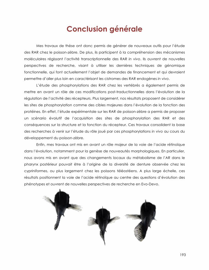
193
Conclusion générale
Mes travaux de thèse ont donc permis de générer de nouveaux outils pour l’étude
des RAR chez le poisson-zèbre. De plus, ils participent à la compréhension des mécanismes
moléculaires régissant l’activité transcriptionnelle des RAR in vivo. Ils ouvrent de nouvelles
perspectives de recherche, visant à utiliser les dernières techniques de génomique
fonctionnelle, qui font actuellement l’objet de demandes de financement et qui devraient
permettre d’aller plus loin en caractérisant les cistromes des RAR endogènes in vivo.
L’étude des phosphorylations des RAR chez les vertébrés a également permis de
mettre en avant un rôle de ces modifications post-traductionnelles dans l’évolution de la
régulation de l’activité des récepteurs. Plus largement, nos résultats proposent de considérer
les sites de phosphorylation comme des cibles majeures dans l’évolution de la fonction des
protéines. En effet, l’étude expérimentale sur les RAR de poisson-zèbre a permis de proposer
un scénario évolutif de l’acquisition des sites de phosphorylation des RAR et des
conséquences sur la structure et la fonction du récepteur. Ces travaux consolident la base
des recherches à venir sur l’étude du rôle joué par ces phosphorylations in vivo au cours du
développement du poisson-zèbre.
Enfin, mes travaux ont mis en avant un rôle majeur de la voie de l’acide rétinoïque
dans l’évolution, notamment pour la genèse de nouveautés morphologiques. En particulier,
nous avons mis en avant que des changements locaux du métabolisme de l’AR dans le
pharynx postérieur pouvait être à l’origine de la diversité de denture observée chez les
cypriniformes, ou plus largement chez les poissons téléostéens. A plus large échelle, ces
résultats positionnent la voie de l’acide rétinoïque au centre des questions d’évolution des
phénotypes et ouvrent de nouvelles perspectives de recherche en Evo-Devo.

194

195
Annexes
Lalevée et al., FASEB 2010…………………………………………………………………………………page 195 Lalevée et al., JBC 2011…………………………….......…………………………………………………page 207 Ferry et al., PNAS 2011………………………………………………………………………………………page 221

196
197
The FASEB Journal • Research Communication
Vinexin!, an atypical “sensor” of retinoic acid receptor" signaling: union and sequestration, separation, andphosphorylation
Sebastien Lalevee, Gaetan Bour,1 Marc Quinternet, Eric Samarut, Pascal Kessler,Marc Vitorino,2 Nathalie Bruck, Marc-Andre Delsuc, Jean-Luc Vonesch, Bruno Kieffer,and Cecile Rochette-Egly3
Institut de Genetique et de Biologie Moleculaire et Cellulaire (IGBMC), Institut National de la Santeet de la Recherche Medicale (INSERM), U596, and Centre National de la Recherche Scientifique(CNRS), Unite Mixte de Recherche (UMR) 7104, Universite de Strasbourg, Illkirch, France
ABSTRACT The transcriptional activity of nuclearretinoic acid receptors (RARs) relies on the associa-tion/dissociation of coregulators at the ligand-bindingdomain. However, we determined that the N-terminaldomain (NTD) also plays a role through its phosphor-ylation, and we isolated vinexin!, a cytoskeleton pro-tein with three SH3 domains, as a new partner of theRAR" NTD. Here we deciphered the mechanism of theinteraction and its role in RAR"-mediated transcrip-tion. By combining molecular and biophysical (surfaceplasmon resonance, NMR, and fluorescence resonanceenergy transfer) approaches, we demonstrated that thethird SH3 domain of vinexin! interacts with a proline-rich domain (PRD) located in RAR" NTD and thatphosphorylation at a serine located in the PRD abro-gates the interaction. The affinity of the interaction wasalso evaluated. In vivo, vinexin! represses RAR"-medi-ated transcription and we dissected the underlyingmechanism in chromatin immunoprecipitation experi-ments performed with F9 cells expressing RAR" wildtype or mutated at the phosphorylation site. In theabsence of retinoic acid (RA), vinexin! does not oc-cupy RAR" target gene promoters and sequesters non-phosphorylated RAR" out of promoters. In response toRA, RAR" becomes phosphorylated and dissociatesfrom vinexin!. This separation allows RAR" to occupypromoters. This is the first report of an RAR corepres-sor association/dissociation out of promoters and reg-ulated by phosphorylation.—Lalevee, S., Bour, G.,Quinternet, M., Samarut, E., Kessler, P., Vitorino, M.,Bruck, N., Delsuc, M.-A., Vonesch, J.-L., Kieffer, B.,Rochette-Egly, C. Vinexin!, an atypical “sensor” ofretinoic acid receptor " signaling: union and sequestra-tion, separation, and phosphorylation. FASEB J. 24,4523–4534 (2010). www.fasebj.org
Key Words: nuclear receptors ! coregulators ! retinoic acid! transcription
Retinoic acid (RA) influences the differentiation,proliferation, and apoptosis of a variety of cell typesthrough modifications in the expression of target genes(1). The RA response is mediated by two classes of
nuclear receptors, the retinoic acid receptors (RARs)(!, ", and #) and the retinoid X receptors (RXRs) (!,", and #), which function as ligand-dependent het-erodimeric RAR/RXR transcription activators (2, 3).Gene induction by RAR/RXR heterodimers relies on acomplex network of dynamic interactions with coregu-latory proteins (for review, see ref. 4). Indeed, after theRA signal, RAR/RXR heterodimers bound at RA re-sponse elements (RAREs) undergo major structuralrearrangements, which initiate an ordered and coordi-nated dissociation and/or recruitment of a series ofcoregulator complexes with different enzymatic activi-ties including histone acetyl and methyl transferases,and DNA-dependent ATPases. At the end, these eventsalter the chromatin structure surrounding the pro-moter of target genes and pave the way for the recruit-ment of the transcription machinery including RNApolymerase II and general transcription factors. Thisnetwork, which is directed by the C-terminal AF-2domain (helix 12) located in the ligand-binding do-main (LBD), was typically described for the RAR!subtype.
In addition to this scenario, it emerged that theN-terminal domain (NTD) of RARs, although of natu-rally disordered structure (5–7), also plays a role,adding more complexity to RAR-mediated transcrip-tion mechanisms. In this context, we recently high-lighted a novel regulation mechanism for the RAR#subtype. Indeed, we found that the NTD of RAR# isphosphorylated (8, 9) and interacts with vinexin" (10),a protein with three SH3 domains, which belongs to thevinexin/CAP/ponsin/ArgBP2 family of adaptor pro-teins (11, 12). Although generally associated as a scaf-folding protein to complexes involved in cytoskeletonorganization and signal transduction, vinexin" is alsoone of a growing number of actin-binding proteins that
1 Current address: IRCAD, 67000 Strasbourg, France.2 Current address: Astex Therapeutics, Cambridge CB4
0QA, UK.3 Correspondence: IGBMC, 1 rue Laurent Fries, BP 10142,
67404 Illkirch Cedex, France. E-mail: [email protected]: 10.1096/fj.10-160572
45230892-6638/10/0024-4523 © FASEB
198
are present in the nucleus and modulate transcription(13), and we found that vinexin! represses RAR"-mediated transcription (10).
Here, we aimed to decipher the underlying mecha-nism of vinexin! interaction with RAR", by combiningmolecular, cellular, and biophysical approaches. Wedemonstrated that the interaction involves the thirdC-terminal SH3 domain of vinexin! and a proline-richdomain (PRD), which is located in the NTD of RAR"and contains phosphorylation sites. The affinity of theinteraction and the influence of phosphorylation weredetermined in surface plasmon resonance (SPR) andNMR experiments. We reveal a completely novel regu-lation mechanism that is distinct from the classic core-pressor network. In this new model, vinexin! repressestranscription through sequestering RAR" out of genepromoters. Then the release of vinexin! that occurs onRAR" phosphorylation, in response to RA, allows ligan-ded and phosphorylated RAR" to occupy the promot-ers of target genes and to initiate transcription.
MATERIALS AND METHODS
Plasmids and peptides
The following plasmids have been described previously (8,10, 14): hRAR"1WT, S79A, S77A, S77A/S79A, #NTD, and#H12 in pSG5; hRAR"1, hRAR$1, and hRAR!2 in pGEX-2T; FLAG-vinexin! in the pCX vector; and the RAR!2-CATreporter gene. The vectors encoding vinexin! deleted foreach SH3 domain, the glutathione S-transferase (GST)-RAR"S77E, S79E, and S77E/S79E chimera and the pSG5RAR"1 mutants P78A, P80A, P82A, P84A, R85A, and R88A,were constructed by double PCR amplification. The pSG5RAR"S77E, RAR"S79E, and RAR"S77E/S79E vectors wereobtained by subcloning each mutant from pGEX2T topSG5. GST-vinexin! and GST-V-SH3-3 were constructed bysubcloning the corresponding fragments amplified by PCRinto pGEX2T.
Cyan fluorescent protein (CFP)-RAR" and yellow fluores-cent protein (YFP)-vinexin! constructs were made by firstintroducing the fragment corresponding to the fluoro-chromes into the HA-FLAG-pCX vector. Then the SH3-3domain of vinexin!, RAR"WT, RAR"S79A, or RAR"S79E orthe AB regions of RAR"S79A were amplified by PCR andligated in-frame at the C-terminal end of the fluorochrome.RAR"S79A and RAR"(AB)S79A were also introduced at theN-terminal end of CFP.
Phospho and nonphospho peptides were synthesized insolid phase using Fmoc chemistry (model 431A peptidesynthesizer; Applied Biosystems, Foster City, CA, USA), puri-fied by reversed-phase HPLC, and verified by electrosprayionization-tandem mass spectrometry.
Antibodies
Polyclonal antibodies recognizing RAR" [RP"(F)] weredescribed previously (8) and purified on SulfoLink gelcolumns (Pierce Chemical, Rockford, IL, USA) coupled tothe corresponding immunizing peptide. Monoclonal anti-bodies against human vinexin! and cyclin H have beendescribed previously (10, 15). Polyclonal antibodies against!-actin and the cdk7 subunit of TFIIH (C-19) were fromSanta Cruz Biotechnology (Santa Cruz, CA, USA). Anti-FLAG monoclonal antibodies were from Sigma-Aldrich (St.
Louis, MO, USA). Monoclonal antibodies recognizing spe-cifically RAR" phosphorylated at position S79 (RAR"-P-S79) were generated by immunization of BALB/c micewith synthetic phosphopeptides coupled to ovalbumin ac-cording to standard procedures.
Recombinant protein expression and purification
Transformed BL21-DE3 cells were grown in minimal mediumsupplemented with 15NH4Cl, to OD600 %0.6, and proteinexpression was induced using 1 mM isopropyl !-d-thiogalac-toside for 8 h. After centrifugation, cells were resuspended ina minimal volume of 20 mM Tris (pH 7.8), containing 200mM NaCl and 1 mM DTT, and sonicated. The GST fusionproteins were isolated using a GSTrap HP affinity column(GE Healthcare, Little Chalfont, UK). Then, the GST part wascleaved with thrombin and removed by exclusion size chro-matography on a HiLoad 16/60 Superdex 75 PG column (GEHealthcare) in 20 mM Tris (pH 7.0), containing 100 mMNaCl. Fractions containing purified 15N-labeled V-SH3-3 werepooled and concentrated using a Vivaspin system (SartoriusStedim Biotech, Aubagne, France).
NMR spectroscopy and chemical shift perturbation
NMR experiments were performed in 50-&l capillaries at300K on a DRX600 spectrometer (Bruker, Newark, DE, USA)equipped with a cryoprobe. Several 15N-1H heteronuclearsingle quantum correlation (HSQC) spectra were recorded tofollow the titration of V-SH3-3 with synthetic peptides corre-sponding to the PRD of RAR" or RAR$. All peptides were firstdesalted and recovered in 100 mM phosphate buffer (pH 7.0)using NAP-5 columns (Pharmacia Biotech, Uppsala, Sweden).The final NMR samples contained 4 mM Tris, 20 mM NaCl,and 80 mM phosphate buffer. Titrations were done with 54&M uniformly 15N-labeled V-SH3-3 to which peptides wereadded. Composite 15N and 1H chemical shifts were used asdescribed in ref. 16, using the 5 most perturbed 15N-1Hcorrelations that were added for averaging of statistical errorsin the Kd (dissociation constant) calculations. Two-parameternonlinear curve fitting was performed using in-house Pythonscripts to get the Kd values. The statistical errors wereestimated from Monte Carlo simulations.
SPR measurements
SPR experiments were performed with a BIAcore T100 instru-ment (Biacore AB, Uppsala, Sweden) and research gradeCM5 sensor chips (17). The ligand (GST-V-SH3-3) was cap-tured via anti-GST antibodies that were immobilized on thesensor surface using standard amine coupling proceduresand following the manufacturer’s instructions. Synthetic pep-tides corresponding to the PRD of RARs were diluted inrunning buffer [10 mM Hepes (pH 7.4), 150 mM NaCl, 3.4mM EDTA, and 0.005% (v/v) P20] and injected over thesurface in a continuous flow at 25°C.
Cell lines, immunoblotting, immunoprecipitation, andimmunofluorescence
COS-1 cells and the different F9 cell lines were cultured andtransiently transfected as in ref. 10. When 80–90% confluent,cells were treated with RA (10'7 M) after 24 h in a mediumcontaining 1% dextran-charcoal-treated FCS. Extract prepa-ration, immunoprecipitation, immunoblotting, and immuno-fluorescence analyses were described previously (10).
4524 Vol. 24 November 2010 LALEVEE ET AL.The FASEB Journal ! www.fasebj.org
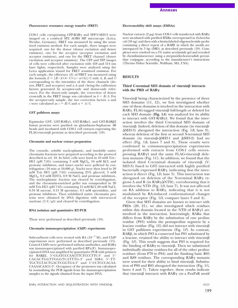
199
Fluorescence resonance energy transfer (FRET)
COS-1 cells coexpressing CFP-RAR! and YFP-V-SH3-3 wereimaged on a confocal SP2 AOBS MP microscope (Leica,Wetzlar, Germany). FRET was measured by using the sensi-tized emission method. For each sample, three images wereacquired: one for the donor (donor excitation and donoremission), one for the acceptor (acceptor excitation andacceptor emission), and one for the FRET channel (donorexcitation and acceptor emission). The CFP and YFP imagesof cells were collected after excitation with 458 and 514 nmlaser lights, respectively. Samples were analyzed using theLeica application wizard for FRET sensitized emission. Foreach sample, the efficiency (E) of FRET was measured usingthe formula E " [B#A$b#C$(c#a$b)]/C with A, B, and Ccorresponding to the intensities of the three channels (do-nor, FRET, and acceptor) and a, b, and c being the calibrationfactors generated by acceptor-only and donor-only refer-ences. For the donor-only sample, the correction of donorcross-talk in the FRET image was calculated as b " B/A. Forthe acceptor-only sample, the two correction factors a andc were calculated as c " B/C and a " A/C.
GST pulldown assays
Equimolar GST, GST-RAR!1, GST-RAR%1, and GST-RAR&2fusion proteins were purified on glutathione-Sepharose 4Bbeads and incubated with COS-1 cell extracts expressing theFLAG-vinexin& proteins as described previously (10).
Chromatin and nuclear extract preparation
The cytosolic, soluble nucleoplasmic, and insoluble nativechromatin fractions were prepared according to the protocoldescribed in ref. 18. In brief, cells were lysed in 10 mM Tris !HCl (pH 7.65) containing 3 mM MgCl2, 10 mM KCl, andprotease inhibitors, and intact nuclei were pelleted by cen-trifugation (10 min at 10,000 g). Nuclei were then lysed in 20mM Tris HCl (pH 7.65) containing 25% glycerol, 3 mMMgCl2, 0.2 mM EDTA, 0.9 M NaCl, and protease inhibitors.The nucleoplasmic fraction was cleared by centrifugation,and the chromatin-enriched pellet was resuspended in 20mM Tris HCl (pH 7.65) containing 15 mM KCl, 60 mM NaCl,0.34 M sucrose, 0.15 M spermine, 0.5 mM spermidine, andprotease inhibitors. Then solubilized native chromatin pro-teins were obtained by DNA digestion with micrococcalnuclease (5 U/'l) and cleared by centrifugation.
RNA isolation and quantitative RT-PCR
These were performed as described previously (10).
Chromatin immunoprecipitation (ChIP) experiments
Subconfluent cells were treated with RA (10#7 M), and ChIPexperiments were performed as described previously (15).Control ChIPs were performed without antibodies, and RAR!was immunoprecipitated with purified RP!(F). Immunopre-cipitated DNA was amplified by quantitative PCR with primersfor RAR&2, 5(-CGATCCCAAGTTCTCCCTTC-3( and 5(-CAGACTGGTTGGGTCATTTG-3( and 36B4, 5( -TT-TGCTGTACTGACTCGGTGA-3( and 5(-CCTCCCACAA-CAAAACAACC-3(. Occupancy of the promoters was calculatedby normalizing the PCR signals from the immunoprecipitationsamples to the signals obtained from the input DNA.
Electromobility shift assays (EMSAs)
Nuclear extracts (2 'g) from COS-1 cells transfected with RAR!were incubated with purified RXR% overexpressed in Escherichiacoli (50 ng) and then with a biotin-labeled oligonucleotide probecontaining a direct repeat of a RARE in which the motifs areinterspaced by 5 bp (DR5) as described previously (19). Com-plexes were resolved on a 5% native acrylamide gel and revealedby chemiluminescence using a streptavidin-horseradish peroxi-dase conjugate, according to the manufacturer’s instructions(Thermo Fisher Scientific, Waltham, MA, USA).
RESULTS
Third C-terminal SH3 domain of vinexin! interactswith the PRD of RAR"
Vinexin& being characterized by the presence of threeSH3 domains (11, 12), we first investigated whetherone of these domains is involved in the interaction withRAR!. FLAG-tagged vinexin& full-length or deleted foreach SH3 domain (Fig. 1A) was analyzed for its abilityto interact with GST-RAR!1. We found that the inter-action involves the third C-terminal SH3 domain ofvinexin&. Indeed, deletion of this domain (in vinexin&-)SH3-3) abrogated the interaction (Fig. 1B, lane 9),whereas deletion of the first or second N-terminal SH3domain (in vinexin&-)SH3-1 and )SH3-2) had noeffect (Fig. 1B, lanes 7 and 8). These results wereconfirmed in coimmunoprecipitation experimentsperformed with extracts from COS-1 cells overex-pressing RAR!1 and the same FLAG-vinexin& dele-tion mutants (Fig. 1C). In addition, we found that theisolated third C-terminal domain of vinexin& (V-SH3-3) fused to GST interacted with highly purifiedbacterially expressed RAR!, indicating that the inter-action is direct (Fig. 1D, lane 3). This interaction wasabrogated on deletion of the N-terminal RAR! re-gions A and B (in RAR!)NTD), corroborating that itinvolves the NTD (Fig. 1D, lane 7). It was not affectedon RA addition to RAR!, indicating that it is notmodulated by RA-induced conformational changesof the receptor (Fig. 1D, compare lanes 3 and 4).
Given that SH3 domains are known to interact withPRDs (20, 21), we also investigated which residueswithin this domain located in the NTD of RAR!1 areinvolved in the interaction. Interestingly, RAR% thatdiffers from RAR! by the substitution of one prolineresidue (P83) within the pentaproline segment by aleucine residue (Fig. 1E) did not interact with vinexin&in GST pulldown experiments (Fig. 1F). In contrast,RAR&, in which P83 is conserved but P81 substituted bya leucine, retained the ability to interact with vinexin&(Fig. 1F). This result suggests that P83 is required forthe binding of RAR! to vinexin&. Then we substitutedindividually alanine residues for all of the other prolineresidues (from P78 to P84) and the flanking basic R85and K88 residues. The corresponding RAR! mutantswere tested for their ability to bind vinexin&. Substitu-tion of P80 and R85 abrogated the interaction (Fig. 1G,lanes 4 and 7). Taken together, these results indicatethat vinexin& interacts with RAR! via a PxxPxR motif
4525RAR! INTERACTION AND SEQUESTRATION WITH VINEXIN&
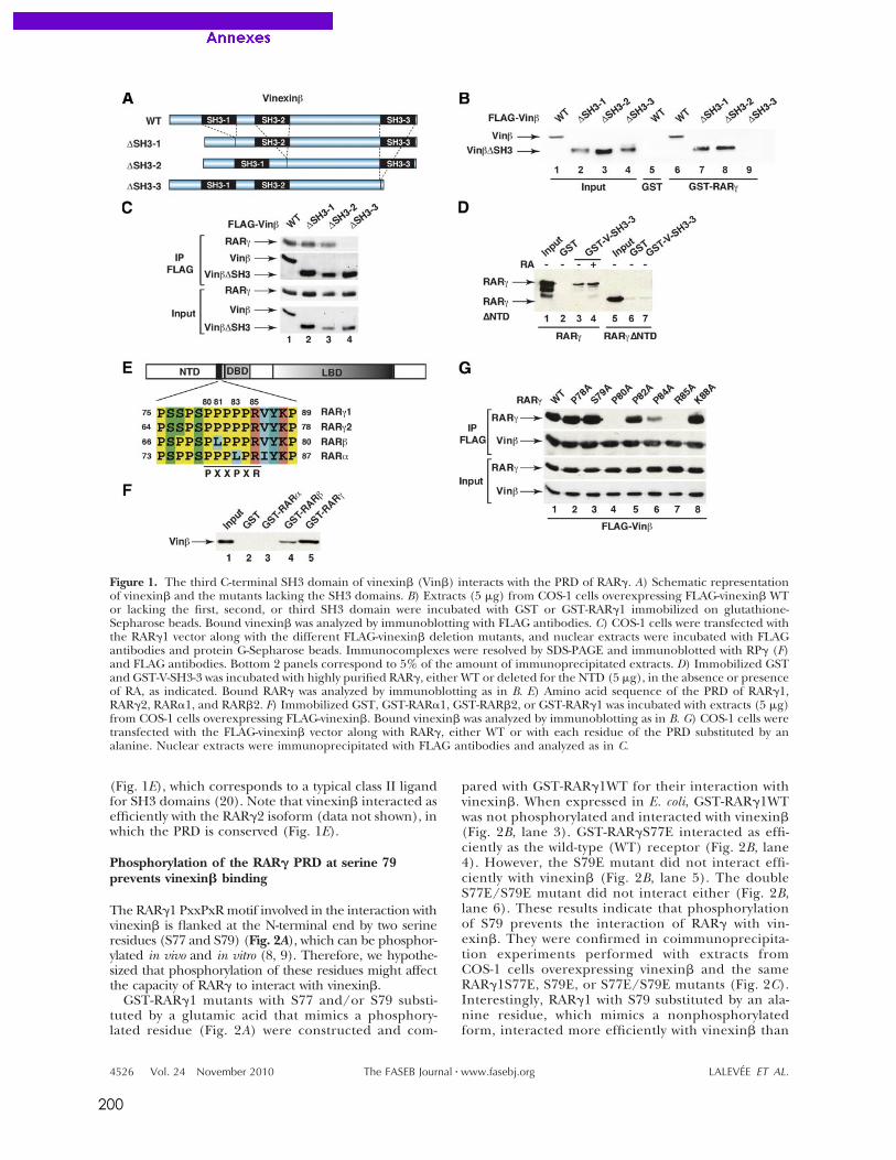
200
(Fig. 1E), which corresponds to a typical class II ligandfor SH3 domains (20). Note that vinexin! interacted asefficiently with the RAR"2 isoform (data not shown), inwhich the PRD is conserved (Fig. 1E).
Phosphorylation of the RAR! PRD at serine 79prevents vinexin" binding
The RAR"1 PxxPxR motif involved in the interaction withvinexin! is flanked at the N-terminal end by two serineresidues (S77 and S79) (Fig. 2A), which can be phosphor-ylated in vivo and in vitro (8, 9). Therefore, we hypothe-sized that phosphorylation of these residues might affectthe capacity of RAR" to interact with vinexin!.
GST-RAR"1 mutants with S77 and/or S79 substi-tuted by a glutamic acid that mimics a phosphory-lated residue (Fig. 2A) were constructed and com-
pared with GST-RAR"1WT for their interaction withvinexin!. When expressed in E. coli, GST-RAR"1WTwas not phosphorylated and interacted with vinexin!(Fig. 2B, lane 3). GST-RAR"S77E interacted as effi-ciently as the wild-type (WT) receptor (Fig. 2B, lane4). However, the S79E mutant did not interact effi-ciently with vinexin! (Fig. 2B, lane 5). The doubleS77E/S79E mutant did not interact either (Fig. 2B,lane 6). These results indicate that phosphorylationof S79 prevents the interaction of RAR" with vin-exin!. They were confirmed in coimmunoprecipita-tion experiments performed with extracts fromCOS-1 cells overexpressing vinexin! and the sameRAR"1S77E, S79E, or S77E/S79E mutants (Fig. 2C).Interestingly, RAR"1 with S79 substituted by an ala-nine residue, which mimics a nonphosphorylatedform, interacted more efficiently with vinexin! than
Figure 1. The third C-terminal SH3 domain of vinexin! (Vin!) interacts with the PRD of RAR". A) Schematic representationof vinexin! and the mutants lacking the SH3 domains. B) Extracts (5 #g) from COS-1 cells overexpressing FLAG-vinexin! WTor lacking the first, second, or third SH3 domain were incubated with GST or GST-RAR"1 immobilized on glutathione-Sepharose beads. Bound vinexin! was analyzed by immunoblotting with FLAG antibodies. C) COS-1 cells were transfected withthe RAR"1 vector along with the different FLAG-vinexin! deletion mutants, and nuclear extracts were incubated with FLAGantibodies and protein G-Sepharose beads. Immunocomplexes were resolved by SDS-PAGE and immunoblotted with RP" (F)and FLAG antibodies. Bottom 2 panels correspond to 5% of the amount of immunoprecipitated extracts. D) Immobilized GSTand GST-V-SH3-3 was incubated with highly purified RAR", either WT or deleted for the NTD (5 #g), in the absence or presenceof RA, as indicated. Bound RAR" was analyzed by immunoblotting as in B. E) Amino acid sequence of the PRD of RAR"1,RAR"2, RAR$1, and RAR!2. F) Immobilized GST, GST-RAR$1, GST-RAR!2, or GST-RAR"1 was incubated with extracts (5 #g)from COS-1 cells overexpressing FLAG-vinexin!. Bound vinexin! was analyzed by immunoblotting as in B. G) COS-1 cells weretransfected with the FLAG-vinexin! vector along with RAR", either WT or with each residue of the PRD substituted by analanine. Nuclear extracts were immunoprecipitated with FLAG antibodies and analyzed as in C.
4526 Vol. 24 November 2010 LALEVEE ET AL.The FASEB Journal ! www.fasebj.org
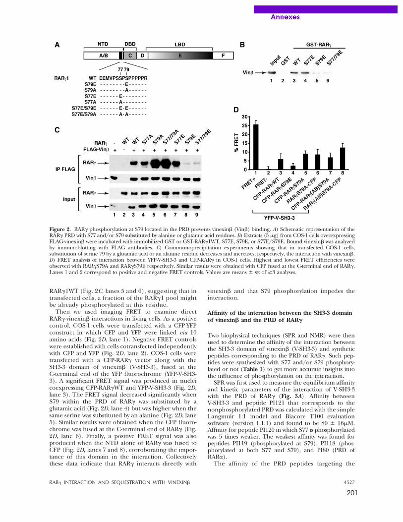
201
RAR!1WT (Fig. 2C, lanes 5 and 6), suggesting that intransfected cells, a fraction of the RAR!1 pool mightbe already phosphorylated at this residue.
Then we used imaging FRET to examine directRAR!-vinexin" interactions in living cells. As a positivecontrol, COS-1 cells were transfected with a CFP-YFPconstruct in which CFP and YFP were linked via 10amino acids (Fig. 2D, lane 1). Negative FRET controlswere established with cells cotransfected independentlywith CFP and YFP (Fig. 2D, lane 2). COS-1 cells weretransfected with a CFP-RAR! vector along with theSH3-3 domain of vinexin" (V-SH3-3), fused at theC-terminal end of the YFP fluorochrome (YFP-V-SH3-3). A significant FRET signal was produced in nucleicoexpressing CFP-RAR!WT and YFP-V-SH3-3 (Fig. 2D,lane 3). The FRET signal decreased significantly whenS79 within the PRD of RAR! was substituted by aglutamic acid (Fig. 2D, lane 4) but was higher when thesame serine was substituted by an alanine (Fig. 2D, lane5). Similar results were obtained when the CFP fluoro-chrome was fused at the C-terminal end of RAR! (Fig.2D, lane 6). Finally, a positive FRET signal was alsoproduced when the NTD alone of RAR! was fused toCFP (Fig. 2D, lanes 7 and 8), corroborating the impor-tance of this domain in the interaction. Collectivelythese data indicate that RAR! interacts directly with
vinexin" and that S79 phosphorylation impedes theinteraction.
Affinity of the interaction between the SH3-3 domainof vinexin! and the PRD of RAR"
Two biophysical techniques (SPR and NMR) were thenused to determine the affinity of the interaction betweenthe SH3-3 domain of vinexin" (V-SH3-3) and syntheticpeptides corresponding to the PRD of RAR!. Such pep-tides were synthesized with S77 and/or S79 phosphory-lated or not (Table 1) to get more accurate insights intothe influence of phosphorylation on the interaction.
SPR was first used to measure the equilibrium affinityand kinetic parameters of the interaction of V-SH3-3with the PRD of RAR! (Fig. 3A). Affinity betweenV-SH3-3 and peptide PI121 that corresponds to thenonphosphorylated PRD was calculated with the simpleLangmuir 1:1 model and Biacore T100 evaluationsoftware (version 1.1.1) and found to be 80 # 16$M.Affinity for peptide PI120 in which S77 is phosphorylatedwas 5 times weaker. The weakest affinity was found forpeptides PI119 (phosphorylated at S79), PI118 (phos-phorylated at both S77 and S79), and PI80 (PRD ofRAR%).
The affinity of the PRD peptides targeting the
Figure 2. RAR! phosphorylation at S79 located in the PRD prevents vinexin" (Vin") binding. A) Schematic representation of theRAR! PRD with S77 and/or S79 substituted by alanine or glutamic acid residues. B) Extracts (5 $g) from COS-1 cells overexpressingFLAG-vinexin" were incubated with immobilized GST or GST-RAR!1WT, S77E, S79E, or S77E/S79E. Bound vinexin" was analyzedby immunoblotting with FLAG antibodies. C) Coimmunoprecipitation experiments showing that in transfected COS-1 cells,substitution of serine 79 by a glutamic acid or an alanine residue decreases and increases, respectively, the interaction with vinexin".D) FRET analysis of interaction between YFP-V-SH3-3 and CFP-RAR! in COS-1 cells. Highest and lowest FRET efficiencies wereobserved with RAR!S79A and RAR!S79E respectively. Similar results were obtained with CFP fused at the C-terminal end of RAR!.Lanes 1 and 2 correspond to positive and negative FRET controls. Values are means # se of !3 analyses.
4527RAR! INTERACTION AND SEQUESTRATION WITH VINEXIN"
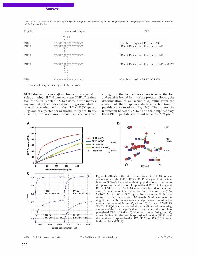
202
SH3-3 domain of vinexin! was further investigated insolution using 1H-15N heteronuclear NMR. The titra-tion of the 15N labeled V-SH3-3 domain with increas-ing amounts of peptides led to a progressive shift ofa set of correlation peaks in the 1H-15N HSQC spectra(Fig. 3B), as expected for weak affinity ligands. In thissituation, the resonance frequencies are weighted
averages of the frequencies characterizing the freeand peptide-bound forms of the protein, allowing thedetermination of an accurate Kd value from theanalysis of the frequency shifts as a function ofpeptide concentrations (Fig. 3C). The Kd for theinteraction between V-SH3-3 and the nonphosphory-lated PI121 peptide was found to be 37 " 9 #M, a
TABLE 1. Amino acid sequence of the synthetic peptides corresponding to the phosphorylated or nonphosphorylated proline-rich domainsof RAR$ and RAR%
Peptide Amino acid sequence PRD
77 79¦ ¦
PI121 EEMVPSSPSPPPPPRVYK Nonphosphorylated PRD of RAR$PI120 EEMVPSSPSPPPPPRVYK PRD of RAR$ phosphorylated at S77
¦P
PI119 EEMVPSSPSPPPPPRVYK PRD of RAR$ phosphorylated at S79¦P
PI118 EEMVPSSPSPPPPPRVYK PRD of RAR$ phosphorylated at S77 and S79¦ ¦P P77
¦PI80 EEIVPSPPSPPPLPRIYK Nonphosphorylated PRD of RAR%
Amino acid sequences are given in 1-letter codes.
Figure 3. Affinity of the interaction between the SH3-3 domainof vinexin! and the PRD of RAR$. A) SPR analysis of interactionbetween GST-V-SH3-3 and synthetic peptides corresponding tothe phosphorylated or nonphosphorylated PRD of RAR$ andRAR%. GST and GST-V-SH3-3 were immobilized on a sensorchip. Peptides were injected at various concentrations (0.5–5&10'4 M) for 60 s. GST signal [relative units (RU)] wassubtracted from the GST-V-SH3-3 signals. Nonlinear curve fit-ting of the equilibrium responses vs. peptide concentration wasused to derive equilibrium Kd values. B) Extract of V-SH3-31H-15N HSQC spectra recorded on addition of increasingamounts of the PI121 peptide that corresponds to the nonphos-phorylated PRD of RAR$. C) Nonlinear curve fitting and Kdvalues obtained for the nonphosphorylated peptide (PI121) andthe peptides phosphorylated at S77 (PI120) or S79 (PI119) or atboth positions (PI118).
4528 Vol. 24 November 2010 LALEVEE ET AL.The FASEB Journal ! www.fasebj.org
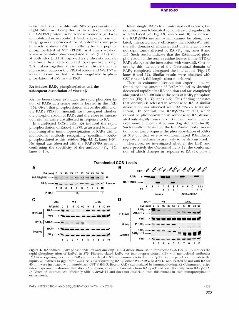
203
value that is compatible with SPR experiments, theslight difference being due to the different state ofthe V-SH3-3 protein in both measurements (surface-immobilized vs. in solution). Such a Kd value is in therange generally observed for SH3 domains and pro-line-rich peptides (20). The affinity for the peptidephosphorylated at S77 (PI120) is 4 times weaker,whereas peptides phosphorylated at S79 (PI119) andat both sites (PI118) displayed a significant decreasein affinity (by a factor of 8 and 15, respectively) (Fig.3C). Taken together, these results indicate that theinteraction between the PRD of RAR! and V-SH3-3 isweak and confirm that it is down-regulated by phos-phorylation of S79 in the PRD.
RA induces RAR! phosphorylation and thesubsequent dissociation of vinexin"
RA has been shown to induce the rapid phosphoryla-tion of RAR" at a serine residue located in the PRD(15). Given that phosphorylation affects the affinity ofthe RAR! PRD for vinexin#, we investigated whetherthe phosphorylation of RAR! and therefore its interac-tion with vinexin# are affected in response to RA.
In transfected COS-1 cells, RA induced the rapidphosphorylation of RAR!1 at S79, as assessed by immu-noblotting after immunoprecipitation of RAR! with amonoclonal antibody recognizing specifically RAR!phosphorylated at this residue (Fig. 4A, C, lanes 1–5).No signal was observed with the RAR!S79A mutant,confirming the specificity of the antibody (Fig. 4C,lanes 6–10).
Interestingly, RAR! from untreated cell extracts, butnot RAR! from RA-treated cells, interacted significantlywith GST-V-SH3-3 (Fig. 4B, lanes 7 and 10). In contrast,the RAR!S79A mutant, which cannot be phosphory-lated, interacted more efficiently than RAR!WT withthe SH3 domain of vinexin#, and this interaction wasnot significantly affected by RA (Fig. 4B, lanes 8 and11). Such results indicate that the RA-induced phos-phorylation of the serine residue located in the NTD ofRAR! abrogates the interaction with vinexin#. Corrob-orating this, deletion of the N-terminal domain ofRAR! completely abrogated the interaction (Fig. 4B,lanes 9 and 12). Similar results were obtained withGST-vinexin# full-length (data not shown)
Then in coimmunoprecipitation experiments, wefound that the amount of RAR! bound to vinexin#decreased rapidly after RA addition and was completelyabrogated at 30–60 min at the peak of RAR! phosphor-ylation (Fig. 4C, D, lanes 1–5). This finding indicatesthat vinexin# is released in response to RA. A similardissociation was observed with RAR!S77A (data notshown). In contrast, the RAR!S79A mutant, whichcannot be phosphorylated in response to RA, dissoci-ated only slightly from vinexin# at 5 min and interactedeven more efficiently at 60 min (Fig. 4C, lanes 6–10).Such results indicate that the full RA-induced dissocia-tion of vinexin# requires the phosphorylation of RAR!at S79 but that in vivo additional rapid RA-inducedregulatory mechanisms are likely to be also involved.
Therefore, we investigated whether the LBD andmore precisely the C-terminal helix 12, the conforma-tion of which changes in response to RA (4), plays a
Figure 4. RA induces RAR! phosphorylation and vinexin# (Vin#) dissociation. A) In transfected COS-1 cells, RA induces therapid phosphorylation of RAR!1 at S79. Phosphorylated RAR! was immunoprecipitated (IP) with monoclonal antibodies(MAb) recognizing specifically RAR! phosphorylated at S79 and immunoblotted with RP!(F). Bottom panel corresponds to theinputs. B) Extracts (5 $g) from COS-1 cells overexpressing RAR!, either WT, S79A, or %NTD, and treated or not with RA for45 min were incubated with immobilized GST-V-SH3-3. Bound RAR! was analyzed by immunoblotting. C) Coimmunoprecipi-tation experiments showing that after RA addition, vinexin# dissociates from RAR!WT and less efficiently from RAR!S79A.D) Vinexin# interacts less efficiently with RAR!%H12 and does not dissociate from this mutant in coimmunoprecipitationexperiments.
4529RAR! INTERACTION AND SEQUESTRATION WITH VINEXIN#
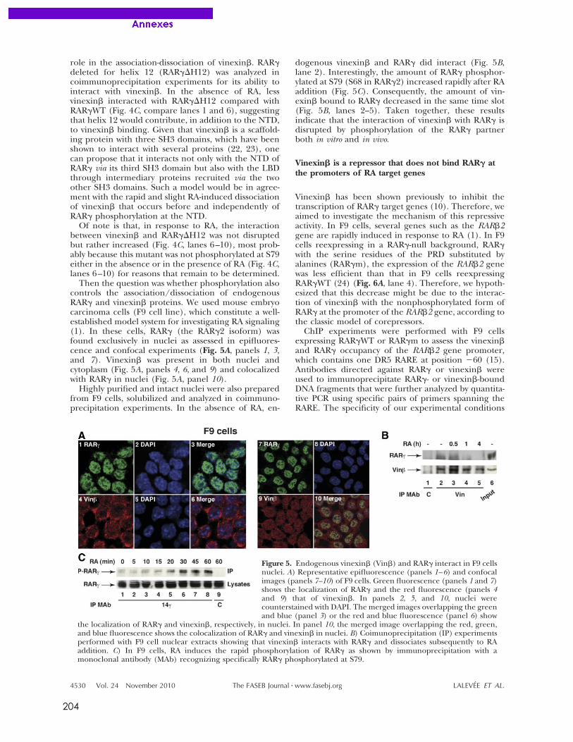
204
role in the association-dissociation of vinexin!. RAR"deleted for helix 12 (RAR"#H12) was analyzed incoimmunoprecipitation experiments for its ability tointeract with vinexin!. In the absence of RA, lessvinexin! interacted with RAR"#H12 compared withRAR"WT (Fig. 4C, compare lanes 1 and 6), suggestingthat helix 12 would contribute, in addition to the NTD,to vinexin! binding. Given that vinexin! is a scaffold-ing protein with three SH3 domains, which have beenshown to interact with several proteins (22, 23), onecan propose that it interacts not only with the NTD ofRAR" via its third SH3 domain but also with the LBDthrough intermediary proteins recruited via the twoother SH3 domains. Such a model would be in agree-ment with the rapid and slight RA-induced dissociationof vinexin! that occurs before and independently ofRAR" phosphorylation at the NTD.
Of note is that, in response to RA, the interactionbetween vinexin! and RAR"#H12 was not disruptedbut rather increased (Fig. 4C, lanes 6–10), most prob-ably because this mutant was not phosphorylated at S79either in the absence or in the presence of RA (Fig. 4C,lanes 6–10) for reasons that remain to be determined.
Then the question was whether phosphorylation alsocontrols the association/dissociation of endogenousRAR" and vinexin! proteins. We used mouse embryocarcinoma cells (F9 cell line), which constitute a well-established model system for investigating RA signaling(1). In these cells, RAR" (the RAR"2 isoform) wasfound exclusively in nuclei as assessed in epifluores-cence and confocal experiments (Fig. 5A, panels 1, 3,and 7). Vinexin! was present in both nuclei andcytoplasm (Fig. 5A, panels 4, 6, and 9) and colocalizedwith RAR" in nuclei (Fig. 5A, panel 10).
Highly purified and intact nuclei were also preparedfrom F9 cells, solubilized and analyzed in coimmuno-precipitation experiments. In the absence of RA, en-
dogenous vinexin! and RAR" did interact (Fig. 5B,lane 2). Interestingly, the amount of RAR" phosphor-ylated at S79 (S68 in RAR"2) increased rapidly after RAaddition (Fig. 5C). Consequently, the amount of vin-exin! bound to RAR" decreased in the same time slot(Fig. 5B, lanes 2–5). Taken together, these resultsindicate that the interaction of vinexin! with RAR" isdisrupted by phosphorylation of the RAR" partnerboth in vitro and in vivo.
Vinexin! is a repressor that does not bind RAR" atthe promoters of RA target genes
Vinexin! has been shown previously to inhibit thetranscription of RAR" target genes (10). Therefore, weaimed to investigate the mechanism of this repressiveactivity. In F9 cells, several genes such as the RAR!2gene are rapidly induced in response to RA (1). In F9cells reexpressing in a RAR"-null background, RAR"with the serine residues of the PRD substituted byalanines (RAR"m), the expression of the RAR!2 genewas less efficient than that in F9 cells reexpressingRAR"WT (24) (Fig. 6A, lane 4). Therefore, we hypoth-esized that this decrease might be due to the interac-tion of vinexin! with the nonphosphorylated form ofRAR" at the promoter of the RAR!2 gene, according tothe classic model of corepressors.
ChIP experiments were performed with F9 cellsexpressing RAR"WT or RAR"m to assess the vinexin!and RAR" occupancy of the RAR!2 gene promoter,which contains one DR5 RARE at position $60 (15).Antibodies directed against RAR" or vinexin! wereused to immunoprecipitate RAR"- or vinexin!-boundDNA fragments that were further analyzed by quantita-tive PCR using specific pairs of primers spanning theRARE. The specificity of our experimental conditions
Figure 5. Endogenous vinexin! (Vin!) and RAR" interact in F9 cellsnuclei. A) Representative epifluorescence (panels 1–6) and confocalimages (panels 7–10) of F9 cells. Green fluorescence (panels 1 and 7)shows the localization of RAR" and the red fluorescence (panels 4and 9) that of vinexin!. In panels 2, 5, and 10, nuclei werecounterstained with DAPI. The merged images overlapping the greenand blue (panel 3) or the red and blue fluorescence (panel 6) show
the localization of RAR" and vinexin!, respectively, in nuclei. In panel 10, the merged image overlapping the red, green,and blue fluorescence shows the colocalization of RAR" and vinexin! in nuclei. B) Coimunoprecipitation (IP) experimentsperformed with F9 cell nuclear extracts showing that vinexin! interacts with RAR" and dissociates subsequently to RAaddition. C) In F9 cells, RA induces the rapid phosphorylation of RAR" as shown by immunoprecipitation with amonoclonal antibody (MAb) recognizing specifically RAR" phosphorylated at S79.
4530 Vol. 24 November 2010 LALEVEE ET AL.The FASEB Journal ! www.fasebj.org
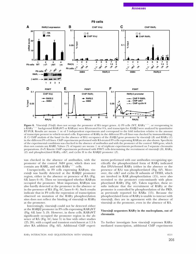
205
was checked in the absence of antibodies, with thepromoter of the control 36B4 gene, which does notcontain any RARE, and with RAR!"/" cells.
Unexpectedly, in F9 cells expressing RAR!m, vin-exin# was hardly detected at the RAR#2 promoterregion, either in the absence or presence of RA (Fig.6B, lanes 6–8). Then we investigated whether RAR!moccupied the promoter. Most important, RAR!m wasalso hardly detected at the promoter in the absence orin the presence of RA (Fig. 6C, lanes 6–8). Such resultsindicate that in F9 cells the repression of transcriptionobserved on mutation of the RAR! phosphorylationsites does not reflect the binding of vinexin# to RAR!at the promoter.
Interestingly, vinexin# could not be detected eitherat the RAR#2 promoter in F9 cells expressing RAR!WT(Fig. 6B, lane 5; D). However, in these cells, RAR!WTsignificantly occupied the promoter region in the ab-sence of RA (Fig. 6C, lane 5) in line with other studies(25, 26), with a rapid and transient enrichment at 1.5 hafter RA addition (Fig. 6E). Additional ChIP experi-
ments performed with our antibodies recognizing spe-cifically the phosphorylated form of RAR! indicatedthat DNA-bound RAR! (either in the absence or thepresence of RA) was phosphorylated (Fig. 6F). More-over, the cdk7 and cyclin H subunits of TFIIH, whichare involved in RAR phosphorylation (15), were alsorecruited to the promoter concomitantly with phos-phorylated RAR! (Fig. 6F). Taken together, these re-sults indicate that the recruitment of RAR! at thepromoter is controlled by phosphorylation of the PRD,as previously reported for RAR$ (15). Because thephosphorylated form of RAR! is unable to interact withvinexin#, they are in agreement with the absence ofvinexin# at the promoter, even in the absence of RA.
Vinexin! sequesters RAR" in the nucleoplasm, out ofchromatin
To further investigate how vinexin# represses RAR!-mediated transcription, additional ChIP experiments
Figure 6. Vinexin# (Vin#) does not occupy the promoter of RA target genes. A) F9 cells (WT, RAR!"/", or reexpressing ina RAR!"/" background RAR!WT or RAR!m) were RA-treated for 8 h, and transcripts for RAR#2 were analyzed by quantitativeRT-PCR. Results are means % se of 3 independent experiments and correspond to the fold induction relative to the amountof transcripts present in vehicle-treated cells. Expression of RAR! in the different F9 cell lines was checked by immunoblotting.B, C) ChIP analysis of the basal (in the absence of RA) occupancy of the RAR#2 gene promoter by vinexin# (B) and RAR! (C)in the different F9 cell lines. ChIP experiments performed with RA-treated F9 cells expressing RAR!m are also shown. Specificityof the experimental conditions was checked in the absence of antibodies and with the promoter of the control 36B4 gene, whichdoes not contain any RARE. Values (% of inputs) are means % se of triplicate experiments performed on 3 separate chromatinpreparations. D–F) Kinetic ChIP experiments performed with F9WT cells determining the recruitment of vinexin# (D), RAR!,(E) and phosphorylated RAR!, cdk7, and cyclin H to the RAR#2 promoter (F).
4531RAR! INTERACTION AND SEQUESTRATION WITH VINEXIN#
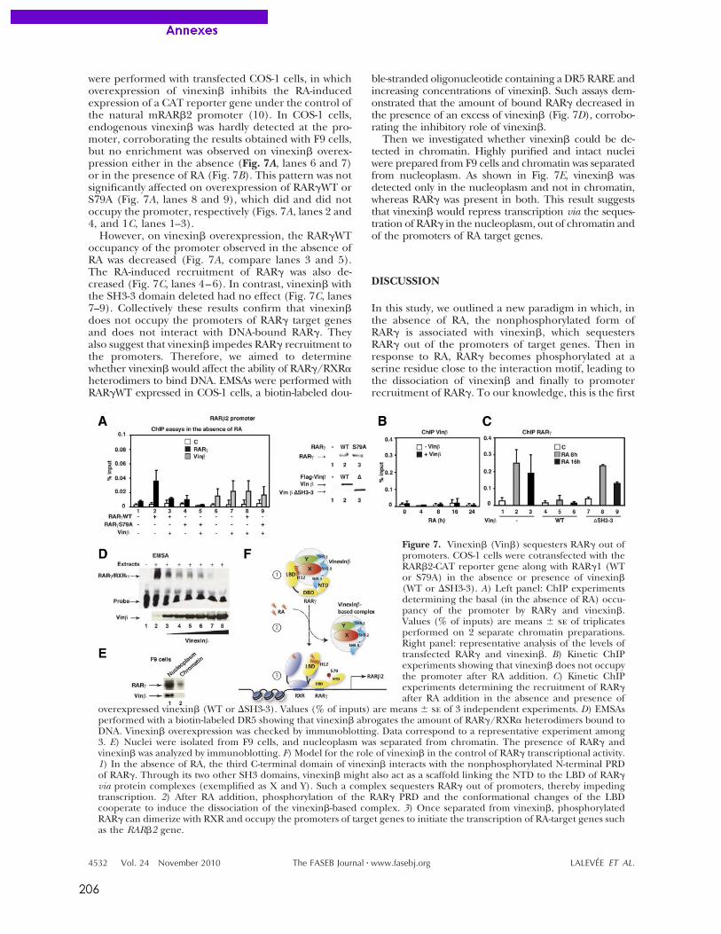
206
were performed with transfected COS-1 cells, in whichoverexpression of vinexin! inhibits the RA-inducedexpression of a CAT reporter gene under the control ofthe natural mRAR!2 promoter (10). In COS-1 cells,endogenous vinexin! was hardly detected at the pro-moter, corroborating the results obtained with F9 cells,but no enrichment was observed on vinexin! overex-pression either in the absence (Fig. 7A, lanes 6 and 7)or in the presence of RA (Fig. 7B). This pattern was notsignificantly affected on overexpression of RAR"WT orS79A (Fig. 7A, lanes 8 and 9), which did and did notoccupy the promoter, respectively (Figs. 7A, lanes 2 and4, and 1C, lanes 1–3).
However, on vinexin! overexpression, the RAR"WToccupancy of the promoter observed in the absence ofRA was decreased (Fig. 7A, compare lanes 3 and 5).The RA-induced recruitment of RAR" was also de-creased (Fig. 7C, lanes 4–6). In contrast, vinexin! withthe SH3-3 domain deleted had no effect (Fig. 7C, lanes7–9). Collectively these results confirm that vinexin!does not occupy the promoters of RAR" target genesand does not interact with DNA-bound RAR". Theyalso suggest that vinexin! impedes RAR" recruitment tothe promoters. Therefore, we aimed to determinewhether vinexin! would affect the ability of RAR"/RXR#heterodimers to bind DNA. EMSAs were performed withRAR"WT expressed in COS-1 cells, a biotin-labeled dou-
ble-stranded oligonucleotide containing a DR5 RARE andincreasing concentrations of vinexin!. Such assays dem-onstrated that the amount of bound RAR" decreased inthe presence of an excess of vinexin! (Fig. 7D), corrobo-rating the inhibitory role of vinexin!.
Then we investigated whether vinexin! could be de-tected in chromatin. Highly purified and intact nucleiwere prepared from F9 cells and chromatin was separatedfrom nucleoplasm. As shown in Fig. 7E, vinexin! wasdetected only in the nucleoplasm and not in chromatin,whereas RAR" was present in both. This result suggeststhat vinexin! would repress transcription via the seques-tration of RAR" in the nucleoplasm, out of chromatin andof the promoters of RA target genes.
DISCUSSION
In this study, we outlined a new paradigm in which, inthe absence of RA, the nonphosphorylated form ofRAR" is associated with vinexin!, which sequestersRAR" out of the promoters of target genes. Then inresponse to RA, RAR" becomes phosphorylated at aserine residue close to the interaction motif, leading tothe dissociation of vinexin! and finally to promoterrecruitment of RAR". To our knowledge, this is the first
Figure 7. Vinexin! (Vin!) sequesters RAR" out ofpromoters. COS-1 cells were cotransfected with theRAR!2-CAT reporter gene along with RAR"1 (WTor S79A) in the absence or presence of vinexin!(WT or $SH3-3). A) Left panel: ChIP experimentsdetermining the basal (in the absence of RA) occu-pancy of the promoter by RAR" and vinexin!.Values (% of inputs) are means % se of triplicatesperformed on 2 separate chromatin preparations.Right panel: representative analysis of the levels oftransfected RAR" and vinexin!. B) Kinetic ChIPexperiments showing that vinexin! does not occupythe promoter after RA addition. C) Kinetic ChIPexperiments determining the recruitment of RAR"after RA addition in the absence and presence of
overexpressed vinexin! (WT or $SH3-3). Values (% of inputs) are means % se of 3 independent experiments. D) EMSAsperformed with a biotin-labeled DR5 showing that vinexin! abrogates the amount of RAR"/RXR# heterodimers bound toDNA. Vinexin! overexpression was checked by immunoblotting. Data correspond to a representative experiment among3. E) Nuclei were isolated from F9 cells, and nucleoplasm was separated from chromatin. The presence of RAR" andvinexin! was analyzed by immunoblotting. F) Model for the role of vinexin! in the control of RAR" transcriptional activity.1) In the absence of RA, the third C-terminal domain of vinexin! interacts with the nonphosphorylated N-terminal PRDof RAR". Through its two other SH3 domains, vinexin! might also act as a scaffold linking the NTD to the LBD of RAR"via protein complexes (exemplified as X and Y). Such a complex sequesters RAR" out of promoters, thereby impedingtranscription. 2) After RA addition, phosphorylation of the RAR" PRD and the conformational changes of the LBDcooperate to induce the dissociation of the vinexin!-based complex. 3) Once separated from vinexin!, phosphorylatedRAR" can dimerize with RXR and occupy the promoters of target genes to initiate the transcription of RA-target genes suchas the RAR!2 gene.
4532 Vol. 24 November 2010 LALEVEE ET AL.The FASEB Journal ! www.fasebj.org
207
report of a RAR corepressor association/dissociationregulated by phosphorylation processes and occurringout of promoters. It was an unexpected finding andsheds light on a new level of complexity in the fine-tuning of RAR!-mediated transcription.
Vinexin" is a protein with 3 SH3 domains. The uniqueproperty of such domains is to recognize PRDs withoutrequiring high affinities (20). Such a PRD is present in theNTD of the RAR! subtype and contains phosphorylationsites. Here we combined classic molecular and cellularapproaches with biophysical techniques to decipher theparameters of the interaction. We have shown that thethird C-terminal domain of vinexin" (SH3-3) interactswith a consensus PxxPxR motif (a classic class II ligand)located in the PRD of RAR!. Interestingly, although wellconserved between RARs, the PRD of the RAR# subtypediffers from that of RAR! at one residue within thePxxPxR motif. Such a discrepancy might be the basis ofthe specificity of the interaction of vinexin" with RAR!.Interestingly, this interaction fulfills the classic criteria ofinteractions between PRDs and their binding partners.Indeed, the SH3-3 domain of vinexin" interacts with thetarget PRD with typical Kd values of $40–80 %M, in linewith the idea that rather weak interactions are requiredfor efficient signaling processes.
Most important, we found that phosphorylation ofRAR! at a serine residue (S79) adjacent to the PxxPxRmotif significantly reduces the binding of vinexin".Moreover, in vivo the amount of RAR! phosphorylatedat S79 increases rapidly in response to RA, and subse-quently vinexin" is released. Given that S79 is locatedin close proximity to the SH3-3 binding surface, it canbe proposed that phosphorylation of this residue prop-agates a signal to this surface, promoting the dissocia-tion of vinexin".
An important clue in the present study is that vin-exin" represses RAR!-mediated transcription, in linewith its ability to interact with the nonphosphorylatedform of RAR!, which is transcriptionally deficient. Thefirst hypothesis was that this repression might resultfrom the binding of vinexin" to RAR! at the promoterof target genes, according to the model of classiccorepressors. Unexpectedly, no significant amounts ofvinexin" could be detected at promoters, either inabsence or presence of RA. Moreover, vinexin" wasfound to inhibit RAR! binding to DNA. Finally, takingadvantage of F9 cells expressing RAR!WT or mutatedat the phosphorylation sites and of antibodies recogniz-ing specifically the phosphorylated form of RAR!,DNA-bound RAR! was phosphorylated. Such data areindeed not compatible with the presence of vinexin" atthe promoter and lead to the conclusion that therepressive activity of vinexin" does not occur throughits binding to RAR! at the promoters of target genesand thus differs from that of the classic corepressors.
In line with this result, we found that, in contrast toRAR!, vinexin" could not be detected in chromatineither in the absence or the presence of RA. In fact,vinexin" colocalized with RAR! in the nucleoplasm.This finding suggests that vinexin" would sequesterthe nonphosphorylated form of RAR! in an inactivestate out of chromatin. The release of vinexin"subsequent to RA binding and to the phosphoryla-
tion of the PRD would increase the amount of RAR!present in chromatin and competent to bind DNAand to activate the expression of the target genes.Accordingly, the RA-induced increase in the RAR!occupancy of promoters occurred in the same timeslot (1 h) as the phosphorylation of RAR! and therelease of vinexin". Thus, vinexin" would act as a“sensor” controlling the amount of transcriptionallyactive RAR!. Moreover, phosphorylation of the PRD,which is in close proximity to the DNA-bindingdomain (DBD) appears to play an important role inthe recruitment of RAR! to DNA, as describedpreviously for RAR# (15). Given the close proximityof the phosphorylation site to the DBD, one cannotexclude the possibility that phosphorylation in-creases the affinity of RAR! for RAREs by an alloste-ric effect that has yet to be understood. Experimentsare in progress to address this hypothesis.
Then the question is, how does vinexin" sequesterRAR! in the nucleoplasm? Interestingly, vinexin" is ascaffolding protein with 3 SH3 domains, which havebeen shown to interact with several proteins such asactin and several actin-binding proteins (22, 23), whichhave an increasingly important role in the regulation oftranscription, some enhancing and others repressingthe transcriptional activity of nuclear receptors (13).Moreover, a direct interaction between vinexin" andSAFB2 a novel nuclear receptor corepressor, has beendescribed (27). All of these proteins might be interest-ing candidates for maintaining RAR! in an inactivestate. Because, according to our results, vinexin" alsoseems to interact weakly with the LBD of RAR!, one canpropose that vinexin" and RAR! belong to a multipro-tein complex in which vinexin" interacts directly withthe NTD of RAR! through its third SH3 domain andindirectly with the LBD of RAR! via its two other SH3domains and intermediary proteins (see model in Fig.7F). Such a hypothesis is in agreement with the weakaffinity of the direct interaction between RAR! and theSH3-3 domain of vinexin" and with our observationthat in vivo the dissociation of vinexin" that occurs inresponse to RA requires not only the phosphorylationof RAR! but also additional rapid RA-induced eventsthat may be either conformational changes or addi-tional phosphorylation processes affecting the RAR!LBD and/or protein partners.
A final supporting result for the proposed model isthe fact that helix 12 of RAR! has been shown to adopt,even in the absence of ligand, a constitutively closedconformation that approximates the conformation ofliganded RAR# but is not compatible with the bindingof the classic corepressors and coactivators (28–30).Thus, vinexin"-based complexes might be an alterna-tive for controlling negatively the transcriptional activ-ity of RAR!. The characterization of the proteinsassociated with the RAR!/vinexin" complex is inprogress and will shed light on the mechanism ofregulation of RAR! transcriptional activity.
The authors thank Dr. N. Kioka (Kyoto University, Kyoto,Japan) for the gift of the vinexin" vector, M. Oulad Abdel-ghani (IGBMC) for the mouse monoclonal antibodies, and P.Eberling (IGBMC) for the synthetic peptides. The authors are
4533RAR! INTERACTION AND SEQUESTRATION WITH VINEXIN"
208
also grateful to D. Altschuh and the Biacore platform (UMR7100 Illkirch). The authors warmly acknowledge Regis Lutz-ing, Julie Goepp, Hussein Raad, Xiaojun Gao, Anne Chapelle,and members of the cell culture facilities for help. We givespecial thanks to Christine Ferry and Ali Hamiche for fruitfuldiscussions and suggestions. This work was supported byfunds from Centre National de la Recherche Scientifique,INSERM, the Agence Nationale pour la Recherche (ANR-05-BLAN-0390-02 and ANR-09-BLAN-0297-01), the Associationpour la Recherche sur le Cancer (ARC-07-1-3169), the Founda-tion pour la Recherche Médicale (FRM; DEQ 20090515423),and the Institut National du Cancer (INCa-PL06-095 and PL07-96099). S.L. was supported by the Ministere de l’EnseignementSuperieur et de la Recherche and ARC.
REFERENCES
1. Bour, G., Taneja, R., and Rochette-Egly, C. (2006) Mouseembryocarcinoma F9 cells and retinoic acid. A model to studythe molecular mechanisms of endodermal differentiation. InNuclear Receptors in Development, Vol. 16 (Taneja, R., ed) pp.211–253, Elsevier, New York
2. Germain, P., Chambon, P., Eichele, G., Evans, R. M., Lazar,M. A., Leid, M., De Lera, A. R., Lotan, R., Mangelsdorf, D. J., andGronemeyer, H. (2006) International Union of Pharmacology.LXIII. Retinoid X receptors. Pharmacol. Rev. 58, 760–772
3. Germain, P., Chambon, P., Eichele, G., Evans, R. M., Lazar,M. A., Leid, M., De Lera, A. R., Lotan, R., Mangelsdorf, D. J., andGronemeyer, H. (2006) International Union of Pharmacology.LX. Retinoic acid receptors. Pharmacol. Rev. 58, 712–725
4. Rochette-Egly, C., and Germain, P. (2009) Dynamic and com-binatorial control of gene expression by nuclear retinoic acidreceptors. Nucl. Recept. Signal. 7, e005
5. Dyson, H. J., and Wright, P. E. (2005) Intrinsically unstructuredproteins and their functions. Nat. Rev. Mol. Cell Biol. 6, 197–208
6. Lavery, D. N., and McEwan, I. J. (2005) Structure and functionof steroid receptor AF1 transactivation domains: induction ofactive conformations. Biochem. J. 391, 449–464
7. Liu, J., Perumal, N. B., Oldfield, C. J., Su, E. W., Uversky, V. N.,and Dunker, A. K. (2006) Intrinsic disorder in transcriptionfactors. Biochemistry 45, 6873–6888
8. Bastien, J., Adam-Stitah, S., Riedl, T., Egly, J. M., Chambon, P.,and Rochette-Egly, C. (2000) TFIIH interacts with the retinoicacid receptor ! and phosphorylates its AF-1-activating domainthrough cdk7. J. Biol. Chem. 275, 21896–21904
9. Gianni, M., Bauer, A., Garattini, E., Chambon, P., and Rochette-Egly, C. (2002) Phosphorylation by p38MAPK and recruitmentof SUG-1 are required for RA-induced RAR! degradation andtransactivation. EMBO J. 21, 3760–3769
10. Bour, G., Plassat, J. L., Bauer, A., Lalevee, S., and Rochette-Egly,C. (2005) Vinexin " interacts with the non-phosphorylated AF-1domain of retinoid receptor ! (RAR!) and represses RAR!-mediated transcription. J. Biol. Chem. 280, 17027–17037
11. Kioka, N., Ueda, K., and Amachi, T. (2002) Vinexin, CAP/ponsin,ArgBP2: a novel adaptor protein family regulating cytoskeletalorganization and signal transduction. Cell Struct. Funct. 27, 1–7
12. Zhang, J., Li, X., Yao, B., Shen, W., Sun, H., Xu, C., Wu, J., andShi, Y. (2007) Solution structure of the first SH3 domain ofhuman vinexin and its interaction with vinculin peptides. Bio-chem. Biophys. Res. Commun. 357, 931–937
13. Zheng, B., Han, M., Bernier, M., and Wen, J. K. (2009) Nuclearactin and actin-binding proteins in the regulation of transcrip-tion and gene expression. FEBS J. 276, 2669–2685
14. Plassat, J., Penna, L., Chambon, P., and Rochette-Egly, C. (2000)The conserved amphipatic #-helical core motif of RAR! and
RAR# activating domains is indispensable for RA-induced dif-ferentiation of F9 cells. J. Cell Sci. 113, 2887–2895
15. Bruck, N., Vitoux, D., Ferry, C., Duong, V., Bauer, A., de The,H., and Rochette-Egly, C. (2009) A coordinated phosphoryla-tion cascade initiated by p38MAPK/MSK1 directs RAR# totarget promoters. EMBO J. 28, 34–47
16. Williamson, R. A., Carr, M. D., Frenkiel, T. A., Feeney, J., andFreedman, R. B. (1997) Mapping the binding site for matrixmetalloproteinase on the N-terminal domain of the tissueinhibitor of metalloproteinases-2 by NMR chemical shift pertur-bation. Biochemistry 36, 13882–13889
17. Altschuh, D., Oncul, S., and Demchenko, A. P. (2006) Fluores-cence sensing of intermolecular interactions and developmentof direct molecular biosensors. J. Mol. Recognit. 19, 459–477
18. Ouararhni, K., Hadj-Slimane, R., Ait-Si-Ali, S., Robin, P., Mietton,F., Harel-Bellan, A., Dimitrov, S., and Hamiche, A. (2006) Thehistone variant mH2A1.1 interferes with transcription by down-regulating PARP-1 enzymatic activity. Genes Dev. 20, 3324–3336
19. Gaillard, E., Bruck, N., Brelivet, Y., Bour, G., Lalevee, S., Bauer, A.,Poch, O., Moras, D., and Rochette-Egly, C. (2006) Phosphorylationby protein kinase A potentiates retinoic acid receptor a activity bymeans of increasing interaction with and phosphorylation by cyclinH/cdk7. Proc. Natl. Acad. Sci. U. S. A. 103, 9548–9553
20. Ball, L. J., Kuhne, R., Schneider-Mergener, J., and Oschkinat, H.(2005) Recognition of proline-rich motifs by protein-protein-interaction domains. Angew. Chem. Int. Ed. Engl. 44, 2852–2869
21. Kay, B. K., Williamson, M. P., and Sudol, M. (2000) Theimportance of being proline: the interaction of proline-richmotifs in signaling proteins with their cognate domains. FASEBJ. 14, 231–241
22. Mitsushima, M., Sezaki, T., Akahane, R., Ueda, K., Suetsugu, S.,Takenawa, T., and Kioka, N. (2006) Protein kinase A-dependentincrease in WAVE2 expression induced by the focal adhesionprotein vinexin. Genes Cells 11, 281–292
23. Mitsushima, M., Takahashi, H., Shishido, T., Ueda, K., andKioka, N. (2006) Abl kinase interacts with and phosphorylatesvinexin. FEBS Lett. 580, 4288–4295
24. Taneja, R., Rochette-Egly, C., Plassat, J. L., Penna, L., Gaub,M. P., and Chambon, P. (1997) Phosphorylation of activationfunctions AF-1 and AF-2 of RAR# and RAR! is indispensable fordifferentiation of F9 cells upon retinoic acid and cAMP treat-ment. EMBO J. 16, 6452–6465
25. Gillespie, R. F., and Gudas, L. J. (2007) Retinoic acid receptorisotype specificity in F9 teratocarcinoma stem cells results fromthe differential recruitment of coregulators to retinoic responseelements. J. Biol. Chem. 282, 33421–33434
26. Gillespie, R. F., and Gudas, L. J. (2007) Retinoid regulatedassociation of transcriptional co-regulators and the polycombgroup protein SUZ12 with the retinoic acid response elementsof Hoxa1, RAR"2, and Cyp26A1 in F9 embryonal carcinoma cells.J. Mol. Biol. 372, 298–316
27. Townson, S. M., Dobrzycka, K. M., Lee, A. V., Air, M., Deng, W.,Kang, K., Jiang, S., Kioka, N., Michaelis, K., and Oesterreich, S.(2003) SAFB2, a new scaffold attachment factor homolog andestrogen receptor corepressor. J. Biol. Chem. 278, 20059–20068
28. Farboud, B., Hauksdottir, H., Wu, Y., and Privalsky, M. L. (2003)Isotype-restricted corepressor recruitment: a constitutivelyclosed helix 12 conformation in retinoic acid receptors " and !interferes with corepressor recruitment and prevents transcrip-tional repression. Mol. Cell. Biol. 23, 2844–2858
29. Privalsky, M. L. (2004) The role of corepressors in transcrip-tional regulation by nuclear hormone receptors. Annu. Rev.Physiol. 66, 315–360
30. Hauksdottir, H., Farboud, B., and Privalsky, M. L. (2003)Retinoic acid receptors " and ! do not repress, but insteadactivate target gene transcription in both the absence andpresence of hormone ligand. Mol. Endocrinol. 17, 373–385
Received for publication March 29, 2010.Accepted for publication July 1, 2010.
4534 Vol. 24 November 2010 LALEVEE ET AL.The FASEB Journal ! www.fasebj.org
209
Genome-wide in Silico Identification of New Conservedand Functional Retinoic Acid Receptor ResponseElements (Direct Repeats Separated by 5 bp)*□S
Received for publication, May 22, 2011, and in revised form, July 28, 2011 Published, JBC Papers in Press, July 29, 2011, DOI 10.1074/jbc.M111.263681
Sebastien Lalevee‡¶1,2, Yannick N. Anno§2, Amandine Chatagnon¶!, Eric Samarut‡, Olivier Poch§, Vincent Laudet**,Gerard Benoit¶!, Odile Lecompte§, and Cecile Rochette-Egly‡3
From the ‡Department of Functional Genomics and Cancer and §Department of Integrated Structural Biology, Institut deGenetique et de Biologie Moleculaire et Cellulaire, INSERM U596 and CNRS UMR7104, Universite de Strasbourg, F_67404 IllkirchCedex, ¶CNRS UMR5534, F-69622 Villeurbanne, !Universite Lyon 1, UMR5534, F-69622 Villeurbanne, and **Institut de GenomiqueFonctionelle de Lyon, UMR 5242, Institut National de la Recherche Agronomique, Universite de Lyon, Ecole Normale Superieure deLyon, 69364 Lyon Cedex 07, France
Background: Retinoic acid (RA) receptors regulate gene expression through binding-specific response elements (RAREs).Results:AcollectionofnewDR5RAREs located!10kb fromTSSsandconservedamong6vertebrates speciesormorehasbeenamassed.Conclusion:We provide a wider knowledge base for analyzing RA target genes.Significance: The RA response of the conserved target genes differs between species and tissues.
Thenuclearretinoicacidreceptors interactwithspecific retinoicacid (RA) response elements (RAREs) located in the promoters oftarget genes to orchestrate transcriptional networks involved incell growthanddifferentiation.Herewedescribeagenome-wide insilico analysis of consensus DR5 RAREs based on the recurrentRGKTSA motifs. More than 15,000 DR5 RAREs were identifiedandanalyzed for their localization andconservation in vertebrates.We selected 138 elements located!10 kb from transcription startsites and gene ends and conserved acrossmore than 6 species.Wealso validated the functionality of these RAREs by analyzing theirability to bind retinoic acid receptors (ChIP sequencing experi-ments) as well as the RA regulation of the corresponding genes(RNA sequencing and quantitative real time PCR experiments).Such a strategy provided a global set of high confidence RAREsexpanding the known experimentally validated RAREs repertoireassociated to a series of new genes involved in cell signaling, devel-opment,andtumorsuppression.Finally, thepresentworkprovidesa valuable knowledge base for the analysis of a wider range of RA-target genes in different species.
Retinoic acid (RA)4 is an active derivative of vitamin A thatinfluences a range of essential biological processes such as
development and homeostasis (1–4). RA exerts its actionthrough nuclear RA receptors (RARs), which are typical ligand-dependent regulators of transcriptionwith a centralDNAbind-ing domain linked to a ligand binding domain (for review, seeRefs. 5 and 6). In response to RA signaling, RARs het-erodimerizewith retinoidX receptors (RXRs) and occupy char-acteristic RA response elements (RAREs) located in the pro-moter of target genes involved in cell proliferation anddifferentiation. RXR/RAR heterodimer occupancy at cognateresponse elements is commonly a determinant of transcrip-tional responsiveness.Within a given cell type, binding of RXR/RAR heterodimers to RAREs can either up- or down-regulatetranscription in a gene-specificmanner. RAREsare composedoftwo direct repeats of a core hexameric motif (A/G)G(G/T)TCA.The classical RARE is a 5-bp-spaced direct repeat (referred to as aDR5), but RXR/RAR heterodimers can also bind to direct repeatsseparated by 2 bp (DR2) or 1 bp (DR1) (6, 7).The development of high throughput technologies such as
DNA microarrays revealed that within a given cell type or tis-sue, the RA response is composed of a huge and complex net-work of responsive genes (8–10). However, such techniquescould not discriminate between direct primary and secondarytarget genes (which are modulated by the product of a primarytarget gene rather than by RXR/RAR heterodimers), and only afew of the RA target genes contained identified RAREs. Morerecently, chromatin immunoprecipitation coupled with arrayhybridization (ChIP-chip) allowed the identification of new RARbinding loci (11, 12). However, whether such loci bind RARsdirectly or indirectly through other bound factors could not beeasily discriminated. Moreover, the identified loci do not corre-spond to the full repertoire, as the arrays do not represent all pos-sible regions in a genome. The nascent genome-wide ChIP-seq(chromatin immunoprecipitation coupled with deep sequencing)technology should expand the repertoire of potential high affinityresponse elements (13, 14). Nevertheless, although powerful, suchChIP-based approaches are highly cell context-specific.
* This work was supported by funds from CNRS, INSERM, the Association pourla Recherche sur le Cancer (ARC 3169), the Agence Nationale pour laRecherche (ANR-05-BLAN-0390-02 and ANR-09-BLAN-0127-01), the Fon-dation pour la Recherche Medicale (DEQ20090515423), and the InstitutNational du Cancer (INCa-PLO7-96099 and PL09-194).
□S The on-line version of this article (available at http://www.jbc.org) containssupplemental Tables S1–S5.
1 Supported by the Association pour la Recherche sur le Cancer (ARC). Presentaddress: Laboratory for Prenatal Medicine, Dept. of Biomedicine, Univer-sity Hospital Basel, 4031 Basel, Switzerland.
2 Supported by the Ministere de l’Enseignement Superieur et de la Recherche.3 To whom correspondence should be addressed: 1 rue Laurent Fries, BP
10142, 67404 Illkirch Cedex, France. Tel.: 33-3-88-65-34-59; Fax: 33-3-88-65-32-01; E-mail: [email protected].
4 The abbreviations used are: RA, retinoic acid; RAR, RA receptor; RXR, retinoidX receptor; DR, direct repeat; RARE, RA response element; TSS, transcrip-tion start site; qPCR, quantitative PCR; seq, sequencing.
THE JOURNAL OF BIOLOGICAL CHEMISTRY VOL. 286, NO. 38, pp. 33322–33334, September 23, 2011© 2011 by The American Society for Biochemistry and Molecular Biology, Inc. Printed in the U.S.A.
33322 JOURNAL OF BIOLOGICAL CHEMISTRY VOLUME 286 • NUMBER 38 • SEPTEMBER 23, 2011
at ECOLE NO
RMALE, on Novem
ber 5, 2012www.jbc.org
Downloaded from
http://www.jbc.org/content/suppl/2011/07/29/M111.263681.DC1.html Supplemental Material can be found at:
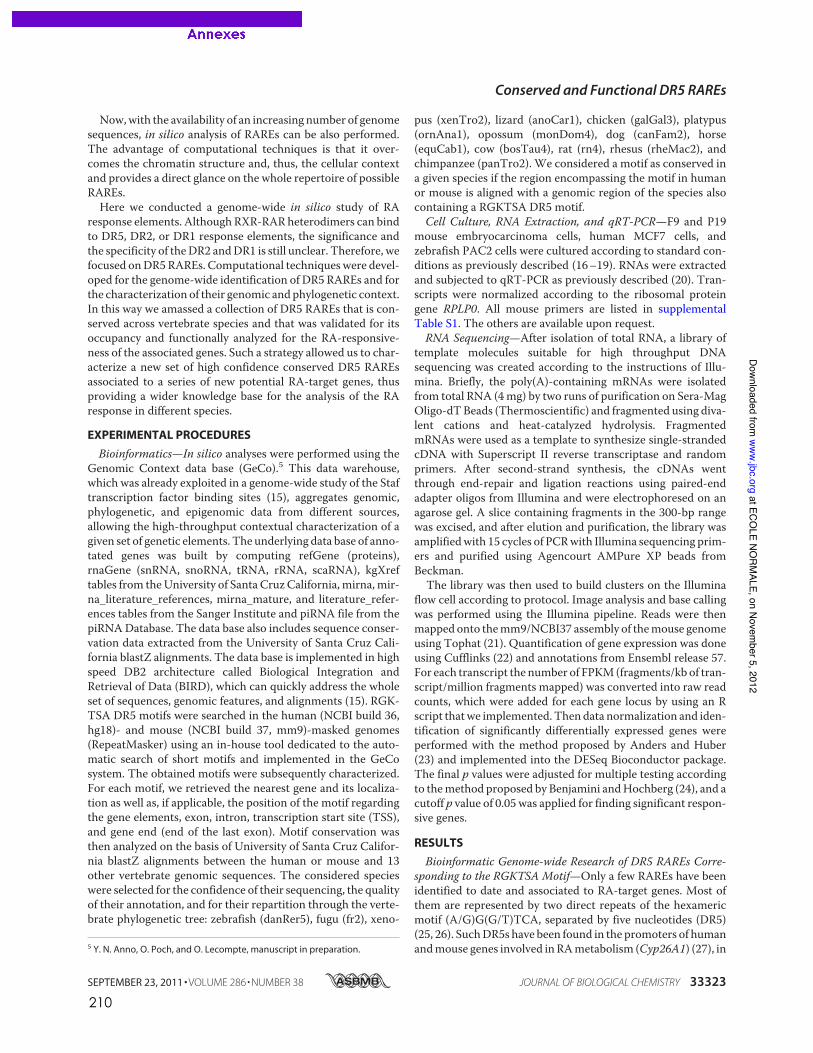
210
Now,with the availability of an increasing number of genomesequences, in silico analysis of RAREs can be also performed.The advantage of computational techniques is that it over-comes the chromatin structure and, thus, the cellular contextand provides a direct glance on the whole repertoire of possibleRAREs.Here we conducted a genome-wide in silico study of RA
response elements. Although RXR-RAR heterodimers can bindto DR5, DR2, or DR1 response elements, the significance andthe specificity of theDR2 andDR1 is still unclear. Therefore, wefocused onDR5RAREs. Computational techniqueswere devel-oped for the genome-wide identification of DR5 RAREs and forthe characterization of their genomic andphylogenetic context.In this way we amassed a collection of DR5 RAREs that is con-served across vertebrate species and that was validated for itsoccupancy and functionally analyzed for the RA-responsive-ness of the associated genes. Such a strategy allowed us to char-acterize a new set of high confidence conserved DR5 RAREsassociated to a series of new potential RA-target genes, thusproviding a wider knowledge base for the analysis of the RAresponse in different species.
EXPERIMENTAL PROCEDURES
Bioinformatics—In silico analyses were performed using theGenomic Context data base (GeCo).5 This data warehouse,which was already exploited in a genome-wide study of the Staftranscription factor binding sites (15), aggregates genomic,phylogenetic, and epigenomic data from different sources,allowing the high-throughput contextual characterization of agiven set of genetic elements. The underlying data base of anno-tated genes was built by computing refGene (proteins),rnaGene (snRNA, snoRNA, tRNA, rRNA, scaRNA), kgXreftables from theUniversity of Santa Cruz California,mirna,mir-na_literature_references, mirna_mature, and literature_refer-ences tables from the Sanger Institute and piRNA file from thepiRNADatabase. The data base also includes sequence conser-vation data extracted from the University of Santa Cruz Cali-fornia blastZ alignments. The data base is implemented in highspeed DB2 architecture called Biological Integration andRetrieval of Data (BIRD), which can quickly address the wholeset of sequences, genomic features, and alignments (15). RGK-TSA DR5 motifs were searched in the human (NCBI build 36,hg18)- and mouse (NCBI build 37, mm9)-masked genomes(RepeatMasker) using an in-house tool dedicated to the auto-matic search of short motifs and implemented in the GeCosystem. The obtained motifs were subsequently characterized.For each motif, we retrieved the nearest gene and its localiza-tion as well as, if applicable, the position of the motif regardingthe gene elements, exon, intron, transcription start site (TSS),and gene end (end of the last exon). Motif conservation wasthen analyzed on the basis of University of Santa Cruz Califor-nia blastZ alignments between the human or mouse and 13other vertebrate genomic sequences. The considered specieswere selected for the confidence of their sequencing, the qualityof their annotation, and for their repartition through the verte-brate phylogenetic tree: zebrafish (danRer5), fugu (fr2), xeno-
pus (xenTro2), lizard (anoCar1), chicken (galGal3), platypus(ornAna1), opossum (monDom4), dog (canFam2), horse(equCab1), cow (bosTau4), rat (rn4), rhesus (rheMac2), andchimpanzee (panTro2). We considered a motif as conserved ina given species if the region encompassing the motif in humanor mouse is aligned with a genomic region of the species alsocontaining a RGKTSA DR5 motif.Cell Culture, RNA Extraction, and qRT-PCR—F9 and P19
mouse embryocarcinoma cells, human MCF7 cells, andzebrafish PAC2 cells were cultured according to standard con-ditions as previously described (16–19). RNAs were extractedand subjected to qRT-PCR as previously described (20). Tran-scripts were normalized according to the ribosomal proteingene RPLP0. All mouse primers are listed in supplementalTable S1. The others are available upon request.RNA Sequencing—After isolation of total RNA, a library of
template molecules suitable for high throughput DNAsequencing was created according to the instructions of Illu-mina. Briefly, the poly(A)-containing mRNAs were isolatedfrom total RNA (4mg) by two runs of purification on Sera-MagOligo-dTBeads (Thermoscientific) and fragmented using diva-lent cations and heat-catalyzed hydrolysis. FragmentedmRNAs were used as a template to synthesize single-strandedcDNA with Superscript II reverse transcriptase and randomprimers. After second-strand synthesis, the cDNAs wentthrough end-repair and ligation reactions using paired-endadapter oligos from Illumina and were electrophoresed on anagarose gel. A slice containing fragments in the 300-bp rangewas excised, and after elution and purification, the library wasamplifiedwith 15 cycles of PCRwith Illumina sequencing prim-ers and purified using Agencourt AMPure XP beads fromBeckman.The library was then used to build clusters on the Illumina
flow cell according to protocol. Image analysis and base callingwas performed using the Illumina pipeline. Reads were thenmapped onto themm9/NCBI37 assembly of themouse genomeusing Tophat (21). Quantification of gene expression was doneusing Cufflinks (22) and annotations from Ensembl release 57.For each transcript the number of FPKM(fragments/kb of tran-script/million fragments mapped) was converted into raw readcounts, which were added for each gene locus by using an Rscript thatwe implemented. Thendata normalization and iden-tification of significantly differentially expressed genes wereperformed with the method proposed by Anders and Huber(23) and implemented into the DESeq Bioconductor package.The final p values were adjusted for multiple testing accordingto themethod proposed by Benjamini andHochberg (24), and acutoff p value of 0.05 was applied for finding significant respon-sive genes.
RESULTS
Bioinformatic Genome-wide Research of DR5 RAREs Corre-sponding to the RGKTSA Motif—Only a few RAREs have beenidentified to date and associated to RA-target genes. Most ofthem are represented by two direct repeats of the hexamericmotif (A/G)G(G/T)TCA, separated by five nucleotides (DR5)(25, 26). SuchDR5s have been found in the promoters of humanandmouse genes involved inRAmetabolism (Cyp26A1) (27), in5 Y. N. Anno, O. Poch, and O. Lecompte, manuscript in preparation.
Conserved and Functional DR5 RAREs
SEPTEMBER 23, 2011 • VOLUME 286 • NUMBER 38 JOURNAL OF BIOLOGICAL CHEMISTRY 33323
at ECOLE NO
RMALE, on Novem
ber 5, 2012www.jbc.org
Downloaded from
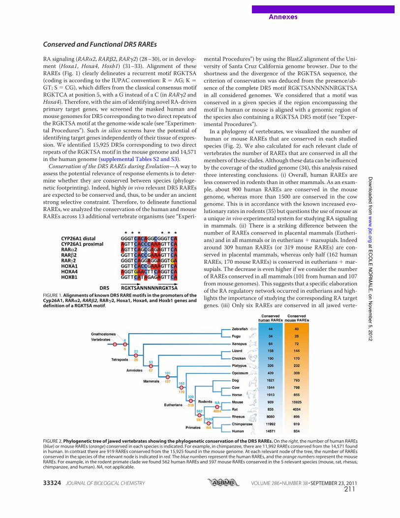
211
RA signaling (RAR!2, RAR"2, RAR#2) (28–30), or in develop-ment (Hoxa1, Hoxa4, Hoxb1) (31–33). Alignment of theseRAREs (Fig. 1) clearly delineates a recurrent motif RGKTSA(coding is according to the IUPAC convention: R ! AG; K !GT; S ! CG), which differs from the classical consensus motifRGKTCA at position 5, with a G instead of a C (in RAR#2 andHoxa4). Therefore, with the aim of identifying novel RA-drivenprimary target genes, we screened the masked human andmouse genomes for DR5 corresponding to two direct repeats ofthe RGKTSAmotif at the genome-wide scale (see “Experimen-tal Procedures”). Such in silico screens have the potential ofidentifying target genes independently of their tissue of expres-sion. We identified 15,925 DR5s corresponding to two directrepeats of the RGKTSAmotif in the mouse genome and 14,571in the human genome (supplemental Tables S2 and S3).Conservation of the DR5 RAREs during Evolution—A way to
assess the potential relevance of response elements is to deter-mine whether they are conserved between species (phyloge-netic footprinting). Indeed, highly in vivo relevant DR5 RAREsare expected to be conserved and, thus, to be under an ancientstrong selective constraint. Therefore, to delineate functionalRAREs, we analyzed the conservation of the human andmouseRAREs across 13 additional vertebrate organisms (see “Experi-
mental Procedures”) by using the BlastZ alignment of the Uni-versity of Santa Cruz California genome browser. Due to theshortness and the divergence of the RGKTSA sequence, thecriterion of conservation was deduced from the presence/ab-sence of the complete DR5 motif RGKTSANNNNNRGKTSAin all considered genomes. We considered that a motif wasconserved in a given species if the region encompassing themotif in human or mouse is aligned with a genomic region ofthe species also containing a RGKTSA DR5 motif (see “Exper-imental Procedures”).In a phylogeny of vertebrates, we visualized the number of
human or mouse RAREs that are conserved in each studiedspecies (Fig. 2). We also calculated for each relevant clade ofvertebrates the number of RAREs that are conserved in all themembers of these clades. Although these data can be influencedby the coverage of the studied genome (34), this analysis raisedthree interesting conclusions. (i) Overall, human RAREs areless conserved in rodents than in other mammals. As an exam-ple, about 900 human RAREs are conserved in the mousegenome, whereas more than 1500 are conserved in the cowgenome. This is in accordance with the known increased evo-lutionary rates in rodents (35) but questions the use ofmouse asa unique in vivo experimental system for studying RA signalingin mammals. (ii) There is a striking difference between thenumber of RAREs conserved in placental mammals (Eutheri-ans) and in all mammals or in eutherians " marsupials. Indeedaround 309 human RAREs (or 319 mouse RAREs) are con-served in placental mammals, whereas only half (162 humanRAREs, 170 mouse RAREs) is conserved in eutherians " mar-supials. The decrease is even higher if we consider the numberof RAREs conserved in all mammals (101 from human and 107frommouse genomes). This suggests that a specific elaborationof the RA regulatory network occurred in eutherians and high-lights the importance of studying the corresponding RA targetgenes. (iii) Only six RAREs are conserved in all jawed verte-
FIGURE 1. Alignments of known DR5 RARE motifs in the promoters of theCyp26A1, RAR!2, RAR"2, RAR#2, Hoxa1, Hoxa4, and Hoxb1 genes anddefinition of a RGKTSA motif.
FIGURE 2. Phylogenetic tree of jawed vertebrates showing the phylogenetic conservation of the DR5 RAREs. On the right, the number of human RAREs(blue) or mouse RAREs (orange) conserved in each species is indicated. For example, in chimpanzee, there are 11,992 RAREs conserved from the 14,571 foundin human. In contrast there are 919 RAREs conserved from the 15,925 found in the mouse genome. At each relevant node of the tree, the number of RAREsconserved in the species of the relevant node is indicated in red. The blue numbers represent the human RAREs, and the orange numbers represent the mouseRAREs. For example, in the rodent primate clade we found 562 human RAREs and 597 mouse RAREs conserved in the 5 relevant species (mouse, rat, rhesus,chimpanzee, and human). NA, not applicable.
Conserved and Functional DR5 RAREs
33324 JOURNAL OF BIOLOGICAL CHEMISTRY VOLUME 286 • NUMBER 38 • SEPTEMBER 23, 2011
at ECOLE NO
RMALE, on Novem
ber 5, 2012www.jbc.org
Downloaded from
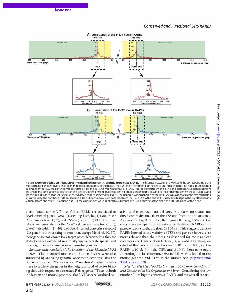
212
brates (gnathostomes). Three of these RAREs are associated todevelopmental genes, Dach1 (Daschung homolog 1) (36), Meis1(Meis homeobox 1) (37), and TSHZ3 (Teashirt 3) (38). The threeothers are associated to the Gria2 (glutamate receptor 2) (39),Lphn2 (latrophilin 2) (40), and Paqr3 (an adiponectin receptor)(41) genes. It is interesting to note that, except Meis1 (8, 10, 37),these gene arenot knownRARtarget genes.Nevertheless, they arelikely to be RA-regulated in virtually any vertebrate species andthus might be considered as new interesting models.Genome-wide Analysis of the Location of the Identified DR5
RAREs—The identified mouse and human RAREs were alsoannotated by analyzing genome-wide their locations using theGeCo system (see “Experimental Procedures”), which allowsusers to retrieve the genes in the neighborhood of factor bind-ing sites with respect to annotated Refseq genes.5 Then, in boththe human and mouse genomes, the RAREs were localized rel-
ative to the nearest matched gene boundary: upstream anddownstream distance from the TSS and from the end of genes.As shown in Fig. 3, A and B, the regions flanking TSSs and theends of genes depict the highest concentration of RAREs com-paredwith the further regions (!500 kb). This suggests that theRAREs located in the vicinity of TSSs and gene ends would bemore relevant than the others, as described for most nuclearreceptors and transcription factors (14, 42–46). Therefore, weselected the RAREs located between "10 and #10 kb, i.e. theRAREs !10 kb from the TSSs and !10 kb from gene ends.According to this criterion, 3862 RAREs were selected in themouse genome and 3429 in the human one (supplementalTables S2 and S3).Selection of a List of RAREs Located!10 kb fromGene Limits
and Conserved in Six Organisms orMore—Considering the lownumber (6) of highly conserved RAREs and the overall reparti-
FIGURE 3. Genome-wide distribution of the identified human (A) and mouse (B) DR5 RAREs. The distance between the RARE and the corresponding genewas calculated by identifying its proximity to both boundaries of the genes, the TSS, and the end (end of the last exon). Following this rule for a RARE locatedupstream of the TSS, the distance was calculated from the TSS and was negative. For a RARE located downstream of a gene, the distance was calculated fromthe end of the gene and was positive. In the case of a RARE present inside the gene, both distances to the TSS and to the end of the gene were calculated, andthe minimal distance in absolute value, called dTSS*, was considered (cf. Fig. 5). The genome-wide mapping of the RARE versus a canonical gene was calculatedby cumulating the number of hits present in a 1-kb sliding window from each side from the TSS or from the end of the gene (the first point being attributed to500 bp before and after TSS or gene end). These calculations were applied to a distance of 500 kb outside of the gene and 100 kb inside of the gene.
Conserved and Functional DR5 RAREs
SEPTEMBER 23, 2011 • VOLUME 286 • NUMBER 38 JOURNAL OF BIOLOGICAL CHEMISTRY 33325
at ECOLE NO
RMALE, on Novem
ber 5, 2012www.jbc.org
Downloaded from
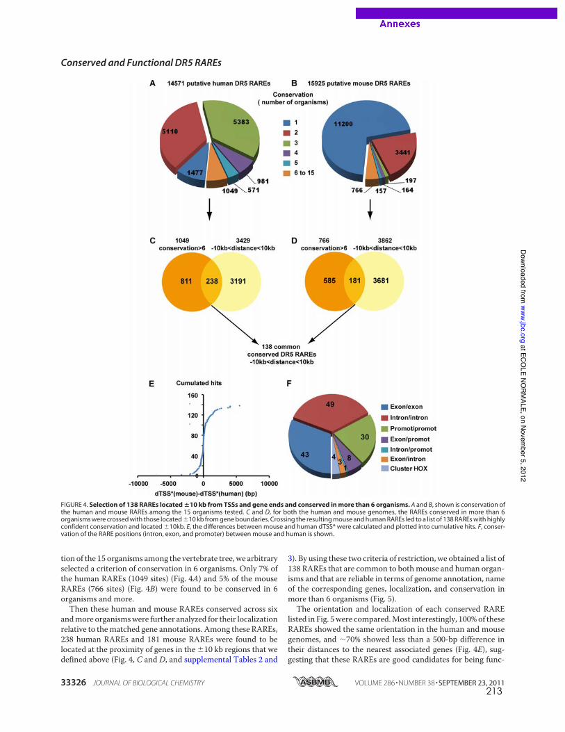
213
tion of the 15 organisms among the vertebrate tree, we arbitraryselected a criterion of conservation in 6 organisms. Only 7% ofthe human RAREs (1049 sites) (Fig. 4A) and 5% of the mouseRAREs (766 sites) (Fig. 4B) were found to be conserved in 6organisms and more.Then these human and mouse RAREs conserved across six
andmore organismswere further analyzed for their localizationrelative to thematched gene annotations. Among these RAREs,238 human RAREs and 181 mouse RAREs were found to belocated at the proximity of genes in the !10 kb regions that wedefined above (Fig. 4, C and D, and supplemental Tables 2 and
3). By using these two criteria of restriction, we obtained a list of138 RAREs that are common to bothmouse and human organ-isms and that are reliable in terms of genome annotation, nameof the corresponding genes, localization, and conservation inmore than 6 organisms (Fig. 5).The orientation and localization of each conserved RARE
listed in Fig. 5were compared.Most interestingly, 100%of theseRAREs showed the same orientation in the human and mousegenomes, and "70% showed less than a 500-bp difference intheir distances to the nearest associated genes (Fig. 4E), sug-gesting that these RAREs are good candidates for being func-
FIGURE 4. Selection of 138 RAREs located !10 kb from TSSs and gene ends and conserved in more than 6 organisms. A and B, shown is conservation ofthe human and mouse RAREs among the 15 organisms tested. C and D, for both the human and mouse genomes, the RAREs conserved in more than 6organisms were crossed with those located !10 kb from gene boundaries. Crossing the resulting mouse and human RAREs led to a list of 138 RAREs with highlyconfident conservation and located !10kb. E, the differences between mouse and human dTSS* were calculated and plotted into cumulative hits. F, conser-vation of the RARE positions (intron, exon, and promoter) between mouse and human is shown.
Conserved and Functional DR5 RAREs
33326 JOURNAL OF BIOLOGICAL CHEMISTRY VOLUME 286 • NUMBER 38 • SEPTEMBER 23, 2011
at ECOLE NO
RMALE, on Novem
ber 5, 2012www.jbc.org
Downloaded from
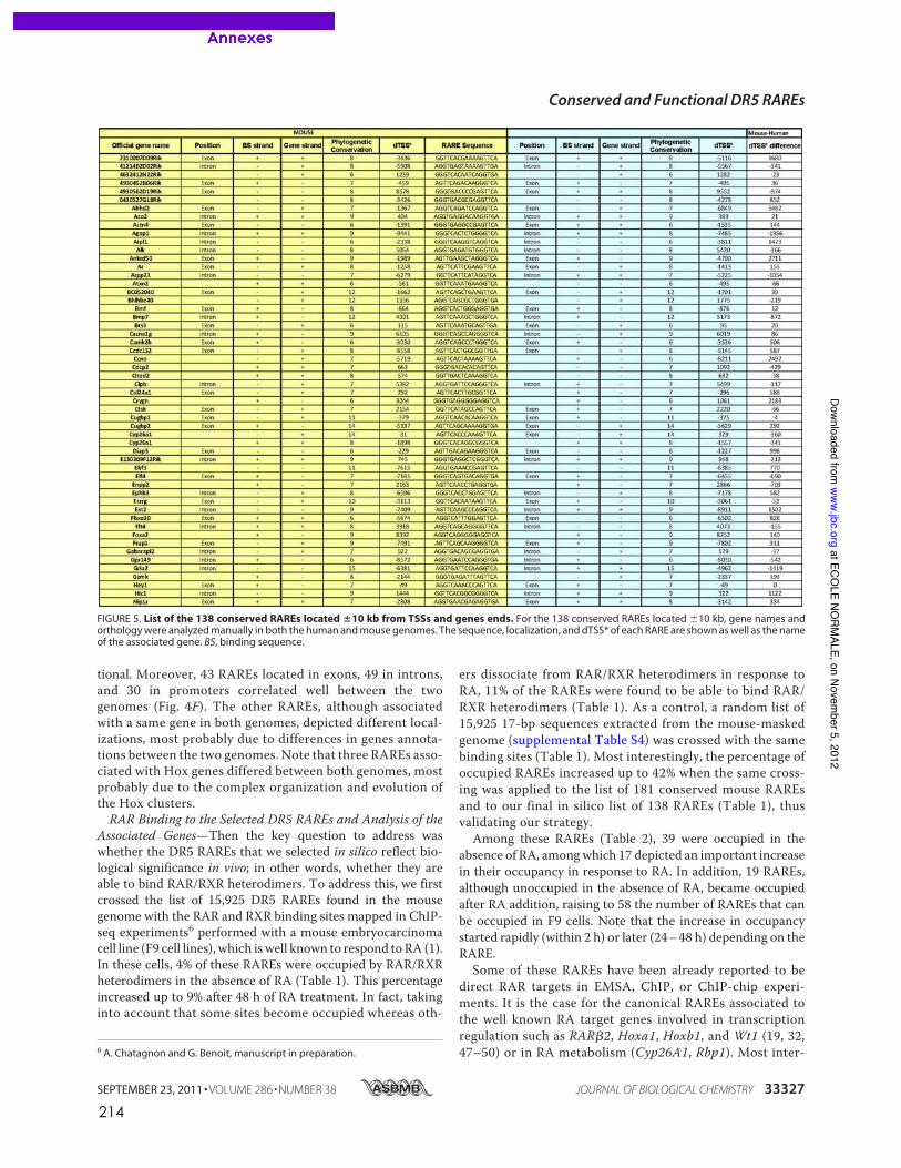
214
tional. Moreover, 43 RAREs located in exons, 49 in introns,and 30 in promoters correlated well between the twogenomes (Fig. 4F). The other RAREs, although associatedwith a same gene in both genomes, depicted different local-izations, most probably due to differences in genes annota-tions between the two genomes. Note that three RAREs asso-ciated with Hox genes differed between both genomes, mostprobably due to the complex organization and evolution ofthe Hox clusters.RAR Binding to the Selected DR5 RAREs and Analysis of the
Associated Genes—Then the key question to address waswhether the DR5 RAREs that we selected in silico reflect bio-logical significance in vivo; in other words, whether they areable to bind RAR/RXR heterodimers. To address this, we firstcrossed the list of 15,925 DR5 RAREs found in the mousegenome with the RAR and RXR binding sites mapped in ChIP-seq experiments6 performed with a mouse embryocarcinomacell line (F9 cell lines), which iswell known to respond toRA (1).In these cells, 4% of these RAREs were occupied by RAR/RXRheterodimers in the absence of RA (Table 1). This percentageincreased up to 9% after 48 h of RA treatment. In fact, takinginto account that some sites become occupied whereas oth-
ers dissociate from RAR/RXR heterodimers in response toRA, 11% of the RAREs were found to be able to bind RAR/RXR heterodimers (Table 1). As a control, a random list of15,925 17-bp sequences extracted from the mouse-maskedgenome (supplemental Table S4) was crossed with the samebinding sites (Table 1). Most interestingly, the percentage ofoccupied RAREs increased up to 42% when the same cross-ing was applied to the list of 181 conserved mouse RAREsand to our final in silico list of 138 RAREs (Table 1), thusvalidating our strategy.Among these RAREs (Table 2), 39 were occupied in the
absence of RA, amongwhich 17 depicted an important increasein their occupancy in response to RA. In addition, 19 RAREs,although unoccupied in the absence of RA, became occupiedafter RA addition, raising to 58 the number of RAREs that canbe occupied in F9 cells. Note that the increase in occupancystarted rapidly (within 2 h) or later (24–48 h) depending on theRARE.Some of these RAREs have been already reported to be
direct RAR targets in EMSA, ChIP, or ChIP-chip experi-ments. It is the case for the canonical RAREs associated tothe well known RA target genes involved in transcriptionregulation such as RAR!2, Hoxa1, Hoxb1, and Wt1 (19, 32,47–50) or in RA metabolism (Cyp26A1, Rbp1). Most inter-6 A. Chatagnon and G. Benoit, manuscript in preparation.
FIGURE 5. List of the 138 conserved RAREs located !10 kb from TSSs and genes ends. For the 138 conserved RAREs located !10 kb, gene names andorthology were analyzed manually in both the human and mouse genomes. The sequence, localization, and dTSS* of each RARE are shown as well as the nameof the associated gene. BS, binding sequence.
Conserved and Functional DR5 RAREs
SEPTEMBER 23, 2011 • VOLUME 286 • NUMBER 38 JOURNAL OF BIOLOGICAL CHEMISTRY 33327
at ECOLE NO
RMALE, on Novem
ber 5, 2012www.jbc.org
Downloaded from
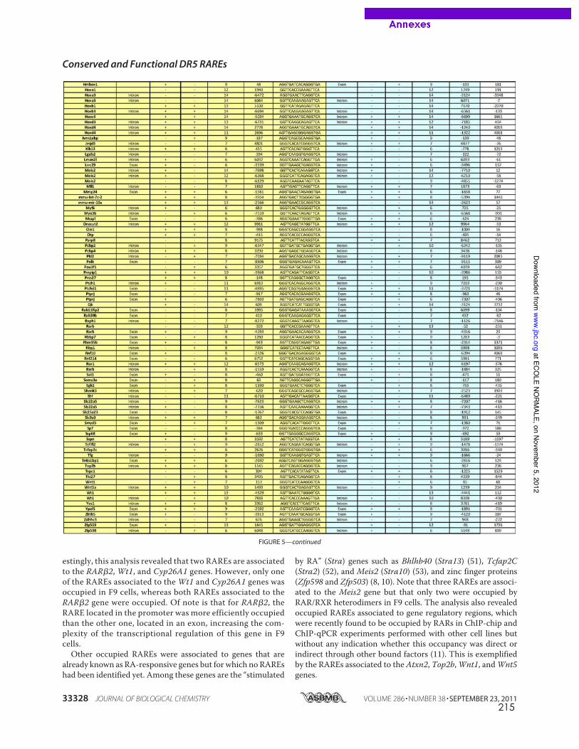
215
estingly, this analysis revealed that two RAREs are associatedto the RAR!2, Wt1, and Cyp26A1 genes. However, only oneof the RAREs associated to the Wt1 and Cyp26A1 genes wasoccupied in F9 cells, whereas both RAREs associated to theRAR!2 gene were occupied. Of note is that for RAR!2, theRARE located in the promoter was more efficiently occupiedthan the other one, located in an exon, increasing the com-plexity of the transcriptional regulation of this gene in F9cells.Other occupied RAREs were associated to genes that are
already known as RA-responsive genes but for which no RAREshad been identified yet. Among these genes are the “stimulated
by RA” (Stra) genes such as Bhlhb40 (Stra13) (51), Tcfap2C(Stra2) (52), and Meis2 (Stra10) (53), and zinc finger proteins(Zfp598 and Zfp503) (8, 10). Note that three RAREs are associ-ated to the Meis2 gene but that only two were occupied byRAR/RXR heterodimers in F9 cells. The analysis also revealedoccupied RAREs associated to gene regulatory regions, whichwere recently found to be occupied by RARs in ChIP-chip andChIP-qPCR experiments performed with other cell lines butwithout any indication whether this occupancy was direct orindirect through other bound factors (11). This is exemplifiedby the RAREs associated to the Atxn2, Top2b,Wnt1, andWnt5genes.
FIGURE 5—continued
Conserved and Functional DR5 RAREs
33328 JOURNAL OF BIOLOGICAL CHEMISTRY VOLUME 286 • NUMBER 38 • SEPTEMBER 23, 2011
at ECOLE NO
RMALE, on Novem
ber 5, 2012www.jbc.org
Downloaded from
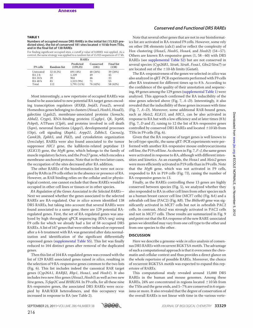
216
Most interestingly, a new repertoire of occupied RAREs wasfound to be associated to new potential RA target genes encod-ing transcription regulators (RXR!, Jmjd3, Foxa2), severalHomeobox genes belonging to clusters (Hoxa3,Hoxb3,Hoxd3),galectins (Lgals2), membrane-associated proteins (Sema3e,Abhd2, Crygn), RNA-binding proteins (Cugbp1, Qk, Srp68,Pcbp4), ATPases (Clpb), and proteins involved in cell death(Sspn), neuronal functions (Agap1), developmental processes(Otp), cell signaling (Raph1, Arpp21, Zdhhc3, Cacna1g,Camk2b, Ephb3, and Pld2), and cytoskeleton organization(Ivns1abp). RAREs were also found associated to the tumorsuppressor HIC1 gene, the kallikrein-related peptidase 13(KLK13) gene, the Myf6 gene, which belongs to the family ofmuscle regulatory factors, and thePrss27 gene, which encodes amembrane-anchored protease.Note that in the two latter cases,the occupation of the sites decreased after RA addition.The other RAREs of the bioinformatics list were not occu-
pied by RARs in F9 cells either in the absence or presence of RA.However, as RAR binding relies on the cellular and/or physio-logical context, one cannot exclude that these RAREs would beoccupied in other cell lines or tissues or in other species.RA Regulation of the Genes Associated to the Selected RAREs—
Next we assessed whether the genes associated to the selectedRAREs are RA-regulated. Our in silico screen identified 138DR5 RAREs, but taking into account that several RAREs werefound associated to a same gene, there are 129 potential RA-regulated genes. First, the set of RA-regulated genes was ana-lyzed by high throughput qPCR sequencing (RNA-seq) usingF9 cells for which we already had a list of 58 occupied DR5RAREs.A list of 167 genes thatwere either induced or repressedafter a 4-h treatment with RAwas generated after data normal-ization and identification of the significant differentiallyexpressed genes (supplemental Table S5). This list was finallyreduced to 164 distinct genes after removal of the duplicatedgenes.Then this list of 164 RA-regulated geneswas crossedwith the
list of 129 RARE-associated genes raised in silico, resulting inthe selection of 9 RA-responsive genes common to the two lists(Fig. 6). This list includes indeed the canonical RAR targetgenes (Cyp26A1, RAR!2, Rbp1, Hoxa1, and Hoxb1). It alsoincludes two newHox genes (Hoxa3,Hoxb3) as well as two newStra genes, Tcfap2C and BHBLH4. In F9 cells, for all these nineRA-responsive genes, the associated DR5 RAREs were occu-pied by RAR/RXR heterodimers, and this occupancy wasincreased in response to RA (see Table 2).
Note that several other genes that are not in our bioinformat-ics list are activated in RA-treated F9 cells. However, some relyon other DR elements (cdx1) and/or reflect the complexity ofHox clustering (Hoxa5, Hoxb5, Hoxa4, and Hoxb2) (54–57).Others are known RA-responsive genes (1, 58–60) with DR5RAREs (see supplemental Table S2) but are not conserved inseveral species (Cyp26B1, Stra6, Stra8, Foxa1, Gbx2/Stra7) orare located out of the !10-kb limits (Gata6).The RA-responsiveness of the genes we selected in silicowas
also analyzed in qRT-PCR experiments performedwith F9 cellsafter RA treatment for different times up to 8 h. According tothe confidence of the quality of their annotation and sequenc-ing, 49 genes among the 129 genes (supplemental Table 1) wereanalyzed. This approach confirmed the RA inducibility of thenine genes selected above (Fig. 7, A–D). Interestingly, it alsorevealed that the inducibility of these genes increases with time(Fig. 7, A–D). Moreover, some additional RAR-bound genes,such as Meis2, KLK13, and HIC1, can be also activated inresponse to RA but with a low efficiency and at later times (8 h)(Fig. 7, D and E), raising to 12 the list of RA-responsive genescontrolled by conserved DR5 RAREs and located !10 kb fromTSSs in F9 cells (Fig. 6).Given that the RA response of target genes is well known to
be cell type-specific, the same qRT-PCR experiments were per-formed with another RA-responsive mouse embryocarcinomacell line, the P19 cell line. As shown in Fig. 7,F–J, the same geneswere activated in response to RA, althoughwith different inten-sities and kinetics. As an example, the Hoxa1 andMeis2 genesweremore efficiently activated in P19 cells than in F9 cells.Notethat the Myf6 gene, which was not activated in F9 cells,responded to RA in P19 cells (Fig. 7I), raising the number ofRA-responsive genes to 13.Finally, as the RAREs controlling these 13 genes are highly
conserved between species (Fig. 5), we analyzed whether theyalso responded to RA in other cell lines from other species suchas a human breast cancer cell line (MCF7 cells) (Fig. 8A) and azebrafish cell line (PAC2) (Fig. 8B). The Bhlhe40 gene was sig-nificantly activated in MCF7 cells but not in zebrafish PAC2cells. In contrast, Meis2 was strongly activated in PAC2 cellsand not in MCF7 cells. These results are summarized in Fig. 9and point out that the RA response of the newRARE-associatedgenes we identifiedmay vary fromone cell type to the other andfrom one species to the other.
DISCUSSION
Here we describe a genome-wide in silico analysis of consen-susDR5RAREswith recurrent RGKTSAmotifs. The advantageof such a computational approach is that it overcomes the chro-matin and cellular context and thus provides a direct glance onthe whole repertoire of possible RAREs. Moreover, the choiceof recurrent RGKTSA motifs was expected to expand this rep-ertoire of RAREs.This computational study revealed around 15,000 DR5
RAREs in the human and mouse genomes. Among theseRAREs, 24% are concentrated in regions located !10 kb fromtheTSSs and the gene ends, and 5–7% are conserved in 6 organ-isms ormore. It also revealed that the degree of conservation ofthe overall RAREs is not linear with time in the various verte-
TABLE 1Numbers of occupied mouse DR5 RAREs in the initial list (15,925 pre-dicted sites), the list of conserved 181 sites located !10 kb from TSSs,and in the final list of 138 RAREsFor finding significant occupied sites, a cutoff p value of 0,00001 was applied. As acontrol, the same strategy was applied to a random list of 15,925 sequences of 17 kb.
F9 cells
RAREs
Random listPredicted(159,25)
Conserved(181)
Final list(138)
Untreated 32 (0.2%) 691 (4%) 48 (26%) 39 (28%)RA 2 h 62 1,109 49 45RA 24 h 39 964 46 33RA 48 h 85 1,523 (9%) 71 55Total 112 1,791 (11%) 76 (42%) 58 (42%)
Conserved and Functional DR5 RAREs
SEPTEMBER 23, 2011 • VOLUME 286 • NUMBER 38 JOURNAL OF BIOLOGICAL CHEMISTRY 33329
at ECOLE NO
RMALE, on Novem
ber 5, 2012www.jbc.org
Downloaded from
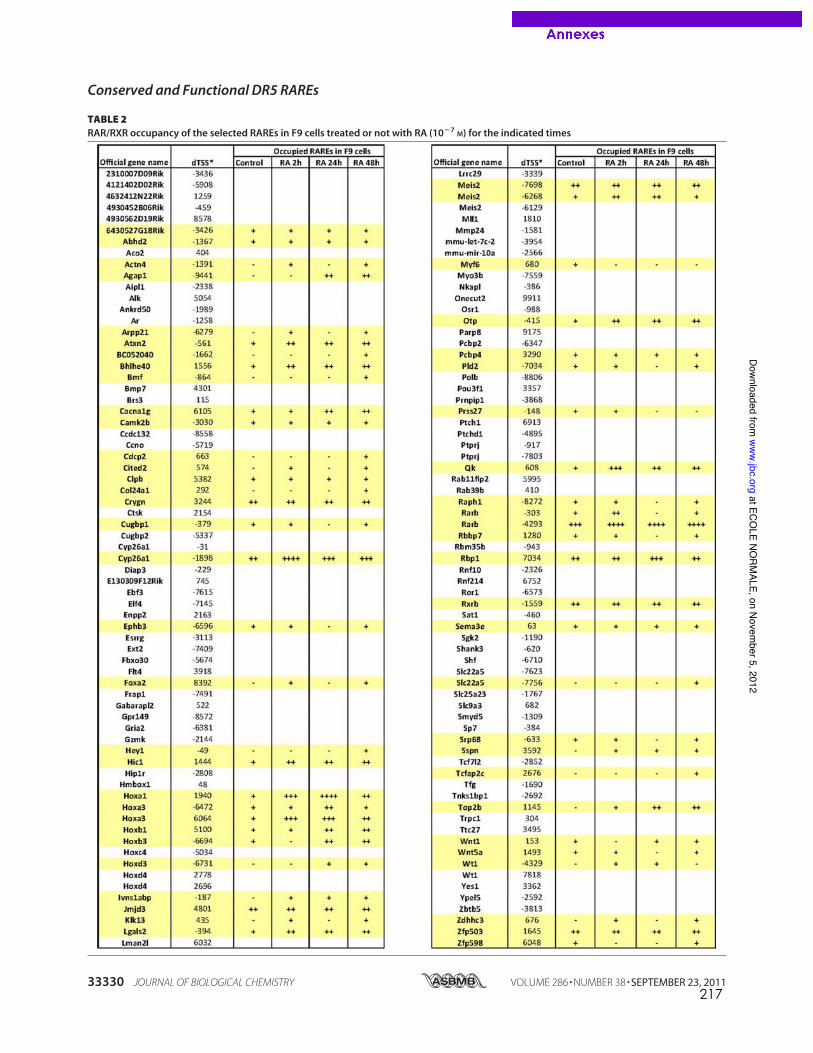
217
TABLE 2RAR/RXR occupancy of the selected RAREs in F9 cells treated or not with RA (10!7
M) for the indicated times
Conserved and Functional DR5 RAREs
33330 JOURNAL OF BIOLOGICAL CHEMISTRY VOLUME 286 • NUMBER 38 • SEPTEMBER 23, 2011
at ECOLE NO
RMALE, on Novem
ber 5, 2012www.jbc.org
Downloaded from
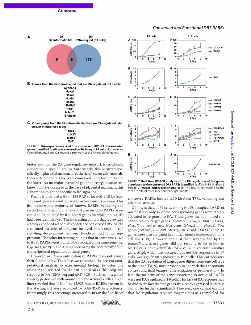
218
brates and that the RA gene regulatory network is specificallyelaborated in specific groups. Surprisingly, this occurred spe-cifically in placentalmammals (eutherians) versus allmammals.Indeed, 3-foldmore RAREs are conserved in the former than inthe latter. As no major events of genomic reorganization areknown to have occurred at the base of placental mammals, thiselaboration might be specific to RA signaling.Finally it provided a list of 138 RAREs located !10 kb from
TSSs and gene ends and conserved in 6 organisms ormore. Thislist includes the majority of known RAREs, validating therestrictive criteria of our analysis. It also includes RAREs asso-ciated to “stimulated by RA” (Stra) genes for which no RAREshad been identified yet. The interesting point is that it provideda newly expanded set of high confidence conservedDR5RAREsassociated to a series of new genes involved in transcription, cellsignaling, development, neuronal functions, and tumor sup-pression. The other interesting point is that in some cases, twoto three RAREs were found to be associated to a same gene (e.g.Cyp26A1, RAR!2, andMeis2), increasing the complexity of thetranscriptional regulation of these genes.However, in silico identification of RAREs does not assure
their functionality. Therefore, we combined the present com-putational analysis to experimental biology to determinewhether the selected RAREs can bind RARs (ChIP-seq) andrespond to RA (RNA-seq and qRT-PCR). Such an integratedstrategy performed withmouse embryocarcinoma cells (F9 cellline) revealed that 11% of the 15,925 mouse RAREs present inthe starting list were occupied by RAR/RXR heterodimers.Interestingly, this percentage increased to 40% in the final list of
conserved RAREs located !10 kb from TSSs, validating ourselection strategy.Of note is that, in F9 cells, among the 58 occupied RAREs of
our final list, only 12 of the corresponding genes were rapidlyactivated in response to RA. These genes include indeed thecanonical RA target genes (Cyp26A1, RAR!2, Rbp1, Hoxa1,Hoxb1) as well as new Hox genes (Hoxa3 and Hoxb3), Stragenes (Tcfap2c, Bhlhe40, Meis2), HIC1, and KLK13. These 12genes were also activated in another mouse embryocarcinomacell line (P19). However, some of them (exemplified by theBhlhe40 and Meis2 genes) did not respond to RA in humanMCF7 cells or in zebrafish PAC2 cells. In contrast, anothergene, Myf6, which was occupied but not RA-responsive in F9cells, was significantly induced in P19 cells. This corroboratesthat the RA regulation of target genes differs from one cell typeto the other (Fig. 9), most probably in line with their chromatincontext and final feature (differentiation or proliferation). Infact, the majority of the genes associated to occupied RAREswere not RA-regulated in F9 cells. This lack of RA responsemaybe due to the fact that the genes are already expressed (and thuscannot be further stimulated). However, one cannot excludethat RA regulation requires longer times as exemplified for
FIGURE 6. RA-responsiveness of the conserved DR5 RARE-associatedgenes identified in silico as assessed by RNA-seq in F9 cells. A, shown areVenn diagrams. B and C, shown is a summary of the RA-regulated genes.
FIGURE 7. Real time RT-PCR analysis of the RA regulation of the genesassociated to the conserved DR5 RAREs identified in silico in F9 (A–E) andP19 (F–J) mouse embryocarcinoma cells. The results correspond to themean ! S.D. of three independent experiments.
Conserved and Functional DR5 RAREs
SEPTEMBER 23, 2011 • VOLUME 286 • NUMBER 38 JOURNAL OF BIOLOGICAL CHEMISTRY 33331
at ECOLE NO
RMALE, on Novem
ber 5, 2012www.jbc.org
Downloaded from
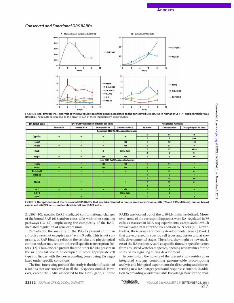
219
Zfp503 (10), specific RARE-mediated conformational changesof the bound RAR (61), and/or cross-talks with other signalingpathways (12, 62), emphasizing the complexity of the RAR-mediated regulation of gene expression.Remarkably, the majority of the RAREs present in our in
silico list were not occupied in vivo in F9 cells. This is not sur-prising, as RAR binding relies on the cellular and physiologicalcontext and/ormay require other cell specific transcription fac-tors (12). Thus, one can predict that the other RAREs present inthe in silico list would be occupied in other appropriate celltypes or tissues with the corresponding genes being RA regu-lated under specific conditions.The final interesting point of this study is the identification of
6 RAREs that are conserved in all the 15 species studied. How-ever, except the RARE associated to the Gria2 gene, all these
RAREs are located out of the !10-kb limits we defined. More-over, none of the corresponding genes were RA-regulated in F9cells, as assessed in RNA-seq experiments, exceptMeis1, whichwas activated 24 h after the RA addition to F9 cells (10). Never-theless, these genes are mostly developmental genes (36–41)that are expressed in specific cell types and tissues and at spe-cific developmental stages. Therefore, theymight be newmark-ers of the RA response, valid at specific times, in specific tissuesfrom any jawed vertebrate species, opening new avenues for thestudy of RA signaling during development.In conclusion, the novelty of the present study resides in an
integrated strategy combining genome-wide biocomputinganalysis and biological experiments for discovering and charac-terizing new RAR target genes and response elements. In addi-tion to providing a wider valuable knowledge base for the anal-
FIGURE 8. Real time RT-PCR analysis of the RA regulation of the genes associated to the conserved DR5 RAREs in human MCF7 (A) and zebrafish PAC2(B) cells. The results correspond to the mean ! S.D. of three independent experiments.
FIGURE 9. Recapitulation of the conserved DR5 RAREs that are RA-activated in mouse embryocarcinoma cells (F9 and P19 cell lines), human breastcancer cells (MCF7 cells), and a zebrafish cell line (PAC2 cells).
Conserved and Functional DR5 RAREs
33332 JOURNAL OF BIOLOGICAL CHEMISTRY VOLUME 286 • NUMBER 38 • SEPTEMBER 23, 2011
at ECOLE NO
RMALE, on Novem
ber 5, 2012www.jbc.org
Downloaded from
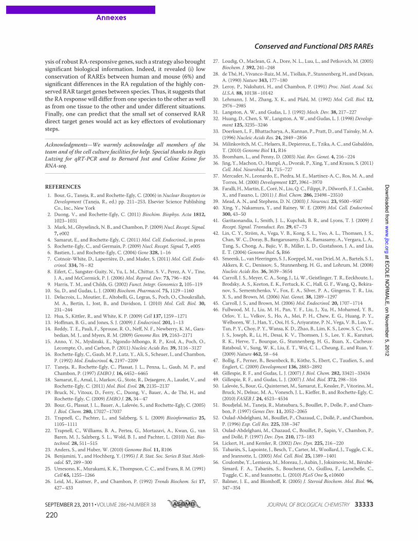
220
ysis of robust RA-responsive genes, such a strategy also broughtsignificant biological information. Indeed, it revealed (i) lowconservation of RAREs between human and mouse (6%) andsignificant differences in the RA regulation of the highly con-served RAR target genes between species. Thus, it suggests thatthe RA response will differ from one species to the other as wellas from one tissue to the other and under different situations.Finally, one can predict that the small set of conserved RARdirect target genes would act as key effectors of evolutionarysteps.
Acknowledgments—We warmly acknowledge all members of theteam and of the cell culture facilities for help. Special thanks to RegisLutzing for qRT-PCR and to Bernard Jost and Celine Keime forRNA-seq.
REFERENCES1. Bour, G., Taneja, R., and Rochette-Egly, C. (2006) in Nuclear Receptors in
Development (Taneja, R., ed.) pp. 211–253, Elsevier Science PublishingCo., Inc., New York
2. Duong, V., and Rochette-Egly, C. (2011) Biochim. Biophys. Acta 1812,1023–1031
3. Mark,M., Ghyselinck, N. B., and Chambon, P. (2009)Nucl. Recept. Signal.7, e002
4. Samarut, E., and Rochette-Egly, C. (2011)Mol. Cell. Endocrinol., in press5. Rochette-Egly, C., and Germain, P. (2009) Nucl. Recept. Signal. 7, e0056. Bastien, J., and Rochette-Egly, C. (2004) Gene 328, 1–167. Cotnoir-White, D., Laperriere, D., and Mader, S. (2011) Mol. Cell. Endo-
crinol. 334, 76–828. Eifert, C., Sangster-Guity, N., Yu, L. M., Chittur, S. V., Perez, A. V., Tine,
J. A., and McCormick, P. J. (2006)Mol. Reprod. Dev. 73, 796–8249. Harris, T. M., and Childs, G. (2002) Funct. Integr. Genomics 2, 105–11910. Su, D., and Gudas, L. J. (2008) Biochem. Pharmacol. 75, 1129–116011. Delacroix, L., Moutier, E., Altobelli, G., Legras, S., Poch, O., Choukrallah,
M. A., Bertin, I., Jost, B., and Davidson, I. (2010) Mol. Cell. Biol. 30,231–244
12. Hua, S., Kittler, R., and White, K. P. (2009) Cell 137, 1259–127113. Hoffman, B. G., and Jones, S. J. (2009) J. Endocrinol. 201, 1–1314. Reddy, T. E., Pauli, F., Sprouse, R. O., Neff, N. F., Newberry, K. M., Gara-
bedian, M. J., and Myers, R. M. (2009) Genome Res. 19, 2163–217115. Anno, Y. N., Myslinski, E., Ngondo-Mbongo, R. P., Krol, A., Poch, O.,
Lecompte, O., and Carbon, P. (2011) Nucleic Acids Res. 39, 3116–312716. Rochette-Egly, C., Gaub, M. P., Lutz, Y., Ali, S., Scheuer, I., and Chambon,
P. (1992)Mol. Endocrinol. 6, 2197–220917. Taneja, R., Rochette-Egly, C., Plassat, J. L., Penna, L., Gaub, M. P., and
Chambon, P. (1997) EMBO J. 16, 6452–646518. Samarut, E., Amal, I., Markov, G., Stote, R., Dejaegere, A., Laudet, V., and
Rochette-Egly, C. (2011)Mol. Biol. Evol. 28, 2135–213719. Bruck, N., Vitoux, D., Ferry, C., Duong, V., Bauer, A., de The, H., and
Rochette-Egly, C. (2009) EMBO J. 28, 34–4720. Bour, G., Plassat, J. L., Bauer, A., Lalevee, S., and Rochette-Egly, C. (2005)
J. Biol. Chem. 280, 17027–1703721. Trapnell, C., Pachter, L., and Salzberg, S. L. (2009) Bioinformatics 25,
1105–111122. Trapnell, C., Williams, B. A., Pertea, G., Mortazavi, A., Kwan, G., van
Baren, M. J., Salzberg, S. L., Wold, B. J., and Pachter, L. (2010) Nat. Bio-technol. 28, 511–515
23. Anders, S., and Huber, W. (2010) Genome Biol. 11, R10624. Benjamini, Y., and Hochberg, Y. (1995) J. R. Stat. Soc. Series B Stat. Meth-
odol. 57, 289–30025. Umesono, K., Murakami, K. K., Thompson, C. C., and Evans, R. M. (1991)
Cell 65, 1255–126626. Leid, M., Kastner, P., and Chambon, P. (1992) Trends Biochem. Sci 17,
427–433
27. Loudig, O., Maclean, G. A., Dore, N. L., Luu, L., and Petkovich, M. (2005)Biochem. J. 392, 241–248
28. deThe, H., Vivanco-Ruiz,M.M., Tiollais, P., Stunnenberg,H., andDejean,A. (1990) Nature 343, 177–180
29. Leroy, P., Nakshatri, H., and Chambon, P. (1991) Proc. Natl. Acad. Sci.U.S.A. 88, 10138–10142
30. Lehmann, J. M., Zhang, X. K., and Pfahl, M. (1992) Mol. Cell. Biol. 12,2976–2985
31. Langston, A. W., and Gudas, L. J. (1992)Mech. Dev. 38, 217–22732. Huang, D., Chen, S. W., Langston, A.W., and Gudas, L. J. (1998)Develop-
ment 125, 3235–324633. Doerksen, L. F., Bhattacharya, A., Kannan, P., Pratt, D., and Tainsky,M. A.
(1996) Nucleic Acids Res. 24, 2849–285634. Milinkovitch,M.C.,Helaers, R., Depiereux, E., Tzika, A. C., andGabaldon,
T. (2010) Genome Biol 11, R1635. Bromham, L., and Penny, D. (2003) Nat. Rev. Genet. 4, 216–22436. Jing, Y., Machon, O., Hampl, A., Dvorak, P., Xing, Y., and Krauss, S. (2011)
Cell. Mol. Neurobiol. 31, 715–72737. Mercader, N., Leonardo, E., Piedra, M. E., Martínez-A, C., Ros, M. A., and
Torres, M. (2000) Development 127, 3961–397038. Faralli, H.,Martin, E., Core, N., Liu,Q. C., Filippi, P., Dilworth, F. J., Caubit,
X., and Fasano, L. (2011) J. Biol. Chem. 286, 23498–2351039. Mead, A. N., and Stephens, D. N. (2003) J. Neurosci. 23, 9500–950740. Xing, Y., Nakamura, Y., and Rainey, W. E. (2009) Mol. Cell. Endocrinol.
300, 43–5041. Garitaonandia, I., Smith, J. L., Kupchak, B. R., and Lyons, T. J. (2009) J.
Recept. Signal. Transduct. Res. 29, 67–7342. Lin, C. Y., Strom, A., Vega, V. B., Kong, S. L., Yeo, A. L., Thomsen, J. S.,
Chan,W. C., Doray, B., Bangarusamy, D. K., Ramasamy, A., Vergara, L. A.,Tang, S., Chong, A., Bajic, V. B., Miller, L. D., Gustafsson, J. A., and Liu,E. T. (2004) Genome Biol. 5, R66
43. Smeenk, L., vanHeeringen, S. J., Koeppel,M., vanDriel,M.A., Bartels, S. J.,Akkers, R. C., Denissov, S., Stunnenberg, H. G., and Lohrum, M. (2008)Nucleic Acids Res. 36, 3639–3654
44. Carroll, J. S., Meyer, C. A., Song, J., Li, W., Geistlinger, T. R., Eeckhoute, J.,Brodsky, A. S., Keeton, E. K., Fertuck, K. C., Hall, G. F., Wang, Q., Bekira-nov, S., Sementchenko, V., Fox, E. A., Silver, P. A., Gingeras, T. R., Liu,X. S., and Brown, M. (2006) Nat. Genet. 38, 1289–1297
45. Carroll, J. S., and Brown, M. (2006)Mol. Endocrinol. 20, 1707–171446. Fullwood, M. J., Liu, M. H., Pan, Y. F., Liu, J., Xu, H., Mohamed, Y. B.,
Orlov, Y. L., Velkov, S., Ho, A., Mei, P. H., Chew, E. G., Huang, P. Y.,Welboren,W. J., Han, Y., Ooi, H. S., Ariyaratne, P. N., Vega, V. B., Luo, Y.,Tan, P. Y., Choy, P. Y.,Wansa, K. D., Zhao, B., Lim, K. S., Leow, S. C., Yow,J. S., Joseph, R., Li, H., Desai, K. V., Thomsen, J. S., Lee, Y. K., Karuturi,R. K., Herve, T., Bourque, G., Stunnenberg, H. G., Ruan, X., Cacheux-Rataboul, V., Sung, W. K., Liu, E. T., Wei, C. L., Cheung, E., and Ruan, Y.(2009) Nature 462, 58–64
47. Bollig, F., Perner, B., Besenbeck, B., Kothe, S., Ebert, C., Taudien, S., andEnglert, C. (2009) Development 136, 2883–2892
48. Gillespie, R. F., and Gudas, L. J. (2007) J. Biol. Chem. 282, 33421–3343449. Gillespie, R. F., and Gudas, L. J. (2007) J. Mol. Biol. 372, 298–31650. Lalevee, S., Bour, G., Quinternet,M., Samarut, E., Kessler, P., Vitorino,M.,
Bruck, N., Delsuc, M. A., Vonesch, J. L., Kieffer, B., and Rochette-Egly, C.(2010) FASEB J. 24, 4523–4534
51. Boudjelal, M., Taneja, R., Matsubara, S., Bouillet, P., Dolle, P., and Cham-bon, P. (1997) Genes Dev. 11, 2052–2065
52. Oulad-Abdelghani, M., Bouillet, P., Chazaud, C., Dolle, P., and Chambon,P. (1996) Exp. Cell Res. 225, 338–347
53. Oulad-Abdelghani, M., Chazaud, C., Bouillet, P., Sapin, V., Chambon, P.,and Dolle, P. (1997) Dev. Dyn. 210, 173–183
54. Lickert, H., and Kemler, R. (2002) Dev. Dyn. 225, 216–22055. Tabaries, S., Lapointe, J., Besch, T., Carter, M., Woollard, J., Tuggle, C. K.,
and Jeannotte, L. (2005)Mol. Cell. Biol. 25, 1389–140156. Coulombe, Y., Lemieux,M.,Moreau, J., Aubin, J., Joksimovic, M., Berube-
Simard, F. A., Tabaries, S., Boucherat, O., Guillou, F., Larochelle, C.,Tuggle, C. K., and Jeannotte, L. (2010) PLoS One 5, e10600
57. Balmer, J. E., and Blomhoff, R. (2005) J. Steroid Biochem. Mol. Biol. 96,347–354
Conserved and Functional DR5 RAREs
SEPTEMBER 23, 2011 • VOLUME 286 • NUMBER 38 JOURNAL OF BIOLOGICAL CHEMISTRY 33333
at ECOLE NO
RMALE, on Novem
ber 5, 2012www.jbc.org
Downloaded from
221
58. Bouillet, P., Sapin, V., Chazaud, C., Messaddeq, N., Decimo, D., Dolle, P.,and Chambon, P. (1997)Mech. Dev. 63, 173–186
59. Bouillet, P., Chazaud, C., Oulad-Abdelghani, M., Dolle, P., and Chambon,P. (1995) Dev. Dyn. 204, 372–382
60. Oulad-Abdelghani, M., Bouillet, P., Decimo, D., Gansmuller, A., Hey-berger, S., Dolle, P., Bronner, S., Lutz, Y., and Chambon, P. (1996) J. Cell
Biol. 135, 469–47761. Meijsing, S. H., Pufall, M. A., So, A. Y., Bates, D. L., Chen, L., and
Yamamoto, K. R. (2009) Science 324, 407–41062. Ross-Innes, C. S., Stark, R., Holmes, K. A., Schmidt, D., Spyrou, C., Russell,
R., Massie, C. E., Vowler, S. L., Eldridge, M., and Carroll, J. S. (2010)GenesDev. 24, 171–182
Conserved and Functional DR5 RAREs
33334 JOURNAL OF BIOLOGICAL CHEMISTRY VOLUME 286 • NUMBER 38 • SEPTEMBER 23, 2011
at ECOLE NO
RMALE, on Novem
ber 5, 2012www.jbc.org
Downloaded from

222
223
Cullin 3 mediates SRC-3 ubiquitination anddegradation to control the retinoic acid responseChristine Ferrya, Samia Gaouara, Benoit Fischerb, Marcel Boeglinc, Nicodeme Pauld, Eric Samaruta, Aleksandr Piskunova,Gabriella Pankotai-Bodoa, Laurent Brinob, and Cecile Rochette-Eglya,1
aDepartment of Functional Genomics and Cancer, bHigh Throughput Screening Facility, dBioinformatics Platform; and cImaging Platform, Institut deGénétique et de Biologie Moléculaire et Cellulaire, Institut National de la Santé et de la Recherche Médicale U964, Centre National de la RechercheScientifique, Unité Mixte de Recherche 7104, Université de Strasbourg, BP 10142, 67404 Illkirch Cedex, France
Edited by Johan Auwerx, Ecole Polytechnique Federale de Lausanne, Lausanne, Switzerland, and accepted by the Editorial Board November 1, 2011 (receivedfor review February 16, 2011)
SRC-3 is an important coactivator of nuclear receptors including theretinoic acid (RA) receptor α. Most of SRC-3 functions are facilitatedby changes in theposttranslational codeof theprotein that involvesmainly phosphorylation and ubiquitination. We recently reportedthat SRC-3 is degraded by the proteasome in response to RA. Here,byusinganRNAiE3-ubiquitin ligaseentry screen,we identifiedCUL-3 and RBX1 as components of the E3 ubiquitin ligase involved in theRA-induced ubiquitination and subsequent degradation of SRC-3.We also show that the RA-induced ubiquitination of SRC-3 dependson its prior phosphorylation at serine 860 that promotes binding ofthe CUL-3–based E3 ligase in the nucleus. Finally, phosphorylation,ubiquitination, and degradation of SRC-3 cooperate to control thedynamics of transcription. In all, this process participates to the anti-proliferative effect of RA.
Retinoic acid (RA) influences cell differentiation, prolifer-ation, and apoptosis through modifications in the expression
of target genes. The transcription of RA target genes is a highlycoordinated process that requires a well-defined cross-talkamong RA nuclear receptors (RARs), basal transcription ma-chinery, and several transcriptional coregulators including thep160 family of coactivators (SRC-1, SRC-2, and SRC-3) (1). Foreach transcriptional component, there is a fine-tuned code ofposttranslational modifications that control their activity, part-ners’ association/dissociation, localization, and turnover (2, 3).This regulation is especially true for the coactivator SRC-3,which is a key regulator of nuclear receptors, metabolic ho-meostasis, and cell proliferation. Indeed, much of its function isfacilitated through changes in the posttranslational code of theprotein including phosphorylation and several types of post-translational modifications (2, 4, 5).In response to RA, SRC-3 binds to RARs and then recruits
a battery of coregulatory proteins such as chromatin remodelersand modifiers that act in a coordinated and combinatorialmanner to decompact chromatin and direct the transcriptionalmachinery to the promoter. Recently, we demonstrated that, inresponse to RA, SRC-3 is degraded by the proteasome (6, 7).However, the underlying mechanism of SRC-3 degradation andits link with the transcription of RA target genes was still unclear.Here, in a high-throughput screen based on the use of a siRNAthematic library and chemical transfection to create transientgene knockdown in MCF7 cells, we identified cullin 3 (CUL-3)and the Ring protein RBX1 as components of the E3 ligasecomplex involved in SRC-3 ubiquitination and degradation. Wealso show that SRC-3 degradation is involved in the transcriptionof RAR target genes and in the antiproliferative action of RA,through a phosphorylation-dependent ubiquitination “code.”
ResultsCUL-3–Based E3 Ligase Controls RA-Induced Degradation of SRC-3.Given that in humanMCF7 breast cancer cells, SRC-3 is degradedin response to RA by the 26S proteasome (Fig. 1A) (6), weaddressed whether this process is regulated by ubiquitination.In immunoprecipitation experiments, SRC-3 was constitutivelyubiquitinated in agreement with other reports (4) and ubiqui-
tinated SRC-3 accumulated in the presence of the proteasomeinhibitor MG132 (Fig. 1B). Ubiquitination was also enhanced inresponse to RA, either in the absence or presence of MG132(Fig. 1B).Then we aimed at investigating which E3-ubiquitin ligase is
involved in the RA-induced ubiquitination and degradation ofSRC-3. We performed a high-throughput screen based on theuse of a siRNA thematic library to create transient gene knock-down in MCF7 cells. The screen was based on the immunoflu-orescence analysis of SRC-3 with specific antibodies. Throughcombining the imaging of cells in microtiter plates with powerfulimage analysis algorithms, the screen determines whether si-lencing of a specific E3 ligase reverses the RA-induced degra-dation of SRC-3.First, the technique was validated by checking that the signal
disappears upon knockdown of SRC-3 with specific siRNAs (Fig.1C). Then kinetic experiments performed after RA additionindicated that SRC-3 degradation occurs within 3–5 h (Fig. 1D).This degradation process was reversed by siRNAs targetingsubunits of the 20S core proteasome (PSMB1 and PSMB2) orthe SUG-1 subunit of the 19S subcomplex (Fig. 1E) corrobo-rating that it involves the 26S proteasome.For the screen, we used a library of 111 siRNAs with four
different siRNAs per target (Dataset S1). Upon statistical anal-ysis of SRC-3 nuclear intensities displayed in the transfectedcells, we determined two lists of candidate genes, differing by thelevel of selection stringency (α = 1.5 and α = 2 for maximumstringency). Seven potential hits validated by at least two siRNAs(α = 2) were found, among which CUL-3 and RBX1 were highlysignificant (P values) and validated by 3 and 4 siRNAs, re-spectively (Dataset S1 and Fig. 1F). However, the screen did notidentify with high confidence any of the other E3 ligases (CUL-1-skp1-Fbw7α and E6AP) previously reported to regulate SRC-3degradation (4, 8) (Fig. 1F). Finally, CUL-3 silencing also re-versed the increase in SRC-3 ubiquitination observed at 2.5 hafter RA addition (before degradation) (Fig. 1G). In conclusion,our screen indicates that the RA-induced ubiquitination anddegradation of SRC-3 involves a cullin-RING Ligase (CRL)assembled with CUL-3 (CRL3).
SRC-3 Is Phosphorylated at S860 Before Degradation. CRLs consistof three core components: a cullin scaffold protein, a RINGdomain protein (RBX1) that recruits the E2 conjugating enzyme,and a substrate-adaptor protein that binds target proteins (9).Given that CRL3s recognize substrates containing serine-rich
Author contributions: C.R.-E. designed research; C.F., S.G., M.B., E.S., A.P., and G.P.-B.performed research; C.F., B.F., M.B., N.P., and A.P. contributed new reagents/analytictools; L.B. analyzed data; and C.R.-E. wrote the paper.
The authors declare no conflict of interest.
This article is a PNAS Direct Submission. J.A. is a guest editor invited by the EditorialBoard.1To whom correspondence should be addressed. E-mail: [email protected].
This article contains supporting information online at www.pnas.org/lookup/suppl/doi:10.1073/pnas.1102572108/-/DCSupplemental.
www.pnas.org/cgi/doi/10.1073/pnas.1102572108 PNAS | December 20, 2011 | vol. 108 | no. 51 | 20603–20608
CELL
BIOLO
GY
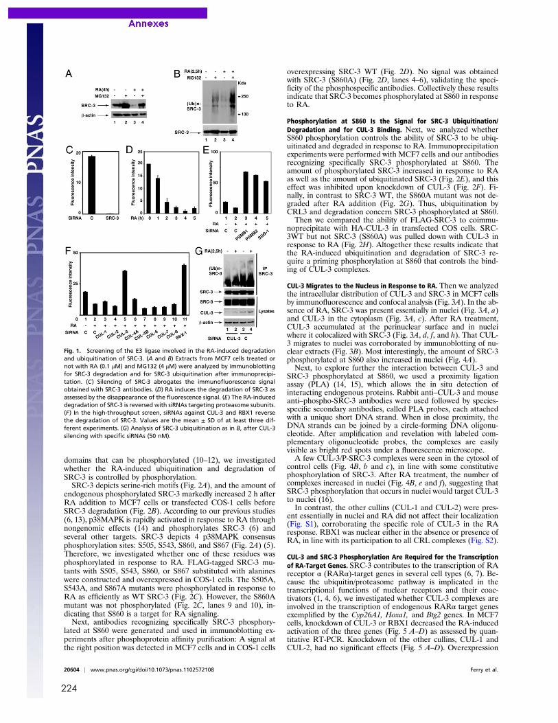
224
domains that can be phosphorylated (10–12), we investigatedwhether the RA-induced ubiquitination and degradation ofSRC-3 is controlled by phosphorylation.SRC-3 depicts serine-rich motifs (Fig. 2A), and the amount of
endogenous phosphorylated SRC-3 markedly increased 2 h afterRA addition to MCF7 cells or transfected COS-1 cells beforeSRC-3 degradation (Fig. 2B). According to our previous studies(6, 13), p38MAPK is rapidly activated in response to RA throughnongenomic effects (14) and phosphorylates SRC-3 (6) andseveral other targets. SRC-3 depicts 4 p38MAPK consensusphosphorylation sites: S505, S543, S860, and S867 (Fig. 2A) (5).Therefore, we investigated whether one of these residues wasphosphorylated in response to RA. FLAG-tagged SRC-3 mu-tants with S505, S543, S860, or S867 substituted with alanineswere constructed and overexpressed in COS-1 cells. The S505A,S543A, and S867A mutants were phosphorylated in response toRA as efficiently as WT SRC-3 (Fig. 2C). However, the S860Amutant was not phosphorylated (Fig. 2C, lanes 9 and 10), in-dicating that S860 is a target for RA signaling.Next, antibodies recognizing specifically SRC-3 phosphory-
lated at S860 were generated and used in immunoblotting ex-periments after phosphoprotein affinity purification: A signal atthe right position was detected in MCF7 cells and in COS-1 cells
overexpressing SRC-3 WT (Fig. 2D). No signal was obtainedwith SRC-3 (S860A) (Fig. 2D, lanes 4–6), validating the speci-ficity of the phosphospecific antibodies. Collectively these resultsindicate that SRC-3 becomes phosphorylated at S860 in responseto RA.
Phosphorylation at S860 Is the Signal for SRC-3 Ubiquitination/Degradation and for CUL-3 Binding. Next, we analyzed whetherS860 phosphorylation controls the ability of SRC-3 to be ubiq-uitinated and degraded in response to RA. Immunoprecipitationexperiments were performed with MCF7 cells and our antibodiesrecognizing specifically SRC-3 phosphorylated at S860. Theamount of phosphorylated SRC-3 increased in response to RAas well as the amount of ubiquitinated SRC-3 (Fig. 2E), and thiseffect was inhibited upon knockdown of CUL-3 (Fig. 2F). Fi-nally, in contrast to SRC-3 WT, the S860A mutant was not de-graded after RA addition (Fig. 2G). Thus, ubiquitination byCRL3 and degradation concern SRC-3 phosphorylated at S860.Then we compared the ability of FLAG-SRC-3 to coimmu-
noprecipitate with HA-CUL-3 in transfected COS cells. SRC-3WT but not SRC-3 (S860A) was pulled down with CUL-3 inresponse to RA (Fig. 2H). Altogether these results indicate thatthe RA-induced ubiquitination and degradation of SRC-3 re-quire a priming phosphorylation at S860 that controls the bind-ing of CUL-3 complexes.
CUL-3 Migrates to the Nucleus in Response to RA. Then we analyzedthe intracellular distribution of CUL-3 and SRC-3 in MCF7 cellsby immunofluorescence and confocal analysis (Fig. 3A). In the ab-sence of RA, SRC-3 was present essentially in nuclei (Fig. 3A, a)and CUL-3 in the cytoplasm (Fig. 3A, c). After RA treatment,CUL-3 accumulated at the perinuclear surface and in nucleiwhere it colocalized with SRC-3 (Fig. 3A, d, f, and h). That CUL-3 migrates to nuclei was corroborated by immunoblotting of nu-clear extracts (Fig. 3B). Most interestingly, the amount of SRC-3phosphorylated at S860 also increased in nuclei (Fig. 4A).Next, to explore further the interaction between CUL-3 and
SRC-3 phosphorylated at S860, we used a proximity ligationassay (PLA) (14, 15), which allows the in situ detection ofinteracting endogenous proteins. Rabbit anti–CUL-3 and mouseanti–phospho-SRC-3 antibodies were used followed by species-specific secondary antibodies, called PLA probes, each attachedwith a unique short DNA strand. When in close proximity, theDNA strands can be joined by a circle-forming DNA oligonu-cleotide. After amplification and revelation with labeled com-plementary oligonucleotide probes, the complexes are easilyvisible as bright red spots under a fluorescence microscope.A few CUL-3/P-SRC-3 complexes were seen in the cytosol of
control cells (Fig. 4B, b and c), in line with some constitutivephosphorylation of SRC-3. After RA treatment, the number ofcomplexes increased in nuclei (Fig. 4B, e and f), suggesting thatSRC-3 phosphorylation that occurs in nuclei would target CUL-3to nuclei (16).In contrast, the other cullins (CUL-1 and CUL-2) were pres-
ent essentially in nuclei and RA did not affect their localization(Fig. S1), corroborating the specific role of CUL-3 in the RAresponse. RBX1 was nuclear either in the absence or presence ofRA, in line with its participation to all CRL complexes (Fig. S2).
CUL-3 and SRC-3 Phosphorylation Are Required for the Transcriptionof RA-Target Genes. SRC-3 contributes to the transcription of RAreceptor α (RARα)-target genes in several cell types (6, 7). Be-cause the ubiquitin/proteasome pathway is implicated in thetranscriptional functions of nuclear receptors and their coac-tivators (1, 4, 6), we investigated whether CUL-3 complexes areinvolved in the transcription of endogenous RARα target genesexemplified by the Cyp26A1, Hoxa1, and Btg2 genes. In MCF7cells, knockdown of CUL-3 or RBX1 decreased the RA-inducedactivation of the three genes (Fig. 5 A–D) as assessed by quan-titative RT-PCR. Knockdown of the other cullins, CUL-1 andCUL-2, had no significant effects (Fig. 5 A–D). Overexpression
Fig. 1. Screening of the E3 ligase involved in the RA-induced degradationand ubiquitination of SRC-3. (A and B) Extracts from MCF7 cells treated ornot with RA (0.1 μM) and MG132 (4 μM) were analyzed by immunoblottingfor SRC-3 degradation and for SRC-3 ubiquitination after immunoprecipi-tation. (C) Silencing of SRC-3 abrogates the immunofluorescence signalobtained with SRC-3 antibodies. (D) RA induces the degradation of SRC-3 asassessed by the disappearance of the fluorescence signal. (E) The RA-induceddegradation of SRC-3 is reversed with siRNAs targeting proteasome subunits.(F) In the high-throughput screen, siRNAs against CUL-3 and RBX1 reversethe degradation of SRC-3. Values are the mean ± SD of at least three dif-ferent experiments. (G) Analysis of SRC-3 ubiquitination as in B, after CUL-3silencing with specific siRNAs (50 nM).
20604 | www.pnas.org/cgi/doi/10.1073/pnas.1102572108 Ferry et al.
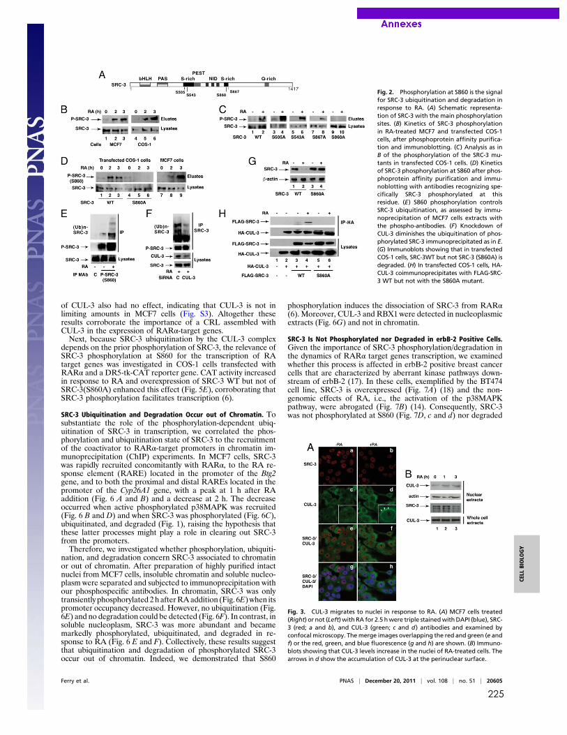
225
of CUL-3 also had no effect, indicating that CUL-3 is not inlimiting amounts in MCF7 cells (Fig. S3). Altogether theseresults corroborate the importance of a CRL assembled withCUL-3 in the expression of RARα-target genes.Next, because SRC-3 ubiquitination by the CUL-3 complex
depends on the prior phosphorylation of SRC-3, the relevance ofSRC-3 phosphorylation at S860 for the transcription of RAtarget genes was investigated in COS-1 cells transfected withRARα and a DR5-tk-CAT reporter gene. CAT activity increasedin response to RA and overexpression of SRC-3 WT but not ofSRC-3(S860A) enhanced this effect (Fig. 5E), corroborating thatSRC-3 phosphorylation facilitates transcription (6).
SRC-3 Ubiquitination and Degradation Occur out of Chromatin. Tosubstantiate the role of the phosphorylation-dependent ubiq-uitination of SRC-3 in transcription, we correlated the phos-phorylation and ubiquitination state of SRC-3 to the recruitmentof the coactivator to RARα-target promoters in chromatin im-munoprecipitation (ChIP) experiments. In MCF7 cells, SRC-3was rapidly recruited concomitantly with RARα, to the RA re-sponse element (RARE) located in the promoter of the Btg2gene, and to both the proximal and distal RAREs located in thepromoter of the Cyp26A1 gene, with a peak at 1 h after RAaddition (Fig. 6 A and B) and a decrease at 2 h. The decreaseoccurred when active phosphorylated p38MAPK was recruited(Fig. 6 B and D) and when SRC-3 was phosphorylated (Fig. 6C),ubiquitinated, and degraded (Fig. 1), raising the hypothesis thatthese latter processes might play a role in clearing out SRC-3from the promoters.Therefore, we investigated whether phosphorylation, ubiquiti-
nation, and degradation concern SRC-3 associated to chromatinor out of chromatin. After preparation of highly purified intactnuclei from MCF7 cells, insoluble chromatin and soluble nucleo-plasm were separated and subjected to immunoprecipitation withour phosphospecific antibodies. In chromatin, SRC-3 was onlytransiently phosphorylated 2h afterRAaddition (Fig. 6E)when itspromoter occupancy decreased. However, no ubiquitination (Fig.6E) and no degradation could be detected (Fig. 6F). In contrast, insoluble nucleoplasm, SRC-3 was more abundant and becamemarkedly phosphorylated, ubiquitinated, and degraded in re-sponse to RA (Fig. 6 E and F). Collectively, these results suggestthat ubiquitination and degradation of phosphorylated SRC-3occur out of chromatin. Indeed, we demonstrated that S860
phosphorylation induces the dissociation of SRC-3 from RARα(6). Moreover, CUL-3 and RBX1 were detected in nucleoplasmicextracts (Fig. 6G) and not in chromatin.
SRC-3 Is Not Phosphorylated nor Degraded in erbB-2 Positive Cells.Given the importance of SRC-3 phosphorylation/degradation inthe dynamics of RARα target genes transcription, we examinedwhether this process is affected in erbB-2 positive breast cancercells that are characterized by aberrant kinase pathways down-stream of erbB-2 (17). In these cells, exemplified by the BT474cell line, SRC-3 is overexpressed (Fig. 7A) (18) and the non-genomic effects of RA, i.e., the activation of the p38MAPKpathway, were abrogated (Fig. 7B) (14). Consequently, SRC-3was not phosphorylated at S860 (Fig. 7D, c and d) nor degraded
Fig. 2. Phosphorylation at S860 is the signalfor SRC-3 ubiquitination and degradation inresponse to RA. (A) Schematic representa-tion of SRC-3 with the main phosphorylationsites. (B) Kinetics of SRC-3 phosphorylationin RA-treated MCF7 and transfected COS-1cells, after phosphoprotein affinity purifica-tion and immunoblotting. (C) Analysis as inB of the phosphorylation of the SRC-3 mu-tants in transfected COS-1 cells. (D) Kineticsof SRC-3 phosphorylation at S860 after phos-phoprotein affinity purification and immu-noblotting with antibodies recognizing spe-cifically SRC-3 phosphorylated at thisresidue. (E) S860 phosphorylation controlsSRC-3 ubiquitination, as assessed by immu-noprecipitation of MCF7 cells extracts withthe phospho-antibodies. (F) Knockdown ofCUL-3 diminishes the ubiquitination of phos-phorylated SRC-3 immunoprecipitated as in E.(G) Immunoblots showing that in transfectedCOS-1 cells, SRC-3WT but not SRC-3 (S860A) isdegraded. (H) In transfected COS-1 cells, HA-CUL-3 coimmunoprecipitates with FLAG-SRC-3 WT but not with the S860A mutant.
Fig. 3. CUL-3 migrates to nuclei in response to RA. (A) MCF7 cells treated(Right) or not (Left) with RA for 2.5 hwere triple stainedwithDAPI (blue), SRC-3 (red; a and b), and CUL-3 (green; c and d) antibodies and examined byconfocal microscopy. Themerge images overlapping the red and green (e andf) or the red, green, and blue fluorescence (g and h) are shown. (B) Immuno-blots showing that CUL-3 levels increase in the nuclei of RA-treated cells. Thearrows in d show the accumulation of CUL-3 at the perinuclear surface.
Ferry et al. PNAS | December 20, 2011 | vol. 108 | no. 51 | 20605
CELL
BIOLO
GY
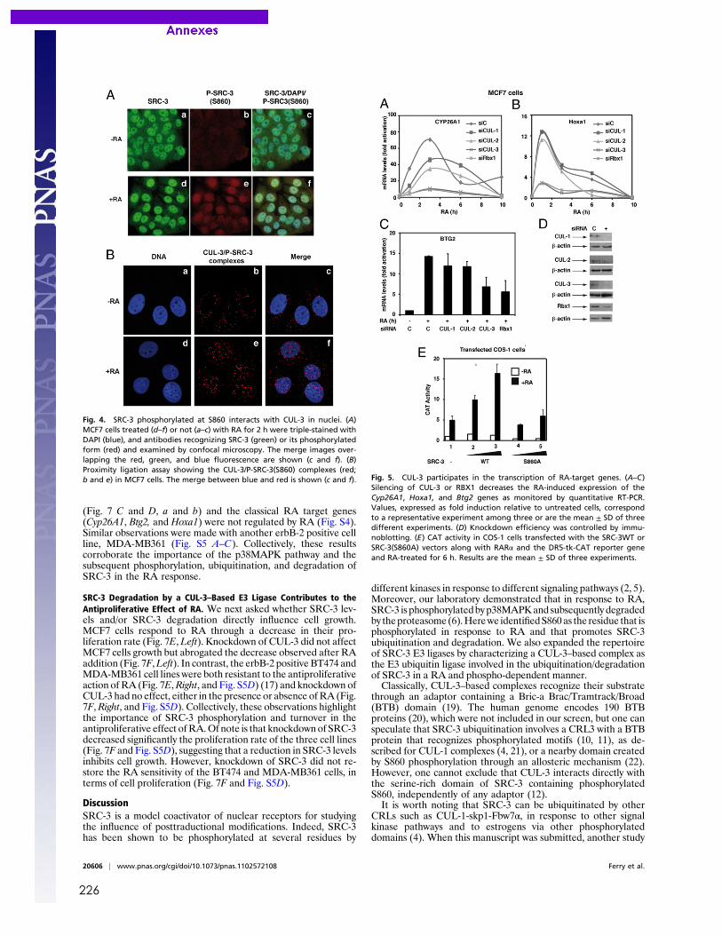
226
(Fig. 7 C and D, a and b) and the classical RA target genes(Cyp26A1, Btg2, and Hoxa1) were not regulated by RA (Fig. S4).Similar observations were made with another erbB-2 positive cellline, MDA-MB361 (Fig. S5 A–C). Collectively, these resultscorroborate the importance of the p38MAPK pathway and thesubsequent phosphorylation, ubiquitination, and degradation ofSRC-3 in the RA response.
SRC-3 Degradation by a CUL-3–Based E3 Ligase Contributes to theAntiproliferative Effect of RA. We next asked whether SRC-3 lev-els and/or SRC-3 degradation directly influence cell growth.MCF7 cells respond to RA through a decrease in their pro-liferation rate (Fig. 7E,Left). Knockdown of CUL-3 did not affectMCF7 cells growth but abrogated the decrease observed after RAaddition (Fig. 7F,Left). In contrast, the erbB-2 positive BT474 andMDA-MB361 cell lines were both resistant to the antiproliferativeaction of RA (Fig. 7E,Right, and Fig. S5D) (17) and knockdown ofCUL-3 had no effect, either in the presence or absence ofRA (Fig.7F,Right, and Fig. S5D). Collectively, these observations highlightthe importance of SRC-3 phosphorylation and turnover in theantiproliferative effect ofRA.Of note is that knockdown of SRC-3decreased significantly the proliferation rate of the three cell lines(Fig. 7F and Fig. S5D), suggesting that a reduction in SRC-3 levelsinhibits cell growth. However, knockdown of SRC-3 did not re-store the RA sensitivity of the BT474 and MDA-MB361 cells, interms of cell proliferation (Fig. 7F and Fig. S5D).
DiscussionSRC-3 is a model coactivator of nuclear receptors for studyingthe influence of posttraductional modifications. Indeed, SRC-3has been shown to be phosphorylated at several residues by
different kinases in response to different signaling pathways (2, 5).Moreover, our laboratory demonstrated that in response to RA,SRC-3 is phosphorylatedbyp38MAPKand subsequently degradedby theproteasome(6).Herewe identifiedS860 as the residue that isphosphorylated in response to RA and that promotes SRC-3ubiquitination and degradation. We also expanded the repertoireof SRC-3 E3 ligases by characterizing a CUL-3–based complex asthe E3 ubiquitin ligase involved in the ubiquitination/degradationof SRC-3 in a RA and phospho-dependent manner.Classically, CUL-3–based complexes recognize their substrate
through an adaptor containing a Bric-a Brac/Tramtrack/Broad(BTB) domain (19). The human genome encodes 190 BTBproteins (20), which were not included in our screen, but one canspeculate that SRC-3 ubiquitination involves a CRL3 with a BTBprotein that recognizes phosphorylated motifs (10, 11), as de-scribed for CUL-1 complexes (4, 21), or a nearby domain createdby S860 phosphorylation through an allosteric mechanism (22).However, one cannot exclude that CUL-3 interacts directly withthe serine-rich domain of SRC-3 containing phosphorylatedS860, independently of any adaptor (12).It is worth noting that SRC-3 can be ubiquitinated by other
CRLs such as CUL-1-skp1-Fbw7α, in response to other signalkinase pathways and to estrogens via other phosphorylateddomains (4). When this manuscript was submitted, another study
Fig. 4. SRC-3 phosphorylated at S860 interacts with CUL-3 in nuclei. (A)MCF7 cells treated (d–f) or not (a–c) with RA for 2 h were triple-stained withDAPI (blue), and antibodies recognizing SRC-3 (green) or its phosphorylatedform (red) and examined by confocal microscopy. The merge images over-lapping the red, green, and blue fluorescence are shown (c and f). (B)Proximity ligation assay showing the CUL-3/P-SRC-3(S860) complexes (red;b and e) in MCF7 cells. The merge between blue and red is shown (c and f). Fig. 5. CUL-3 participates in the transcription of RA-target genes. (A–C)
Silencing of CUL-3 or RBX1 decreases the RA-induced expression of theCyp26A1, Hoxa1, and Btg2 genes as monitored by quantitative RT-PCR.Values, expressed as fold induction relative to untreated cells, correspondto a representative experiment among three or are the mean ± SD of threedifferent experiments. (D) Knockdown efficiency was controlled by immu-noblotting. (E) CAT activity in COS-1 cells transfected with the SRC-3WT orSRC-3(S860A) vectors along with RARα and the DR5-tk-CAT reporter geneand RA-treated for 6 h. Results are the mean ± SD of three experiments.
20606 | www.pnas.org/cgi/doi/10.1073/pnas.1102572108 Ferry et al.
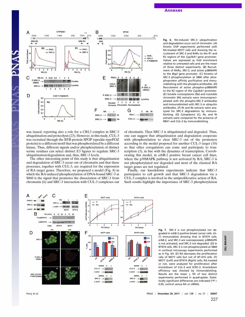
227
was issued, reporting also a role for a CRL3 complex in SRC-3ubiquitination and proteolysis (23). However, in this study, CUL-3was recruited through the BTB protein SPOP (speckle-typePOZprotein) to a differentmotif that was phosphorylated by a differentkinase. Thus, different signals and/or phosphorylation of distinctserine residues can select distinct E3 ligases to regulate SRC-3ubiquitination/degradation and, thus, SRC-3 levels.The other interesting point of this study is that ubiquitination
and degradation of SRC-3 occur out of chromatin and that theseprocesses, together with CUL-3, are required for the expressionof RA target genes. Therefore, we proposed a model (Fig. 8) inwhich the RA-induced phosphorylation of DNA-bound SRC-3 atS860 is the signal that promotes the dissociation of SRC-3 fromchromatin (6) and SRC-3 interaction with CUL-3 complexes out
of chromatin. Then SRC-3 is ubiquitinated and degraded. Thus,one can suggest that ubiquitination and degradation cooperatewith phosphorylation to clear SRC-3 out of the promotersaccording to the model proposed for another CUL-3 target (10)so that other coregulators can come and participate to tran-scription (3), in line with the dynamics of transcription. Corrob-orating this model, in erbB-2 positive breast cancer cell lines,where the p38MAPK pathway is not activated by RA, SRC-3 isnot phosphorylated nor degraded and most of the classical RAtarget genes are not regulated.Finally, our knockdown experiments indicate that SRC-3
participates to cell growth and that SRC-3 degradation via aCUL-3 complex is involved in the antiproliferative action of RA.Such results highlight the importance of SRC-3 phosphorylation
Fig. 6. RA-induced SRC-3 ubiquitinationand degradation occur out of chromatin. (A)Kinetic ChIP experiments performed withRA-treated MCF7 cells and showing the re-cruitment of SRC-3 and RARα to the R1 andR2 regions of the Cyp26A1 gene promoter.Values are expressed as fold enrichmentrelative to untreated cells and are the meanof three distinct experiments. (B) Recruit-ment of RARα, SRC-3, and active p38MAPKto the Btg2 gene promoter. (C) Kinetics ofSRC-3 phosphorylation at S860 after phos-phoprotein affinity purification and immu-noblotting with the phospho-antibodies. (D)Recruitment of active phosphor-p38MAPKto the R2 region of the Cyp26A1 promoter.(E) Soluble nucleoplasmic (Ns) and insolublechromatin (Ni) extracts were immunopreci-pitated with the phospho-SRC-3 antibodiesand immunoblotted with SRC-3 or ubiquitinantibodies. (F) Ni and Ns extracts were ana-lyzed for SRC-3 degradation by immuno-blotting. (G) Cytoplasmic (C), Ns, and Niextracts were compared for the presence ofRBX1 and CUL-3 by immunoblotting.
Fig. 7. SRC-3 is not phosphorylated nor de-graded in erbB-2 positive breast cancer cells. (A–C) Immunoblots showing that in BT474 cells,erbB-2, and SRC-3 are overexpressed, p38MAPKis not activated, and SRC-3 not degraded. (D) InBT474 cells, SRC-3 is not phosphorylated at S860in confocal microscopy experiments performedas in Fig. 4A. (E) RA decreases the proliferationrate of MCF7 cells but not of BT-474 cells. (F)MCF7 (Left) and BT474 (Right) cells, RA-treatedor not, were analyzed for proliferation afterknockdown of CUL-3 and SCRC-3. Knockdownefficiency was checked by immunoblotting.Results are the mean ± SD of two distinctexperiments performed in quadruplate. Statis-tically significant differences are indicated (*P <0.05, control versus RA or siRNA).
Ferry et al. PNAS | December 20, 2011 | vol. 108 | no. 51 | 20607
CELL
BIOLO
GY
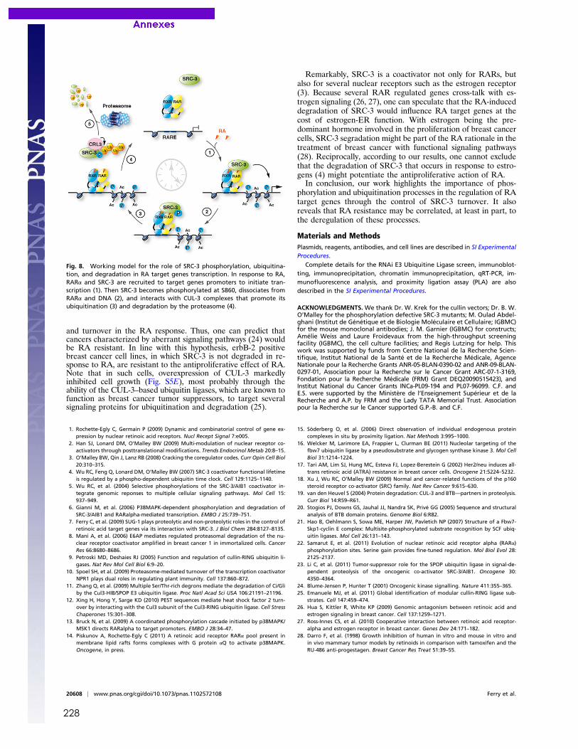
228
and turnover in the RA response. Thus, one can predict thatcancers characterized by aberrant signaling pathways (24) wouldbe RA resistant. In line with this hypothesis, erbB-2 positivebreast cancer cell lines, in which SRC-3 is not degraded in re-sponse to RA, are resistant to the antiproliferative effect of RA.Note that in such cells, overexpression of CUL-3 markedlyinhibited cell growth (Fig. S5E), most probably through theability of the CUL-3–based ubiquitin ligases, which are known tofunction as breast cancer tumor suppressors, to target severalsignaling proteins for ubiquitination and degradation (25).
Remarkably, SRC-3 is a coactivator not only for RARs, butalso for several nuclear receptors such as the estrogen receptor(3). Because several RAR regulated genes cross-talk with es-trogen signaling (26, 27), one can speculate that the RA-induceddegradation of SRC-3 would influence RA target genes at thecost of estrogen-ER function. With estrogen being the pre-dominant hormone involved in the proliferation of breast cancercells, SRC-3 segradation might be part of the RA rationale in thetreatment of breast cancer with functional signaling pathways(28). Reciprocally, according to our results, one cannot excludethat the degradation of SRC-3 that occurs in response to estro-gens (4) might potentiate the antiproliferative action of RA.In conclusion, our work highlights the importance of phos-
phorylation and ubiquitination processes in the regulation of RAtarget genes through the control of SRC-3 turnover. It alsoreveals that RA resistance may be correlated, at least in part, tothe deregulation of these processes.
Materials and MethodsPlasmids, reagents, antibodies, and cell lines are described in SI ExperimentalProcedures.
Complete details for the RNAi E3 Ubiquitine Ligase screen, immunoblot-ting, immunoprecipitation, chromatin immunoprecipitation, qRT-PCR, im-munofluorescence analysis, and proximity ligation assay (PLA) are alsodescribed in the SI Experimental Procedures.
ACKNOWLEDGMENTS. We thank Dr. W. Krek for the cullin vectors; Dr. B. W.O’Malley for the phosphorylation defective SRC-3 mutants; M. Oulad Abdel-ghani (Institut de Génétique et de Biologie Moléculaire et Cellulaire; IGBMC)for the mouse monoclonal antibodies; J. M. Garnier (IGBMC) for constructs;Amélie Weiss and Laure Froidevaux from the high-throughput screeningfacility (IGBMC), the cell culture facilities; and Regis Lutzing for help. Thiswork was supported by funds from Centre National de la Recherche Scien-tifique, Institut National de la Santé et de la Recherche Médicale, AgenceNationale pour la Recherche Grants ANR-05-BLAN-0390-02 and ANR-09-BLAN-0297-01, Association pour la Recherche sur le Cancer Grant ARC-07-1-3169,Fondation pour la Recherche Médicale (FRM) Grant DEQ20090515423), andInstitut National du Cancer Grants INCa-PL09-194 and PL07-96099. C.F. andE.S. were supported by the Ministère de l’Enseignement Supérieur et de laRecherche and A.P. by FRM and the Lady TATA Memorial Trust. Associationpour la Recherche sur le Cancer supported G.P.-B. and C.F.
1. Rochette-Egly C, Germain P (2009) Dynamic and combinatorial control of gene ex-pression by nuclear retinoic acid receptors. Nucl Recept Signal 7:e005.
2. Han SJ, Lonard DM, O’Malley BW (2009) Multi-modulation of nuclear receptor co-activators through posttranslational modifications. Trends Endocrinol Metab 20:8–15.
3. O’Malley BW, Qin J, Lanz RB (2008) Cracking the coregulator codes. Curr Opin Cell Biol20:310–315.
4. Wu RC, Feng Q, Lonard DM, O’Malley BW (2007) SRC-3 coactivator functional lifetimeis regulated by a phospho-dependent ubiquitin time clock. Cell 129:1125–1140.
5. Wu RC, et al. (2004) Selective phosphorylations of the SRC-3/AIB1 coactivator in-tegrate genomic reponses to multiple cellular signaling pathways. Mol Cell 15:937–949.
6. Giannì M, et al. (2006) P38MAPK-dependent phosphorylation and degradation ofSRC-3/AIB1 and RARalpha-mediated transcription. EMBO J 25:739–751.
7. Ferry C, et al. (2009) SUG-1 plays proteolytic and non-proteolytic roles in the control ofretinoic acid target genes via its interaction with SRC-3. J Biol Chem 284:8127–8135.
8. Mani A, et al. (2006) E6AP mediates regulated proteasomal degradation of the nu-clear receptor coactivator amplified in breast cancer 1 in immortalized cells. CancerRes 66:8680–8686.
9. Petroski MD, Deshaies RJ (2005) Function and regulation of cullin-RING ubiquitin li-gases. Nat Rev Mol Cell Biol 6:9–20.
10. Spoel SH, et al. (2009) Proteasome-mediated turnover of the transcription coactivatorNPR1 plays dual roles in regulating plant immunity. Cell 137:860–872.
11. Zhang Q, et al. (2009) Multiple Ser/Thr-rich degrons mediate the degradation of Ci/Gliby the Cul3-HIB/SPOP E3 ubiquitin ligase. Proc Natl Acad Sci USA 106:21191–21196.
12. Xing H, Hong Y, Sarge KD (2010) PEST sequences mediate heat shock factor 2 turn-over by interacting with the Cul3 subunit of the Cul3-RING ubiquitin ligase. Cell StressChaperones 15:301–308.
13. Bruck N, et al. (2009) A coordinated phosphorylation cascade initiated by p38MAPK/MSK1 directs RARalpha to target promoters. EMBO J 28:34–47.
14. Piskunov A, Rochette-Egly C (2011) A retinoic acid receptor RARα pool present inmembrane lipid rafts forms complexes with G protein αQ to activate p38MAPK.Oncogene, in press.
15. Söderberg O, et al. (2006) Direct observation of individual endogenous proteincomplexes in situ by proximity ligation. Nat Methods 3:995–1000.
16. Welcker M, Larimore EA, Frappier L, Clurman BE (2011) Nucleolar targeting of thefbw7 ubiquitin ligase by a pseudosubstrate and glycogen synthase kinase 3. Mol CellBiol 31:1214–1224.
17. Tari AM, Lim SJ, Hung MC, Esteva FJ, Lopez-Berestein G (2002) Her2/neu induces all-trans retinoic acid (ATRA) resistance in breast cancer cells. Oncogene 21:5224–5232.
18. Xu J, Wu RC, O’Malley BW (2009) Normal and cancer-related functions of the p160steroid receptor co-activator (SRC) family. Nat Rev Cancer 9:615–630.
19. van den Heuvel S (2004) Protein degradation: CUL-3 and BTB—partners in proteolysis.Curr Biol 14:R59–R61.
20. Stogios PJ, Downs GS, Jauhal JJ, Nandra SK, Privé GG (2005) Sequence and structuralanalysis of BTB domain proteins. Genome Biol 6:R82.
21. Hao B, Oehlmann S, Sowa ME, Harper JW, Pavletich NP (2007) Structure of a Fbw7-Skp1-cyclin E complex: Multisite-phosphorylated substrate recognition by SCF ubiq-uitin ligases. Mol Cell 26:131–143.
22. Samarut E, et al. (2011) Evolution of nuclear retinoic acid receptor alpha (RARα)phosphorylation sites. Serine gain provides fine-tuned regulation. Mol Biol Evol 28:2125–2137.
23. Li C, et al. (2011) Tumor-suppressor role for the SPOP ubiquitin ligase in signal-de-pendent proteolysis of the oncogenic co-activator SRC-3/AIB1. Oncogene 30:4350–4364.
24. Blume-Jensen P, Hunter T (2001) Oncogenic kinase signalling. Nature 411:355–365.25. Emanuele MJ, et al. (2011) Global identification of modular cullin-RING ligase sub-
strates. Cell 147:459–474.26. Hua S, Kittler R, White KP (2009) Genomic antagonism between retinoic acid and
estrogen signaling in breast cancer. Cell 137:1259–1271.27. Ross-Innes CS, et al. (2010) Cooperative interaction between retinoic acid receptor-
alpha and estrogen receptor in breast cancer. Genes Dev 24:171–182.28. Darro F, et al. (1998) Growth inhibition of human in vitro and mouse in vitro and
in vivo mammary tumor models by retinoids in comparison with tamoxifen and theRU-486 anti-progestagen. Breast Cancer Res Treat 51:39–55.
Fig. 8. Working model for the role of SRC-3 phosphorylation, ubiquitina-tion, and degradation in RA target genes transcription. In response to RA,RARα and SRC-3 are recruited to target genes promoters to initiate tran-scription (1). Then SRC-3 becomes phosphorylated at S860, dissociates fromRARα and DNA (2), and interacts with CUL-3 complexes that promote itsubiquitination (3) and degradation by the proteasome (4).
20608 | www.pnas.org/cgi/doi/10.1073/pnas.1102572108 Ferry et al.
229
Supporting InformationFerry et al. 10.1073/pnas.1102572108SI Materials and MethodsPlasmids and Reagents. The B10-HA-FLAG–tagged SRC-3 andRARα vectors and theDR5-tk-CAT reporter gene were described(1, 2). The expression vectors for SRC-3 S505A, S543A, S860A,and S867A were constructed by cloning the cDNA of the corre-sponding mutants provided by B. W. O’Malley (Baylor College ofMedicine, Houston, TX) into a pTL2 vector containing a FLAG-tag. The pCDNA-based expression vectors for CUL-1, CUL-2,andCUL-3with anHAtagwere kindly provided byW.Krek (SwissFederal Institute of Technology, Zurich, Switzerland).
Antibodies. The rabbit polyclonal antibodies used were againstRARα, SRC-3, β-actin, CUL-1, and CUL-2 (all from Santa CruzBiotechnologies), CUL-3 (Sigma-Aldrich), and phospho-p38MAPK (Thr180/Tyr182) (Cell Signaling). Goat polyclonal anti-bodies against RBX1 were from Santa Cruz Biotechnologies.Mouse monoclonal antibodies were against FLAG-tag (Sigma-Aldrich), HA-Tag, SRC-3 (BD-Bioscience), and mono- andpolyubiquitinated conjugates (Enzo Life Sciences). Mousemonoclonal antibodies recognizing SRC-3 phosphorylated atS860 were generated by immunization of BALB/c mice withsynthetic phosphopeptides (3).
RNAi E3-Ubiquitin Ligase Screen.Screening was performed at the high-throughput screening facility of the Institut de Génétique et deBiologie Moléculaire et Cellulaire, using a siRNA library (4 in-dividual siRNA per protein) targeting 111 E3 Ubiquitin Ligasesand associated proteins (Flexiplate siRNA sets; Qiagen) (DatasetS1). Controls were performed with smartpool siRNAs fromDharmacon (Thermo Scientific) [siRNAs against human SRC-3(M-003759-02) and SUG-1 (M-009484-02) and scramble siRNA]or from Qiagen [siRNAs against CUL-1 (Hs_Cul1_5), CUL-2(Hs_Cul2_3), CUL-3 (Hs_Cul3_5), RBX1 (Hs_RBX1_5),PSMB1 (Hs_PSMB1_2), and PSMB2 (Hs_PSMB2_2)]. Foreach target, 3.5 pmol siRNA was reverse transfected in 5,000MCF7 cells per 0.3 cm2 by using Interferin (Polyplus).All screens were performed in 96-well cell culture microplates
with a particular focus on avoidingmicroplate edge effects. SRC-3degradation was analyzed by immunofluorescence 3 d aftersiRNA transfection and 3 h after RA addition. Cells were washed,fixed with 3% paraformaldehyde, permeabilized with 0.1%TritonX-100, blocked with 2% BSA, and incubated with SRC-3 anti-bodies followed by ALEXAFluor 488-conjugated second anti-
bodies (Invitrogen). The screens were achieved owing toa TECAN robotic station (for cell transfection, staining, andimmunocytochemistry) and to a Caliper Twister II robotic armcoupling microplate stacks to the InCELL1000 analyzer micro-scope (GE LifeSciences).
Statistical Analysis of the siRNA Screens. After background-cor-rection and interplate normalization, internal positive and neg-ative plate controls were used to determine a threshold for cellclassification. The distance from negative controls was quantifiedby determining the percent of control (i.e., the percentage ofpositive cells in the test experiment versus the negative control).After multiple testing correction, P values were determined.Then gene selection was based on the one-sided tests procedurewith H0: Xp ≤ α and H1: Xp > α, where Xp is the percent ofcontrol estimator and α is a sensibility parameter (α ≥ 1 and α =2 for high stringency).
Cell Lines, Cell Proliferation, Transfection, Immunoprecipitation, andImmunoblotting. COS-1, MCF7, BT474, and MDA-MB361 cellswere cultured and transiently transfected under standard con-ditions. After RA (10−7 M) addition (4), whole-cell extracts orsubcellular fractions (cytosol, nucleoplasm, and chromatin) wereprepared, immunoblotted, and immunoprecipitated (2, 3). Cellproliferation was analyzed by using the XTT (2,3-bis-(2-methoxy-4-nitro-5-sulfophenyl)-2H-tetrazolium-5-carboxanilide) assay kit(Roche Diagnostics).
Detection of in Vivo Phosphorylated SRC-3. Cell extracts were ap-plied to phosphoprotein purification columns (Qiagen). Columneluates containing protein peaks were concentrated and analyzedby immunoblotting (4).
Immunofluorescence Analysis and Proximity Ligation Assay (PLA).Cells were seeded onto labtek chamberslides (Fisher Scien-tific), proceeded as described for the RNAi screen, and incubatedwith the primary antibodies followed by ALEXAFluor 448- or555-conjugated secondary antibodies or by the PLA probes(Duolink II; Eurogentec) as described (5). Images were edited byusing ImageJ.RNA isolation, quantitative RT-PCR, and chromatin immu-
noprecipitations (ChIP) were performed as described (2, 4).Primer sequences are available upon request.
1. Giannì M, et al. (2006) P38MAPK-dependent phosphorylation and degradation of SRC-3/AIB1 and RARalpha-mediated transcription. EMBO J 25:739e751.
2. Ferry C, et al. (2009) SUG-1 plays proteolytic and non-proteolytic roles in the control ofretinoic acid target genes via its interaction with SRC-3. J Biol Chem 284:8127e8135.
3. Lalevée S, et al. (2010) Vinexinß, an atypical “sensor” of retinoic acid receptor gammasignaling: Union and sequestration, separation, and phosphorylation. FASEB J 24:4523e4534.
4. Bruck N, et al. (2009) A coordinated phosphorylation cascade initiated by p38MAPK/MSK1 directs RARalpha to target promoters. EMBO J 28:34e47.
5. Piskunov A, Rochette-Egly C (2011) A retinoic acid receptor RARα pool present inmembrane lipid rafts forms complexes with G protein αQ to activate p38MAPK.Oncogene, in press.
Ferry et al. www.pnas.org/cgi/content/short/1102572108 1 of 4
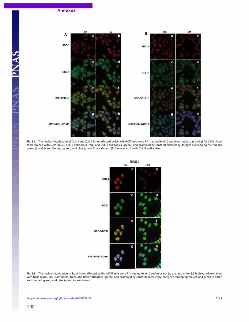
230
Fig. S1. The nuclear localization of CUL-1 and CUL-2 is not affected by RA. (A) MCF7 cells were RA-treated (b, d, f, and h) or not (a, c, e, and g) for 2.5 h, fixed,triple stained with DAPI (blue), SRC-3 antibodies (red), and CUL-1 antibodies (green), and examined by confocal microscopy. Merges overlapping the red andgreen (e and f) and the red, green, and blue (g and h) are shown. (B) Same as in A with CUL-2 antibodies.
Fig. S2. The nuclear localization of Rbx1 is not affected by RA. MCF7 cells were RA-treated (b, d, f, and h) or not (a, c, e, and g) for 2.5 h, fixed, triple stainedwith DAPI (blue), SRC-3 antibodies (red), and Rbx1 antibodies (green), and examined by confocal microscopy. Merges overlapping the red and green (e and f)and the red, green, and blue (g and h) are shown.
Ferry et al. www.pnas.org/cgi/content/short/1102572108 2 of 4
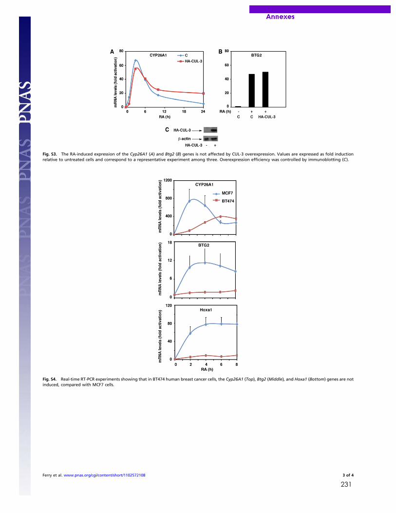
231
Fig. S3. The RA-induced expression of the Cyp26A1 (A) and Btg2 (B) genes is not affected by CUL-3 overexpression. Values are expressed as fold inductionrelative to untreated cells and correspond to a representative experiment among three. Overexpression efficiency was controlled by immunoblotting (C).
Fig. S4. Real-time RT-PCR experiments showing that in BT474 human breast cancer cells, the Cyp26A1 (Top), Btg2 (Middle), and Hoxa1 (Bottom) genes are notinduced, compared with MCF7 cells.
Ferry et al. www.pnas.org/cgi/content/short/1102572108 3 of 4
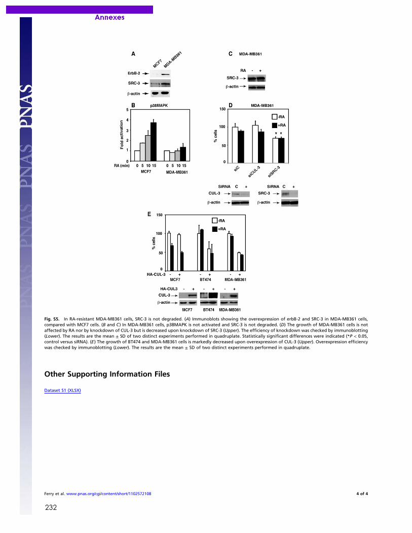
232
Fig. S5. In RA-resistant MDA-MB361 cells, SRC-3 is not degraded. (A) Immunoblots showing the overexpression of erbB-2 and SRC-3 in MDA-MB361 cells,compared with MCF7 cells. (B and C) In MDA-MB361 cells, p38MAPK is not activated and SRC-3 is not degraded. (D) The growth of MDA-MB361 cells is notaffected by RA nor by knockdown of CUL-3 but is decreased upon knockdown of SRC-3 (Upper). The efficiency of knockdown was checked by immunoblotting(Lower). The results are the mean ± SD of two distinct experiments performed in quadruplate. Statistically significant differences were indicated (*P < 0.05,control versus siRNA). (E) The growth of BT474 and MDA-MB361 cells is markedly decreased upon overexpression of CUL-3 (Upper). Overexpression efficiencywas checked by immunoblotting (Lower). The results are the mean ± SD of two distinct experiments performed in quadruplate.
Other Supporting Information Files
Dataset S1 (XLSX)
Ferry et al. www.pnas.org/cgi/content/short/1102572108 4 of 4
233
Bibliographie
Abu-Abed, S., Dolle, P., Metzger, D., Beckett, B., Chambon, P., and Petkovich, M. (2001). The retinoic acid-metabolizing enzyme, CYP26A1, is essential for normal hindbrain patterning, vertebral identity, and development of posterior structures. Genes Dev 15, 226-240. Acampora, D., Merlo, G.R., Paleari, L., Zerega, B., Postiglione, M.P., Mantero, S., Bober, E., Barbieri, O., Simeone, A., and Levi, G. (1999). Craniofacial, vestibular and bone defects in mice lacking the Distal-less-related gene Dlx5. Development 126, 3795-3809. Adam-Stitah, S., Penna, L., Chambon, P., and Rochette-Egly, C. (1999). Hyperphosphorylation of the retinoid X receptor alpha by activated c-Jun NH2-terminal kinases. J Biol Chem 274, 18932-18941. Al Tanoury, Z., Piskunov, A., and Rochette-Egly, C. (2013). Vitamin A and retinoid signaling: genomic and nongenomic effects. J Lipid Res 54, 1761-1775. Albalat, R. (2009). The retinoic acid machinery in invertebrates: ancestral elements and vertebrate innovations. Mol Cell Endocrinol 313, 23-35. Alsayed, Y., Uddin, S., Mahmud, N., Lekmine, F., Kalvakolanu, D.V., Minucci, S., Bokoch, G., and Platanias, L.C. (2001). Activation of Rac1 and the p38 mitogen-activated protein kinase pathway in response to all-trans-retinoic acid. J Biol Chem 276, 4012-4019. Aoto, J., Nam, C.I., Poon, M.M., Ting, P., and Chen, L. (2008). Synaptic signaling by all-trans retinoic acid in homeostatic synaptic plasticity. Neuron 60, 308-320. Baker, R.E., Schnell, S., and Maini, P.K. (2006). A clock and wavefront mechanism for somite formation. Dev Biol 293, 116-126. Bastien, J., Adam-Stitah, S., Plassat, J.L., Chambon, P., and Rochette-Egly, C. (2002). The phosphorylation site located in the A region of retinoic X receptor alpha is required for the antiproliferative effect of retinoic acid (RA) and the activation of RA target genes in F9 cells. Journal of Biological Chemistry 277, 28683-28689. Bastien, J., Adam-Stitah, S., Riedl, T., Egly, J.M., Chambon, P., and Rochette-Egly, C. (2000). TFIIH interacts with the retinoic acid receptor gamma and phosphorylates its AF-1-activating domain through cdk7. Journal of Biological Chemistry 275, 21896-21904. Bastien, J., Plassat, J.L., Payrastre, B., and C., R.-E. (2006). The phosphoinositide 3-kinase/Akt pathway is essential for the retinoic acid-induced differentiation of F9 cells. Oncogene 25, 2040-2047.
234
Begemann, G., and Ingham, P.W. (2000). Developmental regulation of Tbx5 in zebrafish embryogenesis. Mech Dev 90, 299-304. Bentzinger, C.F., Wang, Y.X., and Rudnicki, M.A. (2012). Building muscle: molecular regulation of myogenesis. Cold Spring Harb Perspect Biol 4. Bertrand, S., Thisse, B., Tavares, R., Sachs, L., Chaumot, A., Bardet, P.L., Escriva, H., Duffraisse, M., Marchand, O., Safi, R., et al. (2007). Unexpected novel relational links uncovered by extensive developmental profiling of nuclear receptor expression. PLoS Genet 3, e188. Beverdam, A., Merlo, G.R., Paleari, L., Mantero, S., Genova, F., Barbieri, O., Janvier, P., and Levi, G. (2002). Jaw transformation with gain of symmetry after Dlx5/Dlx6 inactivation: mirror of the past? Genesis 34, 221-227. Bibel, M., Richter, J., Lacroix, E., and Barde, Y.A. (2007). Generation of a defined and uniform population of CNS progenitors and neurons from mouse embryonic stem cells. Nat Protoc 2, 1034-1043. Biddie, S.C., John, S., and Hager, G.L. (2010). Genome-wide mechanisms of nuclear receptor action. Trends Endocrinol Metab 21, 3-9. Bielska, A.A., and Zondlo, N.J. (2006). Hyperphosphorylation of tau induces local polyproline II helix. Biochemistry 45, 5527-5537. Blentic, A., Gale, E., and Maden, M. (2003). Retinoic acid signalling centres in the avian embryo identified by sites of expression of synthesising and catabolising enzymes. Dev Dyn 227, 114-127. Bloom, S., Ledon-Rettig, C., Infante, C., Everly, A., Hanken, J., and Nascone-Yoder, N. (2013). Developmental origins of a novel gut morphology in frogs. Evol Dev 15, 213-223. Boch, J. (2011). TALEs of genome targeting. Nat Biotechnol 29, 135-136. Bour, G., Gaillard, E., Bruck, N., Lalevee, S., Plassat, J.L., Busso, D., Samama, J.P., and Rochette-Egly, C. (2005a). Cyclin H binding to the RAR{alpha} activation function (AF)-2 domain directs phosphorylation of the AF-1 domain by cyclin-dependent kinase 7. Proc Natl Acad Sci U S A 102, 16608-16613. Bour, G., Plassat, J.L., Bauer, A., Lalevee, S., and Rochette-Egly, C. (2005b). Vinexin beta interacts with the non-phosphorylated AF-1 domain of retinoid receptor gamma (RARgamma) and represses RARgamma-mediated transcription. J Biol Chem 280, 17027-17037. Bourguet, W., Germain, P., and Gronemeyer, H. (2000). Nuclear receptor ligand-binding domains: three-dimensional structures, molecular interactions and pharmacological implications. Trends Pharmacol Sci 21, 381-388.
235
Bourguet, W., Ruff, M., Chambon, P., Gronemeyer, H., and Moras, D. (1995). Crystal structure of the ligand-binding domain of the human nuclear receptor RXR-alpha. Nature 375, 377-382. Brelivet, Y., Kammerer, S., Rochel, N., Poch, O., and Moras, D. (2004). Signature of the oligomeric behaviour of nuclear receptors at the sequence and structural level. Embo Reports 5, 423-429. Britz, R., Conway, K.W., and Ruber, L. (2009). Spectacular morphological novelty in a miniature cyprinid fish, Danionella dracula n. sp. Proc Biol Sci 276, 2179-2186. Bruck, N., Bastien, J., Bour, G., Tarrade, A., Plassat, J.L., Bauer, A., Adam-Stitah, S., and Rochette-Egly, C. (2005). Phosphorylation of the Retinoid X Receptor at the Omega loop, modulates the expression of retinoic-acid-target genes with a promoter context specificity. Cellular Signalling 17, 1229-1239. Bruck, N., Vitoux, D., Ferry, C., Duong, V., Bauer, A., de The, H., and Rochette-Egly, C. (2009). A coordinated phosphorylation cascade initiated by p38MAPK/MSK1 directs RARalpha to target promoters. Embo J 28, 34-47. Campo-Paysaa, F., Marletaz, F., Laudet, V., and Schubert, M. (2008). Retinoic acid signaling in development: tissue-specific functions and evolutionary origins. Genesis 46, 640-656. Cañestro, C., Postlethwait, J.H., Gonzàlez-Duarte, R., and Albalat, R. (2006). Is retinoic acid genetic machinery a chordate innovation? Evol Dev 8, 394-406. Carroll, J.S., Liu, X.S., Brodsky, A.S., Li, W., Meyer, C.A., Szary, A.J., Eeckhoute, J., Shao, W., Hestermann, E.V., Geistlinger, T.R., et al. (2005). Chromosome-wide mapping of estrogen receptor binding reveals long-range regulation requiring the forkhead protein FoxA1. Cell 122, 33-43. Carroll, J.S., Meyer, C.A., Song, J., Li, W., Geistlinger, T.R., Eeckhoute, J., Brodsky, A.S., Keeton, E.K., Fertuck, K.C., Hall, G.F., et al. (2006). Genome-wide analysis of estrogen receptor binding sites. Nat Genet 38, 1289-1297. Carroll, S.B. (2005). Endless forms most beautiful: The new science of evo devo and the making of the animal kingdom. (Weidenfeld). Carroll, S.B. (2008). Evo-devo and an expanding evolutionary synthesis: a genetic theory of morphological evolution. Cell 134, 25-36. Carroll, S.B., Grenier, J., and Weatherbee, S.D. (2004). From DNA to Diversity: Molecular Genetics and the Evolution of Animal Design. Chakravarti, D. (2009). Volume 87: Regulatory Mechanisms in Transcriptional Signaling, 1st Edition, Elsevier edn.
236
Chakravarti, D., LaMorte, V.J., Nelson, M.C., Nakajima, T., Schulman, I.G., Juguilon, H., Montminy, M., and Evans, R.M. (1996). Role of CBP/P300 in nuclear receptor signalling. Nature 383, 99-103. Chambers, D., Wilson, L., Maden, M., and Lumsden, A. (2007). RALDH-independent generation of retinoic acid during vertebrate embryogenesis by CYP1B1. Development 134, 1369-1383. Chambon, P. (1996). A decade of molecular biology ot retinoic acid receptors. FASEBJ 10, 940-954. Chan, Y.F., Marks, M.E., Jones, F.C., Villarreal, G., Jr., Shapiro, M.D., Brady, S.D., Southwick, A.M., Absher, D.M., Grimwood, J., Schmutz, J., et al. (2010). Adaptive evolution of pelvic reduction in sticklebacks by recurrent deletion of a Pitx1 enhancer. Science 327, 302-305. Chandra, V., Huang, P., Hamuro, Y., Raghuram, S., Wang, Y., Burris, T.P., and Rastinejad, F. (2008). Structure of the intact PPAR-gamma-RXR- nuclear receptor complex on DNA. Nature 456, 350-356. Chang, C.Y., Norris, J.D., Gron, H., Paige, L.A., Hamilton, P.T., Kenan, D.J., Fowlkes, D., and McDonnell, D.P. (1999). Dissection of the LXXLL nuclear receptor-coactivator interaction motif using combinatorial peptide libraries: Discovery of peptide antagonists of estrogen receptors alpha and beta. Molecular and Cellular Biology 19, 8226-8239. Charite, J., McFadden, D.G., Merlo, G., Levi, G., Clouthier, D.E., Yanagisawa, M., Richardson, J.A., and Olson, E.N. (2001). Role of Dlx6 in regulation of an endothelin-1-dependent, dHAND branchial arch enhancer. Genes Dev 15, 3039-3049. Chatonnet, F., Guyot, R., Benoit, G., and Flamant, F. (2013). Genome-wide analysis of thyroid hormone receptors shared and specific functions in neural cells. Proc Natl Acad Sci U S A 110, E766-775. Chebaro, Y., Amal, I., Rochel, N., Rochette-Egly, C., Stote, R.H., and Dejaegere, A. (2013). Phosphorylation of the retinoic acid receptor alpha induces a mechanical allosteric regulation and changes in internal dynamics. PLoS Comput Biol 9, e1003012. Chen, H., Howald, W.N., and Juchau, M.R. (2000). Biosynthesis of all-trans-retinoic acid from all-trans-retinol: catalysis of all-trans-retinol oxidation by human P-450 cytochromes. Drug Metab Dispos 28, 315-322. Chen, N., and Napoli, J.L. (2008). All-trans-retinoic acid stimulates translation and induces spine formation in hippocampal neurons through a membrane-associated RARalpha. Faseb J 22, 236-245.
237
Chen, N., Onisko, B., and Napoli, J.L. (2008). The nuclear transcription factor RARalpha associates with neuronal RNA granules and suppresses translation. J Biol Chem 283, 20841-20847. Cohen, R.N., Brzostek, S., Kim, B., Chorev, M., Wondisford, F.E., and Hollenberg, A.N. (2001). The specificity of interactions between nuclear hormone receptors and corepressors is mediated by distinct amino acid sequences within the interacting domains. Molecular Endocrinology 15, 1049-1061. Collop, A.H., Broomfield, J.A.S., Chandraratna, R.A.S., Yong, Z., Deimling, S.J., Kolker, S.J., Weeks, D.L., and Drysdale, T.A. (2006). Retinoic acid signaling is essential for formation of the heart tube in Xenopus. Developmental Biology 291, 96-109. Cooke, J., and Zeeman, E.C. (1976). A clock and wavefront model for control of the number of repeated structures during animal morphogenesis. J Theor Biol 58, 455-476. Corcoran, J., Shroot, B., Pizzey, J., and Maden, M. (2000). The role of retinoic acid receptors in neurite outgrowth from different populations of embryonic mouse dorsal root ganglia. J Cell Sci 113 ( Pt 14), 2567-2574. Darimont, B.D., Wagner, R.L., Apriletti, J.W., Stallcup, M.R., Kushner, P.J., Baxter, J.D., Fletterick, R.J., and Yamamoto, K.R. (1998). Structure and specificity of nuclear receptor-coactivator interactions. Genes Dev 12, 3343-3356. de The, H., Vivanco-Ruiz, M.M., Tiollais, P., Stunnenberg, H., and Dejean, A. (1990). Identification of a retinoic acid responsive element in the retinoic acid receptor beta gene. Nature 343, 177-180. Dehal, P., and Boore, J.L. (2005). Two rounds of whole genome duplication in the ancestral vertebrate. PLoS Biol 3, e314. Delacroix, L., Moutier, E., Altobelli, G., Legras, S., Poch, O., Choukrallah, M.A., Bertin, I., Jost, B., and Davidson, I. (2010). Cell-specific interaction of retinoic acid receptors with target genes in mouse embryonic fibroblasts and embryonic stem cells. Mol Cell Biol 30, 231-244. Demarest, S.J., Martinez-Yamout, M., Chung, J., Chen, H., Xu, W., Dyson, H.J., Evans, R.M., and Wright, P.E. (2002). Mutual synergistic folding in recruitment of CBP/p300 by p160 nuclear receptor coactivators. Nature 415, 549-553. Denuce, J.M. (1991). [Teratogenic and metamorphosis inhibiting activity of retinoic acid in Ciona intestinalis]. Z Naturforsch C 46, 1094-1100. Depew, M.J., Simpson, C.A., Morasso, M., and Rubenstein, J.L. (2005). Reassessing the Dlx code: the genetic regulation of branchial arch skeletal pattern and development. J Anat 207, 501-561.
238
Diez del Corral, R., Olivera-Martinez, I., Goriely, A., Gale, E., Maden, M., and Storey, K. (2003). Opposing FGF and retinoid pathways control ventral neural pattern, neuronal differentiation, and segmentation during body axis extension. Neuron 40, 65-79. Dilworth, F.J., and Chambon, P. (2001). Nuclear receptors coordinate the activities of chromatin remodeling complexes and coactivators to facilitate initiation of transcription. Oncogene 20, 3047-3054. Dmetrichuk, J.M., Carlone, R.L., and Spencer, G.E. (2006). Retinoic acid induces neurite outgrowth and growth cone turning in invertebrate neurons. Dev Biol 294, 39-49. Duboc, V., and Logan, M.P. (2011). Regulation of limb bud initiation and limb-type morphology. Dev Dyn 240, 1017-1027. Duboc, V., Röttinger, E., Lapraz, F., Besnardeau, L., and Lepage, T. (2005). Left-right asymmetry in the sea urchin embryo is regulated by nodal signaling on the right side. Dev Cell 9, 147-158. Duboule, D., and Dolle, P. (1989). The Structural and Functional-Organization of the Murine Hox Gene Family Resembles That of Drosophila Homeotic Genes. Embo Journal 8, 1497-1505. Dubrulle, J., McGrew, M.J., and Pourquie, O. (2001). FGF signaling controls somite boundary position and regulates segmentation clock control of spatiotemporal Hox gene activation. Cell 106, 219-232. Dubrulle, J., and Pourquie, O. (2004). Coupling segmentation to axis formation. Development 131, 5783-5793. Dupe, V., Davenne, M., Brocard, J., Dolle, P., Mark, M., Dierich, A., Chambon, P., and Rijli, F.M. (1997). In vivo functional analysis of the Hoxa-1 3' retinoic acid response element (3'RARE). Development 124, 399-410. Dupe, V., and Lumsden, A. (2001). Hindbrain patterning involves graded responses to retinoic acid signalling. Development 128, 2199-2208. Durand, B., Saunders, M., Leroy, P., Leid, M., and Chambon, P. (1992). All-trans and 9-cis retinoic acid induction of CRABPII transcription is mediated by RAR-RXR heterodimers bound to DR1 and DR2 repeated motifs. Cell 71, 73-85. Durston, A.J., Timmermans, J.P., Hage, W.J., Hendriks, H.F., de Vries, N.J., Heideveld, M., and Nieuwkoop, P.D. (1989). Retinoic acid causes an anteroposterior transformation in the developing central nervous system. Nature 340, 140-144. Dyson, H.J., and Wright, P.E. (2005). Intrinsically unstructured proteins and their functions. Nature Reviews Molecular Cell Biology 6, 197-208.
239
Eeckhoute, J., Carroll, J.S., Geistlinger, T.R., Torres-Arzayus, M.I., and Brown, M. (2006). A cell-type-specific transcriptional network required for estrogen regulation of cyclin D1 and cell cycle progression in breast cancer. Genes Dev 20, 2513-2526. Emoto, Y., Wada, H., Okamoto, H., Kudo, A., and Imai, Y. (2005). Retinoic acid-metabolizing enzyme Cyp26a1 is essential for determining territories of hindbrain and spinal cord in zebrafish. Dev Biol 278, 415-427. Epping, M.T., Wang, L.M., Edel, M.J., Carlee, L., Hernandez, M., and Bernards, R. (2005). The human tumor antigen repressor of retinoic acid PRAME is a dominant receptor signaling. Cell 122, 835-847. Erickson, T., Pillay, L.M., and Waskiewicz, A.J. (2011). Zebrafish Tshz3b Negatively Regulates Hox Function in the Developing Hindbrain. Genesis 49, 725-742. Escriva, H., Bertrand, S., Germain, P., Robinson-Rechavi, M., Umbhauer, M., Cartry, J., Duffraisse, M., Holland, L., Gronemeyer, H., and Laudet, V. (2006). Neofunctionalization in vertebrates: the example of retinoic acid receptors. PLoS Genet 2, e102. Escriva, H., Holland, N.D., Gronemeyer, H., Laudet, V., and Holland, L.Z. (2002). The retinoic acid signaling pathway regulates anterior/posterior patterning in the nerve cord and pharynx of amphioxus, a chordate lacking neural crest. Development 129, 2905-2916. Farboud, B., and Privalsky, M.L. (2004). Retinoic acid receptor-alpha is stabilized in a repressive state by its C-terminal, isotype-specific F domain. Molecular Endocrinology 18, 2839-2853. Farooqui, M., Franco, P.J., Thompson, J., Kagechika, H., Chandraratna, R.A., Banaszak, L., and Wei, L.N. (2004). Effects of retinoid ligands on RIP140: molecular interaction with retinoid receptors and biological activity. . Biochemistry 42, 971-979. Feng, L., Hernandez, R.E., Waxman, J.S., Yelon, D., and Moens, C. (2010). Dhrs3a regulates retinoic acid biosynthesis through a feedback inhibition mechanism. Dev Biol 338, 1-14. Fernandes, I., Bastien, Y., Wai, T., Nygard, K., Lin, R., Cormier, O., Lee, H.S., Eng, F., Bertos, N.R., Pelletier, N., et al. (2003). Ligand-dependent nuclear receptor corepressor LCoR functions by histone deacetylase-dependent and -independent mechanisms. Mol Cell 11, 139-150. Ferry, C., Gaouar, S., Fischer, B., Boeglin, M., Paul, N., Samarut, E., Piskunov, A., Pankotai-Bodo, G., Brino, L., and Rochette-Egly, C. (2011). Cullin 3 mediates SRC-3 ubiquitination and degradation to control the retinoic acid response. Proc Natl Acad Sci U S A 108, 20603-20608.
240
Ferry, C., Gianni, M., Lalevee, S., Bruck, N., Plassat, J.L., Raska, I., Jr., Garattini, E., and Rochette-Egly, C. (2009). SUG-1 plays proteolytic and non-proteolytic roles in the control of retinoic acid target genes via its interaction with SRC-3. J Biol Chem 284, 8127-8135. Flajollet, S., Lefebvre, B., Rachez, C., and Lefebvre, P. (2006). Distinct roles of the steroid receptor coactivator 1 and of MED1 in retinoid-induced transcription and cellular differentiation. J Biol Chem 281, 20338-20348. Force, A., Lynch, M., Pickett, F.B., Amores, A., Yan, Y.L., and Postlethwait, J. (1999). Preservation of duplicate genes by complementary, degenerative mutations. Genetics 151, 1531-1545. Freedman, L.P., Luisi, B.F., Korszun, Z.R., Basavappa, R., Sigler, P.B., and Yamamoto, K.R. (1988). The function and structure of the metal coordination sites within the glucocorticoid receptor DNA binding domain. Nature 334, 543-546. Gaillard, E., Bruck, N., Brelivet, Y., Bour, G., Lalevee, S., Bauer, A., Poch, O., Moras, D., and Rochette-Egly, C. (2006). Phosphorylation by PKA potentiates retinoic acid receptor alpha activity by means of increasing interaction with and phosphorylation by cyclin H/cdk7. Proc Natl Acad Sci U S A 103, 9548-9553. Garcia-Fernandez, J., and Holland, P.W. (1994). Archetypal organization of the amphioxus Hox gene cluster. Nature 370, 563-566. Germain, P., Altucci, L., Bourguet, W., Rochette-Egly, C., and Gronemeyer, H. (2003). Nuclear receptor superfamily: Principles of signaling. In Pure Appl Chem,, J. Miyamoto, and J. Burger, eds., pp. 1619-1664. Germain, P., Chambon, P., Eichele, G., Evans, R.M., Lazar, M.A., Leid, M., De Lera, A.R., Lotan, R., Mangelsdorf, D.J., and Gronemeyer, H. (2006a). International Union of Pharmacology. LX. Retinoic acid receptors. Pharmacol Rev 58, 712-725. Germain, P., Chambon, P., Eichele, G., Evans, R.M., Lazar, M.A., Leid, M., De Lera, A.R., Lotan, R., Mangelsdorf, D.J., and Gronemeyer, H. (2006b). International Union of Pharmacology. LXIII. Retinoid X receptors. Pharmacol Rev 58, 760-772. Gettemans, J., Van Impe, K., Delanote, V., Hubert, T., Vandekerckhove, J., and De Corte, V. (2005). Nuclear actin-binding proteins as modulators of gene transcription. Traffic 6, 847-857. Gianni, M., Bauer, A., Garattini, E., Chambon, P., and Rochette-Egly, C. (2002). Phosphorylation by p38MAPK and recruitment of SUG-1 are required for RA-indced RARγ degradation and transactivation. EMBO J 21, 3760-3769. Gianni, M., Tarrade, A., Nigro, E.A., Garattini, E., and Rochette-Egly, C. (2003). The AF-1 and AF-2 domains of RAR gamma 2 and RXR alpha cooperate for triggering the transactivation and the degradation of RAR gamma 2/RXR alpha heterodimers. J Biol Chem 278, 34458-34466.
241
Gibert, Y., Bernard, L., Debiais-Thibaud, M., Bourrat, F., Joly, J.S., Pottin, K., Meyer, A., Retaux, S., Stock, D.W., Jackman, W.R., et al. (2010). Formation of oral and pharyngeal dentition in teleosts depends on differential recruitment of retinoic acid signaling. FASEB J 24, 3298-3309. Glover, J.C., Renaud, J.S., and Rijli, F.M. (2006). Retinoic acid and hindbrain patterning. J Neurobiol 66, 705-725. Godsave, S.F., Koster, C.H., Getahun, A., Mathu, M., Hooiveld, M., van der Wees, J., Hendriks, J., and Durston, A.J. (1998). Graded retinoid responses in the developing hindbrain. Dev Dyn 213, 39-49. Goldbeter, A., and Pourquie, O. (2008). Modeling the segmentation clock as a network of coupled oscillations in the Notch, Wnt and FGF signaling pathways. J Theor Biol 252, 574-585. Gompel, N., Prud'homme, B., Wittkopp, P.J., Kassner, V.A., and Carroll, S.B. (2005). Chance caught on the wing: cis-regulatory evolution and the origin of pigment patterns in Drosophila. Nature 433, 481-487. Graham, A., Papalopulu, N., and Krumlauf, R. (1989). The Murine and Drosophila Homeobox Gene Complexes Have Common Features of Organization and Expression. Cell 57, 367-378. Grandel, H., Lun, K., Rauch, G.J., Rhinn, M., Piotrowski, T., Houart, C., Sordino, P., Kuchler, A.M., Schulte-Merker, S., Geisler, R., et al. (2002). Retinoic acid signalling in the zebrafish embryo is necessary during pre-segmentation stages to pattern the anterior-posterior axis of the CNS and to induce a pectoral fin bud. Development 129, 2851-2865. Grandel, H., and Schulte-Merker, S. (1998). The development of the paired fins in the zebrafish (Danio rerio). Mech Dev 79, 99-120. Grant, P.R., and Grant, B.R. (2011). How and Why Species Multiply: The Radiation of Darwin's Finches. Gronemeyer, H., Gustafsson, J.A., and Laudet, V. (2004). Principles for modulation of the nuclear receptor superfamily. Nat Rev Drug Discov 3, 950-964. Gu, P., LeMenuet, D., Chung, A.C., Mancini, M., Wheeler, D.A., and Cooney, A.J. (2005). Orphan nuclear receptor GCNF is required for the repression of pluripotency genes during retinoic acid-induced embryonic stem cell differentiation. Mol Cell Biol 25, 8507-8519. Guenther, M.G., Barak, O., and Lazar, M.A. (2001). The SMRT and N-CoR corepressors are activating cofactors for histone deacetylase 3. Mol Cell Biol 21, 6091-6101.
242
Guenther, M.G., Lane, W.S., Fischle, W., Verdin, E., Lazar, M.A., and Shiekhattar, R. (2000). A core SMRT corepressor complex containing HDAC3 and TBL1, a WD40-repeat protein linked to deafness. Genes Dev 14, 1048-1057. Gurevich, I., Flores, A.M., and Aneskievich, B.J. (2007). Corepressors of agonist-bound nuclear receptors. Toxicol Appl Pharmacol 223, 288-298. Gutierrez-Mazariegos, J., Theodosiou, M., Campo-Paysaa, F., and Schubert, M. (2011). Vitamin A: A mutifonctional tool for development. Seminars in Cell and Developmental Biology 22, 603-610. Guyot, R., Vincent, S., Bertin, J., Samarut, J., and Ravel-Chapuis, P. (2010). The transforming acidic coiled coil (TACC1) protein modulates the transcriptional activity of the nuclear receptors TR and RAR. BMC Mol Biol 11, 3. Hale, L.A., Tallafuss, A., Yan, Y.L., Dudley, L., Eisen, J.S., and Postlethwait, J.H. (2006). Characterization of the retinoic acid receptor genes raraa, rarab and rarg during zebrafish development. Gene Expr Patterns 6, 546-555. Hamy, F., Verwaerde, P., Helbecque, N., Formstecher, P., and Henichart, J.P. (1991). Nuclear targeting of a viral-cointernalized protein by a short signal sequence from human retinoic acid receptors. Bioconjug Chem 2, 375-378. Heery, D.M., Hoare, S., Hussain, S., Parker, M.G., and Sheppard, H. (2001). Core LXXLL motif sequences in CREB-binding protein, SRC1, and RIP140 define affinity and selectivity for steroid and retinoid receptors. J Biol Chem 276, 6695-6702. Heery, D.M., Kalkhoven, E., Hoare, S., and Parker, M.G. (1997). A signature motif in transcriptional co-activators mediates binding to nuclear receptors. Nature 387, 733-736. Hernandez, R.E., Putzke, A.P., Myers, J.P., Margaretha, L., and Moens, C.B. (2007). Cyp26 enzymes generate the retinoic acid response pattern necessary for hindbrain development. Development 134, 177-187. Hinman, V.F., and Degnan, B.M. (1998). Retinoic acid disrupts anterior ectodermal and endodermal development in ascidian larvae and postlarvae. Dev Genes Evol 208, 336-345. Hirokawa, N., Tanaka, Y., and Okada, Y. (2009). Left-right determination: involvement of molecular motor KIF3, cilia, and nodal flow. Cold Spring Harb Perspect Biol 1, a000802. Holland, L.Z., Albalat, R., Azumi, K., Benito-Gutierrez, E., Blow, M.J., Bronner-Fraser, M., Brunet, F., Butts, T., Candiani, S., Dishaw, L.J., et al. (2008). The amphioxus genome illuminates vertebrate origins and cephalochordate biology. Genome Res 18, 1100-1111. Holland, L.Z., and Holland, N.D. (1996). Expression of AmphiHox-1 and AmphiPax-1 in amphioxus embryos treated with retinoic acid: Insights into evolution and patterning of the chordate nerve cord and pharynx. Development 122, 1829-1838.
243
Howard-Ashby, M., Materna, S.C., Brown, C.T., Chen, L., Cameron, R.A., and Davidson, E.H. (2006). Gene families encoding transcription factors expressed in early development of Strongylocentrotus purpuratus. Dev Biol 300, 90-107. Hu, X., and Lazar, M.A. (1999). The CoRNR motif controls the recruitment of corepressors by nuclear hormone receptors. Nature 402, 93-96. Hua, S., Kittler, R., and White, K.P. (2009). Genomic antagonism between retinoic acid and estrogen signaling in breast cancer. Cell 137, 1259-1271. Huang, S.M., Huang, C.J., Wang, W.M., Kang, J.C., and Hsu, W.C. (2004). The enhancement of nuclear receptor transcriptional activation by a mouse actin-binding protein, alpha actinin 2. J Mol Endocrinol 32, 481-496. Jackman, W.R., Draper, B.W., and Stock, D.W. (2004). Fgf signaling is required for zebrafish tooth development. Developmental Biology 274, 139-157. Jaillon, O., Aury, J.M., Brunet, F., Petit, J.L., Stange-Thomann, N., Mauceli, E., Bouneau, L., Fischer, C., Ozouf-Costaz, C., Bernot, A., et al. (2004). Genome duplication in the teleost fish Tetraodon nigroviridis reveals the early vertebrate proto-karyotype. Nature 431, 946-957. Jernvall, J., and Salazar-Ciudad, I. (2007). The economy of tinkering mammalian teeth. Novartis Found Symp 284, 207-216; discussion 216-224. John, S., Sabo, P.J., Johnson, T.A., Sung, M.H., Biddie, S.C., Lightman, S.L., Voss, T.C., Davis, S.R., Meltzer, P.S., Stamatoyannopoulos, J.A., et al. (2008). Interaction of the glucocorticoid receptor with the chromatin landscape. Mol Cell 29, 611-624. Jones, P.L., and Shi, Y.B. (2003). N-CoR-HDAC Corepressor Complexes: Roles in Transcriptional Regulation by Nuclear Hormone Receptors. In Protein Complexes that Modify Chromatin (Springer Berlin Heidelberg), pp. 237-268. Kambhampati, S., Li, Y., Verma, A., Sassano, A., Majchrzak, B., Deb, D.K., Parmar, S., Giafis, N., Kalvakolanu, D.V., Rahman, A., et al. (2003). Activation of protein kinase C delta by all-trans-retinoic acid. J Biol Chem 278, 32544-32551. Kane, M.A., Folias, A.E., Pingitore, A., Perri, M., Obrochta, K.M., Krois, C.R., Cione, E., Ryu, J.Y., and Napoli, J.L. (2010). Identification of 9-cis-retinoic acid as a pancreas-specific autacoid that attenuates glucose-stimulated insulin secretion. 107 50, 21884-21889. Katsuyama, Y., and Saiga, H. (1998). Retinoic acid affects patterning along the anterior-posterior axis of the ascidian embryo. Dev Growth Differ 40, 413-422. Kawakami, Y., Raya, A., Raya, R.M., Rodriguez-Esteban, C., and Izpisua Belmonte, J.C. (2005). Retinoic acid signalling links left-right asymmetric patterning and bilaterally symmetric somitogenesis in the zebrafish embryo. Nature 435, 165-171.
244
Keidel, S., LeMotte, P., and Apfel, C. (1994). Different agonist- and antagonist-induced conformational changes in retinoic acid receptors analyzed by protease mapping. Mol Cell Biol 14, 287-298. Keriel, A., Stary, A., Sarasin, A., Rochette-Egly, C., and Egly, J.M. (2002). XPD mutations prevent TFIIH-dependent transactivation by nuclear receptors and phosphorylation of RARalpha. Cell 109, 125-135. Kerszberg, M., and Wolpert, L. (2000). A clock and trail model for somite formation, specialization and polarization. J Theor Biol 205, 505-510. King, M.C., and Wilson, A.C. (1975). Evolution at two levels in humans and chimpanzees. Science 188, 107-116. Kininis, M., Chen, B.S., Diehl, A.G., Isaacs, G.D., Zhang, T., Siepel, A.C., Clark, A.G., and Kraus, W.L. (2007). Genomic analyses of transcription factor binding, histone acetylation, and gene expression reveal mechanistically distinct classes of estrogen-regulated promoters. Mol Cell Biol 27, 5090-5104. Klaholz, B.P., Mitschler, A., and Moras, D. (2000). Structural basis for isotype selectivity of the human retinoic acid nuclear receptor. J Mol Biol 302, 155-170. Kopf, E., Plassat, J.L., Vivat, V., de The, H., Chambon, P., and Rochette-Egly, C. (2000). Dimerization with retinoid X receptors and phosphorylation modulate the retinoic acid-induced degradation of retinoic acid receptors alpha and gamma through the ubiquitin-proteasome pathway. J Biol Chem 275, 33280-33288. Kudoh, T., Wilson, S.W., and Dawid, I.B. (2002). Distinct roles for Fgf, Wnt and retinoic acid in posteriorizing the neural ectoderm. Development 129, 4335-4346. Kumar, M., Jordan, N., Melton, D., and Grapin-Botton, A. (2003). Signals from lateral plate mesoderm instruct endoderm toward a pancreatic fate. Dev Biol 259, 109-122. Kumar, M., and Melton, D. (2003). Pancreas specification: a budding question. Curr Opin Genet Dev 13, 401-407. Kurokawa, R., Soderstrom, M., Horlein, A., Halachmi, S., Brown, M., Rosenfeld, M.G., and Glass, C.K. (1995). Polarity-specific activities of retinoic acid receptors determined by a co-repressor. Nature 377, 451-454. Lalevee, S., Anno, Y.N., Chatagnon, A., Samarut, E., Poch, O., Laudet, V., Benoit, G., Lecompte, O., and Rochette-Egly, C. (2011). Genome-wide in silico identification of new conserved and functional retinoic acid receptor response elements (direct repeats separated by 5 bp). J Biol Chem 286, 33322-33334. Lalevee, S., Bour, G., Quinternet, M., Samarut, E., Kessler, P., Vitorino, M., Bruck, N., Delsuc, M.A., Vonesch, J.L., Kieffer, B., et al. (2010). Vinexin{beta}, an atypical "sensor" of
245
retinoic acid receptor {gamma} signaling: union and sequestration, separation, and phosphorylation. FASEB J. Lamason, R.L., Mohideen, M.A., Mest, J.R., Wong, A.C., Norton, H.L., Aros, M.C., Jurynec, M.J., Mao, X., Humphreville, V.R., Humbert, J.E., et al. (2005). SLC24A5, a putative cation exchanger, affects pigmentation in zebrafish and humans. Science 310, 1782-1786. Lammer, E.J., Chen, D.T., Hoar, R.M., Agnish, N.D., Benke, P.J., Braun, J.T., Curry, C.J., Fernhoff, P.M., Grix, A.W., Jr., Lott, I.T., et al. (1985). Retinoic acid embryopathy. N Engl J Med 313, 837-841. Laudet, V., and Gronemeyer, H. (2002). Nuclear Receptor Factsbook (London: Acedemic Press). Lavery, D.N., and McEwan, I.J. (2005). Structure and function of steroid receptor AF1 transactivation domains: induction of active conformations. Biochem J 391, 449-464. Le Romancer, M., Poulard, C., Cohen, P., Sentis, S., Renoir, J.M., and Corbo, L. (2011). Cracking the estrogen receptor's posttranslational code in breast tumors. Endocr Rev 32, 597-622. Lee, H.Y., Suh, Y.A., Robinson, M.J., Clifford, J.L., Hong, W.K., Woodgett, J.R., Cobb, M.H., Mangelsdorf, D.J., and Kurie, J.M. (2000). Stress pathway activation induces phosphorylation of retinoid X receptor. J Biol Chem 275, 32193-32199. Lee, M.S., Kliewer, S.A., Provencal, J., Wright, P.E., and Evans, R.M. (1993). Structure of the retinoid X receptor alpha DNA binding domain: a helix required for homodimeric DNA binding. Science 260, 1117-1121. Lee, Y.H., Campbell, H.D., and Stallcup, M.R. (2004). Developmentally essential protein flightless I is a nuclear receptor coactivator with actin binding activity. Molecular and Cellular Biology 24, 2103-2117. Lefebvre, B., Ozato, K., and Lefebvre, P. (2002). Phosphorylation of histone H3 is functionally linked to retinoic acid receptor beta promoter activation. Embo Reports 3, 335-340. Leid, M., Kastner, P., and Chambon, P. (1992). Multiplicity generates diversity in the retinoic acid signalling pathways. Trends Biochem Sci 17, 427-433. Linville, A., Radtke, K., Waxman, J.S., Yelon, D., and Schilling, T.F. (2009). Combinatorial roles for zebrafish retinoic acid receptors in the hindbrain, limbs and pharyngeal arches. Dev Biol 325, 60-70. Lohnes, D., Mark, M., Mendelsohn, C., Dolle, P., Dierich, A., Gorry, P., Gansmuller, A., and Chambon, P. (1994). Function of the retinoic acid receptors (RARs) during development (I). Craniofacial and skeletal abnormalities in RAR double mutants. Development 120, 2723-2748.
246
Loudig, O., Babichuk, C., White, J., Abu-Abed, S., Mueller, C., and Petkovich, M. (2000). Cytochrome P450RAI(CYP26) promoter: a distinct composite retinoic acid response element underlies the complex regulation of retinoic acid metabolism. Mol Endocrinol 14, 1483-1497. Lumsden, A., and Krumlauf, R. (1996). Patterning the vertebrate neuraxis. Science 274, 1109-1115. Lupien, M., Eeckhoute, J., Meyer, C.A., Krum, S.A., Rhodes, D.R., Liu, X.S., and Brown, M. (2009). Coactivator function defines the active estrogen receptor alpha cistrome. Mol Cell Biol 29, 3413-3423. Maden, M. (2002). Retinoid signalling in the development of the central nervous system. Nat Rev Neurosci 3, 843-853. Maden, M. (2007). Retinoic acid in the development, regeneration and maintenance of the nervous system. Nat Rev Neurosci 8, 755-765. Maden, M., Gale, E., Kostetskii, I., and Zile, M. (1996). Vitamin A-deficient quail embryos have half a hindbrain and other neural defects. Current Biology 6, 417-426. Maghsoodi, B., Poon, M.M., Nam, C.I., Aoto, J., Ting, P., and Chen, L. (2008). Retinoic acid regulates RARalpha-mediated control of translation in dendritic RNA granules during homeostatic synaptic plasticity. Proc Natl Acad Sci U S A 105, 16015-16020. Mallo, M. (1997). Retinoic acid disturbs mouse middle ear development in a stage-dependent fashion. Dev Biol 184, 175-186. Mangelsdorf, D.J., and Evans, R.M. (1995). The RXR heterodimers and orphan receptors. Cell 83, 841-850. Mangelsdorf, D.J., Umesono, K., Kliewer, S.A., Borgmeyer, U., Ong, E.S., and Evans, R.M. (1991). A direct repeat in the cellular retinol-binding protein type II gene confers differential regulation by RXR and RAR. Cell 66, 555-561. Mark, M., Ghyselinck, N.B., and Chambon, P. (2004). Retinoic acid signalling in the development of branchial arches. Curr Opin Genet Dev 14, 591-598. Mark, M., Ghyselinck, N.B., and Chambon, P. (2009). Function of retinoic acid receptors during embryonic development. Nucl Recept Signal 7, e002. Marquez, D.C., Chen, H.W., Curran, E.M., Welshons, W.V., and Pietras, R.J. (2006). Estrogen receptors in membrane lipid rafts and signal transduction in breast cancer. Mol Cell Endocrinol 246, 91-100.
247
Marshall, H., Nonchev, S., Sham, M.H., Muchamore, I., Lumsden, A., and Krumlauf, R. (1992). Retinoic Acid Alters Hindbrain Hox Code and Induces Transformation of Rhombomeres 2-3 into a 4-5 Identity. Nature 360, 737-741. Martens, J.H., Rao, N.A., and Stunnenberg, H.G. (2011). Genome-wide interplay of nuclear receptors with the epigenome. Biochim Biophys Acta 1812, 818-823. Masia, S., Alvarez, S., de Lera, A.R., and Barettino, D. (2007). Rapid, nongenomic actions of retinoic acid on phosphatidylinositol-3-kinase signaling pathway mediated by the retinoic acid receptor. Mol Endocrinol 21, 2391-2402. Matt, N., Ghyselinck, N.B., Wendling, O., Chambon, P., and Mark, M. (2003). Retinoic acid-induced developmental defects are mediated by RARbeta/RXR heterodimers in the pharyngeal endoderm. Development 130, 2083-2093. Matthews, L., Berry, A., Ohanian, V., Ohanian, J., Garside, H., and Ray, D. (2008). Caveolin mediates rapid glucocorticoid effects and couples glucocorticoid action to the antiproliferative program. Mol Endocrinol 22, 1320-1330. McEwan, I.J., Lavery, D., Fischer, K., and Watt, K. (2007). Natural disordered sequences in the amino terminal domain of nuclear receptors: lessons from the androgen and glucocorticoid receptors. Nucl Recept Signal 5, e001. McKenna, N.J., Xu, J., Nawaz, Z., Tsai, S.Y., Tsai, M.J., and O'Malley, B.W. (1999). Nuclear receptor coactivators: multiple enzymes, multiple complexes, multiple functions. J Steroid Biochem Mol Biol 69, 3-12. Meijsing, S.H., Pufall, M.A., So, A.Y., Bates, D.L., Chen, L., and Yamamoto, K.R. (2009). DNA binding site sequence directs glucocorticoid receptor structure and activity. Science 324, 407-410. Meyer, A., and Van de Peer, Y. (2005). From 2R to 3R: evidence for a fish-specific genome duplication (FSGD). Bioessays 27, 937-945. Miloso, M., Villa, D., Crimi, M., Galbiati, S., Donzelli, E., Nicolini, G., and Tredici, G. (2004). Retinoic acid-induced neuritogenesis of human neuroblastoma SH-SY5Y cells is ERK independent and PKC dependent. J Neurosci Res 75, 241-252. Moens, C.B., and Prince, V.E. (2002). Constructing the hindbrain: insights from the zebrafish. Dev Dyn 224, 1-17. Mollard, R., Viville, S., Ward, S.J., Decimo, D., Chambon, P., and Dolle, P. (2000). Tissue-specific expression of retinoic acid receptor isoform transcripts in the mouse embryo. Mech Dev 94, 223-232. Molotkov, A., Molotkova, N., and Duester, G. (2005). Retinoic acid generated by Raldh2 in mesoderm is required for mouse dorsal endodermal pancreas development. Dev Dyn 232, 950-957.
248
Moras, D., and Gronemeyer, H. (1998). The nuclear receptor ligand-binding domain: structure and function. Curr Opin Cell Biol 10, 384-391. Moutier, E., Ye, T., Choukrallah, M.A., Urban, S., Osz, J., Chatagnon, A., Delacroix, L., Langer, D., Rochel, N., Moras, D., et al. (2012). Retinoic acid receptors recognize the mouse genome through binding elements with diverse spacing and topology. J Biol Chem 287, 26328-26341. Murakami, Y., Pasqualetti, M., Takio, Y., Hirano, S., Rijli, F.M., and Kuratani, S. (2004). Segmental development of reticulospinal and branchiomotor neurons in lamprey: insights into the evolution of the vertebrate hindbrain. Development 131, 983-995. Nagatomo, K., and Fujiwara, S. (2003). Expression of Raldh2, Cyp26 and Hox-1 in normal and retinoic acid-treated Ciona intestinalis embryos. Gene Expr Patterns 3, 273-277. Nagpal, S., Friant, S., Nakshatri, H., and Chambon, P. (1993). RARs and RXRs: evidence for two autonomous transactivation functions (AF-1 and AF-2) and heterodimerization in vivo. EMBO J 12, 2349-2360. Nagpal, S., Saunders, M., Kastner, P., Durand, B., Nakshatri, H., and Chambon, P. (1992). Promoter context- and response element-dependent specificity of the transcriptional activation and modulating functions of retinoic acid receptors. Cell 70, 1007-1019. Nagy, L., Kao, H.Y., Love, J.D., Li, C., Banayo, E., Gooch, J.T., Krishna, V., Chatterjee, K., Evans, R.M., and Schwabe, J.W. (1999). Mechanism of corepressor binding and release from nuclear hormone receptors. Genes Dev 13, 3209-3216. Nelson, J.S. (2006). Fishes of the world, Vol 4th edition. Niederreither, K., and Dolle, P. (2008). Retinoic acid in development: towards an integrated view. Nat Rev Genet 9, 541-553. Niederreither, K., McCaffery, P., Drager, U.C., Chambon, P., and Dolle, P. (1997). Restricted expression and retinoic acid-induced downregulation of the retinaldehyde dehydrogenase type 2 (RALDH-2) gene during mouse development. Mech Dev 62, 67-78. Niederreither, K., Vermot, J., Schuhbaur, B., Chambon, P., and Dolle, P. (2000). Retinoic acid synthesis and hindbrain patterning in the mouse embryo. Development 127, 75-85. Nielsen, R., Pedersen, T.A., Hagenbeek, D., Moulos, P., Siersbaek, R., Megens, E., Denissov, S., Borgesen, M., Francoijs, K.J., Mandrup, S., et al. (2008). Genome-wide profiling of PPARgamma:RXR and RNA polymerase II occupancy reveals temporal activation of distinct metabolic pathways and changes in RXR dimer composition during adipogenesis. Genes Dev 22, 2953-2967.
249
Novitch, B.G., Wichterle, H., Jessell, T.M., and Sockanathan, S. (2003). A requirement for retinoic acid-mediated transcriptional activation in ventral neural patterning and motor neuron specification. Neuron 40, 81-95. Ohno, S. (1970). Evolution by gene duplication. Oliveira, E., Casado, M., Raldua, D., Soares, A., Barata, C., and Pina, B. (2013). Retinoic acid receptors' expression and function during zebrafish early development. J Steroid Biochem Mol Biol 138C, 143-151. Olivera-Martinez, I., and Storey, K.G. (2007). Wnt signals provide a timing mechanism for the FGF-retinoid differentiation switch during vertebrate body axis extension. Development 134, 2125-2135. Ollikainen, N., Chandsawangbhuwana, C., and Baker, M.E. (2006). Evolution of the thyroid hormone, retinoic acid, ecdysone and liver X receptors. Integr Comp Biol 46, 815-826. Orlov, I., Rochel, N., Moras, D., and Klaholz, B.P. (2012). Structure of the full human RXR/VDR nuclear receptor heterodimer complex with its DR3 target DNA. EMBO J 31, 291-300. Ostrom, M., Loffler, K.A., Edfalk, S., Selander, L., Dahl, U., Ricordi, C., Jeon, J., Correa-Medina, M., Diez, J., and Edlund, H. (2008). Retinoic Acid Promotes the Generation of Pancreatic Endocrine Progenitor Cells and Their Further Differentiation into beta-Cells. Plos One 3. Pan, F.C., Chen, Y., Bayha, E., and Pieler, T. (2007). Retinoic acid-mediated patterning of the pre-pancreatic endoderm in Xenopus operates via direct and indirect mechanisms. Mech Dev 124, 518-531. Pasco-Viel, E., Charles, C., Chevret, P., Semon, M., Tafforeau, P., Viriot, L., and Laudet, V. (2010). Evolutionary trends of the pharyngeal dentition in Cypriniformes (Actinopterygii: Ostariophysi). PLoS One 5, e11293. Pavri, R., Lewis, B., Kim, T.K., Dilworth, F.J., Erdjument-Bromage, H., Tempst, P., de Murcia, G., Evans, R., Chambon, P., and Reinberg, D. (2005). PARP-1 determines specificity in a retinoid signaling pathway via direct modulation of mediator. Mol Cell 18, 83-96. Pedram, A., Razandi, M., Sainson, R.C., Kim, J.K., Hughes, C.C., and Levin, E.R. (2007). A conserved mechanism for steroid receptor translocation to the plasma membrane. J Biol Chem 282, 22278-22288. Percipalle, P., and Visa, N. (2006). Molecular functions of nuclear actin in transcription. J Cell Biol 172, 967-971.
250
Perissi, V., Scafoglio, C., Zhang, J., Ohgi, K.A., Rose, D.W., Glass, C.K., and Rosenfeld, M.G. (2008). TBL1 and TBLR1 phosphorylation on regulated gene promoters overcomes dual CtBP and NCoR/SMRT transcriptional repression checkpoints. Mol Cell 29, 755-766. Perissi, V., Staszewski, L.M., McInerney, E.M., Kurokawa, R., Krones, A., Rose, D.W., Lambert, M.H., Milburn, M.V., Glass, C.K., and Rosenfeld, M.G. (1999). Molecular determinants of nuclear receptor-corepressor interaction. Genes Dev 13, 3198-3208. Pierani, A., Brenner-Morton, S., Chiang, C., and Jessell, T.M. (1999). A sonic hedgehog-independent, retinoid-activated pathway of neurogenesis in the ventral spinal cord. Cell 97, 903-915. Piskunov, A., and Rochette-Egly, C. (2011a). MSK1 and nuclear receptor signaling. In MSKs, S. Arthur, and L. Vermeulen, eds. (Austin: Landes Biosciences), p. in press. Piskunov, A., and Rochette-Egly, C. (2011b). A retinoic acid receptor RARα pool present in membrane lipid rafts forms complexes with G protein αQ to activate p38MAPK. Oncogene 7. Pogenberg, V., Guichou, J.F., Vivat-Hannah, V., Kammerer, S., Perez, E., Germain, P., de Lera, A.R., Gronemeyer, H., Royer, C.A., and Bourguet, W. (2005). Characterization of the interaction between retinoic acid receptor/retinoid X receptor (RAR/RXR) heterodimers and transcriptional coactivators through structural and fluorescence anisotropy studies. Journal of Biological Chemistry 280, 1625-1633. Poon, M.M., and Chen, L. (2008). Retinoic acid-gated sequence-specific translational control by RARalpha. Proc Natl Acad Sci U S A 105, 20303-20308. Prin, F., and Dhouailly, D. (2004). How and when the regional competence of chick epidermis is established: feathers vs. scutate and reticulate scales, a problem en route to a solution. Int J Dev Biol 48, 137-148. Privalsky, M.L. (2004). The role of corepressors in transcriptional regulation by nuclear hormone receptors. Annu Rev Physiol 66, 315-360. Protas, M.E., Hersey, C., Kochanek, D., Zhou, Y., Wilkens, H., Jeffery, W.R., Zon, L.I., Borowsky, R., and Tabin, C.J. (2006). Genetic analysis of cavefish reveals molecular convergence in the evolution of albinism. Nat Genet 38, 107-111. Quinlan, R., Gale, E., Maden, M., and Graham, A. (2002). Deficits in the posterior pharyngeal endoderm in the absence of retinoids. Dev Dyn 225, 54-60. Ramagopalan, S.V., Heger, A., Berlanga, A.J., Maugeri, N.J., Lincoln, M.R., Burrell, A., Handunnetthi, L., Handel, A.E., Disanto, G., Orton, S.M., et al. (2010). A ChIP-seq defined genome-wide map of vitamin D receptor binding: associations with disease and evolution. Genome Res 20, 1352-1360.
251
Reijntjes, S., Blentic, A., Gale, E., and Maden, M. (2005). The control of morphogen signalling: regulation of the synthesis and catabolism of retinoic acid in the developing embryo. Dev Biol 285, 224-237. Reimand, J., Hui, S., Jain, S., Law, B., and Bader, G.D. (2012). Domain-mediated protein interaction prediction: From genome to network. FEBS Lett 586, 2751-2763. Ren, X., Li, Y., Ma, X., Zheng, L., Xu, Y., and Wang, J. (2007). Activation of p38/MEF2C pathway by all-trans retinoic acid in cardiac myoblasts. Life Sci 81, 89-96. Renaud, J.P., and Moras, D. (2000). Structural studies on nuclear receptors. Cell Mol Life Sci 57, 1748-1769. Renaud, J.P., Rochel, N., Ruff, M., Vivat, V., Chambon, P., Gronemeyer, H., and Moras, D. (1995). Crystal structure of the RAR-gamma ligand-binding domain bound to all-trans retinoic acid. Nature 378, 681-689. Rhinn, M., and Dolle, P. (2012). Retinoic acid signalling during development. Development 139, 843-858. Robinson-Rechavi, M., Carpentier, A.S., Duffraisse, M., and Laudet, V. (2001). How many nuclear hormone receptors are there in the human genome? Trends Genet 17, 554-556. Rochette-Egly, C., Adam, S., Rossignol, M., Egly, J.M., and Chambon, P. (1997). Stimulation of RAR alpha activation function AF-1 through binding to the general transcription factor TFIIH and phosphorylation by CDK7. Cell 90, 97-107. Rochette-Egly, C., and Germain, P. (2009). Dynamic and combinatorial control of gene expression by nuclear retinoic acid receptors (RARs). Nucl Recept Signal 7, e005. Ruest, L.B., Xiang, X., Lim, K.C., Levi, G., and Clouthier, D.E. (2004). Endothelin-A receptor-dependent and -independent signaling pathways in establishing mandibular identity. Development 131, 4413-4423. Sadier, A., Viriot, L., Pantalacci, S., and Laudet, V. (2013). The ectodysplasin pathway: from diseases to adaptations. Trends Genet. Sakai, Y., Meno, C., Fujii, H., Nishino, J., Shiratori, H., Saijoh, Y., Rossant, J., and Hamada, H. (2001). The retinoic acid-inactivating enzyme CYP26 is essential for establishing an uneven distribution of retinoic acid along the anterio-posterior axis within the mouse embryo. Genes & Development 15, 213-225. Samarut, E., and Rochette-Egly, C. (2012). Nuclear retinoic acid receptors: conductors of the retinoic acid symphony during development. Mol Cell Endocrinol 348, 348-360. Santagati, F., and Rijli, F.M. (2003). Cranial neural crest and the building of the vertebrate head. Nat Rev Neurosci 4, 806-818.
252
Schilling, T.F. (2008). Anterior-posterior patterning and segmentation of the vertebrate head. Integr Comp Biol 48, 658-667. Schubert, M., Holland, N.D., Laudet, V., and Holland, L.Z. (2006). A retinoic acid-Hox hierarchy controls both anterior/posterior patterning and neuronal specification in the developing central nervous system of the cephalochordate amphioxus. Developmental Biology 296, 190-202. Schubert, M., Yu, J.K., Holland, N.D., Escriva, H., Laudet, V., and Holland, L.Z. (2005). Retinoic acid signaling acts via Hox1 to establish the posterior limit of the pharynx in the chordate amphioxus. Development 132, 61-73. Seritrakul, P., Samarut, E., Lama, T.T., Gibert, Y., Laudet, V., and Jackman, W.R. (2012). Retinoic acid expands the evolutionarily reduced dentition of zebrafish. FASEB J 26, 5014-5024. Sharpe, C., and Goldstone, K. (2000). Retinoid signalling acts during the gastrula stages to promote primary neurogenesis. Int J Dev Biol 44, 463-470. Shimozono, S., Iimura, T., Kitaguchi, T., Higashijima, S., and Miyawaki, A. (2013). Visualization of an endogenous retinoic acid gradient across embryonic development. Nature 496, 363-366. Simoes-Costa, M.S., Azambuja, A.P., and Xavier-Neto, J. (2008). The search for non-chordate retinoic acid signaling: lessons from chordates. J Exp Zool B Mol Dev Evol 310, 54-72. Sirbu, I.O., and Duester, G. (2006). Retinoic-acid signalling in node ectoderm and posterior neural plate directs left-right patterning of somitic mesoderm. Nat Cell Biol 8, 271-277. Smith, M.M., Fraser, G.J., and Mitsiadis, T.A. (2009). Dental Lamina as Source of Odontogenic Stem Cells: Evolutionary Origins and Developmental Control of Tooth Generation in Gnathostomes. Journal of Experimental Zoology Part B-Molecular and Developmental Evolution 312B, 260-280. Smith, W.C., Nakshatri, H., Leroy, P., Rees, J., and Chambon, P. (1991). A retinoic acid response element is present in the mouse cellular retinol binding protein I (mCRBPI) promoter. EMBO J 10, 2223-2230. Soprano, D.R., and Soprano, K.J. (1995). Retinoids as Teratogens. Annual Review of Nutrition 15, 111-132. Sporn, M.B., Roberts, A.B., and Goodman, D.S. (1994). The Retinoids. Biology, Chemistry, and Medecine. (New York: Raven Press).
253
Srinivas, H., Xia, D., Moore, N.L., Uray, I.P., Kim, H., Ma, L., Weigel, N.L., Brown, P.H., and Kurie, J.M. (2006). Akt phosphorylates and suppresses the transactivation of retinoic acid receptor alpha. Biochem J 395, 653-662. Stafford, D., and Prince, V.E. (2002). Retinoic acid signaling is required for a critical early step in zebrafish pancreatic development. Curr Biol 12, 1215-1220. Stavridis, M.P., Collins, B.J., and Storey, K.G. (2010). Retinoic acid orchestrates fibroblast growth factor signalling to drive embryonic stem cell differentiation. Development 137, 881-890. Stevens, C.E., and Hume, I.D. (2004). Comparative Physiology of the Vertebrate Digestive System. Stock, D.W. (2007). Zebrafish dentition in comparative context. J Exp Zool B Mol Dev Evol 308, 523-549. Stock, D.W., Jackman, W.R., and Trapani, J. (2006). Developmental genetic mechanisms of evolutionary tooth loss in cypriniform fishes. Development 133, 3127-3137. Sun, K., Montana, V., Chellappa, K., Brelivet, Y., Moras, D., Maeda, Y., Parpura, V., Paschal, B.M., and Sladek, F.M. (2007). Phosphorylation of a conserved serine in the deoxyribonucleic acid binding domain of nuclear receptors alters intracellular localization. Mol Endocrinol 21, 1297-1311. Tahayato, A., Dolle, P., and Petkovich, M. (2003). Cyp26C1 encodes a novel retinoic acid-metabolizing enzyme expressed in the hindbrain, inner ear, first branchial arch and tooth buds during murine development. Gene Expr Patterns 3, 449-454. Takechi, M., Adachi, N., Hirai, T., Kuratani, S., and Kuraku, S. (2013). The Dlx genes as clues to vertebrate genomics and craniofacial evolution. Seminars in Cell & Developmental Biology 24, 110-118. Tallafuss, A., Hale, L.A., Yan, Y.L., Dudley, L., Eisen, J.S., and Postlethwait, J.H. (2006). Characterization of retinoid-X receptor genes rxra, rxrba, rxrbb and rxrg during zebrafish development. Gene Expr Patterns 6, 556-565. Tanaka, Y., Okada, Y., and Hirokawa, N. (2005). FGF-induced vesicular release of Sonic hedgehog and retinoic acid in leftward nodal flow is critical for left-right determination. Nature 435, 172-177. Taneja, R., Rochette-Egly, C., Plassat, J.L., Penna, L., Gaub, M.P., and Chambon, P. (1997). Phosphorylation of activation functions AF-1 and AF-2 of RAR alpha and RAR gamma is indispensable for differentiation of F9 cells upon retinoic acid and cAMP treatment. Embo J 16, 6452-6465. Taylor, J.S., and Raes, J. (2004). Duplication and divergence: the evolution of new genes and old ideas. Annu Rev Genet 38, 615-643.
254
Theodosiou, M., Laudet, V., and Schubert, M. (2010). From carrot to clinic: an overview of the retinoic acid signaling pathway. Cell Mol Life Sci 67, 1423-1445. Ting, H.J., and Chang, C. (2008). Actin associated proteins function as androgen receptor coregulators: An implication of androgen receptor's roles in skeletal muscle. Journal of Steroid Biochemistry and Molecular Biology 111, 157-163. Tsukui, T., Capdevila, J., Tamura, K., Ruiz-Lozano, P., Rodriguez-Esteban, C., Yonei-Tamura, S., Magallón, J., Chandraratna, R.A., Chien, K., Blumberg, B., et al. (1999). Multiple left-right asymmetry defects in Shh(-/-) mutant mice unveil a convergence of the shh and retinoic acid pathways in the control of Lefty-1. Proc Natl Acad Sci U S A 96, 11376-11381. Umesono, K., Murakami, K.K., Thompson, C.C., and Evans, R.M. (1991a). Direct repeats as selective reponse elements for the thyroid hormone, retinoic acid, and vitamin D3 receptors. Cell 65, 1255-1266. Umesono, K., Murakami, K.K., Thompson, C.C., and Evans, R.M. (1991b). Direct repeats as selective response elements for the thyroid hormone, retinoic acid, and vitamin D3 receptors. Cell 65, 1255-1266. Veitch, E., Begbie, J., Schilling, T.F., Smith, M.M., and Graham, A. (1999). Pharyngeal arch patterning in the absence of neural crest. Curr Biol 9, 1481-1484. Vermeulen, M., Carrozza, M.J., Lasonder, E., Workman, J.L., Logie, C., and Stunnenberg, H.G. (2004). In vitro targeting reveals intrinsic histone tail specificity of the Sin3/histone deacetylase and N-CoR/SMRT corepressor complexes. Mol Cell Biol 24, 2364-2372. Vermot, J., Gallego Llamas, J., Fraulob, V., Niederreither, K., Chambon, P., and Dolle, P. (2005). Retinoic acid controls the bilateral symmetry of somite formation in the mouse embryo. Science 308, 563-566. Vermot, J., and Pourquie, O. (2005). Retinoic acid coordinates somitogenesis and left-right patterning in vertebrate embryos. Nature 435, 215-220. Vicent, G.P., Ballare, C., Nacht, A.S., Clausell, J., Subtil-Rodriguez, A., Quiles, I., Jordan, A., and Beato, M. (2006). Induction of progesterone target genes requires activation of Erk and Msk kinases and phosphorylation of histone H3. Mol Cell 24, 367-381. Vieux-Rochas, M., Bouhali, K., Baudry, S., Fontaine, A., Coen, L., and Levi, G. (2010). Irreversible Effects of Retinoic Acid Pulse on Xenopus Jaw Morphogenesis: New Insight Into Cranial Neural Crest Specification. Birth Defects Research Part B-Developmental and Reproductive Toxicology 89, 493-503. Vieux-Rochas, M., Coen, L., Sato, T., Kurihara, Y., Gitton, Y., Barbieri, O., Le Blay, K., Merlo, G., Ekker, M., Kurihara, H., et al. (2007). Molecular dynamics of retinoic acid-
255
induced craniofacial malformations: implications for the origin of gnathostome jaws. PLoS One 2, e510. Vucetic, Z., Zhang, Z.P., Zhao, J.H., Wang, F., Soprano, K.J., and Soprano, D.R. (2008). Acinus-S' represses retinoic acid receptor (RAR)-Regulated gene expression through interaction with the B domains of RARs. Molecular and Cellular Biology 28, 2549-2558. Wada, H., Escriva, H., Zhang, S., and Laudet, V. (2006). Conserved RARE localization in amphioxus Hox clusters and implications for Hox code evolution in the vertebrate neural crest. Dev Dyn 235, 1522-1531. Wada, H., Garcia-Fernandez, J., and Holland, P.W. (1999). Colinear and segmental expression of amphioxus Hox genes. Dev Biol 213, 131-141. Waxman, J.S., and Yelon, D. (2007). Comparison of the expression patterns of newly identified zebrafish retinoic acid and retinoid X receptors. Dev Dyn 236, 587-595. Waxman, J.S., and Yelon, D. (2011). Zebrafish retinoic acid receptors function as context-dependent transcriptional activators. Developmental Biology 352, 128-140. Welboren, W.J., van Driel, M.A., Janssen-Megens, E.M., van Heeringen, S.J., Sweep, F.C., Span, P.N., and Stunnenberg, H.G. (2009). ChIP-Seq of ERalpha and RNA polymerase II defines genes differentially responding to ligands. EMBO J 28, 1418-1428. Wellik, D.M. (2007). Hox patterning of the vertebrate axial skeleton. Dev Dyn 236, 2454-2463. Wendling, O., Dennefeld, C., Chambon, P., and Mark, M. (2000). Retinoid signaling is essential for patterning the endoderm of the third and fourth pharyngeal arches. Development 127, 1553-1562. White, J.C., Highland, M., Kaiser, M., and Clagett-Dame, M. (2000). Vitamin A deficiency results in the dose-dependent acquisition of anterior character and shortening of the caudal hindbrain of the rat embryo. Dev Biol 220, 263-284. White, J.H., Fernandes, I., Mader, S., and Yang, X.J. (2004). Corepressor recruitment by agonist-bound nuclear receptors. Vitam Horm 68, 123-143. White, R.J., Nie, Q., Lander, A.D., and Schilling, T.F. (2007). Complex regulation of cyp26a1 creates a robust retinoic acid gradient in the zebrafish embryo. PLoS Biol 5, e304. Wilson, L., Gale, E., Chambers, D., and Maden, M. (2004). Retinoic acid and the control of dorsoventral patterning in the avian spinal cord. Dev Biol 269, 433-446. Wingert, R.A., Selleck, R., Yu, J., Song, H.D., Chen, Z., Song, A., Zhou, Y., Thisse, B., Thisse, C., McMahon, A.P., et al. (2007). The cdx genes and retinoic acid control the positioning and segmentation of the zebrafish pronephros. PLoS Genet 3, 1922-1938.
256
Wong, C.W., and Privalsky, M.L. (1998). Transcriptional silencing is defined by isoform- and heterodimer-specific interactions between nuclear hormone receptors and corepressors. Molecular and Cellular Biology 18, 5724-5733. Wood, H., Pall, G., and Morrisskay, G. (1994). Exposure to Retinoic Acid before or after the Onset of Somitogenesis Reveals Separate Effects on Rhombomeric Segmentation and 3' Hoxb Gene-Expression Domains. Development 120, 2279-2285. Wray, G.A. (2007). The evolutionary significance of cis-regulatory mutations. Nat Rev Genet 8, 206-216. Wright, P.E., and Dyson, H.J. (2009). Linking folding and binding. Curr Opin Struct Biol 19, 31-38. Wuarin, L., Sidell, N., and de Vellis, J. (1990). Retinoids increase perinatal spinal cord neuronal survival and astroglial differentiation. Int J Dev Neurosci 8, 317-326. Yu, J.K., Satou, Y., Holland, N.D., Shin, I.T., Kohara, Y., Satoh, N., Bronner-Fraser, M., and Holland, L.Z. (2007). Axial patterning in cephalochordates and the evolution of the organizer. Nature 445, 613-617. Zákány, J., Kmita, M., Alarcon, P., de la Pompa, J.L., and Duboule, D. (2001). Localized and transient transcription of Hox genes suggests a link between patterning and the segmentation clock. Cell 106, 207-217. Zamir, I., Zhang, J., and Lazar, M.A. (1997). Stoichiometric and steric principles governing repression by nuclear hormone receptors. Genes Dev 11, 835-846. Zhao, J., Zhang, Z., Vucetic, Z., Soprano, K.J., and Soprano, D.R. (2009). HACE1: A novel repressor of RAR transcriptional activity. J Cell Biochem 107, 482-493. Zheng, B., Han, M., Bernier, M., and Wen, J.K. (2009). Nuclear actin and actin-binding proteins in the regulation of transcription and gene expression. FEBS J 276, 2669-2685. Zimmerman, T.L., Thevananther, S., Ghose, R., Burns, A.R., and Karpen, S.J. (2006). Nuclear export of retinoid X receptor alpha in response to interleukin-1beta-mediated cell signaling: roles for JNK and SER260. J Biol Chem 281, 15434-15440.


