Embed Size (px)
Citation preview
Structural and functional insights into thiosulfate oxidation
1 Abbreviations: acc, acceptor; AvTsdA, Allochromatium vinosum thiosulfate dehydrogenase; EPR,
electron paramagnetic resonance; TCEP, Tris(2-carboxyethyl)phosphine; wt, wild type; r.m.s.d., root
mean square deviation
Thiosulfate Dehydrogenase (TsdA) from Allochromatium vinosum: Structural
and Functional Insights into Thiosulfate Oxidation
José A. Brito1*
, Kevin Denkmann2*
, Inês A. C. Pereira1, Margarida Archer
1§, Christiane
Dahl2§
1Instituto de Tecnologia Química e Biológica António Xavier, Universidade Nova de Lisboa
(ITQB-UNL), Oeiras, Portugal
2Institut für Mikrobiologie & Biotechnologie, Rheinische Friedrich-Wilhelms-Universität Bonn,
D-53115 Bonn, Germany
Running title: Structural and functional insights into thiosulfate oxidation
* Both authors contributed equally to this work
§To whom correspondence should be addressed: Christiane Dahl,
2Institut für Mikrobiologie &
Biotechnologie, Rheinische Friedrich-Wilhelms-Universität Bonn, D-53115 Bonn, Germany; E-mail:
[email protected]; Telephone: +49 228 732 119; Fax: + 49 228 737 576; and Margarida Archer,
Instituto de Tecnologia Química e Biológica António Xavier, Universidade Nova de Lisboa, Av. da
República, 2780-157 Oeiras, Portugal, E-mail: [email protected]; Telephone: +351 214 469 747; Fax:
+351 214 433 644
Keywords: thiosulfate dehydrogenase, tetrathionate, Allochromatium vinosum, TsdA, crystal structure
Background: TsdA oxidizes thiosulfate to
tetrathionate and shows unusual histidine-
cysteine axial heme coordination.
Results: Characterization of TsdA variants
provides various snapshots of both heme
environments.
Conclusion: Heme 1 was identified as the
catalytic heme along with a substrate binding
pocket near Cys-96, and a Lys-208/Met-209
ligand switch is observed at heme 2 upon
reduction.
Significance: A novel mechanism for thiosulfate
oxidation is proposed.
ABSTRACT
Although the oxidative condensation of two
thiosulfate anions to tetrathionate constitutes
a well-documented and significant part of the
natural sulfur cycle, little is known about the
enzymes catalyzing this reaction. In the
purple sulfur bacterium Allochromatium (A.)
vinosum, the reaction is catalyzed by the
periplasmic diheme c-type cytochrome
thiosulfate dehydrogenase (TsdA). Here, we
report the crystal structure of the “as-
isolated” form of A. vinosum TsdA to 1.98 Å
resolution, and those of several redox states of
the enzyme to different resolutions. The
protein contains two typical class I c-type
cytochrome domains wrapped around two
hemes axially coordinated by His-53/Cys-96
and His-164/Lys-208. These domains are very
similar suggesting a gene duplication event
during evolution. A ligand switch from Lys-
http://www.jbc.org/cgi/doi/10.1074/jbc.M114.623397The latest version is at JBC Papers in Press. Published on February 11, 2015 as Manuscript M114.623397
Copyright 2015 by The American Society for Biochemistry and Molecular Biology, Inc.
at UL
B B
ON
N / A
BT
MN
L on February 17, 2015
http://ww
w.jbc.org/
Dow
nloaded from
Structural and functional insights into thiosulfate oxidation
2
208 to Met-209 is observed upon reduction of
the enzyme. Cys-96 is an essential residue for
catalysis with the specific activity of the
enzyme being completely abolished in several
TsdA-Cys-96 variants. TsdA-K208N, K208G
and M209G variants were catalytically active
in thiosulfate oxidation as well as in
tetrathionate reduction, pointing to heme 2 as
the electron exit point. In this study, we
provide spectroscopic and structural evidence
that the TsdA reaction cycle involves the
transient presence of heme 1 in the high-spin
state caused by movement of the Sγ atom of
Cys-96 out of the iron coordination sphere.
Based on the presented data, we draw
important conclusions about the enzyme and
propose a possible reaction mechanism for
TsdA.
Thiosulfate dehydrogenases (TsdA) catalyze
the reversible formation of a sulfur sulfur bond
between the sulfane atoms of two thiosulfate
molecules, yielding tetrathionate and releasing
two electrons [equation 1] (1). TsdA homologues
are widespread among bacteria agreeing with
reports of tetrathionate formation not only by
specialized sulfur oxidizers but also by many
chemoorganoheterothrophic bacteria (1,2). In the
purple sulfur bacterium Allochromatium (A.)
vinosum, a γ-proteobacterium and member of the
Chromatiaceae family (3), thiosulfate is oxidized
by two different pathways (4). TsdA catalyzes
the one-step oxidation to tetrathionate [eq. 1] and
the Sox-system in conjunction with the Dsr-
system oxidizes thiosulfate completely to sulfate.
The degradation of thiosulfate via the Sox/Dsr
pathway is initiated by the c-type cytochrome
SoxXA that catalyzes a reaction closely
resembling tetrathionate formation. A sulfur
sulfur bond is formed between the sulfane sulfur
of thiosulfate and a cysteine residue present on
the SoxYZ substrate carrier protein [equation 2]
(5-8).
-O3S-S
- +
-S-SO3
- + 2 accox ↔
-O3S-S-S-SO3
- + 2 accred [eq 1]
SoxZY-S- +
-S-SO3
- + 2 ferricytochrome c →
SoxZY-S-S-SO3- + 2 ferrocytochrome c [eq 2]
In A. vinosum, TsdA is a periplasmic,
monomeric 27.2 kDa diheme c-type cytochrome.
Electronic paramagnetic resonance (EPR)
spectroscopy indicated cysteine and methionine
as sixth distal axial ligands of the two heme
irons. Indeed, Cys-96 was proven to be essential
for catalytic activity (1). At pH 4.0, AvTsdA
exhibits high specific activity (28.600 U mg-1
)
with ferricyanide as artificial electron acceptor.
On the contrary, the maximum specific activity
for the reverse i.e. the tetrathionate-reducing
direction was reported to be very low (22 U
mg-1
). Reduced methylviologen served as
artificial electron donor in these in vitro assays
(2). Taken together these findings indicate that
the A. vinosum enzyme is especially adapted to
catalyzing thiosulfate oxidation.
In c-type cytochromes like TsdA, the heme
moieties are covalently bound to the apoprotein
via thioether linkages. These are formed by two
cysteine residues mostly present in a Cys-X2-
Cys-His motif (9,10). The histidine serves as a
proximal axial ligand to the iron encapsulated in
the porphyrin macrocycle. A few cytochromes
show variations of the heme binding motif to
CX15CH or even CXXCK (11-13). In the
CXXCK motif, the lysine acts as a proximal
heme ligand such as in the catalytically active
site of cytochrome c nitrite reductase (14). In
six-coordinated c-type cytochromes the distal
axial ligand is most commonly a second histidine
or a methionine (15). The combination of
histidine with other ligands is relatively rare. It
includes tyrosine in cyt f (16), the side chain of
lysine in cytochrome c nitrite reductase (17) and
its physiological partner NrfH (18), asparagine
(19) in Sphaeroides Heme Protein (SHP) (19)
and also cysteine. The latter was first discovered
in the SoxXA protein of Rhodovulum (R.)
sulfidophilum (20). In the catalytic subunit
SoxA, the active site heme-ligating cysteine is
modified by a further sulfur atom, i.e. it is
present as a cysteine persulfide possibly arising
from a SoxA-thiocysteine-S-sulfate intermediate
formed during the catalytic cycle (6,21). Other
examples for His/Cys ligated hemes in c-type
cytochromes are the DsrJ protein from A.
vinosum (22) and Desulfovibrio desulfuricans
(23), the green heme protein (GHP) from
Halochromatium salexigens (24), a triheme
cytochrome c from R. sulfidophilum (25), and
at UL
B B
ON
N / A
BT
MN
L on February 17, 2015
http://ww
w.jbc.org/
Dow
nloaded from
Structural and functional insights into thiosulfate oxidation
3
the PsbV2 cytochrome from the cyanobacterium
Thermosynechococcus elongatus (26).
In this work, we crystallized several
recombinant forms of A. vinosum TsdA and
determined the three-dimensional structures of
the as-isolated, dithionite-reduced, and
tetrathionate-soaked proteins as well as of
variants with replacements of putative active site
and heme-iron-ligating residues. Thereby, we
confirmed the proposed axial histidine/cysteine
ligation of heme 1 and found a distal axial lysine
ligation of the second heme in the oxidized state
of the protein. Remarkably, the axial ligand of
heme 2 changes to methionine upon reduction,
as revealed by several X-ray structures.
Assessment of enzymatic activities and
electronic absorption properties for wild type
TsdA and its variants provided further evidence
for unexpected plasticity of heme ligation at both
hemes. We identified the substrate-binding site
in the vicinity of heme 1, close to Cys-96 and
surrounded by other strictly conserved residues.
A catalytic mechanism is herein proposed.
EXPERIMENTAL PROCEDURES
Bacterial strains, media and growth
conditions - The bacterial strains and plasmids
used are described in Table 1. Escherichia (E.)
coli strains were cultivated in NZCYM (27), LB
or 2×YT (28) medium. E. coli DH5α was used
for molecular cloning.
Recombinant DNA techniques - All general
molecular genetics techniques were described
earlier (29). Chromosomal DNA of A. vinosum
was obtained by standard methods as described
in (30). Restriction enzymes, T4 ligase and Pfu
DNA polymerase were obtained from Thermo
Scientific (Schwerte, Germany) and used
according to the manufacturer’s instructions.
Oligonucleotides for cloning were obtained from
Eurofins MWG (Ebersberg, Germany).
Construction of expression plasmids and site
directed mutagenesis - The A. vinosum tsdA gene
coding for the mature protein without the signal
peptide was amplified and cloned as described
earlier (1). Point mutations were introduced into
tsdA by overlap extension (31) using standard
PCR with Pfu DNA polymerase (Thermo
Scientific, Schwerte) and pPRIBAAvtsdA (1) as
the template. For the TsdA-K208G exchange
two fragments were amplified with the following
primers: for the first fragment TsdA_XbaIf and
LysGly_rev, for the second fragment LysGlyfw
and IBA1hind_rev. Both fragments were used as
templates for amplification of the complete tsdA
gene carrying the desired point mutation. In this
step, TsdA_XbaIfopt and IBA1hind_revopt
served as primers. The resulting fragment was
restricted with XbaI and HindIII and cloned into
pPRIBAAvtsdA resulting in plasmid pPR-
IBAAvtsdAsK208G. The following plasmids
were generated applying the same general
strategy: pPR-IBAAvtsdAsK208N, pPR-
IBAAvtsdAsC96G, pPR-IBAAvtsdAsC96H and
pPR-IBAAvtsdAsC96M (Table 1).
Overproduction, purification and preparation
of recombinant TsdA wild type and mutant
proteins - E. coli BL21(DE3) cells containing
pPR-IBAAvtsdAs or one of the tsdA mutant
expression plasmids and pEC86 were cultured in
700 mL NZCYM medium containing ampicillin
100 mg ml-1
and chloramphenicol 25 mg ml-1
in
1-l Erlenmeyer flasks at 37°C and 180 r.p.m. At
OD600 of 0.6, the culture was switched to 25°C
and the cells were harvested after 16 to 20
further hours. Cells were resuspended in 50 mM
BisTris-HCl buffer, pH 6.5 and lysed by
sonication. After removal of insoluble cell
material by centrifugation (10,000 g for 25 min
at 4°C), TsdA wild type or TsdA mutant proteins
were purified by Strep-Tactin affinity
chromatography and gel filtration as described
before (32)
Protein techniques - SDS-PAGE was
performed as described in (33). Heme staining in
acrylamide gels was done as described in (34).
Protein concentration of purified protein was
determined with the BCA-Kit from Pierce
(Rockford, USA).
Enzyme kinetics - Thiosulfate-dependent
ferricyanide reduction was measured by
following the decrease of absorbance at 420 nm
(ε = 1.09 µM cm-1
). 100 mM ammonium acetate
buffer (pH 4.0 or 5.0), 1 mM ferricyanide and
varying concentrations of thiosulfate were pre-
incubated in a 0.5-mL cuvette (Hellma analytics
Mühlheim, Germany) at 30° C for 5 min. Assays
were started by addition of TsdA and data
recorded in a Specord 210 spectrophotometer
(Analytik Jena, Jena, Germany).
Tetrathionate-dependent methylviologen
oxidation was measured by following the
at UL
B B
ON
N / A
BT
MN
L on February 17, 2015
http://ww
w.jbc.org/
Dow
nloaded from
Structural and functional insights into thiosulfate oxidation
4
decrease of absorbance at 585 nm for the
artificial electron donor methylviologen (ε=11.8
µM cm-1
) pre-reduced with titanium (III) citrate.
The latter was prepared as described by Zehnder
and Wuhrmann (35). 100 mM ammonium
acetate buffer, pH 5.0, 300 µM titanium (III)
citrate-reduced methylviologen and 1 µg TsdA
were pre-incubated in a 3-ml cuvette (Hellma
analytics Mühlheim, Germany) at 30°C for 5
min. Assays were started by addition of
tetrathionate in varying concentrations and data
recorded in an Agilent 8453 spectrophotometer
(Agilent Technologies Ratingen, Germany)
under anoxic conditions. When necessary,
control assays without enzyme were run to
calculate the chemical oxidation of
methylviologen by tetrathionate.
Spectroscopic methods UV-Vis spectra were
recorded in a Specord 210 spetrophotometer
(Analytik Jena Jena, Germany) and processed
with Microsoft Excel.
Crystallization and data collection - TsdA
crystallization has been previously reported (32).
In summary, TsdA at a concentration of 8 mg
ml-1
in 20 mM BIS-TRIS-HCl pH 6.5
crystallized in a condition comprising 23.5%
(w/V) PEG 3350, 0.2 M (NH4)2SO4, 0.1 M BIS-
TRIS pH 6.28, and 0.1 M NaI (as additive). 3 µl
drops were set up in an Oryx 6 dispensing robot
(Douglas Instruments), using an MRC Maxi 48-
well crystallization plate (Swissci), by mixing
1.2 µl of protein solution with 1.5 µl precipitant
and 0.3 µl additive, and equilibrated against a
reservoir of 120 µl of crystallization condition.
TsdA mutants and TsdA incubated with ligands
crystallized in similar conditions as the “as
isolated” protein. Other TsdA variants were
obtained by soaking the wt crystals with an
excess of ligands (thiosulfate, tetrathionate and
bisulfite) or reduced with dithionite as described
in Table 3. Crystals were then backsoaked with
the cryo-protection solution. For cryo-protection,
crystals were transferred to a new drop with
higher PEG 3350 concentration (25.4% w/V)
supplemented with 5% (V/V) PEG 400.
Complete X-ray diffraction datasets were
collected at several synchrotrons (see Table 2 for
details).
Data were indexed, integrated and scaled
using XDS and converted to MTZ format with
XDSCONV (36). Rfree flags for all datasets were
created at this stage corresponding to 5% of the
measured reflections for each dataset. For
phasing, a dataset was collected at a wavelength
of 1.722 Å to 1.98 Å resolution with an overall
Rmerge of 9.6% and 92.7% completeness (Table
2), on XALOC beamline at ALBA synchrotron,
Barcelona, Spain.
Structure determination and refinement - The
AutoSol wizard in PHENIX (37) was used to
determine the phases by the Single Anomalous
Dispersion (SAD) method making use of the
anomalous scattering properties of the heme
irons. phenix.xtriage was used to assess the
extent of the anomalous signal that was present
to 2.6 Å resolution. Search for two Fe-atoms
yielded one single solution with five sites
(corresponding to two fully occupied heme Fe
atoms, and three partially occupied iodides),
yielding a Bayes correlation coefficient of 36.6%
for map skew and a figure of merit (FOM) of
37.9%. The electron density map was not readily
interpretable and was subjected to one round of
thorough density modification with the program
RESOLVE (38) as implemented in PHENIX (39).
The map was significantly improved allowing
the recognition of the heme prosthetic groups
and secondary structure elements yielding a
correlation for local r.m.s.d. electron density of
77%. The AutoBuild wizard in PHENIX was
used for automated model building and was able
to build 182 residues, with 148 docked to
sequence with a map-model-correlation
coefficient of 45%. Iterative manual model
building and refinement were carried out in a
cyclic manner with COOT (40) and phenix.refine
(41), until a complete model was built and
refinement convergence achieved (Table 2).
The Ramachandran diagram was plotted with
RAMPAGE (42), and the model was validated
with MolProbity (43), as implemented in
PHENIX.
Since all TsdA crystals were isomorphous,
the remaining structures of TsdA variants were
determined by an initial rigid body refinement
with BUSTER-TNT (44). The heme prosthethic
groups, as well as the coordinating residues,
were removed from the model used in this
refinement and the program macro “Missing
Atoms” together with the “-L” flag (“presence of
an unknown ligand” in BUSTER-TNT), was used
to render clear electron density in those regions.
Cycles of manual model building were
intercalated with crystallographic refinements
at UL
B B
ON
N / A
BT
MN
L on February 17, 2015
http://ww
w.jbc.org/
Dow
nloaded from
Structural and functional insights into thiosulfate oxidation
5
until convergence (Table 2), as described above.
All figures were rendered with PyMOL (45).
RESULTS
Crystallization and structure determination -
The crystal structure of recombinant TsdA from
A. vinosum was solved by Fe-single anomalous
dispersion. Crystals belong to the monoclinic
space C2, with unit cell parameters a = 79.2, b =
69.9, c = 57.9 Å, β = 129.3o. The crystal
structure contains one monomer in the
asymmetric unit, corresponding to a Matthews
coefficient (46) of 2.31 Å3 Da
-1 and a solvent
content around 47%. The “as-isolated” structure
was refined to 1.98 Å with a Rcryst of 13.8% and
Rfree of 19.3%. The model comprises residues
Thr-5 to Pro-210 and Ser-216 to Phe-237, 2
heme molecules, 3 iodide ions, 1 sulfate and 164
water molecules. In this work, we numbered
TsdA residues without integrating the 27-amino
acid signal peptide that is removed during
transport of the protein into the periplasm (1),
i.e. only the residues present in the mature
protein are numbered. The electron density maps
are generally of good quality, except for a
disordered loop between Leu-211 and Asp-215
and the N- and C-terminal amino acid residues
(4 and 15 residues, respectively).
As pointed out previously (32), using NaI as
an additive was absolutely crucial to obtain
single, good diffraction quality crystals for data
collection. Structural analysis shows one iodide
in a hydrophobic pocket formed by Pro-186,
Pro-233 and Pro-235. The presence of this ion at
this position, helps to stabilize the interactions
that occur with a symmetry related molecule,
namely with residues Pro-11, Ala-12/13/14, Leu-
15/16 and Pro-17. Interestingly, the maps show
some disorder in this region with evidence for an
alternate conformation of the main chain around
Pro-17. Remarkably, a second iodide ion is
located near the cysteine coordinating heme 1
and within 4 Å distance to the side-chains of
Cys-96, Arg-82, Arg-92 and Ser-100, the
backbone of Gly-194 and two water molecules.
The third iodide is found nested between the side
chains of Leu-39, Pro-40 and Phe-42 and is only
observed in the “as isolated” structure. Final
model parameters and relevant statistics for all
TsdA structures characterized are shown in
Table 2.
Overall fold of TsdA and similar structures -
TsdA is a heart-shaped molecule with
approximate overall dimensions 35 × 45 × 55 Å3.
The molecule is organized in two domains
related by a pseudo 2-fold symmetry-axis, each
one displaying the typical class I c-type
cytochrome topology (Fig. 1A). Each domain
harbors four alpha helices - residues 20-23 (IN),
49-51 (I’N), 89-98 (IIN), and 111-123 (IIIN), in
the N-terminus, and 149-159 (IC), 161-164 (I’C),
198-208 (IIC) and 219-229 (IIIC), in the C-
terminus - that surround each heme molecule
(Fig. 1B). The two domains superimpose with an
r.m.s.d. of 2.5 Å with 66 structurally aligned
residues that correspond to only 16.7% of
sequence identity. Helices IN/C and IIIN/C intersect
almost perpendicularly to each other, whereas
helices IN/C and IIN/C reside on opposite sides of
the heme plane (Fig. 1A). Interestingly, there is
an insertion of 14 amino acids (residues 35-48),
between helices IN and I’N corresponding to an
extra α-helix in the N-terminal domain that is not
present in the C-terminal domain. Each domain
has an anti-parallel two-stranded β-sheet, but
these do not superimpose with each other being
located in opposite sides of the heme plane. In
summary, these observations suggest that TsdA
resulted from a gene duplication event.
TsdA coordinates were submitted to the
DALI server (47), the highest match being with
the SoxA subunit of R. sulfidophilum SoxAX
complex (PDB entry 1H31), with Z-score of 12.0
and r.m.s.d. of 2.5 Å corresponding to 102
aligned Cα atoms and 22% sequence identity.
Interestingly, the two superimposed domains
correspond to the N-terminal domain of TsdA
with the C-terminal domain of the SoxA subunit
(Fig. 1C). Heme 1 of TsdA superimposes well
with heme 2 of the SoxA subunit, both
displaying the typical saddle distortion common
in c-type cytochromes. Similar to TsdA, SoxAX
also displays a pseudo 2-fold symmetry axis
relating the two heme domains, as discussed
previously (6,21), but this domain arrangement
differs from that of TsdA (Fig. 1C). Figure 1D
highlights the pseudo 2-fold symmetry of both
TsdA and SoxA protein structures. TsdA also
superimposes with other cytochromes, like the
C-terminal domain of cbb3 oxidase subunit III
from Pseudomonas stutzeri (PDB entry 3MK7),
cytochrome c6 from the brown alga Hizikia
fusiformis (PBD entry 2ZBO), and cytochrome
at UL
B B
ON
N / A
BT
MN
L on February 17, 2015
http://ww
w.jbc.org/
Dow
nloaded from
Structural and functional insights into thiosulfate oxidation
6
c6 from the Phaeodactylum tricornutum (PDB
entry 3DMI), which are the next closest hits.
These proteins display Z-scores of 8.6 (cbb3
oxidase), and 8.5 (both cytochromes c6), for 77,
74 and 75 structurally aligned Cα atoms,
respectively. Sequence identities range from 23
to 31 % and superposition occurs with the C-
terminal domain of TsdA, not with its N-
terminus as with SoxA.
Heme coordination in oxidized TsdA - The
structure of the “as-isolated” oxidized TsdA
shows that the two heme groups are covalently
bound to the polypeptide chain through the
thioether bonds of Cys-49 and Cys-52 for heme
1, and Cys-160 and Cys-163 for heme 2. The
heme 1 iron is hexa-coordinated with His-53 as
the proximal axial ligand and Cys-96 as the distal
one, as previously proposed (1) (Fig. 2A and
Table 3). Remarkably, heme 2 iron is axially
coordinated by His-164 and Lys-208 (Fig. 2B
and Table 3). This coordination was quite
unexpected, as this lysine residue is not
conserved in other thiosulfate dehydrogenase
proteins. Instead, an asparagine residue is
usually located at the equivalent position,
followed by a strictly conserved methionine
(Met-209 in A. vinosum TsdA) (1).
The Fe-Fe distance between the two hemes is
around 15.5 Å, with the closest atomic distance
being 4.7 Å. This is close to the range seen in
other two-domain cytochrome c proteins. For
hemes at a distance of <14 Å electron transfer
has been reported to be rapid and essentially
independent of the environment between the
hemes (48). The two hemes form a diheme
perpendicular stacked arrangement, a typical
motif in multiheme cytochromes (49).
Furthermore, both TsdA hemes are quite solvent
exposed with positive electrostatic potential
around the cleft (see below, Fig. 3). Together,
these findings confirm that heme 1 and heme 2
constitute a redox-linked pair with facile electron
flow between them.
Structural properties of heme 1 – Several 3D-
structures of TsdA were obtained, in different
redox states and complexed with ligands, as well
as structures of several TsdA variants (see
below). The heme coordinations in these
structures are summarized in Table 3. The PDB
codes mentioned below refer to this table.
Comparison of the characterized structures
revealed substantial conformational and
chemical heterogeneity around Cys-96, the distal
ligand of heme 1, which is in agreement with
previous EPR data (1). We found that in some
TsdA structures e.g. in the TsdA-K208N
structure shown in Fig. 4A (PDB 4WQC, Table
3) this residue is present in two different
conformations. In the main conformation Cys-96
is directly coordinating the iron through its Sγ,
whereas in the minor conformer this Sγ is tilted
away by ~50o from the heme coordinating
position, and no longer bound to the Fe. The
movement of the sulfur is due solely to rotation
of the cysteine Cα-Cβ bond and does not involve
any major backbone rearrangements.
In several structures Cys-96 is in a
persulfurated state, similarly to what has been
observed previously in the crystal structure of
SoxA (6,7,50). The persulfuration is complete,
for example, in the structure where the protein
was co-crystallized with tetrathionate (PDB
4WQA) or from a crystal-soaked with sodium
bisulfite (Fig. 4B, PDB 4WQB). However, in
some cases the persulfuration of Cys-96 is only
partial, e.g., in the one of the two TsdA-K208G
structures characterized (PDB 4WQD).
Moreover, when the protein is isolated and
crystallized in the presence of TCEP the
persulfuration is not observed (data not shown).
Further heterogeneity was detected for Cys-
96 as in some structures it is present in a double
oxidized sulfinate state (Table 3). The Cys-96
sulfinate state probably results from oxidative
damage during the long crystallization process at
room temperature, as also observed in other
enzymes (51). In the tetrathionate-soaked crystal
the partially persulfurated cysteine is also
oxidized to the sulfinate state (PDB entry
4WQ8), whereas in the dithionite reduced crystal
Cys-96 is found in two different states, either as
a sulfinate or as the unmodified cysteine, and a
sulfide was modeled coordinating the heme,
which is not bound to Cys-96 (Fig. 4C, PDB
entry 4WQ9). This interpretation is based on the
shape and contour of the electron density maps,
its biochemical plausibility and refinement
results. The presence of a reductant (dithionite)
in aerobic conditions leads to generation of
reactive oxygen species that are particularly
damaging and may produce the sulfinate. On the
other hand, if the sulfinate state was already
present before dithionite treatment, this will not
reduce it, as sulfinate is an irreversible oxidation
at UL
B B
ON
N / A
BT
MN
L on February 17, 2015
http://ww
w.jbc.org/
Dow
nloaded from
Structural and functional insights into thiosulfate oxidation
7
state. The sulfide ion coordinating the heme in
the dithionite-reduced structure (4WQ9) induces
a third conformation for Cys-96 (Fig. 4C). This
sulfide most likely originates from dithionite,
which is known to usually contain a mixture of
sulfur compounds.
Close to heme 1, near Cys-96, a conspicuous
cleft is present that is lined by Arg-82, Arg-92
and Arg-197, which lie on the same side of the
heme, with Arg-197 closer to the cleft’s entrance
(Fig. 3B). Arg-197 is not strictly conserved but
replaced by serine in some TsdA sequences (1).
This basic cleft is readily solvent accessible,
providing access to Cys-96 and heme 1.
Noteworthy, the side-chain of strictly conserved
Arg-92 is only 3.6 Å away from the S of Cys-
96 and is also close to Arg-82. A positive charge
appears to be important at the latter position as
sequence comparisons reveal conservation of
either arginine or lysine (1). Since Cys-96 is
essential for TsdA catalytic activity (1), we
propose that this positive groove is the ligand
binding pocket that provides access of
thiosulfate to the active site. A very similar
cluster of basic residues has been found in the
SoxA structure and was also suggested to be the
active site channel for anionic substrates (6).
In some structures, the anomalous difference
map showed a strong blob of electron density
(above 5 sigma contour), inside this active site
cavity and close by Cys-96. This extra density
was modeled as an iodide ion (from the
crystallization buffer) and the refinement
outcome was reasonable. Soaking crystals of
wild type protein with thiosulfate was not
successful in providing a structure with substrate
bound to the active site, most likely because the
high solvent accessibility is linked to a low
residence time of bound substrate. However, a
partially occupied thiosulfate is observed in three
TsdA structures (Table 3): that of the K208N
variant (Fig. 4A, PDB 4WQC), one of the
K208G variants (Fig. 5C, PDB 4WQE), and the
dithionite-reduced wild type protein (Fig. 4C,
PDB 4WQ9). In all three cases, the thiosulfate is
stabilized by H-bonds to Arg-82, Arg-92 and/or
Ser-100. Arg-92 is a strictly conserved residue,
Arg-82 is conservatively substituted by Lys in
some TsdAs (and in SoxA), and Ser-100 is
strictly conserved among TsdAs and is replaced
by Thr in SoxA (1).
In the TsdA-K208N structure (PDB 4WQC),
the thiosulfate molecule was modeled with 75%
occupancy (Fig. 4A). In this structure, the S1-S2
bond of thiosulfate points towards helix IIN
(where Cys-96 lies) and the oxygen atoms are
closer to the heme. The Sγ of Cys-96 was
modeled in two alternate conformations, as
described above. The Fe coordinating Sγ is 3.6 Å
away from S2 and 4.2 Å from the closest oxygen
atom of S2O32-
. In the dithionite-reduced
structure (PDB 4WQ9), the S1-S2 bond of
thiosulfate points towards the heme (Fig. 4C).
This thiosulfate conformation is rotated by
almost 180o relative to that in the TsdA-K208N
(Fig. 4A). We consider this conformation where
the thiosulfate S1-S2 bond points towards the
heme the more likely physiological position for
the substrate (see below).
It is interesting to note that in the three
structures where full persulfuration of Cys-96 is
observed (TsdA-tetrathionate co-crystal, bisulfite
soak and K208G variant II Table 3), a sulfite
molecule is present at the basic cleft instead of
thiosulfate, and in the same spatial position as
the sulfonate group of the thiosulfate molecule in
the dithionite-reduced structure. Moreover, the
position of the S1 atom of thiosulfate is occupied
in these structures by a water molecule.
Therefore, the geometry and spatial arrangement
of the sulfite and the water molecule resemble a
thiosulfate molecule (Fig. 4B, PDB 4WQB and
Fig. 5C, PDB 4WQE). Here, the water oxygen
distances 3.8 and 5.4 Å from the Cys-96 Sγ and
Sδ atoms, respectively. The amino acid residues
stabilizing the sulfite are identical to those
stabilizing thiosulfate. Furthermore, in TsdA-
K208G variant II, Cys-96 is persulfurated with a
partially occupied sulfite in its vicinity and a
thiosulfate further away, close to the clefts’
entrance (Fig. 5C, PDB 4WQE). One oxygen of
this thiosulfate is only 2.7 Å away from the
water molecule that is close to the sulfite and on
the same spatial position as the S1 atom of
thiosulfate present in the two structures depicted
in Figures 4A and 4C (PDB 4QWC and 4QW9).
Based on these structures with various ligands, a
tetrathionate was manually fitted into the TsdA
active site (Fig. 5D).
Site-directed mutagenesis studies of Cys-96 -
The structural data proved our hypothesis that
Cys-96 acts as the sixth distal ligand of heme 1.
We have previously reported studies with a
at UL
B B
ON
N / A
BT
MN
L on February 17, 2015
http://ww
w.jbc.org/
Dow
nloaded from
Structural and functional insights into thiosulfate oxidation
8
TsdA-C96G variant in which the heme ligating
cysteine was exchanged by glycine, an amino
acid not capable of heme coordination (1). Now,
we also replaced Cys-96 by histidine and
methionine, which should be capable of heme
iron coordination. TsdA-C96H and TsdA-C96M
were produced successfully and analyzed by gel
filtration. All three TsdA cysteine mutant
proteins behaved as monomers and showed the
expected size of 28.14 kDa upon SDS-PAGE
(not shown). Electronic absorption spectra
strongly indicated that methionine and histidine
acted as ligands to heme 1 in the respective
variant proteins. Firstly, the Soret peak shifted to
a longer wavelength and increased in intensity
(Fig. 6, Table 4), consistent with replacement of
Cys-96 with a stronger field ligand in both cases
(52). Secondly, spectral features characteristic
for penta-coordinated high-spin heme (52) were
not present. An absorbance band at 695 nm
usually indicating methionine ligation was not
observed for the TsdA-C96M variant (Fig. 6B)
(53). It should be noted however, that some
methionine ligated hemes do not exhibit a peak
at this wavelength (54). As opposed to the other
two variants, TsdA-C96G exhibited a low
intensity spectral feature indicative of the
presence of penta-coordinated high-spin heme
(Fig. 6D), i.e. a band at 630 nm in the oxidized
state (53).
Activity in the thiosulfate-oxidizing as well
as in the tetrathionate-reducing direction was
completely abolished in all of the TsdA Cys-96
variants (Table 5). In addition, it was not
possible to reduce any of the TsdA cysteine
variants with thiosulfate (not shown). All these
findings clearly indicate Cys-96 as an essential
active site residue.
Heme 2 undergoes a ligand switch upon
reduction - Similarly to heme 1, heme 2 is also
quite solvent exposed, although only one of its
propionate groups (bound to the pyrrole rind D),
is oriented towards the solvent with Arg-50 and
His-51 in its vicinity (Fig. 3D). Towards the
interior of the protein, the second propionate arm
and pyrrole ring B stack against a wall of
hydrophobic residues, such as Pro4, Leu-179,
Phe-180 and Leu-211. Interestingly, Leu-179
and Phe-180, shape a protuberance over the
heme making this entrance somewhat narrower
than in heme 1. Lys-208 the distal axial ligand to
heme 2 in oxidized TsdA, and the neighboring
Met-209 are located close to the surface of the
protein followed by a flexible loop (Pro-210 to
Leu-216) that is disordered in all datasets
analyzed.
Most remarkably, when crystals of A.
vinosum TsdA are reduced by soaking with
bisulfite or dithionite, Lys-208 is replaced by the
adjacent Met-209 as the distal axial ligand (Fig.
4D). When Met-209 coordinates heme 2, Lys-
208 switches its position, with the side-chain
now pointing towards a turn between two
helices, helix IIN and another helix not part of the
classical class II c-type cytochrome. The side-
chain of Lys-208 is now within hydrogen
bonding distance to the carboxylate of Glu-63
and main-chain carbonyl oxygen of Ala-64. In
some other TsdA structures the side-chain of
Glu-63 shows a different rotamer or an alternate
conformation, suggesting flexibility around this
residue. Interestingly, no major backbone
conformational changes are observed upon
ligand switch, only the end of helix IIC, where
Lys-208 and Met-209 sit, unwinds along with a
positional adjustment of the following residues
(Figs. 2B and 4C). The loop comprising residues
Leu-211 to Leu-216 is not ordered.
Site-directed mutagenesis of Lys-208 - TsdA
variants were constructed in which the unusual
lysine ligand of heme 2 was replaced not only by
glycine (TsdA-K208G) but also by asparagine
(TsdA-K208N), thereby creating the situation
observed in the vast majority of TsdAs from
other organisms. An asparagine residue at the
Lys-208 equivalent position is highly conserved
among proteobacterial thiosulfate
dehydrogenases and for example found in TsdA
from the Betaproteobacteria Cupriavidus
metallidurans, Thiomonas intermedia and
Sideroxydans lithotrophus, as well as in the
pathogenic ε-proteobacterium Campylobacter
jejuni (1). Even Marichromatium purpuratum, a
member of same γ-proteobacterial family as A.
vinosum, possesses a TsdA homologue
(AHF02870.1) with an Asn residue instead of
Lys-208. The ε-proteobacterium Wolinella
succinogenes is a notable exception and contains
a TsdA homologue (NP_906283.1) with a Thr
residue instead of the highly conserved
asparagine.
Both A. vinosum TsdA Lys-208 variants
behaved as monomers upon analytical gel
filtration and exhibited the expected size of
at UL
B B
ON
N / A
BT
MN
L on February 17, 2015
http://ww
w.jbc.org/
Dow
nloaded from
Structural and functional insights into thiosulfate oxidation
9
28.14 kDa in SDS-PAGE (not shown). Similar
values for Soretred/280 nm for TsdA Lys mutants
and TsdA wild type protein proved full heme
loading (Table 5). Both TsdA Lys-208 variants
were catalytically active in thiosulfate oxidation
as well as in tetrathionate reduction (Table 5),
thus supporting heme 1 as the active site and
suggesting heme 2 as the electron exit point.
In the crystal structures of the TsdA-K208N
and TsdA-K208G variants, His-164 and Met-209
are coordinating heme 2 (Fig. 5A,B), the
environment around the heme 2 distal ligand is
highly disordered and, although modeled,
displays high thermal motion parameters (B
factors) and some unexplained electron density
which we attribute to different conformations of
the loop. It is plausible to assume that the
introduction of an amino acid residue without
any side chain, conferred the terminal portion of
helix IIC with an extra degree of freedom (note
that the disordered loop in the “as isolated”
structure starts at Pro-210), making this loop
extremely flexible and difficult to model.
Nevertheless, there is no doubt that Met209 is the
distal ligand in this TsdA variant. This is
corroborated by electronic absorption
spectroscopy which indicated Met-209 as the
sixth ligand to heme 2 when Lys-208 is replaced
by either glycine or asparagine (Fig. 7, Table 4).
This was evident by the new charge transfer
band around 695 nm indicative of Met-
coordinated heme iron (53) in the oxidized
variant proteins (Fig. 7F,G). The finding that
Met-209 serves as the ligand for the heme 2 iron
in oxidized TsdA-K208N shows that the weak-
field ligand asparagine cannot functionally
replace lysine.
In a last experiment we replaced Met-209 by
glycine. Electronic absorption spectroscopy
indicated the presence of five-coordinated high-
spin heme in this variant due to the presence of a
band at 630 nm (Figure 7H) (53). Activity was
still present in both the thiosulfate-oxidizing and
tetrathionate-reducing directions (Table 5). A
conspicuous difference was observed with regard
to S0.5 for thiosulfate. This value rose by a factor
of five at pH 5.0 (Table 5). A similar effect was
caused by the Lys-208-Gly replacement (Table
5). Both observations hint at a possible
cooperativity between the two hemes that needs
to be further elucidated in the future.
pH dependence of activity and electronic
absorption spectra – Redox transformations of
cysteine-bound hemes are often accompanied by
protonation or deprotonation reactions of the
essential cysteine ligand. Such reactions cannot
be assessed by structural analysis alone and we
therefore performed an independent set of
experiments analyzing the effect of pH on TsdA
activity and electronic absorption spectra.
Consistent with earlier results (1), Vmax for
thiosulfate-dependent reduction of the artificial
electron acceptor ferricyanide proved to be about
five time lower at pH 5.0 than at pH 4.0 (4470
vs. 28.600 U/mg, Table 5) whilst S[0.5] for
thiosulfate decreased from 1.1 mM at pH 4.0 to
0.14 mM at pH 5.0 This results in a 15% higher
value for kcat/S[0.5] at pH 5.0 than at pH 4.0
(Table 5), implying that TsdA is even slightly
more efficient in converting thiosulfate to
tetrathionate at pH 5.0. At pH 5.0, we also
analyzed the tetrathionate reduction ability with
reduced methylviologen as artificial electron
donor of the TsdA wild type protein. The
enzyme exhibited very low specific activity
(Vmax of 82 U/mg) and a relatively high S[0.5] of
1.98 mM for tetrathionate resulting a kcat of 37 s-1
and kcat/S[0.5] = 18.7 (Table 5). These findings
agree with earlier work that identified A.
vinosum TsdA wild type as a more efficient
thiosulfate dehydrogenase than tetrathionate
reductase (2).
In a second approach, we used electronic
absorption spectroscopy to collect experimental
evidence for protonation/deprotonation reactions
of the active site cysteine in TsdA. This is
possible because changes in the protonation state
of thiol (thiolate)-bound hemes are often
associated with large shifts in the Soret spectral
region (54). Indeed, for the reduced A. vinosum
enzyme an increase in pH significantly altered
the electronic spectrum (Fig. 8). The Soret band
maximum of the reduced protein was red-shifted
(from 417 to 420 nm) while the intensity of the
band significantly decreased with increasing pH
(Fig. 8B). This is indicative for the presence of a
stronger field ligand of the heme iron at higher
pH (52). At a lower pH the cysteine thiolate is
probably protonated and the Cys-96 thiol would
be a weaker ligand to the heme (54). For the
oxidized protein, the Soret band maximum
remained at 406 nm even at pH 8.0 (Fig. 8A),
indicating retention of thiolate ligation.
at UL
B B
ON
N / A
BT
MN
L on February 17, 2015
http://ww
w.jbc.org/
Dow
nloaded from
Structural and functional insights into thiosulfate oxidation
10
In the oxidized state of the TsdA wild type
protein, a peak at 634 nm was observed at pH
6.5 and pH 8.0 but not at pH 5.0 (Fig. 8A, inset).
In the completely reduced state of the protein the
band was not present (Figs. 6D and 8B, inset). A
band at the same position has been reported to be
indicative for thiolate-ligated heme (55). This
interpretation is corroborated by the fact that the
634 nm absorbance band is absent in all TsdA
variants with replacements of Cys-96 (Fig. 6D,
Table 3) but present (albeit at about 620 instead
of 634 nm) in the TsdA Lys-208 mutant proteins
(Fig. 7F and 7G, Table 4).
The 634 nm band became more pronounced
when wild-type TsdA was partially reduced with
its natural substrate thiosulfate or by addition of
1 mM sodium dithionite (Fig. 8C), pointing at
the transient presence of high-spin heme (53),
possibly caused by movement of Cys-96 out of
the iron coordination sphere. This conclusion is
supported by the presence of a structural
conformer in which Cys-96 is tilted away from
the heme-coordinating position, as described
above.
In contrast, the 634 nm intermediate state
band observed for the TsdA wild type protein
was completely absent in the Cys-96 mutant
proteins unambiguously allocating this spectral
feature to heme 1 (Fig. 6, Table 4).
DISCUSSION
In this work we performed a thorough
structural and biochemical analysis of a purple
bacterial thiosulfate dehydrogenase. The protein
was found to contain two heme groups both with
unusual axial coordination. In the oxidized state,
heme 1 has a His-53/Cys-96 ligation whilst heme
2 is coordinated by His-164/Lys-208. All tested
TsdA Cys-96 variants were catalytically inactive
both in the thiosulfate-oxidizing as in the
tetrathionate-reducing direction. In contrast, all
TsdA Lys-208 variants retained activity. This
identifies Cys-96 as an active site residue, and
emphasizes a general important role for cysteine
in proteins containing His/Cys ligated hemes
(6,8,22,23). One other example where
replacement of the heme-ligating cysteine
inactivated the protein in vivo is the A. vinosum
triheme cytochrome DsrJ (22). Most data are
available for the heterodimeric SoxXA protein.
Its SoxA subunit is a diheme protein in R.
sulfidophilum and Paracoccus (P.) pantotrophus
(8), while only the heme corresponding to TsdA
heme 1 is present in SoxA from Starkeya (S.)
novella. In all cases, the His/Cys ligated heme
equivalent to TsdA heme 1 has been shown to
participate in the SoxXA catalyzed reaction
(7,20,50,56). However, it is still a matter of
debate to which extent the heme-ligating
cysteine is involved in the reaction and how it
exerts its function. The His/Cys ligation
corresponds to an extremely low redox potential
of the corresponding heme in SoxA and DsrJ
(23,57-59). Values of -432±15 and -479±10 mV
(vs. NHE at pH 7.0) have been reported for the
SoxA hemes in P. pantotropus and S. novella
SoxA (12,14). Thus, considering the Em of +24
mV for the thiosulfate/tetrathionate couple (60),
the Cys coordination of heme 1 as the electron
entry point appears somewhat surprising, as it
would imply a highly unlikely electron transfer
from thiosulfate oxidation to TsdA heme 1.
A positively charged cavity found next to
heme 1 and Cys-96 probably constitutes the
active site of TsdA. In two structures we
observed a bound thiosulfate in the proposed
active site region. This region appears to be
rather similar to that described for SoxA
(6,7,50,61), where in both cases, several arginine
residues are expected to create a strong positive
electrostatic field suited for binding the anionic
substrate thiosulfate. Bamford et al. (21)
suggested that the active site cysteine in SoxA is
stabilized in its thiolate form (or the persulfide
thereof) by its coordination to the ferric c-type
heme. Indeed, a thiolate can be expected to be
preferred over a thiol as a ligand for an oxidized
heme, where the metalloporphyrin dianion unit
has a core charge of +1 (54). Furthermore, it was
proposed that the basic residues in the vicinity of
the active site contribute to stabilization of the
cysteinate form (6). For R. sulfidophilum
SoxXA, it has been discussed that the thiolate
ligand temporarily dissociates from the heme
and covalently binds the thiosulfate substrate,
resulting in a SoxA-thiocysteine-S-sulfate
intermediate state, accompanied by reduction of
the catalytic heme and a second heme present in
the SoxX subunit (6). In this mechanism, the
oxidized thiosulfate would subsequently be
transferred to a cysteine residue present in the
SoxY subunit of the SoxYZ carrier protein.
Catalysis by SoxA would thus essentially follow
a rhodanese-like mechanism involving a
at UL
B B
ON
N / A
BT
MN
L on February 17, 2015
http://ww
w.jbc.org/
Dow
nloaded from
Structural and functional insights into thiosulfate oxidation
11
thiosulfate-transfer reaction. On the other hand, a
S. novella SoxXA variant with a Cys-Met
exchange was still catalytically competent in
vitro and it was concluded that the cysteine is not
absolutely crucial for SoxXA activity, making a
purely rhodanese-like SoxXA reaction
mechanism unlikely (7).
The crystallographic data acquired in our
study are in principle consistent with different
catalytic mechanisms. The thiosulfate molecule
observed in two of the structures (Fig. 4A,C)
closely approaches both, Cys-96 and the heme 1
iron and a sulfur transferase-like mechanisms
similar to what has been suggested for SoxXA
cannot be discounted (6) However, different
interpretations are possible for the results
reported herein. The importance of Cys as an
active site ligand may lie in its abilityto act as a
switch. It could keep the heme in a low redox
potential oxidized state, but would be a weak
enough ligand that it could be moved out of the
coordination sphere when thiosulfate enters the
active site, thus leading to a strong increase in
the heme’s redox potential. This suggestion
corresponds to the alternative Cys-96
conformation observed in some structures.
Combination of two thiosulfate molecules at the
active site, liberating two electrons, could then
proceed with reduction of the two hemes of
TsdA. This suggestion conforms in part to an
alternative reaction mechanism discussed
recently for SoxXA (57). While Bradley et al.
promoted the idea that SoxXA might mediate the
direct coupling of thiosulfate to SoxY without
dissociation of any heme ligands (57), we
consider the possibility that Cys-96 temporarily
exits the heme 1 iron coordination during the
reaction cycle, leading to a change in the redox
potential such that electron transfer into heme 1
is facilitated. The observed switch at heme 2
from His/Lys to His/Met ligation upon reduction,
leading to greater disparity in redox potential
between heme 1 and heme 2 (discussed in more
detail below), could be a further means to drive
the reaction to tetrathionate production. We
showed in this work that the Sγ of Cys-96 can
adopt a second conformation tilted away from
the heme 1 iron and out of its coordination
sphere. TsdA exhibits higher specific activity at
low pH, which is consistent with the idea that in
its protonated form the Sγ of Cys-96 is a weaker
ligand to heme iron that should be more prone to
move into the non-iron coordinating alternate
position, promoting catalysis. Furthermore, we
showed that when we replaced Cys-96 by the
stronger field ligands methionine and histidine
the activity of the enzyme was completely
abolished. For the R. sulfidophilus SoxXA it has
also been proposed that the thiolate ligand to the
active site heme is protonated upon reduction
(20,57). It is difficult to envision how a
protonated cysteine could be a suitable acceptor
for covalent attachment of thiosulfate. .
The structural flexibility of Cys as heme
ligands has been reported for the several proteins
where they are present, including SoxA (7,8,23).
It is a matter of discussion whether this
flexibility is a prerequisite for catalysis (59). In
case of TsdA, the flexibility of the active site
Cys observed by EPR spectroscopy (1), and
especially the movement of Sγ of Cys-96 out of
the heme coordination sphere documented in this
work by structural and spectroscopic evidence, is
highly likely to enable the transient formation of
high-spin iron. Our finding that the TsdA-C96G
variant has no residual activity contradicts a
reaction mechanism that mainly relies on the
transient formation of a pentacoordinated state of
the catalytic heme, because glycine should per se
not be able to act as a heme ligand. On the other
hand, this protein variant presents only a low
intensity high spin signal in the electronic
absorbance spectra, indicating that a major heme
population may have coordination by water or
another exogenous ligand, resulting in a protein
state that is not catalytically competent. Further
experiments are necessary to clarify this
question.
Previously, a critical role for a post-
translational cysteine persulfide modification of
the SoxA heme ligand for SoxXA has been
suggested (6). While in the two structures of
SoxXA from R. sulfidophilum and P.
pantotrophus the cysteines at the active site
hemes were found to be quantitatively modified
to cysteine persulfide, in the S. novella structure,
the coordinating residue was modelled as an
equal mixture of Cys and cysteine persulfide (7).
The detection of the persulfide has been taken as
indication that sulfur substrates, such as
thiosulfate, could indeed become temporarily
bound to the heme-ligating residue and it has
been proposed that incomplete catalysis, i.e.
transfer of solely the thiosulfate sulfone sulfur
at UL
B B
ON
N / A
BT
MN
L on February 17, 2015
http://ww
w.jbc.org/
Dow
nloaded from
Structural and functional insights into thiosulfate oxidation
12
instead of the complete thiosulfate moiety to
SoxYZ, is the cause for persulfuration of the
active site cysteine (6). It should however, be
noted that even recombinant SoxA that had been
produced in E. coli in the absence of thiosulfate
or other Sox pathway proteins carried the
modification, at least in part of the protein
molecules (7,57). The A. vinosum TsdA and all
its variants studied here were recombinantly
expressed in E. coli and we also observed partial
persulfide modification in some structures. It is
noteworthy that in some of these structures we
observe a sulfite molecule at the active site,
which we interpret as the product of reaction
with thiosulfate, possibly present in the growth
medium, leading to formation of the Cys
persulfide and sulfite. The possibility exists that
the modification is not crucial for catalysis, and
that it arises from a side reaction, at least in
TsdA. The Cys-96 thiolate may be prone to
modification when a suitable electron acceptor is
not present.
In the structure derived from a dithionite
reduced crystal, thiosulfate was evident in the
active site pocket with the sulfane group oriented
towards the heme plane (Fig. 4C). Furthermore,
in some other structures the same position is
occupied by a sulfite and a water molecule.
Taken together their arrangement closely
resembles the spatial organization of thiosulfate
with the S1-S2 bond (or the S1 atom of sulfite
and the oxygen atom of water) oriented such that
the sulfane group is pointing towards the heme
plane (Fig. 5C). In none of the structures did we
observe a covalent adduct between the
thiosulfate molecule and Cys-96.
While the back reaction has never been
discussed for the SoxXA-catalyzed reaction, and
it is indeed irrelevant in vivo in this case, it is
proven that TsdAs can be even more adapted for
catalysis of tetrathionate reduction, as for
example the enzyme from Campylobacter jejuni
(2). While it is not clear in detail which
modifications lead to the observed changes in
catalytic efficiencies, it is clear from sequence
comparisons that this does not involve major
changes in the active site residues (1,2). A
mechanism including formation of a covalent
thiocysteine S-sulfate intermediate from
tetrathionate would involve two serial cleavages
of sulfur-sulfur bonds, the first occurring
between the two central atoms of tetrathionate
and the second occurring between Sγ of Cys-96
and the thiosulfate moiety. Our structural data
neither directly exclude nor support such a
mechanism, although it is difficult to envision a
molecule with the size of tetrathionate
approaching Cys-96 close enough for direct
reaction with the thiolate.
In one of the structures for the TsdA-K208G
variant (PDB 4WQE) a thiosulfate molecule
mimicked by sulfite and a water molecule is
oriented close to Cys-96 with the help of Arg-82
and Arg-92, and a second thiosulfate is present
in close vicinity (Fig. 5C). It is striking that a
tetrathionate molecule modeled into this
structure superimposes almost exactly on the
thiosulfate plus water and sulfite molecules (Fig.
5D). Thus, this structure reveals how two
thiosulfate molecules could fit in the active site
before the covalent bond is formed between their
sulfane sulfur atoms.
It is clear from the structural analyses
presented here that TsdA heme 2 is ligated by
Lys-208 in the oxidized state of the enzyme.
Oxidation of two thiosulfate molecules to
tetrathionate releases two electrons that can be
accommodated by the two heme groups of TsdA.
We showed that a switch occurs at heme 2 from
Lys to Met axial ligation upon reduction. Many
six-coordinate heme proteins are known to swap
ligands upon reduction/oxidation, and such
changes affect the redox potential of the hemes
(15,62). Ligand motifs that favor binding to
Fe(III) heme generally result in more negative
reduction potential values than ligand motifs that
favor binding to Fe(II). Soft ligands such as
methionine stabilize reduced iron, which results
in a positive shift in Em values (15,62). On the
basis of these considerations, heme 2 is predicted
to be more positive in potential in the His/Met
than in the His/Lys ligated state. This should
lead to marked hysteresis in electrochemical
measurements (63) and can be experimentally
verified in the future. The ligand change could
thus guide the catalytic electron transfer event
such that the gate for the back reaction is closed
upon reduction of heme 2, because the positive
redox potential of the His/Met ligated heme
hinders its re-oxidation.
Overall, our kinetic, spectroscopic and
structural data lead us to propose the following
mechanism for thiosulfate oxidation (Fig. 9): two
thiosulfate molecules enter the active site, which
at UL
B B
ON
N / A
BT
MN
L on February 17, 2015
http://ww
w.jbc.org/
Dow
nloaded from
Structural and functional insights into thiosulfate oxidation
13
probably induces a movement of the Sγ of Cys-
96 out of the iron coordination sphere. This
ligand movement results in an increase of the
redox potential of heme 1, thus allowing the
sequential uptake of the two electrons resulting
from the conversion of the two thiosulfates to
tetrathionate, leading to the reduction of both
hemes. Upon reduction, heme 2 undergoes a
ligand switch, which increases its redox potential
and hinders the back reaction. The high-potential
iron-sulfur protein (HiPIP) is a likely electron
acceptor for the A. vinosum TsdA in vivo (64).
CONCLUSIONS
In summary, we provide a thorough
functional and structural characterization of A.
vinosum TsdA and propose a catalytic
mechanism for this enzyme. We established the
His/Cys-ligated heme 1 and the His/Lys-ligated
heme 2 as the catalytic and electron transfer
hemes, respectively. The heme 1-ligating Cys-96
was shown to be essential for catalysis and heme
2 was shown to undergo a ligand switch with
Met-209 replacing Lys-208 as an axial ligand to
heme 2 in the reduced state. Further future
experiments, including magnetic circular
dichroism spectroscopy or synchrotron-based X-
ray induced photoreduction experiments coupled
with single crystal microspectrophotometry may
shed light on short-lived intermediates and redox
states of the enzyme and may help to clarify the
reaction mechanism.
ACKNOWLEDGEMENTS
The authors acknowledge the Deutsche
Forschungsgemeinschaft (grant DA 351/7-1,
Germany), Fundação para a Ciência e a
Tecnologia (FCT, grants PTDC/QUI-
BIQ/100591/2008, PTDC/BIA-PRO/118535
/2010 and PEst-OE/EQB/LA0004/2011,
Portugal), German Academic Exchange Service
(DAAD, Germany), Conselho de Reitores das
Universidades Portuguesas (CRUP, Portugal),
and BioStruct-X (proposal 1493), for funding.
JAB is recipient of FCT fellowship
SFRH/BPD/79224/2011. The authors thank the
colleagues of the Macromolecular
Crystallography Unit at ITQB-UNL (Oeiras-
Portugal), for collecting diffraction data and the
beamline scientists at ALBA, PETRA III, ESRF,
SLS and Diamond synchrotrons for technical
support during data collection.
at UL
B B
ON
N / A
BT
MN
L on February 17, 2015
http://ww
w.jbc.org/
Dow
nloaded from
Structural and functional insights into thiosulfate oxidation
14
REFERENCES
1. Denkmann, K., Grein, F., Zigann, R., Siemen, A., Bergmann, J., van Helmont, S., Nicolai,
A., Pereira, I. A. C., and Dahl, C. (2012) Thiosulfate dehydrogenase: a wide-spread
unusual acidophilic c-type cytochrome. Environ. Microbiol. 14, 2673-2688
2. Liu, Y.-W., Denkmann, K., Kosciow, K., Dahl, C., and Kelly, D. J. (2013) Tetrathionate
stimulated growth of Campylobacter jejuni identifies TsdA as a new type of bi-functional
tetrathionate reductase that is widely distributed in bacteria. Mol. Microbiol. 88, 188
3. Imhoff, J. F., Süling, J., and Petri, R. (1998) Phylogenetic relationships among the
Chromatiaceae, their taxonomic reclassification and description of the new genera
Allochromatium, Halochromatium, Isochromatium, Marichromatium, Thiococcus,
Thiohalocapsa, and Thermochromatium. Int. J. Syst. Bacteriol. 48, 1129-1143
4. Hensen, D., Sperling, D., Trüper, H. G., Brune, D. C., and Dahl, C. (2006) Thiosulphate
oxidation in the phototrophic sulphur bacterium Allochromatium vinosum. Mol.
Microbiol. 62, 794-810
5. Friedrich, C. G., Bardischewsky, F., Rother, D., Quentmeier, A., and Fischer, J. (2005)
Prokaryotic sulfur oxidation. Curr. Opin. Microbiol. 8, 253-259
6. Bamford, V. A., Bruno, S., Rasmussen, T., Appia-Ayme, C., Cheesman, M. R., Berks, B.
C., and Hemmings, A. M. (2002) Structural basis for the oxidation of thiosulfate by a
sulfur cycle enzyme. EMBO J. 21, 5599-5610
7. Kilmartin, J. R., Maher, M. J., Krusong, K., Noble, C. J., Hanson, G. R., Bernhardt, P. V.,
Riley, M. J., and Kappler, U. (2011) Insights into structure and function of the active site
of SoxAX cytochromes. J. Biol. Chem. 286, 24872-24881
8. Kappler, U. and Maher, M. J. (2013) The bacterial SoxAX cytochromes. Cell Mol. Life
Sci. 70, 977-992
9. Lemberg, R. and Barrett, J. (1973) Cytochromes, Academic Press, New York
10. Wood, P. M. (1983) Why do c-type cytochromes exist? FEBS Lett. 164, 223-226
11. Simon, J. (2002) Enzymology and bioenergetics of respiratory nitrite ammonification.
FEMS Microbiol. Rev. 26, 285-309
12. Hartshorne, R. S., Kern, M., Meyer, B., Clarke, T. A., Karas, M., Richardson, D. J., and
Simon, J. (2007) A dedicated haem lyase is required for the maturation of a novel
bacterial cytochrome c with unconventional covalent haem binding. Mol. Microbiol. 64,
1049-1060
13. Einsle, O., Stach, P., Messerschmidt, A., Simon, J., Kröger, A., Huber, R., and Kroneck,
P. M. (2000) Cytochrome c nitrite reductase from Wolinella succinogenes. Structure at
1.6 Á resolution, inhibitor binding, and heme-packing motifs. J. Biol. Chem. 275, 39608-
39616
14. Einsle, O., Stach, P., Messerschmidt, A., Klimmek, O., Simon, J., Kröger, A., and
Kroneck, P. M. (2002) Crystallization and preliminary X-ray analysis of the membrane-
bound cytochrome c nitrite reductase complex (NrfHA) from Wolinella succinogenes.
Acta Crystallogr. D. Biol. Crystallogr. 58, 341-342
15. Reedy, C. J. and Gibney, B. R. (2004) Heme protein assemblies. Chem. Rev. 104, 617-
649
16. Martinez, S. E., Huang, D., Ponomarev, M., Cramer, W. A., and Smith, J. L. (1996) The
heme redox center of chloroplast cytochrome f is linked to a buried five-water chain.
Protein Sci. 5, 1081-1092
at UL
B B
ON
N / A
BT
MN
L on February 17, 2015
http://ww
w.jbc.org/
Dow
nloaded from
Structural and functional insights into thiosulfate oxidation
15
17. Einsle, O., Messerschmidt, A., Stach, P., Bourenkov, G. P., Bartunik, H. D., Huber, R.,
and Kroneck, P. M. (1999) Structure of cytochrome c nitrite reductase. Nature 400, 476-
480
18. Rodrigues, M. L., Oliveira, T. F., Pereira, I. A., and Archer, M. (2006) X-ray structure of
the membrane-bound cytochrome c quinol dehydrogenase NrfH reveals novel haem
coordination. EMBO J. 25, 5951-5960
19. Leys, D., Backers, K., Meyer, T. E., Hagen, W. R., Cusanovich, M. A., and van Beeumen,
J. J. (2000) Crystal structures of an oxygen-binding cytochrome c from Rhodobacter
sphaeroides. J. Biol. Chem. 275, 16050-16056
20. Cheesman, M. R., Little, P. J., and Berks, B. C. (2001) Novel heme ligation in a c-type
cytochrome involved in thiosulfate oxidation: EPR and MCD of SoxAX from
Rhodovulum sulfidophilum. Biochemistry 40, 10562-10569
21. Bamford, V. A., Berks, B. C., and Hemmings, A. M. (2002) Novel domain packing in the
crystal structure of a thiosulphate-oxidizing enzyme. Biochem. Soc. Trans. 30, 638-642
22. Grein, F., Venceslau, S. S., Schneider, L., Hildebrandt, P., Todorovic, S., Pereira, I. A. C.,
and Dahl, C. (2010) DsrJ, an essential part of the DsrMKJOP complex in the purple sulfur
bacterium Allochromatium vinosum, is an unusual triheme cytochrome c. Biochemistry
49, 8290-8299
23. Pires, R. H., Venceslau, S. S., Morais, F., Teixeira, M., Xavier, A. V., and Pereira, I. A. C.
(2006) Characterization of the Desulfovibrio desulfuricans ATCC 27774 DsrMKJOP
complex - a membrane-bound redox complex involved in the sulfate respiratory pathway.
Biochemistry 45, 249-262
24. van Driessche, G., Devreese, B., Fitch, J. C., Meyer, T. E., Cusanovich, M. A., and van
Beeumen, J. J. (2006) GHP, a new c-type green heme protein from Halochromatium
salexigens and other proteobacteria. FEBS J. 273, 2801-2811
25. Alric, J., Tsukatani, Y., Yoshida, M., Matsuura, K., Shimada, K., Hienerwadel, R.,
Schoepp-Cothenet, B., Nitschke, W., Nagashima, K. V. P., and Vermeglio, A. (2004)
Structural and functional characterization of the unusual triheme cytochrome bound to the
reaction center of Rhodovulum sulfidophilum. J. Biol. Chem. 279, 26090-26097
26. Suga, M., Lai, T.-L., Sugiura, M., Shen, J.-R., and Boussac, A. (2013) Crystal structure at
1.5 Å resolution of the PsbV2 cytochrome from the cyanobacterium
Thermosynechococcus elongatus. FEBS Lett. 587, 3267
27. Blattner, F. R., Williams, B. G., Blechl, A. E., Denniston-Thompson, K., Faber, H. E.,
Furlong, L., Grunwald, D. J., Kiefer, D. O., Moore, D. D., Schumm, J. W., Sheldon, E. L.,
and Smithies, O. (1977) Charon phages: safer derivatives of bacteriophage lambda for
DNA cloning. Science 196, 161-169
28. Ausubel, F. A., Brent, R., Kingston, R. E., Moore, D. D., Seidman, J. G., Smith, J. A., and
Struhl, K. (1997) Current protocols in molecular biology, John Wiley & Sons, New York
29. Dahl, C., Schulte, A., Stockdreher, Y., Hong, C., Grimm, F., Sander, J., Kim, R., Kim, S.-
H., and Shin, D. H. (2008) Structural and molecular genetic insight into a wide-spread
bacterial sulfur oxidation pathway. J. Mol. Biol. 384, 1287-1300
30. Pott, A. S. and Dahl, C. (1998) Sirohaem-sulfite reductase and other proteins encoded in
the dsr locus of Chromatium vinosum are involved in the oxidation of intracellular sulfur.
Microbiology 144, 1881-1894
31. Horton, R. M. (1995) PCR mediated recombination and mutagenesis: SOEing together
tailor-made genes. Mol. Biotechnol. 3, 93-99
32. Brito, J. A., Guiterres, A., Denkmann, K., Pereira, I. A. C., Dahl, C., and Archer, M.
(2014) Production, crystallization and preliminary crystallographic analysis of
at UL
B B
ON
N / A
BT
MN
L on February 17, 2015
http://ww
w.jbc.org/
Dow
nloaded from
Structural and functional insights into thiosulfate oxidation
16
Allochromatium vinosum TsdA, an unusual acidophilic c-type cytochrome. Acta Cryst.
F70, 1424-1427
33. Dahl, C., Engels, S., Pott-Sperling, A. S., Schulte, A., Sander, J., Lübbe, Y., Deuster, O.,
and Brune, D. C. (2005) Novel genes of the dsr gene cluster and evidence for close
interaction of Dsr proteins during sulfur oxidation in the phototrophic sulfur bacterium
Allochromatium vinosum. J. Bacteriol. 187, 1392-1404
34. Thomas, P. E., Ryan, D., and Levin, W. (1976) Improved staining procedure for detection
of peroxidase-activity of cytochrome P-450 on sodium dodecyl-sulfate polyacrylamide
gels. Anal. Biochem. 75, 168-176
35. Zehnder, A. J. B. and Wuhrmann, K. (1976) Titanium(III) citrate as a nontoxic oxidation-
reduction buffering system for culture of obligate anaerobes. Science 194, 1165-1166
36. Kabsch, W. (2010) XDS. Acta Crystallogr. D Biol. Crystallogr. 66, 125-132
37. Terwilliger, T. C., Adams, P. D., Read, R. J., Mccoy, A. J., Moriarty, N. W., Grosse-
Kunstleve, R. W., Afonine, P. V., Zwart, P. H., and Hung, L. W. (2009) Decision-making
in structure solution using Bayesian estimates of map quality: the PHENIX AutoSol
wizard. Acta Crystallogr. D Biol. Crystallogr. 65, 582-601
38. Terwilliger, T. C. (2003) Statistical density modification using local pattern matching.
Acta Crystallogr. D Biol. Crystallogr. 59, 1688-1701
39. Terwilliger, T. C., Grosse-Kunstleve, R. W., Afonine, P. V., Moriarty, N. W., Zwart, P.
H., Hung, L. W., Read, R. J., and Adams, P. D. (2008) Iterative model building, structure
refinement and density modification with the PHENIX AutoBuild wizard. Acta
Crystallogr. D Biol. Crystallogr. 64, 61-69
40. Emsley, P., Lohkamp, B., Scott, W. G., and Cowtan, K. (2010) Features and development
of Coot. Acta Crystallogr. D Biol. Crystallogr. 66, 486-501
41. Afonine, P. V., Grosse-Kunstleve, R. W., Echols, N., Headd, J. J., Moriarty, N. W.,
Mustyakimov, M., Terwilliger, T. C., Urzhumtsev, A., Zwart, P. H., and Adams, P. D.
(2012) Towards automated crystallographic structure refinement with phenix.refine. Acta
Crystallogr. D Biol. Crystallogr. 68, 352-367
42. Lovell, S. C., Davis, I. W., Arendall, W. B., III, de Bakker, P. I., Word, J. M., Prisant, M.
G., Richardson, J. S., and Richardson, D. C. (2003) Structure validation by Calpha
geometry: phi,psi and Cbeta deviation. Proteins 50, 437-450
43. Chen, V. B., Arendall, W. B., III, Headd, J. J., Keedy, D. A., Immormino, R. M., Kapral,
G. J., Murray, L. W., Richardson, J. S., and Richardson, D. C. (2010) MolProbity: all-
atom structure validation for macromolecular crystallography. Acta Crystallogr. D Biol.
Crystallogr. 66, 12-21
44. Blanc, E., Roversi, P., Vonrhein, C., Flensburg, C., Lea, S. M., and Bricoghe, G. (2004)
Refinement of severely incomplete structures with maximum likelihood in BUSTER-
TNT. Acta Crystallogr. D Biol. Crystallogr. 60, 2210-2221
45. The PyMOL Molecular Graphics System. (2013) version 1.5.0.4, Schrödinger, LLC.
46. Matthews, B. W. (1968) Solvent content of protein crystals. J. Mol. Biol. 33, 491-497
47. Holm, L. and Rosenström, P. (2010) Dali server: conservation mapping in 3D. Nucleic
Acids Res. 38, W545-W549
48. Page, C. C., Moser, C. C., Chen, X., and Dutton, P. L. (1999) Natural engineering
principles of electron tunneling in biological oxidation-reduction. Nature 402, 47-52
49. Pereira, I. A. C. and Xavier, A. V. (2005) Multi-heme cytochromes and enzymes. In King,
R. B., editor, Encyclopedia of of Inorganic and Bioinorganic Chemistry, vol.5 (pp 3360-
3376). John Wiley & Sons, New York
at UL
B B
ON
N / A
BT
MN
L on February 17, 2015
http://ww
w.jbc.org/
Dow
nloaded from
Structural and functional insights into thiosulfate oxidation
17
50. Dambe, T., Quentmeier, A., Rother, D., Friedrich, C., and Scheidig, A. J. (2005) Structure
of the cytochrome complex SoxXA of Paracoccus pantotrophus, a heme enzyme
initiating chemotrophic sulfur oxidation. J. Struct. Biol. 152, 229-234
51. Marques, M. C., Coelho, R., Pereira, I. A. C., and Matias, P. M. (2014) Redox state-
dependent changes in the crystal structure of [NiFeSe] hydrogenase from Desulfovibrio
vulgaris Hildenborough. Int. J. Hydr. Ener. 38, 8664-8682
52. Girvan, H. M., Seward, H. E., Toogood, H. S., Cheesman, M. R., Leys, D., and Munro, A.
W. (2007) Structural and spectroscopic characterization of P450 BM3 mutants with
unprecedented P450 heme iron ligand sets. New heme ligation states influence
conformational equilibria in P450 BM3. J. Biol. Chem. 282, 564-572
53. Miles, C. S., Manson, F. D. C., Reid, G. A., and Chapman, S. K. (1993) Substitution of a
haem-iron axial ligand in flavocytochrome b2. Biochim. Biophys. Acta 1202, 82-86
54. Zhong, F., Lisi, G. P., Collins, D. P., Dawson, J. H., and Pletneva, E. V. (2014) Redox-
dependent stability, protonation, and reactivity of cysteine-bound heme proteins. Proc.
Natl. Acad. Sci. U. S. A 111, E306-E315
55. Marvin, K. A., Kerby, R. L., Youn, H., Roberts, G. P., and Burstyn, J. N. (2008) The
transcription regulator RcoM-2 from Burkholderia xenovorans is a cysteine-ligated
hemoprotein that undergoes a redox-mediated ligand switch. Biochemistry 47, 9016-9028
56. Rother, D. and Friedrich, C. G. (2002) The cytochrome complex SoxXA of Paracoccus
pantotrophus is produced in Escherichia coli and functional in the reconstituted sulfur-
oxidizing enzyme system. Biochim. Biophys. Acta 1598, 65-73
57. Bradley, J. M., Marritt, S. J., Kihlken, M. A., Haynes, K., Hemmings, A. M., Berks, B. C.,
Cheesman, M. R., and Butt, J. N. (2012) Redox and chemical activities of the hemes in
the sulfur oxidation pathway enzyme SoxAX. J. Biol. Chem. 287, 40350-40359
58. Reijerse, E. J., Sommerhalter, M., Hellwig, P., Quentmeier, A., Rother, D., Laurich, C.,
Bothe, E., Lubitz, W., and Friedrich, C. G. (2007) The unusal redox centers of SoxXA, a
novel c-type heme-enzyme essential for chemotrophic sulfur-oxidation of Paracoccus
pantotrophus. Biochemistry 46, 7804-7810
59. Kappler, U., Bernhardt, P. V., Kilmartin, J., Riley, M. J., Teschner, J., McKenzie, K. J.,
and Hanson, G. R. (2008) SoxAX cytochromes, a new type of heme copper protein
involved in bacterial energy generation from sulfur compounds. J. Biol. Chem. 283,
22206-22214
60. Thauer, R. K., Jungermann, K., and Decker, K. (1977) Energy conservation in
chemotrophic anaerobic bacteria. Bacteriol. Rev. 41, 100-180
61. Kappler, U., Aguey-Zinsou, K.-F., Hanson, G. R., Bernhardt, P. V., and McEwan, A. G.
(2004) Cytochrome c551 from Starkeya novella: characterization, spectroscopic
properties, and phylogeny of a diheme protein of the SoxAX family. J. Biol. Chem. 279,
6252-6260
62. Reedy, C. J., Elvekrog, M. M., and Gibney, B. R. (2008) Development of a heme protein
structure electrochemical function database. Nucleic Acids Res. 36, D307-D313
63. Williams, P. A., Fulop, V., Garman, E. F., Saunders, N. F. W., Ferguson, S. J., and Hajdu,
J. (1997) Haem-ligand switching during catalysis in crystals of a nitrogen-cycle enzyme.
Nature 389, 406-412
64. Fukumori, Y. and Yamanaka, T. (1979) A high-potential nonheme iron protein (HiPIP)-
linked, thiosulfate-oxidizing enzyme derived from Chromatium vinosum. Curr.
Microbiol. 3, 117-120
65. Hanahan, D. (1983) Studies on transformation of Escherichia coli with plasmids. J. Mol.
Biol. 166, 557-580
at UL
B B
ON
N / A
BT
MN
L on February 17, 2015
http://ww
w.jbc.org/
Dow
nloaded from
Structural and functional insights into thiosulfate oxidation
18
66. Arslan, E., Schulz, H., Zufferey, R., Kunzler, P., and Thöny-Meyer, L. (1998)
Overproduction of Bradyrhizobium japonicum c-type cytochrome subunits of the cbb3
oxidase in Escherichia coli. Biochem. Biophys. Res. Commun. 251, 744-747
at UL
B B
ON
N / A
BT
MN
L on February 17, 2015
http://ww
w.jbc.org/
Dow
nloaded from
Structural and functional insights into thiosulfate oxidation
19
FIGURE LEGENDS
Figure 1. TsdA structure and comparison with SoxA. A) TsdA overall fold with N-terminal domain
(residues 5-138) represented in blue and C-terminal domain (residues 139-237) in red; the heme prosthetic
groups are colored by atom type (yellow for carbon, blue for nitrogen, red for oxygen and dark red for
iron; this color code will be used in all figures). B) TsdA domains (colored as in A), superposed with the
heme groups displayed in the same colors as their corresponding domains (heme 1 in blue and heme 2 in
red). C) Structural superposition of Allochromatium vinosum TsdA with Rhodovulum sulfidophilum SoxA
for which the N-terminal domain (residues 51-150) is shown in green (corresponding heme group in dark
green), and the C-terminal domain (residues 151-250) in yellow (corresponding heme group in orange).
D) Structural superposition of Allochromatium vinosum TsdA with Rhodovulum sulfidophilum SoxA
individual domains; color code as in B) and C).
Figure 2. Heme coordination of “as isolated” TsdA (PDB code 4QW7). A) Heme 1 is coordinated by
His-53 and Cys-96. B) Heme 2 is coordinated by His-164 and Lys-208. Cartoon representation is shown in
pale grey with heme moieties and coordinating amino acid residues shown in sticks; color code as in Fig.
1 with sulfur atoms in green.
Figure 3. TsdA heme environments. A, C) TsdA electrostatic surface potential highlighting the exposure
of hemes 1 and 2, respectively. B, D) Zoomed view of hemes 1 and 2, respectively; the amino acid
residues forming the cleft´s entrance are labelled.
Figure 4. Vicinity of heme 1 distal side and ligand switch at heme 2. 2mFo-DFc electron density map
contoured at 1σ level around A) heme 1 in TsdA-K208N variant showing an alternate conformation for
Cys-96 and a thiosulfate (PDB code 4QWC) and B) heme 1 from a bisulfite-soaked crystal displaying a
persulfurated cysteine, a sulfite and water molecule (PDB code 4QWB). C) heme 1 from a dithionite-
soaked crystal showing a Cys-96 double conformation, one of which in a sulfinate form, a thiosulfate and
a sulfide ion coordinating the heme (PDB code 4QW9). D) heme 2 from a dithionite-soaked crystal
revealing a ligand switch upon reduction; Lys-208 moves towards the interior of the protein while Met-
209 coordinates heme 2 (PDB code 4QW9).
Figure 5. Heme coordination of TsdA variants. 2mFo-DFc electron density map contoured at 1.5 σ
level around A) Heme 2 in TsdA-K208N (PDB code 4WQC), B) Heme 2 in TsdA-K208G (PDB code
4WQE) and C) Heme 1 in TsdA-K208G (PDB code 4WQE) with a thiosulfate, sulfite and water
molecule. D) same representation as in C) with a modeled tetrathionate ion, represented in thinner sticks,
based on the spatial arrangement of the other observed ions.
Figure 6. Comparison of electronic absorbance spectra for A. vinosum TsdA Cys96 variants.
Complete spectra of recombinant TsdA-C96H (100 µg ml-1
; 3.6 µM; panel A), TsdA-C96M (120 µg ml-1
;
4.3 µM; panel B) and TsdA-C96G (125 µg ml-1
; 4.5 µM; panel C), were recorded for the as isolated (thin
line), oxidized (dotted line) and reduced states (bold line). The region from 550 nm to 750 nm is enlarged
for the oxidized (D) and reduced (E) state of recombinant TsdAwt (250 µg ml-1
; 8.9 µM; bold line), TsdA-
C96G (dashed line), TsdA-C96H (dotted line) and TsdA-C96M (thin line). Oxidation was achieved by
addition of 3.5 µM ferricyanide and reduction by addition of 10 mM sodium dithionite. All spectra were
recorded in 50 mM BisTris-HCl buffer pH 6.5 and set to the same 280 nm and 750 nm values.
Figure 7. Comparison of electronic absorbance spectra for A. vinosum TsdA wild-type and Lys-
208/Met-209 variants. Complete spectra of recombinant TsdA wild-type (160 µg ml-1
; 5.7 µM; panel A),
TsdA-K208G (224.6 µg ml-1
; 8.3 µM; panel B); TsdA-K208N (200 µg ml-1
; 7.4 µM; panel C) and TsdA-
M209G (218 µg ml-1
; 8.1 µM; panel D) were recorded for the as isolated (thin line), oxidized (dotted line)
and reduced states (bold line). Oxidation was achieved by addition of 7 µM ferricyanide and reduction by
addition of 10 mM sodium dithionite. Spectra from 550 nm to 750 nm were recorded for highly
concentrated protein solutions: TsdA wildtype protein (1150 µg ml-1
; 41.1 µM; E), TsdA-K208G (100 µg
at UL
B B
ON
N / A
BT
MN
L on February 17, 2015
http://ww
w.jbc.org/
Dow
nloaded from
Structural and functional insights into thiosulfate oxidation
20
ml-1
; 37 µM; F); TsdA-K208N (1123 µg ml-1
; 41.6 µM; G) and TsdA-M209G (873 µg ml-1
; 32.3 µM;
H). All spectra were taken in 50 mM BisTris-HCl buffer, pH 6.5 and set to the same 280 nm and 750 nm
values.
Figure 8. Comparison of electronic absorption spectra for the A. vinosum TsdA wild type protein at
different pH-values. Spectra of recombinant TsdA wild type protein (160 µg ml-1
; 5.9 µM) at pH 5.0
(thin line), pH 6.5 (dotted line) and pH 8 (bold line) in the oxidized (panel A) and in the reduced state
(panel B) were recorded in 100 mM ammonium acetate buffer (pH 5.0), 50 mM BisTris-HCl buffer (pH
6.5) or 50 mM Tris-HCl buffer (pH 8.0) respectively. Oxidation was achieved by addition of 7 µM
ferricyanide and reduction by addition of 10 mM sodium dithionite. The insets in panels A and B show
details of the 580-750 nm region (640 µg ml-1
; 23.7 µM). (C) Comparison of electronic absorption spectra
(550-750 nm) for A. vinosum wild type TsdA (160 µg ml-1
; 5.9 µM) in the oxidized (thin line), thiosulfate-
reduced (dotted line), partially reduced (dashed line) and completely reduced (bold line) state. Partial
reduction was achieved by addition of 1 mM sodium dithionite, complete reduction by addition of 10 mM
sodium dithionite. For the thiosulfate reduced state 2 mM thiosulfate were added. All spectra were
recorded in 50 mM BisTris-HCl buffer pH 6.5.
Figure 9. Proposed mechanism of TsdA activity. A reaction scheme for the TsdA catalyzed oxidation of
two thiosulfate molecules to tetrathionate is shown. Two thiosulfate molecules enter the active site,
inducing the Sγ atom of Cys-96 to tilt away from the iron coordination sphere. This is probably
accompanied by protonation of Sγ. The two thiosulfates react directly with each other liberating two
electrons that reduce heme 1 and heme 2. Upon reduction heme 2 undergoes a shift from His/Lys to
His/Met ligation. Tetrathionate is released and the hemes are oxidized. HiPIP (high potential iron-sulfur
protein) is a likely in vivo electron acceptor for A. vinosum TsdA (64). Cys-96 is deprotonated and Sγ is
re-established as the ligand to heme 1 iron.
at UL
B B
ON
N / A
BT
MN
L on February 17, 2015
http://ww
w.jbc.org/
Dow
nloaded from
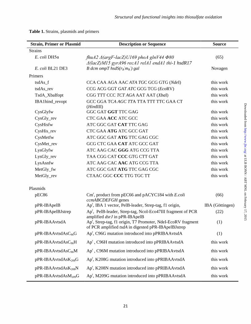
Structural and functional insights into thiosulfate oxidation
21
Table 1. Strains, plasmids and primers
Strain, Primer or Plasmid Description or Sequence Source
Strains
E. coli DH5α fhuA2 Δ(argF-lacZ)U169 phoA glnV44 Φ80
Δ(lacZ)M15 gyrA96 recA1 relA1 endA1 thi-1 hsdR17
(65)
E. coli BL21 DE3 B dcm ompT hsdS(rB-mB
-) gal Novagen
Primers
tsdAs_f CCA CAA AGA AAC ATA TGC GCG GTG (NdeI) this work
tsdAs_rev CCG ACG GGT GAT ATC GCG TCG (EcoRV) this work
TsdA_XbaIfopt CGG TTT CCC TCT AGA AAT AAT (XbaI) this work
IBA1hind_revopt GCC GGA TCA AGC TTA TTA TTT TTC GAA CT
(HindIII)
this work
CysGlyfw GGC GAT GGT TTC GAG this work
CysGly_rev CTC GAA ACC ATC GCC this work
CysHisfw ATC GGC GAT CAT TTC GAG this work
CysHis_rev CTC GAA ATG ATC GCC GAT this work
CysMetfw ATC GGC GAT ATG TTC GAG CGC this work
CysMet_rev GCG CTC GAA CAT ATC GCC GAT this work
LysGlyfw ATC AAG CAC GGG ATG CCG TTA this work
LysGly_rev TAA CGG CAT CCC GTG CTT GAT this work
LysAsnfw ATC AAG CAC AAC ATG CCG TTA this work
MetGly_fw ATC GGC GAT ATG TTC GAG CGC this work
MetGly_rev CTAAC GGC CCC TTG TGC TT this work
Plasmids
pEC86 Cmr, product from pEC66 and pACYC184 with E.coli
ccmABCDEFGH genes
(66)
pPR-IBApelB Apr, IBA 1 vector, PelB-leader, Strep-tag, f1 origin, IBA (Göttingen)
pPR-IBApelBJstrep Apr, PelB-leader, Strep-tag, NcoI-Eco47III fragment of PCR
amplified dsrJ in pPR-IBApelB
(22)
pPR-IBAAvtsdA Apr, Strep-tag, f1 origin, T7 Promoter, NdeI-EcoRV fragment
of PCR amplified tsdA in digested pPR-IBApelBJstrep
(1)
pPR-IBAAvtsdAsC96G Apr, C96G mutation introduced into pPRIBAAvtsdA (1)
pPR-IBAAvtsdAsC96H Apr , C96H mutation introduced into pPRIBAAvtsdA this work
pPR-IBAAvtsdAsC96M Apr , C96M mutation introduced into pPRIBAAvtsdA this work
pPR-IBAAvtsdAsK208G Apr, K208G mutation introduced into pPRIBAAvtsdA this work
pPR-IBAAvtsdAsK208N Apr, K208N mutation introduced into pPRIBAAvtsdA this work
pPR-IBAAvtsdAsM209G Apr, M209G mutation introduced into pPRIBAAvtsdA this work
at UL
B B
ON
N / A
BT
MN
L on February 17, 2015
http://ww
w.jbc.org/
Dow
nloaded from
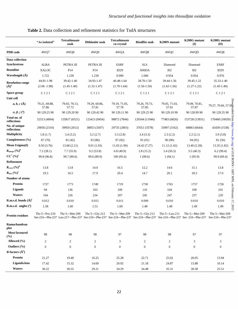
Structural and functional insights into thiosulfate oxidation
22
Table 2. Data collection and refinement statistics for TsdA structures
“As isolated”
Tetrathionate
soak Dithionite soak
Tetrathionate
co-crystal Bisulfite soak K208N mutant
K208G mutant
(I)
K208G mutant
(II)
PDB code 4WQ7 4WQ8 4WQ9 4WQA 4WQB 4WQC 4WQD 4WQE
Data collection
Synchrotron ALBA PETRA III PETRA III ESRF SLS Diamond Diamond ESRF
Beamline XALOC P14 P14 ID29 X06DA I02 I02 ID29
Wavelength (Å) 1.722 1.239 1.239 0.999 1.000 0.954 0.954 0.976
Resolution range
(Å)a
44.81-1.98
(2.06 -1.98)
39.42-1.40
(1.45-1.40)
34.93-1.47
(1.52-1.47)
46.48-1.64
(1.70-1.64)
28.70-1.50
(1.56-1.50)
39.44-1.56
(1.62-1.56)
39.45-1.22
(1.27-1.22)
35.33-1.40
(1.45-1.40)
Space group C 1 2 1 C 1 2 1 C 1 2 1 C 1 2 1 C 1 2 1 C 1 2 1 C 1 2 1 C 1 2 1
Unit cell
a, b, c (Å) 79.21, 69.88,
57.86
79.03, 70.13,
57.72
79.29, 69.86,
57.81
79.19, 71.05,
57.78
79.26, 70.73,
57.85
79.05, 71.03,
57.81
79.09, 70.81,
57.87 79.27, 70.66, 57.95
α, β, γ (o) 90 129.25 90 90 129.20 90 90 129.42 90 90 129.11 90 90 129.25 90 90 129.10 90 90 128.99 90 90 129.35 90
Total no. of
reflections 52513 (4004) 135817 (6521) 123413 (9454) 98872 (7844) 129344 (11946) 77483 (6826) 153728 (13931) 170468 (10020)
No. of unique
reflections 29056 (2310) 39950 (2012) 38853 (3507) 29735 (2803) 37651 (3578) 33997 (3162) 68883 (6644) 43439 (1538)
Multiplicity 1.8 (1.7) 3.4 (3.2) 3.2 (2.7) 3.3 (2.8) 3.4 (3.3) 2.3 (2.2) 2.2 (2.1) 3.9 (3.8)
Completeness (%) 87 (70) 81 (42) 93 (86) 97 (92) 95 (91) 96 (90) 94 (91) 81 (56)
Mean I/sigma(I) 9.50 (3.76) 13.68 (2.21) 9.01 (1.93) 11.03 (1.99) 24.42 (7.27) 11.12 (1.42) 13.40 (2.28) 13.35 (1.82)
Rmerge (%)b 7.2 (28.1) 7.7 (55.9) 9.2 (53.8) 6.8 (49.9) 2.8 (15.2) 5.4 (50.3) 3.5 (40.3) 6.2 (99.4)
CC* (%)c 99.8 (96.8) 99.7 (89.6) 99.6 (89.9) 100 (95.6) 1 (99.6) 1 (94.1) 1 (95.9) 99.9 (69.4)
Refinement
Rcryst (%)d 13.8 13.8 14.0 16.5 12.2 14.6 15.1 13.8
Rfree (%)e 19.3 16.5 17.9 20.4 14.7 20.1 18.3 17.0
Number of atoms
Protein 1727 1771 1749 1719 1730 1763 1757 1726
Ligands 94 136 103 100 110 104 100 101
Waters 164 226 234 207 245 247 257 220
R.m.s.d. bonds (Å)f 0.012 0.010 0.015 0.011 0.009 0.010 0.010 0.010
R.m.s.d. angles (o) 1.58 1.60 1.51 1.60 1.48 1.48 1.49 1.49
Protein residues Thr-5―Pro-210
Ser-216―Phe-237
Thr-5―Met-209
Leu-217―Phe-237
Thr-5―Gly-212
Ser-216―Phe-237
Thr-5―Met-209
Ser-216―Phe-237
Thr-5―Gly-212
Ser-216―Phe-237
Thr-5―Leu-211
Ser-216―Phe-237
Thr-5―Met-209
Ser- 216―Phe-237
Thr-5―Met-209
Ser-216―Phe-237
Ramachandran
plot
Most favoured
(%) 98 98 98 97 98 98 97 97
Allowed (%) 2 2 2 3 2 2 3 3
Outliers (%) 0 0 0 0 0 0 0 0
B-factors (Å2)
Protein 21.27 19.40 16.25 25.28 22.71 25.02 20.05 13.94
Ligands/ions 17.42 15.32 14.69 20.92 21.18 24.87 15.88 10.14
Waters 30.22 30.55 29.21 34.29 34.48 35.31 30.58 25.51
at UL
B B
ON
N / A
BT
MN
L on February 17, 2015
http://ww
w.jbc.org/
Dow
nloaded from
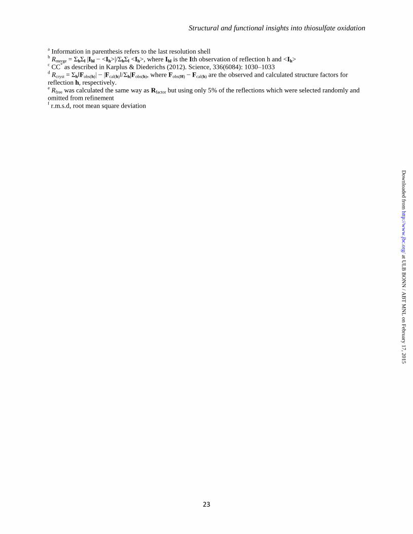
Structural and functional insights into thiosulfate oxidation
23
a Information in parenthesis refers to the last resolution shell
b Rmerge = ΣhΣl |Ihl − <Ih>|/ΣhΣl <Ih>, where Ihl is the Ith observation of reflection h and <Ih>
c CC
* as described in Karplus & Diederichs (2012). Science, 336(6084): 1030–1033
d Rcryst = Σh‖Fobs(h)| − |Fcal(h)‖/Σh|Fobs(h), where Fobs(H) − Fcal(h) are the observed and calculated structure factors for
reflection h, respectively. e Rfree was calculated the same way as Rfactor but using only 5% of the reflections which were selected randomly and
omitted from refinement f r.m.s.d, root mean square deviation
at UL
B B
ON
N / A
BT
MN
L on February 17, 2015
http://ww
w.jbc.org/
Dow
nloaded from
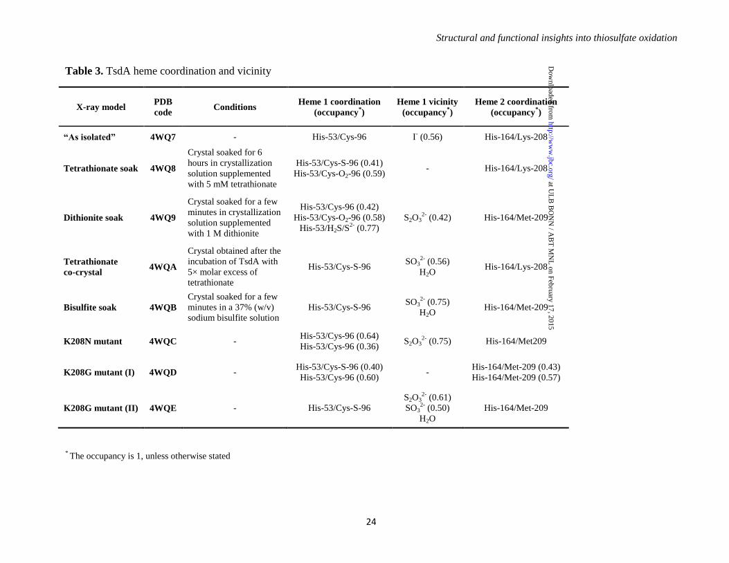
Structural and functional insights into thiosulfate oxidation
24
Table 3. TsdA heme coordination and vicinity
X-ray model PDB
code Conditions
Heme 1 coordination
(occupancy*)
Heme 1 vicinity
(occupancy*)
Heme 2 coordination
(occupancy*)
“As isolated” 4WQ7 - His-53/Cys-96 I- (0.56) His-164/Lys-208
Tetrathionate soak 4WQ8
Crystal soaked for 6
hours in crystallization
solution supplemented
with 5 mM tetrathionate
His-53/Cys-S-96 (0.41)
His-53/Cys-O2-96 (0.59) - His-164/Lys-208
Dithionite soak 4WQ9
Crystal soaked for a few
minutes in crystallization
solution supplemented
with 1 M dithionite
His-53/Cys-96 (0.42)
His-53/Cys-O2-96 (0.58)
His-53/H2S/S2-
(0.77)
S2O32-
(0.42) His-164/Met-209
Tetrathionate
co-crystal 4WQA
Crystal obtained after the
incubation of TsdA with
5× molar excess of
tetrathionate
His-53/Cys-S-96 SO3
2- (0.56)
H2O His-164/Lys-208
Bisulfite soak 4WQB
Crystal soaked for a few
minutes in a 37% (w/v)
sodium bisulfite solution
His-53/Cys-S-96 SO3
2- (0.75)
H2O His-164/Met-209
K208N mutant 4WQC - His-53/Cys-96 (0.64)
His-53/Cys-96 (0.36) S2O3
2- (0.75) His-164/Met209
K208G mutant (I) 4WQD - His-53/Cys-S-96 (0.40)
His-53/Cys-96 (0.60) -
His-164/Met-209 (0.43)
His-164/Met-209 (0.57)
K208G mutant (II) 4WQE - His-53/Cys-S-96
S2O32-
(0.61)
SO32-
(0.50)
H2O
His-164/Met-209
* The occupancy is 1, unless otherwise stated
at UL
B B
ON
N / A
BT
MN
L on February 17, 2015
http://ww
w.jbc.org/
Dow
nloaded from
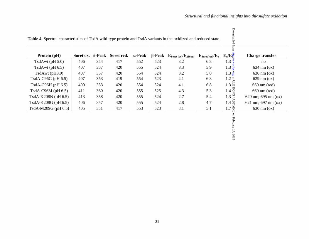
Structural and functional insights into thiosulfate oxidation
25
Table 4. Spectral characteristics of TsdA wild-type protein and TsdA variants in the oxidized and reduced state
Protein (pH) Soret ox. δ-Peak Soret red. α-Peak β-Peak ESoret (ox)/E280nm ESoret(red)/Eα Eα/Eβ Charge transfer
TsdAwt (pH 5.0) 406 354 417 552 523 3.2 6.8 1.3 no
TsdAwt (pH 6.5) 407 357 420 555 524 3.3 5.9 1.3 634 nm (ox)
TsdAwt (pH8.0) 407 357 420 554 524 3.2 5.0 1.3 636 nm (ox)
TsdA-C96G (pH 6.5) 407 353 419 554 523 4.1 6.8 1.2 629 nm (ox)
TsdA-C96H (pH 6.5) 409 353 420 554 524 4.1 6.8 1.3 660 nm (red)
TsdA-C96M (pH 6.5) 411 360 420 555 525 4.3 5.3 1.4 660 nm (red)
TsdA-K208N (pH 6.5) 413 358 420 555 524 2.7 5.4 1.3 620 nm; 695 nm (ox)
TsdA-K208G (pH 6.5) 406 357 420 555 524 2.8 4.7 1.4 621 nm; 697 nm (ox)
TsdA-M209G (pH 6.5) 405 351 417 553 523 3.1 5.1 1.7 630 nm (ox)
at UL
B B
ON
N / A
BT
MN
L on February 17, 2015
http://ww
w.jbc.org/
Dow
nloaded from
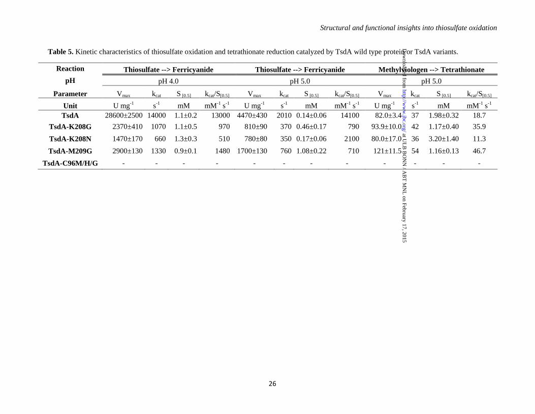
Structural and functional insights into thiosulfate oxidation
26
Table 5. Kinetic characteristics of thiosulfate oxidation and tetrathionate reduction catalyzed by TsdA wild type protein or TsdA variants.
Reaction Thiosulfate --> Ferricyanide Thiosulfate --> Ferricyanide Methylviologen --> Tetrathionate
pH pH 4.0 pH 5.0 pH 5.0
Parameter Vmax kcat S [0.5] kcat/S[0.5] Vmax kcat S [0.5] kcat/S[0.5] Vmax kcat S [0.5] kcat/S[0.5]
Unit U mg-1
s-1
mM mM-1
s-1
U mg-1
s-1
mM mM-1
s-1
U mg-1
s-1
mM mM-1
s-1
TsdA 28600±2500 14000 1.1±0.2 13000 4470±430 2010 0.14±0.06 14100 82.0±3.4 37 1.98±0.32 18.7
TsdA-K208G 2370±410 1070 1.1±0.5 970 810±90 370 0.46±0.17 790 93.9±10.0 42 1.17±0.40 35.9
TsdA-K208N 1470±170 660 1.3±0.3 510 780±80 350 0.17±0.06 2100 80.0±17.0 36 3.20±1.40 11.3
TsdA-M209G 2900±130 1330 0.9±0.1 1480 1700±130 760 1.08±0.22 710 121±11.5 54 1.16±0.13 46.7
TsdA-C96M/H/G - - - - - - - - - - - -
at UL
B B
ON
N / A
BT
MN
L on February 17, 2015
http://ww
w.jbc.org/
Dow
nloaded from
Structural and functional insights into thiosulfate oxidation
27
Figure 1
at UL
B B
ON
N / A
BT
MN
L on February 17, 2015
http://ww
w.jbc.org/
Dow
nloaded from
Structural and functional insights into thiosulfate oxidation
28
Figure 2
at UL
B B
ON
N / A
BT
MN
L on February 17, 2015
http://ww
w.jbc.org/
Dow
nloaded from
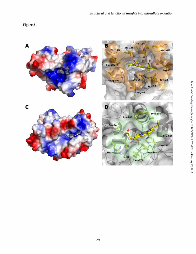
Structural and functional insights into thiosulfate oxidation
29
Figure 3
at UL
B B
ON
N / A
BT
MN
L on February 17, 2015
http://ww
w.jbc.org/
Dow
nloaded from
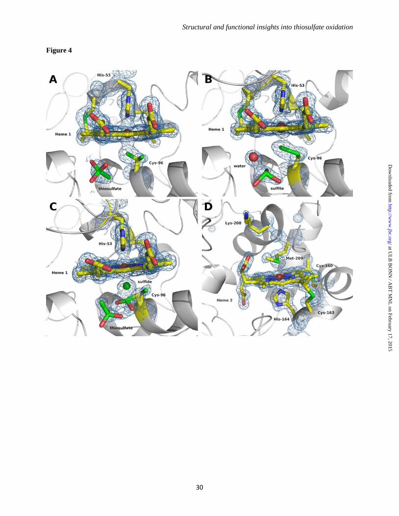
Structural and functional insights into thiosulfate oxidation
30
Figure 4
at UL
B B
ON
N / A
BT
MN
L on February 17, 2015
http://ww
w.jbc.org/
Dow
nloaded from
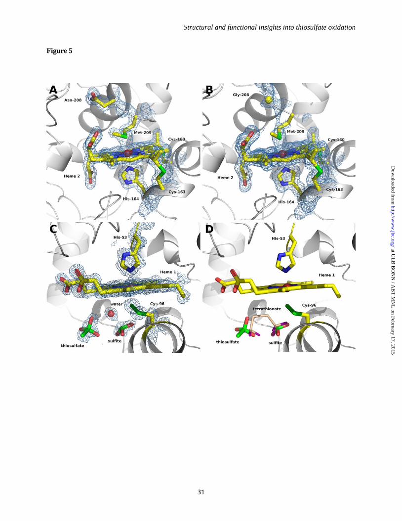
Structural and functional insights into thiosulfate oxidation
31
Figure 5
at UL
B B
ON
N / A
BT
MN
L on February 17, 2015
http://ww
w.jbc.org/
Dow
nloaded from
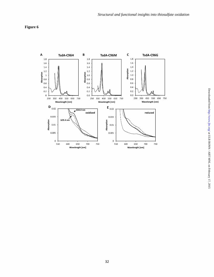
Structural and functional insights into thiosulfate oxidation
32
Figure 6
at UL
B B
ON
N / A
BT
MN
L on February 17, 2015
http://ww
w.jbc.org/
Dow
nloaded from
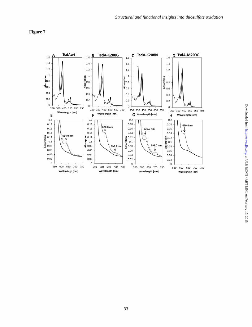
Structural and functional insights into thiosulfate oxidation
33
Figure 7
at UL
B B
ON
N / A
BT
MN
L on February 17, 2015
http://ww
w.jbc.org/
Dow
nloaded from
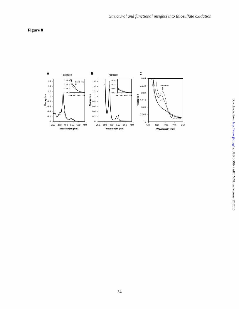
Structural and functional insights into thiosulfate oxidation
34
Figure 8
at UL
B B
ON
N / A
BT
MN
L on February 17, 2015
http://ww
w.jbc.org/
Dow
nloaded from
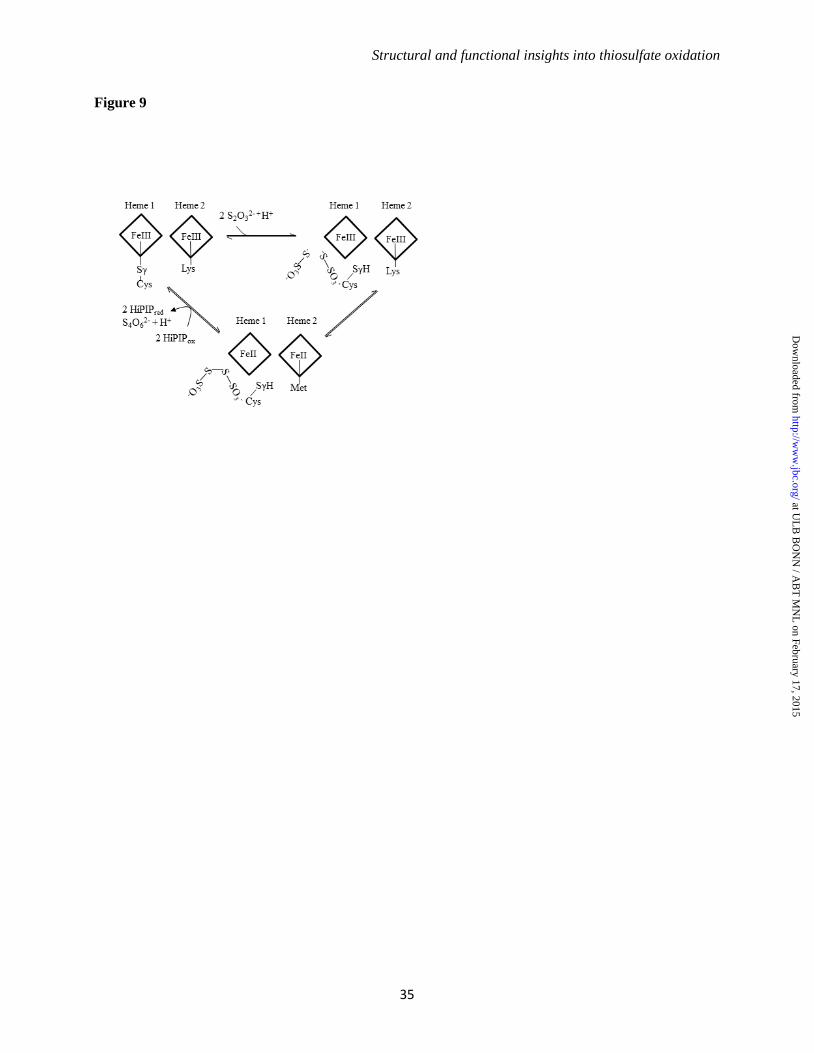
Structural and functional insights into thiosulfate oxidation
35
Figure 9
at UL
B B
ON
N / A
BT
MN
L on February 17, 2015
http://ww
w.jbc.org/
Dow
nloaded from
DahlPereira, Margarida Archer and Christiane José A. Brito, Kevin Denkmann, Ines A. C. OxidationFunctional Insights into Thiosulfate
: Structural andAllochromatium vinosumThiosulfate Dehydrogenase (TsdA) from Protein Structure and Folding:
published online February 11, 2015J. Biol. Chem.
10.1074/jbc.M114.623397Access the most updated version of this article at doi:
.JBC Affinity SitesFind articles, minireviews, Reflections and Classics on similar topics on the
Alerts:
When a correction for this article is posted•
When this article is cited•
to choose from all of JBC's e-mail alertsClick here
http://www.jbc.org/content/early/2015/02/11/jbc.M114.623397.full.html#ref-list-1
This article cites 0 references, 0 of which can be accessed free at
at UL
B B
ON
N / A
BT
MN
L on February 17, 2015
http://ww
w.jbc.org/
Dow
nloaded from
All in-text references underlined in blue are linked to publications on ResearchGate, letting you access and read them immediately.


