Embed Size (px)
Citation preview

www.elsevier.com/locate/yviro
Virology 331 (2
Thogoto virus ML protein suppresses IRF3 function
Stephanie Jenningsa, Luis Martınez-Sobridob, Adolfo Garcıa-Sastreb,
Friedemann Webera, Georg Kochsa,*
aAbteilung Virologie, Institut fur Medizinische Mikrobiologie und Hygiene, Universitat Freiburg, D-79008 Freiburg, GermanybDepartment of Microbiology, Mount Sinai School of Medicine, New York, NY 10029, USA
Received 24 June 2004; returned to author for revision 4 August 2004; accepted 8 October 2004
Available online 5 November 2004
Abstract
The Thogoto virus (THOV) is a member of the family Orthomyxoviridae. It prevents induction of alpha/beta interferons (IFN) in cell
culture and in vivo via the action of the viral ML protein. Phenotypically, the effect of THOV ML resembles that of the NS1 protein of
influenza A virus (FLUAV) in that it blocks the expression of IFN genes. IFN expression depends on IFN regulatory factor 3 (IRF3). Upon
activation, IRF3 forms homodimers and accumulates in the nucleus where it binds the transcriptional coactivator CREB-binding protein
(CBP). Here, we show that expression of ML blocked the transcriptional activity of IRF3 after stimulation by virus infection. Further
biochemical analysis revealed that ML acts by blocking IRF3 dimerization and association with CBP. Surprisingly, however, ML did not
interfere with the nuclear transport of IRF3. Thus, the action of ML differs strikingly from that of FLUAV NS1 that prevents IFN induction
by retaining IRF3 in the cytoplasm.
D 2004 Elsevier Inc. All rights reserved.
Keywords: Orthomyxoviruses; Thogoto virus; ML protein; Influenza virus; NS1 protein; Interferon antagonist; Interferon-regulatory factor 3; IRF3
Introduction
The mammalian interferon (IFN) system represents the
basis of an early host response against viral infections
(Samuel, 2001). Mice lacking a functional type I IFN
receptor (Fiette et al., 1995; Muller et al., 1994) or humans
with defects in IFN receptor signaling (Dupuis et al., 2003)
are highly susceptible to infections with viral pathogens.
0042-6822/$ - see front matter D 2004 Elsevier Inc. All rights reserved.
doi:10.1016/j.virol.2004.10.015
Abbreviations: dsRNA, double-stranded RNA; FF-Luc, firefly lucifer-
ase; FLUAV, influenza A virus; IFN, interferon; M, matrix protein; ML,
matrix protein long; NDV, newcastle disease virus; NP, nucleoprotein; PFU,
plaque forming units; REN-Luc, Renilla luciferase; THOV, Thogoto virus;
VLP, virus-like particle; vRNP, viral ribonucleoprotein complex.
* Corresponding author. Abteilung Virologie, Institut fqr Medizinische
Mikrobiologie und Hygiene, Hermann-Herder-Strasse 11, 79104 Freiburg,
Germany. Fax: +49 761 2036562.
E-mail addresses: [email protected]
(S. Jennings)8 [email protected] (L. Martınez-Sobrido)8
[email protected] (A. Garcıa-Sastre)8
[email protected] (F. Weber)8
[email protected] (G. Kochs).
Upon viral infection, recognition of virus particles (Collins
et al., 2004; Servant et al., 2002) or virus-specific molecular
patterns like single-stranded and double-stranded RNA
(dsRNA) triggers the expression of type I IFNs, mainly
IFNa and h (Alexopoulou et al., 2001; Diebold et al., 2004;
Stark et al., 1998). IFNh, known as the immediate early
IFN, leads via a positive feedback mechanism to the
upregulation of the IFNa genes (Erlandsson et al., 1998;
Marie et al., 1998). Both IFNa and h induce the expression
of IFN-stimulated genes, thus establishing an antiviral state
in cells (Der et al., 1998; Staeheli, 1990).
Production of IFNh is one of the earliest host cell
responses to infection with viral pathogens. The transcrip-
tional induction of the IFNh promoter involves activation of
IFN regulatory factor-3 (IRF3), activating protein-1 (AP1),
and nuclear factor kappa B (NF-nB) (Algarte et al., 1999;
Schafer et al., 1998; Wathelet et al., 1998; Yoneyama et al.,
1998). Among these transcription factors, IRF3 is the most
essential for the immediate early induction of IFNhexpression (Sato et al., 2000). IRF3 is constitutively
expressed and in its inactive, unphosphorylated form
005) 63–72

S. Jennings et al. / Virology 331 (2005) 63–7264
predominantly resides in the cytoplasm. Viral infection
triggers activation of IRF3 through phosphorylation on
serine and threonine residues in its C-terminal part (between
residues 385 and 405) (Lin et al., 1998; Mori et al., 2004;
Yoneyama et al., 1998). The responsible kinases for this
essential activation step were recently characterized as two
unconventional InB kinases, IKKq and TBK1 (Fitzgerald et
al., 2003; Sharma et al., 2003). Activation of IRF3 induces a
sequence of events including nuclear accumulation (Kumar
et al., 2000; Lin et al., 1998), homodimerization, and
association with transcriptional coactivators like the CREB-
binding protein (CBP) in order to gain full transcriptional
activity (Suhara et al., 2000; Weaver et al., 1998; Yang et al.,
2002). The IRF3/CBP holocomplex then binds and activates
target gene promoters containing IRF3 binding sites such as
the IFNh and ISG56 promoter (Grandvaux et al., 2002;
Peters et al., 2002; Wathelet et al., 1998).
Viruses have evolved different strategies to circumvent
the induction of type I IFNs (Goodbourn et al., 2000; Levy
and Garcia-Sastre, 2001; Weber et al., 2003). IRF3 as a key
factor for the induction of the early antiviral host response is
targeted by many viruses (Baigent et al., 2002; Basler et al.,
2003; Bossert et al., 2003; Dauber et al., 2004; Foy et al.,
2003; Graff et al., 2002; Lin et al., 2001; Poole et al., 2002;
Ronco et al., 1998; Xiang et al., 2002). One of the best
studied examples is the NS1 protein of FLUAV that was
shown to prevent activation of the IFNh promoter by
suppressing activation of IRF3 by dsRNA (Talon et al.,
2000). Similar to FLUAV, infection with Thogoto virus
(THOV) attenuates activation of the IFNh promoter
(Hagmaier et al., 2003).
Together with the influenza viruses, THOV belongs to
the family of Orthomyxoviridae (Van Regenmortel et al.,
2000). In contrast to other orthomyxoviruses, THOV is an
arbovirus that replicates both in mammalian and in tick cells
(Jones and Nuttall, 1989). Ticks are the reservoir of THOV
(Nuttall et al., 1995). The virus persistently infects these
animals but is not transmitted horizontally or transovarially
between ticks (Davies et al., 1986), raising the question how
the virus persists in this reservoir. THOV is transmitted to
vertebrates through tick bites. After a viremic phase, the
virus infects other ticks feeding on the same animal,
favoring the spread of the virus back to its arthropod
reservoir. To successfully perform this replication cycle,
THOV has evolved a strategy to inhibit the induction of the
innate immune response in the vertebrate host. The virus has
been shown to effectively suppress the induction of type I
IFN in infected cells and in animals (Hagmaier et al., 2003;
Pichlmair et al., 2004), and therefore prevents induction of
the early antiviral host response that is mainly based on IFN-
stimulated Mx genes (Haller et al., 1995; Pavlovic et al.,
1995).
The genome of THOV consists of six single-stranded
RNA segments of negative polarity that have a coding
capacity for seven proteins: six essential structural proteins
such as the three subunits of the viral RNA-dependent RNA
polymerase, the nucleoprotein (NP), the transmembrane
glycoprotein, the matrix protein (M), and one non-essential
accessory protein, the ML protein (Hagmaier et al., 2003;
Haller and Kochs, 2002). The proteins M and ML are both
encoded by the smallest viral RNA segment. The M
protein is translated from a spliced transcript in which the
stop codon terminating the M reading frame is created by a
splicing event (Kochs et al., 2000), whereas the ML
protein is translated from the full-length, unspliced tran-
script. Thus the 304 amino acid long ML represents an
elongated version of M (266 amino acids) containing 38
additional amino acids at the C terminus. We recently
found that ML functions as an IFN antagonist that prevents
activation of the IFNh promoter (Hagmaier et al., 2003).
Accordingly, a recombinant THOV lacking the ML gene
showed enhanced induction of IFN in infected cells and, in
contrast to the highly virulent wild-type virus, was strongly
attenuated in mice (Hagmaier et al., 2003; Pichlmair et al.,
2004).
Here, we demonstrate that ML targets a distinct step in
IRF3 activation. ML does not interfere with the initial IRF3
activation and nuclear translocation. Instead, it blocks IRF3
dimerization and its interaction with the coactivator CBP.
Therefore, unlike the NS1 protein of FLUAV that prevents
IRF3 activation in the cytoplasm, THOV ML antagonizes
the IFN system by specifically inhibiting IRF3 transcrip-
tional activity in the nucleus.
Results
THOV ML suppresses IRF3-dependent promoter activation
We recently identified the ML protein of THOV as an
IFN antagonist that blocks induction of IFN in infected cells
(Hagmaier et al., 2003). ML is a C-terminally extended
version of THOV M protein containing 38 additional amino
acids. To analyze the activity of ML independent of the viral
context, cDNA expression plasmids encoding THOV M,
THOV ML, or an empty vector were transfected into 293
cells together with a firefly luciferase (FF-Luc) reporter
construct under the control of the IFNh promoter
(Yoneyama et al., 1998). Infection of the transfected cells
with Newcastle disease virus (NDV), a strong IFN inducer
(Bazzigher et al., 1992), led to the expression of FF-Luc,
indicating activation of the IFNh promoter (Fig. 1A).
Coexpression of ML resulted in a 5-fold reduction of
promoter activity compared to the control, thereby confirm-
ing the IFN antagonistic activity of ML (Hagmaier et al.,
2003). M expression, by contrast, had no inhibitory effect.
To elucidate the molecular basis of this inhibitory
activity, we concentrated on IRF3 because this is the most
critical cellular transcription factor that is essential for virus-
induced IFNh expression, whereas the other transcription
factors NFnB and AP-1 have only enhancing effects
(Wathelet et al., 1998). One of the earliest steps of IRF3
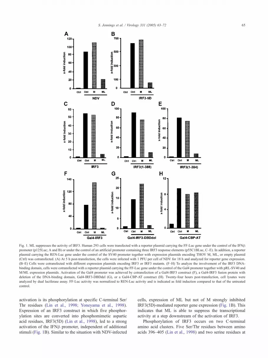
Fig. 1. ML suppresses the activity of IRF3. Human 293 cells were transfected with a reporter plasmid carrying the FF-Luc gene under the control of the IFNhpromoter (p125Luc; A and B) or under the control of an artificial promoter containing three IRF3 response elements (p55C1BLuc, C–E). In addition, a reporter
plasmid carrying the REN-Luc gene under the control of the SV40 promoter together with expression plasmids encoding THOV M, ML, or empty plasmid
(Ctrl) was cotransfected. (A) At 5 h post-transfection, the cells were infected with 1 PFU per cell of NDV for 18 h and analyzed for reporter gene expression.
(B–E) Cells were cotransfected with different expression plasmids encoding IRF3 or IRF3 mutants. (F–H) To analyze the involvement of the IRF3 DNA-
binding domain, cells were cotransfected with a reporter plasmid carrying the FF-Luc gene under the control of the Gal4 promoter together with pRL-SV40 and
M/ML expression plasmids. Activation of the Gal4 promoter was achieved by cotransfection of a Gal4-IRF3 construct (F), a Gal4-IRF3 fusion protein with
deletion of the DNA-binding domain, Gal4-IRF3-DBDdel (G), or a Gal4-CBP-AT construct (H). Twenty-four hours post-transfection, cell lysates were
analyzed by dual luciferase assay. FF-Luc activity was normalized to REN-Luc activity and is indicated as fold induction compared to that of the untreated
control.
S. Jennings et al. / Virology 331 (2005) 63–72 65
activation is its phosphorylation at specific C-terminal Ser/
Thr residues (Lin et al., 1998; Yoneyama et al., 1998).
Expression of an IRF3 construct in which five phosphor-
ylation sites are converted into phosphomimetic aspartic
acid residues, IRF3(5D) (Lin et al., 1998), led to a strong
activation of the IFNh promoter, independent of additional
stimuli (Fig. 1B). Similar to the situation with NDV-infected
cells, expression of ML but not of M strongly inhibited
IRF3(5D)-mediated reporter gene expression (Fig. 1B). This
indicates that ML is able to suppress the transcriptional
activity at a step downstream of the activation of IRF3.
Phosphorylation of IRF3 occurs on two C-terminal
amino acid clusters. Five Ser/Thr residues between amino
acids 396–405 (Lin et al., 1998) and two serine residues at

Fig. 2. ML prevents IRF3 dimerization and association with CBP. 293
cells were infected with 5 PFU per cell of THOVML�, THOVML+,
FLUAV(wt), FLUAVdNS1, or were mock infected for 20 h. (A) Cell
lysates were analyzed for monomers and dimers of IRF3 by non-
denaturing gel electrophoresis followed by Western blotting using an
IRF3-specific antibody. (B) Viral infection was monitored using an
antibody directed against THOV NP or FLUAV NP. (C) The same cell
lysates were used for coimmunoprecipitation with a monoclonal IRF3-
specific antibody and the immunocomplexes were analyzed by Western
blot with a CBP-specific antibody. (D) IRF3 present in the immunocom-
plex was detected with a polyclonal rabbit antiserum directed against
IRF3. (E) The total amount of CBP in individual cell lysates was
monitored using the CBP-specific antibody.
S. Jennings et al. / Virology 331 (2005) 63–7266
amino acids 385/386 (Yoneyama et al., 1998) were
described as the responsible targets of the activating protein
kinases. Therefore, we constructed deletion mutants of IRF3
lacking 39 or 43 amino acids of the C terminus called
IRF3(1–388) and IRF3(1–384). These truncated variants of
IRF3 are active upon overexpression together with an IRF3-
responsive FF-Luc reporter construct containing three IRF3-
binding elements (Yoneyama et al., 1998). Overexpression
of wild-type IRF3 or the C-terminal deletion mutants
strongly stimulated reporter gene expression (Figs. 1C, D,
and E), albeit to lower levels than those in IRF3(5D)
transfection experiments. ML, but not M, was able to
suppress this activation independent of the presence of IRF3
C-terminal phosphorylation sites. These results suggest that
the effect of ML on IRF3 transcriptional activity is
independent of the phosphorylation state of the IRF3 C-
terminus and further implies that the C-terminal regulatory
moiety of IRF3 is not a target of ML action.
Suppression of IRF3 by THOV ML is independent of the
DNA-binding capacity of IRF3
After dimerization and association with transcriptional
coactivators, IRF3 is thought to bind to cognate DNA
elements and stimulate promoter activity (Wathelet et al.,
1998). We therefore tested whether the block of IRF3-
dependent promoter activation by ML is based on an
inhibition of IRF3 DNA binding. To uncouple transcrip-
tional activation by IRF3 from its DNA-binding property,
we used a Gal4-IRF3 fusion protein that transactivates
reporter gene expression by binding to Gal4 promoter
elements (Fitzgerald et al., 2003). As expected, the full-
length Gal4-IRF3 fusion construct induced reporter gene
expression under the control of the Gal4-promoter (Fig. 1F).
Coexpression of THOV M led to an increase in reporter
gene expression, while coexpression of THOV ML com-
pletely abrogated transactivation by Gal4-IRF3 (Fig. 1F).
Accordingly, we tested the effect of ML on a Gal4-IRF3
construct that lacks the complete IRF3 DNA-binding
domain (amino acids 7–107). Coexpression of ML again
suppressed transactivation by the Gal4-IRF3 fusion protein
(Fig. 1G). Since IRF3 activity is dependent on the recruit-
ment of cotransactivators like CBP to the promoter region,
we tested whether transactivation by CBP was also inhibited
by coexpression of ML. As shown in Fig. 1H, neither
THOV M nor ML had any effect on the activity of a Gal4-
CBP fusion protein, indicating that THOV ML does not
affect CBP function. Therefore, THOV ML appears to
specifically affect IRF3 function independent of its DNA-
binding properties.
THOV ML prevents virus-induced IRF3 dimerization and
interaction with CBP
Upon activation, IRF3 forms homodimers that are
required for DNA binding and interaction with transcrip-
tional coactivators (Iwamura et al., 2001). To test the effect
of ML on these steps of IRF3 activation, 293 cells were
infected with recombinant THOVs that either encode the
ML protein THOVML+ or do not encode ML THOVML�(Hagmaier et al., 2003). To analyze the oligomerization state
of IRF3, lysates of infected cells were separated by
nondenaturating gel electrophoresis, and IRF3 monomers
and dimers were detected by Western blot analysis using an
IRF3-specific antibody. As shown in Fig. 2A, THOVML
infection induced IRF3 dimerization, whereas in uninfected
cells, no IRF3 dimers could be detected. Surprisingly, in
cells infected with THOVML+, IRF3 predominantly accu-
mulated in the monomeric fraction and only weak dimer
formation was detectable. Detection of the viral NP suggests
comparable infection with the two different recombinant
viruses (Fig. 2B). In addition, we directly tested the effect of
recombinant ML on the homo-oligomerization of IRF3,
independent of virus infection. 293 cells were cotransfected
with cDNA constructs encoding the constitutively activated
IRF3 variant IRF3(5D) together with M and ML expression
plasmids. Analysis of the cell lysates 24 h later showed a

S. Jennings et al. / Virology 331 (2005) 63–72 67
spontaneous formation of IRF3(5D) dimers, as expected.
Consistent with our experiments with infected cells,
expression of recombinant ML suppressed IRF3(5D)
dimerization whereas expression of M had no effect (data
not shown).
In parallel, we did the same analysis with two strains of
FLUAV (A/PR/8/34) that either expressed or lacked the
IFN-antagonistic protein NS1 (FLUAVdNS1) (Garcia-
Sastre et al., 1998). While FLUAVdNS1 was a strong
inducer of IRF3 activation and resulted in the formation of
IRF3 dimers, wild-type FLUAV infection prevented IRF3
dimerization, similar to the situation in THOVML+-infected
cells (Fig. 2A).
IRF3 dimers recruit the transcriptional coactivator CBP
to gain full activity (Suhara et al., 2000; Weaver et al., 1998;
Yang et al., 2002). Therefore, we considered that ML may
not only prevent IRF3 dimerization but also the association
with CBP. To test this hypothesis, cells were infected with
the recombinant THOVs and FLUAVs for 20 h and then
subjected to immunoprecipitation analysis using an IRF3-
specific antibody. The precipitated immunocomplexes were
analyzed by Western blot using CBP- and IRF3-specific
antibodies. As shown in Fig. 2C, no interaction was
observed between CBP and IRF3 in cells infected with the
wild-type viruses THOVML+ and FLUAV. In contrast, the
mutants THOVML� and FLUAVdNS1 both triggered the
association of CBP with IRF3 (Fig. 2C). Fig. 2D indicates
that comparable levels of IRF3 are present in the immuno-
complexes. Using the anti-CBP antibody, we confirmed that
equal amounts of CBP are present in individual cell lysates
(Fig. 2E). These results demonstrate that the ML protein of
THOV antagonizes IRF3 dimerization and association to
CBP, similar to the NS1 protein of FLUAV.
THOV ML does not affect nuclear accumulation of IRF3
Nuclear translocation of IRF3 is a prerequisite for its
stimulatory activity (Lin et al., 1998). To analyze the effect
of ML on the nuclear accumulation of IRF3, we used two
independent approaches. First, we transfected cells with
THOV M or ML expression constructs together with a GFP-
IRF3-encoding plasmid. Then the cells were infected with
NDV to induce nuclear accumulation of the IRF3 fusion
protein. The cells were then fixed, and the expression of M
and ML was detected by immunofluorescence. As expected,
in uninfected cells, GFP-IRF3 was detected in the cytoplasm
whereas upon NDV infection GFP-IRF3 accumulated in the
nucleus (Fig. 3A). Surprisingly, neither expression of M nor
of ML disturbed the virus-induced nuclear accumulation of
GFP-IRF3 (Fig. 3A). In a second set of experiments, we
used the recombinant THOVs described above. Vero cells
were infected with these viruses and the localization of
endogenous IRF3 as well as the production of viral NP was
analyzed by double immunofluorescence 24 h after infec-
tion. In uninfected cells, IRF3 was detected in the
cytoplasm, whereas THOV infection led to the accumulation
of IRF3 in the cell nucleus (Fig. 3B), indicating that THOV
has the potential to trigger IRF3 activation. Interestingly,
IRF3 nuclear accumulation was observed following infec-
tion with both the ML-expressing and the ML-lacking
THOVs (Fig. 3B). These data suggest that despite the lack
of IRF3 dimerization and the absence of IRF3/CBP
interaction, infection with wild-type THOV induces nuclear
translocation and accumulation of IRF3, indicating that
expression of ML is not able to block this process.
Discussion
In this report, we describe how the THOV protein ML
prevents IFN induction by targeting IRF3, the essential
factor that stimulates early IFN production after virus
infection. ML thereby interferes with the dimerization of
IRF3 and its association with the transcriptional coactivator
CBP, but not with the nuclear accumulation of IRF3.
Virus infection represents a stress signal that activates
the innate immune system. This results in the rapid
expression and secretion of type I IFNs that are respon-
sible for the induction of antiviral host response (Good-
bourn et al., 2000; Stark et al., 1998). One of the earliest
stress responses after virus infection is the activation of
IRF3, the essential factor for IFNh expression (Collins et
al., 2004; Sato et al., 2000). IRF3 is constitutively expressed
and resides in the cytoplasm until activation. According to
the current model (Servant et al., 2002; Yoneyama et al.,
2002), the cell responds to virus infection with the activation
of diverse Ser/Thr protein kinases that activate IRF3.
Phosphorylated IRF3 then dimerizes, translocates into the
nucleus, and associates with the CBP coactivator. This
holocomplex then binds to IRF3-responsive promoters and
stimulates transcription.
Our data clearly show that the ML protein of THOV
prevents activation of IRF3 at a late stage by interfering with
its dimerization and association with the coactivator
molecule CBP. In contrast, the nuclear translocation of
IRF3 was not affected by ML. IRF3 contains an N-terminal
DNA-binding domain (7–107) and a C-terminal trans-
activation domain (110–427) and is normally in an inactive
conformation (Servant et al., 2002; Yang et al., 2002). The
C-terminal part is divided into a linker region (110–198), an
IRF-association domain (199–308), and a C-terminal auto-
inhibitory/serine-rich domain (385–427). The linker region
is the target of MAP- and DNA-dependent protein kinases
(Karpova et al., 2002; Servant et al., 2001) and contains
nuclear import and export signals. The IRF-association
domain is responsible for dimerization and association with
CBP, whereas the C-terminal autoinhibitory/serine-rich
domain is the target of virus-activated protein kinases
(VAK) (Lin et al., 1998; Yoneyama et al., 1998). C-terminal
phosphorylation results in structural changes that allow
dimerization of IRF3 and interaction with CBP via the IRF-
association domain (Qin et al., 2003; Takahasi et al., 2003).

Fig. 3. ML does not prevent nuclear accumulation of IRF3. (A) Vero cells were transfected with a GFP-IRF3 expression plasmid together with expression
plasmids encoding THOVM, THOVML, or empty vector (Ctrl). At 6 h post-transfection, cells were infected with 1 PFU per cell of NDVor mock infected. At
20 h postinfection, expression of M and ML protein was detected by immunofluorescence using an antibody directed against THOV M/ML. GFP-IRF3 (green)
and M or ML (red) localizations were detected by confocal microscopy. (B) Vero cells were infected with 5 PFU per cell of THOV expressing ML
(THOVML+), lacking ML (THOVML�), or mock infected. At 24 h postinfection, cells were fixed and analyzed for localization of endogenous IRF3 (green)
using an IRF3-specific antibody. THOV infection was detected by using an antibody directed against THOV NP (red).
S. Jennings et al. / Virology 331 (2005) 63–7268
Neither substitution of the IRF3 DNA-binding domain by
an unrelated DNA-binding domain nor deletion of the
autoinhibitory C-terminal region prevented the inhibitory
effect of ML on IRF3 function, indicating that phosphory-
lation by VAK or binding to the IRF3 response element is
not the target of ML action. In agreement with this, ML was
also able to inhibit the constitutively active mutant of IRF3
(IRF3-5D), in which a cluster of Ser/Thr residues whose
phosphorylation is required for IRF3 activation was
replaced by Asp, a phosphomimetic amino acid. Since
ML does not interfere with nuclear translocation of IRF3,
the nuclear translocation signal and the putative phosphor-
ylation sites within the linker domain seem not to be
affected by ML. In contrast, ML had a strong effect on IRF3
homo- and hetero-oligomerization with CBP, suggesting
that ML affects the function of the C-terminal IRF-
association domain. Therefore, a straightforward explan-
ation for the suppression of IFNh promoter activation by
THOV would be the existence of an interaction of ML with
the IRF3-association domain, resulting in a block of IRF3
dimerization and of IRF3 binding to CBP. However,
coimmunoprecipitation experiments did not reveal any
direct interaction between ML and IRF3 (data not shown),
suggesting that these two proteins do not interact or that
these interactions are weak or only transient. According to
Kumar et al. (2000), IRF3 nuclear accumulation is a result
of the interaction of IRF3 homodimers with CBP. It is
interesting to note that in our experiments with THOV ML,
IRF3 nuclear accumulation can occur independently of
these interactions.
While the precise mechanism of blocking IRF3-medi-
ated transactivation by ML remains to be elucidated, it is
clear that the C-terminal part of ML plays a critical role.
The M and ML proteins of THOV are expressed from the

S. Jennings et al. / Virology 331 (2005) 63–72 69
same genomic segment. ML is expressed from a colinear
transcript whereas M is encoded by a spliced mRNA of
segment 6 (Kochs et al., 2000). Therefore, the ML protein
represents a 38 amino acid C-terminal elongated version of
the M protein (Hagmaier et al., 2003). Both M and ML are
structural components of the THOV particle. In contrast to
M, however, ML does not perform M protein functions
like inhibition of viral polymerase activity and assistance in
formation of viral particles (Hagmaier et al., 2004; Wagner
et al., 2000). Mutational analysis of ML showed that the
entire C-terminal half of ML is necessary for its IFN
antagonistic activity, indicating that a complex structure of
the ML protein distinct from that of the M protein is
crucial for its IFN antagonistic activity (Hagmaier et al.,
2004).
IRF3 is the target of many different viral IFN antagonists
(Goodbourn et al., 2000; Levy and Garcia-Sastre, 2001;
Weber et al., 2003). In contrast to THOV ML, the proteins
NS1 of FLUAV and E3L of vaccinia virus target an early
step of IRF3 activation. Both proteins are thought to prevent
activation of the respective IRF3 kinases through sequestra-
tion of dsRNA, a potent activator of these kinases (Talon et
al., 2000; Wang et al., 2000; Xiang et al., 2002). For the
NS1 protein, additional antagonistic effects on later events
in the IFN-induced antiviral host response have been
described and appear to be mediated by a general inhibition
of the host mRNA processing (Donelan et al., 2003; Krug et
al., 2003; Seo et al., 2002). Furthermore, VP35 of Ebola
virus (Basler et al., 2003), NS1/NS2 of bovine respiratory
syncytial virus (Bossert et al., 2003), and NS3/4A serine
protease of hepatitis C virus (Foy et al., 2003) inhibit IRF3
activation most likely by targeting cellular components that
stimulate IRF3 phosphorylation and therefore prevent IRF3
activation and nuclear translocation.
Similar to THOV, several other viruses target late steps of
IRF3 activation. Upon BVDV infection, IRF3 translocates
into the nucleus but fails to induce IFN expression (Baigent
et al., 2002). The E6 protein of human papillomavirus-16
directly binds to IRF3 and thereby prevents IRF3 function
(Ronco et al., 1998). The ICP0 protein of Herpes simplex
virus targets a late step of IRF3 activation and, like the
THOV ML, is able to suppress transactivation by the
constitutively active IRF3(5D) (Lin et al., 2004). The vIRF1
encoded by human Herpes virus-8 (HHV8) and the
adenovirus (AdV)-E1A protein prevent the formation of
the holocomplex consisting of IRF3 and CBP. In contrast to
ML that targets IRF3 dimerization but not CBP function
(see Fig. 1H), the IFN antagonists of HHV-8 and AdV
directly interact with CBP to inhibit the formation of the
transcriptionally active IRF3 holocomplex (Juang et al.,
1998; Lin et al., 2001).
Whether ML of THOV directly interacts with IRF3 or
binds to an IRF3-associated cofactor will be the focus of our
future efforts to elucidate the precise mechanism of ML
action. However, our data clearly demonstrate that THOV
uses a strategy to suppress IFN induction that is different
from that of other orthomyxoviruses, revealing the amaz-
ingly broad variation of strategies that viruses have evolved
to circumvent the induction of antiviral host response.
Materials and methods
Cells and viruses
293 cells and Vero cells were maintained in Dulbecco’s
modified Eagle’s medium (DMEM) supplemented with
10% fetal calf serum (FCS). For infection studies, virus
stocks were diluted in DMEM supplemented with 2% FCS
and 20 mM HEPES (pH 7.5). Recombinant Thogoto viruses
(THOV) expressing ML (THOVML+) or lacking ML
(THOVML�) were described previously (Hagmaier et al.,
2003; Wagner et al., 2001). The Newcastle disease virus
(NDV; strain H53) (Bazzigher et al., 1992) stock was grown
on 10-day-old embryonated chicken eggs. Influenza A virus
(FLUAV) A/PR/8/34 (FLUAVwt) and the recombinant
influenza A/PR/8/34 with a deletion in the NS1 gene
(FLUAVdNS1) were described previously (Garcia-Sastre et
al., 1998). FLUAV stocks were diluted in phosphate-
buffered saline (PBS) supplemented with 0.3% bovine
serum albumin prior to infection.
Plasmids
THOV M and ML expression constructs under the
control of the chicken h-actin promoter (pCAGGS) have
been described previously (Hagmaier et al., 2003). Plasmids
expressing a constitutively active IRF3, IRF3(5D) (Lin et
al., 1998), and a fusion protein containing green fluorescent
protein (GFP) N-terminally fused to IRF3, GFP-IRF3, have
been described (Basler et al., 2003). N-terminally HA-
tagged IRF3 constructs in pCAGGS were designed for
expression of full-length human IRF3 (1–427) and C-
terminally truncated forms, IRF3(1–388) and IRF3(1–384),
lacking 39 and 43 C-terminal amino acids, respectively. The
Gal4 promoter studies were performed using a p5xGal4-
AdML-luc reporter plasmid (Wathelet et al., 1998) cotrans-
fected with a Gal4-IRF3 fusion construct, pGal4-IRF3
(Wathelet et al., 1998) (kindly provided by Tom Maniatis),
or a Gal4-CBP-acetyltransferase (AT) fusion construct,
pcDNA3-Gal4-CBP-AT (Bordoli et al., 2001) (kindly
provided by Richard Eckner). The Gal4-IRF3 fusion
construct was deleted in its DNA-binding domain (between
amino acid 7–107), yielding Gal4-IRF3-DBDdel.
Reporter plasmids carrying the firefly luciferase (FF-
Luc) gene under the control of either the IFNh promoter
(p125Luc) or an artificial promoter containing three IRF3-
binding sites (p55C1B-Luc) were kindly provided by
Takashi Fujita (Yoneyama et al., 1998). The reporter
plasmid pRL-SV40 carrying the Renilla luciferase gene
(REN-Luc) under the control of the constitutive SV40
promoter was purchased from Promega.
S. Jennings et al. / Virology 331 (2005) 63–7270
Reporter gene assays
Transient transfection of 293 cells was performed by
using 2 Al Dac-30 (Eurogentec)/Ag DNA in 200 AlOPTIMEM (Gibco-BRL) as described (Hagmaier et al.,
2003). Cells were transfected with 0.5 Ag of either p125Luc
or p55C1B-Luc, together with 0.05 Ag of pRL-SV40, and 1
Ag of the indicated THOV M or ML expression plasmids, in
addition to 1 Ag of the IRF3 expression constructs, when
indicated. At 5 h post-transfection, cells were infected with
1 plaque forming unit (PFU) per cell of NDV or were left
untreated. At 24 h post-transfection, cells were harvested
and lysed in 200 Al of Passive Lysis Buffer (Promega). An
aliquot of 20 Al was used to measure FF-Luc and REN-Luc
activities as described by the manufacturer (Dual-Luciferase
Reporter Assay System, Promega). FF-Luc activities were
normalized to REN-Luc activities and are indicated as fold
induction relative to the untreated control.
For reporter assays based on Gal4-driven reporter gene
expression, 293 cells were transfected with 0.5 Ag of
p5xGal4-AdML-luc, 0.05 Ag of control plasmid pRL-
SV40, 1 Ag of expression plasmids encoding Gal4-IRF3,
Gal4-IRF3-DBDdel, or Gal4-CBP-AT fusion protein, and
expression plasmids for THOV M, THOV ML, or empty
vector. Twenty-four hours post-transfection, cells were lysed
and a dual luciferase assay (Promega) was performed.
Immunofluorescence analysis
To analyze the subcellular localization of IRF3, Vero
cells were grown on coverslips and transfected with 1 Ag of
GFP-IRF3 expression plasmid and 1 Ag of expression
plasmids for THOV M, THOV ML, or empty vector. Five
hours post-transfection, cells were infected with 1 PFU per
cell of NDV or were mock infected. At 20 h post infection,
cells were fixed in 3% paraformaldehyd and permeabilized
with 0.5% Triton X-100. Cells were stained using a
polyclonal antibody specific for THOV M/ML and a Cy3-
conjugated secondary antibody.
To examine the localization of endogenous IRF3 in Vero
cells after infection with the recombinant THOVs, IRF3 was
detected 24 h postinfection using an IRF3-specific poly-
clonal rabbit antibody FL-425 (Santa Cruz), and the signal
was enzymatically amplified by the Tyramide Signal
Amplification (TSA) system (Perkin-Elmer) according to
the manufacturer’s instructions. Infection with THOV was
monitored using a mouse monoclonal antibody directed
against THOV NP and a Cy3-conjugated secondary anti-
body. Samples were examined with a Leica confocal laser
scanning microscope.
IRF3 dimerization assay
293 cells were infected with 5 PFU per cell of the viruses
or were mock infected. At 20 h postinfection, cells were
resuspended in 200 Al lysis buffer containing 50 mM Tris–
HCl (pH 7.5), 150 mM NaCl, 1 mM EDTA, 1% Nonidet P-
40, protease inhibitors (Roche) and phosphatase inhibitors
(Calbiochem), vortexed, incubated on ice for 10 min, and
centrifuged at 4 8C for 5 min at 10000 � g. Aliquots of
10 Ag protein were separated on a 7.5% nondenaturing
polyacrylamide gel with 1% deoxycholate in the cathode
buffer as described (Iwamura et al., 2001). IRF3 monomers
and dimers were detected by Western Blot analysis using a
polyclonal rabbit anti-IRF3 antibody FL-425 (Santa Cruz).
To monitor virus infection, viral NP was detected by
Western blot analysis using polyclonal rabbit antisera
directed against THOV NP or FLUAV NP.
IRF3/CBP coimmunoprecipitation assay
The cell lysates used for IRF3 dimerization assays were
also subjected to coimmunoprecipitation analysis using the
monoclonal mouse anti-IRF3 antibody SL12 (Ronco et al.,
1998) (kindly provided by P.M. Howley). The cell lysates
were preadsorbed with protein G-Sepharose (Amersham
Biosciences) for 1 h at 4 8C, then centrifuged at 10000 � g
for 5 min at 4 8C followed by incubation of the supernatants
with protein G-Sepharose and 5 Al of the monoclonal anti-
IRF3 antibody for 2 h at 4 8C. Sepharose beads were
washed three times with lysis buffer and the bound proteins
were subjected to Western Blot analysis using a polyclonal
rabbit antibody directed against CBP, A-22 (Santa Cruz).
Acknowledgments
This work was supported by grants from the Wissen-
schaftliche Gesellschaft in Freiburg and the Deutsche
Forschungsgemeinschaft (Ko 1579/3-5 and Ko 1579/4-1)
to G.K. and from the NIH (AI46954) to A.G.-S. We would
like to thank Otto Haller and Peter Palese for constant
support and suggestions; Richard Cadagan, Simone Gruber,
and Valentina Wagner for excellent technical assistance; and
Kathrin Hagmaier, Martin Spiegel, and Peter Staeheli for
discussions and critical comments on the manuscript. We
are grateful to Richard Eckner, Takashi Fujita, Peter
Howley, and Tom Maniatis for expression constructs,
reporter plasmids, and antibodies.
This work was conducted by Stephanie Jennings in
partial fulfillment of the requirements for a PhD degree from
the Faculty of Biology of the University of Freiburg,
Germany.
References
Alexopoulou, L., Holt, A.C., Medzhitov, R., Flavell, R.A., 2001.
Recognition of double-stranded RNA and activation of NF-kappaB
by Toll-like receptor 3. Nature 413, 732–738.
Algarte, M., Nguyen, H., Heylbroeck, C., Lin, R., Hiscott, J., 1999.
IkB-mediated inhibition of virus-induced beta interferon transcription.
J. Virol. 73, 2694–2702.

S. Jennings et al. / Virology 331 (2005) 63–72 71
Baigent, S.J., Zhang, G., Fray, M.D., Flick-Smith, H., Goodbourn, S.,
McCauley, J.W., 2002. Inhibition of beta interferon transcription by
noncytopathogenic bovine viral diarrhea virus is through an interferon
regulatory factor 3-dependent mechanism. J. Virol. 76, 8979–8988.
Basler, C., Mikulasova, A., Martinez-Sobrido, L., Paragas, J., Mqhlberger,E., Bray, M., Klenk, H.-D., Palese, P., Garcia-Sastre, A., 2003. The
Ebola virus VP35 protein inhibits activation of interferon regulatory
factor 3. J. Virol. 77, 7945–7956.
Bazzigher, L., Pavlovic, J., Haller, O., Staeheli, P., 1992. Mx genes show
weaker primary response to virus than other interferon-regulated genes.
Virology 186, 154–160.
Bordoli, L., Hqsser, S., Lqthi, U., Netsch, M., Osmani, H., Eckner, R.,
2001. Functional analysis of the p300 acetyltransferase domain: the
PHD finger of p300 but not of CBP is dispensable for enzymatic
activity. Nucleic Acids Res. 29, 4462–4471.
Bossert, B., Marozia, S., Conzelmann, K., 2003. Nonstructural proteins
NS1 and NS2 of bovine respiratory syncytial virus block activation of
interferon regulatory factor 3. J. Virol. 77, 8661–8668.
Collins, S.E., Noyce, R.S., Mossman, K.L., 2004. Innate cellular response
to virus particle entry requires IRF3 but not virus replication. J. Virol.
78, 1706–1717.
Dauber, B., Heins, G., Wolff, T., 2004. The influenza B virus nonstructural
NS1 protein is essential for efficient viral growth and antagonizes beta
interferon induction. J. Virol. 78, 1865–1872.
Davies, C.R., Jones, L.D., Nuttall, P.A., 1986. Experimental studies on the
transmission cycle of Thogoto virus, a candidate orthomyxovirus, in
Rhipicephalus appendiculatus. Am. J. Trop. Hyg. 35, 1256–1262.
Der, S.D., Zhou, A., Williams, B.R.G., Silverman, R.H., 1998. Identi-
fication of genes differentially regulated by interferon alpha, beta, or
gamma using oligonucleotide arrays. Proc. Natl. Acad. Sci. U.S.A. 95,
15623–15628.
Diebold, S.S., Kaisho, T., Hemmi, H., Akira, S., Sousa, C.R., 2004. Innate
antiviral responses by means of TLR7-mediated recognition of single-
stranded RNA. Science 303, 1529–1531.
Donelan, N.R., Basler, C.F., Garcia-Sastre, A., 2003. A recombinant
influenza A virus expressing an RNA-binding-defective NS1 protein
induces high levels of beta interferon and is attenuated in mice. J. Virol.
77, 13257–13266.
Dupuis, S., Jouanguy, E., Al-Hajjar, S., Fieschi, C., Al-Mohsen, I.Z., Al-
Jumaah, S., Yang, K., Chapgier, A., Eidenschenk, C., Eid, P., Al-
Ghonaium, A., Tufenkeji, H., Frayha, H., Al-Gazlan, S., Al-Rayes, H.,
Schreiber, R.D., Gresser, I., Casanova, J.L., 2003. Impaired response to
interferon-alpha/beta and lethal viral disease in human STAT1
deficiency. Nat. Genet. 33, 388–391.
Erlandsson, L., Blumenthal, R., Eloranta, M.L., Engel, H., Alm, G., Weiss,
S., Leanderson, T., 1998. IFN-beta is required for IFN-alpha production
in mouse fibroblasts. Curr. Biol. 8, 223–226.
Fiette, L., Aubert, C., Mqller, U., Huang, S., Aguet, M., Brahic, M.,
Bureau, J.F., 1995. Theiler’s virus infection of 129Sv mice that lack the
interferon alpha/beta or interferon gamma receptors. J. Exp. Med. 181,
2069–2076.
Fitzgerald, K.A., McWhirter, S.M., Faia, K.L., Rowe, D.C., Latz, E.,
Golenbock, D.T., Coyle, A.J., Liao, S.-M., Maniatis, T., 2003. IKKqand TBK1 are essential components of the IRF3 signaling pathway.
Nat. Immunol. 4, 433–491.
Foy, E., Li, K., Wang, C., Sumpter, R., Ikeda, M., Lemon, S.M., Gale, M.,
2003. Regulation of interferon regulatory factor-3 by the hepatitis C
virus serine protease. Science 300, 1145–1148.
Garcia-Sastre, A., Egorov, A., Matassov, D., Brandt, S., Levy, D.E., Durbin,
J.E., Palese, P., Muster, T., 1998. Influenza A virus lacking the NS1
gene replicates in interferon-deficient systems. Virology 252, 324–330.
Goodbourn, S., Didcock, L., Randall, R.E., 2000. Interferons: cell signal-
ling, immune modulation, antiviral responses and virus countermea-
sures. J. Gen. Virol. 81, 2341–2364.
Graff, J.W., Mitzel, D.N., Weisend, C.M., Flenniken, M.L., Hardy, M.E.,
2002. Interferon regulatory factor 3 is a cellular partner of rotavirus
NSP1. J. Virol. 76, 9545–9550.
Grandvaux, N., Servant, M.J., tenOever, B., Sen, G.C., Balachandran, S.,
Barber, G.N., Lin, R., Hiscott, J., 2002. Transcriptional profiling of
interferon regulatory factor 3 target genes: direct involvement in the
regulation of interferon-stimulated genes. J. Virol. 76, 5532–5539.
Hagmaier, K., Jennings, S., Buse, J., Weber, F., Kochs, G., 2003. Novel
gene product of Thogoto virus segment 6 codes for an interferon
antagonist. J. Virol. 77, 2747–2752.
Hagmaier, K., Gelderblom, H.R., Kochs, G., 2004. Functional comparison
of the two gene products of Thogoto virus segment 6. J. Gen. Virol.
85 (in press).
Haller, O., Kochs, G., 2002. Thogotovirus. In: Tidona, C.A., Darai,
G. (Eds.), The Springer Index of Viruses. Springer-Verlag, Berlin,
pp. 615–619.
Haller, O., Frese, M., Rost, D., Nuttall, P., Kochs, G., 1995. Tick-borne
Thogoto virus infection in mice is inhibited by the orthomyxovirus
resistance gene product Mx1. J. Virol. 69, 2596–2601.
Iwamura, T., Yoneyama, M., Yamaguchi, K., Suhara, W., Mori, W., Shiota,
K., Okabe, Y., Namiki, H., Fujita, T., 2001. Induction of IRF-3/-7
kinase and NF-kappaB in response to double-stranded RNA and virus
infection: common and unique pathways. Genes Cells 6, 375–388.
Jones, J.D., Nuttall, P.A., 1989. The effect of virus-immune hosts on
Thogoto virus infection of the tick, Rhipicephalus appendiculatus.
Virus Res. 14, 129–140.
Juang, Y.T., Lowther, W., Kellum, M., Au, W.C., Lin, R., Hiscott, J., Pitha,
P.M., 1998. Primary activation of interferon A and interferon B gene
transcription by interferon regulatory factor 3. Proc. Natl. Acad. Sci.
U.S.A. 95, 9837–9842.
Karpova, A.Y., Trost, M., Murray, J.M., Cantley, L.C., Howley, P.M., 2002.
Interferon regulatory factor-3 is an in vivo target of DNA-PK. Proc.
Natl. Acad. Sci. U.S.A. 99, 2818–2823.
Kochs, G., Weber, F., Gruber, S., Delvendahl, A., Leitz, C., Haller, O.,
2000. Thogoto virus matrix protein is encoded by a spliced mRNA.
J. Virol. 74, 10785–10789.
Krug, R.M., Yuan, W., Noah, D.L., Latham, A.G., 2003. Intracellular
warfare between human influenza viruses and human cells: the roles of
the viral NS1 protein. Virology 309, 181–189.
Kumar, K.P., McBride, K.M., Weaver, B.K., Dingwall, C., Reich, N.C.,
2000. Regulated nuclear-cytoplasmic localization of interferon regu-
latory factor 3, a subunit of double-stranded RNA-activated factor 1.
Mol. Cell. Biol. 20, 4159–4168.
Levy, D.E., Garcia-Sastre, A., 2001. The virus battle: IFN induction of the
antiviral state and mechanisms of viral evasion. Cytokine Growth
Factor Rev. 12, 143–156.
Lin, R., Heylbroeck, C., Pitha, P.M., Hiscott, J., 1998. Virus-dependent
phosphorylation of the IRF-3 transcription factor regulates nuclear
translocation, transactivation potential, and proteasome-mediated deg-
radation. Mol. Cell. Biol. 18, 2986–2996.
Lin, R., Genin, P., Mamane, Y., Sgarbanti, M., Battistini, A., Harrington,
W.J., Barber, G.N., Hiscott, J., 2001. HHV-8 encoded vIRF-1 represses
the interferon antiviral response by blocking IRF-3 recruitment of the
CBP/p300 coactivators. Oncogene 20, 800–811.
Lin, R., Noyce, R.S., Collins, S.E., Everett, R.D., Mossman, K.L., 2004.
The Herpes simplex virus ICP0 RING finger domain inhibits IRF3- and
IRF7-mediated activation of interferon-stimulated genes. J. Virol. 78,
1675–1684.
Marie, I., Durbin, J.E., Levy, D.E., 1998. Differential viral induction of
distinct IFN-alpha genes by positive feedback through IFN regulatory
factor-7. EMBO J. 17, 6660–6669.
Mori, M., Yoneyama, M., Takashi, I., Takahashi, K., Fuyuhiko, I., Fujita,
T., 2004. Identification of Ser 386 of interferon regulatory factor 3 as
critical target for inducible phosphorylation that determines activation.
J. Biol. Chem. 279, 9698–9702.
Mqller, U., Steinhoff, U., Reis, L.F.L., Hemmi, S., Pavlovic, J.,
Zinkernagel, R.M., Aguet, M., 1994. Functional role of type I and
type II interferons in antiviral defense. Science 264, 1918–1921.
Nuttall, P.A., Morse, M.A., Jones, L.D., Portela, A., 1995. Adaptation of
members of the orthomyxoviridae family to transmission by ticks.

S. Jennings et al. / Virology 331 (2005) 63–7272
In: Gibbs, A.J., Calisher, C.H., Garcia-Arenal, F. (Eds.), Molecular
Basis of Virus Evolution. Cambridge Univ. Press, Cambridge,
England, pp. 416–425.
Pavlovic, J., Arzet, H.A., Hefti, H.P., Frese, M., Rost, D., Ernst, B., Kolb,
E., Staeheli, P., Haller, O., 1995. Enhanced virus resistance of
transgenic mice expressing the human MxA protein. J. Virol. 69,
4506–4510.
Peters, K.L., Smith, H.L., Stark, G.R., Sen, G.C., 2002. IRF-3-dependent,
NFkappa B- and JNK-independent activation of the 561 and IFN-beta
genes in response to double-stranded RNA. Proc. Natl. Acad. Sci.
U.S.A. 99, 6322–6327.
Pichlmair, A., Buse, J., Jennings, S., Haller, O., Kochs, G., Staeheli, P.,
2004. Thogoto virus lacking interferon-antagonistic protein ML is
strongly attenuated in newborn Mx1-positive but not Mx1-negative
mice. J. Virol. 78, 11422–11424.
Poole, E., He, B., Lamb, R.A., Randall, R.E., Goodbourn, S., 2002. The V
proteins of simian virus 5 and other paramyxoviruses inhibit induction
of interferon-beta. Virology 303, 33–46.
Qin, B.Y., Liu, C., Lam, S.S., Srinath, H., Delston, R., Correia, J.J.,
Derynck, R., Lin, K., 2003. Crystal structure of IRF-3 reveals
mechanism of autoinhibition and virus-induced phosphoactivation.
Nat. Struct. Biol. 10, 913–921.
Ronco, L.V., Karpova, A.Y., Vidal, M., Howley, P.M., 1998. Human
papillomavirus 16 E6 oncoprotein binds to interferon regulatory factor-
3 and inhibits its transcriptional activity. Genes Dev. 12, 2061–2072.
Samuel, C.E., 2001. Antiviral action of interferons. Clin. Microbiol. Rev.
14, 778–809.
Sato, M., Suemori, H., Hata, N., Asagiri, M., Ogasawera, K., Nakao, K.,
Nakaya, T., Katsuki, M., Noguchi, S., Tanaka, N., Taniguchi, T., 2000.
Distinct and essential roles of transcription factors IRF-3 and IRF-7 in
response to viruses for IFN-alpha/beta gene induction. Immunity 13,
539–548.
Schafer, S.L., Lin, R., Moore, P.A., Hiscott, J., Pitha, P.M., 1998.
Regulation of type I interferon gene expression by interferon regulatory
factor-3. J. Biol. Chem. 273, 2714–2720.
Seo, S.H., Hoffmann, E., Webster, R.G., 2002. Lethal H5N1 influenza
viruses escape host anti-viral cytokine response. Nat. Med. 8, 950–954.
Servant, M.J., tenOever, B., LePage, C., Conti, L., Gessani, S., Julkunen, I.,
Lin, R., Hiscott, J., 2001. Identification of distinct signaling pathways
leading to the phosphorylation of interferon regulatory factor 3. J. Biol.
Chem. 276, 355–363.
Servant, M.J., Grandvaux, N., Hiscott, J., 2002. Multiple signaling
pathways leading to the activation of interferon regulatory factor 3.
Biochem. Pharmacol. 64, 985–992.
Sharma, S., tenOever, B.R., Grandvaux, N., Zhou, G.P., Lin, R., Hiscott, J.,
2003. Triggering the interferon antiviral response through an IKK-
related pathway. Science 300, 1148–1151.
Staeheli, P., 1990. Interferon-induced proteins and the antiviral state. Adv.
Virus Res. 38, 147–200.
Stark, G.R., Kerr, I.M., Williams, B.R., Silverman, R.H., Schreiber, R.D.,
1998. How cells respond to interferons. Annu. Rev. Biochem. 67,
227–264.
Suhara, W., Yoneyama, M., Iwamura, T., Yoshimura, S., Tamura, K.,
Namiki, H., Aimoto, S., Fujita, T., 2000. Analysis of virus-induced
homomeric and heteromeric protein associations between IRF-3 and
coactivator CBP/p300. J. Biochem. 128, 301–307.
Takahasi, K., Suzuki, N.N., Horiuchi, M., Mori, M., Suhara, W., Okabe, Y.,
Fukuhara, Y., Terasawa, H., Akira, S., Fujita, T., Inagaki, F., 2003. X-ray
crystal structure of IRF-3 and its functional implications. Nat. Struct.
Biol. 10, 922–927.
Talon, J., Horvath, C.M., Polley, R., Basler, C.F., Muster, T., Palese, P.,
Garcia-Sastre, A., 2000. Activation of interferon regulatory factor 3
is inhibited by the influenza A virus NS1 protein. J. Virol. 74,
7989–7996.
Van Regenmortel, M.H.V., Fauquet, C.M., Bishop, D.H.L., Carstens, E.B.,
Estes, M.K., Lemon, S.M., Maniloff, J., Mayo, M.A., McGeoch, D.J.,
Pringle, C.R., Wickner, R.B., 2000. Virus taxonomy, nomenclature of
viruses. Seventh Report of the International Committee on Taxonomy
of Viruses. Academic Press, San Diego.
Wagner, E., Engelhardt, O.G., Weber, F., Haller, O., Kochs, G., 2000.
Formation of virus-like particles from cloned cDNA of Thogoto virus.
J. Gen. Virol. 81, 2849–2853.
Wagner, E., Engelhardt, O.G., Gruber, S., Haller, O., Kochs, G., 2001.
Rescue of recombinant Thogoto virus from cloned cDNA. J. Virol. 75,
9282–9286.
Wang, X., Li, M., Zheng, H., Muster, T., Palese, P., Beg, A.A., Garcia-
Sastre, A., 2000. Influenza A virus NS1 protein prevents the
activation of NFkB and induction of type I IFN. J. Virol. 74,
11566–11573.
Wathelet, M.G., Lin, C.H., Parekh, B.S., Ronco, L.V., Howley, P.M.,
Maniatis, T., 1998. Virus infection induces the assembly of coordinately
activated transcription factors on the IFN-h enhancer in vivo. Mol. Cell.
Biol. 1, 507–518.
Weaver, B.K., Kumar, K.P., Reich, N.C., 1998. Interferon regulatory
factor 3 and CREB-binding protein/p300 are subunits of double-
stranded RNA-activated transcription factor DRAF1. Mol. Cell. Biol.
18, 1359–1368.
Weber, F., Kochs, G., Haller, O., Staeheli, P., 2003. Viral evasion of the
interferon system: old viruses, new tricks. J. Interferon Cytokine Res.
23, 209–213.
Xiang, Y., Condit, R.C., Vijaysri, S., Jacobs, B., Williams, B.R.G.,
Silverman, R.H., 2002. Blockade of interferon induction and action
by the E3L double-stranded RNA binding proteins of vaccinia virus.
J. Virol. 76, 5251–5259.
Yang, H., Lin, C.H., Ma, G., Orr, M., Baffi, M.O., Wathelet, M.G.,
2002. Transcriptional activity of interferon regulatory factor (IRF)-3
depends on multiple protein–protein interactions. Eur. J. Biochem.
269, 6142–6151.
Yoneyama, M., Suhara, W., Fukuhara, Y., Fukuda, M., Nishida, E., Fujita,
T., 1998. Direct triggering of the IFN-alpha/beta system by virus
infection: activation of a transcription factor complex containing IRF-3
and CBP/p300. EMBO J. 17, 1087–1095.
Yoneyama, M., Suhara, W., Fujita, T., 2002. Control of IRF-3 activation by
phosphorylation. J. Interferon Cytokine Res. 22, 73–76.


