Embed Size (px)
Citation preview

*Corresponding author: Tel.: #31-205665357; fax: #31-206911856.
E-mail address: [email protected] (M. Beek)
Journal of Biomechanics 33 (2000) 307}316
Three-dimensional "nite element analysis of the humantemporomandibular joint disc
M. Beek*, J.H. Koolstra, L.J. van Ruijven, T.M.G.J. van EijdenDepartment of Functional Anatomy, Academic Center for Dentistry Amsterdam (ACTA), Meibergdreef 15, 1105 AZ Amsterdam, The Netherlands
Received 22 February 1999; accepted 5 September 1999
Abstract
A three-dimensional "nite element model of the articular disc of the human temporomandibular joint has been developed. Thegeometry of the articular cartilage and articular disc surfaces in the joint was measured using a magnetic tracking device. First,polynomial functions were "tted through the coordinates of these scattered measurements. Next, the polynomial description wastransformed into a triangulated description to allow application of an automatic mesher. Finally, a "nite element mesh of the articulardisc was created by "lling the geometry with tetrahedral elements. The articulating surfaces of the mandible and skull were modeledby quadrilateral patches. The "nite element mesh and the patches were combined to create a three-dimensional model in whichunrestricted sliding of the disc between the articulating surfaces was allowed. Simulation of statical joint loading at the closed jawposition predicted that the stress and strain distributions were located primarily in the intermediate zone of the articular disc with thehighest values in the lateral part. Furthermore, it was predicted that considerable deformations occurred for relatively small jointloads and that relatively large variations in the direction of joint loading had little in#uence on the distribution of the deforma-tions. ( 2000 Elsevier Science Ltd. All rights reserved.
Keywords: Temporomandibular joint; Finite element method; Articular disc
1. Introduction
The human mandible is connected to the skull by twotemporomandibular joints. The articulating surfaces ofthese joints are highly incongruent, which provides themandible with a large degree of movability with respectto the skull (Ostry and Flanagan, 1989; Koolstra andVan Eijden, 1999). Between the articulating surfacesa cartilaginous articular disc is situated, which is as-sumed to decrease the contact pressure by increasing thecontact area between the incongruent joint surfaces, sim-ilar to the menisci in the knee joint (Scapino et al., 1996).
Experimental as well as analytic studies have demon-strated, that the human temporomandibular joint isloaded during masticatory function (Hatcher et al., 1986;Smith et al., 1986; Faulkner et al., 1987; Koolstra et al.,1988; Ferrario and Sforza, 1994; Throckmorton andDechow, 1994). Development and degeneration of joint
tissues and overloading caused by parafunctions likebruxism, are supposedly in#uenced by these loads(Mo!et jr. et al., 1964; O'Ryan and Epker, 1984; Nickel etal., 1988; McCormack and Mansour, 1998; Newberry etal., 1998). However, detailed data about the distributionof the loads are still lacking, which means that the maincauses of the processes mentioned cannot be fully under-stood.
Experimental studies regarding the distribution of theloads in the temporomandibular joint have been per-formed in animal models (e.g. Hylander, 1979; Brehnan etal., 1981; Hohl and Tucek, 1982; Boyd et al., 1990). Thenumber of experimental studies is limited, because thejoint is di$cult to reach and the application of experi-mental devices, such as strain gauges, inside the joint willintroduce damage to its tissues, which will in#uence theirmechanical behavior.
Mathematical models of the human masticatory sys-tem including the temporomandibular joint have beendemonstrated to be a powerful tool to predict the loadsacting on this joint. Many studies, however, have over-simpli"ed the geometry of the articulating surfaces andassumed them as being rigid (Koolstra et al., 1988;
0021-9290/00/$ - see front matter ( 2000 Elsevier Science Ltd. All rights reserved.PII: S 0 0 2 1 - 9 2 9 0 ( 9 9 ) 0 0 1 6 8 - 2

Fig. 1. The polynomial reconstructions of the articulating surfaces ofthe condyle and of the skull (mandibular fossa and articular eminence)in the human temporomandibular joint. Both panels display a frontalview of the reconstructions (sup"superior, inf"inferior, med"me-dial, lat"lateral).
Ferrario and Sforza, 1994). Therefore, the tissue deforma-tions and the distribution of loads inside the joint couldnot be analyzed.
The "nite element (FE) method has been proven to bea suitable tool for approximating such mechanical quant-ities in structures with a complex geometry (Huiskes andChao, 1983). Few FE analyses of the temporomandibularjoint including a movable articular disc have been pub-lished (Chen and Xu, 1994; DeVocht et al., 1996; Chen etal., 1998). These analyses, however, were limited to thetwo-dimensional sagittal plane and thus unsuitable toinvestigate, for example, the in#uence of variations of theloading direction out of the sagittal plane or the develop-ment of peak stresses located medially or laterally.
The purpose of the present study was to developa three-dimensional "nite element model of the humantemporomandibular joint in which unrestricted sliding ofa deformable articular disc between the articulating sur-faces was allowed. This model was used to investigate thethree-dimensional load distribution in the articular discduring statical loading tasks. The results might contrib-ute to a better understanding of the normal and abnor-mal functioning of the disc.
2. Materials and methods
2.1. Geometry
The geometry of the model was obtained from theright temporomandibular joint of an embalmed malecadaver (age: 77 yr), showing no abnormalities, usinga magnetic tracking device (Polhemus 3SPACEDigitizer). To avoid disturbing the geometry of the softtissues by applying force with this device during themeasurements, tight "tting plaster casts of the articularsurfaces were made. To ensure that the separate measure-ments of the casts could be combined into one jointmodel afterwards, four reference points were measuredon both the mandible and the skull before separation.Thereafter, the articular capsule was removed and themandible was separated from the skull. This way thearticular surface of the mandibular condyle and the lowersurface of the articular disc, which remained in contactwith the articulating surface of the skull, were revealedand casts could be made. After removing the disc fromthe preparation the articular surface on the skull (fossaplus eminence) was revealed and a third cast could bemade. The casts were sampled in a random sequence withabout 10 000 points with a frequency of 30 Hz and anaccuracy of about 0.1 mm (Luo et al., 1996; Van Ruijvenet al., 1999b).
The articulating surfaces were reconstructed from theunstructured measurements according to Van Ruijven etal. (1999a). Brie#y, this method presumes that the spatialcoordinates of the surfaces could be approximated by
eighth-order polynomial functions of their surface coor-dinates. The coe$cients of these functions were deter-mined simultaneously using a nonlinear least-squaresoptimization method, by minimizing the di!erencebetween the measured spatial coordinates and theirequivalent in the polynomial approximation. This recon-struction procedure "ltered the measurement noise bya factor ten due to the large amount of measurements.The resulting reconstructions of the articulating surfaceson the condyle and the skull are displayed in Fig. 1.
2.2. Finite element mesh
Removal of the disc from the articular surface of theskull deformed its geometry in such a way that directmeasurement of its top surface was unreliable. This lim-ited direct measurement of the articular disc geometry toits bottom surface. In a closed jaw position, the topsurface of the disc is situated against the articulatingsurface of the skull. Therefore, the geometric data of thelatter surface were used to represent this top surface.
Due to the complex geometry of the articular disc, anautomatic mesher (Mentat 3.2, MARC AnalysisResearch Corporation, Palo Alto, USA) was needed tocreate the FE mesh. To be able to apply this mesher, thepolynomial descriptions of the surfaces were transformedto triangular patches. This was performed by applying
308 M. Beek et al. / Journal of Biomechanics 33 (2000) 307}316

Fig. 2. A: typical example of possible deterioration of the surfacerepresentation after transformation from two-dimensional surface co-ordinates to three-dimensional spatial coordinates. The dotted lineidenti"es the mathematical shape of the surface. B: surface representa-tion after correction by the smoothing procedures mentioned in thetext.
Fig. 3. Triangulated reconstructions of the bottom surface and the topsurface of the articular disc in the human temporomandibular joint ina frontal view. Both reconstructions consist of triangles of which themajority has a nearly equilateral shape (sup"superior, inf"inferior,med"medial, lat"lateral).
a two-dimensional Delaunay algorithm (Lee and Schach-ter, 1980; Cavendish et al., 1985; Chew, 1989) upon thetwo-dimensional surface coordinates of the reconstructedsurfaces. After transformation of the patches to thethree-dimensional space, occasionally occurring surfacedistortions (see Fig. 2) were corrected by applying thefollowing procedures. Pairs of adjoining triangles wereredivided according to the shortest diagonal. Edges lar-ger than 1.7 times the mean edge length were subdivided.The new nodes were placed on the surface using thepolynomial description. The resulting triangulated re-constructions of the surfaces are displayed in Fig. 3. Theborders of the top and bottom surfaces of the disc werethen connected to each other by triangular patches of thesame size as those on the surfaces themselves. Afterremoval of a small region on the lateral side of the discthat appeared to be too thin to allow the creation ofa valid mesh, the geometry of the disc was totally sur-rounded by 3028 two-dimensional triangular patchesand was subsequently meshed with three-dimensionallinear hexahedral elements. The mesh consisted of about16 000 elements. The joint surfaces on the condyle andthe skull were modeled by quadrilateral patches.
The local incongruencies due to the #at patches used inthe triangular description, caused point contacts generat-ing large stress gradients and thus resulting in poorconvergence. Therefore, an initial adaptation step wasperformed by moving the articular surface of the condyletowards the articular surface of the skull over a distanceaccording to the incongruencies. During this operation,the elastic modulus of the articular disc was temporarilydecreased considerably. This way, the articular disc re-gained its congruency with the articulating surfaces.Thereafter, the strains and stresses were neutralized andthe articular disc was remeshed. This way, an initial statewith conditions necessary for a proper use of a contactalgorithm was obtained. Fig. 4 displays the FE model inits initial state.
2.3. Simulations
In order to investigate load distributions and deforma-tions in the articular disc during joint loading, simula-tions were performed using the commercially availableFE software MARC K6.2 (MARC Analysis ResearchCorporation, Palo Alto, USA). Because large deforma-tions were expected to occur, the Cauchy stresses and thelogarithmic strains were calculated using the "nite defor-mation theory (MARC, 1996). The contact phenomenonoccurring at the condyle}disc and the disc}fossa interfa-ces, was solved using the Direct Constraint Method, inwhich direct constraints are placed on the motion ofa body in contact using boundary conditions * kin-ematic constraints as well as nodal forces (MARC, 1996).
The material behavior of the articular disc was as-sumed to be linearly elastic. The values of the elasticmodulus and the Poisson's ratio applied were 6 MPa and0.40, respectively. Due to lack of consensus in the litera-ture and to allow comparison with other studies, thevalue of the elastic modulus was chosen between thevalues used by other investigators (Chen and Xu, 1994;DeVocht et al., 1996; Chen et al., 1998). A relatively lowvalue was chosen for the Poisson's ratio, because the #uidinside the disc was supposed to play a minor role indetermining its mechanical behavior in (quasi-)staticalsituations. The joint surfaces on the condyle and the skull
M. Beek et al. / Journal of Biomechanics 33 (2000) 307}316 309
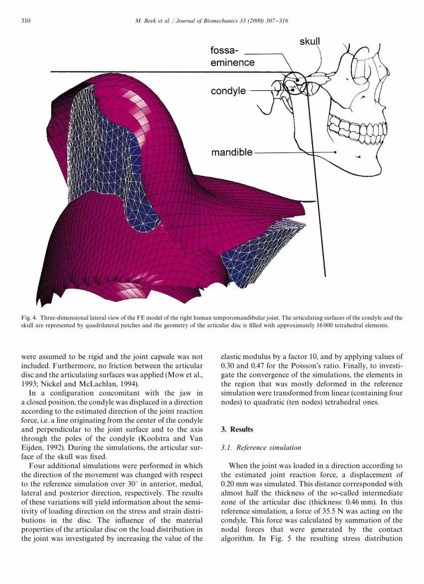
Fig. 4. Three-dimensional lateral view of the FE model of the right human temporomandibular joint. The articulating surfaces of the condyle and theskull are represented by quadrilateral patches and the geometry of the articular disc is "lled with approximately 16 000 tetrahedral elements.
were assumed to be rigid and the joint capsule was notincluded. Furthermore, no friction between the articulardisc and the articulating surfaces was applied (Mow et al.,1993; Nickel and McLachlan, 1994).
In a con"guration concomitant with the jaw ina closed position, the condyle was displaced in a directionaccording to the estimated direction of the joint reactionforce, i.e. a line originating from the center of the condyleand perpendicular to the joint surface and to the axisthrough the poles of the condyle (Koolstra and VanEijden, 1992). During the simulations, the articular sur-face of the skull was "xed.
Four additional simulations were performed in whichthe direction of the movement was changed with respectto the reference simulation over 303 in anterior, medial,lateral and posterior direction, respectively. The resultsof these variations will yield information about the sensi-tivity of loading direction on the stress and strain distri-butions in the disc. The in#uence of the materialproperties of the articular disc on the load distribution inthe joint was investigated by increasing the value of the
elastic modulus by a factor 10, and by applying values of0.30 and 0.47 for the Poisson's ratio. Finally, to investi-gate the convergence of the simulations, the elements inthe region that was mostly deformed in the referencesimulation were transformed from linear (containing fournodes) to quadratic (ten nodes) tetrahedral ones.
3. Results
3.1. Reference simulation
When the joint was loaded in a direction according tothe estimated joint reaction force, a displacement of0.20 mm was simulated. This distance corresponded withalmost half the thickness of the so-called intermediatezone of the articular disc (thickness: 0.46 mm). In thisreference simulation, a force of 35.5 N was acting on thecondyle. This force was calculated by summation of thenodal forces that were generated by the contactalgorithm. In Fig. 5 the resulting stress distribution
310 M. Beek et al. / Journal of Biomechanics 33 (2000) 307}316

Fig. 5. Distribution of the Von Mises stress (pVM
) after nine increments of joint loading in a frontal and inferior view of the disc. The stress isconcentrated in the lateral part of the thin ("intermediate) zone of the articular disc (sup"superior, inf"inferior, med"medial, lat"lateral).
M. Beek et al. / Journal of Biomechanics 33 (2000) 307}316 311

(Von Mises) on the surface of the disc is displayed. Thearticular disc appeared to be loaded predominantly inthe intermediate zone. The largest values of the stresseswere located in the lateral part of this zone. The strainswere not as concentrated as the stresses, but were alsomainly located in the intermediate zone. The maximumvalue of the equivalent elastic strain was 0.44.
3.2. Variation of the direction
A change in the direction of joint loading over 303 hadlittle in#uence on the distribution of strains and stressesin the articular disc (Fig. 6). Only in the cases witha laterally or medially directed load, a small shift of thestrain pattern in the concomitant direction could benoticed visually. However, the direction in#uenced theamount of displacement that was needed to obtain a sim-ilar amount of deformation. This displacement wassmallest (0.18 mm) when the direction of the movementwas changed over 303 in anterior direction and largest(0.34 mm) when the direction was changed in posteriordirection.
3.3. Variation of material properties
A force of 327 N was acting on the condyle aftera displacement of 0.2 mm, when the elastic modulus hada value of 60 MPa. The predicted strains did not di!ersubstantially from the strains predicted for the referencedisc elastic modulus. The predicted stresses increasedproportionally with the elastic modulus.
By comparing the results of the simulations obtainedby application of the three di!erent Poisson's ratiovalues, we observed that the predicted force acting on thecondyle were proportional to the applied Poisson's ratio,when the same amount of displacement was simulated.
3.4. Mesh accuracy
In Fig. 7 the Von Mises stress at node 1512 is displayedas a function of the increment number. This node waslocated on the top surface of the disc in the middle of theregion which bore the highest loads. In this area elementswere transformed from linear to quadratic in order tocheck the convergence. As can be seen, the Von Misesstress at this node predicted in the model containingquadratic elements, increased similarly with the jointload as in the original model. The amount of stress at the"nal increment di!ered by only 0.36% from the referencesimulation.
4. Discussion
The present model, as far as we know, is the "rstthree-dimensional FE model of the temporomandibular
joint disc. The disc was shaped according to its anatom-ical geometry which was sampled with high resolution.The transition zone between the "brous attachment ofthe superior head of the lateral pterygoid muscle and thearticular disc was also included in the present model.This probably explains why the disc of the present modeland that reported by other researchers (e.g., Chen andXu, 1994) had a thicker appearance anteriorly. The discwas allowed to move unrestrictedly between the articula-ting surfaces of the condyle and the skull, only kept inplace by the contact occurring between the disc and thesurfaces.
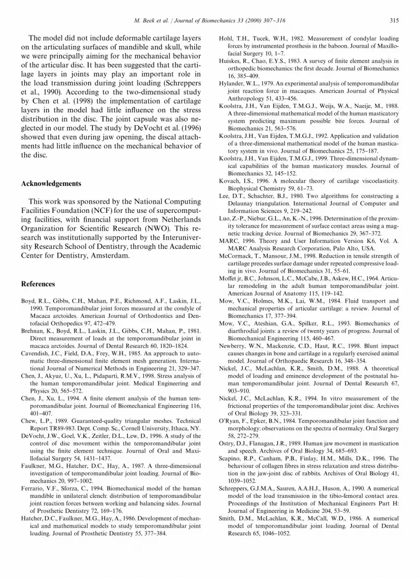
Although movement of the disc was not impeded byligaments or friction, the disc remained in the fossa dur-ing loading in this position. Variation of the loadingdirection over 603 led to only small changes in the stressand strain distributions. Therefore, it can be concludedthat during clenching, the highest deformations are al-ways located in the same region. The sketches in Fig. 8visualize the possible reason why a large variation ofloading direction has little in#uence on the deformationof the disc. Whatever the loading direction, the deforma-tion of the intermediate zone remains large compared tothe other regions in the disc. Fig. 6 might suggest thatduring various, both symmetric as well as asymmetric,loading conditions in a closed jaw position, large stressconcentrations occur predominantly in the lateral side ofthe intermediate zone of the disc. This is supported bya dissection study by Werner et al. (1991), who reportedthat wear leading to perforations was mainly located inthis region.
Thus far, no experimental measurements of stress andstrain distributions in the loaded articular disc havebecome available. Therefore, it is yet impossible to com-pare our predictions with such data. It is also still un-known whether the articular disc is preloaded when thejaw is in the closed position. Therefore, we assumed theinitial loads to be zero and our results should be inter-preted as relative values with respect to the initial con"g-uration.
Comparison of the results with previous "nite elementanalyses (Chen and Xu, 1994; DeVocht et al., 1996; Chenet al., 1998) is di$cult, because these simulations di!ersubstantially from those described in the present study.The studies were two-dimensional and did not describethe strain distributions. Furthermore, they mutually dif-fered in the direction and the amount of the displacementapplied to the condyle, and in the mechanical propertiesof the articular disc. Although the Von Mises stress is nota reliable representative of the mechanical stress ina structure when the deformations are very large, it wasnevertheless calculated in the present study to allowcomparison with the studies mentioned. When we ap-plied an elastic modulus according to Chen and Xu(1994), we obtained almost similar results. DeVocht et al.(1996) prescribed a displacement that was much larger
312 M. Beek et al. / Journal of Biomechanics 33 (2000) 307}316

Fig. 6. In#uence of the direction of the displacement of the condyle on the distribution of the equivalent elastic strain (e%-) in the articular disc. The
displacement was directed 303 anteriorly (A), posteriorly (B), medially (C), or laterally (D) with respect to the reference simulation.
than the ones applied in the studies by Chen and Xu(1994) and Chen et al. (1998) and the present one. Whilethis displacement was almost parallel to the articulatingsurface of the skull only small deformations of the discoccurred.
4.1. Model assumptions
The present "nite element model was based on thegeometry of the right temporomandibular joint of onlyone male cadaver. This particular joint did not deviate
M. Beek et al. / Journal of Biomechanics 33 (2000) 307}316 313
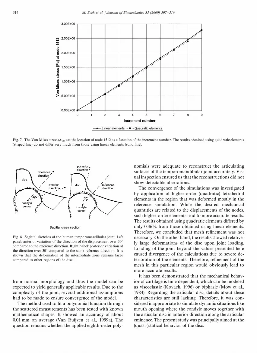
Fig. 7. The Von Mises stress (pVM
) at the location of node 1512 as a function of the increment number. The results obtained using quadratic elements(striped line) do not di!er very much from those using linear elements (solid line).
Fig. 8. Sagittal sketches of the human temporomandibular joint. Leftpanel: anterior variation of the direction of the displacement over 303compared to the reference direction. Right panel: posterior variation ofthe direction over 303 compared to the same reference direction. It isshown that the deformation of the intermediate zone remains largecompared to other regions of the disc.
from normal morphology and thus the model can beexpected to yield generally applicable results. Due to thecomplexity of the joint, several additional assumptionshad to be made to ensure convergence of the model.
The method used to "t a polynomial function throughthe scattered measurements has been tested with knownmathematical shapes. It showed an accuracy of about0.01 mm on average (Van Ruijven et al., 1999a). Thequestion remains whether the applied eighth-order poly-
nomials were adequate to reconstruct the articulatingsurfaces of the temporomandibular joint accurately. Vis-ual inspection ensured us that the reconstructions did notshow detectable aberrations.
The convergence of the simulations was investigatedby application of higher-order (quadratic) tetrahedralelements in the region that was deformed mostly in thereference simulation. While the desired mechanicalquantities are related to the displacements of the nodes,such higher-order elements lead to more accurate results.The results obtained using quadratic elements di!ered byonly 0.36% from those obtained using linear elements.Therefore, we concluded that mesh re"nement was notnecessary. On the other hand, the results showed relative-ly large deformations of the disc upon joint loading.Loading of the joint beyond the values presented herecaused divergence of the calculations due to severe de-terioration of the elements. Therefore, re"nement of themesh in this particular region would obviously lead tomore accurate results.
It has been demonstrated that the mechanical behav-ior of cartilage is time dependent, which can be modeledas viscoelastic (Kovach, 1996) or biphasic (Mow et al.,1984). Regarding the articular disc, details about thesecharacteristics are still lacking. Therefore, it was con-sidered inappropriate to simulate dynamic situations likemouth opening where the condyle moves together withthe articular disc in anterior direction along the articulareminence. The present study was principally aimed at the(quasi-)statical behavior of the disc.
314 M. Beek et al. / Journal of Biomechanics 33 (2000) 307}316
The model did not include deformable cartilage layerson the articulating surfaces of mandible and skull, whilewe were principally aiming for the mechanical behaviorof the articular disc. It has been suggested that the carti-lage layers in joints may play an important role inthe load transmission during joint loading (Schrepperset al., 1990). According to the two-dimensional studyby Chen et al. (1998) the implementation of cartilagelayers in the model had little in#uence on the stressdistribution in the disc. The joint capsule was also ne-glected in our model. The study by DeVocht et al. (1996)showed that even during jaw opening, the discal attach-ments had little in#uence on the mechanical behavior ofthe disc.
Acknowledgements
This work was sponsored by the National ComputingFacilities Foundation (NCF) for the use of supercomput-ing facilities, with "nancial support from NetherlandsOrganization for Scienti"c Research (NWO). This re-search was institutionally supported by the Interuniver-sity Research School of Dentistry, through the AcademicCenter for Dentistry, Amsterdam.
References
Boyd, R.L., Gibbs, C.H., Mahan, P.E., Richmond, A.F., Laskin, J.L.,1990. Temporomandibular joint forces measured at the condyle ofMacaca arctoides. American Journal of Orthodontics and Den-tofacial Orthopedics 97, 472}479.
Brehnan, K., Boyd, R.L., Laskin, J.L., Gibbs, C.H., Mahan, P., 1981.Direct measurement of loads at the temporomandibular joint inmacaca arctoides. Journal of Dental Research 60, 1820}1824.
Cavendish, J.C., Field, D.A., Frey, W.H., 1985. An approach to auto-matic three-dimensional "nite element mesh generation. Interna-tional Journal of Numerical Methods in Engineering 21, 329}347.
Chen, J., Akyuz, U., Xu, L., Pidaparti, R.M.V., 1998. Stress analysis ofthe human temporomandibular joint. Medical Engineering andPhysics 20, 565}572.
Chen, J., Xu, L., 1994. A "nite element analysis of the human tem-poromandibular joint. Journal of Biomechanical Engineering 116,401}407.
Chew, L.P., 1989. Guaranteed-quality triangular meshes. TechnicalReport TR89-983. Dept. Comp. Sc., Cornell University, Ithaca, NY.
DeVocht, J.W., Goel, V.K., Zeitler, D.L., Lew, D., 1996. A study of thecontrol of disc movement within the temporomandibular jointusing the "nite element technique. Journal of Oral and Maxi-llofacial Surgery 54, 1431}1437.
Faulkner, M.G., Hatcher, D.C., Hay, A., 1987. A three-dimensionalinvestigation of temporomandibular joint loading. Journal of Bio-mechanics 20, 997}1002.
Ferrario, V.F., Sforza, C., 1994. Biomechanical model of the humanmandible in unilateral clench: distribution of temporomandibularjoint reaction forces between working and balancing sides. Journalof Prosthetic Dentistry 72, 169}176.
Hatcher, D.C., Faulkner, M.G., Hay, A., 1986. Development of mechan-ical and mathematical models to study temporomandibular jointloading. Journal of Prosthetic Dentistry 55, 377}384.
Hohl, T.H., Tucek, W.H., 1982. Measurement of condylar loadingforces by instrumented prosthesis in the baboon. Journal of Maxillo-facial Surgery 10, 1}7.
Huiskes, R., Chao, E.Y.S., 1983. A survey of "nite element analysis inorthopedic biomechanics: the "rst decade. Journal of Biomechanics16, 385}409.
Hylander, W.L., 1979. An experimental analysis of temporomandibularjoint reaction force in macaques. American Journal of PhysicalAnthropology 51, 433}456.
Koolstra, J.H., Van Eijden, T.M.G.J., Weijs, W.A., Naeije, M., 1988.A three-dimensional mathematical model of the human masticatorysystem predicting maximum possible bite forces. Journal ofBiomechanics 21, 563}576.
Koolstra, J.H., Van Eijden, T.M.G.J., 1992. Application and validationof a three-dimensional mathematical model of the human mastica-tory system in vivo. Journal of Biomechanics 25, 175}187.
Koolstra, J.H., Van Eijden, T.M.G.J., 1999. Three-dimensional dynam-ical capabilities of the human masticatory muscles. Journal ofBiomechanics 32, 145}152.
Kovach, I.S., 1996. A molecular theory of cartilage viscoelasticity.Biophysical Chemistry 59, 61}73.
Lee, D.T., Schachter, B.J., 1980. Two algorithms for constructing aDelaunay triangulation. International Journal of Computer andInformation Sciences 9, 219}242.
Luo, Z.-P., Niebur, G.L., An, K.-N., 1996. Determination of the proxim-ity tolerance for measurement of surface contact areas using a mag-netic tracking device. Journal of Biomechanics 29, 367}372.
MARC, 1996. Theory and User Information Version K6, Vol. A.MARC Analysis Research Corporation, Palo Alto, USA.
McCormack, T., Mansour, J.M., 1998. Reduction in tensile strength ofcartilage precedes surface damage under repeated compressive load-ing in vivo. Journal of Biomechanics 31, 55}61.
Mo!et jr, B.C., Johnson, L.C., McCabe, J.B., Askew, H.C., 1964. Articu-lar remodeling in the adult human temporomandibular joint.American Journal of Anatomy 115, 119}142.
Mow, V.C., Holmes, M.K., Lai, W.M., 1984. Fluid transport andmechanical properties of articular cartilage: a review. Journal ofBiomechanics 17, 377}394.
Mow, V.C., Ateshian, G.A., Spilker, R.L., 1993. Biomechanics ofdiarthrodial joints: a review of twenty years of progress. Journal ofBiomechanical Engineering 115, 460}467.
Newberry, W.N., Mackenzie, C.D., Haut, R.C., 1998. Blunt impactcauses changes in bone and cartilage in a regularly exercised animalmodel. Journal of Orthopaedic Research 16, 348}354.
Nickel, J.C., McLachlan, K.R., Smith, D.M., 1988. A theoreticalmodel of loading and eminence development of the postnatal hu-man temporomandibular joint. Journal of Dental Research 67,903}910.
Nickel, J.C., McLachlan, K.R., 1994. In vitro measurement of thefrictional properties of the temporomandibular joint disc. Archivesof Oral Biology 39, 323}331.
O'Ryan, F., Epker, B.N., 1984. Temporomandibular joint function andmorphology: observations on the spectra of normalcy. Oral Surgery58, 272}279.
Ostry, D.J., Flanagan, J.R., 1989. Human jaw movement in masticationand speech. Archives of Oral Biology 34, 685}693.
Scapino, R.P., Canham, P.B., Finlay, H.M., Mills, D.K., 1996. Thebehaviour of collagen "bres in stress relaxation and stress distribu-tion in the jaw-joint disc of rabbits. Archives of Oral Biology 41,1039}1052.
Schreppers, G.J.M.A., Sauren, A.A.H.J., Huson, A., 1990. A numericalmodel of the load transmission in the tibio-femoral contact area.Proceedings of the Institution of Mechanical Engineers Part H:Journal of Engineering in Medicine 204, 53}59.
Smith, D.M., McLachlan, K.R., McCall, W.D., 1986. A numericalmodel of temporomandibular joint loading. Journal of DentalResearch 65, 1046}1052.
M. Beek et al. / Journal of Biomechanics 33 (2000) 307}316 315

Throckmorton, G.S., Dechow, P.C., 1994. In vitro measurements in thecondylar process of the human mandible. Archives of Oral Biology39, 853}867.
Van Ruijven, L.J., Beek, M., Van Eijden, T.M.G.J., 1999a. Fittingparametrized polynomials with scattered surface data. Journal ofBiomechanics 32, 715}720.
Van Ruijven, L.J., Beek, M., Donker, E., Van Eijden, T.M.G.J., 1999b.The accuracy of joint surface models constructed from dataobtained with an electromagnetic tracking device, submitted forpublication.
Werner, J.A., Tillmann, B., Schleicher, A., 1991. Functional anatomy ofthe temporomandibular joint. Anatomy and Embryology 183, 89}95.
316 M. Beek et al. / Journal of Biomechanics 33 (2000) 307}316


