Embed Size (px)
Citation preview

Three-dimensional in vivo kinematics of an osteoarthriticshoulder before and after total shoulder arthroplasty
Jonathan P. Braman,Department of Orthopaedic Surgery, University of Minnesota, 2450 Riverside Avenue, R200,Minneapolis, MN 55454, USA
Brian M. Thomas,Department of Orthopaedic Surgery, University of Minnesota, 2450 Riverside Avenue, R200,Minneapolis, MN 55454, USA
Robert F. LaPrade,Department of Orthopaedic Surgery, University of Minnesota, 2450 Riverside Avenue, R200,Minneapolis, MN 55454, USA
Vandana Phadke, andDepartment of Physical Medicine and Rehabilitation, University of Minnesota, 420 Delaware St SE,MMC 388, Minneapolis, MN 55455, USA
Paula M. LudewigDepartment of Physical Medicine and Rehabilitation, University of Minnesota, 420 Delaware St SE,MMC 388, Minneapolis, MN 55455, USAJonathan P. Braman: [email protected]
AbstractA case of a patient with glenohumeral joint arthritis is presented which demonstrated the three-dimensional shoulder motion before and after total shoulder arthroplasty. Pre-operative shouldermotion differed compared to normal controls, while post-operatively her glenohumeral-to-scapulothoracic motion ratios were restored to near normal ratios.
KeywordsGlenohumeral joint; Scapula; Biomechanics
IntroductionGlenohumeral osteoarthritis is a relatively uncommon, but debilitating, disease of the shoulder.Pain and restricted range of motion are typical symptoms. Progressive deterioration of articularcartilage within the glenohumeral joint results in changes in the shape and function of theglenohumeral articulation.
Primary treatment consists of activity modification and pain management. In patients with end-stage osteoarthritis, however, total shoulder arthroplasty is indicated. Excellent long-termfunctional outcomes have been reported in patients undergoing shoulder arthroplasty [2,12].
© Springer-Verlag 2010Correspondence to: Jonathan P. Braman, [email protected].
NIH Public AccessAuthor ManuscriptKnee Surg Sports Traumatol Arthrosc. Author manuscript; available in PMC 2011 December 1.
Published in final edited form as:Knee Surg Sports Traumatol Arthrosc. 2010 December ; 18(12): 1774–1778. doi:10.1007/s00167-010-1167-4.
NIH
-PA Author Manuscript
NIH
-PA Author Manuscript
NIH
-PA Author Manuscript

No study to date, however, has documented the in vivo three-dimensional kinematics of anosteoarthritic shoulder preoperatively and following total shoulder arthroplasty.
In previous studies, various techniques including surface sensors and bone pins have been usedto measure kinematics in healthy subjects and those with shoulder pathology [1,3,5,6,9–11].Invasive pins provide better rigid fixation to the osteology and therefore, should improve theaccuracy of shoulder motion analysis. However, the invasive nature of this technique haslimited its use. This case report is from a series of patients analyzed for a larger study. Thispatient ultimately underwent total shoulder arthroplasty. Post-operative data were collectedfrom surface sensors. Therefore, the three-dimensional clavicular, scapular and glenohumeralkinematics of an osteoarthritic shoulder in these planes, using direct bone-fixed tracking isdescribed.
Materials and methodsA 59-year-old woman (1.63 m tall, 79.5 kg, BMI 29.9) participated in an Institutional ReviewBoard-approved study that directly tested in vivo scapular and glenohumeral kinematics ofsubjects with and without shoulder pain using bone-fixed tracking [10]. Utilizing localanesthesia and sterile technique, stainless steel transcortical pins were inserted into her scapularspine, distal clavicle, and lateral humerus via a fluoroscopy-guided handheld drill. Motionsensors were rigidly fixed to the pins using an electromagnetic tracking system (AscensionTechnology Co, Burlington, VT, USA), and a skin marker was attached to the sternum to recordtrunk position. The Flock of Birds system is accurate to 1.8 mm and 0.5 degrees. Kinematicdata were collected for 2 repetitions each of open-chain shoulder forward flexion, abduction,and scapular plane abduction (40 degrees anterior to the coronal plane) without weight in thehand.
The subject’s shoulder motions were described by calculating the scapulohumeral rhythm aswell as graphing glenohumeral, scapular, and clavicular kinematics in various rotational planeswhen compared to average healthy subject data from previous work [1,10].
Approximately 3 months after the pin testing, the subject’s pain continued and she presentedfor clinical treatment. On both the anterior and posterior views, a circumferential humeral headosteophyte was observed, and a smaller glenoid osteophyte was visible on the anterior view(Fig. 1a). Pre-operatively, clinical examination showed 130 degrees of forward elevation, 115degrees of lateral elevation, external rotation at the side of 40 degrees, internal rotation at theside to the greater trochanter, 40 degrees of external rotation in abduction, and −15 degrees ofinternal rotation in abduction. She underwent total shoulder arthroplasty. Surgery wasperformed through a standard deltopectoral approach with a cemented pegged polyethyleneglenoid and an uncemented humeral component (Zimmer, Warsaw, IN USA) (Fig. 1b).
ResultsPre-operatively, the subject had diminished scapulohumeral rhythm ratios compared toasymptomatic subjects tested in previous shoulder motion studies [1,6,10]. Her scapulohumeralrhythm ratios from 30°-to-maximum during abduction, flexion, and scapular plane abductionwere calculated. For every 1 degree of glenohumeral elevation, she had upward scapularrotation of 1.9° in abduction, 0.7° in flexion, and 1.3° in scapular plane abduction. Thiscompared to 0.4°, 0.4°, and 0.4° in healthy individuals. These values yielded scapular rhythmratios of 0.5:1, 1.2:1, and 0.7:1 compared to healthy subject values of 2.1:1, 2.3:1, and 2.2:1.These values demonstrate excess scapular motion or scapular “substitution”.
Moreover, humeral angular elevation ranges relative to the trunk were noticeably lower (100°abduction, 130° flexion and scapular plane abduction) compared to healthy subjects (Fig. 2).
Braman et al. Page 2
Knee Surg Sports Traumatol Arthrosc. Author manuscript; available in PMC 2011 December 1.
NIH
-PA Author Manuscript
NIH
-PA Author Manuscript
NIH
-PA Author Manuscript
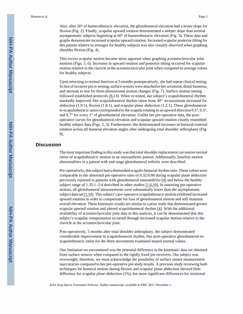
Also, after 50° of humerothoracic elevation, the glenohumeral elevation had a lesser slope forflexion (Fig. 2). Finally, scapular upward rotation demonstrated a steeper slope than normalasymptomatic subjects beginning at 60° of humerothoracic elevation (Fig. 3). These data andgraphs demonstrate increased scapular upward rotation. Increased scapular posterior tilting forthis patient relative to averages for healthy subjects was also visually observed when graphingshoulder flexion (Fig. 4).
This excess scapular motion became more apparent when graphing acromioclavicular jointmotions (Figs. 5, 6). Increases in upward rotation and posterior tilting occurred for scapularmotion relative to the clavicle at the acromioclavicular joint when compared to average valuesfor healthy subjects.
Upon returning to normal function at 5 months postoperatively, she had repeat clinical testing.In lieu of invasive pin re-testing, surface sensors were attached to her acromion, distal humerus,and sternum to test for three-dimensional motion changes (Fig. 7). Surface motion testingfollowed established protocols [8,13]. When re-tested, our subject’s scapulohumeral rhythmmarkedly improved. Her scapulohumeral rhythm ratios from 30° -to-maximum increased forabduction (1.9:1), flexion (1.8:1), and scapular plane abduction (1.2:1). These glenohumeral-to-scapulothoracic ratios corresponded to the scapula rotating in an upward direction 0.5°, 0.5°,and 0.7° for every 1° of glenohumeral elevation. Unlike her pre-operative data, the post-operative curves for glenohumeral elevation and scapular upward rotation closely resembledhealthy subject data (Figs. 2, 3). Furthermore, she demonstrated increases in humeral externalrotation across all humeral elevation angles after undergoing total shoulder arthroplasty (Fig.8).
DiscussionThe most important finding in this study was that total shoulder replacement can restore normalratios of scapulothoracic motion in an osteoarthritic patient. Additionally, baseline motionabnormalities in a patient with end-stage glenohumeral arthritis were described.
Pre-operatively, this subject had a diminished scapulo-humeral rhythm ratio. These values werecomparable to the abnormal pre-operative ratio of 0.32:0.68 during scapular plane abductionpreviously reported in patients with glenohumeral osteoarthritis [4] and below the healthysubject range of 1.35:1–2:4 described in other studies [1,6,10]. In assessing pre-operativemotion, all glenohumeral measurements were substantially lower than the asymptomaticsubject data set [1,10]. This subject’s pre-operative scapulothoracic motion exhibited increasedupward rotation in order to compensate for loss of glenohumeral motion and still maintainoverall elevation. These kinematic results are similar to a prior study that demonstrated greaterscapular upward rotation and altered scapulohumeral rhythm [4]. With the additionalavailability of acromioclavicular joint data in this analysis, it can be demonstrated that thissubject’s scapular compensation occurred through increased scapular motion relative to theclavicle at the acromioclavicular joint.
Post-operatively, 5 months after total shoulder arthroplasty, the subject demonstratedconsiderable improvement in scapulohumeral rhythm. Her post-operative glenohumeral-to-scapulothoracic ratios for the three movements examined neared normal values.
One limitation we encountered was the potential difference in the kinematic data we obtainedfrom surface sensors when compared to the rigidly fixed pin receivers. Our subject wasoverweight; therefore, we must acknowledge the possibility of surface sensor measurementinaccuracies compared to her pre-operative pin study results. A previous study reviewing bothtechniques for humeral motion during flexion and scapular plane abduction showed littledifference for scapular plane abduction (1%), but more significant differences for rotational
Braman et al. Page 3
Knee Surg Sports Traumatol Arthrosc. Author manuscript; available in PMC 2011 December 1.
NIH
-PA Author Manuscript
NIH
-PA Author Manuscript
NIH
-PA Author Manuscript

measurements [9]. Also below 120° of humeral elevation, scapular upward rotationmeasurements demonstrate small errors with surface sensors [7]. Consequently, the primaryfinding of alterations in linear motion and scapular rhythm associated with glenohumeralarthritis and subsequent improvement after arthroplasty makes these data valuable. Improvedunderstanding of shoulder motions in the normal and abnormal state could improve implantdesign, and rehabilitative technique. These data can be used to make perioperativerehabilitation programs address the actual motion abnormalities present in the arthriticshoulder. Furthermore, an understanding of shoulder kinematics may improve assessment ofpatients with shoulder pathology both before and following shoulder surgery.
ConclusionsIn conclusion, we found that shoulder motion differed between a patient with advancedglenohumeral osteoarthritis and healthy individuals. Total shoulder arthroplasty, however,restored the glenohumeral-to-scapulothoracic ratio to normal values. Further research isrecommended to quantify the pre-operative and post-operative in vivo kinematics in patientswith glenohumeral joint arthritis.
AcknowledgmentsThis study was supported by NIH grants K01HD042491 and R03HD053399 from the National Institute of ChildHealth and Human Development. The content is solely the responsibility of the authors and does not necessarilyrepresent the views of the National Institute of Child Health and Human Development or the National Institutes ofHealth. The authors would also like to thank Nicky Kangas RT, Cort J Cieminski PhD, PT, Mike McGinnity RN, andKelley Kyle, CST/CFA for their assistance with various aspects of data collection and analysis for this work.
References1. Braman JP, Engel SC, LaPrade RF, Ludewig PM. In vivo assessment of scapulohumeral rhythm during
unconstrained over head reaching in asymptomatic subjects. J Shoulder Elbow Surg 2010;19:216–223. [PubMed: 19733487]
2. Bryant D, Litchfield R, Sandow M, Gartsman GM, Guyatt G, Kirkley A. A comparison of pain,strength, range of motion, and functional outcomes after hemiarthroplasty and total shoulderarthroplasty in patients with osteoarthritis of the shoulder. A systematic review and meta-analysis. JBone Joint Surg Am 2005;87:1947–1956. [PubMed: 16140808]
3. Doody SG, Freedman L, Waterland JC. Shoulder movements during abduction in the scapular plane.Arch Phys Med Rehabil 1970;51:595–604. [PubMed: 5484648]
4. Fayad F, Roby-Brami A, Yazbeck C, Hanneton S, Lefevre-Colau MM, Gautheron V, Poiraudeau S.Three-dimensional scapular kinematics and scapulohumeral rhythm in patients with glenohumeralosteoarthritis or frozen shoulder. J Biomech 2008;41:326–332. [PubMed: 17949728]
5. Friedman RJ. Prospective analysis of total shoulder arthroplasty biomechanics. Am J Orthop1997;26:265–270. [PubMed: 9113293]
6. Inman VT, Saunders JB, Abbott LC. Observations on the function of the shoulder joint. J Bone JointSurg 1944;26A:1–30.
7. Karduna AR, McClure PW, Michener LA, Sennett B. Dynamic measurements of three-dimensionalscapular kinematics: a validation study. J Biomech Eng 2001;123:184–190. [PubMed: 11340880]
8. Ludewig PM, Cook TM. Alterations in shoulder kinematics and associated muscle activity in peoplewith symptoms of shoulder impingement. Phys Ther 2000;80:276–291. [PubMed: 10696154]
9. Ludewig PM, Cook TM, Shields RK. Comparison of surface sensor and Bone-fixed measurement ofhumeral motion. J Applied Biomech 2002;18:163–170.
10. Ludewig PM, Phadke V, Braman JP, Hassett DR, Cieminski CJ, LaPrade RF. Motion of the shouldercomplex during multiplanar humeral elevation. J Bone Joint Surg Am 2009;91:378–389. [PubMed:19181982]
Braman et al. Page 4
Knee Surg Sports Traumatol Arthrosc. Author manuscript; available in PMC 2011 December 1.
NIH
-PA Author Manuscript
NIH
-PA Author Manuscript
NIH
-PA Author Manuscript

11. McQuade KJ, Hwa Wei S, Smidt GL. Effects of local muscle fatigue on three-dimensionalscapulohumeral rhythm. Clin Biomech 1995;10:144–148.
12. Radnay CS, Setter KJ, Chambers L, Levine WN, Bigliani LU, Ahmad CS. Total shoulder replacementcompared with humeral head replacement for the treatment of primary gleno-humeral osteoarthritis:a systematic review. J Shoulder Elbow Surg 2007;16:396–402. [PubMed: 17582789]
13. Wu G, van der Helm FC, Veeger HE, Makhsous M, Van Roy P, Anglin C, et al. International societyof biomechanics ISB recommendation on definitions of joint coordinate systems of various joints forthe reporting of human joint motion-Part II: shoulder, elbow, wrist and hand. J Biomech2005;38:981–992. [PubMed: 15844264]
Braman et al. Page 5
Knee Surg Sports Traumatol Arthrosc. Author manuscript; available in PMC 2011 December 1.
NIH
-PA Author Manuscript
NIH
-PA Author Manuscript
NIH
-PA Author Manuscript

Fig. 1.Right shoulder AP radiographs: a Pre-operative. b Post-operative
Braman et al. Page 6
Knee Surg Sports Traumatol Arthrosc. Author manuscript; available in PMC 2011 December 1.
NIH
-PA Author Manuscript
NIH
-PA Author Manuscript
NIH
-PA Author Manuscript

Fig. 2.Glenohumeral elevation relative to the scapula during average of two trials of arm elevationin flexion. filled circle pre-operative pin data (in blue), times post-operative surface data (ingreen), open circle mean and standard error of healthy subject data (n = 12) (in red). Negativevalues indicate elevation
Braman et al. Page 7
Knee Surg Sports Traumatol Arthrosc. Author manuscript; available in PMC 2011 December 1.
NIH
-PA Author Manuscript
NIH
-PA Author Manuscript
NIH
-PA Author Manuscript

Fig. 3.Scapular upward rotation relative to the trunk during average of two trials of arm elevation inabduction. filled circle pre-operative pin data (in blue), times post-operative surface data (ingreen), open circle mean and standard error of healthy subject data (n = 12) (in red). Negativevalues indicate upward rotation
Braman et al. Page 8
Knee Surg Sports Traumatol Arthrosc. Author manuscript; available in PMC 2011 December 1.
NIH
-PA Author Manuscript
NIH
-PA Author Manuscript
NIH
-PA Author Manuscript
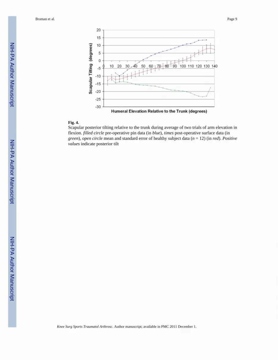
Fig. 4.Scapular posterior tilting relative to the trunk during average of two trials of arm elevation inflexion. filled circle pre-operative pin data (in blue), times post-operative surface data (ingreen), open circle mean and standard error of healthy subject data (n = 12) (in red). Positivevalues indicate posterior tilt
Braman et al. Page 9
Knee Surg Sports Traumatol Arthrosc. Author manuscript; available in PMC 2011 December 1.
NIH
-PA Author Manuscript
NIH
-PA Author Manuscript
NIH
-PA Author Manuscript

Fig. 5.Acromioclavicular posterior tilt relative to the clavicle during average of two trials of armelevation in flexion. filled circle pre-operative pin data (in blue), open circle mean and standarderror of healthy subject data (n = 12) (in red). Positive values indicate posterior tilt
Braman et al. Page 10
Knee Surg Sports Traumatol Arthrosc. Author manuscript; available in PMC 2011 December 1.
NIH
-PA Author Manuscript
NIH
-PA Author Manuscript
NIH
-PA Author Manuscript

Fig. 6.Acromioclavicular upward rotation relative to the clavicle during average of two trials of armelevation in abduction. filled circle pre-operative pin data (in blue), open circle mean andstandard error of healthy subject data (n = 12) (in red). Negative values indicate upward rotation
Braman et al. Page 11
Knee Surg Sports Traumatol Arthrosc. Author manuscript; available in PMC 2011 December 1.
NIH
-PA Author Manuscript
NIH
-PA Author Manuscript
NIH
-PA Author Manuscript

Fig. 7.Subject with post-operative surface testing of right shoulder during scapular plane abduction.The barrier in front of her serves only to control the plane of scapular elevation. There is nopressure against the wall with elevation
Braman et al. Page 12
Knee Surg Sports Traumatol Arthrosc. Author manuscript; available in PMC 2011 December 1.
NIH
-PA Author Manuscript
NIH
-PA Author Manuscript
NIH
-PA Author Manuscript

Fig. 8.Glenohumeral external rotation relative to the scapula during average of two trials of armelevation in abduction. filled circle pre-operative pin data (in blue), times post-operative surfacedata (in green), open circle mean and standard error of healthy subject data (n = 12) (in red).Negative values indicate external rotation
Braman et al. Page 13
Knee Surg Sports Traumatol Arthrosc. Author manuscript; available in PMC 2011 December 1.
NIH
-PA Author Manuscript
NIH
-PA Author Manuscript
NIH
-PA Author Manuscript


