Embed Size (px)
Citation preview
RESEARCH ARTICLE Open Access
Three genes expressing Kunitz domains in theepididymis are related to genes of WFDC-typeprotease inhibitors and semen coagulum proteinsin spite of lacking similarity between their proteinproductsAdam Clauss1,3, Margareta Persson1, Hans Lilja1,2 and Åke Lundwall1*
Abstract
Background: We have previously identified a locus on human chromosome 20q13.1, encompassing related genesof postulated WFDC-type protease inhibitors and semen coagulum proteins. Three of the genes with WFDC motifalso coded for the Kunitz-type protease inhibitor motif. In this report, we have reinvestigated the locus forhomologous genes encoding Kunitz motif only. The identified genes have been analyzed with respect to structure,expression and function.
Results: We identified three novel genes; SPINT3, SPINT4 and SPINT5, and the structure of their transcripts weredetermined by sequencing of DNA generated by rapid amplification of cDNA ends. Each gene encodes a Kunitzdomain preceded by a typical signal peptide sequence, which indicates that the proteins of 7.6, 8.7, and 9.7 kDaare secreted. Analysis of transcripts in 26 tissues showed that the genes predominantly are expressed in theepididymis. The recombinantly produced proteins could not inhibit the amidolytic activity of trypsin, chymotrypsin,plasmin, thrombin, coagulation factor Xa, elastase, urokinase and prostate specific antigen, whereas similarly madebovine pancreatic trypsin inhibitor (BPTI) had the same bioactivity as the protein isolated from bovine pancreas.
Conclusions: The similar organization, chromosomal location and site of expression, suggests that the novel genesare homologous with the genes of WFDC-type protease inhibitors and semen coagulum proteins, despite the lackof similarity in primary structure of their protein products. Their restricted expression to the epididymis suggeststhat they could be important for male reproduction. The recombinantly produced proteins are presumablybioactive, as demonstrated with similarly made BPTI, but may have a narrower spectrum of inhibition, as indicatedby the lacking activity against eight proteases with differing specificity. Another possibility is that they have lost theprotease inhibiting properties, which is typical of Kunitz domains, in favor of hitherto unknown functions.
BackgroundThe mammalian semen coagulum proteins are a hetero-geneous collection of proteins secreted at very high con-centration by the seminal vesicles. There are twohomologous semen coagulum proteins, denoted seme-nogelin I (SEMG1) and semenogelin II (SEMG2), inmost primates [1-3]. Duplication of tandem repeats of
60 amino acid residues in both SEMG1 and SEMG2 isresponsible for most of the size heterogeneity of seme-nogelin molecules in primates, but the frequent occur-rence of premature stop codons is also a contributingfactor [1,4,5]. It has also been reported that the struc-ture of semenogelin molecules can be affected by geneconversion [6].The murine seminal vesicles secrete six proteins, Svs1-
Svs6, at high concentration [7-9]. It has been shown thatthe genes of Svs2-Svs6, and SEMG1 and SEMG2 arehomologous, in spite of lacking similarity between their
* Correspondence: [email protected] University, Department of Laboratory Medicine, Clinical Chemistry,Skåne University Hospital, SE-205 02 Malmö, SwedenFull list of author information is available at the end of the article
Clauss et al. BMC Biochemistry 2011, 12:55http://www.biomedcentral.com/1471-2091/12/55
© 2011 Clauss et al; licensee BioMed Central Ltd. This is an Open Access article distributed under the terms of the Creative CommonsAttribution License (http://creativecommons.org/licenses/by/2.0), which permits unrestricted use, distribution, and reproduction inany medium, provided the original work is properly cited.
protein products [10,11]. This apparent paradox isexplained by rapid evolution of single exon that codesfor most of the gene product. The homologous genesare composed of three exons, the first of which encodesthe signal peptide and the very first residues of thesecreted protein, the second codes for the remainder ofthe protein and also carries a few 3’ non-translatednucleotides, whereas the third exon only has 3’ non-translated nucleotides and holds the poly-adenylationsignal. Sequence comparison shows that the first andlast exons are conserved in these genes, whereas the sec-ond exon is not [10]. Differing selection of splice accep-tor sites and species unique repeat expansion have beenproposed to explain the diversity of the second exon[10].The proposed evolutionary mechanisms suggest that
the seminal vesicle-transcribed genes might be relatedto genes with similar organization that code forsecreted, but structurally very different proteins. Thishypothesis led to the discovery that the genes of theelastase inhibitors elafin and secretory leukocyte pro-tease inhibitor (SLPI), and the predominating seminalvesicle-secreted proteins are homologous, in spite oflacking similarity between their protein products [12].Elafin and SLPI belong to the whey acidic proteinfour-disulfide core (WFDC) family of small proteaseinhibitors, which interacts with cognate proteases bywhat is known as the standard mechanism, by whichthe protease-binding loop of the inhibitors interactswith the catalytic site of proteases in a similar way assubstrates [13].With the advent of nucleotide sequences from the
Human Genome Project, it became possible to assignthe chromosomal location of genes in detail. Analysis ofthe human semenogelin locus on chromosome 20q12-13.1 showed that PI3, the gene of elafin, and SLPI areflanking SEMG1 and SEMG2 and an extended analysisof the chromosomal region identified 12 additional,homologous genes encompassing WFDC motif [14,15].Most of the WFDC genes and Svs2-Svs6 are conservedat the homologous chromosomal region in the mouse,which suggests that the semen coagulum proteins prob-ably originate from a WFDC gene [16]. Many of theWFDC genes are with certain specificity expressed inthe epididymis and display accelerated evolution, whichmight indicate that they are of importance in malereproduction [16].Three of the novel genes with WFDC motifs, WFDC8,
SPINLW1, and WFDC6 also carry the motif of Kunitz-type protease inhibitors [14]. Similar to the WFDCdomain, the conserved Kunitz domain is present in sev-eral proteins that inhibit proteases by the standardmechanism [13]. A well-known and much studied exam-ple is the bovine pancreatic trypsin inhibitor (BPTI), also
known as aprotinin and until recently marketed as ananti-bleeding drug under the name of Trasylol®. Kunitzdomains are also present in important protease inhibi-tors, such as the tissue factor pathway inhibitor, whichregulates blood coagulation, and the light chain, alsoknown as bikunin, of the multifunctional inter-alphatrypsin inhibitor [17-19].We have in several previous studies, as outlined above,
demonstrated that many of the genes at the semenoge-lin/WFDC locus are expressed in the male reproductivetract and display accelerated evolution by a variety ofmechanisms. Most compelling are the findings suggest-ing that a WFDC gene gave rise to the genes of thesemen coagulum proteins, as it also rises the questionwhether there could be still more genes at the locus onchromosome 20 that have gone through a similar meta-morphosis. Our finding that the protease inhibitor locuson chromosome 20 carries several genes that code forinhibitors containing WFDC motifs only, while othergenes encode products containing both WFDC andKunitz motifs, raised the question as to whether therealso exists homologous genes encoding putative inhibi-tors with Kunitz motifs alone. In this report, we havesearched the locus for novel genes encoding proteinswith Kunitz motifs. The identified genes have been ana-lyzed with respect to structure, expression and function.
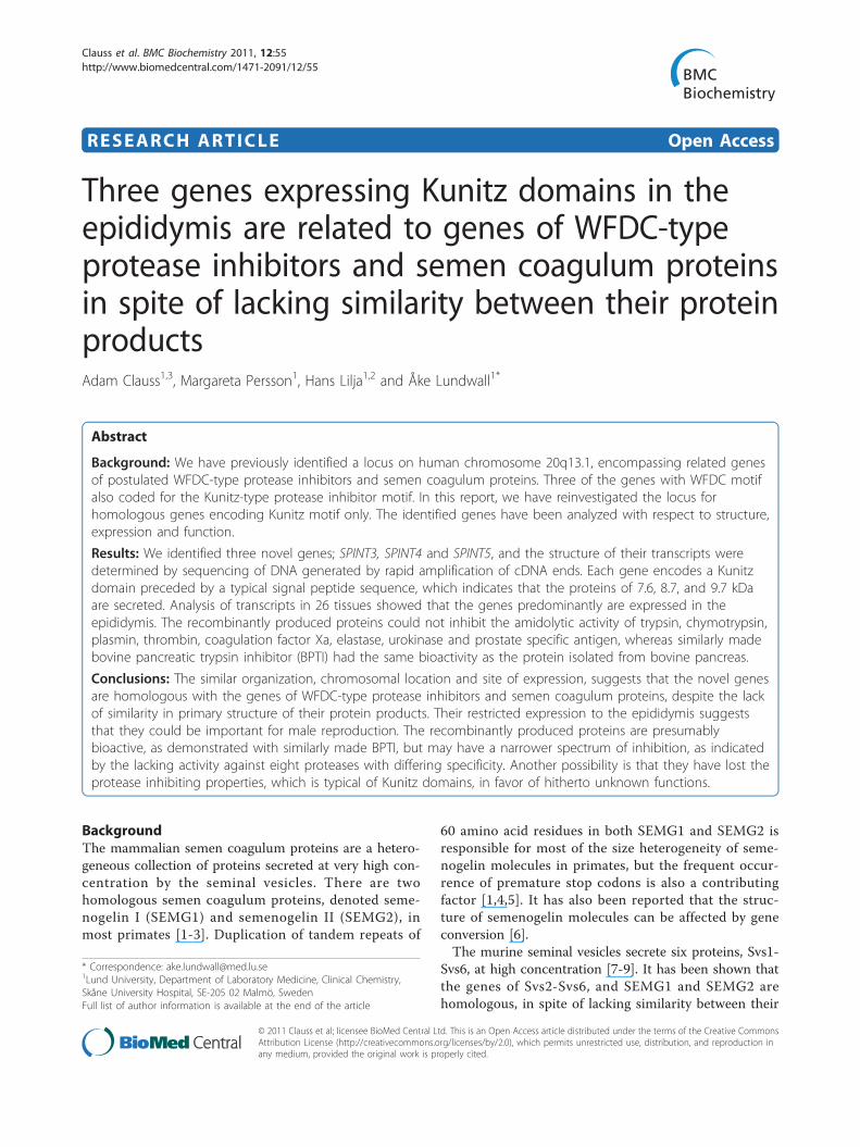
ResultsIdentification of three novel genes encoding KunitzdomainsThe nucleotide sequence of 1.8 Mb centered on the 0.7Mb WFDC locus on chromosome 20q13.1 was trans-lated in 6 reading frames and screened for Kunitzmotifs. A total of six Kunitz motifs were detected, threeof which were associated with proteins containing bothKunitz and WFDC motifs; the previously characterizedSPINLW1, WFDC8 and WFDC6 [14,20]. The humanWFDC locus consists of two subloci and the six genesencoding Kunitz motifs are all located at the telomericsublocus (Figure 1). The first of the novel genes,SPINT3, is located 34 kb on the telomeric side ofWFDC2 and 19 kb on the centromeric side of WFDC6.We located SPINT4 and SPINT5 next to each other,separated by 25 kb of intergenic DNA, with SPINT4located 20 kb on the telomeric side of WFDC13 andSPINT5 25 kb on the centromeric side of WFDC3.
Structure of transcriptsThe nucleotide sequences of full-length transcripts ofthe novel genes were obtained from overlappingsequences generated by 3’, and 5’ RACE with cDNAfrom epididymis. The procedure yielded a single tran-script for SPINT3, whereas two transcripts were asso-ciated with SPINT4 and presumably three with SPINT5.
Clauss et al. BMC Biochemistry 2011, 12:55http://www.biomedcentral.com/1471-2091/12/55
Page 2 of 13
All genes give rise to transcripts that encode a Kunitzdomain preceded by a signal peptide of 24 amino acidresidues, indicating that the protein products aresecreted. There are no signals for N-linked glycosylationin the primary structures, which suggests that the pro-teins are secreted without carbohydrate chains. Sizes ofexons and introns are given in Table 1.The SPINT3 transcript of 476 bp [GenBank:
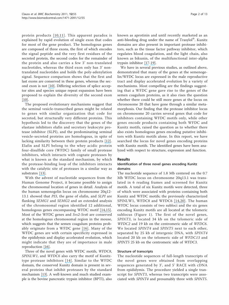
AY372172] terminates in a poly-A tail that is precededby an unconventional poly-adenylation signal, TATAAA,which notably is identical to the canonical TATA-boxsequence in eukaryotic genes (Figure 2A). It gives rise toa polypeptide of 89 amino acid residues that will be pro-cessed to a secreted protein of 7.6 kDa. The gene is notpreceded by a conventional TATA-box, but there is anA and T rich sequence, TAAAAT, 26 bp upstream tothe transcription initiation site.The two SPINT4 transcripts are 380 [GenBank:
AY372173] and 514 [GenBank: AY372174] nucleotidesrespectively. The difference between the transcripts isfound in the 3’ non-translated part, which in the smallertranscript consists of 61 nucleotides that overlap withthose in the longer transcript of 194 nucleotides (Figure
2B). The conventional poly-adenylation signal,AATAAA, used in the shorter transcript, is skipped inthe longer transcript for an unconventional poly-adeny-lation signal, AATGAAA, located further downstream.Both transcripts give rise to a polypeptide of 99 aminoacid residues, which is processed to a protein of 8.7kDa, encompassing a Kunitz motif that is lacking thefifth Cys of the consensus motif. Normally this wouldyield a free thiol group in the third Cys, the bindingpartner of the fifth Cys in the motif. However, there isanother Cys located upstream to the Kunitz motif thatpotentially can form a disulphide with the third Cys ofthe motif (Figure 2B). There is a TATA-like sequence,TATAAC, preceding the gene by 25 bp.It was more difficult to obtain RACE products from
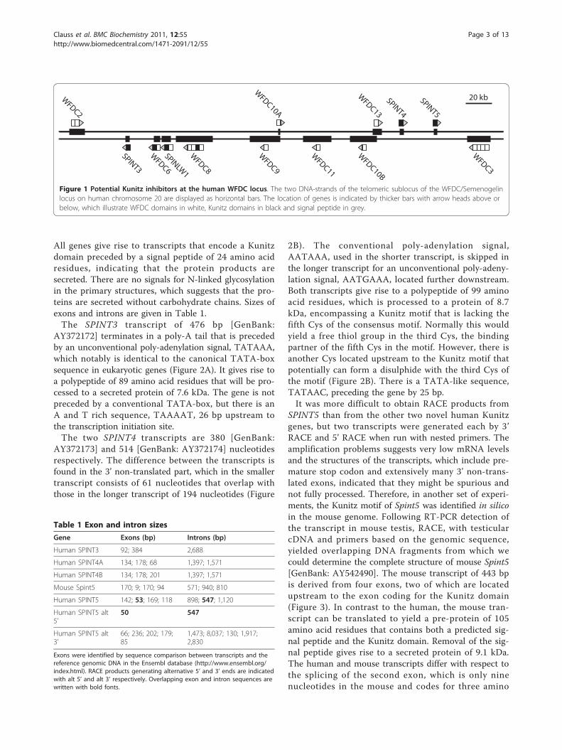
SPINT5 than from the other two novel human Kunitzgenes, but two transcripts were generated each by 3’RACE and 5’ RACE when run with nested primers. Theamplification problems suggests very low mRNA levelsand the structures of the transcripts, which include pre-mature stop codon and extensively many 3’ non-trans-lated exons, indicated that they might be spurious andnot fully processed. Therefore, in another set of experi-ments, the Kunitz motif of Spint5 was identified in silicoin the mouse genome. Following RT-PCR detection ofthe transcript in mouse testis, RACE, with testicularcDNA and primers based on the genomic sequence,yielded overlapping DNA fragments from which wecould determine the complete structure of mouse Spint5[GenBank: AY542490]. The mouse transcript of 443 bpis derived from four exons, two of which are locatedupstream to the exon coding for the Kunitz domain(Figure 3). In contrast to the human, the mouse tran-script can be translated to yield a pre-protein of 105amino acid residues that contains both a predicted sig-nal peptide and the Kunitz domain. Removal of the sig-nal peptide gives rise to a secreted protein of 9.1 kDa.The human and mouse transcripts differ with respect tothe splicing of the second exon, which is only ninenucleotides in the mouse and codes for three amino
WFDC2
WFDC6
WFDC8
WFDC10B
WFDC11
WFDC10A
WFDC9
WFDC13
WFDC3
SPINT4
SPINT5
SPINLW1
SPINT3
20 kb
Figure 1 Potential Kunitz inhibitors at the human WFDC locus. The two DNA-strands of the telomeric sublocus of the WFDC/Semenogelinlocus on human chromosome 20 are displayed as horizontal bars. The location of genes is indicated by thicker bars with arrow heads above orbelow, which illustrate WFDC domains in white, Kunitz domains in black and signal peptide in grey.
Table 1 Exon and intron sizes
Gene Exons (bp) Introns (bp)
Human SPINT3 92; 384 2,688
Human SPINT4A 134; 178; 68 1,397; 1,571
Human SPINT4B 134; 178; 201 1,397; 1,571
Mouse Spint5 170; 9; 170; 94 571; 940; 810
Human SPINT5 142; 53; 169; 118 898; 547; 1,120
Human SPINT5 alt5’
50 547
Human SPINT5 alt3’
66; 236; 202; 179;85
1,473; 8,037; 130; 1,917;2,830
Exons were identified by sequence comparison between transcripts and thereference genomic DNA in the Ensembl database (http://www.ensembl.org/index.html). RACE products generating alternative 5’ and 3’ ends are indicatedwith alt 5’ and alt 3’ respectively. Overlapping exon and intron sequences arewritten with bold fonts.
Clauss et al. BMC Biochemistry 2011, 12:55http://www.biomedcentral.com/1471-2091/12/55
Page 3 of 13
acid residues. The splice acceptor site of the mousetranscript is structurally conserved in the human gen-ome and, if used, it could generate a translation productencompassing both a signal peptide and a secreted pro-tein of 9.7 kDa with a Kunitz domain (Figure 3).The RACE procedure also yielded products for alterna-
tive 5’ and 3’ ends of human SPINT5. The sequence of ashorter 5’ RACE product overlaps with the human tran-script that is orthologous with mouse Spint5, but startsthree nucleotides downstream of the splice acceptor siteof the second exon (Figure 3). This transcript couldpotentially generate an intracellular Kunitz domain, as itis lacking the coding information of the signal peptide.The alternative 3’ end is longer and derived from 5 exonslocated 3’ to the exon with the Kunitz motif.
Expression of transcripts in normal human tissuesGene expression was analyzed by RT-PCR using RNAfrom a panel of 26 tissue specimens that previously hadbeen screened for genes encoding WFDC domains [14].Tissues that generated PCR products with the correctsize of spliced transcripts were taken to quantitative ana-lysis using real time PCR. The housekeeping genes CSTBand APRT, which were used for normalization of thePCR data, generated transcripts with melting
temperatures of 80°C and 81.5°C and yielded Ct values inthe range of 23-25 and 25-27 respectively. SPINT3 andSPINT4 gave rise to transcripts with melting tempera-tures of 76.5°C and 74°C and with cDNA from epididy-mis they generated Ct values that were similar or slightlylower than those of CSTB, but for the remainder of tis-sues the Ct values were 32 or higher; in a few tissuestranscript was not even detectable. Different primer com-binations were tested in order to measure the expressionof SPINT5, but a detectable and specific signal was onlyobtained with a primer pair that primed in the Kunitzdomain and in the last exon of the transcript; it yielded aCt value of 35-36 in the epididymis. The efficacy of thePCR reactions was analyzed by linear regression of Ctvalues generated by serial dilution of epididymal cDNA(Table 2). An efficacy of 2 was assumed for SPINT5, aslow levels prohibited analysis of its transcript by linearregression. Calculation of the relative expression showsthat the level of SPINT3 and SPINT4 transcripts in theepididymis exceeds those in other tissues by more thanthree orders of magnitude (Table 3).
Recombinant expression and functional analysisRecombinant expression yielded proteins of around 27kDa, which contain the Kunitz domains fused to vector
M Q L Q A S L S F L L I L T L C L E L R S E L A RCTGGCTGAGTGGCACCATGCAGCTTCAGGCCTCTCTCTCGTTTCTCCTGATTCTCACTCTCTGCCTAGAGCTTCGATCAGAACTAGCACG D T I K D L L P N V C A F P M E K G P C Q T Y M T R W F F NAGACACTATCAAGGATCTCCTCCCAAATGTATGCGCTTTTCCTATGGAAAAGGGCCCTTGTCAAACCTACATGACGCGATGGTTTTTCAA F E T G E C E L F A Y G G C G G N S N N F L R K E K C E K FCTTTGAAACTGGTGAATGTGAGTTATTTGCTTACGGAGGCTGCGGAGGCAACAGCAACAACTTTTTGAGGAAAGAAAAATGTGAGAAATT C K F TCTGCAAGTTCACCTGATTTTCTAACAAGAACACAGCCCTCCATGGATTCGGGATTGCTCTGAGGGCCATAGAAGGCATTTGCGTGTGTGTGTGTGTGTGAACAAGAGGTTCATTCCTCTACCCCCACATTTATTCTCCCTGATGTGCTCCTACAAGTGCTTCATCTCTGTGCCTGAAATTCTATAAATGTTGCAATCATG
M K S A K L G F L L R F F I F C S L N T L L L GGCCTGCTGCTTGCTGCACCATGAAGTCTGCCAAGCTGGGATTTCTTCTAAGATTCTTCATCTTCTGCTCATTGAATACCCTGTTATTGGG G V N K I A E K I C G D L K D P C K L D M N F G S C Y E V HTGGTGTTAATAAAATTGCGGAGAAGATATGTGGAGACCTCAAAGATCCCTGCAAATTGGACATGAATTTTGGAAGCTGCTATGAAGTTCA F R Y F Y N R T S K R C E T F V F S G C N G N L N N F K L KCTTTAGATATTTCTACAACAGAACCTCCAAAAGATGTGAAACTTTTGTCTTCTCCGGCTGTAATGGCAACCTTAACAACTTCAAGCTTAA I E R E V A C V A K Y K P P RAATAGAACGTGAAGTAGCCTGTGTTGCAAAATACAAACCACCGAGGTGAGAGGATGTGAACTCATGAAGTTGTCTGCTGCACCATCCGAAATAAAGACACAAGAAAATTCAGACTGATTTTGAAAATCTTTGTAATATTTCCATAATGCTTTAAGCTTCCATATGTTTGCTATTTTCCTGACCCTAGTTTTGCCTTTCCTGGAAATTAACTGTATGATCATTAGAATGAAAGAGTCTTTCTGTC
A
B
Figure 2 Structure of SPINT3 and SPINT4. The nucleotide sequences of transcripts are given with translation written above for (A) SPINT3 and(B) SPINT4. The arrows indicate location of the predicted signal peptide cleavage sites. The two Cys in SPINT4, which might form anunconventional disulphide bond, are boxed. The nucleotides present in the longer SPINT4 transcript, but not in the shorter, are italicized.
Clauss et al. BMC Biochemistry 2011, 12:55http://www.biomedcentral.com/1471-2091/12/55
Page 4 of 13
encoded thioredoxin. Experiments with BPTI and BSTI,which served as positive controls, yielded around 5 mgof recombinant protein from 1 L of cultured bacteriaafter purification on Ni-chelate column. The isolatedproteins were highly pure and appeared as single bandson SDS-PAGE under both reducing and non-reducingconditions. The isolated proteins were functionallyactive, as demonstrated by the inhibition of trypsin,plasmin and chymotrypsin by approximately equimolar
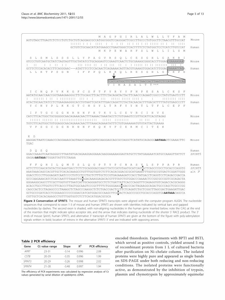
M A G P G I R L A L W L L T F A MAGTGTAGCTTGAGTCTCTCCTGTCTGCTGTCAGGAGCGCCATGGCGGGTCCAGGGATTCGCCTTGCCCTGTGGCTTCTAACGTTTGCCAT |||||| | | | |||| | | | || || | || | || ||||| || | || |||| GCTGTCTGCAACATCATGAAGCCTGAATGGGCTCACTTTTCTCTATGGCTCCTCATCTTGTCCAT Human M K P E W A H F S L W L L I L S M
S L S M L S S S L L Y P A G V R S Q L C E S G H L G A K R IGTCCCTGTCAATGCTATCTAGTAGTTTGCTATATCCTGCAGGAGTCCGAAGTCAACTCTGCGAAAGCGGGCACCTTGGAGCGAAAAGAAT Mouse| || | | || | | ||| |||| || | || ||| || | | ||||| || || ||||||| ||| |GCTTCTCCACACACCTTCAGGGAAC---ATATTTCCTCCACAACTCAGAAAACAGTTACGTGAAAGTGGACATCTTGGAGTAAAACCAGA Human L L H T P S G N I F P P Q L R K Q L R E S G H L G V K P E
TCATCATGGCTTCTGCTCTAACCACTTAATCTCCATGGTCTCAGV I M A S A L T T ***
I C N Q P V K K G F C S F T F Y R Y Y F N P E S A L C E S FAATATGCAACCAACCGGTAAAGAAGGGCTTCTGCAGCTTCACTTTCTACAGATACTACTTCAACCCAGAATCGGCCCTATGTGAGTCCTT Mouse| |||||| | || | ||||||||| ||| | | ||| ||||| ||||| |||| | | |||||||| ||||ATACTGCAACTATCCTCTGAAGAAGGGCACCTGTAATTCATATTTGACCAGATTCTACTACAACACTTTGACATTTTTATGTGAGCCCTT Human Y C N Y P L K K G T C N S Y L T R F Y Y N T L T F L C E P F
I F T G C G G N R N N F K T K Y L C E V R C I H V E CATCTTCACTGGCTGCGGGGGCAACAGAAACAACTTTAAAACTAAATACCTCTGTGAAGTCCGTTGCATTCACGTAGAG Mouse |||||| |||||| || ||||||||||||||||||||| ||||| |||||||| || ||| | | |TGTCTTCAGTGGCTGTGGAGGCAACAGAAACAACTTTAAACAGAAATATTTCTGTGAAAAAATGTGTATTACTGAAAA Human V F S G C G G N R N N F K Q K Y F C E K M C I T E K
K DAAGGACTAATCCAAACCCAGGAAAGCAGTAAGCGAAGGATGCAAGGAGCAGCGCCAGGCTCATATGCAGAGGGAATAAAGTCGAAACTTC MouseTGAC
D Q S Q HumanGGACCAAAGTCAATAGGGCCTTAATATGACAGAAGGAGGAAACAAGGAAAAAGGAGTATGCTCTATGAAAAATCATGCCAAAGTTATTCT SPINT5GAGAAAATAAATTGGATTATTTCTAAAA
F F Q H I L Q M T S L Q G P T Y P C N A S V L S P P A P R HumanATTCTTCCAACATATACTGCAAATGACCTCTCTGCAGGGACCAACCTATCCATGTAATGCATCAGTACTCAGCCCGCCTGCACCCAGGTG SPINT5AAATAAACAGCCACGTTGCTCACACAAAGCCTGTTTGGTGGTCTCTTCACACGGACGCGCATGAAATTTGGTGCCGTGACTCGGATTGGG alt 3’GGACCTCCCTTGGGAGATCAATCCCCTGTCCTCCTGCTCTTTGCTCCGTGAGAAAGATCCACCTATGACCTCAGGTCCTCAGACCGACCAGCCCAAGAAACATCTCACCAATTTTAAATTGGAGACAAAGGAGACACGTTTTATCTGTGGACCCAAAACTCCGGCGCCGGTCGCAGACTAGGGAAGGCAGCCTTCGCTTGGTGTTTAATCATTGCAGGGATGCCTCTCTGATTATTCACCCAGGTTTCAGAGGTGTCAGACCACGCAGGGACACCTGCCTTGGTCCTTCACCCTTAGTGGCAAGTCCCGCTTTTCTGGGGAAGGGGACCCACTAGAAGGCAGACTGCCCAGCTCGCCCGGCAGCCACTCCTAGAGCCCCTAAAGCTCTAGCCCAAGGCTCTCTGACCGACTCCTTCTCAGATCTGCTCGGCTTAGCGACTGAAGATTGACGCTGCCCGATCACCTCGGAAGCCCCCCGGACCATCATGGACGCCAAGCTTCAGACTCAGCCCGCCTGCACCCAGGTGAAATAAACAGCCACGTTGCTCACACAAAGCCTGTTTGGTGGTCTCTTCACATGGACGCGCA
Mouse
Figure 3 Conservation of SPINT5. The mouse and human SPINT5 transcripts were aligned with the computer program ALIGN. The nucleotidesequences that correspond to exon 1-3 of mouse and human SPINT5 are shown with identities indicated by vertical bars and gappednucleotides by dashes. The second exon is shaded, with non-aligning nucleotides in the human gene inserted below; note the CAG at the endof the insertion that might indicate splice acceptor site, and the arrow that indicates starting nucleotide of the shorter 5’ RACE product. The 3’ends of mouse Spint5, human SPINT5, and alternative 3’ transcript of human SPINT5 are given at the bottom of the figure with poly-adenylationsignals written in bold; location of introns in the alternative SPINT5 3’ end are indicated with opposing arrows.
Table 2 PCR efficiency
Gene Ct value range Slope R2 PCR efficiency
APRT 23-31 -3.14 0.996 2.08
CSTB 20-29 -3.35 0.996 1.99
SPINT3 20-29 -3.26 0.998 2.02
SPINT4 21-28 -3.48 0.997 1.94
The efficiency of PCR experiments was calculated by regression analysis of Ctvalues generated by serial dilution of epididymis cDNA.
Clauss et al. BMC Biochemistry 2011, 12:55http://www.biomedcentral.com/1471-2091/12/55
Page 5 of 13
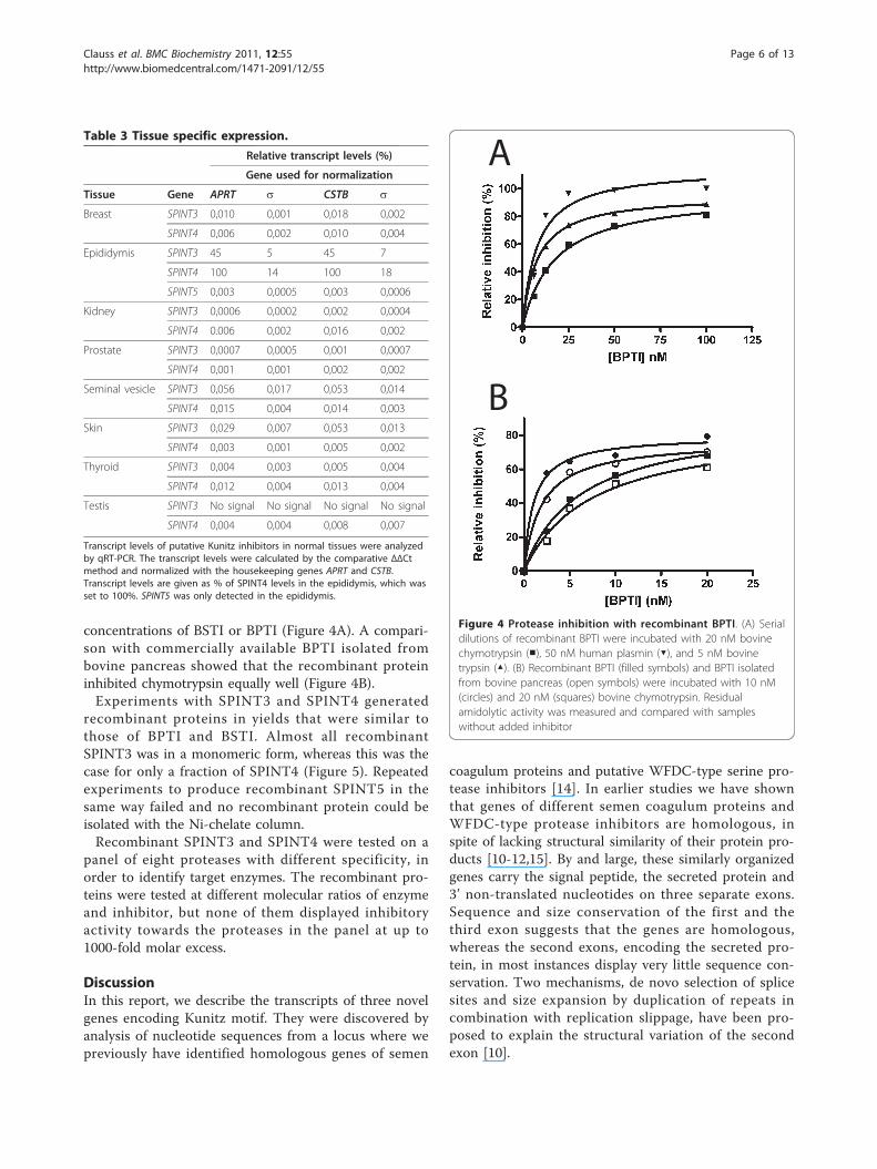
concentrations of BSTI or BPTI (Figure 4A). A compari-son with commercially available BPTI isolated frombovine pancreas showed that the recombinant proteininhibited chymotrypsin equally well (Figure 4B).Experiments with SPINT3 and SPINT4 generated
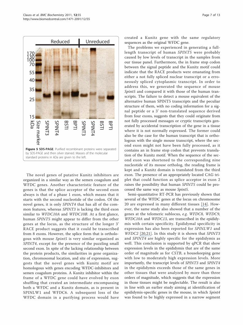
recombinant proteins in yields that were similar tothose of BPTI and BSTI. Almost all recombinantSPINT3 was in a monomeric form, whereas this was thecase for only a fraction of SPINT4 (Figure 5). Repeatedexperiments to produce recombinant SPINT5 in thesame way failed and no recombinant protein could beisolated with the Ni-chelate column.Recombinant SPINT3 and SPINT4 were tested on a
panel of eight proteases with different specificity, inorder to identify target enzymes. The recombinant pro-teins were tested at different molecular ratios of enzymeand inhibitor, but none of them displayed inhibitoryactivity towards the proteases in the panel at up to1000-fold molar excess.
DiscussionIn this report, we describe the transcripts of three novelgenes encoding Kunitz motif. They were discovered byanalysis of nucleotide sequences from a locus where wepreviously have identified homologous genes of semen
coagulum proteins and putative WFDC-type serine pro-tease inhibitors [14]. In earlier studies we have shownthat genes of different semen coagulum proteins andWFDC-type protease inhibitors are homologous, inspite of lacking structural similarity of their protein pro-ducts [10-12,15]. By and large, these similarly organizedgenes carry the signal peptide, the secreted protein and3’ non-translated nucleotides on three separate exons.Sequence and size conservation of the first and thethird exon suggests that the genes are homologous,whereas the second exons, encoding the secreted pro-tein, in most instances display very little sequence con-servation. Two mechanisms, de novo selection of splicesites and size expansion by duplication of repeats incombination with replication slippage, have been pro-posed to explain the structural variation of the secondexon [10].
Table 3 Tissue specific expression.
Relative transcript levels (%)
Gene used for normalization
Tissue Gene APRT s CSTB s
Breast SPINT3 0,010 0,001 0,018 0,002
SPINT4 0,006 0,002 0,010 0,004
Epididymis SPINT3 45 5 45 7
SPINT4 100 14 100 18
SPINT5 0,003 0,0005 0,003 0,0006
Kidney SPINT3 0,0006 0,0002 0,002 0,0004
SPINT4 0.006 0,002 0,016 0,002
Prostate SPINT3 0,0007 0,0005 0,001 0,0007
SPINT4 0,001 0,001 0,002 0,002
Seminal vesicle SPINT3 0,056 0,017 0,053 0,014
SPINT4 0,015 0,004 0,014 0,003
Skin SPINT3 0,029 0,007 0,053 0,013
SPINT4 0,003 0,001 0,005 0,002
Thyroid SPINT3 0,004 0,003 0,005 0,004
SPINT4 0,012 0,004 0,013 0,004
Testis SPINT3 No signal No signal No signal No signal
SPINT4 0,004 0,004 0,008 0,007
Transcript levels of putative Kunitz inhibitors in normal tissues were analyzedby qRT-PCR. The transcript levels were calculated by the comparative ΔΔCtmethod and normalized with the housekeeping genes APRT and CSTB.Transcript levels are given as % of SPINT4 levels in the epididymis, which wasset to 100%. SPINT5 was only detected in the epididymis.
A
B
Figure 4 Protease inhibition with recombinant BPTI. (A) Serialdilutions of recombinant BPTI were incubated with 20 nM bovinechymotrypsin (■), 50 nM human plasmin (▼), and 5 nM bovinetrypsin (▲). (B) Recombinant BPTI (filled symbols) and BPTI isolatedfrom bovine pancreas (open symbols) were incubated with 10 nM(circles) and 20 nM (squares) bovine chymotrypsin. Residualamidolytic activity was measured and compared with sampleswithout added inhibitor
Clauss et al. BMC Biochemistry 2011, 12:55http://www.biomedcentral.com/1471-2091/12/55
Page 6 of 13
The novel genes of putative Kunitz inhibitors areorganized in a similar way as the semen coagulum andWFDC genes. Another characteristic feature of thegenes is that the splice acceptor of the second exonalways is that of a phase 1 exon, which means that itstarts with the second nucleotide of the codon. Of thenovel genes, it is only SPINT4 that has all of the com-mon features, whereas SPINT3 is lacking the third exonsimilar to WFDC10A and WFDC10B. At a first glance,human SPINT5 might appear to differ from the othergenes at the locus, as the structure of the longer 3’RACE product suggests that it could be transcribedfrom 8 exons. However, the splice form that is ortholo-gous with mouse Spint5 is very similar organized asSPINT4, except for the presence of the puzzling smallsecond exon. In spite of the lacking relationship betweenthe protein products, the similarities in gene organiza-tion, chromosomal location, and site of expression, sug-gests that the novel genes with Kunitz motif arehomologous with genes encoding WFDC-inhibitors andsemen coagulum proteins. A Kunitz inhibitor within theframe of a WFDC gene could have evolved by exonshuffling that created an intermediate encompassingboth a WFDC and a Kunitz domain, as is present inSPINLW1 and WFDC6. A subsequent loss of theWFDC domain in a purifying process would have
created a Kunitz gene with the same regulatorysequences as the original WFDC gene.The problems we experienced in generating a full-
length transcript of human SPINT5 were probablycaused by low levels of transcript in the samples fromour tissue panel. Furthermore, the in frame stop codonbetween the signal peptide and the Kunitz motif couldindicate that the RACE products were emanating fromeither a not fully spliced nuclear transcript or a erro-neously spliced cytoplasmic transcript. In order toaddress this, we generated the sequence of mouseSpint5 and compared it with those of the human tran-scripts. The failure to detect a mouse equivalent of thealternative human SPINT5 transcripts and the peculiarstructure of them, with no coding information for a sig-nal peptide or a 3’ non-translated sequence derivedfrom four exons, suggests that they could originate fromnot fully processed messages or cryptic transcripts gen-erated by accidental transcription of the gene in a tissuewhere it is not normally expressed. The former couldalso be the case for the human transcript that is ortho-logous with the single mouse transcript, where the sec-ond exon might not have been fully processed, as itcontains an in frame stop codon that prevents transla-tion of the Kunitz motif. When the sequence of the sec-ond exon was shortened to the corresponding ninenucleotide of its mouse ortholog, the reading frame iskept and a Kunitz domain is translated from the thirdexon. The presence of an appropriately located CAG tri-plet that could function as splice acceptor in exon 2raises the possibility that human SPINT5 could be pro-cessed the same way as mouse Spint5.Semi-quantitative RT-PCR has previously shown that
several of the WFDC genes at the locus on chromosome20 are expressed in many different tissues [14]. How-ever, the same study also showed that a number of thegenes at the telomeric sublocus, e.g. WFDC8, WFDC9,WFDC10A and WFDC13, are transcribed in the epididy-mis with certain specificity. Epididymal specificity inexpression has also been reported for SPINLW1 andWFDC2 [20,21]. In this study it is shown that SPINT3and SPINT4 are highly specific for the epididymis aswell. This conclusion is supported by qPCR that showexpression levels in the epididymis that are of the sameorder of magnitude as for CSTB, a housekeeping genewith low to moderately high expression levels. Moreimportantly, the transcript levels of SPINT3 and SPINT4in the epididymis exceeds those of the same genes inother tissues that were analyzed by more than threeorders of magnitude, which suggests that the expressionin those tissues might be neglectable. The result is alsoin line with an earlier study aiming at identification ofepididymis-specific genes in the mouse, in which Spint4was found to be highly expressed in a narrow segment
Size marker
SPINT3
SPINT4
BSTI
BPTI
SPINT3
SPINT4
BSTI
BPTI
Reduced Unreduced
15
25
10070
5540
35
130170
kDa
Figure 5 SDS-PAGE. Purified recombinant proteins were separatedby SDS-PAGE and then silver stained. Masses of the molecularstandard proteins in kDa are given to the left.
Clauss et al. BMC Biochemistry 2011, 12:55http://www.biomedcentral.com/1471-2091/12/55
Page 7 of 13
encompassing distal caput and proximal corpus of epidi-dymis [22].Transcripts of SPINT5 were only detected in the epi-
didymis, but at a level that is at least four orders ofmagnitude lower than for SPINT3 and SPINT4. In ana-logy with the discussion above, the low transcript levelwould indicate that under normal conditions SPINT5presumably is of less importance in the tissues that wereanalyzed in this study. From this follows that SPINT5probably is expressed either at a relatively rare anatomi-cal site or at a developmental stage that is not coveredby this investigation. In ruminants, it has been shownthat several proteins with Kunitz motifs are expressedand secreted by the conceptus prior to, or at the timeof, trophoblast implantation [23,24]. Although, there aremajor differences in the implantation process betweenruminants and many other mammals, e.g. in the timing,it is not unlikely that Kunitz inhibitors could beexpressed at the time of trophoblast implantation alsoin humans, and one interesting candidate to studywould be SPINT5.Protein domains are building blocks that usually fold
to explicit three dimensional structures, which manytimes have a specific function. The Kunitz domain con-sists of 50-60 amino acid residues, six of which are Cysthat form three disulphide bonds and have characteristiclocations in the primary structure. Studies on recombi-nant Kunitz domains expressed in E. coli show thatproper folding and disulphide formation can be achievedeither by directing the recombinant protein to the peri-plasmic space or by refolding of the protein extractedfrom inclusion bodies [17,25]. Our approach was toexpress the recombinant protein as a part of a largerfusion product with thioredoxin, that would generatehigh solubility and support the proper folding of theKunitz domain, and by using the E. coli K-12 sub-strainOrigami(DE3), formation of disulphide bonds in thecytoplasm would be favored. Studies with recombinantBPTI and BSTI showed that the proteins isolated fromthe cytoplasm were almost exclusively in a monomericstate, which suggests correct disulphide formation andfolding of the Kunitz domain. Furthermore, the recom-binant inhibitors were as functionally active as BPTI iso-lated from bovine pancreas, something that alsoindicates that they are correctly folded. Expression ofSPINT3 yielded an exclusively monomeric protein in asimilar way as BPTI and BSTI, which suggests that theKunitz domain was properly folded. Recombinantexpression of SPINT4 yields high molecular mass com-plexes that probably consist of misfolded peptide chainsheld together by disulphide bonds in an unorganizedway, but also a substantial fraction of monomers thatpresumably are correctly folded. The structure ofSPINT4 differs from that of the consensus Kunitz
domain. The latter contains three conserved disulphidebridges, but in SPINT4 there will only be two, as oneCys is missing. The absent disulphide would have linkedthe C-terminal alpha-helix to one of the beta-strands atthe core of the molecule. Earlier experiment has shownthat the folding of the Kunitz domain is only minimallyaffected by removal of this bridge and would not affectthe protease-binding loop that is situated at the oppositeside of the molecule [26]. The location of a Cys amino-terminal to the Kunitz domain in SPINT4 suggests thatthe monomeric form will have either two free thiolgroups, or more likely a disulphide bridge betweenthem. The crystal structure of BPTI shows that theamino-terminus of the protein is located relatively closeto the third Cys of the Kunitz motif. Therefore, it is notunlikely that a disulfide bridge could be formed inSPINT4 without affecting the protease-binding loop,which is located at the other end of the molecule. Thiswould create a strand of 6 amino acid residues thatlinks the protein’s amino-terminal part to the third Cysof the Kunitz motif.Despite several attempts, we did not succeed to isolate
recombinant SPINT5 from the cytoplasm of Origami(DE3) cells. It is not clear whether the failure was dueto inherent instability of the protein product or whetherthe problems are due to technical problems with ourconstruct, but future studies will presumably give ananswer.The experiments with recombinant BPTI demon-
strated, not only that the Kunitz domain can be cor-rectly folded in Origami(DE3) cells, but also that thefusion product with thioredoxin could be an equally effi-cient protease inhibitor as BPTI isolated from a naturalsource. It is therefore likely that recombinant SPINT3and SPINT4 would be functional as well. They weretested by a set of proteolytic enzymes that represent dif-ferent catalytic specificities, but unfortunately, we didnot succeed to identify a target protease. The aminoacid residues in BPTI that interact with target proteaseshave been characterized in detail and homologous resi-dues can be identified in other Kunitz domains bysequence alignment [27]. There are several residues thatare important for protease-binding, but those flankingthe scissile bond, particularly the residue at the P1 posi-tion, are vital for the target- protease specificity [28,29].The probable scissile bonds in SPINT3 and SPINT4 arebetween Gln and Thr, and Tyr and Glu, respectively(Figure 6). One might then have expected both of themto inhibit PSA, which has been demonstrated to cleaveC-terminal to both Gln and Tyr in SEMG1 and SEMG2[30]. There was no inhibition and perhaps the interac-tion with the inhibitor protein was prevented by the kal-likrein loop in PSA, which restricts the access to thecatalytic cleft [31]. In chymotrypsin there is no such
Clauss et al. BMC Biochemistry 2011, 12:55http://www.biomedcentral.com/1471-2091/12/55
Page 8 of 13
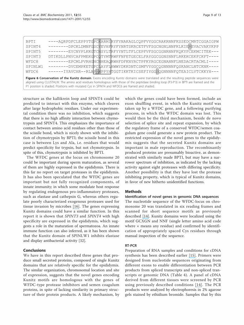
structure as the kallikrein loop and SPINT4 could bepredicted to interact with this enzyme, which cleavesafter large hydrophobic residues. Under our experimen-tal condition there was no inhibition, which suggeststhat there is no high affinity interaction between chymo-trypsin and SPINT4. This emphasizes the importance ofcontact between amino acid residues other than those ofthe scissile bond, which is nicely shown with the inhibi-tion of chymotrypsin by BPTI; the scissile bond in thiscase is between Lys and Ala, i.e. residues that wouldpredict specificity for trypsin, but not chymotrypsin. Inspite of this, chymotrypsin is inhibited by BPTI.The WFDC genes at the locus on chromosome 20
could be important during sperm maturation, as severalof them are highly expressed in the epididymis. There isthis far no report on target proteases in the epididymis.It has also been speculated that the WFDC genes areimportant but not fully recognized components, ofinnate immunity; in which some modulate host responseby regulating endogenous pro-inflammatory proteases,such as elastase and cathepsin G, whereas others regu-late poorly characterized exogenous proteases used fortissue invasion by microbes [16]. The genes expressingKunitz domains could have a similar function. In thisreport it is shown that SPINT3 and SPINT4 with highspecificity are expressed in the epididymis, which sug-gests a role in the maturation of spermatozoa. An innateimmune function can also inferred, as it has been shownthat the Kunitz domain of SPINLW1 inhibits elastaseand display antibacterial activity [32].
ConclusionsWe have in this report described three genes that pro-duce small secreted proteins, composed of single Kunitzdomains that are relatively specific for the epididymis.The similar organization, chromosomal location and siteof expression, suggests that the novel genes encodingKunitz motifs are homologous with the genes ofWFDC-type protease inhibitors and semen coagulumproteins, in spite of lacking similarity in primary struc-ture of their protein products. A likely mechanism, by
which the genes could have been formed, include anexon shuffling event, in which the Kunitz motif wastaken up by a WFDC gene, and a following purifyingprocess, in which the WFDC domain was lost. Thiswould then be the third mechanism, beside de novoselection of splice site and repeat expansion, by whichthe regulatory frame of a conserved WFDC/semen coa-gulum gene could generate a new protein product. Therestricted expression of the novel genes to the epididy-mis suggests that the secreted Kunitz domains areimportant in male reproduction. The recombinantlyproduced proteins are presumably bioactive, as demon-strated with similarly made BPTI, but may have a nar-rower spectrum of inhibition, as indicated by the lackingactivity against eight proteases with differing specificity.Another possibility is that they have lost the proteaseinhibiting property, which is typical of Kunitz domains,in favor of new hitherto unidentified functions.
MethodsIdentification of novel genes in genomic DNA sequencesThe nucleotide sequence of the WFDC-locus on chro-mosome 20 was translated in six reading frames andscanned for short sequence motifs as previouslydescribed [14]. Kunitz domains were localized using themotif GCXGN and NNF (single letter amino acid codewhere × means any residue) and confirmed by identifi-cation of appropriately spaced Cys residues throughmanual inspection of the sequence.
RT-PCRPreparation of RNA samples and conditions for cDNAsynthesis has been described earlier [15]. Primers weredesigned from nucleotide sequences originating fromdifferent exons to enable differentiation between PCRproducts from spliced transcripts and non-spliced tran-scripts or genomic DNA (Table 4). A panel of cDNAderived from different tissues were screened by PCRusing previously described conditions [14]. The PCRproducts were analyzed by electrophoresis in 2% agarosegels stained by ethidium bromide. Samples that by this
BPTI ----AQRPDFCLEPPYTGPCKARMIRYFYNAKAGLCQPFVYGGCRAKRNNFKSSEDCMRTCGGAIGPWSPINT4 --------DPCKLDMNFGSCYEVHFRYFYNRTSKRCETFVFSGCNGNLNNFKLKIEREVACVAKYKPPSPINT5 --------EYCNYPLKKGTCNSYLTRFYYNTLTFLCEPFVFSGCGGNRNNFKQKYFCEKMCITEK---SPINT3 DTIKDLLPNVCAFPMEKGPCQTYMTRWFFNFETGECELFAYGGCGGNSNNFLRKEKCEKFCKFT----WFDC8 --------EPCMLPVRHGNCNHEAQRWHFDFKNYRCTPFKYRGCEGNANNFLNEDACRTACMLI----SPINLW1 --------DVCEMPKETGPCLAYFLHWWYDKKDNTCSMFVYGGCQGNNNNFQSKANCLNTCKNK----WFDC6 ------IYAVCHR--RLAPAWPPYHTGGTIKKTKICSEFIYGGSQGNNNNFQTEAICLVTCKKYH---
Figure 6 Conservation of the Kunitz domain. Exons encoding Kunitz domains were translated and the resulting peptide sequences werealigned using CLUSTALW. The amino acid residues homologous with those of the peptidase binding loop (P3-P’3) in BPTI are framed and theP1 position is shaded. Positions with mutated Cys in SPINT4 and WFDC6 are framed and shaded.
Clauss et al. BMC Biochemistry 2011, 12:55http://www.biomedcentral.com/1471-2091/12/55
Page 9 of 13
procedure generated a PCR product from a spliced tran-script were taken to quantitative PCR using real timedetection.
Quantitative RT-PCR (qRT-PCR)The cDNA for qRT-PCR was made from DNAase-freeRNA using reagents and protocol provided by the sup-plier (Fermentas, Helsingborg, Sweden). Briefly, 3 μg ofRNA was treated at 37°C for 30 min with 1 unit ofRNase-free DNase in 10 μl of 10 mM Tris-HCl pH 7.5,2.5 mM MgCl2 and 0.1 mM CaCl2. Following additionof 1 μl of 25 mM EDTA, the enzyme was inactivated byincubation at 65°C for 10 min. The material was thentaken to oligo(dT)18-primed cDNA synthesis in avolume of 20 μl using the RevertAid H Minus FirstStrand cDNA Synthesis kit (Fermentas) for 1 h at 42°C.Finally, the reverse transcriptase was inactivated by heat-ing to 70°C for 5 min, and the samples were diluted
with H2O to 200 μl and stored at -20°C until used.Negative controls were produced in the same way, butwith the reverse transcriptase omitted.The PCR was done in MicroAmp Optical 384-Well
Reaction Plates (Applied Biosystems, Stockholm, Swe-den) using the 7900 HT Fast Real-Time PCR System(Applied Biosystems). PCR primers were selected suchthat the resultant transcript should be less than 150 bp(Table 4). The reaction mix was made from 3 μl ofdiluted cDNA, 2 μl of primers at 5 μM each, and 5 μl ofFast SYBR Green Master Mix (Applied Biosystems).Samples were analyzed in quadruplicates. The cyclingconditions consisted of an initial activation step at 95°Cfor 20 s, followed by 40 cycles of denaturation at 95°Cfor 1 s and annealing and extension at 60°C for 20 s.Following PCR the homogeneity of the products werechecked by DNA melting analysis. The real time PCRdata were analyzed by the Sequence Detection System
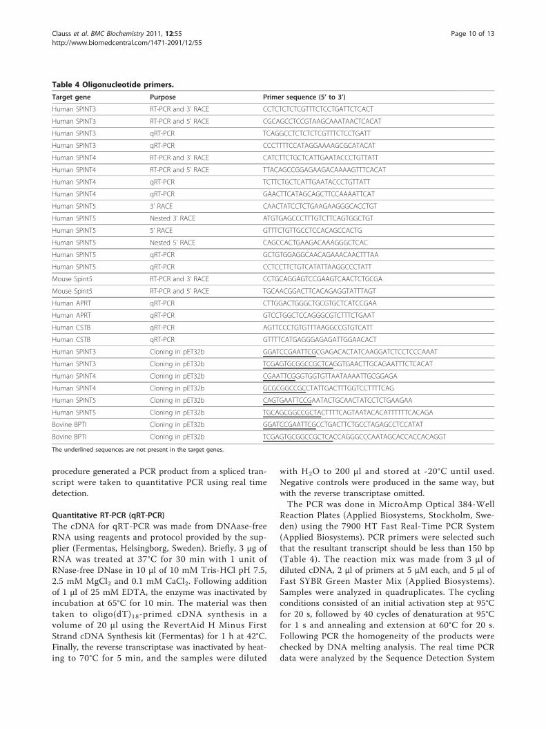
Table 4 Oligonucleotide primers.
Target gene Purpose Primer sequence (5’ to 3’)
Human SPINT3 RT-PCR and 3’ RACE CCTCTCTCTCGTTTCTCCTGATTCTCACT
Human SPINT3 RT-PCR and 5’ RACE CGCAGCCTCCGTAAGCAAATAACTCACAT
Human SPINT3 qRT-PCR TCAGGCCTCTCTCTCGTTTCTCCTGATT
Human SPINT3 qRT-PCR CCCTTTTCCATAGGAAAAGCGCATACAT
Human SPINT4 RT-PCR and 3’ RACE CATCTTCTGCTCATTGAATACCCTGTTATT
Human SPINT4 RT-PCR and 5’ RACE TTACAGCCGGAGAAGACAAAAGTTTCACAT
Human SPINT4 qRT-PCR TCTTCTGCTCATTGAATACCCTGTTATT
Human SPINT4 qRT-PCR GAACTTCATAGCAGCTTCCAAAATTCAT
Human SPINT5 3’ RACE CAACTATCCTCTGAAGAAGGGCACCTGT
Human SPINT5 Nested 3’ RACE ATGTGAGCCCTTTGTCTTCAGTGGCTGT
Human SPINT5 5’ RACE GTTTCTGTTGCCTCCACAGCCACTG
Human SPINT5 Nested 5’ RACE CAGCCACTGAAGACAAAGGGCTCAC
Human SPINT5 qRT-PCR GCTGTGGAGGCAACAGAAACAACTTTAA
Human SPINT5 qRT-PCR CCTCCTTCTGTCATATTAAGGCCCTATT
Mouse Spint5 RT-PCR and 3’ RACE CCTGCAGGAGTCCGAAGTCAACTCTGCGA
Mouse Spint5 RT-PCR and 5’ RACE TGCAACGGACTTCACAGAGGTATTTAGT
Human APRT qRT-PCR CTTGGACTGGGCTGCGTGCTCATCCGAA
Human APRT qRT-PCR GTCCTGGCTCCAGGGCGTCTTTCTGAAT
Human CSTB qRT-PCR AGTTCCCTGTGTTTAAGGCCGTGTCATT
Human CSTB qRT-PCR GTTTTCATGAGGGAGAGATTGGAACACT
Human SPINT3 Cloning in pET32b GGATCCGAATTCGCGAGACACTATCAAGGATCTCCTCCCAAAT
Human SPINT3 Cloning in pET32b TCGAGTGCGGCCGCTCAGGTGAACTTGCAGAATTTCTCACAT
Human SPINT4 Cloning in pET32b CGAATTCGGGTGGTGTTAATAAAATTGCGGAGA
Human SPINT4 Cloning in pET32b GCGCGGCCGCCTATTGACTTTGGTCCTTTTCAG
Human SPINT5 Cloning in pET32b CAGTGAATTCCGAATACTGCAACTATCCTCTGAAGAA
Human SPINT5 Cloning in pET32b TGCAGCGGCCGCTACTTTTCAGTAATACACATTTTTTCACAGA
Bovine BPTI Cloning in pET32b GGATCCGAATTCGCCTGACTTCTGCCTAGAGCCTCCATAT
Bovine BPTI Cloning in pET32b TCGAGTGCGGCCGCTCACCAGGGCCCAATAGCACCACCACAGGT
The underlined sequences are not present in the target genes.
Clauss et al. BMC Biochemistry 2011, 12:55http://www.biomedcentral.com/1471-2091/12/55
Page 10 of 13
2.3 software. The relative amount of transcripts was cal-culated by the comparative ΔΔCt method and normal-ized with values of the housekeeping genes CSTB andAPRT [33,34]. The PCR efficiency was calculated fromCt values generated by serially diluted epididymiscDNA.
Rapid amplification of cDNA ends (RACE)Synthesis of RACE ready cDNA and touch down PCRwas done with the SMART or the SMARTer RACEcDNA Amplification Kits (Clontech, In vitro, Stockholm,Sweden) and RNA from human epididymis or mousetestis, essentially as described [14]. The primers werethe same as used for RT-PCR, except for experiment onhuman SPINT5, in which RACE products were gener-ated with the SMARTer RACE cDNA amplification kitand two rounds of PCR using nested primers, as recom-mended in the protocol supplied with the kit. TheRACE products were purified using JetQuick (Genomed,Saween Werner AB, Malmö, Sweden) or Nucleotrap(Clontech) and subjected to DNA sequencing using theBig Dye Terminator Cycle Sequencing Ready ReactionKit 3.0 and a 3730 DNA Analyzer (Applied Biosystems).The sequences were analyzed with the Sequence Analy-sis 5.1 program (Applied Biosystems) and bioinformaticstools available at the EBI web server (http://www.ebi.ac.uk/Tools/). Translations of transcripts were screened forsignal peptides using the SignalP 3.0 server [35].Sequences were deposited with the Genbank SequenceDatabase and are available under accession numbersAY372172, AY372173, AY372174, and AY542490.
Construction of expression plasmidsTranscripts for expression of Kunitz domains in theprokaryotic expression vector pET32b were generatedby PCR using primers that contain restriction enzymerecognition sites for EcoRI and NotI (Table 4). The PCRprotocol was the same as for RT-PCR and as templateserved human epididymal cDNA, and human andbovine genomic DNA. The PCR that was run in orderto generate the control plasmid encoding BPTI alsoyielded transcript for the homologous bovine spleentrypsin inhibitor (BSTI) [36]. The restricted PCR pro-ducts were ligated into plasmids using the Rapid DNALigation Kit (Fermentas) and different chemo-competentbacteria were transformed and grown with appropriateantibiotics. Plasmids were purified by QIAprep SpinMiniPrep Kit (Qiagen AB, Sollentuna, Sweden) from 1.5ml of overnight cultures grown in LB medium contain-ing appropriate antibiotics, and then digested withrestriction enzyme and analyzed by electrophoresis in2% agarose gels containing ethidium bromide (1 μg/ml).Plasmids containing inserts of correct size were verifiedby DNA sequencing.
Protein expressionOrigami(DE3) bacteria containing plasmids designed forprokaryotic expression were grown at 37°C in 500 ml ofLB medium containing antibiotics until mid-logarithmicphase. The culture was chilled and the synthesis ofrecombinant protein was induced by addition of isopro-pyl-b-D-thiogalactopyranoside (IPTG) to a final concen-tration of 1 mM, where after growth continued at 30°Cfor 5 hours. Bacteria were harvested by centrifugation at10,000 × g for 10 minutes at 4°C. The resulting pelletwas washed in 50 ml of 20 mM Tris-HCl, 0.5 M NaCl,pH 7.5 and then resuspended in the same buffer con-taining 1 mg/ml of lysozyme. The suspension was made1 mM with respect to PMSF and then incubated on icefor 30 min. Next, the bacterial suspension was sonicatedwith an Ultrasonic processor XL 2020 (Misonix inc.,Farmingdale, NY, USA) for 30 minutes with 10 s burstseparated by intervals of 10 s, using the power setting 3.Cell debris was removed by centrifugation at 16,000 × gfor 20 min. The supernatant was dialyzed against 50mM Na-phosphate pH 8.0, 0.3 M NaCl and then passedthrough a 0.45 micron filter (Millipore AB, Solna,Sweden).The recombinant proteins were purified on a 12 ml
column of Ni-NTA superflow (Qiagen) that was run at2 ml/min in 50 mM Na-phosphate, 0.3 M NaCl and 10mM imidazole, pH 8.0. Proteins were eluted by increas-ing the concentration of imidazole to 100 mM or 300mM.
Protease inhibitionThe amidolytic activity of proteases was measured withpurified enzymes and synthetic peptide substrate. Theassays were run in microtiter plates with enzyme, inhibi-tor and peptide substrate in wells containing 100 μl of50 mM Tris-HCl, pH 8.0, 0.15 M NaCl and 0.05%Tween 20. Enzymatic activity was measured at 37°C in aViktor2 instrument (Wallac, Upplands Väsby, Sweden)and displayed as the time-dependent increase in absor-bance at 405 nm. Inhibitors at various concentrationswere incubated with proteases for 15 min at 37°C in theassay buffer. Reactions were initiated by addition of pep-tide substrate. The following concentrations of proteases(purchased from Sigma-Aldrich Sweden AB, Stockholm,Sweden) were used: 5 nM of bovine trypsin, 10 and 20nM of bovine chymotrypsin, 50 nM human plasmin, 10nM human urokinase, 15 nM bovine thrombin, 15 nMbovine Factor Xa, 10 nm human leukocyte elastase, and20 nM porcine pancreatic elastase. Analysis was alsomade with 10 nM human prostate-specific antigen(PSA), purified from human seminal plasma by affinitychromatography using Mab 2E9 [37]. The activity oftrypsin, plasmin, urokinase, thrombin and Factor Xawere measured with the peptide substrates S-2366,
Clauss et al. BMC Biochemistry 2011, 12:55http://www.biomedcentral.com/1471-2091/12/55
Page 11 of 13
pyroGlu-Pro-Arg-pNA (Chromogenix, Mölndal, Swe-den), chymotryptic activity with S-2586, MeO-Suc-Arg-Pro-Tyr-pNA (Chromogenix,), elastase activity with N-Suc-Ala-Ala-Ala-pNA (VWR International AB, Stock-holm, Sweden) and, PSA activity with Mu-Ser-Ser-Tyr-Tyr-AMC (custom made by MedProbe, Lund, Sweden).The inhibition was calculated as the decrease in enzy-matic activity between samples pre-incubated with inhi-bitor and buffer only. The kinetic data was analyzed anddisplayed using the computer program GraphPad Prism,version 4.00 for Windows (GraphPad Software, SanDiego CA, USA)
Acknowledgements and fundingThis work was supported in parts by grants from the Alfred Österlund Trust,MAS Cancer Foundation, The Swedish Cancer Society project no. 0345, TheSwedish Research Council (Medicine) project no. 20095, Fundación FedericoSA, and The Sidney Kimmel Center for Prostate and Urologic Cancers.
Author details1Lund University, Department of Laboratory Medicine, Clinical Chemistry,Skåne University Hospital, SE-205 02 Malmö, Sweden. 2Departments ofClinical Laboratories, Surgery, and Medicine, Memorial Sloan-Kettering CancerCenter, 1275 York Avenue, New York, NY, 10065, NY, USA. 3KarolinskaInstitute, Department of Oncology-Pathology, Karolinska Hospital, SE-171 76Stockholm, Sweden.
Authors’ contributionsAC designed and carried out experimental procedures and drafted themanuscript; MP designed and carried out experimental procedures, HL wasinvolved in planning and design of experiments and reviewed andcontributed substantive edits of the manuscript, ÅL initiated the study,identified the novel gens, contributed to the experimental design, made theqPCR and wrote the final manuscript. All authors have read and approvedthe final manuscript.
Received: 19 April 2011 Accepted: 11 October 2011Published: 11 October 2011
References1. Hurle B, Swanson W, Green ED: Comparative sequence analyses reveal
rapid and divergent evolutionary changes of the WFDC locus in theprimate lineage. Genome Res 2007, 17(3):276-286.
2. Lilja H, Abrahamsson PA, Lundwall A: Semenogelin, the predominantprotein in human semen. Primary structure and identification of closelyrelated proteins in the male accessory sex glands and on thespermatozoa. J Biol Chem 1989, 264(3):1894-1900.
3. Lilja H, Lundwall A: Molecular cloning of epididymal and seminalvesicular transcripts encoding a semenogelin-related protein. Proc NatlAcad Sci USA 1992, 89(10):4559-4563.
4. Jensen-Seaman MI, Li WH: Evolution of the hominoid semenogelin genes,the major proteins of ejaculated semen. J Mol Evol 2003, 57(3):261-270.
5. Ulvsback M, Lundwall A: Cloning of the semenogelin II gene of therhesus monkey. Duplications of 360 bp extend the coding region inman, rhesus monkey and baboon. Eur J Biochem 1997, 245(1):25-31.
6. Valtonen-Andre C, Olsson AY, Kullberg M, Nayudu PL, Lundwall A: TheCommon Marmoset (Callithrix jacchus) Has Two Very SimilarSemenogelin Genes as the Result of Gene Conversion. Biol Reprod 2007,76(4):604-610.
7. Higgins SJ, Burchell JM, Mainwaring WI: Androgen-dependent synthesis ofbasic secretory proteins by the rat seminal vesicle. Biochem J 1976,158(2):271-282.
8. Lundwall A, Peter A, Lovgren J, Lilja H, Malm J: Chemical characterizationof the predominant proteins secreted by mouse seminal vesicles. Eur JBiochem 1997, 249(1):39-44.
9. Ostrowski MC, Kistler MK, Kistler WS: Purification and cell-free synthesis ofa major protein from rat seminal vesicle secretion. A potential markerfor androgen action. J Biol Chem 1979, 254(2):383-390.
10. Lundwall A, Lazure C: A novel gene family encoding proteins with highlydiffering structure because of a rapidly evolving exon. FEBS Lett 1995,374(1):53-56.
11. Lundwall A: The cloning of a rapidly evolving seminal-vesicle-transcribedgene encoding the major clot-forming protein of mouse semen. Eur JBiochem 1996, 235(1-2):424-430.
12. Lundwall A, Ulvsback M: The gene of the protease inhibitor SKALP/elafinis a member of the REST gene family. Biochem Biophys Res Commun 1996,221(2):323-327.
13. Krowarsch D, Cierpicki T, Jelen F, Otlewski J: Canonical protein inhibitorsof serine proteases. Cell Mol Life Sci 2003, 60(11):2427-2444.
14. Clauss A, Lilja H, Lundwall A: A locus on human chromosome 20 containsseveral genes expressing protease inhibitor domains with homology towhey acidic protein. Biochem J 2002, 368(Pt 1):233-242.
15. Lundwall A, Clauss A: Identification of a novel protease inhibitor genethat is highly expressed in the prostate. Biochem Biophys Res Commun2002, 290(1):452-456.
16. Clauss A, Lilja H, Lundwall A: The evolution of a genetic locus encodingsmall serine proteinase inhibitors. Biochem Biophys Res Commun 2005,333(2):383-389.
17. Burgering MJ, Orbons LP, van der Doelen A, Mulders J, Theunissen HJ,Grootenhuis PD, Bode W, Huber R, Stubbs MT: The second Kunitz domainof human tissue factor pathway inhibitor: cloning, structuredetermination and interaction with factor Xa. J Mol Biol 1997,269(3):395-407.
18. Gebhard W, Hochstrasser K, Fritz H, Enghild JJ, Pizzo SV, Salvesen G:Structure of inter-alpha-inhibitor (inter-alpha-trypsin inhibitor) and pre-alpha-inhibitor: current state and proposition of a new terminology. BiolChem Hoppe Seyler 1990, 371 Suppl:13-22.
19. Petersen LC, Bjorn SE, Olsen OH, Nordfang O, Norris F, Norris K: Inhibitoryproperties of separate recombinant Kunitz-type-protease-inhibitordomains from tissue-factor-pathway inhibitor. Eur J Biochem 1996, 235(1-2):310-316.
20. Richardson RT, Sivashanmugam P, Hall SH, Hamil KG, Moore PA, Ruben SM,French FS, O’Rand M: Cloning and sequencing of human Eppin: a novelfamily of protease inhibitors expressed in the epididymis and testis.Gene 2001, 270(1-2):93-102.
21. Kirchhoff C, Habben I, Ivell R, Krull N: A major human epididymis-specificcDNA encodes a protein with sequence homology to extracellularproteinase inhibitors. Biol Reprod 1991, 45(2):350-357.
22. Penttinen J, Pujianto DA, Sipila P, Huhtaniemi I, Poutanen M: Discovery insilico and characterization in vitro of novel genes exclusively expressedin the mouse epididymis. Mol Endocrinol 2003, 17(11):2138-2151.
23. Kramer KK, Duffy JY, Klemann SW, Bixby JA, Low BG, Pope WF, Roberts RM:Selective cloning of cDNA for secretory proteins of early embryos.Identification of a transiently expressed kunitz domain protein frompreimplantation sheep trophoblast. J Biol Chem 1994, 269(10):7255-7261.
24. MacLean JA, Chakrabarty A, Xie S, Bixby JA, Roberts RM, Green JA: Familyof Kunitz proteins from trophoblast: expression of the trophoblastKunitz domain proteins (TKDP) in cattle and sheep. Mol Reprod Dev 2003,65(1):30-40.
25. Krokoszynska I, Dadlez M, Otlewski J: Structure of single-disulfide variantsof bovine pancreatic trypsin inhibitor (BPTI) as probed by their bindingto bovine beta-trypsin. J Mol Biol 1998, 275(3):503-513.
26. Eigenbrot C, Randal M, Kossiakoff AA: Structural effects induced byremoval of a disulfide-bridge: the X-ray structure of the C30A/C51Amutant of basic pancreatic trypsin inhibitor at 1.6 A. Protein Eng 1990,3(7):591-598.
27. Huber R, Kukla D, Bode W, Schwager P, Bartels K, Deisenhofer J,Steigemann W: Structure of the complex formed by bovine trypsin andbovine pancreatic trypsin inhibitor. II. Crystallographic refinement at 1.9A resolution. J Mol Biol 1974, 89(1):73-101.
28. Castro MJ, Anderson S: Alanine point-mutations in the reactive region ofbovine pancreatic trypsin inhibitor: effects on the kinetics andthermodynamics of binding to beta-trypsin and alpha-chymotrypsin.Biochemistry 1996, 35(35):11435-11446.
29. Otlewski J, Jaskolski M, Buczek O, Cierpicki T, Czapinska H, Krowarsch D,Smalas AO, Stachowiak D, Szpineta A, Dadlez M: Structure-function
Clauss et al. BMC Biochemistry 2011, 12:55http://www.biomedcentral.com/1471-2091/12/55
Page 12 of 13
relationship of serine protease-protein inhibitor interaction. Acta BiochimPol 2001, 48(2):419-428.
30. Malm J, Hellman J, Hogg P, Lilja H: Enzymatic action of prostate-specificantigen (PSA or hK3): substrate specificity and regulation by Zn(2+), atight-binding inhibitor. Prostate 2000, 45(2):132-139.
31. Menez R, Michel S, Muller BH, Bossus M, Ducancel F, Jolivet-Reynaud C,Stura EA: Crystal structure of a ternary complex between humanprostate-specific antigen, its substrate acyl intermediate and anactivating antibody. J Mol Biol 2008, 376(4):1021-1033.
32. McCrudden MT, Dafforn TR, Houston DF, Turkington PT, Timson DJ:Functional domains of the human epididymal protease inhibitor, eppin.FEBS J 2008, 275(8):1742-1750.
33. Eisenberg E, Levanon EY: Human housekeeping genes are compact.Trends Genet 2003, 19(7):362-365.
34. Bookout AL, Cummins CL, Mangelsdorf DJ, Pesola JM, Kramer MF: High-throughput real-time quantitative reverse transcription PCR. Curr ProtocMol Biol 2006, Chapter 15:Unit 15 18.
35. Bendtsen JD, Nielsen H, von Heijne G, Brunak S: Improved prediction ofsignal peptides: SignalP 3.0. J Mol Biol 2004, 340(4):783-795.
36. Creighton TE, Charles IG: Sequences of the genes and polypeptideprecursors for two bovine protease inhibitors. J Mol Biol 1987,194(1):11-22.
37. Piironen T, Villoutreix BO, Becker C, Hollingsworth K, Vihinen M, Bridon D,Qiu X, Rapp J, Dowell B, Lovgren T, et al: Determination and analysis ofantigenic epitopes of prostate specific antigen (PSA) and humanglandular kallikrein 2 (hK2) using synthetic peptides and computermodeling. Protein Sci 1998, 7(2):259-269.
doi:10.1186/1471-2091-12-55Cite this article as: Clauss et al.: Three genes expressing Kunitz domainsin the epididymis are related to genes of WFDC-type proteaseinhibitors and semen coagulum proteins in spite of lacking similaritybetween their protein products. BMC Biochemistry 2011 12:55.
Submit your next manuscript to BioMed Centraland take full advantage of:
• Convenient online submission
• Thorough peer review
• No space constraints or color figure charges
• Immediate publication on acceptance
• Inclusion in PubMed, CAS, Scopus and Google Scholar
• Research which is freely available for redistribution
Submit your manuscript at www.biomedcentral.com/submit
Clauss et al. BMC Biochemistry 2011, 12:55http://www.biomedcentral.com/1471-2091/12/55
Page 13 of 13


