Embed Size (px)
Citation preview

Progress in Biophysics & Molecular Biology 81 (2003) 1–44
Review
Tight junction proteins
L. Gonz!alez-Mariscal*, A. Betanzos, P. Nava, B.E. Jaramillo
Department of Physiology, Biophysics and Neuroscience, Center for Research and Advanced Studies (CINVESTAV),
Ave. Polit!ecnico Nacional 2508, M!exico DF, 07000, Mexico
Abstract
A fundamental function of epithelia and endothelia is to separate different compartments within theorganism and to regulate the exchange of substances between them. The tight junction (TJ) constitutes thebarrier both to the passage of ions and molecules through the paracellular pathway and to the movement ofproteins and lipids between the apical and the basolateral domains of the plasma membrane. In recent yearsmore than 40 different proteins have been discovered to be located at the TJs of epithelia, endothelia andmyelinated cells. This unprecedented expansion of information has changed our view of TJs from merely aparacellular barrier to a complex structure involved in signaling cascades that control cell growth anddifferentiation. Both cortical and transmembrane proteins integrate TJs. Among the former are scaffoldingproteins containing PDZ domains, tumor suppressors, transcription factors and proteins involved in vesicletransport. To date two components of the TJ filaments have been identified: occludin and claudin. The latteris a protein family with more than 20 members. Both occludin and claudins are integral proteins capable ofinteracting adhesively with complementary molecules on adjacent cells and of co-polymerizing laterally.These advancements in the knowledge of the molecular structure of TJ support previous physiological modelsthat exhibited TJ as dynamic structures that present distinct permeability and morphological characteristicsin different tissues and in response to changing natural, pathological or experimental conditions.r 2003 Elsevier Science Ltd. All rights reserved.
Keywords: Tight junctions; Claudin; Occludin; JAM, MAGUK, ZO; Cingulin
Contents
1. Introduction . . . . . . . . . . . . . . . . . . . . . . . . . . . . . . . . . . . . . . . . . . . 2
2. Transmembrane proteins of the TJ . . . . . . . . . . . . . . . . . . . . . . . . . . . . . . . . 5
2.1. TJ tetraspan proteins found in epithelial and endothelial cells . . . . . . . . . . . . . . . 5
2.1.1. Occludin . . . . . . . . . . . . . . . . . . . . . . . . . . . . . . . . . . . . . . 5
2.1.2. Claudins . . . . . . . . . . . . . . . . . . . . . . . . . . . . . . . . . . . . . . 7
*Corresponding author. Tel.: +52-55-57-47-70-00 x 5110 or 5155; fax: +52-55-57-47-70-00 x 5702.
E-mail address: [email protected] (L. Gonz!alez-Mariscal).
0079-6107/03/$ - see front matter r 2003 Elsevier Science Ltd. All rights reserved.
PII: S 0 0 7 9 - 6 1 0 7 ( 0 2 ) 0 0 0 3 7 - 8

1. Introduction
In multicellular organisms fluids with different molecular compositions (e.g. urine, milk, gastricjuice, blood, etc.) are contained in compartments delineated by epithelia (e.g. renal tubules) andendothelia (blood vessels). These cellular sheets constitute the frontier between the organisminternal milieu and the compartments contents.
Epithelia and endothelia have tight junctions (TJ) that regulate the passage of ions, water andmolecules through the paracellular pathway. This characteristic is generally referred to as the gateproperty of the TJ. The establishment of TJ at the uppermost portion of the lateral plasmamembranes is the result of a polarized insertion of proteins, yet TJ act as a fence that maintainscell polarity, by blocking the free diffusion of proteins and lipids between the apical andbasolateral domains of the plasma membrane.
2.2. TJ tetraspan proteins found within myelin sheaths . . . . . . . . . . . . . . . . . . . . 10
2.2.1. OSP/claudin 11 . . . . . . . . . . . . . . . . . . . . . . . . . . . . . . . . . 10
2.2.2. PMP22/gas-3 . . . . . . . . . . . . . . . . . . . . . . . . . . . . . . . . . . . 10
2.2.3. OAP-1/TSPAN-3 . . . . . . . . . . . . . . . . . . . . . . . . . . . . . . . . 11
2.3. TJ proteins that belong to the immunoglobulin superfamily . . . . . . . . . . . . . . . 11
2.3.1. JAM . . . . . . . . . . . . . . . . . . . . . . . . . . . . . . . . . . . . . . . 11
2.3.2. CAR . . . . . . . . . . . . . . . . . . . . . . . . . . . . . . . . . . . . . . . 13
2.3.3. P0 . . . . . . . . . . . . . . . . . . . . . . . . . . . . . . . . . . . . . . . . 13
3. Plaque proteins of the TJ . . . . . . . . . . . . . . . . . . . . . . . . . . . . . . . . . . . . 14
3.1. PDZ-containing proteins . . . . . . . . . . . . . . . . . . . . . . . . . . . . . . . . . 14
3.1.1. The MAGUK proteins of the TJ . . . . . . . . . . . . . . . . . . . . . . . . 14
3.1.2. MAGI, the MAGUK inverted proteins of the TJ . . . . . . . . . . . . . . . . 20
3.1.3. PAR proteins of the TJ . . . . . . . . . . . . . . . . . . . . . . . . . . . . . 21
3.1.4. MUPP1 . . . . . . . . . . . . . . . . . . . . . . . . . . . . . . . . . . . . . 22
3.1.5. AF-6/Afadin . . . . . . . . . . . . . . . . . . . . . . . . . . . . . . . . . . . 22
3.1.6. PATJ . . . . . . . . . . . . . . . . . . . . . . . . . . . . . . . . . . . . . . . 23
3.2. TJ proteins lacking PDZ domains . . . . . . . . . . . . . . . . . . . . . . . . . . . . 24
3.2.1. Cingulin . . . . . . . . . . . . . . . . . . . . . . . . . . . . . . . . . . . . . 24
3.2.2. Symplekin . . . . . . . . . . . . . . . . . . . . . . . . . . . . . . . . . . . . 26
3.2.3. 7H6 antigen/barmotin . . . . . . . . . . . . . . . . . . . . . . . . . . . . . . 26
3.2.4. Rab proteins . . . . . . . . . . . . . . . . . . . . . . . . . . . . . . . . . . . 26
3.2.5. Pilt . . . . . . . . . . . . . . . . . . . . . . . . . . . . . . . . . . . . . . . . 27
3.2.6. JEAP . . . . . . . . . . . . . . . . . . . . . . . . . . . . . . . . . . . . . . . 27
3.2.7. huASH1 . . . . . . . . . . . . . . . . . . . . . . . . . . . . . . . . . . . . . 27
3.2.8. Heterotrimeric G proteins . . . . . . . . . . . . . . . . . . . . . . . . . . . . 28
4. Molecular assembly of the TJ . . . . . . . . . . . . . . . . . . . . . . . . . . . . . . . . . 28
4.1. Assembly of TJ molecules in epithelial and endothelial monolayers . . . . . . . . . . . 28
4.2. Molecular maturation of TJ during trophoectoderm differentiation . . . . . . . . . . . 29
4.3. TJ assembly during early Xenopus development . . . . . . . . . . . . . . . . . . . . . 29
5. Conclusions and future perspectives . . . . . . . . . . . . . . . . . . . . . . . . . . . . . . 30
Acknowledgements . . . . . . . . . . . . . . . . . . . . . . . . . . . . . . . . . . . . . . . . . 31
References . . . . . . . . . . . . . . . . . . . . . . . . . . . . . . . . . . . . . . . . . . . . . . 31
L. Gonz !alez-Mariscal et al. / Progress in Biophysics & Molecular Biology 81 (2003) 1–442

On ultrathin section electron micrographs, TJs are viewed as a series of fusion points betweenthe outer leaflets of the membrane of adjacent cells. At these ‘‘kissing’’ points, the intercellularspace is completely obliterated. On freeze-fracture replica electron micrographs TJs appear as anetwork of continuous and anastomosing filaments on the protoplasmic face (P) of the plasmamembrane, with complementary grooves on the exoplasmic face (E) (Gonzalez-Mariscal et al.,2001) (Fig. 1).
Two models have been developed to explain the chemical nature of TJ strands. In the proteinmodel, the filaments are formed by integral membrane proteins that associate with a partner in theapposing membrane of the adjacent cell. In the lipid model instead, strands are proposed to beformed of cylindrical micelles with the polar groups of the lipids directed inwards, and thehydrophobic tails immersed in the lipid matrix of the plasma membrane of both contacting cells(Kachar and Reese, 1982; Pinto and Kachar, 1982). The lipid model is however inconsistent withthe observation that changing the total composition of phospholipids, sphingolipids andcholesterol in epithelial cells does not alter the appearance of TJ strands nor the gate or fencefunction of TJ (Calderon et al., 1998). Nevertheless, a rapid reduction of cell cholesterol bymethyl-b-cyclodextrin has been reported to generate a decline in transepithelial electricalresistance (TER), an increased mannitol flux and an augmented number of TJ particles associatedwith the E face (Francis et al., 1999). These results therefore suggest that although the TJ strandsmight be composed of proteins, the structure is sensitive to rapid changes on its lipidicenvironment.
In recent years, a constellation of cortical and integral proteins of the TJ has beendiscovered. Of the former, 16 different molecules have so far been identified. Some functionas scaffolds, that link the integral proteins of the TJ to the actin cytoskeleton, while othersact as cross linkers of transmembrane junctional proteins. Still others are involved invesicular trafficking to the TJ or in cell signaling through their association to kinases andRas. Exciting new information has revealed that some submembranous junctional proteins evenhave a role in gene expression due to their nuclear shuttling and specific binding to transcriptionfactors.
At the TJ three integral proteins are found: occludin, claudins and JAM. The formertwo constitute the backbone of TJ strands while JAM appears to be important for theroutine trafficking of T-lymphocytes, neutrophiles and dentritic cells from the lymphoidand vascular compartments to the tissues during immune surveillance and inflammatoryresponses.
TJs were initially described in epithelia and endothelia. However recent observations havedemonstrated that they are also present in myelinated cells. Thus, during development of thecentral (CNS) and peripheral nervous system (PNS), oligodendrocyte and Schwann cells stopdividing and respectively start to wrap axons in a loose spiral, resulting in the typicalmultilamellar structure that electrically isolates the axon and allows saltatory conduction toproceed. TJs present between the glia and the axons and within the myelin sheaths have proteinsfound in the TJs of epithelia and endothelia as well as other nervous system specific molecules thatwill also be described in this review.
Since a wealth of information on TJ proteins has emerged in recent times, and keeping updatedon so many different proteins has become a hard task, we have written this review, to serve as aguide for readers interested in the field of cell–cell adhesion.
L. Gonz !alez-Mariscal et al. / Progress in Biophysics & Molecular Biology 81 (2003) 1–44 3

Fig. 1. Structure of tight junctions. (A) Freeze-fracture electron microscopic image of TJs in an epithelial monolayer.
TJs appear as a network of anastomosing strands on the P face (P) with complementary grooves on the E face (E). Scale
bar, 100 nm: (B) Ultrathin section of a TJ. Ruthenium red added to the apical surface of epithelial monolayers cannot
pass beyond the tight junction (arrow). Scale bar, 10 nm (courtesy of Dr. Bibiana Ch!avez de Ram!ırez).
L. Gonz !alez-Mariscal et al. / Progress in Biophysics & Molecular Biology 81 (2003) 1–444

2. Transmembrane proteins of the TJ
2.1. TJ tetraspan proteins found in epithelial and endothelial cells
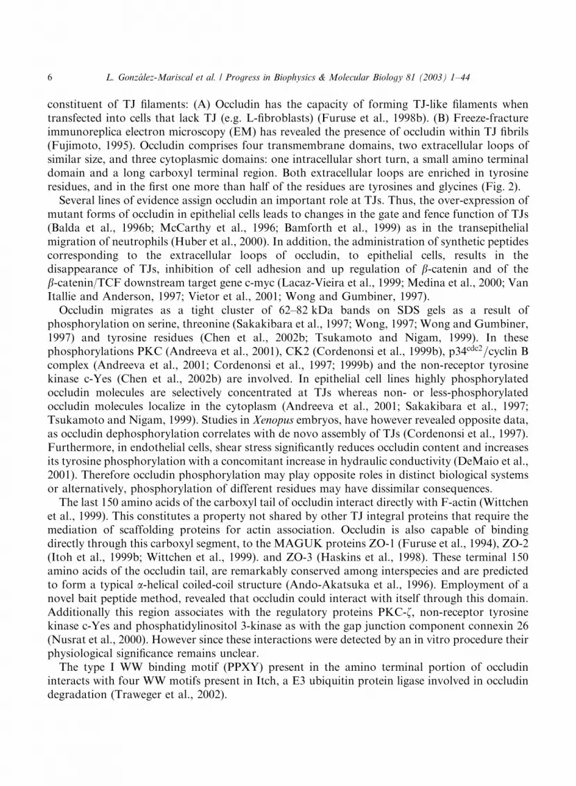
In this section we will refer to integral TJ proteins whose structure predicts four transmembraneregions with two extracellular domains, and with both amino and carboxyl terminal ends orientedtowards the cytoplasm. While certain TJ tetraspans have both extracellular loops ofapproximately the same size (e.g. occludin), some have one extracellular loop larger than theother (e.g. claudins and tetraspanins) (Fig. 2).
2.1.1. OccludinThe name of this integral protein of the TJ derives from the Latin word ‘‘occludere’’ which
means to occlude (Furuse et al., 1993). Two crucial lines of evidence have shown that occludin is a
Occludin TetraspaninClaudin
254 aaCOOH
21-42 aaCOOH149 aa
NH2
10 aa
45 aa 44 aa
NH2
51 aa14 aa
13 aa
9-24 aaNH2
C
4-40aaCOOH
4 aa
13-30 aa
78-150aa
C
C
C
G
XX
P
YG
G
G Y
Y GG
-
--
++
+
+
+-
Y
Y
Y
Y
Y
Y
C
Fig. 2. Schematic representation of tetraspan proteins of the TJ. Tetraspan proteins have four transmembrane regions,
two extracellular domains, and their amino and carboxyl terminal ends are oriented towards the intracellular region.
Both extracellular loops of occludin are of approximately the same size, lack charged residues and are very rich in
tyrosine (Y). More than half of the first loop residues are tyrosines and glycines (G). In claudins the first extracellular
loop is longer than the second one. Both claudin loops display a number of charged residues ðþ;�Þ which are expected
to influence the passage of ions through the paracellular space. Tetraspanins have a longer second extracellular loop
and can be further differentiated from claudins, by the presence within the long loop of CCG and PXXCC motifs as
well as two or four cysteine residues (C), one of which is consistently located 11 residues from the predicted start of the
fourth transmembrane domain.
L. Gonz !alez-Mariscal et al. / Progress in Biophysics & Molecular Biology 81 (2003) 1–44 5
constituent of TJ filaments: (A) Occludin has the capacity of forming TJ-like filaments whentransfected into cells that lack TJ (e.g. L-fibroblasts) (Furuse et al., 1998b). (B) Freeze-fractureimmunoreplica electron microscopy (EM) has revealed the presence of occludin within TJ fibrils(Fujimoto, 1995). Occludin comprises four transmembrane domains, two extracellular loops ofsimilar size, and three cytoplasmic domains: one intracellular short turn, a small amino terminaldomain and a long carboxyl terminal region. Both extracellular loops are enriched in tyrosineresidues, and in the first one more than half of the residues are tyrosines and glycines (Fig. 2).
Several lines of evidence assign occludin an important role at TJs. Thus, the over-expression ofmutant forms of occludin in epithelial cells leads to changes in the gate and fence function of TJs(Balda et al., 1996b; McCarthy et al., 1996; Bamforth et al., 1999) as in the transepithelialmigration of neutrophils (Huber et al., 2000). In addition, the administration of synthetic peptidescorresponding to the extracellular loops of occludin, to epithelial cells, results in thedisappearance of TJs, inhibition of cell adhesion and up regulation of b-catenin and of theb-catenin/TCF downstream target gene c-myc (Lacaz-Vieira et al., 1999; Medina et al., 2000; VanItallie and Anderson, 1997; Vietor et al., 2001; Wong and Gumbiner, 1997).
Occludin migrates as a tight cluster of 62–82 kDa bands on SDS gels as a result ofphosphorylation on serine, threonine (Sakakibara et al., 1997; Wong, 1997; Wong and Gumbiner,1997) and tyrosine residues (Chen et al., 2002b; Tsukamoto and Nigam, 1999). In thesephosphorylations PKC (Andreeva et al., 2001), CK2 (Cordenonsi et al., 1999b), p34cdc2=cyclin Bcomplex (Andreeva et al., 2001; Cordenonsi et al., 1997; 1999b) and the non-receptor tyrosinekinase c-Yes (Chen et al., 2002b) are involved. In epithelial cell lines highly phosphorylatedoccludin molecules are selectively concentrated at TJs whereas non- or less-phosphorylatedoccludin molecules localize in the cytoplasm (Andreeva et al., 2001; Sakakibara et al., 1997;Tsukamoto and Nigam, 1999). Studies in Xenopus embryos, have however revealed opposite data,as occludin dephosphorylation correlates with de novo assembly of TJs (Cordenonsi et al., 1997).Furthermore, in endothelial cells, shear stress significantly reduces occludin content and increasesits tyrosine phosphorylation with a concomitant increase in hydraulic conductivity (DeMaio et al.,2001). Therefore occludin phosphorylation may play opposite roles in distinct biological systemsor alternatively, phosphorylation of different residues may have dissimilar consequences.
The last 150 amino acids of the carboxyl tail of occludin interact directly with F-actin (Wittchenet al., 1999). This constitutes a property not shared by other TJ integral proteins that require themediation of scaffolding proteins for actin association. Occludin is also capable of bindingdirectly through this carboxyl segment, to the MAGUK proteins ZO-1 (Furuse et al., 1994), ZO-2(Itoh et al., 1999b; Wittchen et al., 1999). and ZO-3 (Haskins et al., 1998). These terminal 150amino acids of the occludin tail, are remarkably conserved among interspecies and are predictedto form a typical a-helical coiled-coil structure (Ando-Akatsuka et al., 1996). Employment of anovel bait peptide method, revealed that occludin could interact with itself through this domain.Additionally this region associates with the regulatory proteins PKC-z; non-receptor tyrosinekinase c-Yes and phosphatidylinositol 3-kinase as with the gap junction component connexin 26(Nusrat et al., 2000). However since these interactions were detected by an in vitro procedure theirphysiological significance remains unclear.
The type I WW binding motif (PPXY) present in the amino terminal portion of occludininteracts with four WW motifs present in Itch, a E3 ubiquitin protein ligase involved in occludindegradation (Traweger et al., 2002).
L. Gonz !alez-Mariscal et al. / Progress in Biophysics & Molecular Biology 81 (2003) 1–446

Recently an occludin related gene (ORG) has been identified on the Y chromosome ofDrosophila melanogaster (Carvalho et al., 2001), and two alternatively splicing forms of occludinhave lately been described (Muresan et al., 2000). Isoform 1B contains a unique N terminalsequence of 56 amino acids, whose function remains unknown.
Although occludin is a clear constituent of TJ filaments, and its abundance is related to thedegree of sealing of epithelia (e.g. more in distal than in the proximal segments of the nephron)(Gonzalez-Mariscal et al., 2000), its precise role in TJ remains unclear, specially after observingthat occludin knock out mice display well developed TJ (Saitou et al., 2000).
2.1.2. Claudins
The paradoxical results obtained with occludin deficient mice described above, led Tsukita andco-workers to search for other integral components of TJ. Using the same liver fraction employedto identify occludin, and by means of a sucrose step gradient, a single 22 kDa band wasdiscovered as a putative novel TJ integral protein. Peptide sequencing revealed two proteins in thisband that were subsequently named claudin 1 and 2 (Furuse et al., 1998a). The name claudinderives from the Latin word ‘‘claudere’’ which means to close.
By data base searching and cDNA and genomic cloning the claudin family has expanded to 24members (Table 1) (Tsukita et al., 2001). All claudins encode 20–27 kDa proteins with fourtransmembrane domains, two extracellular loops where the first one is significantly longer thanthe second one, and a short carboxyl intracellular tail (Fig. 2). The last amino acids of this tail arehighly conserved within the family and constitute PDZ binding motifs: claudins 1–9 and 17 S/TYV, claudins 10 and 15 AYV, claudin 11 AHV, claudin 12 HTT, claudin 13 LDV, claudins 14,18 and 20 DYV, claudin 16 TRV, and claudin 19 DRV. Through these motifs claudins are linkedto the TJ PDZ containing proteins ZO-1, ZO-2, ZO-3 (Itoh et al., 1999a), PATJ (Roh et al.,2002a) and MUPP1 (Hamazaki et al., 2002).
When individual claudins 1–3, 5 or 11 were introduced into mouse L fibroblasts,intramembrane strands appeared in freeze-fracture replicas (Furuse et al., 1998b; Morita et al.,1999a, b). Thus suggesting that claudins constitute the backbone of TJ strands. Different claudinspecies are capable of generating different freeze-fracture patterns. Thus, transfection of claudins1 or 3 generates continuous smooth intramembrane strands on the protoplasmic surface (P face)of the replicas (Furuse et al., 1999), whereas claudins 2 or 5 form discontinuous chains of particlesassociated to the exoplasmic face (E face) (Morita et al., 1999b). Transfection with claudin 11generates instead parallel intramembrane strands on the P face that scarcely branch (Morita et al.,1999a).
Heterogeneous claudins can interact within a single TJ strand. For example, by immunoreplicaEM the co-incorporation of distinct transfected claudins into individual intramembrane strandshas been confirmed. The particular combination of claudins within a TJ strand might give rise todifferent freeze-fracture patterns. Thus strands formed with claudins 1 and 3 are continuous andassociated to the P face, while strands formed with claudins 1 and 2 or 3 and 2 have evenlyscattered particles in the E face grooves. At the paracellular space the extracellular loops ofdifferent species of claudins belonging to neighboring cells can also interact, except in somecombinations. Thus, when L transfectant singly expressing claudin 1, 2 or 3 were co-cultured,claudin 3 strands associated with claudin 1 and 2 strands of the apposing cell, whereas claudin 1did not interact with claudin 2 strands (Furuse et al., 1999).
L. Gonz !alez-Mariscal et al. / Progress in Biophysics & Molecular Biology 81 (2003) 1–44 7

Claudins depict a differential distribution in distinct tissues, supporting the idea that they areresponsible for the ample variety in electrical resistance and paracellular ionic selectivity displayedby epithelia and endothelia. The nephron is a good model to exemplify this point, since it isintegrated by tubules with low and high TER (6 O cm2 in proximal segments vs. 870–2000 O cm2
in the collecting duct) that specialize in the resorption of specific ions. Northern blot analysis hasrevealed the expression of all claudins except 6, 9, 13 and 14 in a total kidney extract. However,the study of kidney cryosections (Enck et al., 2001; Kiuchi-Saishin et al., 2002) or of micro-dissected tubules (Reyes et al., 2002) has revealed the differential distribution of these proteins:claudins 5 and 15 in endothelia, claudins 2, 10 and 11 at the proximal segment, claudins 1, 3 and 8at the distal tubule and claudins 1, 3, 4 and 8 at the collecting segment.
The expression of different type of claudins appears to be finely tuned during development. Forexample: (A) Claudin 6 is present in embryonic epithelia (Turksen and Troy, 2001) and its overexpression in transgenic mice generates a defective epidermal permeability barrier (Turksen andTroy, 2002). It has been proposed that the unstable temperature control and dehydration
Table 1
Characteristic features of different claudins
Claudin Distinctive characteristics
1 Present in high-resistance epithelia (e.g. collecting segment) and absent in leaky epithelia (e.g. proximal
tubule) (Reyes et al., 2002). Crucial for the mammalian epidermal barrier (Furuse et al., 2002). Absent in
most human breast cancer cell lines (Hoevel et al., 2002).
2 Present in leaky epithelia (e.g. proximal tubule) and absent in tight epithelia (e.g. collecting segment)
(Reyes et al., 2002; Enck et al., 2001). Present in the choroids plexus epithelium (Wolburg et al., 2001).
3 Also known as RVP1 (Briehl and Miesfeld, 1991). Present in the tighter segments of the nephron (Kiuchi-
Saishin et al., 2002). Its expression is elevated in regressing ventral prostate and in prostate
adenocarcinomas (Long et al., 2001). Capable of CPE binding (Sonoda et al., 1999).
4 Its expression decreases paracellular conductance through a selective decrease in sodium permeability
(Van Itallie et al., 2001). Present in the tighter segments of the nephron (Kiuchi-Saishin et al., 2002). Over
expressed in pancreatic and gastrointestinal tumors (Michl et al., 2001). The selective CPE binding gave
rise to its alternative name CPE-R (Sonoda et al., 1999).
5 Receives the alternative name of TMVCF, as it is frequently deleted in Velo cardio facial syndrome
(Sirotkin et al., 1997). Constitutes TJ strands in endothelial cells (Morita et al., 1999b). Transiently
expressed during the development of the retinal pigment epithelium (Kojima et al., 2002).
6 Present in embryonic epithelia (Turksen and Troy, 2001). Its over expression in transgenic mice generates
a defective epidermal permeability barrier (Turksen and Troy, 2002).
7 Down regulated in head and neck squamous cell carcinomas (Al Moustafa et al., 2002).
8 Present in the tighter segments of the nephron (Kiuchi-Saishin et al., 2002).
11 Also named OSP. Present in oligodendrocytes and Sertoli cells (Morita et al., 1999a).
14 Expressed in the sensory epithelium of the organ of Corti. Mutations in the gene cause autosomal
recessive deafness (Wilcox et al., 2001).
15 Present in endothelial cells (Kiuchi-Saishin et al., 2002).
16 Also known as Paracellin-1. Critical for Mg2þ and Ca2þ resorption in the human thick ascending limb of
Henle (Blanchard et al., 2001; Simon et al., 1999).
18 A downstream target gene for the T/EBP/NKX2.1 homeodomain transcription factor. Expressed in lung
and stomach (Niimi et al., 2001).
Claudins 9, 10, 12, 13, 17 and 19–24 have not been yet well characterized.
L. Gonz !alez-Mariscal et al. / Progress in Biophysics & Molecular Biology 81 (2003) 1–448

frequently observed in premature infants, might be related to the expression of this claudin in theirepidermis. (B) Claudin 5 is transiently expressed during the development of the retinal pigmentepithelium (Kojima et al., 2002). (C) Claudin 11 is expressed in Sertoli cells, immediately after thepeak of expression of the sex determining region in the Y gene (Hellani et al., 2000).
Claudin 16 is mutated in human patients with hypomagnesemia hypercalciuria syndrome(HHS) (Simon et al., 1999). These patients manifest a selective defect in paracellular Mg2þ andCa2þ reabsorption in the thick ascending limb of Henle (TAL), while maintaining an intact NaClresorption ability (Blanchard et al., 2001). Hence claudin 16 might function as a paracellularchannel selective for Mg2þ and Ca2þ (Goodenough and Wong, 1999). Other claudins have alsoproved to be ionic selective. Such is the case of claudin 4, that when transfected into epithelialcells, decreases the paracellular conductance through a selective decrease in Naþ permeabilitywithout a significant effect on Cl� permeability (Van Itallie et al., 2001). The proposal of ionchannels or pores within the TJ strands is more than two decades old, and arose with Claude’sobservation that TER increases with the number of TJ strands present in the epithelia, not in alinear fashion as would be expected from the addition of resistors in series, but exponentially(Claude, 1978; Gonzalez-Mariscal et al., 2001). The ionic selectivity at the TJ could therefore bedetermined by the specific claudins that constitute the pore. On analyzing the extracellular loopsof claudins an enormous variability in distribution and number of charged residues is found. Forexample the isoelectric points of the first loop range from 4.17 in claudin 16 to 10.49 in claudin 14,and in the second extracellular loop from 4.05 in claudins 2, 7, 10 and 14 to 10.5 in claudin 13.Based on the pKIs of the extracellular loops sequences, claudin 16 is predicted to act as a cationpore, whereas claudins 4, 11 and 17 should function as anionic channels (Mitic and Van Itallie,2001).
Variations in the tightness of the TJ appear to be determined by the combination and mixingratios of different claudin species. Thus when MDCK cells expressing claudin 1 and 4 wereincubated with the claudin 4 binding protein, Clostridium perfringens enterotoxin (CPE), claudin 4was selectively removed from TJs, generating a significant decrease in TER (Sonoda et al., 1999).When claudin-2 instead was introduced into high-resistance MDCK cells (MDCK I), their TJsbecame leaky and were similar functionally and morphologically to those in low-resistance cells(MDCK II), which normally contain high levels of claudin 2 (Furuse et al., 2001).
The role of claudins in carcinogenesis is controversial. Thus, claudin 4 is over-expressed inpancreatic cancer and gastrointestinal tumors. Treatment with TGFb or CPE, leads to asignificant reduction of tumor growth (Michl et al., 2001), thus suggesting that proteins involvedin cell–cell contacts such as claudins may facilitate processes of invasion and migration. On theother hand certain claudins remain low or undetectable in a number of tumors and cancer celllines. For example claudin 1 expression is lost in most human breast cancers without presentingalterations in its promoter or coding sequences (Hoevel et al., 2002; Kramer et al., 2000), andclaudin 7 is down regulated in head and neck squamous cell carcinomas (Al Moustafa et al.,2002).
The crucial task of claudins in the gate function of TJs is highlighted by the following evidence:(A) In the mammalian epidermis, claudin-1 co-localizes with occludin in the most apical regionsof the second layer of the stratum granulosum, while claudin 4 is present in deeper layers of thestratum. In claudin 1 deficient mice, the epidermal barrier is severely affected leading todehydration, wrinkled skin and death of the animals within 1 day of birth. In these mice the
L. Gonz !alez-Mariscal et al. / Progress in Biophysics & Molecular Biology 81 (2003) 1–44 9
occludin positive and claudin-1 deficient skin layers allow the passage of paracellular tracers,suggesting that the combination of claudin-1 and occludin is needed for the establishment of aneffective paracellular barrier (Furuse et al., 2002). (B) In human breast cancer cells that have lostthe expression of claudin-1, transfection of this claudin decreases the paracellular flux of tracersdespite the absence of occludin (Hoevel et al., 2002).
2.2. TJ tetraspan proteins found within myelin sheaths
2.2.1. OSP/claudin 11OSP/claudin 11 is a crucial component of TJs in CNS myelin and between Sertoli cells (Gow
et al., 1999; Spector et al., 1998). Claudin 11 null mice have no TJ in their oligodendrocytes andSertoli cells, show slow CNS conductance, hind limb weakness, and sterility in male animals (Gowet al., 1999).
In contrast to conventional TJ that are formed by networks of anastomosing strands, thosefound in the CNS myelin and in Sertoli cells are comprised by parallel filaments (Southwood andGow, 2001), thus suggesting that claudin 11 polymerization restricts the formation of branchingTJ fibrils.
OSP/claudin 11 is the third most abundant central nervous system (CNS) myelin protein(Bronstein et al., 1997). During prenatal development OSP/claudin 11 is profuse in developingmeninges and mesenchymal cells, especially around regions of chondrocyte formation (Bronsteinet al., 2000). Postnatally, it is only expressed in oligodendrocytes and testis. In adult animals,expression of OSP/claudin 11 by the testis is inhibited by the hormone FSH and the cytokineTNFa (Hellani et al., 2000).
Although abundant evidence supports a major role for claudin 11 at the TJ of Sertoli cells, theparticipation of occludin in these junctions cannot be ruled out. In fact, the administration of asynthetic peptide corresponding to the second extracellular loop of occludin perturbs the blood–testis barrier and reversibly disrupts spermatogenesis (Chung et al., 2001). Therefore bothoccludin and claudin 11 might be needed for Sertoli TJ to develop.
2.2.2. PMP22/gas-3
The peripheral myelin protein PMP22/gas-3 was originally identified as a growth arrest specificprotein of fibroblasts (Schneider et al., 1988). PMP22/gas-3 is a 22 kDa tetraspan glycoprotein of160 amino acids. PMP22/gas-3 expression is closely synchronized with Schwann cellsdifferentiation and localizes in the myelin sheath (Baechner et al., 1995). It has been characterizedas a strong adhesive component for compact myelin formation. Deletions, duplications ormutations of PMP22/gas-3 account for the majority of heritable demyelinating peripheralneuropathies in mice (Trembler) and humans including Charcot–Marie-Tooth disease type IA,Dejerine Sottas syndrome and heredity neuropathy with liability to pressure palsies (HNPP)(Suter and Nave, 1999). PMP22/gas-3 mRNA has been detected in a variety of non-neural tissues,the epithelial cells of the lungs and intestine being the higher expressers (Baechner et al., 1995).
In epithelial cells PMP22/gas-3 co-localizes with occludin and ZO-1 at the TJs, and its over-expression in L cell fibroblasts mediates the formation of ZO-1 positive intercellular junctions(Notterpek et al., 2001). Therefore it is tempting to speculate that PMP22/gas-3 plays a role in theestablishment and maintenance of TJs in epithelia and within the Schwann cell membrane. The
L. Gonz !alez-Mariscal et al. / Progress in Biophysics & Molecular Biology 81 (2003) 1–4410

amino acid sequence and predicted structure have posed the question of whether PMP22/gas-3 isa claudin family member that functions as the peripheral nervous system (PNS) homologue ofclaudin 11. In this respect it should be pointed that: (1) the sequence of PMP22/gas-3 is fairlyshorter compared to those of claudins (160 vs. 207–264 amino acids); (2) PMP22/gas-3 has a weakhomology even with claudin-10 to which sequence it resembles the most (25% identity); (3)claudin family members display at their carboxyl termini the PDZ binding motifs YV or fXf(where X denotes any amino acid and f a hydrophobic one), while PMP22/gas-3 ends with RE,which is not a consensus for PDZ binding; and (4) PMP22/gas-3 expressing cells do not show anyhomophilic cell adhesion (Takeda et al., 2001). Therefore it has been suggested that PMP22/gas-3does not contribute by itself to form and maintain the compact myelin sheath, and instead does sovia its heterophilic interaction with P0, a member of the immunoglobulin superfamily(Berditchevski, 2001), known to form compact myelin sheaths by homophilic adhesion (D’Ursoet al., 1990).
PMP22/gas-3 has recently been described as a member of the evolving epithelial membraneprotein family (EMP1–3) which appears to function in regulating cell growth and differentiation.Since members of this family, are somehow similar to the claudin family, it has been suggestedthat they derive from a common ancestor (Jetten and Suter, 2000).
2.2.3. OAP-1/TSPAN-3
OAP-1/TSPAN-3 is a tetraspanin of 28 kDa with 254 amino acids. Tetraspanins can bedifferentiated from other tetraspan proteins such as claudins, for having their first extracellularloop shorter than the second one. They are also characterized by the presence within the longextracellular loop, of CCG and PXXCC motifs as well as of two or four cysteine residues, one ofwhich is consistently located 11 residues from the predicted start of the fourth TM domain(Berditchevski, 2001) (Fig. 2).
OAP-1/TSPAN-3 forms a complex with b1 integrin and OSP/claudin-11 within myelin sheathsthat regulates proliferation and migration of oligodendrocytes (Tiwari-Woodruff et al., 2001).
2.3. TJ proteins that belong to the immunoglobulin superfamily
2.3.1. JAM
The junctional adhesion molecule JAM is a glycosilated 43 kDa protein found at the TJs ofepithelial and endothelial cells. It has three distinct structural domains: an extracellular region of215 amino acids that contains two variable type Ig domains; a single transmembrane domain, anda short intracellular tail (45 aa) that features a classical type II PDZ binding motif (Martin-Paduraet al., 1998). Through its carboxyl termini JAM interacts with the PDZ domains of AF6 (Ebnetet al., 2000), ASIP/Par-3 (1st domain) (Itoh et al., 2001; Ebnet et al., 2001) and ZO-1 (domains 2and 3) (Bazzoni et al., 2000). JAM co-immunoprecipitates with cingulin, and this associationrequires the amino terminal globular head of cingulin (Bazzoni et al., 2000; Ebnet et al., 2000).
The X-ray structure of JAM suggests a homophilic adhesion model in which U-shaped JAMdimmers stick out almost perpendicular to the cell surface. Contact is established between the firstvariable type amino terminal loops that lie almost parallel to the cell surface (Fig. 3) (Kostrewaet al., 2001).
L. Gonz !alez-Mariscal et al. / Progress in Biophysics & Molecular Biology 81 (2003) 1–44 11

Antibodies against JAM inhibit TER recovery in a transient calcium depletion assay,suggesting the participation of JAM in TJ sealing. The same antibodies have no effect whenapplied to confluent monolayers with well-formed TJ, thus indicating the inaccessibility of JAMwithin the sealed TJs (Liu et al., 2000).
In freeze-fracture immunoreplicas JAM shows an intimate spatial relationship with TJ strands.However, JAM transfection into fibroblasts does not generate the appearance of TJ fibrils (Itohet al., 2001). This can be explained by the observation that integral membrane proteins with asingle membrane-spanning domain like JAM, cannot be detected as intramembrane particles(IMP) in freeze-fracture replicas. Therefore JAM molecules in epithelial cells may associatelaterally as dimmers, that in turn could aggregate with TJ strands made of linear polymers ofclaudin and occludin.
JAM transfection instead generates the appearance of IMP devoid areas in the freeze-fracturereplicas. This pattern is remarkable, as it resembles the in vivo appearance of the membraneduring the beginning of TJ assembly, described by several groups more than two decades ago
COOH COOH
COOH COOH COOH
NH2 NH2
NH2NH2
NH2NH2
215
aa45
aa COOH
*
*
*
Fig. 3. Homophilic model of interaction of JAM molecules. U-shaped JAM dimmers (indicated with a discontinuous
green line) stick out almost perpendicular to the cell surface, while their first amino terminal loops (red) lie almost
parallel to the cell surface and contact each other in a common central plane (asterisks). Paracellular JAM interactions
occur between the first loops of JAM molecules located in apposing cell membranes (arrows). JAM network is thus
constructed by repeating the structural motif of the U-shaped dimmers over several neighboring cells.
L. Gonz !alez-Mariscal et al. / Progress in Biophysics & Molecular Biology 81 (2003) 1–4412
(Humbert et al., 1976; Montesano et al., 1975; Tice et al., 1977). The development of this patterncould speculatively suggest a role for JAM in restricting the free diffusion of proteins within themembrane. This is a fundamental characteristic of TJs and was described long before themolecular components of the TJ were first identified (Dragsten et al., 1981; Mandel et al., 1993).
Endothelial TJs in addition to their role in regulating solute permeability, serve to impedeleukocyte egress. However, during inflammation leukocytes traverse the microvasculature. Therole of JAM in this process is revealed by the ability of a neutralizing antibody to modulatemonocyte transmigration through the vessel wall (Lechner et al., 2000; Martin-Padura et al.,1998). New JAMs have recently been described, suggesting the existence of a JAM protein family.JAM2/VE-JAM present at the cellular borders of venules and vessels, functions as an adhesionprotein capable of capturing human T cells (Cunningham et al., 2000; Palmeri et al., 2000). Itdisplays homo and heterotypic interactions. The latter happen on T cells, when JAM3 functionsas the counter receptor of JAM2 (Arrate et al., 2001).
Most recently JAM has also been described as a receptor for Reovirus attachment protein s1(Barton et al., 2001; Tyler et al., 2001).
2.3.2. CARThe coxsackievirus and adenovirus receptor (CAR) is a 46 kDa integral membrane protein with
one transmembrane region, a long cytoplasmic tail, and an extracellular region composed of twoIg-like domains (Tomko et al., 1997).
CAR seems to be a functional component of TJs since: (A) In epithelial cells it co-immunoprecipitates with ZO-1 and co-localizes with it at the TJ. The carboxyl terminal domain ofCAR contains the type I PDZ binding motif SXV, that could account for the observed JAM/ZO-1interaction. (B) In transfected fibroblasts, CAR mediates homotypic cell aggregation and recruitsZO-1 to cell–cell contacts. (C) CAR over expression in epithelial cells leads to an increase in TERaccompanied by a reduced passage of macromolecules through the paracellular pathway (Cohenet al., 2001).
CAR binds to IgG and IgM present in serum (Carson and Chapman, 2001) and is overexpressed at sites of inflammation (Ito et al., 2000). Therefore it is tempting to speculate that CARlike JAM might participate in the transmigration of cells of the immune system. However, anidentity lower than 30% is maintained between the extracellular regions of CAR and JAM.
2.3.3. P0Protein 0 (P0) is the major myelin protein of the PNS. Mutations in the P0 gene, cause the
demyelinating peripheral neuropathy Charcot–Marie-Tooth disease, the more severe Dejerine–Sottas syndrome and congenital hypomyelination.
In transfected epithelial cells, P0 behaves as a homophilic adhesion molecule (D’Urso et al.,1990; Filbin et al., 1990). This PNS protein is capable of triggering epithelial reversion incarcinoma cells, highlighting its importance as a cell adhesion molecule (Doyle et al., 1995).
Together with the tetraspan PMP22, P0 is involved in the formation and compaction of myelin.These two proteins co-localize at the intercellular borders of transfected epithelial cells and whenPMP22 and P0 are expressed in separate but neighboring epithelial cells, P0 is recruited at theapposed plasma membrane of the PMP22 expressor cell (D’Urso et al., 1999). Crystallographic
L. Gonz !alez-Mariscal et al. / Progress in Biophysics & Molecular Biology 81 (2003) 1–44 13

studies of P0 explain this PMP22/P0 heterotypic interaction, proposing a tetrameric arrangementof P0 molecules arranged around a central hole that accommodates PMP22 (Shapiro et al., 1996).
The interaction between two distinct types of myelin proteins, opens the possibility that otherheterotypic contacts might be present in epithelial TJ, for example between JAM and claudins oroccludin. However it should be pointed that the crystallographic structure predicted for JAM is ofa U-shaped dimmer instead of a tetramer.
3. Plaque proteins of the TJ
3.1. PDZ-containing proteins
PDZ are 80–90 amino acid modules that bind to specific motifs [e.g. S/TXV, FXF; for a reviewsee (Bezprozvanny and Maximov, 2001; Songyang et al., 1997)] found at the carboxyl terminalend of several proteins, although some PDZ domains are capable of recognizing internal motifs(Shieh and Zhu, 1996). The PDZ motif also mediates interactions with PDZ motifs in otherproteins, thus this module in the neural NO synthase (nNOS) binds to the PDZ domain of PSD-95 (Brenman et al., 1996). At the TJ, ZO-1 associates through its second PDZ to the second PDZpresent in ZO-2 and ZO-3 as will be described below in further detail. PDZ domains are criticalfor the clustering and anchoring of transmembrane proteins (Kim et al., 1995). Thus proteins thatcontain multiple PDZ domains (PSD95/DLG/ZO-1) function as scaffolds that bring togethercytoskeletal, signaling, and integral proteins at specific regions of the plasma membrane (Fig. 4).
3.1.1. The MAGUK proteins of the TJThe race for the discovery of the molecular components of the TJ started with the identification
by Daniel Goodenough and Mark Mooseker groups of a 225 kDa protein associated to the TJ,and consequently named ZO-1 (zonula occludens 1) (Stevenson et al., 1986). When the cDNA ofZO-1 was unraveled, its homology with the tumor suppressor protein disc large (Dlg) ofDrosophila and with the postsynaptic density protein PSD95/SAP90 was recognized (Itoh et al.,1993; Willott et al., 1993). Later, when the sequence of the other ZO molecules of the TJ, namelyZO-2 (Jesaitis and Goodenough, 1994) and ZO-3 (Haskins et al., 1998) was acknowledged, itbecame clear they too belonged to a protein family named MAGUK (membrane associatedguanylate kinase homologues). Proteins in this family are recognized for having structurallyconserved PDZ, SH3 and GK domains.
SH3 are 50–70 amino acid and non-catalytic protein domains that bind to GK modules or toligands at least seven residues in length that contain a PXXP sequence. The GK module ishomologous to the enzyme guanylate kinase that catalyzes the conversion of GMP to GDP at theexpense of ATP. However since the sequence of ZO proteins does not predict binding neither toGMP nor to ATP, the GK module in these proteins is assumed to be enzymatically inactive.Instead protein binding properties have been ascribed to this module (Kim et al., 1997). It has alsobeen hypothesized that the GK domain could activate G-protein coupled pathways. In thisrespect it should be mentioned that TJ assembly is regulated by G proteins (Balda et al., 1991;Saha et al., 2001).
L. Gonz !alez-Mariscal et al. / Progress in Biophysics & Molecular Biology 81 (2003) 1–4414

TJ proteins, particularly ZO-1 and ZO-2, also contain a long carboxyl terminal region with anacidic module, a proline-rich domain and several alternative splicing sites. This area absent inother MAGUK proteins might be responsible for the unique properties of MAGUK TJmolecules. In fact transfection with ZO-1 mutants that maintain the MAGUK core but lack the
PDZ1 PDZ2 PDZ3+ SH3 GK - PR
claudins ZO-1occludin actin, 4.1
cingulin, atypical PKC, AP-1 and C/EBP
PDZ1 PDZ2 PDZ3+ SH3 GK -PR
claudins ZO-1
occludin and cinguin
ZO-1
ZO-2
ZO-3
MAGI-1
MAGI-2
PDZ1 PDZ2 PDZ3 PDZ5PDZ4PDZ0 GK WW
GEP
PDZ1 PDZ2 PDZ3 PDZ5PDZ4PDZ0 GK WW
PTEN
MAGI-3PDZ1 PDZ2 PDZ3 PDZ5PDZ4PDZ0 GK WW
PTEN
MUPP1PDZ1
AF-6PDZmyosin VDRBD1 PRRBD2 kinesin D PRPR actin BD
ZO-1cingulin
JAM
PATJ
CAR, cingulin and AF-6
PDZ1 PDZ2 PDZ3+ SH3 GK - PR
claudins ZO-2, ZO-3 ZONAB ZAK
occludin actinJAM 4.1
claudinsJAM
PDZ2 PDZ3 PDZ4 PDZ5 PDZ6 PDZ7 PDZ8 PDZ9 PDZ10 PDZ11 PDZ12 PDZ13
PDZ1 PDZ2 PDZ3 PDZ4 PDZ5 PDZ6 PDZ7 PDZ8
actin
PAR-3
PAR-6PDZ1
atypical PKC
CR1 CR2 CRIB
PDZ1 PDZ2 PDZ3
atypical PKC
CR3CR1
JAM
ZO-3 claudins
PATJ
PDZ9 PDZ10MRE
Pals1
Pals1PDZ1 SH3 GKU1 L27N L27C 4.1 B
PATJ, MUPP1 CRB1
MRE
Pals1
Fig. 4. PDZ-containing proteins found at the TJ. PDZ domains are represented by ovals while the other domains are
all schematized by dotted boxes. Intermolecular associations with other TJ and cytoskeletal proteins are indicated with
brackets.
L. Gonz !alez-Mariscal et al. / Progress in Biophysics & Molecular Biology 81 (2003) 1–44 15

carboxyl region generates a transformation from epithelia to a mesenchymal like type (Ryeomet al., 2000).
3.1.1.1. ZO-1. ZO-1 is a 210–225 kDa protein found at the submembranous domain of TJs inepithelia and endothelia. Cells that do not form TJs such as fibroblasts show ZO-1 disperse in thecytoplasm and concentrated at cadherin-based adherens junctions (Itoh et al., 1993) throughinteractions with a-catenin and the nectin–afadin system (Yokoyama et al., 2001).
At the TJ ZO-1 is associated through its first PDZ domain to the carboxyl terminal end ofclaudins (Itoh et al., 1999a), by the second and third PDZs to JAM (Ebnet et al., 2000) and by itsGK module to occludin (Fanning et al., 1998; Schmidt et al., 2001). ZO-1 immunoprecipitateswith CAR, a protein that contains PDZ and SH3 recognition motifs (Cohen et al., 2001). ZO-2and ZO-3 independently associate to ZO-1 through a PDZ-2/PDZ-2 interaction (Wittchen et al.,1999). ZO-1 binds to the actin cytoskeleton (Fanning et al., 1998; Itoh et al., 1997; Wittchen et al.,1999) and to actin binding protein 4.1 (Mattagajasingh et al., 2000) through its carboxyl terminalend. Other cortical proteins of the TJ such as AF-6 (Yamamoto et al., 1997) and cingulin(Cordenonsi et al., 1999a) bind to ZO-1. ZO-1 associates to the adherens junction proteins a-catenin (Itoh et al., 1997) and to the gap junction proteins connexins 43 (Barker et al., 2002;Toyofuku et al., 1998) and 45 (Kausalya et al., 2001).
ZO-1 is a phosphoprotein, however the effect of phosphorylation will remain controversial,until studies on the participation of different kinases over distinct residues on the protein, clarifythe results so far obtained. ZO-1 in low resistance cells is significantly more phosphorylated thanin high-resistance monolayers (Stevenson et al., 1989), and hypoxia in brain micro-vessels inducesan enhanced phosphorylation of ZO-1 that correlates with a decreased expression anddislocalization of ZO-1 (Fischer et al., 2002). However, a low phosphorylated ZO-1 has beendetected in cells that lack TJs or have them disassembled due to lack of calcium (Howarth et al.,1994). With regards to tyrosine phosphorylation, some recent studies have demonstrated thatvascular endothelial growth factor increases paracellular permeability and augments ZO-1tyrosine phosphorylation (Antonetti et al., 1999). However, in A431 cells, epidermal growthfactor induces tyrosine phosphorylation of ZO-1 and concentrates this protein at TJs (Van Itallieet al., 1995) and during TJ assembly ZO-1 becomes tyrosine phosphorylated (Chen et al., 2000).
ZO-1 associates and is a substrate of ZAK, a serine/threonine kinase (Balda et al., 1996a) andof PKC (Avila-Flores et al., 2001). MAPK signaling pathway regulates tyrosine phosphorylationof ZO-1, as MEK1 inhibition in Ras transformed epithelial cells restores epithelial morphologyand increases tyrosine phosphorylation of ZO-1 and occludin (Chen et al., 2000).
Three alternative splicing domains have been identified in ZO-1, all of which are located at thecarboxyl region of the molecule. The first named motif a is an 80 amino acid domain (Balda andAnderson, 1993). In epithelia and endothelia both the aþ and a� isoforms are expressed. Yet, theaþ is quantitatively more abundant in epithelia while the opposite is true for endothelia (Baldaand Anderson, 1993; Underwood et al., 1999). These isoforms seem to perform different roles.For example, the a� isoform is present in cells that display no TER like podocytes (Balda andAnderson, 1993; Kurihara et al., 1992), in Sertoli cells (Balda and Anderson, 1993), whosejunctions can be described as dynamic since they move along the entire lateral cell border andbreak and reseal around migrating spermatocytes, and in mouse blastomeres that lack TJs.Instead, the aþ isoform is expressed later upon the formation of the blastocoele and the
L. Gonz !alez-Mariscal et al. / Progress in Biophysics & Molecular Biology 81 (2003) 1–4416

development of TJs (Sheth et al., 1997). Therefore the aþ isoform appears to be related to theestablishment of functional TJs (Table 2), while the a� is related to structurally dynamicjunctions.
The other alternative splicing domains identified in ZO-1 are b1; b2 and g; with respective motifsof 7, 20 and 45 amino acids. Although they are expressed in a variety of tissues their functionalsignificance still remains unclear (Gonzalez-Mariscal et al., 1999).
The sequence of ZO-1 contains two putative nuclear export signals (NES) and three nuclearexport signal (NES) (Gonzalez-Mariscal et al., 1999), thus suggesting shuttling of ZO-1 betweenthe nucleus and the plasma membrane (Islas et al., 2002). Furthermore, cells with decreasedcell–cell contact, such as those in sparse or mechanically injured monolayers, display a strongpresence of ZO-1 at the nuclei (Gottardi et al., 1996). ZO-1 specifically interacts through itsSH3 domain, with a Y box transcription factor named ZONAB, which binds to promotersequences of cell cycle regulators. This interaction modulates paracellular permeability and geneexpression in reporter assays (Balda and Matter, 2000), speculatively suggesting that ZOmolecules establish a cross talk between the nucleus and the TJ that balances epithelial celldifferentiation and growth.
Numerous studies employing cytokines, hormones and growth factors have been done, thatrelate ZO-1 abundance with the degree of tightness of the junction. For example, IL-15 up-regulates ZO-1 and fastens intestinal monolayers (Nishiyama et al., 2001), while IL-3 and IL-4 inlung epithelia, decrease ZO-1 expression and the barrier function of TJs (Ahdieh et al., 2001).Pathogens and their toxins also modify ZO-1 expression an epithelial permeability. Thus,Entamoeba hystolytica, alters the TER and paracellular flow of enteric monolayers and inducesdegradation of ZO-1 (Leroy et al., 2000), and Clostridium difficile toxin A increases paracellularpermeability of colonic epithelia and delocalizes ZO-1 from the TJ (Chen et al., 2002a). Incontrast, glycoprotein E of Varicella-Zoster increases translocation of ZO-1 to the cell membraneand augments the TER (Mo et al., 2000). Many of these correlations should however be takenwith caution as changes in ZO-1 expression do not necessarily imply that this protein is the directtarget of the treatment employed. Instead alterations in ZO-1 could arise as a consequence of themodification of another key TJ component. One of the few cases in which the direct participationof ZO-1 in the development of tighter junctions has been demonstrated is constituted by the studyof glucocorticoid treatment in trabecular endothelial cells of the eye. In this case, inhibition ofZO-1 expression with an specific antisense, abolished the dexamethasone-induced increase inresistance, supporting the idea that ZO-1 is involved in development and maintenance of TER(Underwood et al., 1999).
The role played by ZO-1 in tumorigenesis remains widely unexplored. However, ZO-1 isstarting to be considered a tumor suppressor since deletions or mutations in its gene produceovergrowth, and down regulation of its expression is found coupled to breast cancer progression(Hoover et al., 1998). The participation of ZO-1 in tumor suppression is complex, as manyadditional factors appear to be intertwined. For example, in breast cancer cells, insulin-likegrowth factor I receptor (IGF-IR) induces E-cadherin mediated cell–cell adhesion by up-regulating ZO-1. The expression of IGF-IR and ZO-1 increase growth and survival of theprimary tumor but in contrast, may reduce cell metastasis (Mauro et al., 2001). Vitamin D3promotes the differentiation of colon carcinoma cells by the induction of E-cadherin and ZO-1,inhibition of b-catenin signaling and translocation of ZO-1 from the nucleus to the plasma
L. Gonz !alez-Mariscal et al. / Progress in Biophysics & Molecular Biology 81 (2003) 1–44 17

membrane (Palmer et al., 2001). In opposition to the above observations, ZO-1 is over expressedin primary and metastasic pancreatic cells (Kleeff et al., 2001). The reason for this astonishingdifference is not known, but reveals that ZO-1 may act as a tumor suppressor only in specificcancers.
Table 2
Appearance of TJ proteins during embryonic development
Protein Stage at which first detected Assembly into TJ Model References
Claudin-1 ND 32-cell stage Mouse Fleming et al. (2001)
Claudin-5 Barely detectable on
embryonic day 5 (early
stage)
Embryonic day 10 (near the
beginning of the
intermediate stage)
Chick
RPE
Kojima et al. (2002)
Xcla Throughout all embryonic
stages
Blastula stage Xenopus Brizuela et al. (2001)
Occludin 72–75 kDa band,
throughout all embryonic
stages, decreases from late
blastocyst onwards 65–
67 kDa band, throughout
all embryonic stages,
increases from early
blastocyst onwards 58 kDa
band, throughout all
embryonic stages, decreases
from compact 8-cell
embryos onwards
Early 32-cell stage, just
prior to blastocele
cavitation
Mouse Sheth et al. (2000b)
Throughout all embryonic
stages
2-cell stage after cingulin
and ZO-1 incorporation
Xenopus Fesenko et al. (2000)
JAM ND 8-cell stage Mouse Fleming et al. (2001)
ZO-1 a� Throughout all embryonic
stages
Punctuate staining at
compact 8-cell embryos
Mouse Sheth et al. (1997)
Throughout all embryonic
stages
2-cell stage, after cingulin
incorporation
Xenopus Fesenko et al. (2000)
ZO-1 aþ Beginning of the blastocyst
stage
32-cell stage, just prior to
the early blastocyst stage
Mouse Sheth et al. (1997)
Throughout all embryonic
stages
2-cell stage, after cingulin
incorporation
Xenopus Fesenko et al. (2000)
AF-6 ND Observed at 7.5 days post-
coitum
Mouse Zhadanov et al. (1999)
Cingulin Throughout all embryonic
stages
16-cell stage Mouse Javed et al. (1993)
Throughout all embryonic
stages
First cell division, 2-cell
stage
Xenopus Cardellini et al. (1996)
Rab13 Throughout all embryonic
stages
Punctuate staining at
compact 8-cell embryos
Mouse Sheth et al. (2000a)
ND, not determined; RPE, retinal pigment epithelium; Xcla, Xenopus claudin.
L. Gonz !alez-Mariscal et al. / Progress in Biophysics & Molecular Biology 81 (2003) 1–4418

In Drosophila, a ZO-1 homologue named Tamou, is involved in the signaling pathway thatactivates the expression of the repressor gene emc which participates in neural development(Takahisa et al., 1996).
3.1.1.2. ZO-2. ZO-2, a 160 kDa molecule, was originally identified as a TJ protein due to its co-immunoprecipitation with ZO-1 (Gumbiner et al., 1991). Further studies demonstrated thatthis association proceeds through the respective second PDZ domains of each molecule (Wittchenet al., 1999). ZO-2 interacts as well with other tight and adherens junction associated mole-cules: with claudin by its first PDZ module (Itoh et al., 1999a), with occludin by its GKregion (Itoh et al., 1999b), and with cingulin (Cordenonsi et al., 1999a; D’Atri et al., 2002) anda-catenin (Itoh et al., 1999b). The proline-rich domain of ZO-2, located at the carboxyl terminalend of the protein binds to actin (Wittchen et al., 1999) and to protein 4.1 (Mattagajasingh et al.,2000).
The sequence of ZO-2 contains NLS (Gonzalez-Mariscal et al., 1999) and NES (Islas et al.,2002). In sparse monolayers ZO-2 is conspicuously present at the nucleus in speckles where it co-localizes with splicing factor SC35 (Islas et al., 2002). Recent evidence has indicated that ZO-2associates both at the nuclei and TJ with transcription factors Fos, Jun and C/EBP (Betanzoset al., 2001). These results thus suggest a role for ZO-2 in signaling to the nucleus the adhesionstate of the monolayer.
Although tyrosine phosphorylation of ZO-2 has been reported in v-src transfected epithelialcells (Takeda and Tsukita, 1995), and six putative tyrosine phosphorylation sites are presentin ZO-2 sequence, the two-dimensional phosphoaminoacid analysis of native ZO-2 doesnot reveal tyrosine phosphorylated residues in confluent monolayers nor in those withdisassembled junctions due to calcium chelation (Avila-Flores et al., 2001). Instead ZO-2 issignificantly phosphorylated in serine and threonine residues specially when TJs are eitherabsent or disassembled due to Ca2þ removal. This increased phosphorylation is due to the actionof both cAMP-dependent protein kinase (PKA) and PKC, particularly by the atypical isoforms land z:
ZO-2 has recently been identified as a candidate tumor suppressor protein. This assertionresponds to the observation that ZO-2 expression is either lost or significantly decreased in themajority of breast cancer lines and adenocarcinomas, although it is mostly present in coloncancers and prostate carcinomas (Chlenski et al., 2000). The ZO-2 gene employs two alternativepromoters that give rise to two ZO-2 isoforms that differ at their amino terminal portion by 23amino acids. Although both isoforms are present in normal tissues, the longer one is absent inmost pancreatic cancers (Chlenski et al., 1999a, b). Moreover, over-expression of ZO-2 suppressesthe neoplastic growth of cells activated by Ras V12, polyomavirus middle T protein andadenovirus type 9 oncogenic determinant E4. The mechanism underlying this tumor growth arrestis still poorly understood, however, sequestration of ZO-2 in the cytoplasm with tumorigenicproteins is observed (Glaunsinger et al., 2001).
3.1.1.3. ZO-3. ZO-3 was originally identified as a 130 kDa phosphoprotein which co-immunoprecipitates with the ZO-1/ZO-2 complex (Balda et al., 1993). Further studies howeverdemonstrated that this interaction proceeds through association between the second PDZdomains of ZO-1 and ZO-3, while no direct binding appears to take place, at least under low
L. Gonz !alez-Mariscal et al. / Progress in Biophysics & Molecular Biology 81 (2003) 1–44 19

stringency conditions, between ZO-3 and ZO-2 (Wittchen et al., 1999). ZO-3 associates by its firstPDZ to claudins (Itoh et al., 1999a) and via both its amino and carboxyl terminal halves tooccludin (Haskins et al., 1998) and cingulin (Cordenonsi et al., 1999a; Wittchen et al., 2000). Thecarboxyl terminal end of ZO-3 contains the class I PDZ binding motif TDL, that binds to the 6thPDZ domain of PATJ (Roh et al., 2002a). In contrast to ZO-1 and ZO-2, the amino terminal halfof ZO-3 associates to actin (Wittchen et al., 2000). ZO-3 associates to the PDZ binding motifpresent in Connexin 45 (Kausalya et al., 2001), suggesting it might have a role in the targeting orlocalization of gap junctions to specialized domains of the plasma membrane. ZO-3 does notposses the long carboxyl tail that characterizes ZO-1 and ZO-2. Instead in ZO-3 the proline-richregion typical of ZO proteins is located between the second and third PDZ domains (Haskinset al., 1998).
The sequence of ZO-3 contains two putative bipartite NLS (Gonzalez-Mariscal et al., 1999)and one NES (Islas et al., 2002), although no studies have yet reported its presence at thenuclei.
Transfection with the amino terminal half of ZO-3 (1–3 PDZ domains) delays the assembly oftight and adherens junction (Wittchen et al., 2000), suggesting that the carboxyl terminal half thatassociates with occludin and cingulin is crucial for TJ to assemble.
3.1.1.4. Pals1. In Caenorhabditis elegans, three PDZ containing proteins Lin-2, Lin-7 and Lin-10,are necessary for the basolateral targeting of the Let-23 growth factor receptor (Kaech et al.,1998). Pals1 is a recently discovered protein associated with Lin-7, that localizes at epithelial TJ(Kamberov et al., 2000; Roh et al., 2002b). It is a MAGUK protein that contains one PDZmodule, SH3 and GK regions. Between the latter two a 4.1 binding domain is found. Pals1 isdifferent from other TJ MAGUKs, as it lacks the acidic and proline-rich domains present in ZOproteins, and instead contains two Lin-7 binding modules termed L27 domains. The L27Cmodule binds Lin-7, while the L27N domain targets Pals1 to TJ by binding to the MRE region ofPATJ and MUPP1, both recently discovered PDZ-containing proteins of the TJ. The extremeamino terminal region of Pals1 contains a 125 amino acid domain that bears no similarity to otherproteins and is thus referred to as unknown 1 (U1).
At the TJ, Pals1 exists in a ternary complex with PATJ and the human Crumbs homo-logue CRB1 (Roh et al., 2002b). Crumbs together with Disc lost (Dlt) functions as an apicalpolarity determinant in Drosophila. In mammalian epithelia, Pals1 interacts directly with CRB1and thus serves as an adaptor protein, mediating the indirect interaction between CRB1 andPATJ.
3.1.2. MAGI, the MAGUK inverted proteins of the TJThe MAGUK inverted proteins named MAGI have three unique structural features: (A) they
contain six PDZ domains, one located at the amino terminus and the rest at the carboxyl terminaldomain, (B) the SH3 region is replaced by two WW domains, and (C) the GK domain is foundafter the first PDZ module at the amino terminus of the protein. Three members of this familyhave been so far described.
3.1.2.1. MAGI-1/BAP-1. This protein co-localizes with ZO-1 at the TJ of epithelial cells (Ideet al., 1999b). MAGI-1 appears to be a tumor suppressor, since the tumorigenic potential of the
L. Gonz !alez-Mariscal et al. / Progress in Biophysics & Molecular Biology 81 (2003) 1–4420

viral oncoproteins of the human adenovirus type 9 (E4-ORF1) and the high-risk humanpapillomaviruses (E6) depends on their ability to sequester or to target MAGI-1 for degradation(Glaunsinger et al., 2000). MAGI-1 interacts at the TJ with the signaling molecule GEP. Thelatter is a GDP/GTP exchange protein specific for the small G protein RAP (Mino et al., 2000). Atthe glomerular podocytes MAGI-1 associates with the transmembrane glycoprotein megalin(Patrie et al., 2001). Three splicing variants of MAGI-1 have been characterized: MAGI-1a, -1band -1c. MAGI-1a is found in soluble and insoluble cellular fractions. MAGI-1b localizes to thebasolateral membrane of epithelial cells and forms complexes with b-catenin and E-cadherinduring junction formation (Dobrosotskaya et al., 1997). MAGI-1c contains three bipartitenuclear localization signals and is predominantly found at the nucleus of epithelial cells(Dobrosotskaya and James, 2000). At the neuromuscular junction MAGI-1c interacts withMuSK, a tyrosine kinase receptor active in differentiation (Strochlic et al., 2001).
3.1.2.2. MAGI-2. MAGI-2 is found at synaptic junctions where it functions as a scaffoldingprotein (MAGI-2/S-SCAM, synaptic scaffolding molecule). The identification of MAGI-2 ligandsis important for the elucidation of the structure of synaptic junctions. It interacts through its GKregion, with SAPAP (SAP90 associated protein), and by its PDZ domains with NMDA receptors,neuroligins, MAGUIN (membrane associated guanylate kinase-interacting protein-1), b1-adrenergic receptor, and GEP (Hirao et al., 1998; Ohtsuka et al., 1999; Xu et al., 2001; Yaoet al., 1999). MAGI-2 associates with atrophin-1 (MAGI-2/AIP1, atrophin interacting protein), aprotein with a polyglutamine repeat expansion, which is responsible for dentatorubral andpallidoluysian atrophy (Wood et al., 1998). In epithelial cells MAGI-2 localizes the TJ, where itforms a complex through its second PDZ repeat with the carboxyl terminal end of PTEN. Thelatter is a tumor suppressor that functions as a catalyst for the removal of 3-phosphate fromphosphatidylinoditol 3,4,5,-triphosphate. This phospholipid is a product of PI3-kinase, and isinvolved in the activation of the protooncogene AKT/PKB that suppresses apoptosis (Wu et al.,2000a). Phosphorylation of PTEN tail causes a conformational change that results in the maskingof the PDZ binding domain (Vazquez et al., 2001). MAGI-2 also binds b and d catenin (Ide et al.,1999a; Kawajiri et al., 2000). Three isoforms of MAGI-2, a; b and g have been characterized(Hirao et al., 2000).
3.1.2.3. MAGI-3. MAGI-3 localizes in epithelial cells at the TJs where it binds through its secondPDZ domains to the tumor suppressor PTEN. In the synaptic junction MAGI-3 associatesthrough its fifth PDZ repeat to the NMDA receptor (Wu et al., 2000b).
3.1.3. PAR proteins of the TJPAR are partitioning-defective proteins, required for embryonic polarity. In C. elegans for
example, PAR-3 localizes asymmetrically at the anterior periphery of one cell embryos. Mutationsin PAR-3 alter the polarized distribution of other proteins involved in cell fate determination andthe orientation of the mitotic spindles in successive cell cycles (Bowerman et al., 1997; Ebnet et al.,2001; Guo and Kemphues, 1996).
In epithelial cells PAR-3, a three PDZ containing protein, localizes at TJs, where it directlybinds through its second PDZ domain to the carboxyl terminus of JAM (Itoh et al., 2001). PAR-3forms a complex with PAR-6, a one PDZ possessing molecule with a CRIB domain (Johansson
L. Gonz !alez-Mariscal et al. / Progress in Biophysics & Molecular Biology 81 (2003) 1–44 21

et al., 2000), and atypical PKCs l and z (PAR-3/ASIP, atypical PKC isotype-specificinteracting protein) (Izumi et al., 1998). Expression of a dominant negative mutant atypicalPKC causes mislocalization of PAR-3 and cell surface polarity impairments, suggesting a rolefor the PAR protein complex in the apico-basal polarization of epithelial cells (Suzuki et al.,2001).
Par-6 inhibits TJ reassembly after junctional disruption induced by Ca2þ depletion, but doesnot inhibit adherens junction formation. The amino terminal fragment of PKC z; which binds toPAR-6, also inhibits TJ assembly (Gao et al., 2002). In accordance, we have observed that theMAGUK protein ZO-2 is a phosphorylation target for atypical PKCs, and that thephosphorylated state of ZO-2 restrains its capacity to operate at the junctional complex (Avila-Flores et al., 2001).
PAR-6 is a binding partner for the Rho GTPases Cdc42-GTP and Rac1 (Johansson et al.,2000). Hence PAR-6 is a key adaptor that links Rac1, Cdc42 and atypical PKCs to PAR-3.Binding of Cdc42-GTP to PAR-6 enhances the activity of the atypical PKCs (Yamanaka et al.,2001), and thus the activated Cdc42 disrupts TJs (Gao et al., 2002).
Recruitment of the PAR-3/PAR-6/CDC-42/aPKC complex to the TJs appears to be mediatedby the tethering of PAR-3 to JAM (Itoh et al., 2001).
3.1.4. MUPP1
MUPP1 is a protein that contains 13 PDZ domains (multi-PDZ domain protein 1); therefore itis suggested to function as a multivalent scaffold protein. It is exclusively concentrated at TJswhere it interacts through its PDZ 10 with claudins and by its PDZ 9 with JAM (Hamazaki et al.,2002). Thus, MUPP1 functions as a cross linker between claudin-based TJ strands and JAMoligomers in TJs. Other integral proteins might also be tethered to the claudin-based TJ strandsthrough MUPP1 molecules. In fact, a serotonin receptor (Ullmer et al., 1998), the protooncogenethat encodes the receptor for the stem cell factor named c-Kit (Mancini et al., 2000) and themembrane spanning proteoglycan NG2 (Barritt et al., 2000) respectively bind to MUPP1’s PDZdomains 10, 10 and 1, although their recruitment to TJs remains unclear.
Within the amino terminus of MUPP1 a novel protein–protein interaction domain has beenfound. Since this domain has the ability to bind and recruit MAGUK protein Pals1 to TJ, it hasbeen named MAGUK recruitment domain (MRE) (Roh et al., 2002b).
The major oncogenic determinant for human adenovirus type 9 (E4-ORF1) aberrantlysequesters MUPP1 within the cellular cytoplasm, whereas the high-risk human papilloma virusesdeterminant HPV-18 E6 targets MUPP1 for degradation. Consequently MUPP1 is proposed tobe negatively involved in regulating cellular proliferation (Lee et al., 2000).
3.1.5. AF-6/Afadin
AF-6 is the ALL-1 fusion partner at chromosome 6. The ALL-1/AF6 chimeric protein is acritical product associated with acute human leukemia (Prasad et al., 1993).
AF-6 is a 205 kDa multidomain protein that contains two Ras-binding domains within theamino terminus, followed by kinesin and myosin like domains, and a PDZ module at the middleof the protein. At the carboxyl terminal AF-6 contains three proline-rich domains followed by aF-actin binding region. Af-6 is a component of tight (Yamamoto et al., 1997) and adherens(Mandai et al., 1997) junctions.
L. Gonz !alez-Mariscal et al. / Progress in Biophysics & Molecular Biology 81 (2003) 1–4422

AF-6 is a target of Ras and of several related proteins (Rap1A, Rit, Rin and M-Ras) (Boettneret al., 2000; Quilliam et al., 1999; Shao et al., 1999; Yamamoto et al., 1997). ZO-1 interacts withthe Ras binding domains of AF-6 and this association is inhibited by activated Ras. The overexpression of activated Ras perturbs cell–cell contacts and decreases the accumulation of AF-6and ZO-1 at the cell borders (Yamamoto et al., 1997). AF-6 also interacts with cingulin, anotherTJ plaque protein (Cordenonsi et al., 1999a).
AF-6 can directly associate through its PDZ domain with the TJ integral protein JAM. Sinceboth AF-6 and ZO-1 associate to the PDZ type II binding motif of JAM (Ebnet et al., 2000), thesecomplexes are mutually exclusive and should therefore be involved in different functions. The factthat AF-6 is transiently expressed at the cell contacts of epithelial lines suggests, that this proteinmight be more important for the formation of junctions that for the maintenance of stablejunctional complexes.
AF-6 is also a constituent of a novel cell–cell adhesion system named NAP, which localizes atadherens junctions (Asakura et al., 1999). The NAP complex is composed of nectin, afadin andponsin. Nectin, whose name derives from the Latin word ‘‘necto’’ meaning ‘‘to connect’’ is amolecule member of the immunoglobulin superfamily, previously identified as a poliovirus andalpha herpes virus receptor. It associates to afadin through its carboxyl terminal PDZ bindingmotif (Takahashi et al., 1999). Ponsin, whose name derives from the Latin word ‘‘pons’’, meaningbridge, is capable of binding to the proline-rich regions of AF-6 through its SH3 domain, and tovinculin. Thus ponsin connects the NAP system with the cadherin–catenin adhesion junctions(Mandai et al., 1999).
AF-6 associates with profilin, a protein that activates monomeric actin units for subsequentpolymerization and participates in cortical actin assembly (Boettner et al., 2000). Thus Af-6through its interaction with profilin could modulate actin modeling at the adhesion complexes.
AF-6 forms a complex with and serves as a substrate for Fam, a deubiquitinating enzymeproduct of the fat facets gene (Taya et al., 1998). Fam probably maintains the stability of cell–cellcontacts by deubiquitinating the components of intercellular adhesions, therefore its recruitmentto tight and adherens junctions through AF-6 might be crucial.
AF-6 is a critical regulator of cell–cell junctions during development. Thus a null mutation inthe Af6 locus disrupts epithelial intercellular junctions and cell polarity during mousedevelopment (Zhadanov et al., 1999). Furthermore, during Drosophila embryogenesis, thereduced expression of ZO-1 and the AF-6 homologue, Canoe, generates a failure in the embryonicdorsal closure (Takahashi et al., 1998).
AF-6 has a splicing variant of 190 kDa (s-AF-6) that lacks the F-actin binding domain and thethird proline-rich domains, and is abundantly expressed in neuronal tissues (Mandai et al., 1997).AF-6 localizes at post-synaptic densities where it interacts and clusters with some Ephrin receptortyrosine kinases (RTKs) (Buchert et al., 1999). AF-6 is a phosphorylation substrate of the Ephreceptor. Since in neural tissues AF-6 accumulates in post-synaptic densities whereas ZO-1localizes at pre-synaptic terminals, it is assumed that in this system they fulfill different roles.
3.1.6. PATJ
PATJ contains 10 PDZ domains and concentrates at the TJ of epithelial cells (Roh et al.,2002b), although it is also found at the apical plasma membrane. Over expression of PATJdisrupts the TJ localization of ZO-1 and ZO-3, thus suggesting that it might be involved in
L. Gonz !alez-Mariscal et al. / Progress in Biophysics & Molecular Biology 81 (2003) 1–44 23

regulating the integrity of TJs. In epithelia antibodies against PATJ recognize proteins of 230,200, 135 and 75 kDa bands (Lemmers et al., 2002).
PATJ was cloned by homology to Drosophila INAD and thus named human INAD-likeprotein (hINADl) (Philipp and Flockerzi, 1997). INAD is a protein with 5 PDZ domains thatparticipates in photo-transduction in Drosophila. Recent findings however, have shown that itssimilarity is stronger for Dlt. The latter is a 4 PDZ containing protein that co-localizes at theapical region with dCrumbs, and together play a crucial role in regulating cell polarity inDrosophila. The organization of PATJ is similar to MUPP1’s, thus it has been suggested that theyare paralogues.
PATJ recruits Pals1 to TJ (Pals1 associated tight junctions protein) through the specificinteraction between the L27N domain of latter and the MRE domain of PATJ. Pals1/PATJinteraction is not crucial for PATJ targeting to TJs. The 6th and 8th PDZ modules of PATJinteract with ZO-3 and claudin-1, respectively, through the type I PDZ binding domains presentin the carboxyl terminal ends of these proteins. While the interaction with the former appears tobe crucial for PATJ targeting to TJ, deleting the 8th domain has little effect on PATJ localization.Therefore PATJ is proposed to be recruited to TJ by its association to ZO-3 (Roh et al., 2002a).
3.2. TJ proteins lacking PDZ domains
Several submembranous proteins of the TJ do not contain PDZ domains. Some are involved invesicular trafficking to the TJ, other bridge integral TJ proteins to the actin myosin cytoskeleton,certain are transcription factors or proteins with nuclear functions, while the task of others stillremains unclear (Table 3).
3.2.1. CingulinCingulin was named from the Latin word ‘‘cingere’’ which means to encircle. It is a 140–
160 kDa protein that localizes at the TJ submembranous region of epithelial and endothelial cells(Citi et al., 1988). Cingulin has globular head and tail domains and a central a-helical rod region.The latter is responsible for the formation of coiled-coil parallel dimmers which can furtheraggregate though intermolecular interactions. The globular head of cingulin interacts with ZO-2(residues 150–295 of Xenopus cingulin), ZO-3, AF-6, JAM, F-actin and myosin. ZO-3 and myosinare also capable of interaction with the rest of the cingulin molecule (Bazzoni et al., 2000;Cordenonsi et al., 1999a; D’Atri and Citi, 2001; D’Atri et al., 2002). The interaction with ZO-1 iscomplex, since a sequence remarkably conserved at the head of Xenopus (residues 41–55), mouse(residues 43–57) and human (residues 48–62) cingulin denoted as ZIM, is required for ZO-1binding in pulldown experiments (D’Atri et al., 2002). Yet this ZO-1 interaction motif appears notto be sufficient for ZO-1 binding, as GST fusion proteins containing only such cingulin residuesfail to interact with ZO-1. The amino terminal region of cingulin, containing the ZIM domain, iscapable of targeting transfected cingulin to the junctions only when fused to rod-tail sequences.Furthermore, deletion of ZIM does not abolish junctional recruitment of cingulin. These resultstherefore suggest the requirement of multiple protein interactions for junctional localization toproceed. In fibroblasts that lack the molecular context of TJ (e.g. occludin and claudins) butcontain cadherin-based cell–cell adhesion sites with ZO-1, the ZIM domain of cingulin is requiredfor recruitment to cell–cell adhesion sites. The interaction between cingulin and ZO-1 might be
L. Gonz !alez-Mariscal et al. / Progress in Biophysics & Molecular Biology 81 (2003) 1–4424

finely modulated, as over expression of cingulin results in disruption of ZO-1 localization (D’Atriet al., 2002).
Cingulin is phosphorylated in serine residues, but its phosphorylation is unaffected bytreatment with PKC activators or inhibitors (Citi and Denisenko, 1995).
Table 3
TJ cortical proteins lacking PDZ domains
Protein Binding Possible functions References
partner
Cingulin JAM Cross-links TJ proteins with the
actomyosin cytoskeleton
Bazzoni et al. (2000), Cordenonsi et al.
(1999a) and D’Atri and Citi (2001)
ZO-1
ZO-2
ZO-3
AF-6
F-actin
Myosin
Symplekin CstF A house keeping function at the nuclei
related to the polyadenylation of mRNA
Takagaki and Manley (2000)
7H6 ND Maturation of TJ Kimura et al. (1996)
Pilt ND Late incorporation into TJ suggests a
role in junctional maturation
Kawabe et al. (2001)
JEAP ND ND Nishimura et al. (2002)
Rab13 d-PDE Docking/fusion of transport vesicles at
the TJ
Marzesco et al. (1998) and Zahraoui et al.
(1994)
Rab3b ND Docking/fusion of transport vesicles at
the TJ
Sunshine et al. (2000)
Sec6/8
complex
ND Vesicle targeting Grindstaff et al. (1998)
ZONAB ZO-1 Transcription factor, regulation of ErbB-
2 transcription and paracellular
permeability
Balda and Matter (2000)
HuASH1 CRB-3 Transcription factor, adhesion-mediated
signaling to the nucleus.
Nakamura et al. (2000)
AP-1 ZO-2 Transcription factors Betanzos et al. (2001)
C/EBP
PTEN MAGI-2 Lipid phosphatase, tumor supressor. Wu et al. (2000a, b)
MAGI-3 Serine/threonine kinases
Atypical ZO-2 Phosphorylation of ZO-2 serine residues
during junctional disassembly
Avila-Flores et al. (2001)
PKC l; z PAR-3 Regulation of epithelial polarity Izumi et al. (1998)
PAR-6 Regulation of epithelial polarity Suzuki et al. (2001)
Ga-i0 ZO-1 G proteins subunits Denker et al. (1996)
Ga-i2 Increased TER and accelerated TJ
assembly
Saha et al. (1998)
Ga-12 Dodane and Kachar (1996) and Saha
et al. (2001)
Ga-s Saha et al. (2001)
ND, not determined.
L. Gonz !alez-Mariscal et al. / Progress in Biophysics & Molecular Biology 81 (2003) 1–44 25

3.2.2. Symplekin
The name symplekin derives from Greek and means to weave or tie together. This 127 kDaprotein, is found at the TJ of epithelial cells, but is absent from endothelia. Symplekin can also bedetected in the nucleoplasm of a wide range of cells including those devoid of any stable cell–cellcontact (Keon et al., 1996). In Xenopus oocytes symplekin has been located at Cajal bodies.Symplekin contains 4 putative NLS and interacts at the nuclei with CstF and CPSF, bothconstituents of the 30-end cleavage and polyadenylation complex of mRNA precursors. Symplekinalso has a significant similarity with the yeast protein PTA1, that is another component of thepolyadenylation machinery (Hofmann et al., 2002; Takagaki and Manley, 2000). Surprisingly,symplekin is found in the cytoplasm associated to CPSF proteins, suggesting their involvement incytoplasmic polyadenylation and in the regulation of translation.
3.2.3. 7H6 antigen/barmotin
A monoclonal antibody generated against a rat bile canaliculus rich membrane fraction,recognized a novel TJ cortically associated protein named 7H6 antigen (Zhong et al., 1993). This155 kDa protein shows homology to the SMC family. These are alpha helical coiled-coil proteinswith a putative ATPase domain (Ezoe et al., 1995). Due to these structural characteristics antigen7H6 was also named barmotin (Muto et al., 2000). Phosphorylation of 7H6/barmotin is closelyrelated to its localization at the TJs, thus cellular ATP depletion reversibly dissociates 7H6/barmotin from the junction (Zhong et al., 1994a). 7H6/barmotin appearance at TJs correlateswith the maintenance of the paracellular barrier function in epithelial (Zhong et al., 1993),endothelial (Satoh et al., 1996) and mesothelial (Tobioka et al., 1996) cells. Furthermore, in HGF-induced cell spreading (Muto et al., 2000), liver carcinogenesis (Zhong et al., 1994b), primarybiliary cirrhosis (Sakisaka et al., 2001), under treatment with a bacterial lipopolysacharide(Kimura et al., 1997) and upon exposure to Helicobacter pylori (Suzuki et al., 2002), a reducedexpression of 7H6/barmotin is found that correlates with disruption of cellular polarity,adhesiveness and an increased paracellular permeability.
The appearance of 7H6/barmotin at developing TJs suffers a transition from a dottedarrangement to a continuous honeycomb linear appearance only after ZO-1 and occludin havecompletely surrounded the cellular borders (Kimura et al., 1996). Therefore instead of being aconstituting protein of the TJ, 7H6/barmotin might play a role in TJ maintenance andmaturation.
3.2.4. Rab proteinsRab proteins are monomeric molecules that bind GDP/GTP and exhibit an intrinsic GTPase
activity. They constitute a branch of the Ras superfamily of G proteins and are closely related tothe yeast Ypt1 and Sec4 gene products. Rab proteins are involved in vesicle trafficking and thuscycle between cytosolic and membrane bound forms (Novick and Zerial, 1997).
3.2.4.1. Rab13. In fibroblasts Rab13 associates with vesicles throughout the cytoplasm, while inepithelial cells it accumulates at the TJ (Zahraoui et al., 1994). During mammalian developmentRab13 assembles at the apicolateral region of contact between blastomeres from compactiononwards in precise co-localization with ZO-1 a� (Sheth et al., 2000a). Rab13 is an isoprenylatedand geranylgeranylated protein, a property that may facilitate its anchorage to the lipid bilayer of
L. Gonz !alez-Mariscal et al. / Progress in Biophysics & Molecular Biology 81 (2003) 1–4426

the plasma membrane (Joberty et al., 1993). Regulation of membrane association anddisassociation is achieved by Rab13 specific binding to cGMP phosphodiesterase d subunit(d-PDE) (Marzesco et al., 1998). d-PDE exhibits two putative carboxyl PDZ binding motifs thatin principle could interact with the PDZ-containing proteins of the TJ.
Rab13 is highly homologous to mammalian Rab8 and Rab10 and to the yeast Sec4 protein(Elferink et al., 1992; Zahraoui et al., 1994). The latter, controls the assembly of the exocyst, acomplex of several proteins that targets secretory vesicles to specific sites on the plasma membrane(Guo et al., 1999). In polarized epithelial cells the mammalian exocyst (Sec6/8 complex) localizesto the TJ (Grindstaff et al., 1998). By analogy, Rab13 could be involved in docking/fusion oftransport vesicles at the TJ.
3.2.4.2. Rab3B. Rab3B is expressed in astrocytes, Schwann cell lines (Madison et al., 1996),neuroendocrine cells and epithelia. In the latter, Rab3b localizes at the TJ region, from where it isretrieved upon inhibition of cell–cell contact by reduction in extracellular calcium (Weber et al.,1994). In PC12 neuroendocrine cells, Rab3b reorganizes F actin into large filopodia and regulatesZO-1 targeting to the TJ through a processes in which phosphatidylinositol 3-kinase is involved(Sunshine et al., 2000). Rab3B participates in norepinephrine (Sunshine et al., 2000) andgonadotrophins (Tasaka et al., 1998) secretion.
3.2.5. PiltPilt is a 61 kDa protein with a proline-rich domain and no transmembrane segments (Kawabe
et al., 2001). The protein was named Pilt (protein incorporated later into TJs), because itassembled into TJs after claudin-based junctional strands were formed. The aa sequences of theamino and carboxyl terminal regions of Pilt are 43% and 51% identical respectively to those ofBEGAIN. Like BEGAIN, Pilt has a coiled-coil structure in its amino terminal region.
In cadherin-deficient L fibroblasts stably expressing transfected claudin-1 or JAM, andendogenous ZO-1, Pilt is not recruited to the contact sites, suggesting it does not interact directlywith claudins, JAM, or ZO-1. Pilt is highly expressed in tissues that are not rich in TJs, howeverthe significance of this observation is unclear.
Since Pilt localizes at the Golgi complex as well as at the TJs, it may be involved in vesicletrafficking to the TJ.
3.2.6. JEAPJEAP is a 105 kDa peripheral tight junction enriched and associated protein specifically
expressed in exocrine cells (Nishimura et al., 2002). JEAP contains a polyglutamic acid repeat atits amino terminal region, a coiled-coil domain at the middle portion and a PDZ binding motif atthe carboxyl terminal end.
In L fibroblasts stably expressing transfected claudin-1 or JAM, and endogenous ZO-1,transfected JEAP is not recruited to the contact sites, suggesting that as Pilt, it does not interactdirectly with claudins, JAM, or ZO-1.
3.2.7. huASH1huASH1 protein, is a putative transcription factor, encoded by a human homologue of the
Drosophila ash1 gene (absent, small, or homeotic 1) that belongs to the trithorax (trx) group of
L. Gonz !alez-Mariscal et al. / Progress in Biophysics & Molecular Biology 81 (2003) 1–44 27

genes. In Drosophila this protein is believed to activate transcription of target genes throughchromatin remodeling (Papoulas et al., 1998). The protein contains 4 AT hooks that bind DNA, aSET domain and a PHD finger that associate to chromatin-linked proteins and a bromodomainthat interacts with acetylated lysins in histones or other proteins.
huASH1 distributes in epithelial cells in intranuclear speckles and at the TJ region, suggesting itmay be involved in adhesion-mediated signaling (Nakamura et al., 2000).
3.2.8. Heterotrimeric G proteinsGTP binding proteins are membrane signaling systems that cycle between an active (GTP
bound) and an inactive (GDP bound) state. They are formed of three subunits, a; b and g: Over 20different a subunits are expressed in mammals. They have been classified into four classes: Ga-s,Ga-i, Ga-q and Ga-12. Members of the Ga-s group (stimulatory) activate adenyl cyclase and aresubstrates of cholera toxin. Ga-i members (inhibitory) are substrates for pertussis toxin andregulate adenyl cyclase and phosphodiesterase. Ga-q members instead are not affected by eitherbacterial toxins, and regulate particular phospholipase C isoforms. Ga-12 is insensitive to ADP-ribosylation by pertussis toxin and regulates phospholipase A2 activity (Hepler and Gilman, 1992;Strathmann and Simon, 1991).
Initial pharmacological studies performed with a non-hydrolysable GTP analogue, the G proteinactivator ALF4 and pertussis toxin indicated involvement of heterotrimeric G proteins in TJassembly (Balda et al., 1991). Later, Ga-i2 (de Almeida et al., 1994; Denker et al., 1996; Saha et al.,1998), transfected and not endogenously expressed Ga-i0 (Denker et al., 1996) and Ga-12 (Dodaneand Kachar, 1996) were found at cell–cell contacts, co-localizing with ZO-1. The function of Gasubunits on TJ assembly is highlighted with the observation that the over expression of Ga-i2 orexpression of constitutively active mutants of Ga-i2 or Ga-i0 accelerates TJ formation induced byCa2þ and increases TER in confluent monolayers (Saha et al., 1998). Furthermore, transfected Ga-i0 co-immunoprecipitates with ZO-1 and accelerates TJ assembly (Denker et al., 1996).
Stable transfection of epithelial cells with either Ga-s or a constitutively activated mutant,accelerates TJ assembly without generating detectable differences in cAMP. Ga-s distributes alongthe lateral membrane, but a fraction is specifically located at the TJ, and can co-immunoprecipitatewith ZO-1 (Saha et al., 2001). In contrast, treatment of epithelial cells with cholera toxin orforskolin during Ca2þ assembly lowers TER (Balda et al., 1991). The reason for the contradictionbetween the pharmacological and the transfection experiments remains unknown.
4. Molecular assembly of the TJ
4.1. Assembly of TJ molecules in epithelial and endothelial monolayers
During the formation of epithelia, primordial spot-like junctions are formed at the tip of theprotrusions that radiate from neighboring cells. Cadherin, nectin, AF-6 and ZO-1 co-localize atthis primordial junctions (Asakura et al., 1999; Yonemura et al., 1995). As polarization of theepithelia proceeds, claudin and occludin gradually accumulate at these ZO-1 positive spot-likejunctions to form belt-like TJs, while cadherin, nectin and AF-6 are sorted out to form adherensjunctions (Ando-Akatsuka et al., 1999). In fact, AF-6 is only found at the TJ of endothelial cells
L. Gonz !alez-Mariscal et al. / Progress in Biophysics & Molecular Biology 81 (2003) 1–4428

and of recently confluent epithelial monolayers, but not in densely confluent epithelia. AlthoughZO-1 binds JAM and might be important for its cytoskeletal anchoring, ZO-1 is not capable ofrecruiting JAM to TJs. Instead JAM appearance at TJs seems to be dependent on the presence ofAF-6. Therefore densely confluent cells that loose AF-6 from the cell borders, are also devoid ofJAM at those sites. These observations suggest that AF-6 and JAM could be involved in thereversible opening and closing of TJs during monocyte transmigration of epithelia and endothelia(Ebnet et al., 2000). Pilt instead, is incorporated into TJs at the very late stage of junctionformation (Kawabe et al., 2001).
4.2. Molecular maturation of TJ during trophoectoderm differentiation
In vertebrate embryos TJ are present between trophoectoderm cells and are essential forblastocoele cavity formation (Table 2). When the mouse embryo has only eight cells, activation ofthe E-cadherin/catenin adhesion system takes place resulting in compaction of the embryo, nowcalled morula (Vestweber et al., 1987). Through this process the cells become polarized.Formation of TJs in mouse embryos starts with the assembly of ZO-1 a� isoform at theapicolateral contact site of the eight-cell stage (Sheth et al., 1997). This isoform is expressed fromboth maternal and early embryonic (1–2 cell) transcripts. ZO-1 a� first assembles as punctate fociand only from the 16 cell stage on it forms a continuous ring (Fleming et al., 1989). JAM and Rab13 also assemble at the apicolateral junction site after compaction at the eight-cell stage (Fleminget al., 2001; Sheth et al., 2000a).
The second phase of TJ assembly happens during the 16-cell stage, as cingulin co-localizes withZO-1 a� at the apicolateral contact sites (Fleming et al., 1993). Cingulin is expressed from bothmaternal and embryonic genomes, yet it is only embryonically expressed cingulin that assemblesat the nascent TJ site.
The final stage in TJ assembly occurs at the early 32-cell stage. Here, occludin and the ZO-1 aþ
isoform co-assembly at the junction site (Sheth et al., 1997, 2000b). Throughout cleavage,occludin is detected as a set of at least four separate bands with molecular mass ranging between58 and 75 kDa: These different forms of occludin increase or decrease their relative amountsduring cleavage and blastocyst formation and appear to be the result of post-translationalmodifications. One particular band of 65–67 kDa; increases in abundance during cleavage,undergoes phosphorylation close to the time of blastocyst formation and exclusively incorporatesinto the detergent insoluble pool, a fraction where TJ incorporated proteins partition (Sheth et al.,2000b). Therefore post-translational modifications of occludin might be crucial for TJ proteinassembly. Claudin-1 also assembles in this late phase of TJ formation (Fleming et al., 2001).Occludin and claudin-1 assembly is rapidly followed by polarized fluid transport across thetrophoectoderm, onset of blastocoele cavitation and blockade to the passage of electron densetracers and vital dyes. Therefore the incorporation of the 65–67 kDa occludin band and claudin-1to TJ might act as limiting factors in junctional sealing.
4.3. TJ assembly during early Xenopus development
The large size and rapid rate of cellularization of the amphibian egg contrast with thedevelopment of the mammalian pre-implantation embryo. Accumulating evidence indicate that in
L. Gonz !alez-Mariscal et al. / Progress in Biophysics & Molecular Biology 81 (2003) 1–44 29

Xenopus, TJ are assembled from the time of the first cleavage (Cardellini et al., 1996; Fesenkoet al., 2000; Merzdorf et al., 1998). The blastocoele is detected from the two-cell stage on. Incontrast to mammals, E-cadherin is not expressed until Xenopus gastrulation. Instead, twomaternally encoded and related cadherins, EP- and XB/U-cadherin, become localized to thenewly formed basolateral membrane after the first cleavage (Angres et al., 1991; Ginsberg et al.,1991; Herzberg et al., 1991).
The time course of TJ biogenesis in Xenopus consists of a sequential assembly of cingulin, ZO-1and occludin to the apical contact site between blastomeres (Table 2) (Fesenko et al., 2000). Infertilized eggs cingulin is initially found along the egg cortex, and as the onset of the first celldivision starts it is detectable in the cleavage furrow area. Later cingulin staining concentrates atthe border between the newly lateral membrane and the oolemma, while apical cortical stainingdiminishes (Cardellini et al., 1996; Fesenko et al., 2000).
Simultaneous incorporation of aþ and a� isoforms of ZO-1 into nascent TJ occurs after that ofcingulin. Prior to assembly, ZO-1 is detected within large vesicles in the cytoplasm, arrangedperpendicular to the plane of cleavage. These vesicles target ZO-1 to the basolateral membranebefore accumulation of the protein at the TJ site (Fesenko et al., 2000).
Occludin localization at TJ occurs after cingulin and ZO-1 assembly and appears to be targetedfirst to the basolateral surface (Fesenko et al., 2000), in agreement with data from culturedepithelial cells (Matter and Balda, 1998). Therefore the basolateral membrane appears to providea temporal store for ZO-1 and occludin, while the apical surface contributes a similar role forcingulin. Furthermore, lateral membrane formation and the generation of cell surface polarityappears to precede initiation of TJ formation.
Occludin from Xenopus and mice embryos, presents different phosphorylation states. Howeverin contrast to the latter, Xenopus occludin undergoes dephosphorylation rather thanphosphorylation at the onset of junctional assembly (Cordenonsi et al., 1997).
In polarized epithelial cells the site of TJ formation is defined by cell–cell interactions. Forexample inhibition of cell adhesion by low Ca2þ medium or anti E-cadherin antibodies destroysTJ integrity and assembly (Gonzalez-Mariscal et al., 1985; Gumbiner et al., 1988). Similarly in themouse pre-implantation embryo E-cadherin adhesion occurs prior to the spatial organization ofTJ. In contrast, in Xenopus embryos, assembly of TJ proteins at the apical-basolateral membraneboundary is preserved even under the complete absence of intercellular contacts and adhesiongenerated by extracellular Ca2þ depletion or vitelline membrane removal (Cardellini et al., 1996;Fesenko et al., 2000).
5. Conclusions and future perspectives
The discovery of more than 40 different proteins at the TJ has significantly widened our visionof this structure. Now TJs are regarded not only as a paracellular barrier but as site implicated inthe control of cell growth and differentiation. The cortical region of the TJ is a scaffold of proteinsthat coordinates multiple components of signal transduction pathways. One critical question onthis complex is how and in what order it becomes assembled and which elements trigger itsdisassembly. The role that kinases and phosphatases might play in maintaining the stability of thecomplex or in its signaling properties remains scarce. Another important issue that remains to be
L. Gonz !alez-Mariscal et al. / Progress in Biophysics & Molecular Biology 81 (2003) 1–4430

solved is to determine how the cortical proteins of the TJ that travel to the nucleus regulate geneexpression.
The identification of TJ fibril components, occludin and the members of the claudin proteinfamily, has been a turning point in TJ studies. Variations in paracellular permeability and thestructural appearance of TJs can now be understood by considering the varying combinations ofclaudins that can give rise to junctional strands. Critical questions that still remain to be solvedinclude determining if all claudins have properties of both resistive elements and ionic selectivechannels, or if certain claudins such as those expressed in highly permeable epithelia have only thesecond characteristic. Moreover, it would be important to determine if the resistive nature ofclaudins arises solely as a consequence of the closed state of the putative claudin channel.
Although occludin is clearly a constituent of TJ fibrils its role in TJ physiology remains obscure,particularly after the occludin knock out animals exhibited functional TJs. Questions also arise onthe way occludin and claudins might mingle within a junctional fibril.
Although so many questions still remain unanswered, we can unmistakably affirm that therecent burst in the identification of TJ proteins represents a gigantic leap for the field of cell–celladhesion.
Acknowledgements
This work was supported by grants G34511-M and 37846-N from the Mexican NationalCouncil on Science and Technology (CONACYT). Abigail Betanzos, Porfirio Nava and BlancaEstela Jaramillo are recipients of doctoral fellowships from CONACYT (95736, 143090, 143112).
References
Ahdieh, M., Vandenbos, T., Youakim, A., 2001. Lung epithelial barrier function and wound healing are decreased by
IL-4 and IL-13 and enhanced by IFN-gamma. Am. J. Physiol. Cell Physiol. 281, C2029–C2038.
Al Moustafa, A.E., Alaoui-Jamali, M.A., Batist, G., Hernandez-Perez, M., Serruya, C., Alpert, L., Black, M.J., Sladek,
R., Foulkes, W.D., 2002. Identification of genes associated with head and neck carcinogenesis by cDNA microarray
comparison between matched primary normal epithelial and squamous carcinoma cells. Oncogene 21, 2634–2640.
Ando-Akatsuka, Y., Saitou, M., Hirase, T., Kishi, M., Sakakibara, A., Itoh, M., Yonemura, S., Furuse, M., Tsukita,
S., 1996. Interspecies diversity of the occludin sequence: cDNA cloning of human, mouse, dog, and rat–kangaroo
homologues. J. Cell Biol. 133, 43–47.
Ando-Akatsuka, Y., Yonemura, S., Itoh, M., Furuse, M., Tsukita, S., 1999. Differential behavior of E-cadherin and
occludin in their colocalization with ZO-1 during the establishment of epithelial cell polarity. J. Cell Physiol. 179,
115–125.
Andreeva, A.Y., Krause, E., Muller, E.C., Blasig, I.E., Utepbergenov, D.I., 2001. Protein kinase C regulates the
phosphorylation and cellular localization of occludin. J. Biol. Chem. 276, 38480–38486.
Angres, B., Muller, A.H., Kellermann, J., Hausen, P., 1991. Differential expression of two cadherins in Xenopus laevis.
Development 111, 829–844.
Antonetti, D.A., Barber, A.J., Hollinger, L.A., Wolpert, E.B., Gardner, T.W., 1999. Vascular endothelial growth factor
induces rapid phosphorylation of tight junction proteins occludin and zonula occluden 1, A potential mechanism for
vascular permeability in diabetic retinopathy and tumors. J. Biol. Chem. 274, 23463–23467.
Arrate, M.P., Rodriguez, J.M., Tran, T.M., Brock, T.A., Cunningham, S.A., 2001. Cloning of human junctional
adhesion molecule 3 (JAM3) and its identification as the JAM2 counter-receptor. J. Biol. Chem. 276, 45826–45832.
L. Gonz !alez-Mariscal et al. / Progress in Biophysics & Molecular Biology 81 (2003) 1–44 31

Asakura, T., Nakanishi, H., Sakisaka, T., Takahashi, K., Mandai, K., Nishimura, M., Sasaki, T., Takai, Y., 1999.
Similar and differential behaviour between the nectin–afadin–ponsin and cadherin–catenin systems during the
formation and disruption of the polarized junctional alignment in epithelial cells. Genes Cells 4, 573–581.
Avila-Flores, A., Rendon-Huerta, E., Moreno, J., Islas, S., Betanzos, A., Robles-Flores, M., Gonzalez-Mariscal, L.,
2001. Tight-junction protein zonula occludens 2 is a target of phosphorylation by protein kinase C. Biochem. J. 360,
295–304.
Baechner, D., Liehr, T., Hameister, H., Altenberger, H., Grehl, H., Suter, U., Rautenstrauss, B., 1995. Widespread
expression of the peripheral myelin protein-22 gene (PMP22) in neural and non-neural tissues during murine
development. J. Neurosci. Res. 42, 733–741.
Balda, M.S., Anderson, J.M., 1993. Two classes of tight junctions are revealed by ZO-1 isoforms. Am. J. Physiol. 264,
C918–C924.
Balda, M.S., Matter, K., 2000. The tight junction protein ZO-1 and an interacting transcription factor regulate ErbB-2
expression. EMBO J. 19, 2024–2033.
Balda, M.S., Gonzalez-Mariscal, L., Contreras, R.G., Macias-Silva, M., Torres-Marquez, M.E., Garcia-Sainz, J.A.,
Cereijido, M., 1991. Assembly and sealing of tight junctions: possible participation of G- proteins, phospholipase C,
protein kinase C and calmodulin. J. Membr. Biol. 122, 193–202.
Balda, M.S., Gonzalez-Mariscal, L., Matter, K., Cereijido, M., Anderson, J.M., 1993. Assembly of the tight junction:
the role of diacylglycerol. J. Cell Biol. 123, 293–302.
Balda, M.S., Anderson, J.M., Matter, K., 1996a. The SH3 domain of the tight junction protein ZO-1 binds to a serine
protein kinase that phosphorylates a region C-terminal to this domain. FEBS Lett. 399, 326–332.
Balda, M.S., Whitney, J.A., Flores, C., Gonzalez, S., Cereijido, M., Matter, K., 1996b. Functional dissociation of
paracellular permeability and transepithelial electrical resistance and disruption of the apical–basolateral
intramembrane diffusion barrier by expression of a mutant tight junction membrane protein. J. Cell Biol. 134,
1031–1049.
Bamforth, S.D., Kniesel, U., Wolburg, H., Engelhardt, B., Risau, W., 1999. A dominant mutant of occludin disrupts
tight junction structure and function. J. Cell Sci. 112 (Pt. 12), 1879–1888.
Barker, R.J., Price, R.L., Gourdie, R.G., 2002. Increased association of ZO-1 with connexin43 during remodeling of
cardiac gap junctions. Circ. Res. 90, 317–324.
Barritt, D.S., Pearn, M.T., Zisch, A.H., Lee, S.S., Javier, R.T., Pasquale, E.B., Stallcup, W.B., 2000. The multi-PDZ
domain protein MUPP1 is a cytoplasmic ligand for the membrane-spanning proteoglycan NG2. J. Cell Biochem. 79,
213–224.
Barton, E.S., Forrest, J.C., Connolly, J.L., Chappell, J.D., Liu, Y., Schnell, F.J., Nusrat, A., Parkos, C.A., Dermody,
T.S., 2001. Junction adhesion molecule is a receptor for reovirus. Cell. 104, 441–451.
Bazzoni, G., Martinez-Estrada, O.M., Orsenigo, F., Cordenonsi, M., Citi, S., Dejana, E., 2000. Interaction of
junctional adhesion molecule with the tight junction components ZO-1, cingulin, and occludin. J. Biol. Chem. 275,
20520–20526.
Berditchevski, F., 2001. Complexes of tetraspanins with integrins: more than meets the eye. J. Cell Sci. 114, 4143–4151.
Betanzos, A., Amerena, J.A., Azuara, E., Lopez-Bayghen, E., Gonzalez-Mariscal, L., 2001. The tight junction protein
ZO-2 associates with Jun, Fos and C/E[beta]P transcription factors in epithelial cells. Mol. Biol. Cell 12S, 219a.
Bezprozvanny, I., Maximov, A., 2001. Classification of PDZ domains. FEBS Lett. 509, 457–462.
Blanchard, A., Jeunemaitre, X., Coudol, P., Dechaux, M., Froissart, M., May, A., Demontis, R., Fournier, A.,
Paillard, M., Houillier, P., 2001. Paracellin-1 is critical for magnesium and calcium reabsorption in the human thick
ascending limb of Henle. Kidney Int. 59, 2206–2215.
Boettner, B., Govek, E.E., Cross, J., Van Aelst, L., 2000. The junctional multidomain protein AF-6 is a binding partner
of the Rap1A GTPase and associates with the actin cytoskeletal regulator profilin. Proc. Natl. Acad. Sci. USA 97,
9064–9069.
Bowerman, B., Ingram, M.K., Hunter, C.P., 1997. The maternal par genes and the segregation of cell fate specification
activities in early Caenorhabditis elegans embryos. Development 124, 3815–3826.
Brenman, J.E., Chao, D.S., Gee, S.H., McGee, A.W., Craven, S.E., Santillano, D.R., Wu, Z., Huang, F., Xia, H.,
Peters, M.F., Froehner, S.C., Bredt, D.S., 1996. Interaction of nitric oxide synthase with the postsynaptic density
protein PSD-95 and alpha1-syntrophin mediated by PDZ domains. Cell 84, 757–767.
L. Gonz !alez-Mariscal et al. / Progress in Biophysics & Molecular Biology 81 (2003) 1–4432

Briehl, M.M., Miesfeld, R.L., 1991. Isolation and characterization of transcripts induced by androgen withdrawal and
apoptotic cell death in the rat ventral prostate. Mol. Endocrinol. 5, 1381–1388.
Brizuela, B.J., Wessely, O., De Robertis, E.M., 2001. Overexpression of the Xenopus tight-junction protein claudin
causes randomization of the left–right body axis. Dev. Biol. 230, 217–229.
Bronstein, J.M., Micevych, P.E., Chen, K., 1997. Oligodendrocyte-specific protein (OSP) is a major component of CNS
myelin. J. Neurosci. Res. 50, 713–720.
Bronstein, J.M., Chen, K., Tiwari-Woodruff, S., Kornblum, H.I., 2000. Developmental expression of OSP/claudin-11.
J. Neurosci. Res. 60, 284–290.
Buchert, M., Schneider, S., Meskenaite, V., Adams, M.T., Canaani, E., Baechi, T., Moelling, K., Hovens, C.M., 1999.
The junction-associated protein AF-6 interacts and clusters with specific Eph receptor tyrosine kinases at specialized
sites of cell–cell contact in the brain. J. Cell Biol. 144, 361–371.
Calderon, V., Lazaro, A., Contreras, R.G., Shoshani, L., Flores-Maldonado, C., Gonzalez-Mariscal, L., Zampighi, G.,
Cereijido, M., 1998. Tight junctions and the experimental modifications of lipid content. J. Membr. Biol. 164, 59–69.
Cardellini, P., Davanzo, G., Citi, S., 1996. Tight junctions in early amphibian development: detection of junctional
cingulin from the 2-cell stage and its localization at the boundary of distinct membrane domains in dividing
blastomeres in low calcium. Dev. Dyn. 207, 104–113.
Carson, S.D., Chapman, N.M., 2001. Coxsackievirus and adenovirus receptor (CAR) binds immunoglobulins.
Biochemistry 40, 14324–14329.
Carvalho, A.B., Dobo, B.A., Vibranovski, M.D., Clark, A.G., 2001. Identification of five new genes on the Y
chromosome of Drosophila melanogaster. Proc. Natl. Acad. Sci. USA 98, 13225–13230.
Chen, Y., Lu, Q., Schneeberger, E.E., Goodenough, D.A., 2000. Restoration of tight junction structure and barrier
function by down-regulation of the nitogen-activated protein kinase pathway in ras-transformed Madin–Darby
canine kidney cells. Mol. Biol. Cell. 11, 849–862.
Chen, M.L., Pothoulakis, C., LaMont, J.T., 2002a. Protein kinase C signaling regulates ZO-1 translocation
and increased paracellular flux of T84 colonocytes exposed to Clostridium difficile toxin A. J. Biol. Chem. 277,
4247–4254.
Chen, Y.H., Lu, Q., Goodenough, D.A., Jeansonne, B., 2002b. Nonreceptor tyrosine kinase c-Yes interacts with
occludin during tight jnction formation in canine kidney epithelial cells. Mol. Biol. Cell 13, 1227–1237.
Chlenski, A., Ketels, K.V., Engeriser, J.L., Talamonti, M.S., Tsao, M.S., Koutnikova, H., Oyasu, R., Scarpelli, D.G.,
1999a. zo-2 gene alternative promoters in normal and neoplastic human pancreatic duct cells. Int. J. Cancer 83,
349–358.
Chlenski, A., Ketels, K.V., Tsao, M.S., Talamonti, M.S., Anderson, M.R., Oyasu, R., Scarpelli, D.G., 1999b. Tight
junction protein ZO-2 is differentially expressed in normal pancreatic ducts compared to human pancreatic
adenocarcinoma. Int. J. Cancer 82, 137–144.
Chlenski, A., Ketels, K.V., Korovaitseva, G.I., Talamonti, M.S., Oyasu, R., Scarpelli, D.G., 2000. Organization
and expression of the human zo-2 gene (tjp-2) in normal and neoplastic tissues. Biochim. Biophys. Acta. 1493,
319–324.
Chung, N.P., Mruk, D., Mo, M.Y., Lee, W.M., Cheng, C.Y., 2001. A 22-amino acid synthetic peptide corresponding to
the second extracellular loop of rat occludin perturbs the blood–testis barrier and disrupts spermatogenesis
reversibly in vivo. Biol. Reprod. 65, 1340–1351.
Citi, S., Denisenko, N., 1995. Phosphorylation of the tight junction protein cingulin and the effects of protein kinase
inhibitors and activators in MDCK epithelial cells. J. Cell Sci. 108 (Pt. 8), 2917–2926.
Citi, S., Sabanay, H., Jakes, R., Geiger, B., Kendrick-Jones, J., 1988. Cingulin, a new peripheral component of tight
junctions. Nature 333, 272–276.
Claude, P., 1978. Morphological factors influencing transepithelial permeability: a model for the resistance of the
zonula occludens. J. Membr. Biol. 39, 219–232.
Cohen, C.J., Shieh, J.T., Pickles, R.J., Okegawa, T., Hsieh, J.T., Bergelson, J.M., 2001. The coxsackievirus
and adenovirus receptor is a transmembrane component of the tight junction. Proc. Natl. Acad. Sci. USA 98,
15191–15196.
Cordenonsi, M., Mazzon, E., De Rigo, L., Baraldo, S., Meggio, F., Citi, S., 1997. Occludin dephosphorylation in early
development of Xenopus laevis. J. Cell Sci. 110 (Pt. 24), 3131–3139.
L. Gonz !alez-Mariscal et al. / Progress in Biophysics & Molecular Biology 81 (2003) 1–44 33

Cordenonsi, M., D’Atri, F., Hammar, E., Parry, D.A., Kendrick-Jones, J., Shore, D., Citi, S., 1999a. Cingulin
contains globular and coiled-coil domains and interacts with ZO-1, ZO-2, ZO-3, and myosin. J. Cell Biol. 147,
1569–1582.
Cordenonsi, M., Turco, F., D’Atri, F., Hammar, E., Martinucci, G., Meggio, F., Citi, S., 1999b. Xenopus laevis
occludin. Identification of in vitro phosphorylation sites by protein kinase CK2 and association with cingulin. Eur.
J. Biochem. 264, 374–384.
Cunningham, S.A., Arrate, M.P., Rodriguez, J.M., Bjercke, R.J., Vanderslice, P., Morris, A.P., Brock, T.A., 2000. A
novel protein with homology to the junctional adhesion molecule. Characterization of leukocyte interactions. J. Biol.
Chem. 275, 34750–34756.
D’Atri, F., Citi, S., 2001. Cingulin interacts with F-actin in vitro. FEBS Lett. 507, 21–24.
D’Atri, F., Nadalutti, F., Citi, S., 2002. Evidence for a functional interaction between cingulin and ZO-1 in cultured
cells. J. Biol. Chem. 277, 27757–27764.
D’Urso, D., Brophy, P.J., Staugaitis, S.M., Gillespie, C.S., Frey, A.B., Stempak, J.G., Colman, D.R., 1990. Protein
zero of peripheral nerve myelin: biosynthesis, membrane insertion, and evidence for homotypic interaction. Neuron
4, 449–460.
D’Urso, D., Ehrhardt, P., Muller, H.W., 1999. Peripheral myelin protein 22 and protein zero: a novel association in
peripheral nervous system myelin. J. Neurosci. 19, 3396–3403.
de Almeida, J.B., Holtzman, E.J., Peters, P., Ercolani, L., Ausiello, D.A., Stow, J.L., 1994. Targeting of chimeric G
alpha i proteins to specific membrane domains. J. Cell Sci. 107 (Pt. 3), 507–515.
DeMaio, L., Chang, Y.S., Gardner, T.W., Tarbell, J.M., Antonetti, D.A., 2001. Shear stress regulates occludin content
and phosphorylation. Am. J. Physiol. Heart Circ. Physiol. 281, H105–H113.
Denker, B.M., Saha, C., Khawaja, S., Nigam, S.K., 1996. Involvement of a heterotrimeric G protein alpha subunit in
tight junction biogenesis. J. Biol. Chem. 271, 25750–25753.
Dobrosotskaya, I., Guy, R.K., James, G.L., 1997. MAGI-1, a membrane-associated guanylate kinase with a unique
arrangement of protein–protein interaction domains. J. Biol. Chem. 272, 31589–31597.
Dobrosotskaya, I.Y., James, G.L., 2000. MAGI-1 interacts with beta-catenin and is associated with cell–cell adhesion
structures. Biochem. Biophys. Res. Commun. 270, 903–909.
Dodane, V., Kachar, B., 1996. Identification of isoforms of G proteins and PKC that colocalize with tight junctions.
J. Membr. Biol. 149, 199–209.
Doyle, J.P., Stempak, J.G., Cowin, P., Colman, D.R., D’Urso, D., 1995. Protein zero, a nervous system adhesion
molecule, triggers epithelial reversion in host carcinoma cells. J. Cell Biol. 131, 465–482.
Dragsten, P.R., Blumenthal, R., Handler, J.S., 1981. Membrane asymmetry in epithelia: is the tight junction a barrier to
diffusion in the plasma membrane? Nature 294, 718–722.
Ebnet, K., Schulz, C.U., Meyer Zu Brickwedde, M.K., Pendl, G.G., Vestweber, D., 2000. Junctional adhesion molecule
interacts with the PDZ domain-containing proteins AF-6 and ZO-1. J. Biol. Chem. 275, 27979–27988.
Ebnet, K., Suzuki, A., Horikoshi, Y., Hirose, T., Meyer Zu Brickwedde, M.K., Ohno, S., Vestweber, D., 2001. The
cell polarity protein ASIP/PAR-3 directly associates with junctional adhesion molecule (JAM). EMBO J. 20,
3738–3748.
Elferink, L.A., Anzai, K., Scheller, R.H., 1992. rab15, a novel low molecular weight GTP-binding protein specifically
expressed in rat brain. J. Biol. Chem. 267, 22693.
Enck, A.H., Berger, U.V., Yu, A.S., 2001. Claudin-2 is selectively expressed in proximal nephron in mouse kidney. Am.
J. Physiol. Renal Physiol. 281, F966–F974.
Ezoe, E., Kokai, Y., Konishi, Y., Kuwahara, K., Zhong, Y., Enomoto, K., Sawada, N., Hirata, K., Mori, M., 1995.
Isolation of cDNA encoding 7H6-reactive polypeptide defines a new class of protein with [alpha]-helical coiled-coil
structure and DA-box similar to yeast chromosomal segregation proteins. Tumor Res. 30, 21–36.
Fanning, A.S., Jameson, B.J., Jesaitis, L.A., Anderson, J.M., 1998. The tight junction protein ZO-1 establishes a link
between the transmembrane protein occludin and the actin cytoskeleton. J. Biol. Chem. 273, 29745–29753.
Fesenko, I., Kurth, T., Sheth, B., Fleming, T.P., Citi, S., Hausen, P., 2000. Tight junction biogenesis in the early
Xenopus embryo. Mech. Dev. 96, 51–65.
Filbin, M.T., Walsh, F.S., Trapp, B.D., Pizzey, J.A., Tennekoon, G.I., 1990. Role of myelin P0 protein as a homophilic
adhesion molecule. Nature 344, 871–872.
L. Gonz !alez-Mariscal et al. / Progress in Biophysics & Molecular Biology 81 (2003) 1–4434

Fischer, S., Wobben, M., Marti, H.H., Renz, D., Schaper, W., 2002. Hypoxia-induced hyperpermeability in brain
microvessel endothelial cells involves VEGF-mediated changes in the expression of zonula occludens-1. Microvasc.
Res. 63, 70–80.
Fleming, T.P., McConnell, J., Johnson, M.H., Stevenson, B.R., 1989. Development of tight junctions de novo
in the mouse early embryo: control of assembly of the tight junction-specific protein, ZO-1. J. Cell Biol. 108,
1407–1418.
Fleming, T.P., Hay, M., Javed, Q., Citi, S., 1993. Localisation of tight junction protein cingulin is temporally and
spatially regulated during early mouse development. Development 117, 1135–1144.
Fleming, T.P., Sheth, B., Thomas, F., Fesenko, I., Eckert, J., 2001. Developmental assembly of the tight junction. In:
Cereijido, M., Anderson, J. (Eds.), Tight Junctions, Boca Raton, pp. 285–303.
Francis, S.A., Kelly, J.M., McCormack, J., Rogers, R.A., Lai, J., Schneeberger, E.E., Lynch, R.D., 1999. Rapid
reduction of MDCK cell cholesterol by methyl-beta-cyclodextrin alters steady state transepithelial electrical
resistance. Eur. J. Cell Biol. 78, 473–484.
Fujimoto, K., 1995. Freeze-fracture replica electron microscopy combined with SDS digestion for cytochemical labeling
of integral membrane proteins. Application to the immunogold labeling of intercellular junctional complexes. J. Cell
Sci. 108 (Pt. 11), 3443–3449.
Furuse, M., Hirase, T., Itoh, M., Nagafuchi, A., Yonemura, S., Tsukita, S., Tsukita, S., 1993. Occludin: a novel integral
membrane protein localizing at tight junctions. J. Cell Biol. 123, 1777–1788.
Furuse, M., Itoh, M., Hirase, T., Nagafuchi, A., Yonemura, S., Tsukita, S., Tsukita, S., 1994. Direct association of
occludin with ZO-1 and its possible involvement in the localization of occludin at tight junctions. J. Cell Biol. 127,
1617–1626.
Furuse, M., Fujita, K., Hiiragi, T., Fujimoto, K., Tsukita, S., 1998a. Claudin-1 and -2: novel integral membrane
proteins localizing at tight junctions with no sequence similarity to occludin. J. Cell Biol. 141, 1539–1550.
Furuse, M., Sasaki, H., Fujimoto, K., Tsukita, S., 1998b. A single gene product, claudin-1 or -2, reconstitutes tight
junction strands and recruits occludin in fibroblasts. J. Cell Biol. 143, 391–401.
Furuse, M., Sasaki, H., Tsukita, S., 1999. Manner of interaction of heterogeneous claudin species within and between
tight junction strands. J. Cell Biol. 147, 891–903.
Furuse, M., Furuse, K., Sasaki, H., Tsukita, S., 2001. Conversion of zonulae occludentes from tight to leaky strand
type by introducing claudin-2 into Madin–Darby canine kidney I cells. J. Cell Biol. 153, 263–272.
Furuse, M., Hata, M., Furuse, K., Yoshida, Y., Haratake, A., Sugitani, Y., Noda, T., Kubo, A., Tsukita, S., 2002.
Claudin-based tight junctions are crucial for the mammalian epidermal barrier: a lesson from claudin-1-deficient
mice. J. Cell Biol. 156, 1099–1111.
Gao, L., Joberty, G., Macara, I.G., 2002. Assembly of epithelial tight junctions is negatively regulated by Par6. Curr.
Biol. 12, 221–225.
Ginsberg, D., DeSimone, D., Geiger, B., 1991. Expression of a novel cadherin (EP-cadherin) in unfertilized eggs and
early Xenopus embryos. Development 111, 315–325.
Glaunsinger, B.A., Lee, S.S., Thomas, M., Banks, L., Javier, R., 2000. Interactions of the PDZ-protein MAGI-1 with
adenovirus E4-ORF1 and high-risk papillomavirus E6 oncoproteins. Oncogene 19, 5270–5280.
Glaunsinger, B.A., Weiss, R.S., Lee, S.S., Javier, R., 2001. Link of the unique oncogenic properties of adenovirus type 9
E4-ORF1 to a select interaction with the candidate tumor suppressor protein ZO-2. EMBO J. 20, 5578–5586.
Gonzalez-Mariscal, L., Chavez, D.R., Cereijido, M., 1985. Tight junction formation in cultured epithelial cells
(MDCK). J. Membr. Biol. 86, 113–125.
Gonzalez-Mariscal, L., Islas, S., Contreras, R.G., Garcia-Villegas, M.R., Betanzos, A., Vega, J., Diaz-Quinonez, A.,
Martin-Orozco, N., Ortiz-Navarrete, V., Cereijido, M., Valdes, J., 1999. Molecular characterization of the tight
junction protein ZO-1 in MDCK cells. Exp. Cell Res. 248, 97–109.
Gonzalez-Mariscal, L., Namorado, M.C., Martin, D., Luna, J., Alarcon, L., Islas, S., Valencia, L., Muriel, P., Ponce,
L., Reyes, J.L., 2000. Tight junction proteins ZO-1, ZO-2, and occludin along isolated renal tubules. Kidney Int. 57,
2386–2402.
Gonzalez-Mariscal, L., Avila-Flores, A., Betanzos, A., 2001. The relationship between structure and function of tight
junctions. In: Cereijido, M., Anderson, J.M. (Eds.), Tight Junctions, Boca Raton, pp. 89–119.
Goodenough, D.A., Wong, V., 1999. Paracellular channels!. Science 285, 62.
L. Gonz !alez-Mariscal et al. / Progress in Biophysics & Molecular Biology 81 (2003) 1–44 35

Gottardi, C.J., Arpin, M., Fanning, A.S., Louvard, D., 1996. The junction-associated protein, zonula occludens-1,
localizes to the nucleus before the maturation and during the remodeling of cell–cell contacts. Proc. Natl. Acad. Sci.
USA 93, 10779–10784.
Gow, A., Southwood, C.M., Li, J.S., Pariali, M., Riordan, G.P., Brodie, S.E., Danias, J., Bronstein, J.M., Kachar, B.,
Lazzarini, R.A., 1999. CNS myelin and sertoli cell tight junction strands are absent in Osp/claudin-11 null mice. Cell
99, 649–659.
Grindstaff, K.K., Yeaman, C., Anandasabapathy, N., Hsu, S.C., Rodriguez-Boulan, E., Scheller, R.H., Nelson, W.J.,
1998. Sec6/8 complex is recruited to cell–cell contacts and specifies transport vesicle delivery to the basal-lateral
membrane in epithelial cells. Cell 93, 731–740.
Gumbiner, B., Stevenson, B., Grimaldi, A., 1988. The role of the cell adhesion molecule uvomorulin in the formation
and maintenance of the epithelial junctional complex. J. Cell Biol. 107, 1575–1587.
Gumbiner, B., Lowenkopf, T., Apatira, D., 1991. Identification of a 160-kDa polypeptide that binds to the tight
junction protein ZO-1. Proc. Natl. Acad. Sci. USA 88, 3460–3464.
Guo, S., Kemphues, K.J., 1996. Molecular genetics of asymmetric cleavage in the early Caenorhabditis elegans embryo.
Curr. Opin. Genet. Dev. 6, 408–415.
Guo, W., Roth, D., Walch-Solimena, C., Novick, P., 1999. The exocyst is an effector for Sec4p, targeting secretory
vesicles to sites of exocytosis. EMBO J. 18, 1071–1080.
Hamazaki, Y., Itoh, M., Sasaki, H., Furuse, M., Tsukita, S., 2002. Multi-PDZ domain protein 1 (MUPP1) is
concentrated at tight junctions through its possible interaction with claudin-1 and junctional adhesion molecule.
J. Biol. Chem. 277, 455–461.
Haskins, J., Gu, L., Wittchen, E.S., Hibbard, J., Stevenson, B.R., 1998. ZO-3, a novel member of the MAGUK protein
family found at the tight junction, interacts with ZO-1 and occludin. J. Cell Biol. 141, 199–208.
Hellani, A., Ji, J., Mauduit, C., Deschildre, C., Tabone, E., Benahmed, M., 2000. Developmental and hormonal
regulation of the expression of oligodendrocyte-specific protein/claudin 11 in mouse testis. Endocrinology 141,
3012–3019.
Hepler, J.R., Gilman, A.G., 1992. G proteins. Trends Biochem. Sci. 17, 383–387.
Herzberg, F., Wildermuth, V., Wedlich, D., 1991. Expression of XBcad, a novel cadherin, during oogenesis and early
development of Xenopus. Mech. Dev. 35, 33–42.
Hirao, K., Hata, Y., Ide, N., Takeuchi, M., Irie, M., Yao, I., Deguchi, M., Toyoda, A., Sudhof, T.C., Takai, Y., 1998.
A novel multiple PDZ domain-containing molecule interacting with N-methyl-d-aspartate receptors and neuronal
cell adhesion proteins. J. Biol. Chem. 273, 21105–21110.
Hirao, K., Hata, Y., Yao, I., Deguchi, M., Kawabe, H., Mizoguchi, A., Takai, Y., 2000. Three isoforms of synaptic
scaffolding molecule and their characterization. Multimerization between the isoforms and their interaction with
N-methyl-d-aspartate receptors and SAP90/PSD-95- associated protein. J. Biol. Chem. 275, 2966–2972.
Hoevel, T., Macek, R., Mundigl, O., Swisshelm, K., Kubbies, M., 2002. Expression and targeting of the tight junction
protein CLDN1 in CLDN1-negative human breast tumor cells. J. Cell Physiol. 191, 60–68.
Hofmann, I., Schnolzer, M., Kaufmann, I., Franke, W.W., 2002. Symplekin, a constitutive protein of karyo- and
cytoplasmic particles involved in mRNA biogenesis in Xenopus laevis oocytes. Mol. Biol. Cell 13, 1665–1676.
Hoover, K.B., Liao, S.Y., Bryant, P.J., 1998. Loss of the tight junction MAGUK ZO-1 in breast cancer: relationship to
glandular differentiation and loss of heterozygosity. Am. J. Pathol. 153, 1767–1773.
Howarth, A.G., Singer, K.L., Stevenson, B.R., 1994. Analysis of the distribution and phosphorylation state of ZO-1 in
MDCK and nonepithelial cells. J. Membr. Biol. 137, 261–270.
Huber, D., Balda, M.S., Matter, K., 2000. Occludin modulates transepithelial migration of neutrophils. J. Biol. Chem.
275, 5773–5778.
Humbert, F., Montesano, R., Perrelet, A., Orci, L., 1976. Junctions in developing human and rat kidney: a freeze-
fracture study. J. Ultrastruct. Res. 56, 202–214.
Ide, N., Hata, Y., Deguchi, M., Hirao, K., Yao, I., Takai, Y., 1999a. Interaction of S-SCAM with neural plakophilin-
related Armadillo-repeat protein/delta-catenin. Biochem. Biophys. Res. Commun. 256, 456–461.
Ide, N., Hata, Y., Nishioka, H., Hirao, K., Yao, I., Deguchi, M., Mizoguchi, A., Nishimori, H., Tokino, T.,
Nakamura, Y., Takai, Y., 1999b. Localization of membrane-associated guanylate kinase (MAGI)-1/BAI-associated
protein (BAP) 1 at tight junctions of epithelial cells. Oncogene 18, 7810–7815.
L. Gonz !alez-Mariscal et al. / Progress in Biophysics & Molecular Biology 81 (2003) 1–4436

Islas, S., Vega, J., Ponce, L., Gonzalez-Mariscal, L., 2002. Nuclear localization of the tight junction protein ZO-2 in
epithelial cells. Exp. Cell Res. 274, 138–148.
Ito, M., Kodama, M., Masuko, M., Yamaura, M., Fuse, K., Uesugi, Y., Hirono, S., Okura, Y., Kato, K., Hotta, Y.,
Honda, T., Kuwano, R., Aizawa, Y., 2000. Expression of coxsackievirus and adenovirus receptor in hearts of rats
with experimental autoimmune myocarditis. Circ. Res. 86, 275–280.
Itoh, M., Nagafuchi, A., Yonemura, S., Kitani-Yasuda, T., Tsukita, S., Tsukita, S., 1993. The 220-kD protein
colocalizing with cadherins in non-epithelial cells is identical to ZO-1, a tight junction-associated protein in epithelial
cells: cDNA cloning and immunoelectron microscopy. J. Cell Biol. 121, 491–502.
Itoh, M., Nagafuchi, A., Moroi, S., Tsukita, S., 1997. Involvement of ZO-1 in cadherin-based cell adhesion through its
direct binding to alpha catenin and actin filaments. J. Cell Biol. 138, 181–192.
Itoh, M., Furuse, M., Morita, K., Kubota, K., Saitou, M., Tsukita, S., 1999a. Direct binding of three tight
junction-associated MAGUKs, ZO-1, ZO-2, and ZO-3, with the COOH termini of claudins. J. Cell Biol. 147,
1351–1363.
Itoh, M., Morita, K., Tsukita, S., 1999b. Characterization of ZO-2 as a MAGUK family member associated with tight
as well as adherens junctions with a binding affinity to occludin and alpha catenin. J. Biol. Chem. 274, 5981–5986.
Itoh, M., Sasaki, H., Furuse, M., Ozaki, H., Kita, T., Tsukita, S., 2001. Junctional adhesion molecule (JAM) binds to
PAR-3: a possible mechanism for the recruitment of PAR-3 to tight junctions. J. Cell Biol. 154, 491–497.
Izumi, Y., Hirose, T., Tamai, Y., Hirai, S., Nagashima, Y., Fujimoto, T., Tabuse, Y., Kemphues, K.J., Ohno, S., 1998.
An atypical PKC directly associates and colocalizes at the epithelial tight junction with ASIP, a mammalian
homologue of Caenorhabditis elegans polarity protein PAR-3. J. Cell Biol. 143, 95–106.
Javed, Q., Fleming, T.P., Hay, M., Citi, S., 1993. Tight junction protein cingulin is expressed by maternal and
embryonic genomes during early mouse development. Development 117, 1145–1151.
Jesaitis, L.A., Goodenough, D.A., 1994. Molecular characterization and tissue distribution of ZO-2, a tight junction
protein homologous to ZO-1 and the Drosophila discs-large tumor suppressor protein. J. Cell Biol. 124, 949–961.
Jetten, A.M., Suter, U., 2000. The peripheral myelin protein 22 and epithelial membrane protein family. Prog. Nucleic
Acid Res. Mol. Biol. 64, 97–129.
Joberty, G., Tavitian, A., Zahraoui, A., 1993. Isoprenylation of Rab proteins possessing a C-terminal CaaX motif.
FEBS Lett. 330, 323–328.
Johansson, A., Driessens, M., Aspenstrom, P., 2000. The mammalian homologue of the Caenorhabditis elegans polarity
protein PAR-6 is a binding partner for the Rho GTPases Cdc42 and Rac1. J. Cell Sci. 113 (Pt. 18), 3267–3275.
Kachar, B., Reese, T.S., 1982. Evidence for the lipidic nature of tight junction strands. Nature 296, 464–466.
Kaech, S.M., Whitfield, C.W., Kim, S.K., 1998. The LIN-2/LIN-7/LIN-10 complex mediates basolateral membrane
localization of the C. elegans EGF receptor LET-23 in vulval epithelial cells. Cell 94, 761–771.
Kamberov, E., Makarova, O., Roh, M., Liu, A., Karnak, D., Straight, S., Margolis, B., 2000. Molecular cloning and
characterization of Pals, proteins associated with mLin-7. J. Biol. Chem. 275, 11425–11431.
Kausalya, P.J., Reichert, M., Hunziker, W., 2001. Connexin45 directly binds to ZO-1 and localizes to the tight junction
region in epithelial MDCK cells. FEBS Lett. 505, 92–96.
Kawabe, H., Nakanishi, H., Asada, M., Fukuhara, A., Morimoto, K., Takeuchi, M., Takai, Y., 2001. Pilt, a novel
peripheral membrane protein at tight junctions in epithelial cells. J. Biol. Chem. 276, 48350–48355.
Kawajiri, A., Itoh, N., Fukata, M., Nakagawa, M., Yamaga, M., Iwamatsu, A., Kaibuchi, K., 2000. Identification of a
novel beta-catenin-interacting protein. Biochem. Biophys. Res. Commun. 273, 712–717.
Keon, B.H., Schafer, S., Kuhn, C., Grund, C., Franke, W.W., 1996. Symplekin, a novel type of tight junction plaque
protein. J. Cell Biol. 134, 1003–1018.
Kim, E., Niethammer, M., Rothschild, A., Jan, Y.N., Sheng, M., 1995. Clustering of Shaker-type K+ channels by
interaction with a family of membrane-associated guanylate kinases. Nature 378, 85–88.
Kim, E., Naisbitt, S., Hsueh, Y.P., Rao, A., Rothschild, A., Craig, A.M., Sheng, M., 1997. GKAP, a novel synaptic
protein that interacts with the guanylate kinase-like domain of the PSD-95/SAP90 family of channel clustering
molecules. J. Cell Biol. 136, 669–678.
Kimura, M., Sawada, N., Kimura, H., Isomura, H., Hirata, K., Mori, M., 1996. Comparison between the distribution
of 7H6 tight junction-associated antigen and occludin during the development of chick intestine. Cell Struct. Funct.
21, 91–96.
L. Gonz !alez-Mariscal et al. / Progress in Biophysics & Molecular Biology 81 (2003) 1–44 37

Kimura, H., Sawada, N., Tobioka, H., Isomura, H., Kokai, Y., Hirata, K., Mori, M., 1997. Bacterial
lipopolysaccharide reduced intestinal barrier function and altered localization of 7H6 antigen in IEC-6 rat
intestinal crypt cells. J. Cell Physiol. 171, 284–290.
Kiuchi-Saishin, Y., Gotoh, S., Furuse, M., Takasuga, A., Tano, Y., Tsukita, S., 2002. Differential expression
patterns of claudins, tight junction membrane proteins, in mouse nephron segments. J. Am. Soc. Nephrol. 13,
875–886.
Kleeff, J., Shi, X., Bode, H.P., Hoover, K., Shrikhande, S., Bryant, P.J., Korc, M., Buchler, M.W., Friess, H., 2001.
Altered expression and localization of the tight junction protein ZO-1 in primary and metastatic pancreatic cancer.
Pancreas 23, 259–265.
Kojima, S., Rahner, C., Peng, S., Rizzolo, L.J., 2002. Claudin 5 is transiently expressed during the development of the
retinal pigment epithelium. J. Membr. Biol. 186, 81–88.
Kostrewa, D., Brockhaus, M., D’Arcy, A., Dale, G.E., Nelboeck, P., Schmid, G., Mueller, F., Bazzoni, G., Dejana, E.,
Bartfai, T., Winkler, F.K., Hennig, M., 2001. X-ray structure of junctional adhesion molecule: structural basis for
homophilic adhesion via a novel dimerization motif. EMBO J. 20, 4391–4398.
Kramer, F., White, K., Kubbies, M., Swisshelm, K., Weber, B.H., 2000. Genomic organization of claudin-1 and its
assessment in hereditary and sporadic breast cancer. Hum. Genet. 107, 249–256.
Kurihara, H., Anderson, J.M., Farquhar, M.G., 1992. Diversity among tight junctions in rat kidney: glomerular slit
diaphragms and endothelial junctions express only one isoform of the tight junction protein ZO-1. Proc. Natl. Acad.
Sci. USA 89, 7075–7079.
Lacaz-Vieira, F., Jaeger, M.M., Farshori, P., Kachar, B., 1999. Small synthetic peptides homologous to segments of the
first external loop of occludin impair tight junction resealing. J. Membr. Biol. 168, 289–297.
Lechner, F., Sahrbacher, U., Suter, T., Frei, K., Brockhaus, M., Koedel, U., Fontana, A., 2000. Antibodies to the
junctional adhesion molecule cause disruption of endothelial cells and do not prevent leukocyte influx into the
meninges after viral or bacterial infection. J. Infect. Dis. 182, 978–982.
Lee, S.S., Glaunsinger, B., Mantovani, F., Banks, L., Javier, R.T., 2000. Multi-PDZ domain protein MUPP1 is a
cellular target for both adenovirus E4-ORF1 and high-risk papillomavirus type 18 E6 oncoproteins. J. Virol. 74,
9680–9693.
Lemmers, C., Medina, E., Delgrossi, M.H., Michel, D., Arsanto, J.P., Le Bivic, A., 2002. hINADl/PATJ, a homolog
of Discs lost, interacts with crumbs and localizes to tight junctions in human epithelial cells. J. Biol. Chem. 277,
25408–25415.
Leroy, A., Lauwaet, T., De Bruyne, G., Cornelissen, M., Mareel, M., 2000. Entamoeba histolytica disturbs the tight
junction complex in human enteric T84 cell layers. FASEB J. 14, 1139–1146.
Liu, Y., Nusrat, A., Schnell, F.J., Reaves, T.A., Walsh, S., Pochet, M., Parkos, C.A., 2000. Human junction adhesion
molecule regulates tight junction resealing in epithelia. J. Cell Sci. 113 (Pt. 13), 2363–2374.
Long, H., Crean, C.D., Lee, W.H., Cummings, O.W., Gabig, T.G., 2001. Expression of Clostridium perfringens
enterotoxin receptors claudin-3 and claudin-4 in prostate cancer epithelium. Cancer Res. 61, 7878–7881.
Madison, D.L., Kruger, W.H., Kim, T., Pfeiffer, S.E., 1996. Differential expression of rab3 isoforms in
oligodendrocytes and astrocytes. J. Neurosci. Res. 45, 258–268.
Mancini, A., Koch, A., Stefan, M., Niemann, H., Tamura, T., 2000. The direct association of the multiple PDZ domain
containing proteins (MUPP-1) with the human c-Kit C-terminus is regulated by tyrosine kinase activity. FEBS Lett.
482, 54–58.
Mandai, K., Nakanishi, H., Satoh, A., Obaishi, H., Wada, M., Nishioka, H., Itoh, M., Mizoguchi, A., Aoki, T.,
Fujimoto, T., Matsuda, Y., Tsukita, S., Takai, Y., 1997. Afadin: a novel actin filament-binding protein with one
PDZ domain localized at cadherin-based cell-to-cell adherens junction. J. Cell Biol. 139, 517–528.
Mandai, K., Nakanishi, H., Satoh, A., Takahashi, K., Satoh, K., Nishioka, H., Mizoguchi, A., Takai, Y., 1999. Ponsin/
SH3P12: an l-afadin- and vinculin-binding protein localized at cell–cell and cell–matrix adherens junctions. J. Cell
Biol. 144, 1001–1017.
Mandel, L.J., Bacallao, R., Zampighi, G., 1993. Uncoupling of the molecular ‘fence’ and paracellular ‘gate’ functions in
epithelial tight junctions. Nature 361, 552–555.
Martin-Padura, I., Lostaglio, S., Schneemann, M., Williams, L., Romano, M., Fruscella, P., Panzeri, C., Stoppacciaro,
A., Ruco, L., Villa, A., Simmons, D., Dejana, E., 1998. Junctional adhesion molecule, a novel member of the
L. Gonz !alez-Mariscal et al. / Progress in Biophysics & Molecular Biology 81 (2003) 1–4438

immunoglobulin superfamily that distributes at intercellular junctions and modulates monocyte transmigration.
J. Cell Biol. 142, 117–127.
Marzesco, A.M., Galli, T., Louvard, D., Zahraoui, A., 1998. The rod cGMP phosphodiesterase delta subunit
dissociates the small GTPase Rab13 from membranes. J. Biol. Chem. 273, 22340–22345.
Mattagajasingh, S.N., Huang, S.C., Hartenstein, J.S., Benz Jr., E.J., 2000. Characterization of the interaction between
protein 4.1R and ZO-2. A possible link between the tight junction and the actin cytoskeleton. J. Biol. Chem. 275,
30573–30585.
Matter, K., Balda, M.S., 1998. Biogenesis of tight junctions: the C-terminal domain of occludin mediates basolateral
targeting. J. Cell Sci. 111 (Pt. 4), 511–519.
Mauro, L., Bartucci, M., Morelli, C., Ando’, S., Surmacz, E., 2001. IGF-I receptor-induced cell–cell adhesion of MCF-
7 breast cancer cells requires the expression of junction protein ZO-1. J. Biol. Chem. 276, 39892–39897.
McCarthy, K.M., Skare, I.B., Stankewich, M.C., Furuse, M., Tsukita, S., Rogers, R.A., Lynch, R.D.,
Schneeberger, E.E., 1996. Occludin is a functional component of the tight junction. J. Cell Sci. 109 (Pt. 9),
2287–2298.
Medina, R., Rahner, C., Mitic, L.L., Anderson, J.M., Van Itallie, C.M., 2000. Occludin localization at the tight
junction requires the second extracellular loop. J. Membr. Biol. 178, 235–247.
Merzdorf, C.S., Chen, Y.H., Goodenough, D.A., 1998. Formation of functional tight junctions in Xenopus embryos.
Dev. Biol. 195, 187–203.
Michl, P., Buchholz, M., Rolke, M., Kunsch, S., Lohr, M., McClane, B., Tsukita, S., Leder, G., Adler, G., Gress, T.M.,
2001. Claudin-4: a new target for pancreatic cancer treatment using Clostridium perfringens enterotoxin.
Gastroenterology 121, 678–684.
Mino, A., Ohtsuka, T., Inoue, E., Takai, Y., 2000. Membrane-associated guanylate kinase with inverted orientation
(MAGI)-1/brain angiogenesis inhibitor 1-associated protein (BAP1) as a scaffolding molecule for Rap small G
protein GDP/GTP exchange protein at tight junctions. Genes Cells 5, 1009–1016.
Mitic, L., Van Itallie, C.M., 2001. Occludin and claudins: transmembrane proteins of the tight junction. In: Cereijido,
M., Anderson, J.M. (Eds.), Tight Junctions. Boca Raton, pp. 213–230.
Mo, C., Schneeberger, E.E., Arvin, A.M., 2000. Glycoprotein E of varicella-zoster virus enhances cell–cell contact in
polarized epithelial cells. J. Virol. 74, 11377–11387.
Montesano, R., Friend, D.S., Perrelet, A., Orci, L., 1975. In vivo assembly of tight junctions in fetal rat liver. J. Cell
Biol. 67, 310–319.
Morita, K., Sasaki, H., Fujimoto, K., Furuse, M., Tsukita, S., 1999a. Claudin-11/OSP-based tight junctions of myelin
sheaths in brain and Sertoli cells in testis. J. Cell Biol. 145, 579–588.
Morita, K., Sasaki, H., Furuse, M., Tsukita, S., 1999b. Endothelial claudin: claudin-5/TMVCF constitutes tight
junction strands in endothelial cells. J. Cell Biol. 147, 185–194.
Muresan, Z., Paul, D.L., Goodenough, D.A., 2000. Occludin 1B, a variant of the tight junction protein occludin. Mol.
Biol. Cell. 11, 627–634.
Muto, S., Sato, Y., Umeki, Y., Yoshida, K., Yoshioka, T., Nishikawa, Y., Nakamura, T., Mori, M., Koyama, K.,
Enomoto, K., 2000. HGF/SF-induced spreading of MDCK cells correlates with disappearance of barmotin/7H6, a
tight junction-associated protein, from the cell membrane. Cell Biol. Int. 24, 439–446.
Nakamura, T., Blechman, J., Tada, S., Rozovskaia, T., Itoyama, T., Bullrich, F., Mazo, A., Croce, C.M., Geiger, B.,
Canaani, E., 2000. huASH1 protein, a putative transcription factor encoded by a human homologue of
the Drosophila ash1 gene, localizes to both nuclei and cell–cell tight junctions. Proc. Natl. Acad. Sci. USA 97,
7284–7289.
Niimi, T., Nagashima, K., Ward, J.M., Minoo, P., Zimonjic, D.B., Popescu, N.C., Kimura, S., 2001. claudin-18, a
novel downstream target gene for the T/EBP/NKX2.1 homeodomain transcription factor, encodes lung- and
stomach-specific isoforms through alternative splicing. Mol. Cell Biol. 21, 7380–7390.
Nishimura, M., Kakizaki, M., Ono, Y., Morimoto, K., Takeuchi, M., Inoue, Y., Imai, T., Takai, Y., 2002. JEAP, a
novel component of tight junctions in exocrine cells. J. Biol. Chem. 277, 5583–5587.
Nishiyama, R., Sakaguchi, T., Kinugasa, T., Gu, X., MacDermott, R.P., Podolsky, D.K., Reinecker, H.C., 2001.
Interleukin-2 receptor beta subunit-dependent and -independent regulation of intestinal epithelial tight junctions.
J. Biol. Chem. 276, 35571–35580.
L. Gonz !alez-Mariscal et al. / Progress in Biophysics & Molecular Biology 81 (2003) 1–44 39

Notterpek, L., Roux, K.J., Amici, S.A., Yazdanpour, A., Rahner, C., Fletcher, B.S., 2001. Peripheral myelin protein 22
is a constituent of intercellular junctions in epithelia. Proc. Natl. Acad. Sci. USA 98, 14404–14409.
Novick, P., Zerial, M., 1997. The diversity of Rab proteins in vesicle transport. Curr. Opin. Cell Biol. 9, 496–504.
Nusrat, A., Chen, J.A., Foley, C.S., Liang, T.W., Tom, J., Cromwell, M., Quan, C., Mrsny, R.J., 2000. The coiled-coil
domain of occludin can act to organize structural and functional elements of the epithelial tight junction. J. Biol.
Chem. 275, 29816–29822.
Ohtsuka, T., Hata, Y., Ide, N., Yasuda, T., Inoue, E., Inoue, T., Mizoguchi, A., Takai, Y., 1999. nRap GEP: a novel
neural GDP/GTP exchange protein for rap1 small G protein that interacts with synaptic scaffolding molecule
(S-SCAM). Biochem. Biophys. Res. Commun. 265, 38–44.
Palmer, H.G., Gonzalez-Sancho, J.M., Espada, J., Berciano, M.T., Puig, I., Baulida, J., Quintanilla, M., Cano, A., de
Herreros, A.G., Lafarga, M., Munoz, A., 2001. Vitamin D(3) promotes the differentiation of colon carcinoma cells
by the induction of E-cadherin and the inhibition of beta-catenin signaling. J. Cell Biol. 154, 369–387.
Palmeri, D., van Zante, A., Huang, C.C., Hemmerich, S., Rosen, S.D., 2000. Vascular endothelial junction-associated
molecule, a novel member of the immunoglobulin superfamily, is localized to intercellular boundaries of endothelial
cells. J. Biol. Chem. 275, 19139–19145.
Papoulas, O., Beek, S.J., Moseley, S.L., McCallum, C.M., Sarte, M., Shearn, A., Tamkun, J.W., 1998. The Drosophila
trithorax group proteins BRM, ASH1 and ASH2 are subunits of distinct protein complexes. Development 125,
3955–3966.
Patrie, K.M., Drescher, A.J., Goyal, M., Wiggins, R.C., Margolis, B., 2001. The membrane-associated guanylate
kinase protein MAGI-1 binds megalin and is present in glomerular podocytes. J. Am. Soc. Nephrol. 12,
667–677.
Philipp, S., Flockerzi, V., 1997. Molecular characterization of a novel human PDZ domain protein with homology to
INAD from Drosophila melanogaster. FEBS Lett. 413, 243–248.
Pinto, d.S., Kachar, B., 1982. On tight-junction structure. Cell 28, 441–450.
Prasad, R., Gu, Y., Alder, H., Nakamura, T., Canaani, O., Saito, H., Huebner, K., Gale, R.P., Nowell, P.C.,
Kuriyama, K., 1993. Cloning of the ALL-1 fusion partner, the AF-6 gene, involved in acute myeloid leukemias with
the t(6;11) chromosome translocation. Cancer Res. 53, 5624–5628.
Quilliam, L.A., Castro, A.F., Rogers-Graham, K.S., Martin, C.B., Der, C.J., Bi, C., 1999. M-Ras/R-Ras3, a
transforming ras protein regulated by Sos1, GRF1, and p120 Ras GTPase-activating protein, interacts with the
putative Ras effector AF6. J. Biol. Chem. 274, 23850–23857.
Reyes, J.L., Lamas, M., Martin, D., Namorado, M.C., Islas, S., Luna, J., Tauc, M., Gonzalez-Mariscal, L., 2002. The
renal segmental distribution of claudins changes with development. Kidney Int. 62, 476–487.
Roh, M.H., Liu, C.J., Laurinec, S., Margolis, B., 2002a. The carboxy-terminus of zona Occludens-3 binds and recruits a
mammalian homologue of discs lost to tight junctions. J. Biol. Chem.
Roh, M.H., Makarova, O., Liu, C.J., Shin, K., Lee, S., Laurinec, S., Goyal, M., Wiggins, R., Margolis, B., 2002b. The
Maguk protein, Pals1, functions as an adapter, linking mammalian homologues of Crumbs and Discs Lost. J. Cell
Biol. 157, 161–172.
Ryeom, S.W., Paul, D., Goodenough, D.A., 2000. Truncation mutants of the tight junction protein ZO-1 disrupt
corneal epithelial cell morphology. Mol. Biol. Cell. 11, 1687–1696.
Saha, C., Nigam, S.K., Denker, B.M., 1998. Involvement of Galphai2 in the maintenance and biogenesis of epithelial
cell tight junctions. J. Biol. Chem. 273, 21629–21633.
Saha, C., Nigam, S.K., Denker, B.M., 2001. Expanding role of G proteins in tight junction regulation: Galpha(s)
stimulates TJ assembly. Biochem. Biophys. Res. Commun. 285, 250–256.
Saitou, M., Furuse, M., Sasaki, H., Schulzke, J.D., Fromm, M., Takano, H., Noda, T., Tsukita, S., 2000.
Complex phenotype of mice lacking occludin, a component of tight junction strands. Mol. Biol. Cell. 11,
4131–4142.
Sakakibara, A., Furuse, M., Saitou, M., Ando-Akatsuka, Y., Tsukita, S., 1997. Possible involvement of
phosphorylation of occludin in tight junction formation. J. Cell Biol. 137, 1393–1401.
Sakisaka, S., Kawaguchi, T., Taniguchi, E., Hanada, S., Sasatomi, K., Koga, H., Harada, M., Kimura, R., Sata, M.,
Sawada, N., Mori, M., Todo, S., Kurohiji, T., 2001. Alterations in tight junctions differ between primary biliary
cirrhosis and primary sclerosing cholangitis. Hepatology 33, 1460–1468.
L. Gonz !alez-Mariscal et al. / Progress in Biophysics & Molecular Biology 81 (2003) 1–4440

Satoh, H., Zhong, Y., Isomura, H., Saitoh, M., Enomoto, K., Sawada, N., Mori, M., 1996. Localization of 7H6 tight
junction-associated antigen along the cell border of vascular endothelial cells correlates with paracellular barrier
function against ions, large molecules, and cancer cells. Exp. Cell Res. 222, 269–274.
Schmidt, A., Utepbergenov, D.I., Krause, G., Blasig, I.E., 2001. Use of surface plasmon resonance for real-time
analysis of the interaction of ZO-1 and occludin. Biochem. Biophys. Res. Commun. 288, 1194–1199.
Schneider, C., King, R.M., Philipson, L., 1988. Genes specifically expressed at growth arrest of mammalian cells. Cell
54, 787–793.
Shao, H., Kadono-Okuda, K., Finlin, B.S., Andres, D.A., 1999. Biochemical characterization of the Ras-related
GTPases Rit and Rin. Arch. Biochem. Biophys. 371, 207–219.
Shapiro, L., Doyle, J.P., Hensley, P., Colman, D.R., Hendrickson, W.A., 1996. Crystal structure of the extracellular
domain from P0, the major structural protein of peripheral nerve myelin. Neuron 17, 435–449.
Sheth, B., Fesenko, I., Collins, J.E., Moran, B., Wild, A.E., Anderson, J.M., Fleming, T.P., 1997. Tight junction
assembly during mouse blastocyst formation is regulated by late expression of ZO-1 alpha+ isoform. Development
124, 2027–2037.
Sheth, B., Fontaine, J.J., Ponza, E., McCallum, A., Page, A., Citi, S., Louvard, D., Zahraoui, A., Fleming, T.P., 2000a.
Differentiation of the epithelial apical junctional complex during mouse preimplantation development: a role for
rab13 in the early maturation of the tight junction. Mech. Dev. 97, 93–104.
Sheth, B., Moran, B., Anderson, J.M., Fleming, T.P., 2000b. Post-translational control of occludin membrane assembly
in mouse trophectoderm: a mechanism to regulate timing of tight junction biogenesis and blastocyst formation.
Development 127, 831–840.
Shieh, B.H., Zhu, M.Y., 1996. Regulation of the TRP Ca2+ channel by INAD in Drosophila photoreceptors. Neuron
16, 991–998.
Simon, D.B., Lu, Y., Choate, K.A., Velazquez, H., Al Sabban, E., Praga, M., Casari, G., Bettinelli, A., Colussi, G.,
Rodriguez-Soriano, J., McCredie, D., Milford, D., Sanjad, S., Lifton, R.P., 1999. Paracellin-1, a renal tight junction
protein required for paracellular Mg2þ resorption. Science 285, 103–106.
Sirotkin, H., Morrow, B., Saint-Jore, B., Puech, A., Das, G.R., Patanjali, S.R., Skoultchi, A., Weissman, S.M.,
Kucherlapati, R., 1997. Identification, characterization, and precise mapping of a human gene encoding a novel
membrane-spanning protein from the 22q11 region deleted in velo-cardio-facial syndrome. Genomics 42, 245–251.
Songyang, Z., Fanning, A.S., Fu, C., Xu, J., Marfatia, S.M., Chishti, A.H., Crompton, A., Chan, A.C., Anderson,
J.M., Cantley, L.C., 1997. Recognition of unique carboxyl-terminal motifs by distinct PDZ domains. Science 275,
73–77.
Sonoda, N., Furuse, M., Sasaki, H., Yonemura, S., Katahira, J., Horiguchi, Y., Tsukita, S., 1999. Clostridium
perfringens enterotoxin fragment removes specific claudins from tight junction strands: evidence for direct
involvement of claudins in tight junction barrier. J. Cell Biol. 147, 195–204.
Southwood, C.M., Gow, A., 2001. Functions of OSP/claudin-11-containing parallel tight junctions: implications
from the knockout mouse. In: Cereijido, M., Anderson, J.M. (Eds.), Tight Junctions. CRC Press, Boca Raton,
pp. 723–745.
Spector, D.L., Goldman, R.D., Leinwand, L.A., 1998. Preparation of nuclei from tissues and suspension cells. In: Cells:
a Laboratory Manual. CSHL Press, Cold Spring Harbor, pp. 43.1–43.6.
Stevenson, B.R., Siliciano, J.D., Mooseker, M.S., Goodenough, D.A., 1986. Identification of ZO-1: a high molecular
weight polypeptide associated with the tight junction (zonula occludens) in a variety of epithelia. J. Cell Biol. 103,
755–766.
Stevenson, B.R., Anderson, J.M., Braun, I.D., Mooseker, M.S., 1989. Phosphorylation of the tight-junction protein
ZO-1 in two strains of Madin–Darby canine kidney cells which differ in transepithelial resistance. Biochem. J. 263,
597–599.
Strathmann, M.P., Simon, M.I., 1991. G alpha 12 and G alpha 13 subunits define a fourth class of G protein alpha
subunits. Proc. Natl. Acad. Sci. USA 88, 5582–5586.
Strochlic, L., Cartaud, A., Labas, V., Hoch, W., Rossier, J., Cartaud, J., 2001. MAGI-1c: a synaptic MAGUK
interacting with muSK at the vertebrate neuromuscular junction. J. Cell Biol. 153, 1127–1132.
Sunshine, C., Francis, S., Kirk, K.L., 2000. Rab3B regulates ZO-1 targeting and actin organization in PC12
neuroendocrine cells. Exp. Cell Res. 257, 1–10.
L. Gonz !alez-Mariscal et al. / Progress in Biophysics & Molecular Biology 81 (2003) 1–44 41

Suter, U., Nave, K.A., 1999. Transgenic mouse models of CMT1A and HNPP. Ann. N. Y. Acad. Sci. 883, 247–253.
Suzuki, A., Yamanaka, T., Hirose, T., Manabe, N., Mizuno, K., Shimizu, M., Akimoto, K., Izumi, Y., Ohnishi, T.,
Ohno, S., 2001. Atypical protein kinase C is involved in the evolutionarily conserved par protein complex and plays
a critical role in establishing epithelia-specific junctional structures. J. Cell Biol. 152, 1183–1196.
Suzuki, K.K., Kokai, Y., Sawada, N., Takakuwa, R., Kuwahara, K., Isogai, E., Isogai, H., Mori, M., 2002. SS1
Helicobacter pylori disrupts the paracellular barrier of the gastric mucosa and leads to neutrophilic gastritis in mice.
Virchows Arch. 440, 318–324.
Takagaki, Y., Manley, J.L., 2000. Complex protein interactions within the human polyadenylation machinery identify a
novel component. Mol. Cell Biol. 20, 1515–1525.
Takahashi, K., Matsuo, T., Katsube, T., Ueda, R., Yamamoto, D., 1998. Direct binding between two PDZ domain
proteins Canoe and ZO-1 and their roles in regulation of the jun N-terminal kinase pathway in Drosophila
morphogenesis. Mech. Dev. 78, 97–111.
Takahashi, K., Nakanishi, H., Miyahara, M., Mandai, K., Satoh, K., Satoh, A., Nishioka, H., Aoki, J., Nomoto, A.,
Mizoguchi, A., Takai, Y., 1999. Nectin/PRR: an immunoglobulin-like cell adhesion molecule recruited to cadherin-
based adherens junctions through interaction with Afadin, a PDZ domain-containing protein. J. Cell Biol. 145,
539–549.
Takahisa, M., Togashi, S., Suzuki, T., Kobayashi, M., Murayama, A., Kondo, K., Miyake, T., Ueda, R., 1996. The
Drosophila tamou gene, a component of the activating pathway of extramacrochaetae expression, encodes a protein
homologous to mammalian cell–cell junction-associated protein ZO-1. Genes Dev. 10, 1783–1795.
Takeda, H., Tsukita, S., 1995. Effects of tyrosine phosphorylation on tight junctions in temperature- sensitive v-src-
transfected MDCK cells. Cell Struct. Funct. 20, 387–393.
Takeda, Y., Notsu, T., Kitamura, K., Uyemura, K., 2001. Functional analysis for peripheral myelin protein PASII/
PMP22: is it a member of claudin superfamily? Neurochem. Res. 26, 599–607.
Tasaka, K., Masumoto, N., Mizuki, J., Ikebuchi, Y., Ohmichi, M., Kurachi, H., Miyake, A., Murata, Y., 1998. Rab3B
is essential for GnRH-induced gonadotrophin release from anterior pituitary cells. J. Endocrinol. 157, 267–274.
Taya, S., Yamamoto, T., Kano, K., Kawano, Y., Iwamatsu, A., Tsuchiya, T., Tanaka, K., Kanai-Azuma, M., Wood,
S.A., Mattick, J.S., Kaibuchi, K., 1998. The Ras target AF-6 is a substrate of the fam deubiquitinating enzyme.
J. Cell Biol. 142, 1053–1062.
Tice, L.W., Carter, R.L., Cahill, M.C., 1977. Tracer and freeze fracture observations on developing tight junctions in
fetal rat thyroid. Tissue Cell 9, 395–417.
Tiwari-Woodruff, S.K., Buznikov, A.G., Vu, T.Q., Micevych, P.E., Chen, K., Kornblum, H.I., Bronstein, J.M., 2001.
OSP/claudin-11 forms a complex with a novel member of the tetraspanin super family and beta1 integrin and
regulates proliferation and migration of oligodendrocytes. J. Cell Biol. 153, 295–305.
Tobioka, H., Sawada, N., Zhong, Y., Mori, M., 1996. Enhanced paracellular barrier function of rat mesothelial cells
partially protects against cancer cell penetration. Br. J. Cancer 74, 439–445.
Tomko, R.P., Xu, R., Philipson, L., 1997. HCAR and MCAR: the human and mouse cellular receptors for subgroup C
adenoviruses and group B coxsackieviruses. Proc. Natl. Acad. Sci. USA 94, 3352–3356.
Toyofuku, T., Yabuki, M., Otsu, K., Kuzuya, T., Hori, M., Tada, M., 1998. Direct association of the gap junction
protein connexin-43 with ZO-1 in cardiac myocytes. J. Biol. Chem. 273, 12725–12731.
Traweger, A., Fang, D., Liu, Y.C., Stelzhammer, W., Krizbai, I.A., Fresser, F., Bauer, H.C., Bauer, H., 2002. The tight
junction-specific protein occludin is a functional target of the E3 ubiquitin-protein ligase itch. J. Biol. Chem. 277,
10201–10208.
Tsukamoto, T., Nigam, S.K., 1999. Role of tyrosine phosphorylation in the reassembly of occludin and other tight
junction proteins. Am. J. Physiol. 276, F737–F750.
Tsukita, S., Furuse, M., Itoh, M., 2001. Multifunctional strands in tight junctions. Nat. Rev. Mol. Cell Biol. 2, 285–293.
Turksen, K., Troy, T.C., 2001. Claudin-6: a novel tight junction molecule is developmentally regulated in mouse
embryonic epithelium. Dev. Dyn. 222, 292–300.
Turksen, K., Troy, T.C., 2002. Permeability barrier dysfunction in transgenic mice overexpressing claudin 6.
Development 129, 1775–1784.
Tyler, K.L., Clarke, P., DeBiasi, R.L., Kominsky, D., Poggioli, G.J., 2001. Reoviruses and the host cell. Trends
Microbiol. 9, 560–564.
L. Gonz !alez-Mariscal et al. / Progress in Biophysics & Molecular Biology 81 (2003) 1–4442

Ullmer, C., Schmuck, K., Figge, A., Lubbert, H., 1998. Cloning and characterization of MUPP1, a novel PDZ domain
protein. FEBS Lett. 424, 63–68.
Underwood, J.L., Murphy, C.G., Chen, J., Franse-Carman, L., Wood, I., Epstein, D.L., Alvarado, J.A., 1999.
Glucocorticoids regulate transendothelial fluid flow resistance and formation of intercellular junctions. Am.
J. Physiol. 277, C330–C342.
Van Itallie, C.M., Anderson, J.M., 1997. Occludin confers adhesiveness when expressed in fibroblasts. J. Cell Sci. 110
(Pt. 9), 1113–1121.
Van Itallie, C.M., Balda, M.S., Anderson, J.M., 1995. Epidermal growth factor induces tyrosine phosphorylation and
reorganization of the tight junction protein ZO-1 in A431 cells. J. Cell Sci. 108 (Pt. 4), 1735–1742.
Van Itallie, C., Rahner, C., Anderson, J.M., 2001. Regulated expression of claudin-4 decreases paracellular
conductance through a selective decrease in sodium permeability. J. Clin. Invest. 107, 1319–1327.
Vazquez, F., Grossman, S.R., Takahashi, Y., Rokas, M.V., Nakamura, N., Sellers, W.R., 2001. Phosphorylation of the
PTEN tail acts as an inhibitory switch by preventing its recruitment into a protein complex. J. Biol. Chem. 276,
48627–48630.
Vestweber, D., Gossler, A., Boller, K., Kemler, R., 1987. Expression and distribution of cell adhesion molecule
uvomorulin in mouse preimplantation embryos. Dev. Biol. 124, 451–456.
Vietor, I., Bader, T., Paiha, K., Huber, L.A., 2001. Perturbation of the tight junction permeability barrier by occludin
loop peptides activates beta-catenin/TCF/LEF-mediated transcription. EMBO Rep. 2, 306–312.
Weber, E., Berta, G., Tousson, A., St John, P., Green, M.W., Gopalokrishnan, U., Jilling, T., Sorscher, E.J., Elton,
T.S., Abrahamson, D.R., 1994. Expression and polarized targeting of a rab3 isoform in epithelial cells. J. Cell Biol.
125, 583–594.
Wilcox, E.R., Burton, Q.L., Naz, S., Riazuddin, S., Smith, T.N., Ploplis, B., Belyantseva, I., Ben Yosef, T.,
Liburd, N.A., Morell, R.J., Kachar, B., Wu, D.K., Griffith, A.J., Riazuddin, S., Friedman, T.B., 2001. Mutations
in the gene encoding tight junction claudin-14 cause autosomal recessive deafness DFNB29. Cell 104,
165–172.
Willott, E., Balda, M.S., Fanning, A.S., Jameson, B., Van Itallie, C., Anderson, J.M., 1993. The tight junction protein
ZO-1 is homologous to the Drosophila discs-large tumor suppressor protein of septate junctions. Proc. Natl. Acad.
Sci. USA 90, 7834–7838.
Wittchen, E.S., Haskins, J., Stevenson, B.R., 1999. Protein interactions at the tight junction. Actin has multiple
binding partners, and ZO-1 forms independent complexes with ZO-2 and ZO-3. J. Biol. Chem. 274,
35179–35185.
Wittchen, E.S., Haskins, J., Stevenson, B.R., 2000. Exogenous expression of the amino-terminal half of the tight
junction protein ZO-3 perturbs junctional complex assembly. J. Cell Biol. 151, 825–836.
Wolburg, H., Wolburg-Buchholz, K., Liebner, S., Engelhardt, B., 2001. Claudin-1, claudin-2 and claudin-11 are present
in tight junctions of choroid plexus epithelium of the mouse. Neurosci. Lett. 307, 77–80.
Wong, V., 1997. Phosphorylation of occludin correlates with occludin localization and function at the tight junction.
Am. J. Physiol. 273, C1859–C1867.
Wong, V., Gumbiner, B.M., 1997. A synthetic peptide corresponding to the extracellular domain of occludin perturbs
the tight junction permeability barrier. J. Cell Biol. 136, 399–409.
Wood, J.D., Yuan, J., Margolis, R.L., Colomer, V., Duan, K., Kushi, J., Kaminsky, Z., Kleiderlein, J.J., Sharp, A.H.,
Ross, C.A., 1998. Atrophin-1, the DRPLA gene product, interacts with two families of WW domain-containing
proteins. Mol. Cell Neurosci. 11, 149–160.
Wu, X., Hepner, K., Castelino-Prabhu, S., Do, D., Kaye, M.B., Yuan, X.J., Wood, J., Ross, C., Sawyers, C.L., Whang,
Y.E., 2000a. Evidence for regulation of the PTEN tumor suppressor by a membrane-localized multi-PDZ domain
containing scaffold protein MAGI-2. Proc. Natl. Acad. Sci. USA 97, 4233–4238.
Wu, Y., Dowbenko, D., Spencer, S., Laura, R., Lee, J., Gu, Q., Lasky, L.A., 2000b. Interaction of the tumor
suppressor PTEN/MMAC with a PDZ domain of MAGI3, a novel membrane-associated guanylate kinase. J. Biol.
Chem. 275, 21477–21485.
Xu, J., Paquet, M., Lau, A.G., Wood, J.D., Ross, C.A., Hall, R.A., 2001. Beta 1-adrenergic receptor association with
the synaptic scaffolding protein membrane-associated guanylate kinase inverted-2 (MAGI-2). Differential
regulation of receptor internalization by MAGI-2 and PSD-95. J. Biol. Chem. 276, 41310–41317.
L. Gonz !alez-Mariscal et al. / Progress in Biophysics & Molecular Biology 81 (2003) 1–44 43

Yamamoto, T., Harada, N., Kano, K., Taya, S., Canaani, E., Matsuura, Y., Mizoguchi, A., Ide, C., Kaibuchi, K.,
1997. The Ras target AF-6 interacts with ZO-1 and serves as a peripheral component of tight junctions in epithelial
cells. J. Cell Biol. 139, 785–795.
Yamanaka, T., Horikoshi, Y., Suzuki, A., Sugiyama, Y., Kitamura, K., Maniwa, R., Nagai, Y., Yamashita, A., Hirose,
T., Ishikawa, H., Ohno, S., 2001. PAR-6 regulates aPKC activity in a novel way and mediates cell–cell contact-
induced formation of the epithelial junctional complex. Genes Cells 6, 721–731.
Yao, I., Hata, Y., Ide, N., Hirao, K., Deguchi, M., Nishioka, H., Mizoguchi, A., Takai, Y., 1999. MAGUIN, a novel
neuronal membrane-associated guanylate kinase-interacting protein. J. Biol. Chem. 274, 11889–11896.
Yokoyama, S., Tachibana, K., Nakanishi, H., Yamamoto, Y., Irie, K., Mandai, K., Nagafuchi, A., Monden, M.,
Takai, Y., 2001. Alpha-catenin-independent recruitment of ZO-1 to nectin-based cell–cell adhesion sites through
afadin. Mol. Biol. Cell. 12, 1595–1609.
Yonemura, S., Itoh, M., Nagafuchi, A., Tsukita, S., 1995. Cell-to-cell adherens junction formation and actin filament
organization: similarities and differences between non-polarized fibroblasts and polarized epithelial cells. J. Cell Sci.
108 (Pt. 1), 127–142.
Zahraoui, A., Joberty, G., Arpin, M., Fontaine, J.J., Hellio, R., Tavitian, A., Louvard, D., 1994. A small rab GTPase is
distributed in cytoplasmic vesicles in non polarized cells but colocalizes with the tight junction marker ZO-1 in
polarized epithelial cells. J. Cell Biol. 124, 101–115.
Zhadanov, A.B., Provance Jr., D.W., Speer, C.A., Coffin, J.D., Goss, D., Blixt, J.A., Reichert, C.M., Mercer, J.A.,
1999. Absence of the tight junctional protein AF-6 disrupts epithelial cell–cell junctions and cell polarity during
mouse development. Curr. Biol. 9, 880–888.
Zhong, Y., Saitoh, T., Minase, T., Sawada, N., Enomoto, K., Mori, M., 1993. Monoclonal antibody 7H6 reacts with a
novel tight junction-associated protein distinct from ZO-1, cingulin and ZO-2. J. Cell Biol. 120, 477–483.
Zhong, Y., Enomoto, K., Isomura, H., Sawada, N., Minase, T., Oyamada, M., Konishi, Y., Mori, M., 1994a.
Localization of the 7H6 antigen at tight junctions correlates with the paracellular barrier function of MDCK cells.
Exp. Cell Res. 214, 614–620.
Zhong, Y., Enomoto, K., Tobioka, H., Konishi, Y., Satoh, M., Mori, M., 1994b. Sequential decrease in tight junctions
as revealed by 7H6 tight junction-associated protein during rat hepatocarcinogenesis. Jpn. J. Cancer Res. 85,
351–356.
L. Gonz !alez-Mariscal et al. / Progress in Biophysics & Molecular Biology 81 (2003) 1–4444


