Embed Size (px)
Citation preview
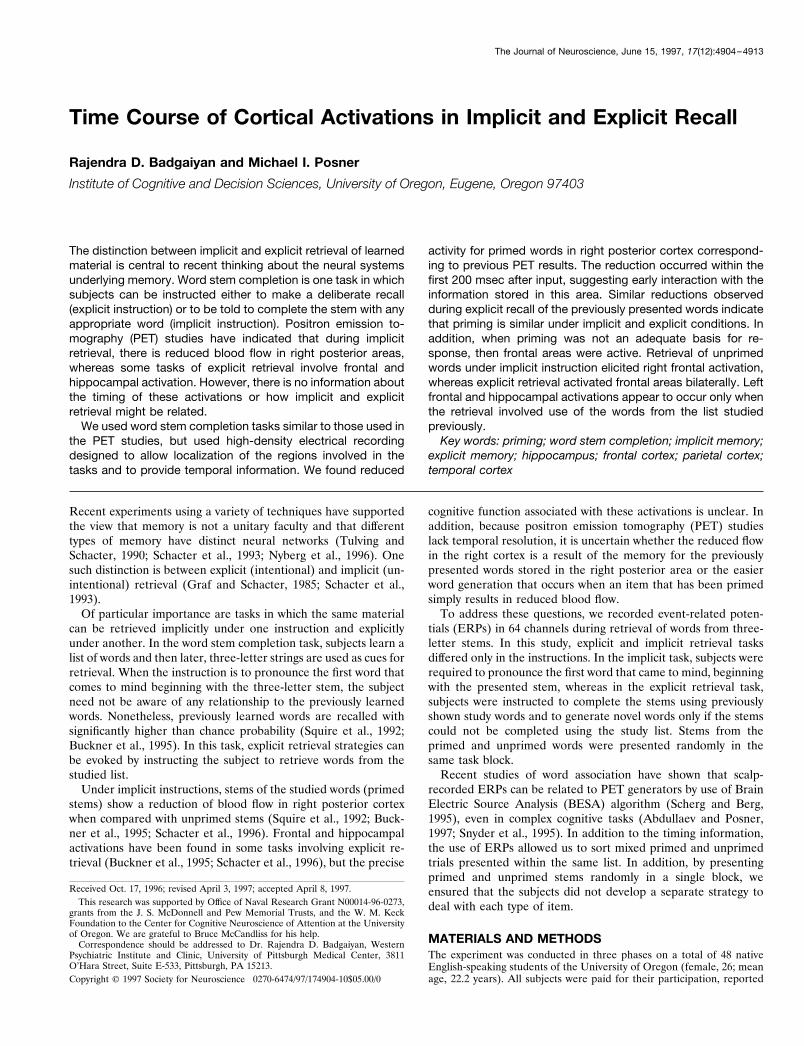
Time Course of Cortical Activations in Implicit and Explicit Recall
Rajendra D. Badgaiyan and Michael I. Posner
Institute of Cognitive and Decision Sciences, University of Oregon, Eugene, Oregon 97403
The distinction between implicit and explicit retrieval of learnedmaterial is central to recent thinking about the neural systemsunderlying memory. Word stem completion is one task in whichsubjects can be instructed either to make a deliberate recall(explicit instruction) or to be told to complete the stem with anyappropriate word (implicit instruction). Positron emission to-mography (PET) studies have indicated that during implicitretrieval, there is reduced blood flow in right posterior areas,whereas some tasks of explicit retrieval involve frontal andhippocampal activation. However, there is no information aboutthe timing of these activations or how implicit and explicitretrieval might be related.
We used word stem completion tasks similar to those used inthe PET studies, but used high-density electrical recordingdesigned to allow localization of the regions involved in thetasks and to provide temporal information. We found reduced
activity for primed words in right posterior cortex correspond-ing to previous PET results. The reduction occurred within thefirst 200 msec after input, suggesting early interaction with theinformation stored in this area. Similar reductions observedduring explicit recall of the previously presented words indicatethat priming is similar under implicit and explicit conditions. Inaddition, when priming was not an adequate basis for re-sponse, then frontal areas were active. Retrieval of unprimedwords under implicit instruction elicited right frontal activation,whereas explicit retrieval activated frontal areas bilaterally. Leftfrontal and hippocampal activations appear to occur only whenthe retrieval involved use of the words from the list studiedpreviously.
Key words: priming; word stem completion; implicit memory;explicit memory; hippocampus; frontal cortex; parietal cortex;temporal cortex
Recent experiments using a variety of techniques have supportedthe view that memory is not a unitary faculty and that differenttypes of memory have distinct neural networks (Tulving andSchacter, 1990; Schacter et al., 1993; Nyberg et al., 1996). Onesuch distinction is between explicit (intentional) and implicit (un-intentional) retrieval (Graf and Schacter, 1985; Schacter et al.,1993).
Of particular importance are tasks in which the same materialcan be retrieved implicitly under one instruction and explicitlyunder another. In the word stem completion task, subjects learn alist of words and then later, three-letter strings are used as cues forretrieval. When the instruction is to pronounce the first word thatcomes to mind beginning with the three-letter stem, the subjectneed not be aware of any relationship to the previously learnedwords. Nonetheless, previously learned words are recalled withsignificantly higher than chance probability (Squire et al., 1992;Buckner et al., 1995). In this task, explicit retrieval strategies canbe evoked by instructing the subject to retrieve words from thestudied list.
Under implicit instructions, stems of the studied words (primedstems) show a reduction of blood flow in right posterior cortexwhen compared with unprimed stems (Squire et al., 1992; Buck-ner et al., 1995; Schacter et al., 1996). Frontal and hippocampalactivations have been found in some tasks involving explicit re-trieval (Buckner et al., 1995; Schacter et al., 1996), but the precise
cognitive function associated with these activations is unclear. Inaddition, because positron emission tomography (PET) studieslack temporal resolution, it is uncertain whether the reduced flowin the right cortex is a result of the memory for the previouslypresented words stored in the right posterior area or the easierword generation that occurs when an item that has been primedsimply results in reduced blood flow.
To address these questions, we recorded event-related poten-tials (ERPs) in 64 channels during retrieval of words from three-letter stems. In this study, explicit and implicit retrieval tasksdiffered only in the instructions. In the implicit task, subjects wererequired to pronounce the first word that came to mind, beginningwith the presented stem, whereas in the explicit retrieval task,subjects were instructed to complete the stems using previouslyshown study words and to generate novel words only if the stemscould not be completed using the study list. Stems from theprimed and unprimed words were presented randomly in thesame task block.
Recent studies of word association have shown that scalp-recorded ERPs can be related to PET generators by use of BrainElectric Source Analysis (BESA) algorithm (Scherg and Berg,1995), even in complex cognitive tasks (Abdullaev and Posner,1997; Snyder et al., 1995). In addition to the timing information,the use of ERPs allowed us to sort mixed primed and unprimedtrials presented within the same list. In addition, by presentingprimed and unprimed stems randomly in a single block, weensured that the subjects did not develop a separate strategy todeal with each type of item.
MATERIALS AND METHODSThe experiment was conducted in three phases on a total of 48 nativeEnglish-speaking students of the University of Oregon (female, 26; meanage, 22.2 years). All subjects were paid for their participation, reported
Received Oct. 17, 1996; revised April 3, 1997; accepted April 8, 1997.This research was supported by Office of Naval Research Grant N00014-96-0273,
grants from the J. S. McDonnell and Pew Memorial Trusts, and the W. M. KeckFoundation to the Center for Cognitive Neuroscience of Attention at the Universityof Oregon. We are grateful to Bruce McCandliss for his help.
Correspondence should be addressed to Dr. Rajendra D. Badgaiyan, WesternPsychiatric Institute and Clinic, University of Pittsburgh Medical Center, 3811O’Hara Street, Suite E-533, Pittsburgh, PA 15213.Copyright © 1997 Society for Neuroscience 0270-6474/97/174904-10$05.00/0
The Journal of Neuroscience, June 15, 1997, 17(12):4904–4913
normal or corrected-to-normal vision, and were right-handed as assessedby Edinburgh handedness inventory questionnaires (Raczkowski et al.,1974).
In the first phase, ERPs were recorded in 24 subjects and grandaveraged waveform generated. In the second phase, this experiment wasrepeated on 16 subjects to confirm replicability of the results. In the firsttwo phases, subjects were tested first for implicit recall and then forexplicit recall on similar paradigms. In the third phase, eight subjects weretested only for explicit recall to verify that previous exposure to theimplicit task did not affect the activity during the explicit task.
For this experiment, a list of 450 words was prepared in such a way thatno two words had similar first three letters (stem) and each stem made atleast 10 words in Webster’s New Collegiate Dictionary. Length of wordsvaried between four and nine letters. These words were randomized, and90 of them were separated for use as filler words that were presentedbefore and after each study list to minimize primacy and recency effects.The remaining words were randomly divided in 12 lists of 30 words. Six ofthese were presented as study lists and were used to study implicit orexplicit recall, whereas the remaining lists were used for the control tasks.
After informed consent was obtained, the EEG electrode net wasapplied and the subject was seated in a sound-attenuated chamber. In thefirst two phases, the experiment began with three implicit memory blocks.Each of these blocks had a word-reading stage and a stem-completionstage (Table 1). In the word reading stage, a list of 45 words waspresented in upper case (black on white background). Each word wasdisplayed for 3 sec on the center of a computer monitor. The first 9 andthe last 6 words were drawn from the filler list, and 30 words from one ofthe study lists were shown between the fillers. Subjects were asked toassess whether the word would be easy or difficult for a child to under-stand and to respond by pressing either a right or left key. This task wasintroduced to ensure that the presented words were attended. After aninterval of 2 min, the stem-completion stage began. In this stage, 60 lowercase three-letter strings were shown for 3 sec each. Half of the stringswere the first three letters of the previously presented study list, and theremaining strings were derived from one of the three control lists. Noneof the control stems could be completed using any of the study words.Stems from the two lists were presented in random order, and the task ofthe subject was to pronounce the first word that came to mind, beginningwith the stem. They were advised not to use proper nouns and to avoidblinking before making a response. Subjects were also instructed not tomake any body movements not associated with the response. Pronouncedwords were recorded on a tape recorder, and the voice onset latency wasdetermined using a microphone channel connected to a voice-operatedrelay. While the subjects were completing stems, a scalp EEG wasrecorded using a 64 channel EEG electrode net (Tucker et al., 1994).
There were three implicit memory blocks. Each block contained stemsfrom the PRIMING condition, in which stems were derived from thewords presented during word-reading stage, and the PRIMING BASE-LINE condition, in which no previously learned word could complete thestem. At the end of the implicit memory blocks, subjects were allowed abreak for ;5 min before beginning the three explicit memory blocks.
The explicit memory blocks had the same structure as the implicitmemory blocks, but had different words/stems and instructions. In thestem-completion stage, subjects were informed that it was possible tocomplete some of the stems using the list of words shown in the word-reading stage and were instructed to use the study list words for com-pleting stems whenever possible. They were allowed to make guesses andwere told to come up with a novel word only if they could not recall anystudy list word beginning with the given stem. Tasks involving stems fromthe study list were designated as RECALL, and those involving stemsfrom the control list were named RECALL BASELINE. Each subject
was tested in three such blocks. Control list words were never shown tothe subjects in any of the implicit or explicit memory blocks.
In the first two phases, implicit memory blocks always preceded explicitmemory to reduce explicit recall effort during implicit memory blocks. Inthe third phase, subjects were tested only for explicit memory.
During the stem completion stage, a 1 sec EEG epoch beginning 184msec before stem onset and sampled at 250 samples/sec was collected.The EEG samples were collected with a 0.15–50 Hz bandpass from 64electrode sites referenced to the right mastoid channel. The signals werebaseline corrected, edited off-line for exclusion of ocular and motionartifacts, and referenced algebraically to an average reference derivation.The averaged ERP data were filtered digitally with a 50 Hz low-pass filterto reduce electromyographic contamination. The ERPs were averagedseparately for each of four conditions, PRIMING, PRIMING BASE-LINE, RECALL, and RECALL BASELINE. For comparing two condi-tions, difference waves were analyzed statistically using nonparametricWilcoxon signed-rank tests for each of the 4 msec samples. Whereappropriate, ANOVA was used on selected windows. Two-dimensionalspherical spline interpolated head surface images were created for thedifference waves at 4 msec intervals to depict the temporal continuity andtopography of statistical significance of difference by t test. Differencewaves were also analyzed using BESA algorithm (Scherg and Berg, 1995)to determine topography of the best-fitting dipoles.
Grand averaged waveforms were generated separately for each condi-tion for all subjects who participated in the study, and the differenceswere considered significant only when the probability was p , 0.05.
RESULTSThe words produced during the stem-completion stage were an-alyzed to assess whether there was a difference between implicitand explicit instructions. For the words actually presented duringthe word-reading stage, 42.2% were produced during the stem-completion stage under implicit instruction and 47.4% underexplicit instruction. This difference was significant by t test ( p ,0.01). Subjects completed 18.1% of the items with words from thecontrol list. Because subjects were never shown the control lists,generating items from this list had to occur only by chance.
In the implicit memory blocks, mean latency for completing astem with a word that was on the previously studied list (PRIM-ING) was 1293 msec, whereas for words not on the list (PRIM-ING BASELINE), mean latency was 1311 msec. In the explicitmemory blocks, latencies were longer than under implicit condi-tions. For primed words (RECALL), this was 1452 msec, and forunprimed (RECALL BASELINE), 1508 msec.
The longer reaction times and higher percentages of recall fromthe list under explicit instructions indicate an effort by subjects torecall items from the list. The lower response times and higherpercentage of correct response for primed items indicate thathaving an item on the previously studied list increased the likeli-hood that it would be generated for stem completion irrespectiveof instructions.
ERPs recorded using a 64 channel electrode net were averagereferenced, and separate grand average waveforms were plottedfor each condition of the study. The channels found to havesignificant variation under any two conditions in phase 1 were
Table 1. Scheme of experimental paradigm
Word-reading stage(Stage 1)
Stem-completion stage(Stage 2) Condition
Implicit memory 45 Upper case words 30 Lower case stems (primed) PRIMING(3 Blocks) Filler 1–9 and 40–45 30 Lower case stems (unprimed) PRIMING BASELINE
Study list 10–39Explicit memory 45 Upper case words 30 Lower case stems (primed) RECALL(3 Blocks) Filler 1–9 and 40–45 30 Lower case stems (unprimed) RECALL BASELINE
Study list 10–39
Badgaiyan and Posner • Cortical Activations in Recall J. Neurosci., June 15, 1997, 17(12):4904–4913 4905
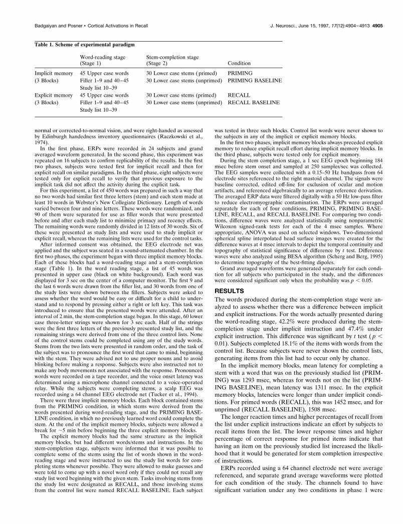
selected for additional analyses in phases 2 and 3. Topographiclocation of these channels is shown in Figure 1. ERPs of thechannels in each of the four regions (left frontal, right frontal,right parietotemporal, and medial frontoparietal) were collapsed,and these collapsed ERPs represented potentials in the respectiveareas.
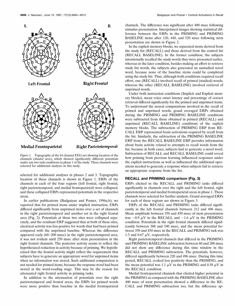
In earlier publications (Badgaiyan and Posner, 1996a,b), wereported that for primed items under implicit instruction, ERPsdiffered significantly from unprimed items over a set of channelsin the right parietotemporal and another set in the right frontalarea (Fig. 2). Potentials at these two sites were collapsed sepa-rately, and the resultant ERP suggested that at both of these sites,electrical activity was less positive for words that had been primedcompared with the unprimed baseline. Whereas the differenceappeared early (60–200 msec) in the right parietotemporal area,it was not evident until 250 msec after stem presentation in theright frontal channels. The posterior activity seems to reflect thehypothesized reduction in activity because of priming. We hypoth-esized that the frontal activity might reflect the requirement thatsubjects have to generate an appropriate word for unprimed itemswhen no information was stored. Such additional computation isnot needed for primed items, because the response word had beenstored at the word-reading stage. This may be the reason forattenuated right frontal activity in priming tasks.
In addition to the attenuation of potentials over the rightparietotemporal and frontal areas, the ERPs for primed wordswere more positive than baseline in the medial frontoparietal
channels. The difference was significant after 400 msec followingstimulus presentation. Interpolated images showing statistical dif-ference between the ERPs in the PRIMING and PRIMINGBASELINE items after 120, 440, and 520 msec following stempresentation are shown in Figure 2.
In the explicit memory blocks, we separated stems derived fromthe study list (RECALL) and those derived from the control list(RECALL BASELINE). In the former condition, the subjectsintentionally recalled the study words they were presented earlier,whereas in the later condition, besides making an effort to retrievestudy list words, the subjects also generated an unstudied novelword, because none of the baseline stems could be completedusing the study list. Thus, although both conditions required recalleffort, one (RECALL) involved recall of primed (studied) words,whereas the other (RECALL BASELINE) involved retrieval ofunprimed words.
Under both instruction conditions (Implicit and Explicit mem-ory blocks), mean voice onset latency and percentage of correctretrieval differed significantly for the primed and unprimed items.To understand the neural computations involved in the recall ofprimed and unprimed words, grand averaged ERPs obtainedduring the PRIMING and PRIMING BASELINE conditionswere subtracted from those obtained in primed (RECALL) andunprimed (RECALL BASELINE) conditions of the explicitmemory blocks. The subtraction of PRIMING ERP from RE-CALL ERP represented brain activations required by recall fromthe list. Similarly, the subtraction of the PRIMING BASELINEERP from the RECALL BASELINE ERP provides informationabout brain activity related to attempts to recall words from thelist, because in both cases, subjects had to generate a novel word.Subtraction of RECALL and RECALL BASELINE could revealhow priming from previous learning influenced responses underthe explicit instructions as well as influenced the additional oper-ations needed to generate a response when subjects fail to retrievean appropriate response from the list.
RECALL and PRIMING comparison (Fig. 3)ERPs elicited in the RECALL and PRIMING tasks differedsignificantly in channels over the right and the left frontal, rightparietotemporal and medial frontoparietal areas in phase 1. Thesechannels were selected for further analysis. Grand averaged ERPsfor each of these regions are shown in Figure 3.
ERPs of the RECALL and PRIMING tasks differed signifi-cantly in the left frontal channels between 312 and 496 msec.Mean amplitude between 350 and 450 msec of stem presentationwas 20.9 mV in the RECALL and 21.6 mV in the PRIMINGcondition. Potentials in the right frontal channels differed signif-icantly between 308 and 540 msec, and the mean potential be-tween 350 and 450 msec in the RECALL and PRIMING task was1.3 and 0.67 mV, respectively.
Right parietotemporal channels that differed in the PRIMINGand PRIMING BASELINE subtraction between 60 and 200 msecdid not show any difference during this time window in theRECALL and PRIMING subtraction. The potentials, however,differed significantly between 220 and 496 msec. During this timeperiod, RECALL evoked less positivity than the PRIMING, andthe mean potential was 1.1 mV in the PRIMING and 0.58 mV inthe RECALL condition.
Medial frontoparietal channels that elicited higher potential inthe PRIMING as compared with the PRIMING BASELINE after400 msec of stem presentation showed a difference in the RE-CALL and PRIMING subtraction too, but the difference ap-
Figure 1. Topography of the 64 channel EEG net showing location of thechannels (shaded area), which showed significantly different potentialsunder any two task conditions in phase 1 of the study. These channels wereselected for additional analyses in this study.
4906 J. Neurosci., June 15, 1997, 17(12):4904–4913 Badgaiyan and Posner • Cortical Activations in Recall
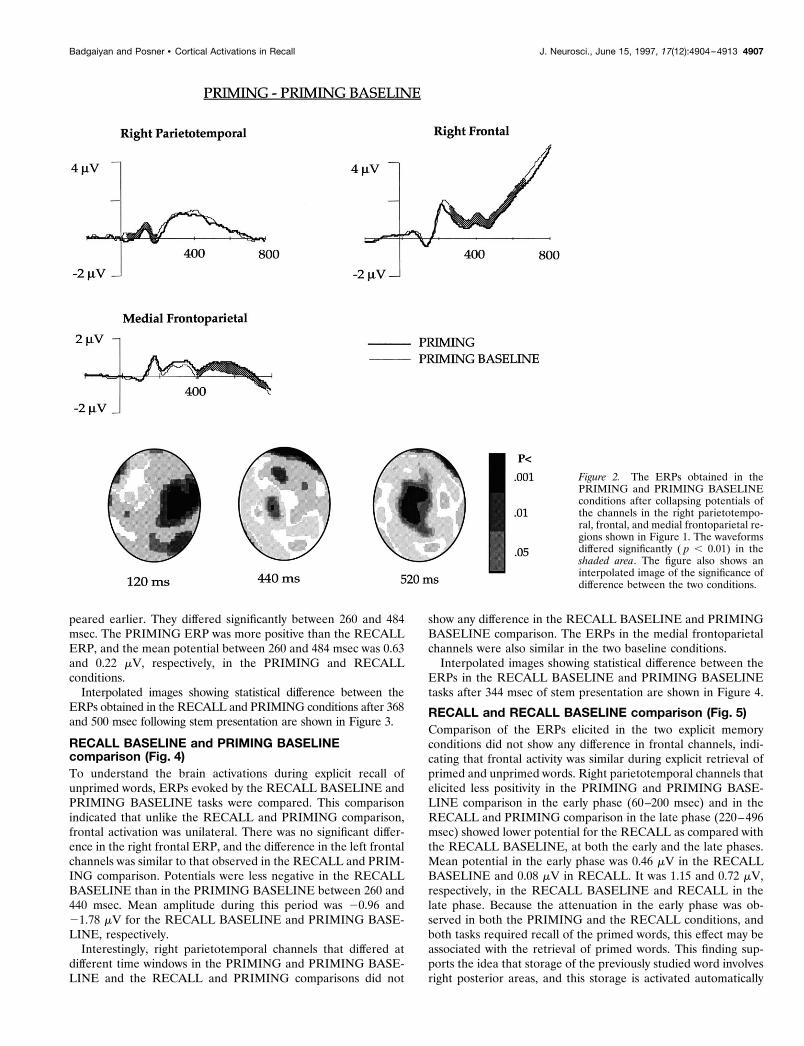
peared earlier. They differed significantly between 260 and 484msec. The PRIMING ERP was more positive than the RECALLERP, and the mean potential between 260 and 484 msec was 0.63and 0.22 mV, respectively, in the PRIMING and RECALLconditions.
Interpolated images showing statistical difference between theERPs obtained in the RECALL and PRIMING conditions after 368and 500 msec following stem presentation are shown in Figure 3.
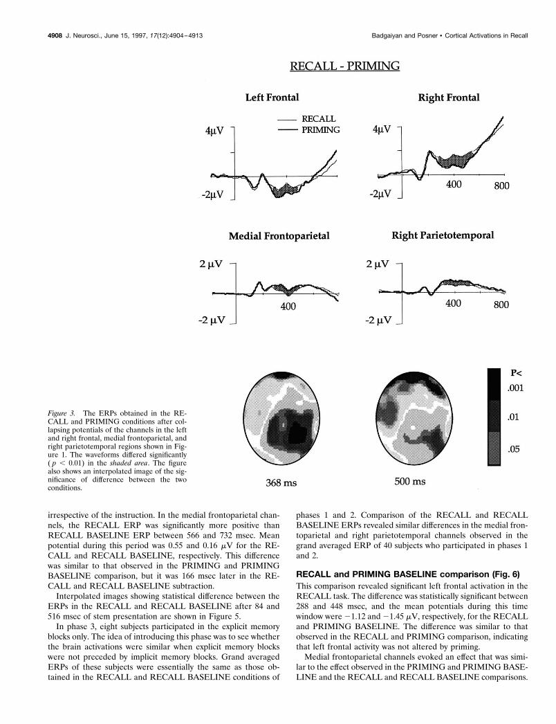
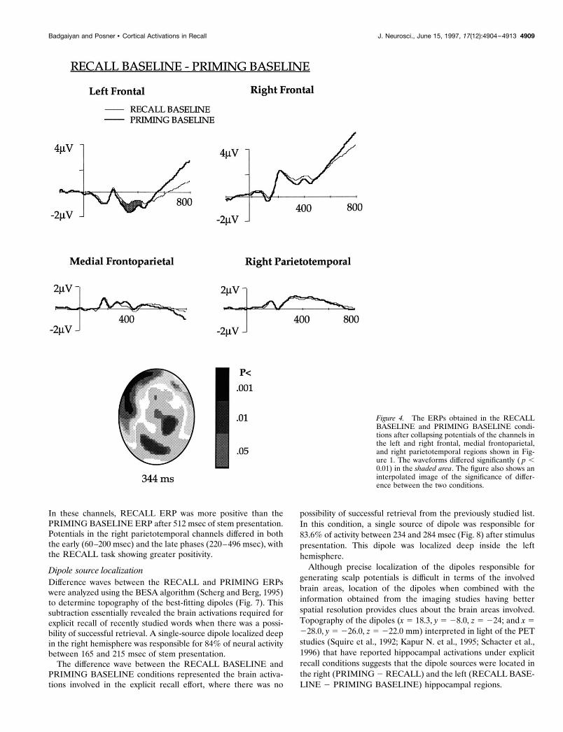
RECALL BASELINE and PRIMING BASELINEcomparison (Fig. 4)To understand the brain activations during explicit recall ofunprimed words, ERPs evoked by the RECALL BASELINE andPRIMING BASELINE tasks were compared. This comparisonindicated that unlike the RECALL and PRIMING comparison,frontal activation was unilateral. There was no significant differ-ence in the right frontal ERP, and the difference in the left frontalchannels was similar to that observed in the RECALL and PRIM-ING comparison. Potentials were less negative in the RECALLBASELINE than in the PRIMING BASELINE between 260 and440 msec. Mean amplitude during this period was 20.96 and21.78 mV for the RECALL BASELINE and PRIMING BASE-LINE, respectively.
Interestingly, right parietotemporal channels that differed atdifferent time windows in the PRIMING and PRIMING BASE-LINE and the RECALL and PRIMING comparisons did not
show any difference in the RECALL BASELINE and PRIMINGBASELINE comparison. The ERPs in the medial frontoparietalchannels were also similar in the two baseline conditions.
Interpolated images showing statistical difference between theERPs in the RECALL BASELINE and PRIMING BASELINEtasks after 344 msec of stem presentation are shown in Figure 4.
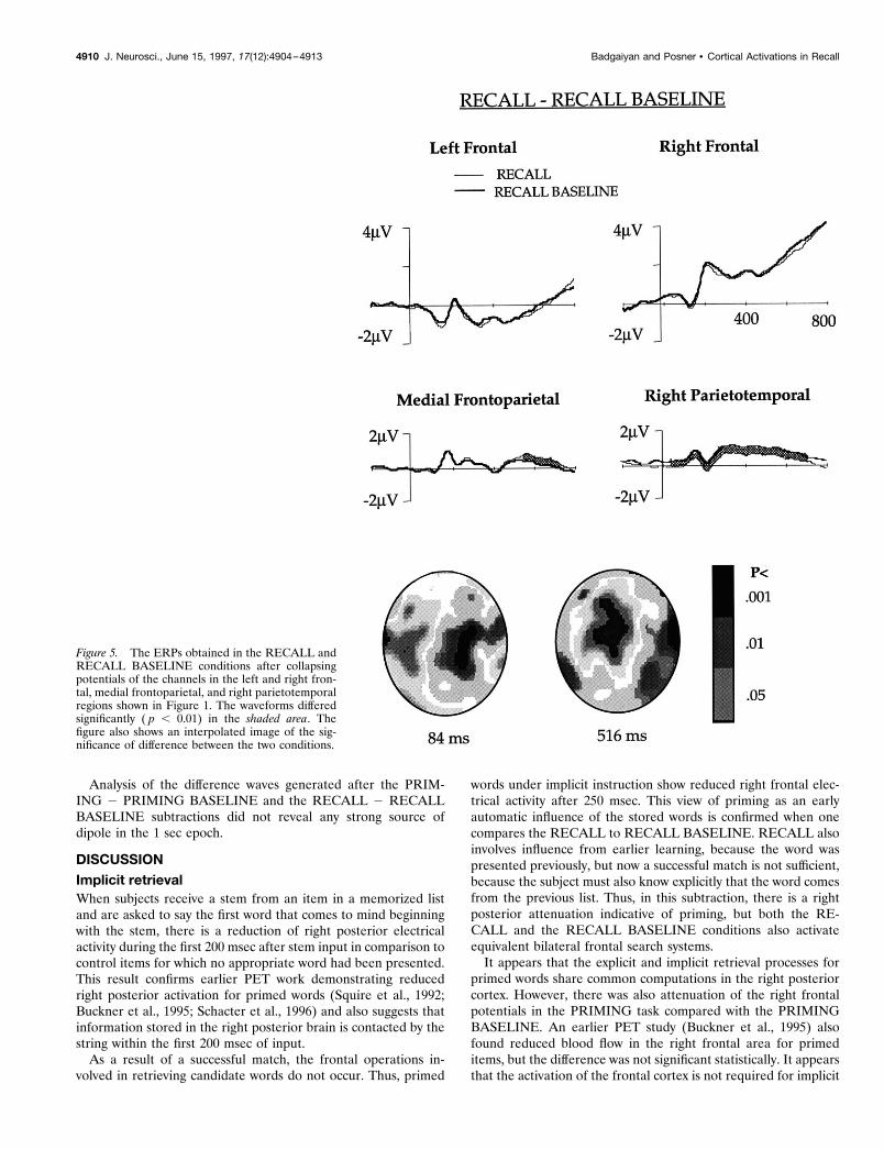
RECALL and RECALL BASELINE comparison (Fig. 5)Comparison of the ERPs elicited in the two explicit memoryconditions did not show any difference in frontal channels, indi-cating that frontal activity was similar during explicit retrieval ofprimed and unprimed words. Right parietotemporal channels thatelicited less positivity in the PRIMING and PRIMING BASE-LINE comparison in the early phase (60–200 msec) and in theRECALL and PRIMING comparison in the late phase (220–496msec) showed lower potential for the RECALL as compared withthe RECALL BASELINE, at both the early and the late phases.Mean potential in the early phase was 0.46 mV in the RECALLBASELINE and 0.08 mV in RECALL. It was 1.15 and 0.72 mV,respectively, in the RECALL BASELINE and RECALL in thelate phase. Because the attenuation in the early phase was ob-served in both the PRIMING and the RECALL conditions, andboth tasks required recall of the primed words, this effect may beassociated with the retrieval of primed words. This finding sup-ports the idea that storage of the previously studied word involvesright posterior areas, and this storage is activated automatically
Figure 2. The ERPs obtained in thePRIMING and PRIMING BASELINEconditions after collapsing potentials ofthe channels in the right parietotempo-ral, frontal, and medial frontoparietal re-gions shown in Figure 1. The waveformsdiffered significantly ( p , 0.01) in theshaded area. The figure also shows aninterpolated image of the significance ofdifference between the two conditions.
Badgaiyan and Posner • Cortical Activations in Recall J. Neurosci., June 15, 1997, 17(12):4904–4913 4907
irrespective of the instruction. In the medial frontoparietal chan-nels, the RECALL ERP was significantly more positive thanRECALL BASELINE ERP between 566 and 732 msec. Meanpotential during this period was 0.55 and 0.16 mV for the RE-CALL and RECALL BASELINE, respectively. This differencewas similar to that observed in the PRIMING and PRIMINGBASELINE comparison, but it was 166 msec later in the RE-CALL and RECALL BASELINE subtraction.
Interpolated images showing statistical difference between theERPs in the RECALL and RECALL BASELINE after 84 and516 msec of stem presentation are shown in Figure 5.
In phase 3, eight subjects participated in the explicit memoryblocks only. The idea of introducing this phase was to see whetherthe brain activations were similar when explicit memory blockswere not preceded by implicit memory blocks. Grand averagedERPs of these subjects were essentially the same as those ob-tained in the RECALL and RECALL BASELINE conditions of
phases 1 and 2. Comparison of the RECALL and RECALLBASELINE ERPs revealed similar differences in the medial fron-toparietal and right parietotemporal channels observed in thegrand averaged ERP of 40 subjects who participated in phases 1and 2.
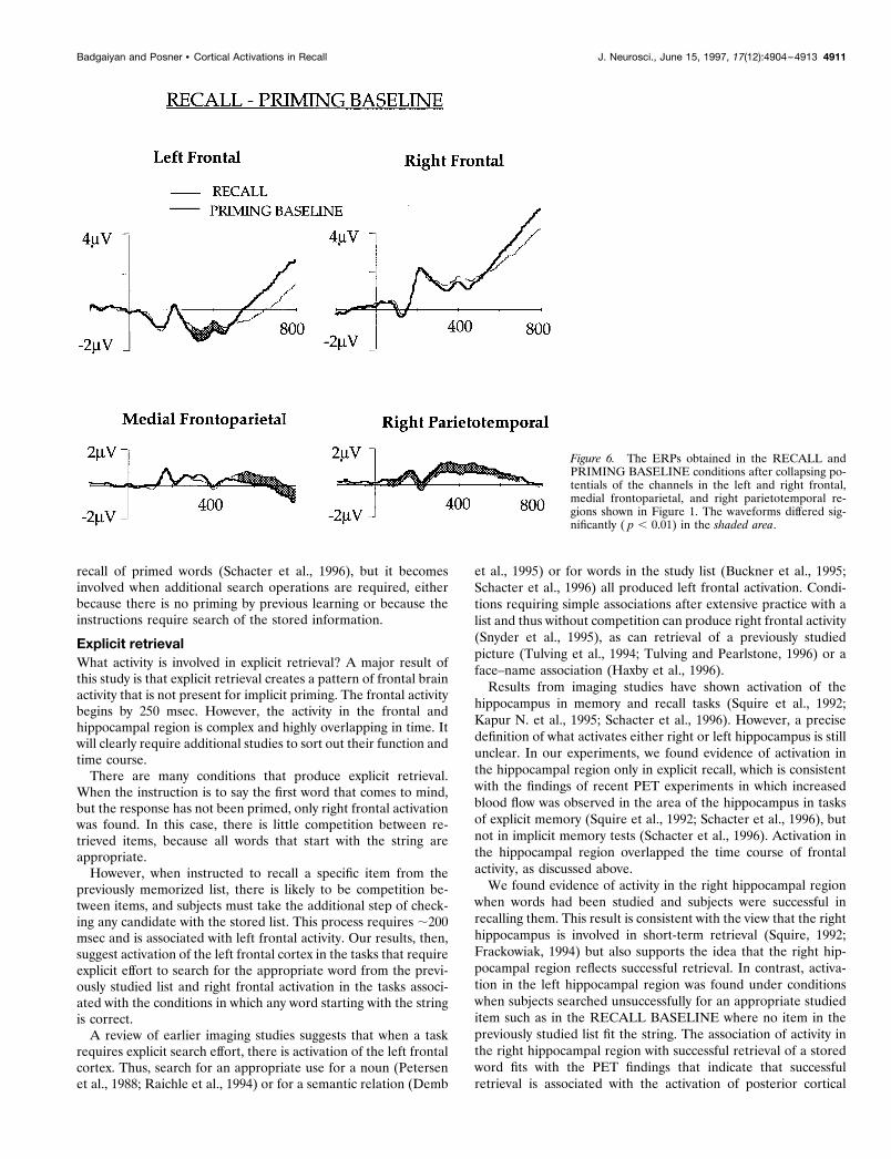
RECALL and PRIMING BASELINE comparison (Fig. 6)This comparison revealed significant left frontal activation in theRECALL task. The difference was statistically significant between288 and 448 msec, and the mean potentials during this timewindow were 21.12 and 21.45 mV, respectively, for the RECALLand PRIMING BASELINE. The difference was similar to thatobserved in the RECALL and PRIMING comparison, indicatingthat left frontal activity was not altered by priming.
Medial frontoparietal channels evoked an effect that was simi-lar to the effect observed in the PRIMING and PRIMING BASE-LINE and the RECALL and RECALL BASELINE comparisons.
Figure 3. The ERPs obtained in the RE-CALL and PRIMING conditions after col-lapsing potentials of the channels in the leftand right frontal, medial frontoparietal, andright parietotemporal regions shown in Fig-ure 1. The waveforms differed significantly( p , 0.01) in the shaded area. The figurealso shows an interpolated image of the sig-nificance of difference between the twoconditions.
4908 J. Neurosci., June 15, 1997, 17(12):4904–4913 Badgaiyan and Posner • Cortical Activations in Recall
In these channels, RECALL ERP was more positive than thePRIMING BASELINE ERP after 512 msec of stem presentation.Potentials in the right parietotemporal channels differed in boththe early (60–200 msec) and the late phases (220–496 msec), withthe RECALL task showing greater positivity.
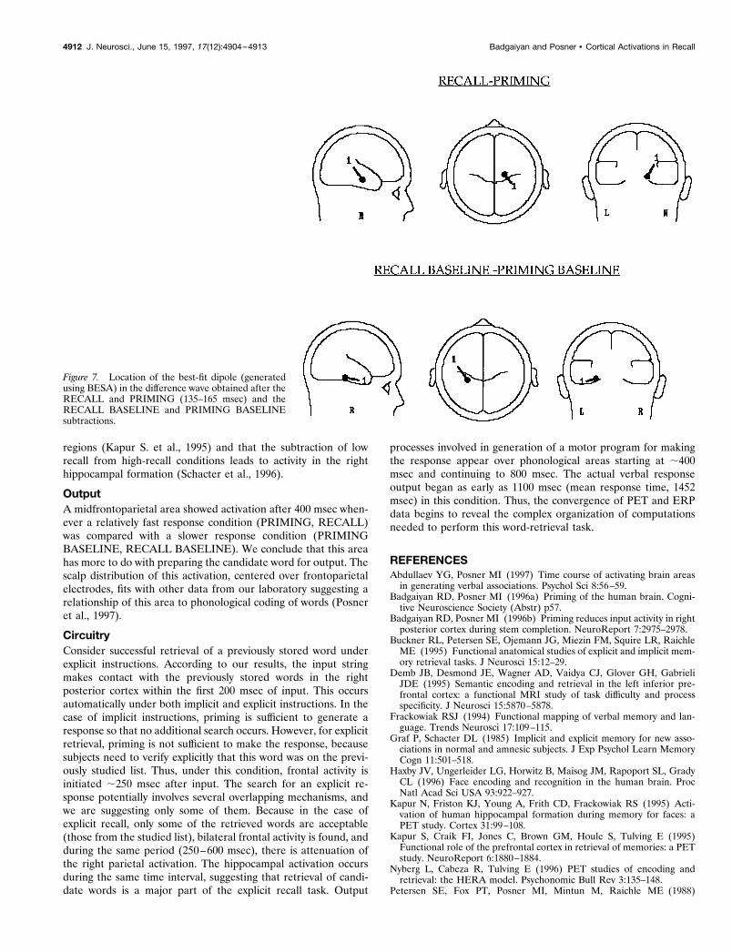
Dipole source localizationDifference waves between the RECALL and PRIMING ERPswere analyzed using the BESA algorithm (Scherg and Berg, 1995)to determine topography of the best-fitting dipoles (Fig. 7). Thissubtraction essentially revealed the brain activations required forexplicit recall of recently studied words when there was a possi-bility of successful retrieval. A single-source dipole localized deepin the right hemisphere was responsible for 84% of neural activitybetween 165 and 215 msec of stem presentation.
The difference wave between the RECALL BASELINE andPRIMING BASELINE conditions represented the brain activa-tions involved in the explicit recall effort, where there was no
possibility of successful retrieval from the previously studied list.In this condition, a single source of dipole was responsible for83.6% of activity between 234 and 284 msec (Fig. 8) after stimuluspresentation. This dipole was localized deep inside the lefthemisphere.
Although precise localization of the dipoles responsible forgenerating scalp potentials is difficult in terms of the involvedbrain areas, location of the dipoles when combined with theinformation obtained from the imaging studies having betterspatial resolution provides clues about the brain areas involved.Topography of the dipoles (x 5 18.3, y 5 28.0, z 5 224; and x 5228.0, y 5 226.0, z 5 222.0 mm) interpreted in light of the PETstudies (Squire et al., 1992; Kapur N. et al., 1995; Schacter et al.,1996) that have reported hippocampal activations under explicitrecall conditions suggests that the dipole sources were located inthe right (PRIMING 2 RECALL) and the left (RECALL BASE-LINE 2 PRIMING BASELINE) hippocampal regions.
Figure 4. The ERPs obtained in the RECALLBASELINE and PRIMING BASELINE condi-tions after collapsing potentials of the channels inthe left and right frontal, medial frontoparietal,and right parietotemporal regions shown in Fig-ure 1. The waveforms differed significantly ( p ,0.01) in the shaded area. The figure also shows aninterpolated image of the significance of differ-ence between the two conditions.
Badgaiyan and Posner • Cortical Activations in Recall J. Neurosci., June 15, 1997, 17(12):4904–4913 4909
Analysis of the difference waves generated after the PRIM-ING 2 PRIMING BASELINE and the RECALL 2 RECALLBASELINE subtractions did not reveal any strong source ofdipole in the 1 sec epoch.
DISCUSSIONImplicit retrievalWhen subjects receive a stem from an item in a memorized listand are asked to say the first word that comes to mind beginningwith the stem, there is a reduction of right posterior electricalactivity during the first 200 msec after stem input in comparison tocontrol items for which no appropriate word had been presented.This result confirms earlier PET work demonstrating reducedright posterior activation for primed words (Squire et al., 1992;Buckner et al., 1995; Schacter et al., 1996) and also suggests thatinformation stored in the right posterior brain is contacted by thestring within the first 200 msec of input.
As a result of a successful match, the frontal operations in-volved in retrieving candidate words do not occur. Thus, primed
words under implicit instruction show reduced right frontal elec-trical activity after 250 msec. This view of priming as an earlyautomatic influence of the stored words is confirmed when onecompares the RECALL to RECALL BASELINE. RECALL alsoinvolves influence from earlier learning, because the word waspresented previously, but now a successful match is not sufficient,because the subject must also know explicitly that the word comesfrom the previous list. Thus, in this subtraction, there is a rightposterior attenuation indicative of priming, but both the RE-CALL and the RECALL BASELINE conditions also activateequivalent bilateral frontal search systems.
It appears that the explicit and implicit retrieval processes forprimed words share common computations in the right posteriorcortex. However, there was also attenuation of the right frontalpotentials in the PRIMING task compared with the PRIMINGBASELINE. An earlier PET study (Buckner et al., 1995) alsofound reduced blood flow in the right frontal area for primeditems, but the difference was not significant statistically. It appearsthat the activation of the frontal cortex is not required for implicit
Figure 5. The ERPs obtained in the RECALL andRECALL BASELINE conditions after collapsingpotentials of the channels in the left and right fron-tal, medial frontoparietal, and right parietotemporalregions shown in Figure 1. The waveforms differedsignificantly ( p , 0.01) in the shaded area. Thefigure also shows an interpolated image of the sig-nificance of difference between the two conditions.
4910 J. Neurosci., June 15, 1997, 17(12):4904–4913 Badgaiyan and Posner • Cortical Activations in Recall
recall of primed words (Schacter et al., 1996), but it becomesinvolved when additional search operations are required, eitherbecause there is no priming by previous learning or because theinstructions require search of the stored information.
Explicit retrievalWhat activity is involved in explicit retrieval? A major result ofthis study is that explicit retrieval creates a pattern of frontal brainactivity that is not present for implicit priming. The frontal activitybegins by 250 msec. However, the activity in the frontal andhippocampal region is complex and highly overlapping in time. Itwill clearly require additional studies to sort out their function andtime course.
There are many conditions that produce explicit retrieval.When the instruction is to say the first word that comes to mind,but the response has not been primed, only right frontal activationwas found. In this case, there is little competition between re-trieved items, because all words that start with the string areappropriate.
However, when instructed to recall a specific item from thepreviously memorized list, there is likely to be competition be-tween items, and subjects must take the additional step of check-ing any candidate with the stored list. This process requires ;200msec and is associated with left frontal activity. Our results, then,suggest activation of the left frontal cortex in the tasks that requireexplicit effort to search for the appropriate word from the previ-ously studied list and right frontal activation in the tasks associ-ated with the conditions in which any word starting with the stringis correct.
A review of earlier imaging studies suggests that when a taskrequires explicit search effort, there is activation of the left frontalcortex. Thus, search for an appropriate use for a noun (Petersenet al., 1988; Raichle et al., 1994) or for a semantic relation (Demb
et al., 1995) or for words in the study list (Buckner et al., 1995;Schacter et al., 1996) all produced left frontal activation. Condi-tions requiring simple associations after extensive practice with alist and thus without competition can produce right frontal activity(Snyder et al., 1995), as can retrieval of a previously studiedpicture (Tulving et al., 1994; Tulving and Pearlstone, 1996) or aface–name association (Haxby et al., 1996).
Results from imaging studies have shown activation of thehippocampus in memory and recall tasks (Squire et al., 1992;Kapur N. et al., 1995; Schacter et al., 1996). However, a precisedefinition of what activates either right or left hippocampus is stillunclear. In our experiments, we found evidence of activation inthe hippocampal region only in explicit recall, which is consistentwith the findings of recent PET experiments in which increasedblood flow was observed in the area of the hippocampus in tasksof explicit memory (Squire et al., 1992; Schacter et al., 1996), butnot in implicit memory tests (Schacter et al., 1996). Activation inthe hippocampal region overlapped the time course of frontalactivity, as discussed above.
We found evidence of activity in the right hippocampal regionwhen words had been studied and subjects were successful inrecalling them. This result is consistent with the view that the righthippocampus is involved in short-term retrieval (Squire, 1992;Frackowiak, 1994) but also supports the idea that the right hip-pocampal region reflects successful retrieval. In contrast, activa-tion in the left hippocampal region was found under conditionswhen subjects searched unsuccessfully for an appropriate studieditem such as in the RECALL BASELINE where no item in thepreviously studied list fit the string. The association of activity inthe right hippocampal region with successful retrieval of a storedword fits with the PET findings that indicate that successfulretrieval is associated with the activation of posterior cortical
Figure 6. The ERPs obtained in the RECALL andPRIMING BASELINE conditions after collapsing po-tentials of the channels in the left and right frontal,medial frontoparietal, and right parietotemporal re-gions shown in Figure 1. The waveforms differed sig-nificantly ( p , 0.01) in the shaded area.
Badgaiyan and Posner • Cortical Activations in Recall J. Neurosci., June 15, 1997, 17(12):4904–4913 4911
regions (Kapur S. et al., 1995) and that the subtraction of lowrecall from high-recall conditions leads to activity in the righthippocampal formation (Schacter et al., 1996).
OutputA midfrontoparietal area showed activation after 400 msec when-ever a relatively fast response condition (PRIMING, RECALL)was compared with a slower response condition (PRIMINGBASELINE, RECALL BASELINE). We conclude that this areahas more to do with preparing the candidate word for output. Thescalp distribution of this activation, centered over frontoparietalelectrodes, fits with other data from our laboratory suggesting arelationship of this area to phonological coding of words (Posneret al., 1997).
CircuitryConsider successful retrieval of a previously stored word underexplicit instructions. According to our results, the input stringmakes contact with the previously stored words in the rightposterior cortex within the first 200 msec of input. This occursautomatically under both implicit and explicit instructions. In thecase of implicit instructions, priming is sufficient to generate aresponse so that no additional search occurs. However, for explicitretrieval, priming is not sufficient to make the response, becausesubjects need to verify explicitly that this word was on the previ-ously studied list. Thus, under this condition, frontal activity isinitiated ;250 msec after input. The search for an explicit re-sponse potentially involves several overlapping mechanisms, andwe are suggesting only some of them. Because in the case ofexplicit recall, only some of the retrieved words are acceptable(those from the studied list), bilateral frontal activity is found, andduring the same period (250–600 msec), there is attenuation ofthe right parietal activation. The hippocampal activation occursduring the same time interval, suggesting that retrieval of candi-date words is a major part of the explicit recall task. Output
processes involved in generation of a motor program for makingthe response appear over phonological areas starting at ;400msec and continuing to 800 msec. The actual verbal responseoutput began as early as 1100 msec (mean response time, 1452msec) in this condition. Thus, the convergence of PET and ERPdata begins to reveal the complex organization of computationsneeded to perform this word-retrieval task.
REFERENCESAbdullaev YG, Posner MI (1997) Time course of activating brain areas
in generating verbal associations. Psychol Sci 8:56–59.Badgaiyan RD, Posner MI (1996a) Priming of the human brain. Cogni-
tive Neuroscience Society (Abstr) p57.Badgaiyan RD, Posner MI (1996b) Priming reduces input activity in right
posterior cortex during stem completion. NeuroReport 7:2975–2978.Buckner RL, Petersen SE, Ojemann JG, Miezin FM, Squire LR, Raichle
ME (1995) Functional anatomical studies of explicit and implicit mem-ory retrieval tasks. J Neurosci 15:12–29.
Demb JB, Desmond JE, Wagner AD, Vaidya CJ, Glover GH, GabrieliJDE (1995) Semantic encoding and retrieval in the left inferior pre-frontal cortex: a functional MRI study of task difficulty and processspecificity. J Neurosci 15:5870–5878.
Frackowiak RSJ (1994) Functional mapping of verbal memory and lan-guage. Trends Neurosci 17:109–115.
Graf P, Schacter DL (1985) Implicit and explicit memory for new asso-ciations in normal and amnesic subjects. J Exp Psychol Learn MemoryCogn 11:501–518.
Haxby JV, Ungerleider LG, Horwitz B, Maisog JM, Rapoport SL, GradyCL (1996) Face encoding and recognition in the human brain. ProcNatl Acad Sci USA 93:922–927.
Kapur N, Friston KJ, Young A, Frith CD, Frackowiak RS (1995) Acti-vation of human hippocampal formation during memory for faces: aPET study. Cortex 31:99–108.
Kapur S, Craik FI, Jones C, Brown GM, Houle S, Tulving E (1995)Functional role of the prefrontal cortex in retrieval of memories: a PETstudy. NeuroReport 6:1880–1884.
Nyberg L, Cabeza R, Tulving E (1996) PET studies of encoding andretrieval: the HERA model. Psychonomic Bull Rev 3:135–148.
Petersen SE, Fox PT, Posner MI, Mintun M, Raichle ME (1988)
Figure 7. Location of the best-fit dipole (generatedusing BESA) in the difference wave obtained after theRECALL and PRIMING (135–165 msec) and theRECALL BASELINE and PRIMING BASELINEsubtractions.
4912 J. Neurosci., June 15, 1997, 17(12):4904–4913 Badgaiyan and Posner • Cortical Activations in Recall
Positron emission tomography studies of the cortical anatomy of single-word processing. Nature 331:585–589.
Posner MI, Abdullaev YG, McCandliss BD, Sereno S (1997) Anatomy,circuitry and plasticity of word reading. In: Visual and attentionalprocesses in reading and dyslexia (Everatt J, ed). London: Routledge.
Raczkowski D, Kalatm JW, Nebes R (1974) Reliability and validity ofsome right handed questionnaire items. Neuropsychology 6:43–47.
Raichle ME, Fiez JA, Videen TO, MacLeod A-MK, Pardo JV, Fox PT,Petersen SE (1994) Practice-related changes in human brain func-tional anatomy during nonmotor learning. Cereb Cortex 4:8–26.
Schacter DL, Peter-Chieu C-Y, Ochsner KN (1993) Implicit memory: aselective review. Annu Rev Neurosci 16:159–182.
Schacter DL, Alpert NM, Savage CR, Rauch SL, Albert MS (1996)Conscious recollection and the human hippocampal formation: evi-dence from positron emission tomography. Proc Natl Acad Sci USA93:321–325.
Scherg M, Berg P (1995) Brain electric source analysis handbook. Hern-don, VA: Neuroscan.
Snyder A, Abdullaev YG, Posner MI, Raichle ME (1995) Scalp electricalpotentials reflect regional cerebral blood flow responses during process-ing of written words. Proc Natl Acad Sci USA 92:1689–1693.
Squire LR (1992) Memory and the hippocampus: a synthesis from find-ings with rats, monkeys, and humans. Psychol Rev 99:195–231.
Squire LR, Ojemann JG, Miezin FM, Petersen SE, Videen TO, RaichleME (1992) Activation of the hippocampus in normal humans: a func-tional anatomical study of memory. Proc Natl Acad Sci USA89:1837–1841.
Tucker DM, Liotti M, Potts GF, Russell GS, Posner MI (1994) Spatio-temporal analysis of brain electrical fields. Hum Brain Mapp 1:134–152.
Tulving E, Pearlstone Z (1996) Availability versus accessibility of infor-mation in memory for words. J Verbal Learn Verbal Behav 5:381–391.
Tulving E, Schacter DL (1990) Priming and human memory systems.Science 247:301–306.
Tulving E, Markowitsch HJ, Kapur S, Habib R, Houle S (1994) Noveltyencoding networks in the human brain: positron emission tomographydata. NeuroReport 5:525–536.
Badgaiyan and Posner • Cortical Activations in Recall J. Neurosci., June 15, 1997, 17(12):4904–4913 4913