Embed Size (px)
Citation preview

www.elsevier.com/locate/neuint
Neurochemistry International 52 (2008) 447–456
TNF is a key mediator of septic encephalopathy acting through its
receptor, TNF receptor-1§
Jessy J. Alexander *, Alexander Jacob, Patrick Cunningham,Lauren Hensley, Richard J. Quigg
Department of Medicine, University of Chicago, 5841 South Maryland Avenue, MC5100, Chicago, IL 60637, USA
Received 4 January 2007; received in revised form 17 July 2007; accepted 9 August 2007
Available online 17 August 2007
Abstract
In this study, we demonstrate that mice deficient in TNFR1 (TNFR1�/�) were resistant to LPS-induced encephalopathy. Systemic
administration of lipopolysaccharide (LPS) induces a widespread inflammatory response similar to that observed in sepsis. Following LPS
administration TNFR1�/� mice had less caspase-dependent apoptosis in brain cells and fewer neutrophils infiltrating the brain ( p < 0.039),
compared to control C57Bl6 (TNFR1+/+) mice. TNFR1-dependent increase in aquaporin (AQP)-4 mRNA and protein expression was observed
with a concomitant increase in water content, in brain (18% increase in C57Bl6 mice treated with LPS versus those treated with saline), similar to
cerebral edema observed in sepsis. Furthermore, absence of TNFR1 partially but significantly reduced the activation of astrocytes, as shown by
immunofluorescence and markedly inhibited iNOS mRNA expression ( p < 0.01).
Septic encephalopathy is a devastating complication of sepsis. Although, considerable work has been done to identify the mechanism causing
the pathological alterations in this setting, the culprit still remains an enigma. Our results demonstrate for the first time that endotoxemia leads to
inflammation in brain, with alteration in blood–brain barrier, up-regulation of AQP4 and associated edema, neutrophil infiltration, astrocytosis, as
well as apoptotic cellular death, all of which appear to be mediated by TNF-a signaling through TNFR1.
# 2007 Elsevier Ltd. All rights reserved.
Keywords: Rodent; Septic encephalopathy; Lipopolysaccharide; Apoptosis; Aquaporin 4
Sepsis is a condition characterized by uncontrolled infection
and affects many organs including brain (Green et al., 2004),
with an attendant high mortality rate. Administration of
bacterial endotoxin (LPS), a cell wall component of gram-
negative bacteria, to mice cause pathogenesis, mimicking what
occurs in clinical sepsis (Gardenfors et al., 2002; Radzivil et al.,
1990). Septic encephalopathy could be due to multiple factors
including inflammatory cells and their mediators, reduced
cerebral blood flow, disruption of the blood–brain barrier
(BBB), cerebral edema and inflammation (Papadopoulos et al.,
2000). One of the mediators of inflammation that play a key role
in sepsis, tumor necrosis factor (TNF-a), is increased in
circulation following LPS administration (Tsao et al., 2001;
§ This work was supported by National Institutes of Health Grants
R01DK41873, R01DK55357, and R01AI43579.
* Corresponding author.
E-mail address: [email protected] (J.J. Alexander).
0197-0186/$ – see front matter # 2007 Elsevier Ltd. All rights reserved.
doi:10.1016/j.neuint.2007.08.006
Merrill and Benveniste, 1996). Serum TNF-a is an acute phase
response and does not correlate with the alterations in brain,
during stroke (Intiso et al., 2004). On the other hand, TNF-a is
also produced in brain, by microglia and astrocytes, both of
which are activated by LPS. LPS disrupts the BBB (Tsao et al.,
2001; Gurney et al., 2006), which leads to increased infiltration
of blood cells into brain. TNF-a has also been shown to
stimulate the oxidative burst of neutrophils (Granert et al.,
1994; Wispelwey et al., 1989) and to up-regulate the expression
of adhesion molecules (Amrani et al., 2000; Marshall et al.,
2003; Schafers et al., 2002; Yang et al., 2005) that mediate
neutrophil recruitment to tissues following an inflammatory
stimulus. TNF-a participates in many pathophysiological
conditions in the central nervous system (CNS) (Pan et al.,
1997), including neurodegenerative diseases (Alexander et al.,
2005, 2007). The significance of TNF-a in these neuropatho-
logical conditions is not totally understood, since it has
potentially both detrimental and beneficial roles in the CNS.
The role of TNF-a is manifold and range from apoptosis and

J.J. Alexander et al. / Neurochemistry International 52 (2008) 447–456448
astrogliosis, a prominent feature of inflammation in brain, to the
regulation of iNOS synthesis and edema. On the other hand,
several lines of evidence show that TNF-a can also exhibit
regenerating or neuroprotective effects (Loddick and Rothwell,
1999; Allan and Rothwell, 2001). TNF-a acts through two
distinct receptors: TNFR1, which mediates most of the
inflammatory actions of TNF-a, and TNFR2, which has been
proposed to have an anti-inflammatory role (Dopp et al., 1997).
Although both TNF-a receptors occur in brain, TNFR1 seems
to be abundantly expressed in the brain and shows constitutive
expression in astrocytes (Dopp et al., 1997) and the
microvasculature (Nadeau and Rivest, 1999), while TNFR2
is observed only on stimulation in brain.
Astrocytes are the major glial cell population within the
CNS and play important physiological roles in brain
functions. Astrocytes react to various neurodegenerative
insults and diseases, leading to vigorous astrogliosis (Eng
et al., 1992). On activation, astrocytes express an enhanced
level of glial fibrillary acidic protein (GFAP) proportional to
the severity of astroglial activation (Eng et al., 1992, 2000;
Eng and Ghirnikar, 1994). However, the mechanism by which
astroglial expression of GFAP is increased in neurodegen-
erative CNS remains unclear (Norenberg, 1996, 1998).
Several lines of evidence demonstrate that NO plays a key
role in regulating the expression of GFAP in astrocytes
(Askalan et al., 2006; Enkhbaatar et al., 2006; Hawkins et al.,
1998). On the other hand activated astrocytes express
inducible nitric oxide (NO) synthase (iNOS) leading to an
excessive amount of NO, a molecule implicated in many
neurodegenerative and neuroinflammatory conditions (Lee
et al., 2004; Choi et al., 2005; Kubes et al., 1993). NO could
induce apoptosis in astrocytes by p-53 and Bax-dependent
mechanisms (Yung et al., 2004). Furthermore, aquaporin
(AQP) 4, the most abundant water channel in the CNS is
present on the astrocytes (Vizuete et al., 1999; Rao et al.,
2003) and plays a crucial role in brain edema formation
observed after CNS insults or in disease (Manley et al., 2004;
Badaut et al., 2002; Alexander et al., 2003). Edema could lead
to increased intracranial pressure, resulting in brain herniation
and finally death (Norenberg et al., 2005).
Septic encephalopathy although common and devastating,
the role of the different mechanisms behind this occurrence,
still remains unclear. Although cytokines such as TNF-a are
key mediators of sepsis (Lucas et al., 1997), knowledge as to
their effects in brain is still being investigated, in the setting of
endotoxemia. As many of the central consequences of TNF-a
are mediated by the TNFR1 receptor (Gimenez et al., 2004; Yin
et al., 2004), the present investigation attempts to gain further
insights into the role of this receptor in endotoxin-induced
toxicity, in brain.
1. Experimental procedures
1.1. Animals
Mice with targeted deletion of TNFR1 (TNFR1�/�) were obtained from The
Jackson Laboratory (Bar Harbor, ME) and used for experiments at 8 weeks of
age. Because these mice are on a C57BL/6 background, age-matched C57BL/6
mice were used as controls. The animals were maintained with 12 h light and
dark cycles with free access to food and water. One group was given LPS (from
Escherichia coli, serotype 055:B55, Sigma–Aldrich, St. Louis, MO., Lot #
127H4097) as a single injection of 0.15 mg i.p. and another group received the
saline vehicle as control. The mice were sacrificed 8 h later and their brains
harvested. The animals were maintained and experiments were performed in
accordance with the guidelines set by the University of Chicago Institutional
Animal Care and Use Committee.
1.2. Tissue processing
Brains were isolated from animals immediately upon sacrifice. Cerebellum
and brain stem were discarded. The anterior region of the cerebral cortex was
snap frozen for immunofluorescence (IF) microscopy and the rest were pro-
cessed for RNA and genomic DNA isolation. For immunohistochemistry, 4-mm
brain cryostat sections were fixed with ether/ethanol, incubated with 0.3% H2O2
for 30 min, and blocked with dilute horse serum. Sections were stained for
neutrophils by sequential incubation with rat anti-mouse neutrophil (mAb 7/4;
Serotec, Raleigh, NC) at a 1/60 dilution for 30 min followed by HRP-con-
jugated rabbit anti-rat IgG (Sigma–Aldrich) at a 1/60 dilution for 30 min and
diaminobenzidine reagent (Vector Laboratories, Burlingame, CA) for 10 min. A
blinded observer counted the number of neutrophils per high-power field and
recorded the average of 10 fields for each sample.
1.3. LM-PCR
Brains were harvested and immediately frozen at �80 8C. Later, a small
piece was thawed, from which DNA was purified (DNeasy DNA purification
system; Qiagen, Valencia, CA). The extent of DNA laddering was amplified and
detected by ligase-mediated (LM)-PCR (Clontech Laboratories, Palo Alto, CA)
according to the manufacturer’s instructions, as follows. DNA isolated from
each animal was incubated with the supplied primer targets and T4 DNA ligase
for 18 h at 16 8C. Twenty micrograms of this ligated DNA was then used as the
substrate for PCR, using supplied primers and Advantage DNA polymerase
(Clontech Laboratories) for 23 cycles of 94 8C for 1 min and at 72 8C for 3 min.
The reaction product for each animal was run on a 1.3% agarose gel, and
ethidium bromide-stained bands were detected with UV light illumination.
1.4. Assay of caspase-3 activity
Brains were homogenized in cell lysis buffer (25 mM HEPES pH 7.4, 2 mM
DTT, 5 mM EDTA, and 10 mM digitonin). Lysates were then incubated on ice
for 15 min and centrifuged at 10,000 � g for 10 min at 4 8C and protein
concentrations were determined using a BCA assay. Lysates (soluble super-
natants) were used immediately or stored at�80 8C. Aliquots of protein (50 mg)
were incubated at 37 8C with assay buffer (50 mm HEPES pH 7.4, 100 mm
NaCl, 0.1% CHAPS, 10 mm dithiothreitol, 1 mM EDTA, 10% glycerol) and
200 mM Ac-DEVD-pNA (Biomol). Hydrolysis of the DEVD-AFC substrate
was followed for 15 min by fluorimetry of the released AFC (excitation 400 nm,
emission 505 nm) and activity calculated from the slope.
1.5. Brain water content
We determined the brain water content in normal and LPS-treated mice. The
brains were removed, weighed immediately and then kept at 300 8C for 48 h.
The brains were weighed during this process at 24 h and then again at 48 h to
make sure they had reached a consistent weight. The percentage of brain water
in the tissue was calculated as (wet wt. � dry wt.) � 100/wet wt.
1.6. Blood–brain barrier integrity
The BBB integrity was determined in normal, LPS-treated C57Bl6 and
TNFR1-deficient mice. One percent Evans blue in saline was injected through
the tail vein and the mice were sacrificed 5 min later. The brains were removed,
snap frozen. Cryosections (4 mm thickness) were obtained and observed using a
Zeiss microscope at 40�.

Table 1
The sequences of primers and probes for the different genes synthesized by integrated DNA technologies (Coralville, IA) and synthegen (Houston, TX)
Gene Forward primer 50–30 Reverse primer 50–30 Probe 50–30
FAM/TAMRA dyes
GAPDH GGCAAATTCAAGGCACAGT AGATGGTGATGGGCTTCCC GGCC GAGAATGGGAAGCTTGTCATC
iNOS CAGCTGGGCTGTACAAACCTT TGAATGTGATGTTTGCTTCGG CGGGCAGCCTGTGAGACCTTTGA
Gene Forward primer 50–30 Reverse primer 50–30
SYBR green incorporation
TNF-a CCGATGGGTTGTACCTTGTC GTGGGTGAGGAGCACGTAGT
TNFR1 GACCGGGAGAAGAGGGATAG GTTCCTTTGTGGCACTTGGT
AQP4 AGGTGCCCTTTATGAGTATG CTTCCCTTCTTCTCTTCTCC
GAPDH GGCAAATTCAACGGCACAGT AGATGGTGATGGGCTTCCC
J.J. Alexander et al. / Neurochemistry International 52 (2008) 447–456 449
1.7. IF microscopy
Cryostat sections (4 mm) were fixed with ethanol:ether and ethanol and IF
microscopy was done using hamster anti-mouse TNFR1 (BD PharMingen, San
Diego, CA), rabbit anti-rat AQP4 (Chemicon International, Temecula, CA) and
rabbit anti-cow GFAP (Dako) at dilutions of 1:60. The staining was detected
using FITC-labeled anti-hamster or anti-rabbit antibody at a dilution of 1:100.
No staining was observed in controls slides treated with a rat or rabbit IgG
followed by FITC-labeled anti-rabbit antibody. Sections were observed using a
Zeiss microscope at 400�. The slides were scored in a blinded fashion from 0 to
4 (0 indicating no staining and 4 the most intense staining).
1.8. Quantitative (q) RT-PCR
RNA from brains was isolated using TriZol reagent (Life Technologies,
Grand Island, NY). cDNA was prepared in an analogous fashion from brains of
mice (TNFR1�/� and TNFR1+/+) sacrificed 8 h after LPS injection and
compared with C57Bl6 mice treated with saline (n = 6 per group). Real-time
RT-PCR was performed for iNOS, and AQP4 as follows. A portion of brain from
each mouse was frozen at�80 8C, from which total RNAwas purified using the
RNeasy Mini RNA purification kit (Qiagen). To remove all traces of genomic
DNA, samples were then treated with RNase-free RQ1 DNase (1 U per 4 mg
RNA; Promega, Madison, WI) in 10 ml reaction buffer (final concentration,
40 mM Tris–HCl, 10 mM MgSO4, and 1 mM CaCl2 (pH 8)) at 37 8C for
30 min. This was followed by addition of 1 ml 20 mM EGTA (pH 8) to stop the
reaction and incubation at 65 8C for 10 min to inactivate the DNase. cDNA was
generated from RNA using random hexamers as primers with the SuperScript
Fig. 1. TNF-a is significantly increased in circulation by LPS-treatment. Serum
TNF-a was determined by ELISA. At baseline, circulating levels of TNF-a
were undetectable using a sensitive ELISA technique. Two hours after LPS
injection, there was a profound (25-fold) increase in serum TNF-a. n = 6 per
group. *p < 0.001 vs. saline-treated.
first-strand synthesis kit (Life Technologies), according to the manufacturer’s
instructions. Real-time PCR was performed using a Smart Cycler (Cepheid,
Sunnyvale, CA). For iNOS and GAPDH measurements, probes were labeled at
the 50 end with the reporter dye molecule FAM (6-carboxyfluorescein; emission
lmax = 518 nm) and at the 30 end with the quencher dye molecule TAMRA (6-
carboxytetramethyl-rhodamine; emission lmax = 582 nm). Each reaction was
conducted in a total volume of 25 ml with 1� TaqMan Master Mix (PE Applied
Biosystems, Foster City, CA), 3 ml sample or standard cDNA, primers at
200 nM each, and probe at 100 nM. PCR was conducted with a hot start at
95 8C (5 min), followed by 45 cycles of 95 8C for 15 s and 60 8C for 30 s. For
each sample, the number of cycles required to generate a given threshold signal
(Ct) was recorded. Using a standard curve generated from serial dilutions of
splenic cDNA, the ratio of iNOS expression relative to GAPDH expression was
calculated for each experimental animal and normalized relative to an average
of ratios from TNFR1+/+ brain unexposed to LPS. Measurements of TNF-a
TNFR1 and AQP4 mRNA expression relative to GAPDH were performed in an
analogous fashion, but using the SybrGreen intercalating dye method with
HotStar DNA polymerase (PE Applied Biosystems) instead of labeled probes.
Reaction conditions were according to the manufacturer’s instructions. The
reaction temperature profiles for TNF-a, TNFR1 and AQP4 were as described
above, except that the annealing temperature was 58 8C (Table 1).
1.9. Statistics
Data were analyzed with Minitab software (State College, PA). Unless noted
otherwise, data are given as the mean � S.E.M. Groups were compared by two-
sample t test. A value of p � 0.05 was considered significant.
2. Results
2.1. TNF-a mediates septic encephalopathy by acting on
its receptor, TNFR1 in brain
TNF-a is released systemically into circulation after LPS
administration (Fig. 1). TNF-a could mediate LPS-induced
septic encephalopathy, either indirectly by stimulating the
release of other proinflammatory cytokines or by acting directly
on its receptor, TNFR1 in brain (Schafers et al., 2002; Merrill
and Benveniste, 1996). Therefore, to determine the role of
TNF-a and its receptor, TNFR1 as mediators in septic
encephalopathy, it was important to know whether their
expression was altered in endotoxemia. Using real-time RT-
PCR, we found both TNF-a and TNFR1 mRNA to be increased
in brain by a factor of 6 and 3 after LPS administration
compared to saline-treated controls (n = 6 per group;
p < 0.001) (Fig. 2). Furthermore, by IF, TNFR1 expression
was significantly elevated in endotoxin treated mice, especially

Fig. 2. Upregulated brain mRNA expression of TNF-a and its receptor, TNFR1
in endotoxemia. mRNA expression of TNF-a and TNFR1 were quantified by
qRT-PCR and normalized to expression of GAPDH. Systemic administration of
LPS caused a significant increase in expression of TNF-a (7-fold) and TNFR1
(4-fold). n = 6 per group. *p < 0.01 vs. saline-treated.
J.J. Alexander et al. / Neurochemistry International 52 (2008) 447–456450
in the hippocampal region (Fig. 3A). TNFR1�/� brain sections
served as negative controls. As reported earlier, TNFR1 is
expressed on the surface of the astrocytes (Fig. 3B). Shown is
immunofluorescence (IF) micrographs of paraformaldehyde-
fixed cortical sections stained with (a) GFAP or (b) TNFR1 and
(c) merge.
2.2. TNFR1 mediates LPS-induced neutrophil infiltration
in the brain
Neutrophils have been implicated in the pathogenesis of
LPS-induced injury of the brain. TNFR1 was shown to be a
major factor involved in the recruitment of inflammatory cells.
We confirmed that neutrophils infiltrated the brain following
LPS administration, but to a significantly lesser extent in
TNFR1�/� mice compared with TNFR1+/+ controls (Fig. 4)
(2.15 � 1.3 versus 6.6 � 1.9 neutrophils per high-power field
in TNFR1�/� and TNFR1+/+ mice, respectively; p < 0.01).
C57Bl6 mice treated with saline had very few neutrophils in
brain (0.65 � 0.6). This supports a role for TNF-mediated
neutrophil recruitment in the pathogenesis of LPS-induced
septic shock.
2.3. LPS causes up-regulation ofTNFR1 dependent iNOS
expression
iNOS has been shown to be strongly up-regulated in a wide
variety of tissues, including the brain, after administration of
LPS (Brown and Bal-Price, 2003; Lee et al., 1995, 2004).
Previous studies have reported that iNOS activation requires
autocrine stimulation by TNF-a (Bi et al., 2005; Marcus et al.,
2003). By real-time qRT-PCR, expression of iNOS mRNA in
the brain was markedly up-regulated in TNFR1+/+ mice given
LPS compared with the saline-treated C57Bl6 mice
(1.2 � 0.19- and 3.2 � 0.74-fold increases in control and
TNFR1+/+ mice, respectively; p < 0.01) and remained closer to
normal in the TNFR1�/�mice (Fig. 5). Our results indicate that
TNF-a induced increased expression of iNOS through its
receptor, TNFR1.
2.4. Absence of TNFR1 partially abrogates LPS-induced
astrocytosis
Activation of astroglia, also known as astrocytosis, occurs in
many neurodegenerative diseases and in response to CNS
insults. To determine the role of TNF-a in this CNS response,
we assessed GFAP expression in mice sufficient (Fig. 6B) and
deficient in TNFR1 (Fig. 6C) treated with endotoxin and
compared them to control C57Bl6 treated with saline (Fig. 6A).
GFAP expression was significantly increased in mice treated
with endotoxin compared to the saline-treated controls. The
absence of TNFR1 reduced the increase in GFAP expression
but did not prevent it completely indicating that TNF-a may be
one of the factors that induce astrogliosis through its receptor,
TNFR1.
2.5. Mice deficient in TNFR1 are protected against
apoptosis that occurs in endotoxin-induced encephalopathy
NO and other cytokines induce apoptosis in neurons which
are remarkably sensitive. In endotoxemic mice, there were an
increased number of cells in the hippocampus that were
shrunken and had condensed cellular structure consistent with
ongoing apoptosis (Fig. 7). To examine whether TNFR1
regulates apoptosis in brain and to quantify the extent of
apoptosis in the brain after systemic LPS administration, LM-
PCR was performed to amplify fragmented DNA from brains of
C57Bl6 mice treated with saline and C57Bl6 and TNFR1�/�
mice treated with endotoxin (Fig. 8). Apoptosis was
significantly increased in brains from mice treated with
endotoxin compared to the saline-treated controls. TNFR1�/
� mice were resistant to LPS-induced apoptosis (n = 6 per
group). Concomitant with these results, in response to systemic
LPS administration caspase-3, the ‘‘destroyer enzyme’’
common to both extrinsic and intrinsic apoptotic pathways
was increased 4 fold in brain compared to the saline-treated
controls ( p < 0.01). This increase was significantly attenuated
in the absence of TNFR1 (Fig. 9). Therefore, TNF-a, acting
through TNFR1, is a key mediator of LPS-induced encephalo-
pathy.
2.6. Endotoxemia increases brain AQP4 expression and
water content
Cerebral edema can occur as a result of a number of factors
that include the increased expression of TNF-a. To determine
whether edema occurred in brains of mice administered LPS,
we measured water content in brains of control and LPS-treated
mice. Water content in brain was increased, by approximately
18% in wild-type mice treated with LPS compared to those
controls treated with saline (Fig. 10). Edema could be due to
both vasogenic and cytotoxic changes. To gain insight into this
possibility, we assessed the BBB integrity. Evans blue was
observed to leak out of the microcapillaries in the LPS-treated
mice forming a halo, indicating that the BBB was compromised
(Fig. 11B) compared to controls (Fig. 11A) and TNFR1-
deficient mice (Fig. 11C). Since AQP4 is the main water

Fig. 3. (A) LPS administration significantly up regulates TNFR1 expression, by immunofluorescence. Representative sections immunostained with anti-TNFR1
followed by FITC-labeled anti-hamster antibody show increased expression of TNFR1 mainly in the hippocampus and cortex of LPS-treated mice (b). Reduced
TNFR1 expression was observed in mice receiving saline (a). No TNFR1 expression is apparent in LPS-induced TNFR1�/� mice (c). n = 6 in each group.
Magnification = 20�. Setting of the microscope was kept strictly unaltered while examining the different groups. The stained sections were scored from 0 to 4 and the
results are given in (d). (B) Dual staining for GFAP (a) and TNFR1 (b) and merge (c) indicate colocalization. TNFR1 is present on the surface of the astrocytes.
Magnification, 40� under oil.
Fig. 4. Neutrophil accumulation in the LPS-induced brain is TNFR1-dependent. (A) Few neutrophils are present in C57Bl6 brain at baseline (0.65 � 0.6). (B)
Following injection of LPS, marked neutrophil accumulation was found in the brains of C57Bl6 mice, primarily in the cerebral cortex, which is shown (6.6 � 1.9). (C)
TNFR1�/� mice had significantly less number of neutrophils in the brain after LPS injection (2.5 � 1.3). Magnification, 200�.
J.J. Alexander et al. / Neurochemistry International 52 (2008) 447–456 451

Fig. 5. Expression of iNOS in LPS-induced brain is regulated by TNFR1.
mRNA expression of the inflammatory mediator, iNOS, was quantified by qRT-
PCR and normalized to expression of GAPDH (n = 6 per group). Significant
increase in expression of iNOS (*p < 0.01) was observed in LPS-treated
C57Bl6 mice compared to their saline-treated counterparts. Absence of TNFR1
prevented the increase of iNOS expression in LPS-treated brain. Each data point
represents an individual animal.
J.J. Alexander et al. / Neurochemistry International 52 (2008) 447–456452
channel protein in brain, we studied its expression in
endotoxemic mice. AQP4 expression as determined at the
mRNA level by qRT-PCR and at the protein level by IF were
significantly increased in LPS-treated mice compared to control
mice treated with saline (Fig. 12). The absence of TNFR1
prevented the LPS induced increase in AQP4 expression in
brain. Of importance is that the mRNA and protein expression
of AQP4 were concordant in all experimental groups.
3. Discussion
Encephalopathy is a common feature in sepsis occurring in
�25% of patients often before failure of other organ systems.
Patients with septic encephalopathy have a higher mortality rate
compared to those without brain involvement, likely reflecting
the severity of disease and the direct adverse effects of brain
involvement. Given the complexity of sepsis, pathogenesis of
septic encephalopathy is multifactorial and includes circulatory
and metabolic derangements (Papadopoulos et al., 2000),
inflammation (Green et al., 2004), and infections of the brain.
Although it has been established that TNF-a is a key mediator in
sepsis and the role of TNF-a in LPS-induced encephalopathy has
been examined, the exact mechanism still remains an enigma.
Fig. 6. LPS causes activation of astrocytes partially through the actions of TNFR1
astrocytes in the hippocampus and cortex of LPS-treated mice (B) while there were
higher magnification. A marked reduction of these activated cells is apparent in L
TNF-a can induce apoptosis directly through its receptor,
TNFR1 that is abundantly present in brain or indirectly through
stimulating glial cells to produce neurotoxins such as excitatory
amino acids. There are different possible mechanisms by which
TNF-a acts through TNFR1 leading to the pathological
alterations observed during sepsis, in brain. Activation of
TNFR1 could increase permeability of the BBB (Pan et al.,
1997), induce expression of cell adhesion molecules in
endothelial cells and astrocytes (Yin et al., 2004; Gimenez
et al., 2004; Henninger et al., 1997), thereby favoring leukocyte
infiltration and inflammatory brain damage, and can play an
important role in inducing apoptosis. After systemic admin-
istration of LPS, a profound systemic up regulation of TNF-a
was observed, similar to earlier studies (Cunningham et al.,
2002). Systemic TNF-a could affect areas with no BBB such as
the CVO and in areas where the BBB is breached as a result of
LPS-treatment. In addition, TNF-a produced in situ could
cause pathological changes. In accord with the results observed
in rat brain (Nadeau and Rivest, 1999), mRNA expression of
both TNF-a and TNFR1 in our studies were unregulated by i.p.
LPS. TNF-a signaling through TNFR1 causes apoptosis via the
extrinsic (death receptor) pathway, leading to caspase-8
activation and subsequently the activation of caspase-3
(Hengartner, 2000; Yuan and Yankner, 2000). Our results
demonstrate that apoptosis in brains of LPS-treated mice was
significantly increased compared to saline-treated controls
while little to no apoptosis over baseline occurred in TNFR1�/�
mice given LPS. These results indicate, that in this setting of
endotoxemia, apoptosis in the brain occurred through the
extrinsic pathway in a TNFR1-dependent fashion, possibly in
glial cells (Jellinger and Stadelmann, 2000a,b). However, other
known triggers of apoptosis, such as NO, or reactive oxygen
intermediates, the latter perhaps derived from infiltrating
neutrophils, could be relevant as well. This is an important
aspect, since we observed definite disruption of the BBB
especially around the microcapillaries, which could alter the
movement of circulating cells and water into the brain.
Experiments to understand the precise apoptotic pathways
involved in LPS-induced encephalopathy, is the subject of
ongoing work.
TNF-a, on induction increases the expression of adhesion
molecules, ICAM-1 and VCAM and alters the BBB integrity
(Henninger et al., 1997). Since increased expression of ICAM-1
on endothelial cells mediates the infiltration of blood-borne
. Representative brain sections immunostained with anti-GFAP show activated
virtually no positive cells in those receiving saline (A). Inset is an astrocyte at
PS-induced TNFR1�/� mice (C). n = 6 in each group. Magnification, 20�.

Fig. 7. Endotoxemia leads to histological changes in the hippocampus. Wild type mice were given saline (A and C) or LPS (B and D) and sacrificed 8 h later for
histological analyses. Shown are representative PAS stained sections, from the dentate gyrus of individual mice. Control mice showed normal architecture, while LPS-
treated mice had evidence for cellular damage, as indicated by arrows. Magnifications, 200� (A and C) and 400 (B and D).
J.J. Alexander et al. / Neurochemistry International 52 (2008) 447–456 453
cells into the brain, this could explain the observed increased
infiltration of neutrophils into the brain. Infiltration of
neutrophils (polymorphonuclear leukocytes) into brain par-
enchyma could induce activation of astrocytes and promote
cerebral inflammation. Activated astrocytes could produce and
release TNF-a, and nitric oxide (NO). Furthermore, TNF-a was
shown to induce production of NO through TNFR1, in cultured
Fig. 8. TNFR1 deficiency significantly reduces endotoxemia-induced apoptosis in
0.15 mg LPS and subjected to LM-PCR and apoptotic DNA laddering was studied
injection compared to the negligible amount of apoptosis occurring at baseline (C57B
markedly reduced in the absence of TNFR1. Each lane contains brain tissue harve
glial cells (Romero et al., 1996; Dong and Benveniste, 2001).
iNOS, the protein that catalyzes the formation of NO in injured
brain regulates neurodegeneration and cellular apoptosis
(Gahm et al., 2006). iNOS mRNA expression was observed
to correlate with clinical severity in experimental animals such
as models of herpes simplex virus type 1 and experimental
allergic encephalitis, suggesting that locally produced NO may
brain. DNA was isolated from brains of C57BL/6 mice treated with saline or
. Significant apoptosis was observed in brains of wildtype mice 8 h after LPS
l6 brain not exposed to LPS). In contrast, the amount of apoptosis after LPS was
sted from an individual animal.
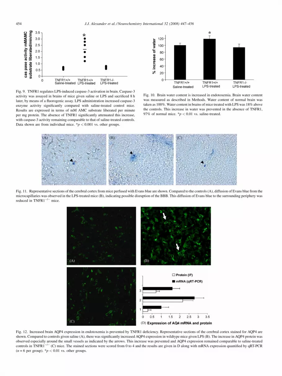
Fig. 9. TNFR1 regulates LPS-induced caspase-3 activation in brain. Caspase-3
activity was assayed in brains of mice given saline or LPS and sacrificed 8 h
later, by means of a fluorogenic assay. LPS administration increased caspase-3
enzyme activity significantly compared with saline-treated control mice.
Results are expressed in terms of mM AMC substrate liberated per minute
per mg protein. The absence of TNFR1 significantly attenuated this increase,
with caspase-3 activity remaining comparable to that of saline-treated controls.
Data shown are from individual mice. *p < 0.001 vs. other groups.
Fig. 10. Brain water content is increased in endotoxemia. Brain water content
was measured as described in Methods. Water content of normal brain was
taken as 100%. Water content in brains of mice treated with LPS was 18% above
the controls. This increase in water was prevented in the absence of TNFR1,
97% of normal mice. *p < 0.01 vs. saline-treated.
Fig. 11. Representative sections of the cerebral cortex from mice perfused with Evans blue are shown. Compared to the controls (A), diffusion of Evans blue from the
microcapillaries was observed in the LPS-treated mice (B), indicating possible disruption of the BBB. This diffusion of Evans blue to the surrounding periphery was
reduced in TNFR1�/� mice.
Fig. 12. Increased brain AQP4 expression in endotoxemia is prevented by TNFR1 deficiency. Representative sections of the cerebral cortex stained for AQP4 are
shown. Compared to controls given saline (A), there was significantly increased AQP4 expression in wildtype mice given LPS (B). The increase in AQP4 protein was
observed especially around the small vessels as indicated by the arrows. This increase was prevented and AQP4 expression remained comparable to saline-treated
controls in TNFR1�/� (C) mice. The stained sections were scored from 0 to 4 and the results are given in D along with mRNA expression quantified by qRT-PCR
(n = 6 per group). *p < 0.01 vs. other groups.
J.J. Alexander et al. / Neurochemistry International 52 (2008) 447–456454

J.J. Alexander et al. / Neurochemistry International 52 (2008) 447–456 455
play an important pathophysiological role during infection and
inflammation of the central nervous system. Consequently,
treatments that control the activation of astroglia may be
effective at diminishing the severity of LPS-induced encepha-
lopathy.
Astrocytes are an integral part of the blood–brain barrier
(BBB), which maintains the internal milieu constant (Davies,
2002; Davson et al., 1993; Davson and Segal, 1995). Increased
expression of the important water channel protein, AQP4 on
astrocytes and endothelial cells could lead to the increased
water content (Alexander et al., 2003; Badaut et al., 2002;
Manley et al., 2004) observed in LPS-induced brains, in our
study, similar to edema seen in clinical patients (Papadopoulos
et al., 2000). Furthermore, in our study, an 18% increase in
brain water content was found in mice that were treated with
LPS compared to mice receiving saline, which was reduced in
LPS-treated TNFR1�/� mice. Although the increase is
significant but not substantial, the brain is located in a defined
space and an alteration in the internal milieu could lead to
traumatic consequences. In clinical patients with septic
encephalopathy the integrity of the BBB is altered with
associated edema. Edema is a significant source of morbidity
and mortality in several brain disorders and therefore an
understanding of its pathophysiology is an important step
towards designing therapeutic strategies not only in sepsis but
in other CNS diseases too.
In summary, our results demonstrate that TNFR1-mediated
signaling is a key mechanism involved in restricting or
resolving, the LPS-induced inflammatory response. Absence of
the TNF-a/TNFR1 pathway could alleviate apoptosis, reduce
neutrophil infiltration, astrogliosis and edema in LPS-induced
brain. Additional experiments need to be designed and
conducted to understand the extent to which these mechanisms
individually contribute to LPS-induced encephalopathy and the
extent to which these processes are interrelated. Furthermore,
our future studies will address the issue of which populations of
cells are dying of apoptosis in this setting. This will lead to a
better understanding of the pathophysiology of septic
encephalopathy and thereby the designing of more effective
therapeutic strategies.
References
Alexander, J.J., Bao, L., Jacob, A., Kraus, D.M., Holers, V.M., Quigg, R.J.,
2003. Administration of the soluble complement inhibitor, Crry-Ig, reduces
inflammation and aquaporin 4 expression in lupus cerebritis. Biochim.
Biophys. Acta 1639, 169–176.
Alexander, J.J., Jacob, A., Bao, L., MacDonald, R.L., Quigg, R.J., 2005.
Complement-dependent apoptosis and inflammatory gene changes in mur-
ine lupus cerebritis. J. Immunol. 175, 8312–8319.
Alexander, J.J., Jacob, A., Vezina, P., Sekine, H., Gilkeson, G.S., Quigg, R.J.,
2007. Absence of functional alternative complement pathway alleviates
lupus cerebritis. Eur. J. Immunol. 37, 1691–1701.
Allan, S.M., Rothwell, N.J., 2001. Cytokines and acute neurodegeneration. Nat.
Rev. Neurosci. 2, 734–744.
Amrani, Y., Lazaar, A.L., Hoffman, R., Amin, K., Ousmer, S., Panettieri Jr.,
R.A., 2000. Activation of p55 tumor necrosis factor-alpha receptor-1
coupled to tumor necrosis factor receptor-associated factor 2 stimulates
intercellular adhesion molecule-1 expression by modulating a thapsigargin-
sensitive pathway in human tracheal smooth muscle cells. Mol. Pharmacol.
58, 237–245.
Askalan, R., Deveber, G., Ho, M., Ma, J., Hawkins, C., 2006. Astrocytic-
inducible nitric oxide synthase in the ischemic developing human brain.
Pediatr. Res. 60, 687–692.
Badaut, J., Lasbennes, F., Magistretti, P.J., Regli, L., 2002. Aquaporins in brain:
distribution, physiology, and pathophysiology. J. Cereb. Blood Flow Metab.
22, 367–378.
Bi, X.L., Yang, J.Y., Dong, Y.X., Wang, J.M., Cui, Y.H., Ikeshima, T., Zhao,
Y.Q., Wu, C.F., 2005. Resveratrol inhibits nitric oxide and TNF-alpha
production by lipopolysaccharide-activated microglia. Int. Immunophar-
macol. 5, 185–193.
Brown, G.C., Bal-Price, A., 2003. Inflammatory neurodegeneration mediated
by nitric oxide, glutamate, and mitochondria. Mol. Neurobiol. 27, 325–355.
Choi, D.K., Lee, H., Jeong, J., Lim, B., Suk, K., 2005. Differential effects of
ethanol on glial signal transduction initiated by lipopolysaccharide and
interferon-gamma. J. Neurosci. Res. 82, 225–231.
Cunningham, P.N., Dyanov, H.M., Park, P., Wang, J., Newell, K.A., Quigg, R.J.,
2002. Acute renal failure in endotoxemia is caused by TNF acting directly
on TNF receptor-1 in kidney. J. Immunol. 168, 5817–5823.
Davies, D.C., 2002. Blood–brain barrier breakdown in septic encephalopathy
and brain tumours. J. Anat. 200, 639–646.
Davson, H., Segal, M.B., 1995. Physiology of the CSF and the Blood–Brain
Barrier. CRC, New York.
Davson, H., Zlokovic, B.V., Rakic, L., Segal, M.B., 1993. An Introduction to the
Blood–Brain Barrier. Macmillan, London.
Dong, Y., Benveniste, E.N., 2001. Immune function of astrocytes. Glia 36, 180–
190.
Dopp, J.M., kenzie-Graham, A., Otero, G.C., Merrill, J.E., 1997. Differential
expression, cytokine modulation, and specific functions of type-1 and type-
2 tumor necrosis factor receptors in rat glia. J. Neuroimmunol. 75, 104–112.
Eng, L.F., Ghirnikar, R.S., 1994. GFAP and astrogliosis. Brain Pathol. 4, 229–
237.
Eng, L.F., Ghirnikar, R.S., Lee, Y.L., 2000. Glial fibrillary acidic protein:
GFAP-thirty-one years (1969–2000). Neurochem. Res. 25, 1439–1451.
Eng, L.F., Yu, A.C., Lee, Y.L., 1992. Astrocytic response to injury. Prog. Brain
Res. 94, 353–365.
Enkhbaatar, P., Cox, R., Traber, L.D., Maybauer, M.O., Maybauer, D.M.,
Nakano, Y.Y., Hawkins, H., Schmalstieg, F., Herndon, D., Traber, D.,
2006. Role of inducible nitric oxide synthase in septic shock. FASEB. J.
20, A1391.
Gahm, C., Holmin, S., Wiklund, P.N., Brundin, L., Mathiesen, T., 2006.
Neuroprotection by selective inhibition of inducible nitric oxide synthase
after experimental brain contusion. J. Neurotrauma 23, 1343–1354.
Gardenfors, A., Nilsson, F., Skagerberg, G., Ungerstedt, U., Nordstrom, C.H.,
2002. Cerebral physiological and biochemical changes during vasogenic
brain oedema induced by intrathecal injection of bacterial lipopolysacchar-
ides in piglets. Acta Neurochir. (Wien) 144, 601–608.
Gimenez, M.A., Sim, J.E., Russell, J.H., 2004. TNFR1-dependent VCAM-1
expression by astrocytes exposes the CNS to destructive inflammation. J.
Neuroimmunol. 151, 116–125.
Granert, C., Raud, J., Xie, X., Lindquist, L., Lindbom, L., 1994. Inhibition of
leukocyte rolling with polysaccharide fucoidin prevents pleocytosis in
experimental meningitis in the rabbit. J. Clin. Invest. 93, 929–936.
Green, R., Scott, L.K., Minagar, A., Conrad, S., 2004. Sepsis associated
encephalopathy (SAE): a review. Front Biosci. 9, 1637–1641.
Gurney, K.J., Estrada, E.Y., Rosenberg, G.A., 2006. Blood–brain barrier dis-
ruption by stromelysin-1 facilitates neutrophil infiltration in neuroinflam-
mation. Neurobiol. Dis. 23, 87–96.
Hawkins, R.D., Son, H., Arancio, O., 1998. Nitric oxide as a retrograde
messenger during long-term potentiation in hippocampus. Nitric Oxide
Brain Dev. Plast. Dis. 118, 155–172.
Hengartner, M.O., 2000. The biochemistry of apoptosis. Nature 407, 770–776.
Henninger, D.D., Panes, J., Eppihimer, M., Russell, J., Gerritsen, M., Anderson,
D.C., Granger, D.N., 1997. Cytokine-induced VCAM-1 and ICAM-1
expression in different organs of the mouse. J. Immunol. 158, 1825–1832.
Intiso, D., Zarrelli, M.M., Lagioia, G., Di, R.F., Checchia De, A.C., Simone, P.,
Tonali, P., Cioffi Dagger, R.P., 2004. Tumor necrosis factor alpha serum

J.J. Alexander et al. / Neurochemistry International 52 (2008) 447–456456
levels and inflammatory response in acute ischemic stroke patients. Neurol.
Sci. 24, 390–396.
Jellinger, K.A., Stadelmann, C., 2000a. Mechanisms of cell death in neurode-
generative disorders. J. Neural Transm. Suppl. 59, 95–114.
Jellinger, K.A., Stadelmann, C.H., 2000b. The enigma of cell death in neuro-
degenerative disorders. J. Neural Transm. Suppl. 21–36.
Kubes, P., Kanwar, S., Niu, X.F., Gaboury, J.P., 1993. Nitric oxide synthesis
inhibition induces leukocyte adhesion via superoxide and mast cells.
FASEB J. 7, 1293–1299.
Lee, S.C., Dickson, D.W., Brosnan, C.F., 1995. Interleukin-1, nitric oxide and
reactive astrocytes. Brain Behav. Immun. 9, 345–354.
Lee, S.M., Yune, T.Y., Kim, S.J., Kim, Y.C., Oh, Y.J., Markelonis, G.J., Oh,
T.H., 2004. Minocycline inhibits apoptotic cell death via attenuation of
TNF-alpha expression following iNOS/NO induction by lipopolysaccharide
in neuron/glia co-cultures. J. Neurochem. 91, 568–578.
Loddick, S.A., Rothwell, N.J., 1999. Mechanisms of tumor necrosis factor alpha
action on neurodegeneration: interaction with insulin-like growth factor-1.
Proc. Natl. Acad. Sci. U.S.A. 96, 9449–9451.
Lucas, R., Lou, J., Morel, D.R., Ricou, B., Suter, P.M., Grau, G.E., 1997. TNF
receptors in the microvascular pathology of acute respiratory distress
syndrome and cerebral malaria. J. Leukoc. Biol. 61, 551–558.
Manley, G.T., Binder, D.K., Papadopoulos, M.C., Verkman, A.S., 2004. New
insights into water transport and edema in the central nervous system from
phenotype analysis of aquaporin-4 null mice. Neuroscience 129, 983–991.
Marcus, J.S., Karackattu, S.L., Fleegal, M.A., Sumners, C., 2003. Cytokine-
stimulated inducible nitric oxide synthase expression in astroglia: role of
Erk mitogen-activated protein kinase and NF-kappaB. Glia 41, 152–160.
Marshall, D., Dangerfield, J.P., Bhatia, V.K., Larbi, K.Y., Nourshargh, S.,
Haskard, D.O., 2003. MRL/lpr lupus-prone mice show exaggerated
ICAM-1-dependent leucocyte adhesion and transendothelial migration in
response to TNF-alpha. Rheumatology (Oxford) 42, 929–934.
Merrill, J.E., Benveniste, E.N., 1996. Cytokines in inflammatory brain lesions:
helpful and harmful. Trends Neurosci. 19, 331–338.
Nadeau, S., Rivest, S., 1999. Effects of circulating tumor necrosis factor on the
neuronal activity and expression of the genes encoding the tumor necrosis
factor receptors (p55 and p75) in the rat brain: a view from the blood–brain
barrier. Neuroscience 93, 1449–1464.
Norenberg, M.D., 1996. Astrocytes in neuronal degeneration. J. Neurochem. 66,
S79.
Norenberg, M.D., 1998. Role of astrocytes in neurologic disease: an introduc-
tion. J. Neurochem. 70, S75.
Norenberg, M.D., Rao, K.V., Jayakumar, A.R., 2005. Mechanisms of ammonia-
induced astrocyte swelling. Metab. Brain Dis. 20, 303–318.
Pan, W., Banks, W.A., Kastin, A.J., 1997. Blood–brain barrier permeability
to ebiratide and TNF in acute spinal cord injury. Exp. Neurol. 146,
367–373.
Papadopoulos, M.C., Davies, D.C., Moss, R.F., Tighe, D., Bennett, E.D., 2000.
Pathophysiology of septic encephalopathy: a review. Crit. Care. Med. 28,
3019–3024.
Radzivil, G.G., Beloborodov, V.B., Bronikin, I., 1990. Hemodynamics and
rheologic properties of the blood in meningococcemia associated with
meningitis and complicated by septic shock and intracranial hypertension.
Anesteziol. Reanimatol. 28–33.
Rao, K.V.R., Chen, M., Simard, J.M., Norenberg, M.D., 2003. Increased
aquaporin-4 expression in ammonia-treated cultured astrocytes. Neurore-
port 14, 2379–2382.
Romero, L.I., Tatro, J.B., Field, J.A., Reichlin, S., 1996. Roles of IL-1 and TNF-
alpha in endotoxin-induced activation of nitric oxide synthase in cultured rat
brain cells. Am. J. Physiol. 270, R326–R332.
Schafers, M., Schmidt, C., Vogel, C., Toyka, K.V., Sommer, C., 2002. Tumor
necrosis factor-alpha (TNF) regulates the expression of ICAM-1 predomi-
nantly through TNF receptor 1 after chronic constriction injury of mouse
sciatic nerve. Acta Neuropathol. (Berl) 104, 197–205.
Tsao, N., Hsu, H.P., Wu, C.M., Liu, C.C., Lei, H.Y., 2001. Tumour necrosis
factor-alpha causes an increase in blood–brain barrier permeability during
sepsis. J. Med. Microbiol. 50, 812–821.
Vizuete, M.L., Venero, J.L., Vargas, C., Ilundain, A.A., Echevarria, M.,
Machado, A., Cano, J., 1999. Differential upregulation of aquaporin-4
mRNA expression in reactive astrocytes after brain injury: potential role
in brain edema. Neurobiol. Dis. 6, 245–258.
Wispelwey, B., Hansen, E.J., Scheld, W.M., 1989. Haemophilus influenzae
outer membrane vesicle-induced blood–brain barrier permeability during
experimental meningitis. Infect. Immun. 57, 2559–2562.
Yang, L., Froio, R.M., Sciuto, T.E., Dvorak, A.M., Alon, R., Luscinskas, F.W.,
2005. ICAM-1 regulates neutrophil adhesion and transcellular migration
of TNF-alpha-activated vascular endothelium under flow. Blood 106,
584–592.
Yin, L., Ohtaki, H., Nakamachi, T., Kudo, Y., Makino, R., Shioda, S., 2004.
Delayed expressed TNFR1 co-localize with ICAM-1 in astrocyte in mice
brain after transient focal ischemia. Neurosci. Lett. 370, 30–35.
Yuan, J., Yankner, B.A., 2000. Apoptosis in the nervous system. Nature 407,
802–809.
Yung, H.W., Bal-Price, A.K., Brown, G.C., Tolkovsky, A.M., 2004. Nitric
oxide-induced cell death of cerebrocortical murine astrocytes is
mediated through p53- and Bax-dependent pathways. J. Neurochem.
89, 812–821.


