Embed Size (px)
Citation preview
Tonsillar application of AT-2 SIV affords partial protection againstrectal challenge with SIVmac239
Panagiotis Vagenas, PhD1, Vennansha G. Williams, BS1, Michael Piatak Jr., PhD2, Julian W.Bess Jr., MS2, Jeffrey D. Lifson, MD2, James L. Blanchard, DVM, PhD3, Agegnehu Gettie,BS4, and Melissa Robbiani, PhD1,#1Center for Biomedical Research, HIV/AIDS Program, Population Council, New York, NY 100652AIDS and Cancer Virus Program, SAIC-Frederick, Inc., National Cancer Institute, Frederick, MD217023Tulane National Primate Research Center, Tulane University Health Sciences Center, Covington,LA 704334Aaron Diamond AIDS Research Center, Rockefeller University, New York, NY 10016
AbstractWhile mucosal responses are important for preventing infections with HIV, the optimal strategiesfor inducing them remain unclear. To evaluate vaccine strategies targeting the oral mucosal lymphoidtissue inductive sites as an approach to provide immunity at distal sites, we vaccinated healthymacaques via the palatine/lingual tonsils with aldrithiol 2 (AT-2) inactivated SIVmac239, combinedwith CpG-C immunostimulatory oligonucleotide (CpG-C ISS-ODN , C274) as the adjuvant.Macaques received 5 doses of C274 or control ODN C661 and AT-2 SIV on the tonsillar tissuesevery 6 weeks before being challenged rectally with SIVmac239, 8 weeks after the last immunization.Although no T or B cell responses were detected in the blood prior to challenge, Ab responses weredetected in the rectum . Immunization with AT-2 SIV significantly reduced the frequency of infectioncompared to non-immunized controls, irrespective of adjuvant. In the vaccinated animals that becameinfected, peak viremias were somewhat reduced. SIV-specific responses were detected in the bloodonce animals became infected with no detectable differences between the differently immunizedgroups and the controls. This work provides evidence that vaccine immunogens applied to the oralmucosal-associated lymphoid tissues can provide benefit against rectal challenge, a finding withimportant implications for mucosal vaccination strategies.
KeywordsSIV; mucosal; vaccine; CpG ISS-ODN
IntroductionA quarter of a century after HIV-1 was shown to be the etiologic agent of AIDS, the pandemichas claimed tens of millions of lives. Despite a global scientific effort, an effective prophylacticvaccine for HIV/AIDS remains elusive. Hopes were dashed again with results from a recent aclinical trial not only showing a lack of efficacy in preventing HIV acquisition, but suggestingan increased risk of infection in some vaccinees 1. These results led to recommendations for a
#Corresponding author: Melissa Robbiani, Population Council, 1230 York Avenue, New York, NY 10065; Tel: (212) 327-7794, Fax:(212) 327-7764, [email protected].
NIH Public AccessAuthor ManuscriptJ Acquir Immune Defic Syndr. Author manuscript; available in PMC 2010 December 1.
Published in final edited form as:J Acquir Immune Defic Syndr. 2009 December 1; 52(4): 433–442. doi:10.1097/QAI.0b013e3181b880f3.
NIH
-PA Author Manuscript
NIH
-PA Author Manuscript
NIH
-PA Author Manuscript
renewed emphasis on basic preclinical research and the need to ”make best use of animal,particularly monkey, models” 2. The benefits of the SIV/macaque model as the principal animalmodel for studying HIV are well documented 3.
HIV infection occurs mainly via mucosal surfaces, suggesting that induction of mucosalimmune responses may be an important property for an effective vaccine. Numerous studiesin rodents have provided evidence that oral/nasal administration induces immunity at distalmucosal sites, as well as systemically. Oral or nasal immunization of mice againstChlamydia or HSV led to protective immunity in the vagina 4-7 and to memory CTL responses8, 9. Mucosal immunization of mice with Tat or Gag-containing vectors led to vaginalprotection against challenge with recombinant HIV protein-expressing vectors 10, 11. A numberof mucosal immunization studies in macaques have also been shown to elicit mucosal andsystemic immunity 12-17.
Aldrithiol 2 (AT-2) inactivated SIV and HIV are attractive vaccine immunogens as they containall of the virion associated proteins in the absence of an infectious virus 18. Previous work hasshown that AT-2 viruses interact authentically with dendritic cells (DCs) 19 and mature DCselicit both CD4+ and CD8+ T cell responses in vitro 20. Moreover, in therapeutic immunizationregimens, injection of autologous mature DCs pulsed with AT-2 SIV or HIV were reported toboost immunity and reduce viral loads in infected macaques 21 and in a preliminary clinicalstudy in humans 22, respectively.
CpG-C immunostimulatory oligonucleotides (ISS-ODNs) activate both plasmacytoid DCs(PDCs) and B cells 23, 24, to potentially augment innate and adaptive immunity elicited againsta vaccine. Similar observations have been made in macaques where CpG-C ISS-ODNs inducedPDC activation, IFNα and IL-12 production, and boosted SIV-specific T cell responses invitro 25, as well as stimulating robust B cell proliferation, survival and activation 26. Injectionof CpG-C ISS-ODNs in macaque lymph nodes also activated both DCs and B cells,demonstrating the ability of CpG-C ISS-ODNs to work in vivo 27. While CpG ISS-ODNs havebeen shown to boost macaque immunity 28-30, there is no evidence for the activity of the morebroadly acting CpG-C ISS-ODNs in this species.
We examined whether applying a combination of AT-2 SIV and CpG-C ISS-ODNs to thetonsillar mucosa (as a controlled way to model targeting the nasal lymphoid tissues) wouldprotect against rectal challenge with infectious SIV. Whilst partial protection by tonsillarapplication of AT-2 SIV was observed, CpG-C ISS-ODNs did not augment this effectsuggesting that alternative adjuvant strategies will be needed to optimize the efficacy ofmucosally applied AT-2 SIV.
Materials and MethodsReagents
CpG-C ISS-ODN C274 and the control ODN C661 were provided by Dynavax Technologies(Berkeley, CA). The sequences were: C274 5′-TCGTCGAACGTTCGAGATGAT-3′ andC661 5′-TGCTTGCAAGCTTGCAAGCA-3′. AT-2 SIV (AT-2 SIVmac239 lot numbers:P4001, P4146, P3876, P3778, P3782; AT-2 SIVmac239ΔV1V2 31 lot number: P3956) and theno virus microvesicle (MV) controls (lot numbers: P3826, P3971), prepared from the samecell line in which the viruses were grown (SUPT1), were provided by the AIDS and CancerVirus Program (NCI-Frederick, Frederick, MD). AT-2 inactivation of virus was performed aspreviously described 32. AT-2 SIV was used at 300ng of p27/ml for all in vitro cultures. MVwere normalized to SIV on total protein (300ng of p27 equivalent/ml). Concanavalin A (ConA;Sigma, St Louis, MO) was used at 1μg/ml.
Vagenas et al. Page 2
J Acquir Immune Defic Syndr. Author manuscript; available in PMC 2010 December 1.
NIH
-PA Author Manuscript
NIH
-PA Author Manuscript
NIH
-PA Author Manuscript
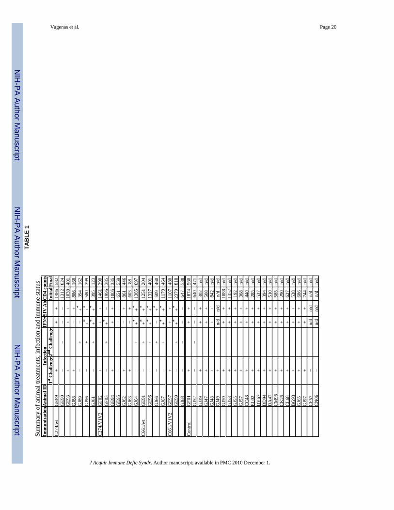
Animals and TreatmentAdult male Chinese Rhesus macaques (Macaca mulatta) were housed at the Tulane NationalPrimate Research Center (TNPRC; Covington, LA). All studies were approved by the AnimalCare and Use Committee of the TNPRC. The animals’ average age at the beginning of thestudy was 5 years and their average weight was 10kg. All animals tested negative for simiantype D retroviruses, simian T cell leukemia virus-1, and SIV prior to use. Animals wereanesthetized prior and during all procedures (10mg ketamine-HCl/kg), in compliance with theregulations detailed under the Animal Welfare Act and in the Guide for the Care and Use ofLaboratory Animals 33, 34. Animals were immunized a total of five times, at six week intervals,by application across the lingual and palatine tonsils of 1mg of CpG-C ISS-ODN C274 or thecontrol ODN C661 mixed with 5μg of p27 of either AT-2 SIVmac239 or AT-2SIVmac239ΔV1V2 31 in a volume of 100μl. The treatment groups included animals immunizedwith the following: C274 and wild-type AT-2 SIVmac239 (C274/wt), C274 and AT-2 SIVmac239ΔV1V2 (C274/V1V2), C661 and wild-type AT-2 SIVmac239 (C661/wt), C661 andAT-2 SIVmac239ΔV1V2 (C661/V1V2), or nothing (non-vaccinated controls). Table 1 listsall study animals. Eight weeks after the final immunization, animals were challenged rectallywith 103 TCID50 of SIVmac239 (TNPRC stock virus propagated in SEB-stimulated rhesusPBMCs; “SIVmac239 RhPBMC 7/29/94”). Once the follow up period was completed, 5months after the initial challenge, uninfected animals were re-immunized once more, re-challenged 8 weeks later and followed up for 6 months (as indicated). Immune responses werefollowed by collecting EDTA-anticoagulated blood and mucosal (oral and rectal) fluidsthroughout the immunization regimen and for up to 6 months after SIV challenge. Mucosalfluids were collected by insertion of a foam pad (approx. size 1×0.5cm ) in the mucosal cavityfor 5min, after which the swab was placed into a tube containing 1ml PBS/1% FCS/penicillin-streptomycin (Cellgro/Mediatech, VA). Blood, fluids and tissue samples were transported tothe laboratory by overnight courier service. Blood was processed as described below and themucosal fluids were spun at 805g for 10min, collecting the supernatant and storing at −80°Cuntil analysis. Upon study termination, animals were sacrificed and standard full necropsy forSIV infected animals was performed. To further assess the in vivo activity of mucosally appliedCpG-C ISS-ODNs, 1mg of C274 or C661 (50μl of 20mg/ml stock) were applied to the tonsilsof healthy infected or uninfected macaques and tonsillar pinch biopsies were collected 24hlater. Cellular activation was monitored by flow cytometry.
Cell isolationMacaque peripheral blood mononuclear cells (PBMCs) were isolated using Ficoll-Hypaquedensity centrifugation (GE Healthcare, Sweden). Cells were cultured in complete RPMI 1640(Cellgro, Springfield, NJ) containing 2mM L-glutamine (GIBCO Life Technologies, GrandIsland, NY) 10mM HEPES (N-2-hydroxyethylpiperazine-N’-2-ethanesulfonic acid; GIBCOLife technologies), 50μM 2-mercaptoethanol (Sigma), penicillin (100U/ml)/streptomycin(100μg/ml) (GIBCO Life Technologies) and 1% heparinized human plasma (InnovativeResearch, Southfield, MI).
Tonsillar biopsies were placed in RPMI (supplemented as above, but with 10% heat inactivatedfetal bovine serum (Mediatech, Manassas, VA) instead of human plasma) containing 200μg/ml gentamycin (GIBCO) for 1 hour at 4°C. The tissue was washed by spinning at 244g for10min and resuspending in medium (RPMI-10% FBS) containing 400U/ml Collagenase D(Roche, Indianapolis, IN) and 10μg/ml DNAse I (Roche) in a tissue-culture dish. The tissuewas broken up using a forceps and a scalpel and incubated at 37°C for 1 hour. It was thenfiltered using a 70μm nylon filter (BD Falcon) and the suspension was spun at 340g for 10min.Cells were then resuspended in RPMI (1% human plasma) and counted.
Vagenas et al. Page 3
J Acquir Immune Defic Syndr. Author manuscript; available in PMC 2010 December 1.
NIH
-PA Author Manuscript
NIH
-PA Author Manuscript
NIH
-PA Author Manuscript
Flow cytometryFour color flow cytometry was used to characterize leukocyte subsets from macaque bloodand tissue. DCs were identified as Lin−HLA-DR+ populations using FITC-conjugated anti-lineage marker Abs (CD3, CD8, CD11b, clones SP34, SK1 and F6.2); CD14, clone M5E2 (BDBiosciences); CD20, clone L27 (BD Biosciences)) and APC-conjugated anti-HLA-DR (cloneG46-6, BD Biosciences). PDCs and MDC-containing fractions were identified as CD123+ andCD123− subsets within the Lin−HLA-DR+ cells, respectively, using PE-conjugated anti-CD123 (clone TU27, BD Biosciences). DC activation status was examined using Cy-conjugated anti-CD86 (clone 2331 FUN1, BD Biosciences) and anti-CD80 (clone L307.4, BDBiosciences). B cells were identified using FITC-conjugated anti-CD20 and their activationstatus was monitored using PE-conjugated anti-CD86 (clone IT2.2, BD Biosciences), -CD80,and -CD40 (clone 5C3, BD Biosciences). T cell subsets were identified using APC-conjugatedanti-CD28 (clone CD28.2, Biolegend) and PE-conjugated CD95 (clone DX2, BDBiosciences).
The appropriate isotype Ig controls were included in all experiments and typically gave MFIsof <1 log. Samples were acquired on a FACSCalibur (BD) and analyzed using FlowJo software(Tree Star, OR).
IFNγ ELISPOTNumbers of IFN-γ spot-forming cells (SFCs) in peripheral blood responding to wild-type AT-2SIVmac239, to AT-2 SIVmac239ΔV1V2 mutant or Gag/Env peptide pools (NIH AIDSResearch and Reference Reagent Program) were measured by ELISPOT 25. Each peptide poolwas made by mixing 10 consecutive peptides (resuspended in 100μl DMSO) at 2μg/ml foreach peptide (0.2% DMSO final concentration). 22 peptide pools were prepared for Env and13 for Gag, covering the entire span of each protein. ConA was used at 1μg/ml as a positivecontrol. Medium, MV, and 0.2% DMSO controls were included for the respective stimuli.
Viral Load and SIV Ab detectionPlasma samples were collected from all animals at all time points of the study by centrifugingwhole blood at 805g for 10min, collecting the clear supernatant, centrifuging again and aliquotsof the supernatants were stored at −80°C. SIV RNA was determined by quantitative RT-PCR35 and SIV-specific Abs were measured by ELISA 36.
Neutralizing Ab activity against SIV was measured in monkey plasma samples using minoradaptations to published protocols 37, 38. Plasma, from weeks 8-10 post-infection, was heatinactivated by incubating at 56°C for 1 hour and then clarified by centrifugation at 956g.Samples were then diluted 5-fold twice in flat-bottom 96-well plates (BD Falcon, NJ). 50TCID50 of SIVmac239 or SIVmac251 were added to each plasma-containing well andincubated for 1 hour at 37°C. 3×10⁵ 174×CEM cells (NIH AIDS Research and ReferenceReagent Program) were added per well and the plates were incubated for 2 weeks at 37°C.50μl of culture medium were exchanged for fresh medium after 7 days. No more plasma wasadded during this period. Samples were run in duplicate. The plates were monitored forcytotoxicity every 3 days and cell-free supernatants were collected at day 14 of culture for p27ELISA (ZeptoMetrix Corporation, NY). Pooled heat-inactivated plasma fromSIVmac239Δnef/wild type-infected animals (healthy, long-term infected) was used as apositive control. Negative controls included plasma from uninfected monkeys, as well as wellswith no plasma added.
SIV-specific IgA was measured in rectal fluids collected at the beginning of the study, as wellas at the last time point prior to challenge (week 26) or after challenge (week 36), by ELISAas previously described 17, with minor modifications. Briefly, 96 well plates (Costar, NY) were
Vagenas et al. Page 4
J Acquir Immune Defic Syndr. Author manuscript; available in PMC 2010 December 1.
NIH
-PA Author Manuscript
NIH
-PA Author Manuscript
NIH
-PA Author Manuscript
coated overnight with lysed SIVmac239 (lot P4145) at 1μg/ml. Plates were blocked with 0.25%gelatin in PBS, the samples were added (100μl/well) and incubated for 2 hours at 37°C.Peroxidase-conjugated goat anti-monkey IgA (Alpha Diagnostic International, San Antonio,TX) was used as the secondary Ab at 1:10,000 and incubated for 2 hours at 37°C. TMBperoxidase substrate solution (KPL, Gaithersburg, MD) was then added (100μl/well) andincubated for 30min at RT. The reaction was stopped with 1M HCl (50μl/well) and theabsorbance was read at 450/650nm. Pooled plasma from SIV mac239Δnef/wild type-infectedanimals (healthy, long-term infected) was used as a positive control. Plasma from uninfectedmonkeys was used as a negative control.
Statistical analysesViral load data were analyzed for statistical significance using the Mann-Whitney test.Frequency of infection data were analyzed using the Fisher’s Exact test. p values <0.05 weretaken as statistically significant.
ResultsTonsillar C274/AT-2 SIV vaccination partially protected against SIV challenge
Knowing that C274 activates macaque DC and B cells, and augments SIV-specific T cellresponses 25-27, we set out to determine if combining C274 and AT-2 SIV would serve as apotent vaccine when applied to the oral mucosal-associated lymphoid tissues (MALT) ofmacaques. This allowed us to control that the vaccine was directly applied to the oral MALT(and not swallowed), to provide a model for future strategies that would target nasal MALT.In addition to comparing C274 as the adjuvant, we compared two vaccine antigens, wild-typeAT-2 SIVmac239 and AT-2 SIVmac239ΔV1V2 31, a mutant of the virus that lacks thehypervariable loops V1 and V2 from the viral envelope. We hypothesized that the deletion ofV1 and V2 in the mutant virus might reveal neutralization sensitive epitopes that would bepresented in this AT-2-treated vaccine, thereby inducing more potent neutralizing Abresponses, whilst inducing a similar cellular response to the wild-type virus.
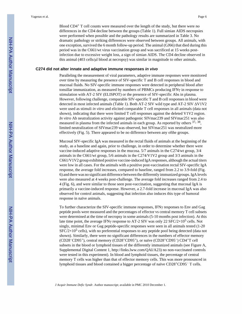
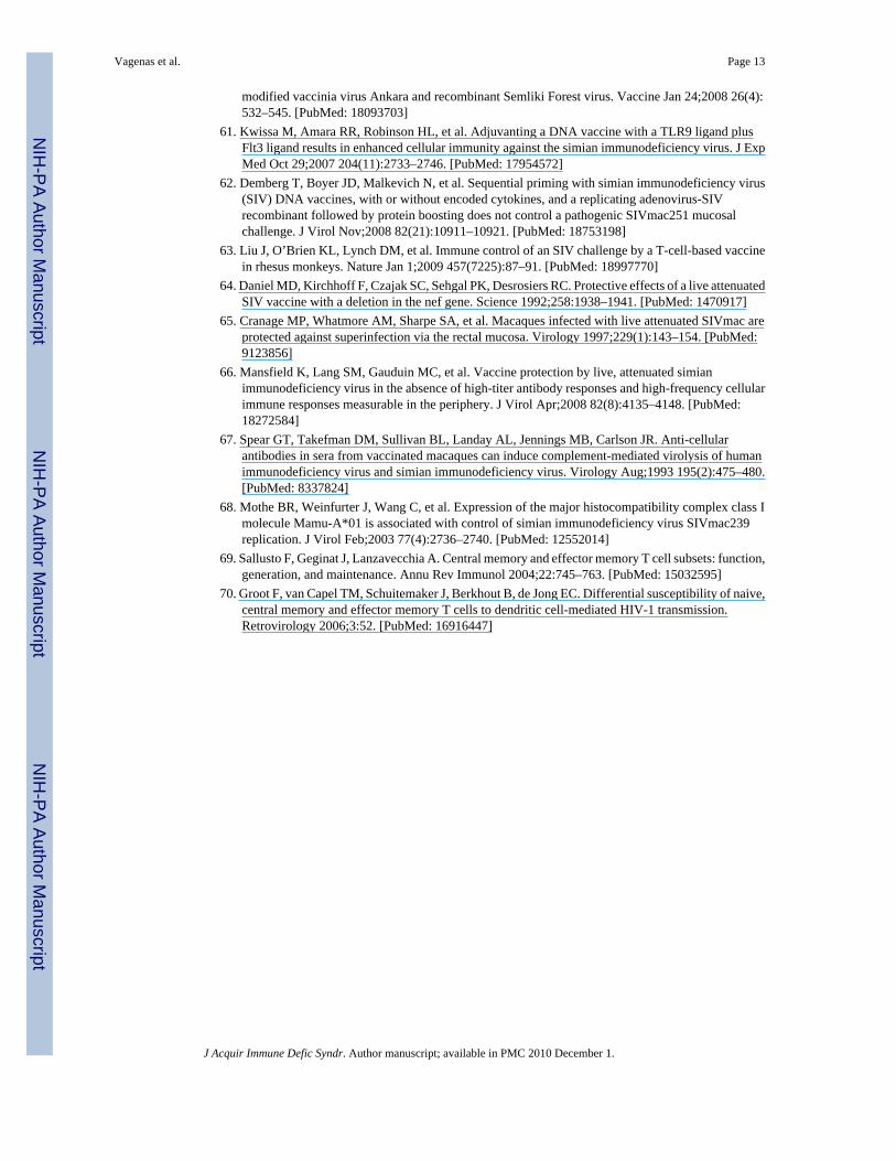
SIV-naïve Chinese rhesus macaques were vaccinated 5 times by applying AT-2 SIV with theindicated ODNs to the tonsillar tissues every 6 weeks (Fig. 1). 8 weeks after the finalimmunization, the animals were challenged rectally with pathogenic SIVmac239. They werefollowed up for a period of 6 months. Vaccinated animals that remained uninfected after 5months of follow up were then re-immunized once more and re-challenged 8 weeks later andfollowed for an additional 6 months to see if they continued to resist infection (Table 1). Whencomparing all challenges and infection outcomes, the vaccinated animal groups exhibited asignificantly lower frequency of infection (average of 53%) compared to the non-vaccinatedcontrol group (83%), independent of the presence of C274 (Fig. 2, Table 1; p<0.03 whencomparing all challenges for each group vs control).
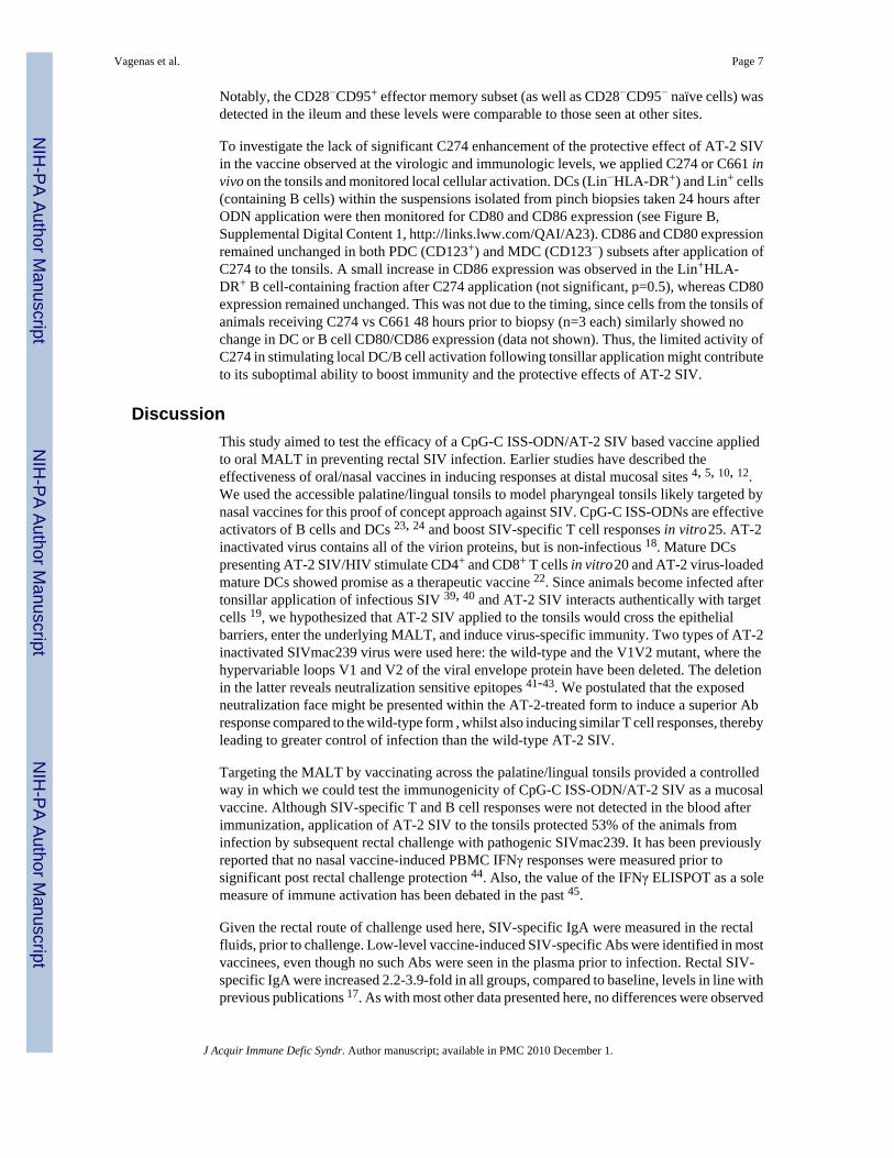
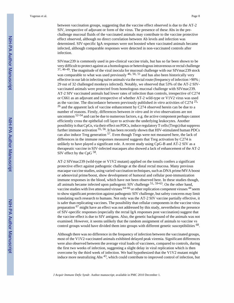
Examination of the plasma viral loads (Fig. 3) revealed that all infected control animals, 6/6infected C274/wt-vaccinated animals, and 2/4 infected C661/wt-vaccinated animals exhibitedpeak viremia 2 weeks post challenge. Peak viremias were delayed by 1-2 weeks in 4/6 infectedC274/V1V2 and 1/2 infected C661/V1V2 vaccinated animals. Closer analysis of acute viremiarevealed that wild type-vaccinated animals showed significantly lower viremia than controls(independent of C274) during the first week of infection (Fig.4, upper panel), but no differencein subsequent weeks. ΔV1V2-vaccinees also had significantly lower viremia than controlsduring the first 2 weeks of infection, with the exception of C661-vaccinees in the first week(Fig.4, lower panel). Set-point viremias (weeks 8 and 10 post-infection) and viremias at timeof necropsy (4-11 months post infection) yielded no significant differences between any of thegroups.
Vagenas et al. Page 5
J Acquir Immune Defic Syndr. Author manuscript; available in PMC 2010 December 1.
NIH
-PA Author Manuscript
NIH
-PA Author Manuscript
NIH
-PA Author Manuscript
Blood CD4+ T cell counts were measured over the length of the study, but there were nodifferences in the CD4 decline between the groups (Table 1). Full simian AIDS necropsieswere performed when possible and the pathology results are summarized in Table 3. Nodramatic pathology or striking differences were observed between groups. All animals, withone exception, survived the 6 month follow-up period. The animal (GJ66) that died during thisperiod was in the C661/wt virus vaccination group and was sacrificed at 15 weeks post-infection due to excessive weight loss, a sign of simian AIDS. The CD4 decline observed inthis animal (403 cells/μl blood at necropsy) was similar in magnitude to other animals.
C274 did not alter innate and adaptive immune responses in vivoParalleling the measurement of viral parameters, adaptive immune responses were monitoredover time by measuring the presence of SIV-specific T and B cell responses in blood andmucosal fluids. No SIV-specific immune responses were detected in peripheral blood aftertonsillar immunization, as measured by numbers of PBMCs producing IFNγ in response tostimulation with AT-2 SIV (ELISPOT) or the presence of SIV-specific Abs in plasma.However, following challenge, comparable SIV-specific T and B cell responses in blood weredetected in most infected animals (Table 1). Both AT-2 SIV wild type and AT-2 SIV ΔV1V2were used as stimuli in vitro and elicited comparable T cell responses in all animals (data notshown), indicating that there were limited T cell responses against the deleted V1V2 region.In vitro Ab neutralization activity against pathogenic SIVmac239 and SIVmac251 was alsomeasured in plasma from the infected animals in each group. As reported by others 37, 38,limited neutralization of SIVmac239 was observed, but SIVmac251 was neutralized moreeffectively (Fig. 5). There appeared to be no difference between any ofthe groups.
Mucosal SIV-specific IgA was measured in the rectal fluids of animals at the beginning of thestudy, as a baseline and again, prior to challenge, in order to determine whether there werevaccine-induced adaptive responses in the mucosa. 5/7 animals in the C274/wt group, 3/4animals in the C661/wt group, 5/6 animals in the C274/V1V2 group and 3/3 animals in theC661/V1V2 group exhibited positive vaccine-induced IgA responses, although the actual titerswere low in all cases. For the animals with a positive post-vaccination rectal SIV-specific IgAresponse, the average fold increases, compared to baseline, ranged from 2.2 to 3.9-fold (Fig.6) and there was no significant difference between the differently immunized groups. IgA levelswere also measured at 4 weeks post-challenge. The average fold increases ranged from 2.4 to4 (Fig. 6), and were similar to those seen post-vaccination, suggesting that mucosal IgA isprimarily a vaccine-induced response. However, a 2.7-fold increase in mucosal IgA was alsoobserved for control animals, suggesting that infection also induces this type of humoralresponse in naïve animals.
To further characterize the SIV-specific immune responses, IFNγ responses to Env and Gagpeptide pools were measured and the percentages of effector vs central memory T cell subsetswere determined at the time of necropsy in some animals (5-10 months post infection). At thislate time point, the average IFNγ response to AT-2 SIV was only 22 SFC/2×105 cells. Notsingly, minimal Env or Gag peptide-specific responses were seen in all animals tested (1-20SFC/2×105 cells), with no preferential responses to any peptide pool being detected (data notshown). Similarly, there were no significant differences in the numbers of effector memory(CD28−CD95+), central memory (CD28+CD95+), or naïve (CD28+CD95−) CD4+T cellsubsets in the blood or lymphoid tissues of the differently immunized animals (see Figure A,Supplemental Digital Content 1, http://links.lww.com/QAI/A23) no non-vaccinated controlswere tested in this experiment). In blood and lymphoid tissues, the percentage of centralmemory T cells was higher than that of effector memory cells. This was more pronounced inlymphoid tissues and blood contained a bigger percentage of naïve CD28+CD95− T cells.
Vagenas et al. Page 6
J Acquir Immune Defic Syndr. Author manuscript; available in PMC 2010 December 1.
NIH
-PA Author Manuscript
NIH
-PA Author Manuscript
NIH
-PA Author Manuscript
Notably, the CD28−CD95+ effector memory subset (as well as CD28−CD95− naïve cells) wasdetected in the ileum and these levels were comparable to those seen at other sites.
To investigate the lack of significant C274 enhancement of the protective effect of AT-2 SIVin the vaccine observed at the virologic and immunologic levels, we applied C274 or C661 invivo on the tonsils and monitored local cellular activation. DCs (Lin−HLA-DR+) and Lin+ cells(containing B cells) within the suspensions isolated from pinch biopsies taken 24 hours afterODN application were then monitored for CD80 and CD86 expression (see Figure B,Supplemental Digital Content 1, http://links.lww.com/QAI/A23). CD86 and CD80 expressionremained unchanged in both PDC (CD123+) and MDC (CD123−) subsets after application ofC274 to the tonsils. A small increase in CD86 expression was observed in the Lin+HLA-DR+ B cell-containing fraction after C274 application (not significant, p=0.5), whereas CD80expression remained unchanged. This was not due to the timing, since cells from the tonsils ofanimals receiving C274 vs C661 48 hours prior to biopsy (n=3 each) similarly showed nochange in DC or B cell CD80/CD86 expression (data not shown). Thus, the limited activity ofC274 in stimulating local DC/B cell activation following tonsillar application might contributeto its suboptimal ability to boost immunity and the protective effects of AT-2 SIV.
DiscussionThis study aimed to test the efficacy of a CpG-C ISS-ODN/AT-2 SIV based vaccine appliedto oral MALT in preventing rectal SIV infection. Earlier studies have described theeffectiveness of oral/nasal vaccines in inducing responses at distal mucosal sites 4, 5, 10, 12.We used the accessible palatine/lingual tonsils to model pharyngeal tonsils likely targeted bynasal vaccines for this proof of concept approach against SIV. CpG-C ISS-ODNs are effectiveactivators of B cells and DCs 23, 24 and boost SIV-specific T cell responses in vitro 25. AT-2inactivated virus contains all of the virion proteins, but is non-infectious 18. Mature DCspresenting AT-2 SIV/HIV stimulate CD4+ and CD8+ T cells in vitro 20 and AT-2 virus-loadedmature DCs showed promise as a therapeutic vaccine 22. Since animals become infected aftertonsillar application of infectious SIV 39, 40 and AT-2 SIV interacts authentically with targetcells 19, we hypothesized that AT-2 SIV applied to the tonsils would cross the epithelialbarriers, enter the underlying MALT, and induce virus-specific immunity. Two types of AT-2inactivated SIVmac239 virus were used here: the wild-type and the V1V2 mutant, where thehypervariable loops V1 and V2 of the viral envelope protein have been deleted. The deletionin the latter reveals neutralization sensitive epitopes 41-43. We postulated that the exposedneutralization face might be presented within the AT-2-treated form to induce a superior Abresponse compared to the wild-type form , whilst also inducing similar T cell responses, therebyleading to greater control of infection than the wild-type AT-2 SIV.
Targeting the MALT by vaccinating across the palatine/lingual tonsils provided a controlledway in which we could test the immunogenicity of CpG-C ISS-ODN/AT-2 SIV as a mucosalvaccine. Although SIV-specific T and B cell responses were not detected in the blood afterimmunization, application of AT-2 SIV to the tonsils protected 53% of the animals frominfection by subsequent rectal challenge with pathogenic SIVmac239. It has been previouslyreported that no nasal vaccine-induced PBMC IFNγ responses were measured prior tosignificant post rectal challenge protection 44. Also, the value of the IFNγ ELISPOT as a solemeasure of immune activation has been debated in the past 45.
Given the rectal route of challenge used here, SIV-specific IgA were measured in the rectalfluids, prior to challenge. Low-level vaccine-induced SIV-specific Abs were identified in mostvaccinees, even though no such Abs were seen in the plasma prior to infection. Rectal SIV-specific IgA were increased 2.2-3.9-fold in all groups, compared to baseline, levels in line withprevious publications 17. As with most other data presented here, no differences were observed
Vagenas et al. Page 7
J Acquir Immune Defic Syndr. Author manuscript; available in PMC 2010 December 1.
NIH
-PA Author Manuscript
NIH
-PA Author Manuscript
NIH
-PA Author Manuscript
between vaccination groups, suggesting that the vaccine effect observed is due to the AT-2SIV, irrespective of adjuvant or form of the virus. The presence of these Abs in the pre-challenge mucosal fluids of the vaccinated animals may contribute to the vaccine protectiveeffect observed, although no direct correlation between Ab levels and infection wasdetermined. SIV-specific IgA responses were not boosted when vaccinated animals becameinfected, although comparable responses were detected in non-vaccinated controls afterinfection.
SIVmac239 is commonly used in pre-clinical vaccine trials, but has so far been shown to bevery difficult to protect against as a homologous or heterologous intravenous or rectal challenge37, 46-49. The magnitude of the viral inocula for mucosal challenge with our SIVmac239 stockwas comparable to what was used previously 46, 50, 51 and has also been historically veryeffective in our lab in infecting naïve animals via the rectal route (frequency of infection >90% ;29 out of 32 challenged monkeys infected). Notably, we observed that 53% of the AT-2 SIV-vaccinated animals were protected from homologous mucosal challenge with SIVmac239.AT-2 SIV vaccinated animals had lower rates of infection than controls, irrespective of C274or C661 as an adjuvant and irrespective of whether AT-2 wild-type or V1V2 virus was usedas the vaccine. The discordance between previously published in vitro activities of C274 25,26 and the apparent lack of vaccine enhancement by C274 observed herein can be due to anumber of reasons. Firstly, differences between in vitro and in vivo observations are notuncommon 52-54 and can be due to numerous factors, e.g. the active component perhaps cannotefficiently cross the epithelial cell layer to activate the underlying leukocytes. Anotherpossibility is that CpGs, via their effect on PDCs, induce regulatory T cells (Tregs) that suppressfurther immune activation 55, 56. It has been recently shown that HIV-stimulated human PDCscan also induce Treg generation 57. Even though Tregs were not measured here, the lack ofdifferences in the immune responses measured suggests that Treg activation by C274 isunlikely to have played a significant role. A recent study using CpG-B and AT-2 SIV as atherapeutic vaccine in SIV-infected macaques also showed a lack of enhancement of the AT-2SIV effect by the CpG 58.
AT-2 SIVmac239 (wild-type or V1V2 mutant) applied on the tonsils confers a significantprotective effect against pathogenic challenge at the distal rectal mucosa. Many previousmacaque vaccine studies, using varied vaccination techniques, such as DNA prime/MVA boostor adenoviral prime/boost, show development of humoral and cellular post-immunizationimmune responses in the blood, which have not been observed here. In these studies though,all animals became infected upon pathogenic SIV challenge 51, 59-63. On the other hand,vaccine studies with live attenuated viruses 64-66 or other replication competent viruses 44 seemto show significant protection against pathogenic SIV challenge, but safety concerns may limittranslating such research to humans. Not only was the AT-2 SIV vaccine partially effective, itis safer than replicating vaccines. The possibility that cellular components in the vaccine viruspreparation 67 might have an effect was not addressed by this study, nevertheless the presenceof SIV-specific responses (especially the rectal IgA responses post vaccination) suggest thatthe vaccine effect is due to SIV antigens. Also, the genetic background of the animals was notexamined. However, it seems unlikely that the random assignment of animals to vaccine vscontrol groups would have divided them into groups with different genetic susceptibilities 68.
Although there was no difference in the frequency of infection between the vaccinated groups,most of the V1V2-vaccinated animals exhibited delayed peak viremia. Significant differenceswere also observed between the average viral loads of vaccinees, compared to controls, duringthe first two weeks of infection, suggesting a slight delay in viral replication which is thenovercome by the third week of infection. We had hypothesized that the V1V2 mutant mightinduce more neutralizing Abs 43, which could contribute to improved control of infection, but
Vagenas et al. Page 8
J Acquir Immune Defic Syndr. Author manuscript; available in PMC 2010 December 1.
NIH
-PA Author Manuscript
NIH
-PA Author Manuscript
NIH
-PA Author Manuscript
there was no difference in the neutralizing Ab activity (in plasma) nor the rectal IgA Abresponses detected between the different groups.
Despite the reduced infection frequency in the AT-2 SIV-vaccinated animals, no considerabledifferences were observed between groups once the animals were infected, as measured byplasma viremia, CD4 counts, numbers of IFNγ-producing cells and disease progression. Therewere also no differences in the distribution of effector and central memory CD4+ T cellsbetween vaccinated groups, with central memory cells dominating in the blood and lymphoidtissues and effector memory cells predominating in the gut, as expected 69, 70.
This study suggests that tonsillar immunization with a non-replicating immunogen can helpprotect against rectal challenge with a highly pathogenic SIV, although we could not correlateSIV-specific immune responses with protection. In addition to reducing the frequency ofinfection, AT-2 SIVΔV1V2 appeared to better limit the initial amplification of infection insome animals. While C274 appeared to have no boosting effect on the AT-2 SIV vaccinationunder this regimen, future studies using this and/or other TLR ligands to augment oral/nasalvaccines represent an exciting strategy to tackle HIV.
Supplementary MaterialRefer to Web version on PubMed Central for supplementary material.
Acknowledgments174×CEM cells were obtained from the NIH AIDS Research and Reference Reagent Program, courtesy of PeterCresswell. SIV Env and Gag peptide pools were obtained from the NIH AIDS Research and Reference ReagentProgram. We thank William Bohn, Jeremy Miller, Terra Schaden-Ireland, Rodman Smith, Robert Imming and ElenaChertova for producing, inactivating, purifying and characterizing AT-2 SIV and MV preparations. We thank JasonMarshall and Gary Van Nest from Dynavax Technologies for the ODNs. We would like to acknowledge the PopulationCouncil Cell Core for flow cytometry assistance and the veterinary staff at the TNPRC for their continued support.We thank Irving Sivin (Population Council) for his assistance with statistical analyses and R. Paul Johnson (NewEngland Primate Research Center, Harvard University) for advice on the Env/Gag peptide pools. We thank membersof our laboratory for their assistance in editing the manuscript and continued help during the course of this study.
Work presented at the International Congress of Immunology, Rio de Janeiro, Brazil, August 2007 and at the ModernMucosal Vaccines, Adjuvants and Microbicides conference, Porto, Portugal, October 2008. This project was fundedby the National Institutes of Health grant R01 DE016256, the base grant RR00164, and in part with federal funds fromthe National Cancer Institute, NIH, under contract N01 CO 124000. MR was a 2002 Elizabeth Glaser Scientist.
References1. Steinbrook R. One step forward, two steps back--will there ever be an AIDS vaccine? N Engl J Med
Dec 27;2007 357(26):2653–2655. [PubMed: 18160684]2. Walker BD, Burton DR. Toward an AIDS vaccine. Science May 9;2008 320(5877):760–764. [PubMed:
18467582]3. Desrosiers RC. The simian immunodeficiency viruses. Ann. Rev. Immunol 1990;8:557–578. [PubMed:
2188674]4. Cui ZD, Tristram D, LaScolea LJ, Kwiatkowski T Jr. Kopti S, Ogra PL. Induction of antibody response
to Chlamydia trachomatis in the genital tract by oral immunization. Infect Immun Apr;1991 59(4):1465–1469. [PubMed: 2004824]
5. Pal S, Peterson EM, de la Maza LM. Intranasal immunization induces long-term protection in miceagainst a Chlamydia trachomatis genital challenge. Infect Immun Dec;1996 64(12):5341–5348.[PubMed: 8945586]
6. Gallichan WS, Johnson DC, Graham FL, Rosenthal KL. Mucosal immunity and protection afterintranasal immunization with recombinant adenovirus expressing herpes simplex virus glycoproteinB. J Infect Dis Sep;1993 168(3):622–629. [PubMed: 8354903]
Vagenas et al. Page 9
J Acquir Immune Defic Syndr. Author manuscript; available in PMC 2010 December 1.
NIH
-PA Author Manuscript
NIH
-PA Author Manuscript
NIH
-PA Author Manuscript
7. Milligan GN, Dudley-McClain KL, Chu CF, Young CG. Efficacy of genital T cell responses to herpessimplex virus type 2 resulting from immunization of the nasal mucosa. Virology Jan 20;2004 318(2):507–515. [PubMed: 14972519]
8. Gallichan WS, Rosenthal KL. Long-lived cytotoxic T lymphocyte memory in mucosal tissues aftermucosal but not systemic immunization. J. Exp. Med 1996;184:1879–1890. [PubMed: 8920875]
9. Gallichan WS, Rosenthal KL. Long-term immunity and protection against herpes simplex virus type2 in the murine female genital tract after mucosal but not systemic immunization. J Infect Dis May;1998 177(5):1155–1161. [PubMed: 9592997]
10. Dumais N, Patrick A, Moss RB, Davis HL, Rosenthal KL. Mucosal immunization with inactivatedhuman immunodeficiency virus plus CpG oligodeoxynucleotides induces genital immune responsesand protection against intravaginal challenge. J Infect Dis Oct 15;2002 186(8):1098–1105. [PubMed:12355360]
11. Adalid-Peralta L, Godot V, Colin C, et al. Stimulation of the primary anti-HIV antibody response byIFN-alpha in patients with acute HIV-1 infection. J Leukoc Biol Apr;2008 83(4):1060–1067.[PubMed: 18182457]
12. Vajdy M, Singh M, Kazzaz J, et al. Mucosal and systemic anti-HIV responses in rhesus macaquesfollowing combinations of intranasal and parenteral immunizations. AIDS Res Hum RetrovirusesNov;2004 20(11):1269–1281. [PubMed: 15588349]
13. Wang SW, Bertley FM, Kozlowski PA, et al. An SHIV DNA/MVA rectal vaccination in macaquesprovides systemic and mucosal virus-specific responses and protection against AIDS. AIDS ResHum Retroviruses Aug;2004 20(8):846–859. [PubMed: 15320989]
14. Pahar B, Cantu MA, Zhao W, et al. Single epitope mucosal vaccine delivered via immuno-stimulatingcomplexes induces low level of immunity against simian-HIV. Vaccine Nov 17;2006 24(4748):6839–6849. [PubMed: 17050045]
15. Bogers WM, Davis D, Baak I, et al. Systemic neutralizing antibodies induced by long intervalmucosally primed systemically boosted immunization correlate with protection from mucosal SHIVchallenge. Virology Dec 20;2008 382(2):217–225. [PubMed: 18947849]
16. Schulte R, Suh YS, Sauermann U, et al. Mucosal prior to systemic application of recombinantadenovirus boosting is more immunogenic than systemic application twice but confers similarprotection against SIV-challenge in DNA vaccine-primed macaques. Virology Jan 20;2009 383(2):300–309. [PubMed: 19027133]
17. Hidajat R, Xiao P, Zhou Q, et al. Correlation of vaccine-elicited systemic and mucosal non-neutralizing antibody activities with reduced acute viremia following intrarectal SIVmac251challenge of rhesus macaques. J Virol Jan;2009 83(2):791–801. [PubMed: 18971271]
18. Lifson JD, Piatak M Jr, Rossio JL, Bess J Jr, Chertova E, Schneider D, Kiser R, Coalter V, Poore B,Imming R, Desrosiers RC, Henderson LE, Arthur LO. Whole inactivated SIV virion vaccines withfunctional envelope glycoproteins: Safety, immunogenicity, and activity against intrarectalchallenge. J Med Primatol 2002;31:205–216. [PubMed: 12390543]
19. Frank I, Piatak MJ, Stoessel H, et al. Infectious and whole inactivated simian immunodeficiencyviruses interact similarly with primate dendritic cells (DCs): Differential intracellular fate of virionsin mature and immature DCs. J. Virol 2002;76(6):2936–2951. [PubMed: 11861860]
20. Frank I, Santos JJ, Mehlhop E, et al. Presentation of exogenous whole inactivated simianimmunodeficiency virus by mature dendritic cells induces CD4+ and CD8+ T cell responses. J. AIDS2003;34(1):7–19.
21. Lu W, Wu X, Lu Y, Guo W, Andrieu JM. Therapeutic dendritic-cell vaccine for simian AIDS. NatMed Jan;2003 9(1):27–32. [PubMed: 12496959]
22. Lu W, Arraes LC, Ferreira WT, Andrieu JM. Therapeutic dendritic-cell vaccine for chronic HIV-1infection. Nat Med Dec;2004 10(12):1359–1365. [PubMed: 15568033]
23. Vollmer J, Weeratna R, Payette P, et al. Characterization of three CpG oligodeoxynucleotide classeswith distinct immunostimulatory activities. Eur J Immunol Jan;2004 34(1):251–262. [PubMed:14971051]
24. Marshall JD, Fearon K, Abbate C, et al. Identification of a novel CpG DNA class and motif thatoptimally stimulate B cell and plasmacytoid dendritic cell functions. J. Leuk. Biol Jun;2003 73(6):781–792.
Vagenas et al. Page 10
J Acquir Immune Defic Syndr. Author manuscript; available in PMC 2010 December 1.
NIH
-PA Author Manuscript
NIH
-PA Author Manuscript
NIH
-PA Author Manuscript
25. Teleshova N, Kenney J, Jones J, et al. CpG-C immunostimulatory oligodeoxyribonucleotideactivation of plasmacytoid dendritic cells in rhesus macaques to augment the activation of IFN-gamma-secreting simian immunodeficiency virus-specific T cells. J Immunol Aug 1;2004 173(3):1647–1657. [PubMed: 15265893]
26. Teleshova N, Kenney J, Van Nest G, et al. CpG-C ISS-ODN activation of blood-derived B cells fromhealthy and chronic immunodeficiency virus-infected macaques. J. Leuk. Biol 2006;79:257–267.
27. Teleshova N, Kenney J, Van Nest G, et al. Local and systemic effects of intranodally injected CpG-C ISS-ODNs in macaques. J. Immunol 2006;177:8531–8541. [PubMed: 17142751]
28. Verthelyi D, Kenney KT, Seder RA, A.A. G, Friedag B, Klinman DM. CpG Oligodeoxynucleotidesas Vaccine Adjuvants in Primates. J. Immunol 2002;168:1659–1663. [PubMed: 11823494]
29. Hartmann G, Weeratna RD, Ballas ZK, et al. Delineation of a CpG PhosphorothionateOligodeoxynucleotide for Activating Primate Immune Responses In Vitro and In Vivo. J. Immunol2000;164:1617–1624. [PubMed: 10640783]
30. Cafaro A, Titti F, Fracasso C, et al. Vaccination with DNA containing tat coding sequences andunmethylated CpG motifs protects cynomolgus monkeys upon infection with simian/humanimmunodeficiency virus (SHIV89.6P). Vaccine Apr 6;2001 19(2022):2862–2877. [PubMed:11282197]
31. Johnson WE, Lifson JD, Lang SM, Johnson RP, Desrosiers RC. Importance of B cell responses forimmunological control of variant strains of simian immunodeficiency virus. J. Virol 2003;77(1):375–381. [PubMed: 12477842]
32. Rossio JL, Esser MT, Suryanarayana K, et al. Inactivation of human immunodeficiency virus type 1infectivity with preservation of conformational and functional integrity of virion surface proteins. J.Virol 1998;72(10):7992–8001. [PubMed: 9733838]
33. Animal Welfare Act and Regulation. Code of Federal Regulations T, Chapter 1, Subchapter A:Animals and Animal Products.
34. Guide for the Care and Use of Laboratory Animals. Committee on Care and Use of LaboratoryAnimals of the Institute of Laboratory Animal Resources UDoHaHS; 1985. p. 1-83.
35. Cline AN, Bess JW, Piatak M Jr. Lifson JD. Highly sensitive SIV plasma viral load assay: practicalconsiderations, realistic performance expectations, and application to reverse engineering of vaccinesfor AIDS. J Med Primatol Oct;2005 34(56):303–312. [PubMed: 16128925]
36. Smith SM, Holland B, Russo C, Dailey PJ, Marx PA, Connor RI. Retrospective analysis of viral loadand SIV antibody responses in rhesus macaques infected with pathogenic SIV: predictive value fordisease progression. AIDS Res. Hum. Retroviruses Dec 10;1999 15(18):1691–1701. [PubMed:10606092]
37. Horton H, Vogel TU, Carter DK, et al. Immunization of rhesus macaques with a DNA prime/modifiedvaccinia virus Ankara boost regimen induces broad simian immunodeficiency virus (SIV)-specificT-cell responses and reduces initial viral replication but does not prevent disease progressionfollowing challenge with pathogenic SIVmac239. J. Virol Jul;2002 76(14):7187–7202. [PubMed:12072518]
38. Cole KS, Rowles JL, Jagerski BA, et al. Evolution of envelope-specific antibody responses inmonkeys experimentally infected or immunized with simian immunodeficiency and its associationwith the development of protective immunity. J. Virol 1997;71:5069–5079. [PubMed: 9188572]
39. Stahl-Hennig C, Steinman RM, Tenner-Racz K, et al. Rapid infection of oral mucosal-associatedlymphoid tissue with simian immunodeficiency virus. Science 1999;285:1261–1265. [PubMed:10455052]
40. Baba TW, Koch J, Mittler ES, et al. Mucosal infection of neonatal rhesus monkeys with cell-freeSIV. AIDS Res. Hum. Retroviruses 1994;10(4):351–357. [PubMed: 8068415]
41. Cao J, Sullivan N, Desjardin E, et al. Replication and neutralization of human immunodeficiencyvirus type 1 lacking the V1 and V2 variable loops of the gp120 envelope glycoprotein. J Virol Dec;1997 71(12):9808–9812. [PubMed: 9371651]
42. Johnson WE, Morgan J, Reitter J, et al. A replication-competent, neutralization-sensitive variant ofsimian immunodeficiency virus lacking 100 amino acids of envelope. J Virol Mar;2002 76(5):2075–2086. [PubMed: 11836385]
Vagenas et al. Page 11
J Acquir Immune Defic Syndr. Author manuscript; available in PMC 2010 December 1.
NIH
-PA Author Manuscript
NIH
-PA Author Manuscript
NIH
-PA Author Manuscript
43. Stamatatos L, Cheng-Mayer C. An envelope modification that renders a primary, neutralization-resistant clade B human immunodeficiency virus type 1 isolate highly susceptible to neutralizationby sera from other clades. J Virol Oct;1998 72(10):7840–7845. [PubMed: 9733820]
44. Zhou Q, Hidajat R, Peng B, et al. Comparative evaluation of oral and intranasal priming withreplication-competent adenovirus 5 host range mutant (Ad5hr)-simian immunodeficiency virus(SIV) recombinant vaccines on immunogenicity and protective efficacy against SIV(mac251).Vaccine Nov 19;2007 25(47):8021–8035. [PubMed: 17935840]
45. Hogrefe WR. Biomarkers and assessment of vaccine responses. Biomarkers Nov;2005 10(Suppl1):S50–57. [PubMed: 16298912]
46. Allen TM, Mortara L, Mothe BR, et al. Tat-vaccinated macaques do not control simianimmunodeficiency virus SIVmac239 replication. J Virol Apr;2002 76(8):4108–4112. [PubMed:11907251]
47. Allen TM, Jing P, Calore B, et al. Effects of cytotoxic T lymphocytes (CTL) directed against a singlesimian immunodeficiency virus (SIV) Gag CTL epitope on the course of SIVmac239 infection. JVirol Oct;2002 76(20):10507–10511. [PubMed: 12239328]
48. Ahmad S, Lohman B, Marthas M, et al. Reduced virus load in rhesus macaques immunized withrecombinant gp160 and challenged with simian immunodeficiency virus. AIDS Res HumRetroviruses Feb;1994 10(2):195–204. [PubMed: 8198872]
49. Mori K, Sugimoto C, Ohgimoto S, et al. Influence of glycosylation on the efficacy of an Env-basedvaccine against simian immunodeficiency virus SIVmac239 in a macaque AIDS model. J Virol Aug;2005 79(16):10386–10396. [PubMed: 16051831]
50. Casimiro DR, Wang F, Schleif WA, et al. Attenuation of simian immunodeficiency virus SIVmac239infection by prophylactic immunization with dna and recombinant adenoviral vaccine vectorsexpressing Gag. J Virol Dec;2005 79(24):15547–15555. [PubMed: 16306625]
51. Kawada M, Tsukamoto T, Yamamoto H, et al. Gag-specific cytotoxic T-lymphocyte-based controlof primary simian immunodeficiency virus replication in a vaccine trial. J Virol Oct;2008 82(20):10199–10206. [PubMed: 18667518]
52. John M, Moore CB, James IR, Mallal SA. Interactive selective pressures of HLA-restricted immuneresponses and antiretroviral drugs on HIV-1. Antivir Ther 2005;10(4):551–555. [PubMed:16038481]
53. Pore N, Gupta AK, Cerniglia GJ, et al. Nelfinavir down-regulates hypoxia-inducible factor 1alphaand VEGF expression and increases tumor oxygenation: implications for radiotherapy. Cancer ResSep 15;2006 66(18):9252–9259. [PubMed: 16982770]
54. Turville SG, Aravantinou M, Miller T, et al. Efficacy of Carraguard®-based microbicides in vivodespite variable in vitro activity. PLos ONE 2008;3(9):e3162. [PubMed: 18776937]
55. Moseman EA, Liang X, Dawson AJ, et al. Human plasmacytoid dendritic cells activated by CpGoligodeoxynucleotides induce the generation of CD4+CD25+ regulatory T cells. J Immunol Oct1;2004 173(7):4433–4442. [PubMed: 15383574]
56. Ito T, Yang M, Wang YH, et al. Plasmacytoid dendritic cells prime IL-10-producing T regulatorycells by inducible costimulator ligand. J Exp Med Jan 22;2007 204(1):105–115. [PubMed: 17200410]
57. Manches O, Munn D, Fallahi A, et al. HIV-activated human plasmacytoid DCs induce Tregs throughan indoleamine 2,3-dioxygenase-dependent mechanism. J Clin Invest Oct;2008 118(10):3431–3439.[PubMed: 18776940]
58. Wang Y, Blozis SA, Lederman M, Krieg A, Landay A, Miller CJ. Enhanced antibody responseselicited by a CpG adjuvant do not improve the protective effect of an aldrithiol-2-inactivated simianimmunodeficiency virus therapeutic AIDS vaccine. Clin Vaccine Immunol Apr;2009 16(4):499–505.[PubMed: 19225080]
59. Manrique M, Micewicz E, Kozlowski PA, et al. DNA-MVA vaccine protection after X4 SHIVchallenge in macaques correlates with day-of-challenge antiviral CD4+ cell-mediated immunitylevels and postchallenge preservation of CD4+ T cell memory. AIDS Res Hum Retroviruses Mar;2008 24(3):505–519. [PubMed: 18373436]
60. Martinon F, Brochard P, Ripaux M, et al. Improved protection against simian immunodeficiencyvirus mucosal challenge in macaques primed with a DNA vaccine and boosted with the recombinant
Vagenas et al. Page 12
J Acquir Immune Defic Syndr. Author manuscript; available in PMC 2010 December 1.
NIH
-PA Author Manuscript
NIH
-PA Author Manuscript
NIH
-PA Author Manuscript
modified vaccinia virus Ankara and recombinant Semliki Forest virus. Vaccine Jan 24;2008 26(4):532–545. [PubMed: 18093703]
61. Kwissa M, Amara RR, Robinson HL, et al. Adjuvanting a DNA vaccine with a TLR9 ligand plusFlt3 ligand results in enhanced cellular immunity against the simian immunodeficiency virus. J ExpMed Oct 29;2007 204(11):2733–2746. [PubMed: 17954572]
62. Demberg T, Boyer JD, Malkevich N, et al. Sequential priming with simian immunodeficiency virus(SIV) DNA vaccines, with or without encoded cytokines, and a replicating adenovirus-SIVrecombinant followed by protein boosting does not control a pathogenic SIVmac251 mucosalchallenge. J Virol Nov;2008 82(21):10911–10921. [PubMed: 18753198]
63. Liu J, O’Brien KL, Lynch DM, et al. Immune control of an SIV challenge by a T-cell-based vaccinein rhesus monkeys. Nature Jan 1;2009 457(7225):87–91. [PubMed: 18997770]
64. Daniel MD, Kirchhoff F, Czajak SC, Sehgal PK, Desrosiers RC. Protective effects of a live attenuatedSIV vaccine with a deletion in the nef gene. Science 1992;258:1938–1941. [PubMed: 1470917]
65. Cranage MP, Whatmore AM, Sharpe SA, et al. Macaques infected with live attenuated SIVmac areprotected against superinfection via the rectal mucosa. Virology 1997;229(1):143–154. [PubMed:9123856]
66. Mansfield K, Lang SM, Gauduin MC, et al. Vaccine protection by live, attenuated simianimmunodeficiency virus in the absence of high-titer antibody responses and high-frequency cellularimmune responses measurable in the periphery. J Virol Apr;2008 82(8):4135–4148. [PubMed:18272584]
67. Spear GT, Takefman DM, Sullivan BL, Landay AL, Jennings MB, Carlson JR. Anti-cellularantibodies in sera from vaccinated macaques can induce complement-mediated virolysis of humanimmunodeficiency virus and simian immunodeficiency virus. Virology Aug;1993 195(2):475–480.[PubMed: 8337824]
68. Mothe BR, Weinfurter J, Wang C, et al. Expression of the major histocompatibility complex class Imolecule Mamu-A*01 is associated with control of simian immunodeficiency virus SIVmac239replication. J Virol Feb;2003 77(4):2736–2740. [PubMed: 12552014]
69. Sallusto F, Geginat J, Lanzavecchia A. Central memory and effector memory T cell subsets: function,generation, and maintenance. Annu Rev Immunol 2004;22:745–763. [PubMed: 15032595]
70. Groot F, van Capel TM, Schuitemaker J, Berkhout B, de Jong EC. Differential susceptibility of naive,central memory and effector memory T cells to dendritic cell-mediated HIV-1 transmission.Retrovirology 2006;3:52. [PubMed: 16916447]
Vagenas et al. Page 13
J Acquir Immune Defic Syndr. Author manuscript; available in PMC 2010 December 1.
NIH
-PA Author Manuscript
NIH
-PA Author Manuscript
NIH
-PA Author Manuscript
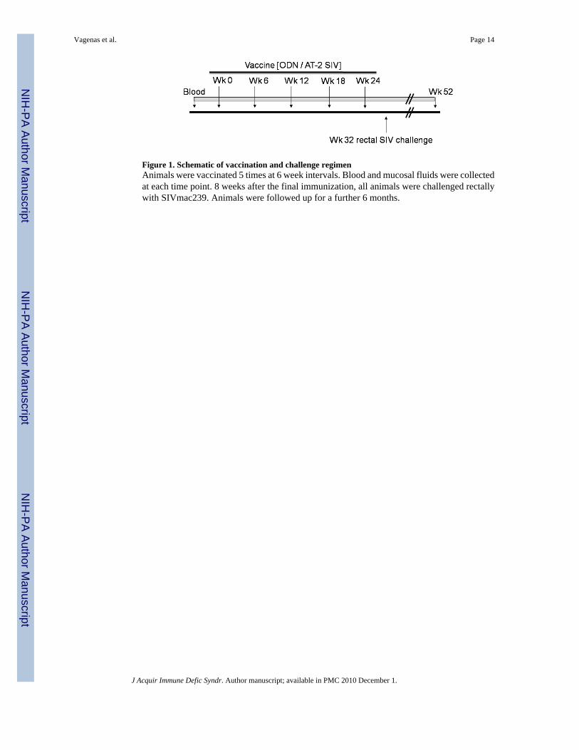
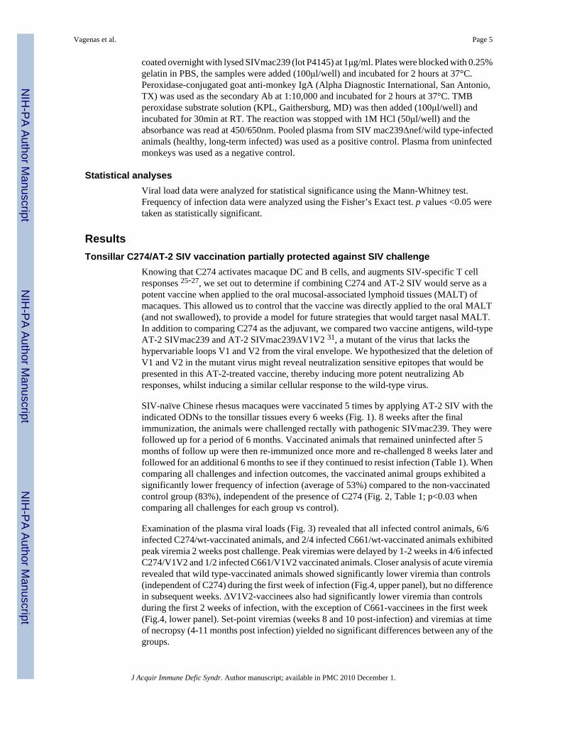
Figure 1. Schematic of vaccination and challenge regimenAnimals were vaccinated 5 times at 6 week intervals. Blood and mucosal fluids were collectedat each time point. 8 weeks after the final immunization, all animals were challenged rectallywith SIVmac239. Animals were followed up for a further 6 months.
Vagenas et al. Page 14
J Acquir Immune Defic Syndr. Author manuscript; available in PMC 2010 December 1.
NIH
-PA Author Manuscript
NIH
-PA Author Manuscript
NIH
-PA Author Manuscript
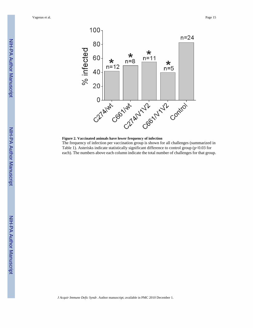
Figure 2. Vaccinated animals have lower frequency of infectionThe frequency of infection per vaccination group is shown for all challenges (summarized inTable 1). Asterisks indicate statistically significant difference to control group (p<0.03 foreach). The numbers above each column indicate the total number of challenges for that group.
Vagenas et al. Page 15
J Acquir Immune Defic Syndr. Author manuscript; available in PMC 2010 December 1.
NIH
-PA Author Manuscript
NIH
-PA Author Manuscript
NIH
-PA Author Manuscript
Figure 3. Individual viral loads of vaccinated and control groups(A) Plasma viral loads (SIV RNA copies/ml) of vaccinated and control monkeys weremeasured by RT-PCR. Each symbol denotes a different macaque challenge in each group. Nx,necropsy (5-10 months post infection). Profiles end at 10 weeks for 18 control animals thatwere moved to a different study at this point, having established the outcome of rectal challengefor infection status as well as peak and set point viremia.
Vagenas et al. Page 16
J Acquir Immune Defic Syndr. Author manuscript; available in PMC 2010 December 1.
NIH
-PA Author Manuscript
NIH
-PA Author Manuscript
NIH
-PA Author Manuscript
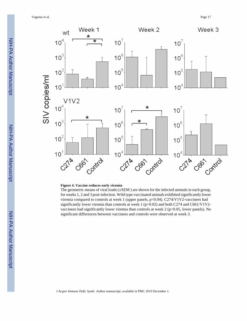
Figure 4. Vaccine reduces early viremiaThe geometric means of viral loads (±SEM ) are shown for the infected animals in each group,for weeks 1, 2 and 3 post-infection. Wild type-vaccinated animals exhibited significantly lowerviremia compared to controls at week 1 (upper panels, p<0.04). C274/V1V2-vaccinees hadsignificantly lower viremia than controls at week 1 (p<0.02) and both C274 and C661/V1V2-vaccinees had significantly lower viremia than controls at week 2 (p<0.05, lower panels). Nosignificant differences between vaccinees and controls were observed at week 3.
Vagenas et al. Page 17
J Acquir Immune Defic Syndr. Author manuscript; available in PMC 2010 December 1.
NIH
-PA Author Manuscript
NIH
-PA Author Manuscript
NIH
-PA Author Manuscript
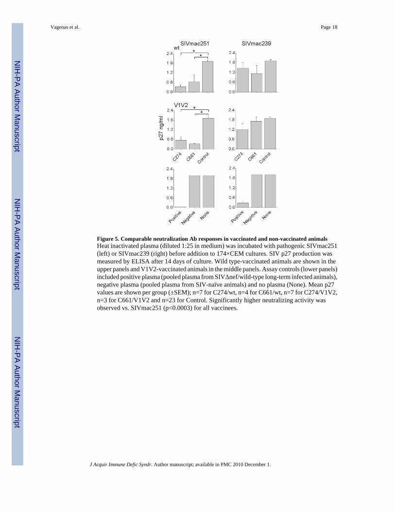
Figure 5. Comparable neutralization Ab responses in vaccinated and non-vaccinated animalsHeat inactivated plasma (diluted 1:25 in medium) was incubated with pathogenic SIVmac251(left) or SIVmac239 (right) before addition to 174×CEM cultures. SIV p27 production wasmeasured by ELISA after 14 days of culture. Wild type-vaccinated animals are shown in theupper panels and V1V2-vaccinated animals in the middle panels. Assay controls (lower panels)included positive plasma (pooled plasma from SIVΔnef/wild-type long-term infected animals),negative plasma (pooled plasma from SIV-naïve animals) and no plasma (None). Mean p27values are shown per group (±SEM); n=7 for C274/wt, n=4 for C661/wt, n=7 for C274/V1V2,n=3 for C661/V1V2 and n=23 for Control. Significantly higher neutralizing activity wasobserved vs. SIVmac251 (p<0.0003) for all vaccinees.
Vagenas et al. Page 18
J Acquir Immune Defic Syndr. Author manuscript; available in PMC 2010 December 1.
NIH
-PA Author Manuscript
NIH
-PA Author Manuscript
NIH
-PA Author Manuscript
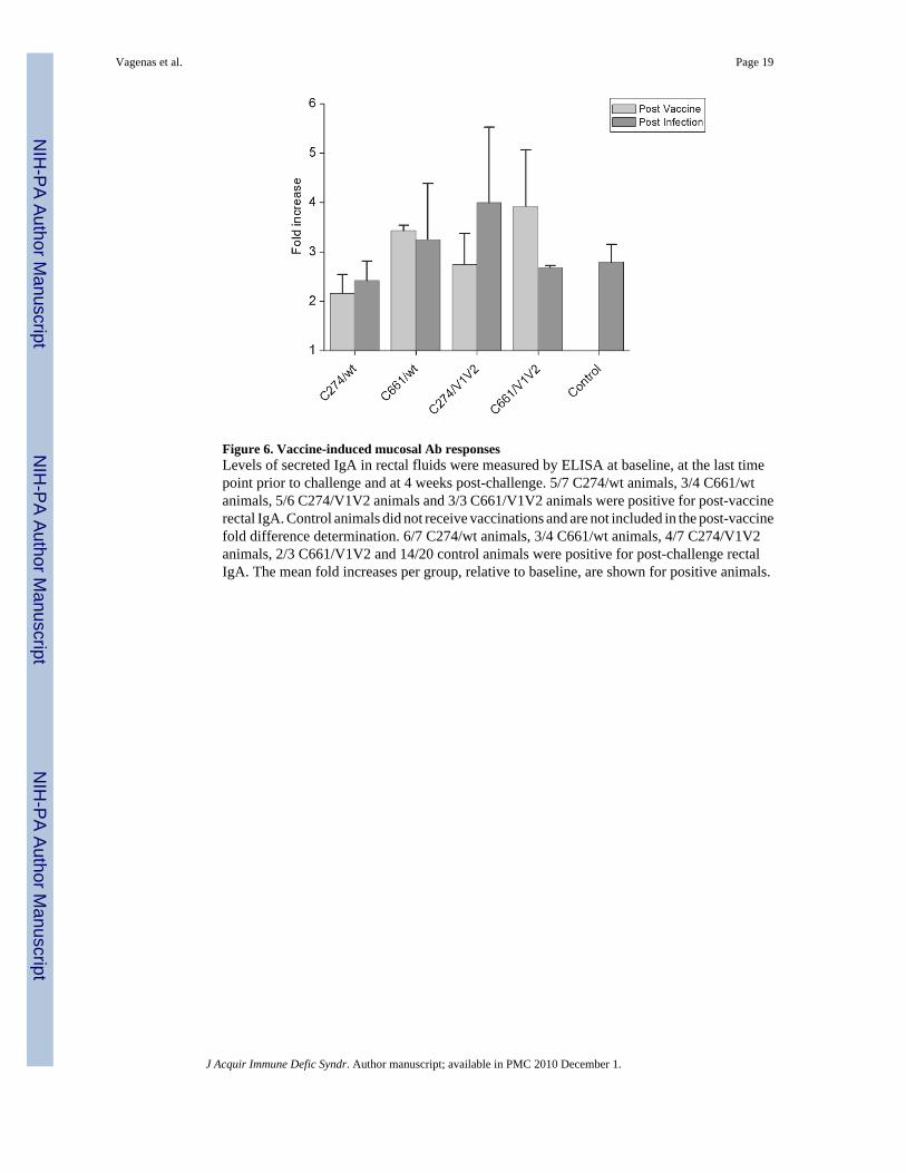
Figure 6. Vaccine-induced mucosal Ab responsesLevels of secreted IgA in rectal fluids were measured by ELISA at baseline, at the last timepoint prior to challenge and at 4 weeks post-challenge. 5/7 C274/wt animals, 3/4 C661/wtanimals, 5/6 C274/V1V2 animals and 3/3 C661/V1V2 animals were positive for post-vaccinerectal IgA. Control animals did not receive vaccinations and are not included in the post-vaccinefold difference determination. 6/7 C274/wt animals, 3/4 C661/wt animals, 4/7 C274/V1V2animals, 2/3 C661/V1V2 and 14/20 control animals were positive for post-challenge rectalIgA. The mean fold increases per group, relative to baseline, are shown for positive animals.
Vagenas et al. Page 19
J Acquir Immune Defic Syndr. Author manuscript; available in PMC 2010 December 1.
NIH
-PA Author Manuscript
NIH
-PA Author Manuscript
NIH
-PA Author Manuscript
NIH
-PA Author Manuscript
NIH
-PA Author Manuscript
NIH
-PA Author Manuscript
Vagenas et al. Page 20TA
BLE
1
Sum
mar
y of
ani
mal
trea
tmen
ts, i
nfec
tion
and
imm
une
stat
usIm
mun
izat
ion
Ani
mal
IDIn
fect
ion
IFNγS
IV A
bC
D4
coun
ts1st
Cha
lleng
e2nd
Cha
lleng
eIn
itial
Fina
lC
274/
wt
GE8
9+
++
1486
582
GE9
0−
−−
−13
1262
4G
E93
++
+10
3940
1G
J88
+−
+88
616
8G
J89
−+
−+*
394
162
GJ9
6−
++*
+*58
039
9G
J61
−+
+*+*
395
123
C27
4/V
1V2
GF0
2+
++
1461
390
GF0
3−
++*
−19
9638
5G
E94
++
+10
0533
5G
E95
−−
−−
651
559
GJ6
2+
−+
861
446
GJ6
3+
−+
603
88G
J64
−+
+*+*
1385
697
C66
1/w
tG
E91
−+
+*+*
1251
204
GE9
6−
++*
+*13
2740
1G
J66
−+
−+*
509
460
GJ6
7−
++*
+*11
7946
4C
661/
V1V
2G
E97
++
+11
0748
0G
E99
−+
+*+*
2379
818
GJ6
8−
−−
−64
753
8C
ontro
lG
F01
++
+18
7456
0G
J52
−−
−−
640
471
GJ3
9+
++
302
n/d
GJ4
7+
++
508
n/d
GJ4
8+
++
842
n/d
GJ4
9+
n/d
n/d
n/d
n/d
GJ5
0+
++
1088
n/d
GJ5
3+
++
1357
n/d
GJ5
5+
++
192
n/d
GJ5
7+
++
368
n/d
CC
48+
++
440
n/d
EL02
++
+28
5n/
dD
V67
++
+53
7n/
dD
D94
++
+39
4n/
dD
A47
++
+51
0n/
dC
M96
++
+58
5n/
dC
K25
++
+29
0n/
dC
L68
++
+62
7n/
dB
G93
++
+53
8n/
dG
J65
++
+68
6n/
dG
J97
++
+74
4n/
dC
F57
−n/
dn/
dn/
dn/
dC
N06
−n/
dn/
dn/
dn/
d
J Acquir Immune Defic Syndr. Author manuscript; available in PMC 2010 December 1.
NIH
-PA Author Manuscript
NIH
-PA Author Manuscript
NIH
-PA Author Manuscript
Vagenas et al. Page 21SI
V-s
peci
fic (A
T-2
SIV
mac
239
and
AT-
2 SI
Vm
ac23
9ΔV
1V2)
IFNγ-
prod
ucin
g ce
lls m
easu
red
by E
LISP
OT
post
infe
ctio
n; a
pos
itive
is g
iven
to a
nim
als w
ith m
ore
than
10
SIV
-spe
cific
SFC
/
2×10
5 ce
lls a
t mor
e th
an o
ne ti
me-
poin
t. To
obt
ain
SIV
-spe
cific
val
ues,
the
num
ber o
f spo
ts in
duce
d by
Sup
T1 M
V w
as su
btra
cted
from
the
SIV
val
ue. S
IV A
b po
sitiv
ity m
easu
red
by E
LISA
on
plas
ma
colle
cted
at 4
and
8 w
eeks
pos
t-inf
ectio
n.
* anim
al b
ecam
e IF
Nγ
or S
IV A
b po
sitiv
e af
ter 2
nd c
halle
nge.
Infe
ctio
n st
atus
was
con
firm
ed b
y R
T-PC
R o
n pl
asm
a sa
mpl
es (s
uppo
rted
by th
e de
tect
ion
of in
fect
ious
viru
s in
PBM
C-1
74×C
EM c
o-
cultu
res)
. Tw
o an
imal
s (G
E90
and
GE9
5) w
ere
chal
leng
ed a
3rd
tim
e (n
ot sh
own)
and
rem
aine
d ne
gativ
e; th
ese
data
are
incl
uded
in la
ter c
alcu
latio
ns. C
D4
coun
ts a
re g
iven
at b
asel
ine
(initi
al) a
ndne
crop
sy (f
inal
) as C
D4
cells
/μl b
lood
(mea
sure
d by
FA
CS
at T
NPR
C).
n/d=
not d
eter
min
ed.
J Acquir Immune Defic Syndr. Author manuscript; available in PMC 2010 December 1.


