Embed Size (px)
Citation preview

Touch to See: Neuropsychological Evidence of a Sensory Mirror System for Touch
Nadia Bolognini1,2, Elena Olgiati1, Annalisa Xaiz1, Lucio Posteraro3, Francesco Ferraro4 and Angelo Maravita1
1Department of Psychology, University of Milano-Bicocca, Milano 20126, Italy, 2Neuropsychological Laboratory, Istituto Auxologico
Italiano, 20122 Milano, Italy, 3Ospedale di Suzzara, Suzzara 46029, Italy and 4Azienda Ospedaliera Carlo Poma, Mantova 46012, Italy
Address correspondence to Dr Nadia Bolognini, Department of Psychology, University of Milano-Bicocca, P.zza Dell’Ateneo Nuovo 1, Ed. U6, 20126
Milano, Italy. Email: [email protected].
The observation of touch can be grounded in the activation of brainareas underpinning direct tactile experience, namely the somato-sensory cortices. What is the behavioral impact of such a mirrorsensory activity on visual perception? To address this issue, weinvestigated the causal interplay between observed and felt touchin right brain--damaged patients, as a function of their underlyingdamaged visual and/or tactile modalities. Patients and healthycontrols underwent a detection task, comprising visual stimulidepicting touches or without a tactile component. Touch and No-touch stimuli were presented in egocentric or allocentricperspectives. Seeing touches, regardless of the viewing perspec-tive, differently affects visual perception depending on whichsensory modality is damaged: In patients with a selective visualdeficit, but without any tactile defect, the sight of touch improvesthe visual impairment; this effect is associated with a lesion to thesupramarginal gyrus. In patients with a tactile deficit, but intactvisual perception, the sight of touch disrupts visual processing,inducing a visual extinction-like phenomenon. This disruptive effectis associated with the damage of the postcentral gyrus. Hence,a damage to the somatosensory system can lead to a dysfunctionalvisual processing, and an intact somatosensory processing can aidvisual perception.
Keywords: crossmodal, mirror neuron system, somatosensory cortex,touch observation, visual processing
Introduction
The human brain is able to implicitly transform the sight of
touch into an inner representation of touch (Gallese 2003;
Keysers et al. 2010). This function might be supported by
a visuo-tactile mirror system dealing with both our personal
experience of touch and the experience of touch observed in
other individuals. Animal and human studies have provided
extensive evidence for the existence of neural circuits in the
sensorimotor system that are involved in the processing of self
as well as others’ body-related experiences (Keysers et al.
2010). Empirical evidence from functional imaging studies in
humans (Keysers et al. 2004; Blakemore et al. 2005; Schaefer
et al. 2005; Ebisch et al. 2008) has revealed the existence of
a visuo-tactile mirror system, which matches observed touch
with felt touch. Even more intriguing is the report that the
mere observation of touch automatically induces the activation
of the neural circuitry that is normally involved in our
experience of touch, such as the primary (SI) and secondary
(SII) somatosensory cortices (Keysers et al. 2004; Blakemore
et al. 2005; Bolognini et al. 2011). This somatosensory activation
during the sight of touch is reminiscent of the mirror
premotor activation following action observation (Rizzolatti
and Craighero 2004), although it involves brain regions
responsible for somatosensory processing rather than action
programming (Keysers et al. 2010).
On a broader behavioral level, this system may be involved in
anticipating the effects of the tactile stimulation on our body in
a feed-forward fashion as well as in understanding the effects of
tactile stimulation experienced by other individuals, as a part of
the broader neural circuitry for embodied simulation (Gallese
2005; Grafton 2009).
So far, the behavioral impact of such a mirror somatosensory
activity on visual perception remains to be determined. This
intriguing question is motivated by findings from both stroke
patients and healthy subjects showing that the mere sight of
tactile events can be sufficient to evoke somatic sensations in
the observer (Halligan et al. 1996; Blakemore et al. 2005). Such
somatosensory experiences might be accomplished by the
activation of somatosensory areas through a mirror simulation
mechanism.
If the vision of a tactile event might automatically be
associated with the activation of the cortical network normally
involved in our own experience of being touched (e.g.,
Blakemore et al. 2005; Keysers et al. 2010), there should be
some difference in the processing of visual stimuli with or
without a tactile component; this difference, in turn, could be
linked to the ability to process tactile stimuli following stroke.
We therefore expected that, if the tactile system is undamaged,
the sight of touch may activate a spared somatosensory
representation. In the case of a visual impairment, this
additional recruitment of the tactile modality would improve
the perception of visual stimuli with a tactile content as
compared with visual stimuli without a tactile content.
Conversely, a damage to the tactile modality might selectively
affect the processing of visual stimuli depicting touch either
by disrupting the mirror simulation of the observed touch or by
activating an impaired somatosensory representation of the
observed touch. In both cases, no advantage would be
expected for visual stimuli depicting a touch as compared
with those without touch. Finally, when both the visual and
tactile modalities are damaged, no crossmodal modulation
would be expected in sensory processing by adding a somatic
dimension to the visual experience; therefore, any difference
between touching and no-touching stimuli should emerge.
To address this issue, the effect of touch observation on
visual perception was studied in right brain--damaged patients
with visual and/or tactile deficits, namely: patients with visual
extinction, which consists in the failure to detect a visual
stimulus presented in the contralesional hemifield when
a competing stimulus is simultaneously presented in the
ipsilesional hemifield (Brozzoli et al. 2006); patients with
tactile deficits on the contralesional hand, that is, tactile
extinction or hypoesthesia; patients with both contralesional
tactile and visual deficits; and patients without sensory (tactile
� The Author 2011. Published by Oxford University Press. All rights reserved.
For permissions, please e-mail: [email protected]
doi:10.1093/cercor/bhr283
Advance Access publication October 10, 2011
Cerebral Cortex September 2012;22:2055– 2064
at Bicocca - B
iblioteca Centrale InterfacoltÃ
on Novem
ber 14, 2012http://cercor.oxfordjournals.org/
Dow
nloaded from

or visual) deficit. A group of healthy participants was also
tested.
Participants performed a visual detection task during which
they had to detect the movement of index fingers that could
touch the thumb of the same hand (Touch condition) or not
(No-touch condition). Egocentric and allocentric viewing
perspectives were used in order to determine whether the
visual processing of body contact may differ according to the
viewing perspective. Since viewing one’s own body being
touched has an impact on somatosensory processing (Taylor-
Clarke et al. 2002), the self-attribution of being touched in the
observer (i.e., egocentric view) might affect visual processing
to a greater extent as compared with the observation of a touch
not attributable to one’s own body (i.e., allocentric view).
Materials and Methods
ParticipantsPatients were recruited from the inpatient population of a Neurological
Rehabilitation Unit (S.C. Riabilitazione Specialistica Neuromotoria,
Azienda Ospedaliera Carlo Poma, Bozzolo, Italy). Fourteen brain-
damaged patients (all right-handed, 8 males; mean age = 68, standard
deviation [SD] = 12; mean educational level = 9, SD = 3) participated in
the study. The inclusion criterion was the presence of a right
hemisphere lesion. All patients had no history of previous neurological
disease or psychiatric disorders. Patients did not show any sign of
cognitive decline (Folstein et al. 1975).
Twelve neurologically unimpaired subjects (all right-handed, 6 males,
mean age = 62, SD = 9; mean educational level = 7, SD = 3) were also
tested, and they served as controls.
All participants gave their informed consent to participate in the
study. The protocol was carried out in accordance with the ethical
standards of the Declaration of Helsinki (British Medical Journal, 1991,
Vol. 302, p. 1194), and it was approved by the ethical committee of the
University of Milano-Bicocca.
Clinical AssessmentContralesional motor, somatosensory, and visual field deficits, including
extinction to tactile and visual stimuli, were assessed by a standard
neurological examination (Bisiach et al. 1983; Haerer 1992; Anderson
et al. 2005). The motor examination of the upper and lower limbs
included the clinical assessment of asymmetric strength deficits. The
patient was asked, with eyes closed, to hold his/her arms out
horizontally (palms up) for 30 s, then observing any sign of weakness
(arm drift or pronation: score: 0--3, 0 = no deficit and 3 = maximum
deficit). For the lower extremity, the supine patient was asked to flex
the thigh on the pelvis 90� and extend the leg on the thigh 90�,maintaining the position for 30 s (score: 0--3) (Bisiach et al. 1983).
Furthermore, none of the patients showed clinical deficit neither of
motor coordination nor of tendon reflexes.
Visual field (upper and lower quadrants) was tested by confrontation.
The patient was instructed to fixate the nose of the examiner, who
performed a conventional manual confrontation test by ‘‘finger
wiggling,’’ keeping the hands at about 20� of visual angle to the right
and left visual fields. The patient had to report the presence and
location (left/right/bilateral) of any perceived movement.
The presence of tactile deficits was assessed with manual stimulation
through brief and light pressure stimuli delivered with the fingertip on
the back of the patient’s hand. The patient had to report which hand
was touched (left, right, or both).
In both the tactile and visual tests, the patients were warned that the
stimulation could be unilateral or bilateral and stimuli were adminis-
tered in 2 consecutive series: First, only 10 unilateral left and 10
unilateral right stimuli (score = 0--10 omissions for each side) were
presented in a random fixed order. In the second sequence, which
aimed at looking for the presence of extinction, 5 unilateral left, 5
unilateral right, and 10 bilateral simultaneous stimuli were delivered
(score = 0--10 omissions in bilateral trials). The presence of left
extinction is indexed by a difference between unilateral left and
bilateral stimuli >20% (Bisiach et al. 1983). For both the visual and
tactile tests, whenever the patient correctly reported the occurrence of
the stimulus, he/she was always able to correctly localize it.
Furthermore, we evaluated the presence of unilateral spatial neglect
(USN), a spatial deficit consisting in the lack of awareness for visual
events located in the space contralateral to the damaged hemisphere
(typically the left hemispace following a right lesion; Bisiach and Vallar
2000) by using the following tests: 1) Cancellation tasks, that is, Line
cancellation, score = 0--40 (Albert 1973); Letter cancellation, score = 0--
104 (Diller and Weinberg 1977); and Bell cancellation, score = 0--35
(Gauthier et al. 1989); 2) Line Bisection, score = mean percentage of
rightward bias (Wilson et al. 1987); 3) Drawing, that is, Clock Drawing
Test, score = 0--12 (Wilson et al. 1987) and 5-element complex drawing,
score = 0--10 (Gainotti et al. 1972); in both tests, performance was
evaluated considering the completeness and symmetrical arrangement
of drawing; and 4) Sentence reading test, score = 0--6 (Pizzamiglio et al.
1992). Personal USN was assessed by asking the patient to touch the
contralesional hand with the ipsilesional hand, score = 0--3 (0 =unimpaired performance; 3 = maximum deficit) (Bisiach et al. 1986).
A more detailed description of these tests and their scoring is described
elsewhere (e.g., Sposito et al. 2010).
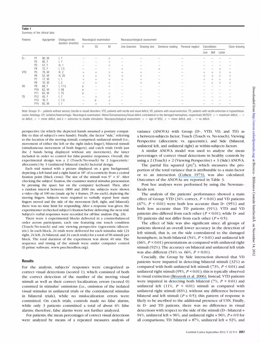
The clinical details of each patient are summarized in Table 1.
Based on the neuropsychological evaluation, patients were divided
into 4 groups, with respect to their damaged sensory processing: 1) 4
patients with contralesional tactile and visual field deficits at the
neurological examination (i.e., visual plus tactile deficit [VTD] group);
these patients also exhibited USN; 2) 3 patients with visual extinction
but without tactile deficits or USN (i.e., VD group); 3) 3 patients with
a tactile deficit on the contralesional hand and no signs of visual deficits
or USN (i.e., TD group); for one of these patients, the deficit was
present only following bilateral stimulation (i.e., tactile extinction),
while for the others, it also followed single contralesional stimulation
(i.e., hypoesthesia); and 4) 4 patients without any sensory (tactile or
visual) disorder (i.e., D– group) (Note that P4 showed only a mild
rightward bias at the line bisection test, at the reading task she made
just one error, and at the Bell cancellation task the errors were not
spatial specific for the contralesional hemispace [omissions on both
the left and right sides]. More important, with respect to the aim of the
present study, she did not exhibit any visual or tactile deficits at the
neurological examination. Hence, P4 was included in the D– group).
Lesion DataFigure 1 shows the mapping of the brain lesions for 11 of 14 patients,
for whom the original brain scan (in all cases, this was a CT scan) was
available. Lesions were mapped using the software MRIcro (Rorden and
Brett 2000). We reconstructed the region of interest (ROI) to define
the location and the size of the lesion for each patient by using
a Template Technique, that is, by manually drawing the lesion on the
standard template from the Montreal Neurological Institute. ROIs were
created by mapping the regions on each and every 2D slice of a 3D
volume. By overlapping the lesions in each group of patients, we could
calculate the voxels in which lesions overlapped, as reported in Table 2.
For the 3 patients for whom the original scan was not available, the
radiological report indicated the following lesion site: P1 = basal ganglia;
P2 = middle cerebral artery stroke, with no sign of focal lesions; and
P10 = parietal and temporal areas.
Experimental TaskThe visual detection task comprised 3 experimental main factors (see
Fig. 2a). The factor ‘‘touch,’’ referring to the kind of stimulus delivered,
comprised the Touch condition, namely the observation of a video clip
depicting the index finger touching the thumb of the same hand at
the end of the movement, and the No-touch condition, namely the
observation of a video clip depicting the index finger making the same
movement as in the Touch condition but without touching the thumb.
In Touch and No-touch conditions, the amount of the movement of the
index finger was matched. The factor ‘‘viewing perspective,’’ referring
to the configuration of the visual stimuli, comprised the allocentric
perspective (in which the depicted hands mimicked the posture of
a putative examiner placed in front of the subject) and the egocentric
Visuo-Tactile Mirror Interactions in Humans d Bolognini et al.2056
at Bicocca - B
iblioteca Centrale InterfacoltÃ
on Novem
ber 14, 2012http://cercor.oxfordjournals.org/
Dow
nloaded from
perspective (in which the depicted hands assumed a posture compat-
ible to that of subject’s own hands). Finally, the factor ‘‘side,’’ referring
to the location of the moving stimuli, comprised: unilateral stimuli (i.e.,
movement of either the left or the right index finger), bilateral stimuli
(simultaneous movement of both fingers), and catch trials (with just
the 2 hands being displayed without any movement), the latter
included in order to control for false-positive responses. Overall, the
experimental design was a 2 (Touch/No-touch) by 2 (egocentric/
allocentric) by 3 (unilateral/bilateral/catch) factorial design.
Each trial started with a picture displayed on a gray background,
depicting a left hand and a right hand at 18� of eccentricity from a central
fixation point (black cross). The size of the stimuli was 9� 3 6�. Afterchecking the subject’ fixation, the examiner started stimulus presentation
by pressing the space bar on the computer keyboard. Then, after
a random interval between 1800 and 2000 ms, subjects were shown
a video clip of 100 ms (made up by 4 frames, 25 ms each), depicting the
moving fingers. Subjects were required to verbally report how many
fingers moved and the side of the movement (left, right, and bilateral);
there was no time limit for responding. After a response was given, the
experimenter rechecked subject’s fixation before delivering the next trial.
Subject’s verbal responses were recorded for off-line analysis (Fig. 2b).
There were 4 experimental blocks delivered in a counterbalanced
order across participants, each one featuring one touch condition
(Touch/No-touch) and one viewing perspective (egocentric/allocen-
tric). In each block, 24 trials were delivered for each stimulus side (24
right, 24 left, 24 bilateral, and 24 catch trials) for a total of 96 stimuli per
block. The total duration of the experiment was about 40 min. The
sequence and timing of the stimuli were under computer control
(E-prime software, www.psychtoolbox.org).
Results
For the analysis, subjects’ responses were categorized as
correct visual detections (scored 1), which consisted of both
the correct detection of the number of the moving visual
stimuli as well as their correct localization; errors (scored 0)
consisted in stimulus’ omissions (i.e., omission of the isolated
visual stimulus in unilateral trials or the contralateral stimulus
in bilateral trials), while no mislocalization errors were
committed. On catch trials, controls made no false alarms,
while only 3 patients committed a total of about 6% false
alarms; therefore, false alarms were not further analyzed.
For patients, the mean percentages of correct visual detections
were analyzed by means of a repeated measures analysis of
variance (ANOVA) with Group (D–, VTD, VD, and TD) as
a between-subjects factor, Touch (Touch vs. No-touch), Viewing
Perspective (allocentric vs. egocentric), and Side (bilateral,
unilateral left, and unilateral right) as within-subjects factors.
A similar ANOVA model was used to analyze the mean
percentages of correct visual detections in healthy controls by
using a 2 (Touch) 3 2 (Viewing Perspective) 3 3 (Side) ANOVA.
The partial Eta squared (pg2), which measures the pro-
portion of the total variance that is attributable to a main factor
or to an interaction (Cohen 1973), was also calculated.
Outcomes of the ANOVAs are reported in Table 3.
Post hoc analyses were performed by using the Newman--
Keuls test.
The analysis of the patients’ performance showed a main
effect of Group: VTD (34% correct, P < 0.01) and VD patients
(67%, P < 0.01) were both less accurate than D– (95%) and
both less accurate than TD patients (91%). VTD and VD
patients also differed from each other (P < 0.01), while D– and
TD patients did not differ from each other (P = 0.5).
The effect of Side was also significant since all groups of
patients showed an overall lower accuracy in the detection of
left stimuli, that is, on the side contralateral to the damaged
hemisphere, in both bilateral (54%, P < 0.01) and unilateral left
(66%, P < 0.01) presentations as compared with unilateral right
stimuli (92%). The accuracy on bilateral and unilateral left trials
was also different (54% vs. 66%, P < 0.01).
Crucially, the Group by Side interaction showed that VD
patients were impaired in detecting bilateral stimuli (32%) as
compared with both unilateral left stimuli (73%, P < 0.01) and
unilateral right stimuli (95%, P < 0.01); this is typically observed
in visual extinction (Brozzoli et al. 2006). Instead, VTD patients
were impaired in detecting both bilateral (7%, P < 0.01) and
unilateral left (11%, P < 0.01) stimuli as compared with
unilateral right stimuli (83%), without any difference between
bilateral and left stimuli (P = 0.5); this pattern of response is
likely to be ascribed to the additional presence of USN. Finally,
in D– and TD patients, there was no difference in visual
detections with respect to the side of the stimuli (D–: bilateral =94%, unilateral left = 96%, and unilateral right = 96%, P = 0.9 for
all comparisons; TD: bilateral = 87%, unilateral left = 92%, and
Table 1Summary of the clinical data
Patients Age/gender Etiology/strokeduration (months)
Neurological examination Neuropsychological assessment
V SS M Line bisection Drawing test Sentence reading Personal neglect Cancellation Clock drawing
Line Bell Letter
D� P1 90, M I, 1 � � þ � � � � � � � �P2 85, F I, 1 � � þ � � � � � � � �P3 57, F H, 1 � � ± � � � � � � � �P4 72, F I, 1.5 � � � þ � ± � � � ± �
VTD P5 79, M H, 2 þ þ ± þ þ � � þ þ ± þP6 52, M H, 20 þ þ þ þ þ � � þ þ þ þP7 77, M I, 1 þ þ þ þ þ � � þ þ þ þP8 70, M I, 2 þ þ þ � þ þ � � þ þ þ
VD P9 68, F I, 17.5 e � þ � � � � � � � �P10 62, M I, 59 e � � � � � � � � � �P11 63, M I, 15 e � þ � � � � � � � �
TD P12 45, F I, 7 � þ þ � � � � � � � �P13 76, F I, 1.5 � e þ -- � � � � � � �P14 65, M I, 1 � þ þ � � � � � � � �
Note: Groups: D�, patients without sensory (tactile or visual) disorders; VTD, patients with tactile and visual deficit; VD, patients with visual extinction; TD, patients with tactile extinction or hypoesthesia.
Lesion Aetiology: I/H, ischemic/haemorrhagic. Neurological examination: Motor/Somatosensory/Visual deficit contralateral to the damaged hemisphere, respectively M/SS/V: þ5 maximum deficit, �5
no deficit, ± 5 minor deficit, and e 5 extinction to double stimulation. Neuropsychological assessment: þ 5 sign of NSU, ± 5 minor deficit, and � 5 no deficit.
Cerebral Cortex September 2012, V 22 N 9 2057
at Bicocca - B
iblioteca Centrale InterfacoltÃ
on Novem
ber 14, 2012http://cercor.oxfordjournals.org/
Dow
nloaded from

unilateral right = 95%, P = 0.6 for all comparisons). These
findings replicate those obtained at the neurological examina-
tion, confirming the appropriateness of our visual task for
assessing unilateral and bilateral visual deficits and supporting
the validity of our allocation of the patients in the different
experimental groups.
The significant main effect of the Viewing Perspective
showed a better performance for the allocentric (73%) as
compared to the egocentric perspective (69%, P < 0.01).
The key finding with respect to our experimental hypothesis
was the significant Group by Touch by Side interaction, which
highlighted how the sight of touch can modulate visual
perception in a fashion that is specific to the patients’ sensory
deficit, as shown in Figure 3a. In VD patients, seeing visual
stimuli depicting a touch improved visual extinction on
bilateral trials (39%, P < 0.01) as compared with bilateral visual
stimuli without the tactile component (24%). For unilateral
left-sided and right-sided stimuli, the difference between
Touch and No-touch conditions was not significant (unilateral
left: Touch = 76% vs. No-touch = 70%, P = 0.07; unilateral right:
Touch = 94% vs. No-touch = 97%, P = 0.9). In striking contrast,
in TD patients, the sight of touch impaired the performance on
bilateral trials in an extinction-like fashion; in other words,
when the tactile modality is affected, seeing touches reduced
the accuracy on bilateral trials as compared with bilateral No-
touch trials (78% vs. 93%, P < 0.01). This effect was specific for
the bilateral stimuli, being absent for unilateral left stimuli
(Touch = 91% vs. No-touch = 93%, P = 0.9) and for unilateral
right stimuli (Touch = 96% vs. No-touch = 95%, P = 0.8).
Instead, in both VTD and D– patients, there was no difference
between Touch and No-touch conditions for all stimulus
locations (i.e., bilateral, unilateral left, and unilateral right; P >
0.2, for all comparisons).
Other effects did not reach significance (Table 3).
Noteworthy, the size effects in the ANOVA showed that the
significant effects were quite consistent, notwithstanding the
comparatively small number of patients (Table 3).
Finally, the analysis of healthy control subjects did not
showed any significant effect (Table 3).
Comparison between Patients and Controls
To assess for the presence of any significant dissociation in
patients’ performance, we further compared the detection
accuracy on bilateral trials in Touch and No-touch conditions
Figure 1. Lesion mapping for each single patient. White areas represent theextension of the lesion of each single patient.
Table 2Number of voxels in each ROI are given for each group of patient
D� VTD TD VD
Precentral gyrus 104 3190 17 330 5003Frontal sup — — 2964 602Frontal mid — 366 1383 2954Frontal inf 76 3574 18 455 5868Rolandic oper 196 8942 10 622 2643Insula 400 9036 12 749 4511Cingulum — 19 27 747Hippocampus — 358 — 143Fusiform 690 1922 — —Occipital mid — 775 204 102Occipital inf 779 — — —Postcentral gyrus — 6079 18 562 3845Parietal sup — — 80 123Parietal inf — 59 2315 1266Supramarginal gyrus — 8883 10 665 8704Angular gyrus — 2580 4228 1270Caudate — — 3501 3432Putamen 1139 7507 7088 7739Pallidum 261 117 1578 1658Thalamus 1519 163 73 1986Heschl — 1740 1657 825Temporal sup — 25 838 19 275 17 568Temporal mid — 29 455 8400 16 367Temporal infer 725 7974 — 3028
Visuo-Tactile Mirror Interactions in Humans d Bolognini et al.2058
at Bicocca - B
iblioteca Centrale InterfacoltÃ
on Novem
ber 14, 2012http://cercor.oxfordjournals.org/
Dow
nloaded from

with that of the healthy controls. The comparison was
performed by t-tests performed following the procedure by
Crawford and Garthwaite (2005). This procedure allows to
estimate the abnormality of the patient’s score at each test (i.e.,
the estimate of the percentage of the control population that
would obtain a lower score). Then, using the same statistical
methodology, we compared the difference between Touch and
No-touch conditions in each patient with that of the control
sample (Crawford and Garthwaite 2005).
Every VD patient was significantly impaired in detecting
bilateral stimuli in both Touch and No-touch conditions (P <
0.05 for all comparisons), but they also showed a significant
difference between these conditions consisting in an advan-
tage for the touching stimuli (difference between the Touch
and No-touch conditions: P9 = 19%, t11 = 1.7, P < 0.05; P10 =13%, t11 = 4.7, P < 0.01; and P11 = 13%, t11 = 5.8, P < 0.01)
(see Fig. 4).
Instead, TD patients showed a selective impairment only in
the Touch condition (P < 0.05) with the performance in No-
touch condition being within the normal range. Crucially, every
TD patient showed a significant difference between the 2
conditions consisting in a disadvantage for the Touch condition
at variance with the VD group (difference between the 2
conditions: P12 = –27%, t11 = 8.6, P < 0.01; P13 = –9%, t11 = 4.3,
P < 0.01; and P14 = –10%, t11 = 2.5, P < 0.01) (Fig. 4).
Finally, VTD patients were impaired in both the Touch and
No-touch conditions (P < 0.01), and they did not showed any
Figure 2. Experimental procedure. (a) Experimental conditions of the visual detection task: No-touch and Touch stimuli, in egocentric or allocentric perspectives, presentedunilaterally (i.e., movement of the left or the right index finger) or bilaterally (simultaneous movement of both fingers). In catch trials, 2 hands were displayed without anymovement (not shown in the figure). (b) Sequence of events: for example, the bilateral stimulus of the Touch/Allocentric condition. With their eyes at fixation, patients had toverbally report the movement of the fingers and the side of the movement.
Table 3F, P level, and pg2 values from the ANOVAs
Factors (df) F P level pg2
Patients Group (3,10) 47.03 0.0001 0.9Viewing Perspective (1,10) 7.02 0.02 0.4Touch (1,10) 0.026 0.8 0.01Side (2,20) 82.53 0.0001 0.9Viewing Perspective 3 Touch (1,10) 0.17 0.7 0.02Viewing Perspective 3 Side (2,20) 0.47 0.6 0.04Touch 3 Side (2,20) 1.16 0.3 0.1Viewing Perspective 3 Touch 3 Side (2,20) 1.13 0.3 0.1Group 3 Viewing Perspective (3,10) 2.15 0.16 0.3Group 3 Touch (3,10) 2.31 0.14 0.4Group 3 Side (6,20) 28.1 0.0001 0.9Group 3 Viewing Perspective 3 Touch (3,10) 0.49 0.7 0.1Group 3 Viewing Perspective 3 Side (6,20) 0.36 0.9 0.1Group 3 Touch 3 Side (6,20) 4.95 0.01 0.6Group 3 Viewing Perspective 3 Touch 3 Side (6,20) 1.07 0.4 0.2
Controls Touch (1,11) 0.49 0.5 0.01Side (2,22) 1.85 0.2 0.04Viewing Perspective 3 Touch (1,11) 1.90 0.2 0.1Viewing Perspective 3 Side (2,22) 0.37 0.7 0.03Touch 3 Side (2,22) 0.09 0.9 0.01Viewing Perspective 3 Touch 3 Side (2,20) 1.07 0.4 0.01
Note: df 5 degrees of freedom.
Cerebral Cortex September 2012, V 22 N 9 2059
at Bicocca - B
iblioteca Centrale InterfacoltÃ
on Novem
ber 14, 2012http://cercor.oxfordjournals.org/
Dow
nloaded from

difference between these 2 conditions (P = 0.2); D– patients
scored normal on both conditions (P = 0.5), without difference
among them (P = 0.7).
Association between the Crossmodal Effect of the Sight ofTouch and the Anatomoclinical Data
Multiple correlations (Pearson correlation) were performed to
examine the relation between the effect induced by the sight
of touch on bilateral trials (facilitation or impairment of
visual detections, i.e., Touch minus No-touch, with positive
values indicating that the sight of touch improves visual
detections and negative values indicating that the sight of
touch impairs visual detections) and the following variables:
age; time from stroke (in months); visual, tactile, and motor
deficit as assessed at the neurological examination; and
performance at the neuropsychological assessment. All the 14
patients were considered in these analyses. Only a significant
correlation was found: The difference between Touch and No-
touch on bilateral trials was positively correlated with the
difference between visual and tactile extinction (tactile
detections on bilateral trials minus visual detections on bilateral
trials) (r = 0.7, P < 0.01). This suggests that the presence of
a tactile impairment, without visual extinction, was associated
with a visual impairment in the Touch condition (i.e., the
greater the tactile deficit, the greater the disruption of visual
perception in the Touch condition). By contrast, the presence
of visual extinction, without tactile extinction, was associated
with a benefit in the Touch condition (i.e., the greater the
extinction of visual rather than tactile stimuli, the greater the
benefit in the Touch condition).
With respect to the lesion profile, correlation analyses were
performed between the change in visual detections between
Touch and No-touch stimuli on bilateral trials and the lesion
size (number of voxels) in each ROI. We considered all the 11
patients for which the lesion reconstruction was made. Only 2
significant effects were found. A negative correlation was found
between the difference between Touch and No-touch stimuli
and the size of the lesion in the postcentral cortex (r = –0.65,
P < 0.04). This means that the bigger the damage to the
postcentral gyrus, the greater the impairment induced by the
sight of touch; conversely, the greater the tactile-induced
benefit, the smaller the damage to the postcentral gyrus.
Additionally, there was a positive correlation between the
difference between Touch and No-touch stimuli and the size of
the lesion in the supramarginal gyrus (r = 0.71, P < 0.02),
suggesting that the larger the damage to this area, the greater
the benefit induced by seeing touches. The results from the
correlation analysis are reported in the Supplementary Tables
S1 and S2.
To further explore the relation between the lesion site and
the dissociation in performance between VD and TD patients,
we looked for those regions that were more damaged (in
percentage) in TD than in VD patients by using a subtraction
method (Rorden and Brett 2000). Figure 3b depicts the
resulting subtraction plot, showing the hotspots indicating
regions that were more damaged in the TD group than in the
VD group. The more damaged areas in TD patients were the
postcentral gyrus, the inferior frontal gyrus, and the precentral
gyrus; instead, in VD patients, the inferior and middle temporal
cortices and the supramarginal gyrus were more largely
involved (see also Table 2).
Discussion
Our findings showed that viewing a touch can differentially
modulate visual perception, depending on the underlying
damaged sensory modality: If the visual modality is affected
(VD patients), the sight of touch can improve the visual
disorder, whereas in case of a damage confined to the tactile
modality (TD patients), the sight of touch impairs visual
perception. These visuo-tactile effects were present only on
bilateral trials, which is the most attentional demanding
condition. This supports the view that the somatosensory
mirror system might produce a behaviorally relevant cross-
modal modulation of visual perception only within a certain
perceptual threshold (Blakemore et al. 2005).
Conversely, patients with both visual and tactile disorders or
patients without visual and tactile disorders did not show any
visual effect induced by the observation of touch. Hence, the
crossmodal modulation of visual perception by touch does not
occur when sensory awareness is deeply impaired across vision
and touch (VTD, who also exhibited USN) or, else, completely
undamaged (D–, as well as in healthy controls or in general for
processing ipsilesional stimuli).
Correlation analyses clearly indicate that the deficit of
sensory processing predicts the effect induced by the sight of
touch: TD is associated to the disruption of visual processing
induced by touch observation, while visual extinction is
associated to the visual improvement induced by touch
observation. Instead, other clinical factors, such as motor- and
USN-related disorders, stroke duration, or age, were not
associated to the effects induced by the sight of touch
(although we must be cautious about absent correlations since
the sample size was pretty small).
The effect of seeing touches was unrelated to the attribution
of the observed touch to oneself (egocentric view) or to
somebody else (allocentric view), in line with previous
evidence (Keysers et al. 2004; Schaefer et al. 2005). Rather,
we found that all groups of patients were more impaired in
detecting visual stimuli seen from an egocentric perspective
than from an allocentric perspective. These findings are
consistent with the idea of a right hemisphere specialization
in the egocentric processing of spatial information (Stein 1989;
Iachini et al. 2009). The right hemisphere also plays a special
role in the ability to distinguish the self from others (Decety
and Chaminade 2003) and the recognition of self body parts
(Frassinetti et al. 2008). Hence, a lesion to the right hemisphere
may have affected any possible advantage/disadvantage by self-
referential processing of the observed hands, regardless of the
tactile component.
Overall, the behavioral results provide compelling support
that the processing of visual stimuli conveying a tactile content
involves different mechanisms as compared with the process-
ing of visual stimuli without a tactile content. Indeed, the
former seems to automatically engage an additional somato-
sensory representation, likely through a mirror simulation
process, in order to build a crossmodal representation of the
seen touch. In patients with visual deficits, but spared
somatosensation, the recruitment of this undamaged represen-
tation in the tactile modality favors their defective visual
perception. This effect may be viewed as a compensatory
crossmodal mechanism for visual processing, activated by the
sight of touch. Conversely, in patients with tactile deficits,
the sight of touch impinges on visual perception negatively.
Visuo-Tactile Mirror Interactions in Humans d Bolognini et al.2060
at Bicocca - B
iblioteca Centrale InterfacoltÃ
on Novem
ber 14, 2012http://cercor.oxfordjournals.org/
Dow
nloaded from

The striking finding is that the damage to the tactile modality
actually reverses the crossmodal effect of the sight of touch,
although does not abolish it, suggesting that mirror system for
touch is spared and still functioning. In fact, the impaired
tactile processing appears now associated to the activation of
a ‘‘disrupted’’ somatosensory representation of the observed
touch, which selectively hampers visual perception of touch,
even if visual processing is undamaged per se.
Worth mentioning, in our design, the blocked nature of
Touch and No-touch trials (which likely provided clues as to
whether a Touch or No-touch trial was about to occur) could
have induced patients to rely more on the visual or tactile
representation to detect the visual stimulus, depending on the
block of trials, hence fostering the difference between Touch
and No-touch conditions. However, this is still perfectly
compatible with the role of a putative imitative mechanism
for touch in biasing the participant’s performance.
With respect to the anatomoclinical correlation of the
present visuo-tactile effects, the lesions of VD and TD patients
differed mainly with respect to the involvement of regions in
the postcentral gyrus, suggesting a likely role of cortical areas
underpinning tactile sensation in matching observed and felt
touch. In fact, the size of the lesion in the postcentral gyrus was
associated with the effect induced by the sight of touch.
A larger damage to the postcentral gyrus corresponded to
a greater visual impairment induced by the tactile component
of the observed visual event; conversely, the sparing of this area
was positively associated with the benefit induced by the sight
of touch. This finding is in line with functional neuroimaging
evidence showing the activation of somatosensory areas by the
sight of touch (Keysers et al. 2004; Blakemore et al. 2005;
Schaefer et al. 2005; Ebisch et al. 2008). Noteworthy, mirror-
touch synesthesia, that is, people reporting on their own skin
the feeling of a touch observed on other person, is associated
with an unusual hyperactivation of somatosensory cortices
during touch observation (Blakemore et al. 2005). Conversely,
complete haptic deafferentation impairs the ability to infer
Figure 3. (a) Modulation of visual perception by the sight of touch in right brain--damaged patients: D�5 no sensory or spatial disorders, VTD5 visual plus tactile deficit, VD5visual deficit, TD5 tactile deficit. Light gray bars 5 accuracy in Touch trials; dark gray bars 5 accuracy in No-touch trials. Asterisks indicate the significant difference in bilateraltrials for Touch and No-touch stimuli in VD and TD patients. Error Bars 5 standard error of the mean. (b) Overlapping of VD and TD lesions. Each bar represents 20% increments:lightest blue areas 5 the maximum percentage of specificity of the VD group for that region; lightest yellow areas 5 maximum percentage of specificity of the TD group for thatregion; and purple 5 regions where there is an identical percent of VD and TD.
Cerebral Cortex September 2012, V 22 N 9 2061
at Bicocca - B
iblioteca Centrale InterfacoltÃ
on Novem
ber 14, 2012http://cercor.oxfordjournals.org/
Dow
nloaded from

someone else’s sensory expectations through the observation
of their actions (Bosbach et al. 2005).
On the other hand, the amount of involvement of the
supramarginal gryus (SMG) was associated to the visual
facilitation induced by the sight of touch. In other words,
touch observation seems to provide a facilitatory effect, which
is likely mediated by crossmodal somatosensory activation, that
can compensate an impaired visual processing due to an SMG
damage. The right SMG is important for strategic orienting of
spatial attention (Perry and Zeki 2000; Corbetta and Shulman
2002), particularly within the visual modality. The temporary
disruption of this area can impair the ability to strategically
orient toward visual stimuli but without affecting the shift of
attention to somatosensory stimuli in the same location
(Chambers et al. 2004). Moreover, the SMG is a crucial site of
multisensory convergence, being involved in the integration of
visual and tactile information for space and body representation
(Macaluso and Maravita 2010). Finally, functional magnetic
resonance imaging evidence in brain-damaged patients shows
that crossmodal visuo-tactile extinction reflects the interaction
of competing information from vision and touch within
surviving multisensory parietal areas along the right SMG (Sarri
et al. 2006). This last finding is also in accordance with the
selectivity of our crossmodal effects for bilateral trials.
Overall, the lesion data suggest that the visuo-tactile effects
observed in the present study may involve a local mechanism
within somatosensory areas as well as the interaction between
somatosensory and posterior parietal areas. First, some SI
neurons react to visual stimuli that are behaviorally associated
with tactile information (Zhou and Fuster 1997), while SII is
involved in visuo-tactile integration of body-related information
(Carlsson et al. 2000; Avikainen et al. 2002). Second, both SI and
SII have afferent and efferent cortical connections with
multisensory regions of the posterior parietal cortex (PPC)
(Keysers et al. 2010). PPC contains neurons sensitive to both
vision and touch (Andersen 1997; Rizzolatti et al. 1997;
Duhamel et al. 1998) and shows strong visuo-tactile inter-
actions for conscious perception, body, and space representa-
tion (Halligan et al. 2003; Maravita et al. 2003; Farne et al. 2005;
Brozzoli et al. 2006; Bolognini and Maravita 2007; Macaluso
and Maravita 2010; Pasalar et al. 2010). Therefore, the cross-
modal influences induced by the sight of touch on visual
processing may arise as a result of an interaction between the
visual input and the tactile input, being the last provided by
the somatosensory cortex through a simulation process.
These interactions may take place in multisensory convergence
zones and/or attentional control structures of the posterior
parietal cortex (Macaluso and Driver 2005; Stein and Stanford
2008).
With respect to the supposed involvement of the mirror
neuron system in the observed crossmodal effects, seminal
studies in monkeys report that the viewing of actions
performed by other individuals activates frontal and parietal
cortical areas (Rizzolatti and Craighero 2004). However, it has
been recently proposed that motor and parietal areas might
play a differential role in simulating, respectively, efferent (i.e.,
motor) and afferent (somatic) components of observed actions.
The visual observation of movements eliciting somatic sensa-
tions in the onlooker, ranging from pain to the sensation of
being touched, activates a large sensorimotor parietal network,
including SI (Avenanti et al. 2007). In this regard, our findings
suggest that the sensory-parietal, but not the motor-frontal,
component of the mirror neuron system is likely to affect the
ability of simulating the afferent (somatic) components of
observed actions.
In future experiments, it would be interesting to include
a control task with nonhuman stimuli depicting touches, in
order to assess whether the visuo-tactile mirroring mechanism
for touch applies to the sight of ‘‘any’’ touch or whether it is
restricted to human contact. There is evidence suggesting that
mirror areas are preferentially activated by ‘‘social’’ touch (i.e.,
the observation of touch to another human, relative to the
contact between objects) and, more generally, when the target
of the action is biological rather than inanimate (Tai et al. 2004;
Blakemore et al. 2005).
In conclusion, the perception of visual events conveying
a tactile content seems to involve not only the visual but also
the somatosensory modality. This is true when there is a visually
based encoding of somatic sensations, such as touch, that is
likely to activate an inner mirror-like simulation of the observed
tactile sensation (Gallese 2003; Blakemore et al. 2005; Keysers
et al. 2010; Bolognini et al. 2011). The pervasiveness of the
functional interplay between vision and touch is such that
a damage to the somatosensory system can lead to a dysfunc-
tional visual processing, whereas an intact somatosensory
mirroring mechanism can even aid an impaired visual percep-
tion. On a broader perspective, the activation of a shared circuit
for visual and somatosensory processing of tactile events might
allow an automatic and implicit coding of the sense of touch
through vision, in line with the theories of embodied simulation
(Rizzolatti and Craighero 2004; Keysers et al. 2010).
Our findings may also have clinical implications. After
ischemic insults, there may be an increase in axonal sprouting,
unmasking of existing connections, and other occurrences of
neuronal plasticity. These changes are likely to create the
opportunity for crossmodal functional rewiring (Ro et al. 2007;
Beauchamp and Ro 2008), which may promote plastic visuo-
tactile interactions, usually masked under normal conditions.
Crossmodal rehabilitation approaches may be useful to prevent
Figure 4. Visual detection accuracy on bilateral trials in healthy controls, VD, and TDpatients. Stars: significant difference between each patient’s performance and that ofthe control group for each Touch and No-touch conditions. Circles: significantdifference between Touch and No-touch conditions in the individual patient ascompared with the same average difference in the control group.
Visuo-Tactile Mirror Interactions in Humans d Bolognini et al.2062
at Bicocca - B
iblioteca Centrale InterfacoltÃ
on Novem
ber 14, 2012http://cercor.oxfordjournals.org/
Dow
nloaded from

the formation of inappropriate crossmodal deficits (as the
visually induced impairment in TD patients) or may subserve
crossmodal compensation (as the improvement in visual
perception induced by the sight of tactile stimuli in VD
patients).
Supplementary Material
Supplementary material can be found at: http://www.cercor.
oxfordjournals.org/
Funding
Fondi di Ateneo per la Ricerca (FAR) from University of Milano-
Bicocca to N.B. and A.M.
Notes
We thank Nicoletta Beschin, Carlo Toneatto, and Laura Melini for
assistance. We are also grateful to the anonymous referees for their
helpful suggestions. Conflict of Interest : None declared.
References
Albert ML. 1973. A simple test of visual neglect. Neurology. 23:658--664.
Andersen RA. 1997. Multimodal integration for the representation of
space in the posterior parietal cortex. Philos Trans R Soc Lond B
Biol Sci. 352:1421--1428.
Anderson NE, Mason DF, Fink JN, Bergin PS, Charleston AJ, Gamble GD.
2005. Detection of focal cerebral hemisphere lesions using the
neurological examination. J Neurol Neurosurg Psychiatry.
76:545--549.
Avenanti A, Bolognini N, Maravita A, Aglioti SM. 2007. Somatic and
motor components of action simulation. Curr Biol. 17:2129--2135.
Avikainen S, Forss N, Hari R. 2002. Modulated activation of the human SI
and SII cortices during observation of hand actions. Neuroimage.
15:640--646.
Beauchamp MS, Ro T. 2008. Neural substrates of sound-touch
synesthesia after a thalamic lesion. J Neurosci. 28:13696--13702.
Bisiach E, Cappa SF, Vallar G. 1983. Guida all’esame neuropsicologico.
Milano (Italy): Raffaello Cortina.
Bisiach E, Perani D, Vallar G, Berti A. 1986. Unilateral neglect: personal
and extra-personal. Neuropsychologia. 24:759--767.
Bisiach E, Vallar G. 2000. Unilateral neglect in humans. In: Boller F,
Grafman J, Rizzolati G, editors. Handbook of neuropsychology.
Amsterdam (the Netherlands): Elsevier. p. 459--502.
Blakemore SJ, Bristow D, Bird G, Frith C, Ward J. 2005. Somatosensory
activations during the observation of touch and a case of vision-
touch synaesthesia. Brain. 128:1571--1583.
Bolognini N, Maravita A. 2007. Proprioceptive alignment of visual and
somatosensory maps in the posterior parietal cortex. Curr Biol.
17:1890--1895.
Bolognini N, Rossetti R, Maravita A, Miniussi C. 2011. Seeing touches in
the somatosensory cortex: a TMS study of the visual perception of
touch. Hum Brain Mapp. doi: 10.1002/hbm.21172.
Bosbach S, Cole J, Prinz W, Knoblich G. 2005. Inferring another’s
expectation from action: the role of peripheral sensation. Nat
Neurosci. 8:1295--1297.
Brozzoli C, Dematte ML, Pavani F, Frassinetti F, Farne A. 2006. Neglect
and extinction: within and between sensory modalities. Restor
Neurol Neurosci. 24:217--232.
Carlsson K, Petrovic P, Skare S, Petersson KM, Ingvar M. 2000. Tickling
expectations: neural processing in anticipation of a sensory
stimulus. J Cogn Neurosci. 12:691--703.
Chambers CD, Stokes MG, Mattingley JB. 2004. Modality-specific control
of strategic spatial attention in parietal cortex. Neuron. 44:925--930.
Cohen J. 1973. Eta-squared and partial Eta-squared in fixed factor
ANOVA designs. Educ Psychol Meas. 33:107--112.
Corbetta M, Shulman GL. 2002. Control of goal-directed and stimulus-
driven attention in the brain. Nat Rev Neurosci. 3:201--215.
Crawford JR, Garthwaite PH. 2005. Testing for suspected impairments
and dissociations in single-case studies in neuropsychology:
evaluation of alternatives using Monte Carlo simulations and revised
tests for dissociations. Neuropsychology. 19:318--331.
Decety J, Chaminade T. 2003. When the self represents the other:
a new cognitive neuroscience view on psychological identification.
Conscious Cogn. 12:577--596.
Diller L, Weinberg J. 1977. Hemi-inattention in rehabilitation. The
evolution of a rational remediation program. In: Weinstein EA,
Friedland RP, editors. Hemi-inattention and hemisphere specializa-
tion. New York: Raven Press. p. 62--82.
Duhamel JR, Colby CL, Goldberg ME. 1998. Ventral intraparietal area of
the macaque: congruent visual and somatic response properties. J
Neurophysiol. 79:126--136.
Ebisch SJ, Perrucci MG, Ferretti A, Del Gratta C, Romani GL, Gallese V.
2008. The sense of touch: embodied simulation in a visuotactile
mirroring mechanism for observed animate or inanimate touch. J
Cogn Neurosci. 20:1611--1623.
Farne A, Dematte ML, Ladavas E. 2005. Neuropsychological evidence of
modular organization of the near peripersonal space. Neurology.
65:1754--1758.
Folstein MF, Folstein SE, McHugh PR. 1975. Mini-mental state. A
practical method for grading the cognitive state of patients for the
clinician. J Psychiatr Res. 12:189--198.
Frassinetti F, Maini M, Romualdi S, Galante E, Avanzi S. 2008. Is it mine?
Hemispheric asymmetries in corporeal self-recognition. J Cogn
Neurosci. 20:1507--1516.
Gainotti G, Messerli P, Tissot R. 1972. Qualitative analysis of unilateral
spatial neglect in relation to laterality of cerebral lesions. J Neurol
Neurosurg Psychiatry. 35:545--550.
Gallese V. 2003. The manifold nature of interpersonal relations: the
quest for a common mechanism. Philos Trans R Soc Lond B Biol Sci.
358:517--528.
Gallese V. 2005. Embodied simulation: from neurons to phenomenal
experience. Phenomenol Cogn Sci. 4:23--48.
Gauthier L, Dehaut F, Joanette Y. 1989. The Bells test: a quantitative and
qualitative test for visual neglect. Int J Clin Neuropsychol. 11:49--54.
Grafton ST. 2009. Embodied cognition and the simulation of action to
understand others. Ann N Y Acad Sci. 1156:97--117.
Haerer AF. 1992. DeJong’s the neurologic examination. Philadelphia:
Lippincott Co.
Halligan PW, Fink GR, Marshall JC, Vallar G. 2003. Spatial cognition:
evidence from visual neglect. Trends Cogn Sci. 7:125--133.
Halligan PW, Hunt M, Marshall JC, Wade DT. 1996. When seeing is
feeling: acquired synaesthesia or phantom touch? Neurocase.
2:21--29.
Iachini T, Ruggiero G, Conson M, Trojano L. 2009. Lateralization of
egocentric and allocentric spatial processing after parietal brain
lesions. Brain Cogn. 69:514--520.
Keysers C, Kaas JH, Gazzola V. 2010. Somatosensation in social
perception. Nat Rev Neurosci. 11:417--428.
Keysers C, Wicker B, Gazzola V, Anton JL, Fogassi L, Gallese V. 2004. A
touching sight: SII/PV activation during the observation and
experience of touch. Neuron. 42:335--346.
Macaluso E, Driver J. 2005. Multisensory spatial interactions: a window
onto functional integration in the human brain. Trends Neurosci.
28:264--271.
Macaluso E, Maravita A. 2010. The representation of space near the
body through touch and vision. Neuropsychologia. 48:782--795.
Maravita A, Spence C, Driver J. 2003. Multisensory integration and the
body schema: close to hand and within reach. Curr Biol. 13:
531--539.
Pasalar S, Ro T, Beauchamp MS. 2010. TMS of posterior parietal cortex
disrupts visual tactile multisensory integration. Eur J Neurosci.
31:1783--1790.
Perry RJ, Zeki S. 2000. The neurology of saccades and covert shifts in
spatial attention: an event-related fMRI study. Brain. 123(Pt
11):2273--2288.
Pizzamiglio L, Antonucci G, Judica A, Montenero P, Razzano C,
Zoccolotti P. 1992. Cognitive rehabilitation of the hemineglect
Cerebral Cortex September 2012, V 22 N 9 2063
at Bicocca - B
iblioteca Centrale InterfacoltÃ
on Novem
ber 14, 2012http://cercor.oxfordjournals.org/
Dow
nloaded from

disorder in chronic patients with unilateral right brain damage. J
Clin Exp Neuropsychol. 14:901--923.
Rizzolatti G, Craighero L. 2004. The mirror-neuron system. Annu Rev
Neurosci. 27:169--192.
Rizzolatti G, Fadiga L, Fogassi L, Gallese V. 1997. The space around us.
Science. 277:190--191.
Ro T, Farne A, Johnson RM, Wedeen V, Chu Z, Wang ZJ, Hunter JV,
Beauchamp MS. 2007. Feeling sounds after a thalamic lesion. Ann
Neurol. 62:433--441.
Rorden C, Brett M. 2000. Stereotaxic display of brain lesions. Behav
Neurol. 12:191--200.
Sarri M, Blankenburg F, Driver J. 2006. Neural correlates of crossmodal
visual-tactile extinction and of tactile awareness revealed by fMRI in
a right-hemisphere stroke patient. Neuropsychologia. 44:2398--2410.
Schaefer M, Heinze HJ, Rotte M. 2005. Seeing the hand being touched
modulates the primary somatosensory cortex. Neuroreport.
16:1101--1105.
Sposito AV, Bolognini N, Vallar G, Posteraro L, Maravita A. 2010. The
spatial encoding of body parts in patients with neglect and
neurologically unimpaired participants. Neuropsychologia.
48:334--340.
Stein BE, Stanford TR. 2008. Multisensory integration: current issues
from the perspective of the single neuron. Nat Rev Neurosci.
9:255--266.
Stein JF. 1989. Representation of egocentric space in the posterior
parietal cortex. Q J Exp Physiol. 74:583--606.
Tai YF, Scherfler C, Brooks DJ, Sawamoto N, Castiello U. 2004. The
human premotor cortex is ‘mirror’ only for biological actions. Curr
Biol. 14:117--120.
Taylor-Clarke M, Kennett S, Haggard P. 2002. Vision modulates
somatosensory cortical processing. Curr Biol. 12:233--236.
Wilson B, Cockburn J, Halligan P. 1987. Development of a behavioral
test of visuospatial neglect. Arch Phys Med Rehabil. 68:98--102.
Zhou YD, Fuster JM. 1997. Neuronal activity of somatosensory cortex in
a cross-modal (visuo-haptic) memory task. Exp Brain Res.
116:551--555.
Visuo-Tactile Mirror Interactions in Humans d Bolognini et al.2064
at Bicocca - B
iblioteca Centrale InterfacoltÃ
on Novem
ber 14, 2012http://cercor.oxfordjournals.org/
Dow
nloaded from